Embed Size (px)
Citation preview

1
Low-dose CT Lung Cancer Screening Guidelines for Pulmonary Nodules Management
Version 2
The Committee for Management of CT-screening-detected Pulmonary Nodules ©2009-2011 The Japanese Society of CT Screening
A pulmonary nodule is defined as a rounded or irregular opacity, well or
poorly defined, measuring up to 3 cm in diameter (1). The shape of a pulmonary nodule may be round, oval, polygonal, irregular, or fusiform (1, 2). Pulmonary nodules detected by low-dose CT lung cancer screening are classified into three types: a homogeneous ground-glass opacity type (pure ground-glass opacity: pure GGO) (nonsold nodule), a GGO with solid component(s) type (mixed GGO) (part-solid nodule), and a solid nodule type on thin-section CT images (1, 3, 4). A GGO is defined as a focal area of increased pulmonary attenuation through which normal parenchymal structures, such as airways, vessels, and interlobular septa, can be seen. A calcified nodule does not require a diagnostic work-up.
A. A schema of the Guidelines is shown in Figure 1.
Low-dose CT Lung Cancer Screening Guidelines for Pulmonary Nodule Management (Version 2)
First Screening CT D: Maximal Diameter TS-CT: Thin-section CT ©The Japanese Society of CT Screening
0 1 3 6 12 24 Months
Screening Site
< 5 mm Screening CT
Screening CT
Hospital Decrease or Disappearance
2nd 3rd 4th 5th
D < 10 mm TS-CT Stable TS-CT Stable TS-CT Stable TS-CT&
Solid nodule*
D** 10 mm
Increase Increase Increase Increase
Nodule 1st 2nd Biopsy
≧ 5 mm TS-CT Mixed GGO TS-CT*** Increase or
or Stable$ Surgery
Increase in Size or Density# Increase Increase
D 15 mm
Pure GGO 10/14 mm¶ 2nd
D < 10 mm TS-CT Stable TS-CT Stable TS-CT∞
Decrease or Disappearance
Screening CT
§
Figure 1.

2
1.Role of the screening site. Radiologists classify pulmonary nodules detected by low-dose CT lung cancer screening according to size. If the size of a nodule on low-dose CT images is ≥ 5 mm, a thin-section CT scan examination in a hospital is recommended. If the size of a nodule on low-dose CT images is < 5 mm, annual low-dose CT screening is recommended. 2. Role of the hospital Radiologists classify nodules on thin-section CT images into three types: a pure GGO type, a mixed GGO type, and a solid nodule type. a. Solid nodule type If the size of the nodule is ≥ 10 mm, a diagnostic work-up is recommended. If the nodule is strongly suspected of being an intrapulmonary lymph node or the size of the nodule is ≥ 5 mm but < 10 mm, follow-up CTs at 3, 6, 12, and 24 months are recommended. Findings of the intrapulmonary lymph node are localization of subpleural area or attachment to the interlobar fissure, polygonal shape, and visualization of an interlobular septum between the pleura and the nodule in the form of a linear opacity. If the nodule increases in size during follow-up, a diagnostic work-up is recommended. If the nodule is still stable at the 24-month follow-up examination, or shrinks or disappears during the follow-up period, annual CT screening at the screening site is recommended. b. Mixed GGO type Version 1 of the Guidelines recommended a diagnostic work-up without further follow-up for mixed GGOs. However, a mixed GGO is sometimes seen on CT scans showing evidence of pneumonia, and in such cases a 3-month follow-up examination is recommended to determine whether the mixed GGO is persistent or not. If the size of a mixed GGO is < 10 mm, follow-up CT is an option instead of resection. c. Pure GGO type If the size of a GGO is ≥ 15 mm, a diagnostic work-up is recommended. If the size of a GGO is ≥ 5 mm but < 10 mm, follow-up CTs at 3, 12, and 24 months are recommended. If the GGO increases in size or in density during follow-up, a diagnostic work-up is recommended. If the size of the GGO is ≥ 10 mm but < 15 mm, follow-up CT or resection depend on the hospital’s criteria. If the GGO increases in size or density during follow-up, a diagnostic work-up is recommended. If the GGO is still stable at the time of the 24-month follow-up examination, further follow-up CT examinations are recommended. If the GGO shrinks without increasing in density, or disappears during the follow-up period, annual CT screening at the screening site is recommended. d. Newly developed nodules If a newly developed nodule is detected during follow-up after baseline CT screening or during repeat CT screening, in principle, follow-up thin-section CT should be performed after 1 month, even if the size of the nodule is < 5 mm. The minimal radiation dose necessary should be used for follow-up CT to evaluate changes in the size or density of the nodule. B. Scanning and reconstruction protocols for low-dose CT lung cancer screening 1) Single-slice CT The scanning protocol used for single slice CT are 120~140 kVp, 20~50 mAs, collimation 10 mm, and helical pitch 2.0, and the CT images are reconstructed at 10-

3
mm intervals and a 10-mm slice thickness. 2) Multislice CT The protocols currently used for scannning and reconstruction at the screening sites are shown in Table 1. Dose modulation, e.g., RealEC and AutoMA, is recommended for low-dose settings. Finally, these Guidelines should be appropriately updated based on new evidence. Table 1. Scanning and Reconstruction Protocols of Multislice CT
Institution A B C D E F G
Number of Detectors 64 4 4 64 4 16 16
kVp 120 120 120 120 120 120 120
mA 50 10-100* 30 30
second/rotation 0.5 0.4 0.5 0.75
mAs 30 30 30 or 24 Auto MA 15 22.5 15
Helical Pitch 6 6 5.5 11
Pitch Factor 0.985 1.375 1.375
Collimation 0.625mm×64 3mm×4 2.5mm×4 0.625mm×64 2mm×4 0.5mm×16 2mm×16
Reconstruction 5 mm 3 mm 2.5 mm 2.5 mm 1 mm 3 mm 5 mm
2 mm 0.62 5mm 2 mm
Lung Fiield * 1500/-500 1600/-600
1600/-600 1600/-600 1600/-
600 1600/-600 2000/-750
Mediastinal * 300/25
* WW/WL

4
Table 2. Exposure Dose
Single slice CT Multislice CT
Standard Low-dose Thin-section CT Standard Low-
dose Thin-section CT
Tube Voltage (kVp) 120 120 120 120 120 120
Tube Current (mA) 150 50 400 200 30 300
second/rotation 1 1 0.75 0.5 0.5 0.5
mAs 150 50 300 100 15 150
Collimation 10 10 2 1×16 1×16 0.5×16
Helical Pitch 1 2 1 0.94 0.94 0.69
Scanning Range ¶ ¶ 4 cm ¶ ¶ 4 cm
Exposure Dose (mSv) 5.7 0.97 2.1 7.1 1.1 3.7
¶ apex to diaphragm The results calculated by using a computer software program specifically designed to estimate exposure doses are shown in Table 2. It should be noted that the exposure dose in Table 2 is a reference value for screenees. CT lung cancer screening should be performed at the lowest dose settings possible, because the screenees are asymptomatic. The minimum exposure dose during a CT lung cancer screening examination with a multislice CT scanner is 0.43 mSv. Reference 1. Hansell DM, Bankier AA, MacMahon H, et al. Fleischner Society: Glossary of Terms for Thoracic Imaging. Radiology 2008;246:697-722. 2.Sone S, Nakayama T, Honda T,et al. CT findings of early-stage small cell lung cancer in a low-dose CT screening programme. Lung Cancer 2007;56:207-215. 3. Nakata M, Saeki H, Takata I, et al. Focal ground-glass opacity detected by low-dose helical CT. Chest 2002;121:1464:1467. 4.Li F, Sone S, Abe H, et al. Malignant versus benign nodules at CT screening for lung cancer: comparison of thin-section CT findings. Radiology 2004; 233:793-798. 5. Jones DG, Shrimpton PC. Normalised organ doses for x-ray CT calculated using Monte Carlo techniques. NRPB-SR250. Chilton, England: National Radiological Protection Board, 1993.

5
Sample Images
Figure A. Coronal view reconstructed from 10-mm-interval CT images acquired during low-dose single slice CT lung cancer screening in which CT scanning was performed at 50 mAs.
Figure B. Coronal view reconstructed from 2-mm-interval, 2-mm slice thickness CT images acquired during low-dose multislice CT lung cancer screening in which scanning protocols were 15 mAs, and 1mm X 16 rows.
6
Figure C. Axial CT images acquired during low-dose multislice CT lung cancer screening in which scanning protocols were 15 mAs, and 2 mm X 4 rows and the CT images were reconstructed at 1-mm intervals, and a 2-mm slice thickness. The CT image on the left was acquired during the baseline screening. The CT images in the center and on the right were taken 6 months later and 12 months later, respectively. The pathological diagnosis of the tumor was adenocarcinoma (Noguchi’s Type E), and the pathological stage was IA. The size of tumor measured in the pathological specimen was 16 mm.
Figure D. Thin-section CT image acquired under scanning protocols of 150 mAs, and 0.5 mm X 16 rows, and reconstructed at 1-mm intervals and a 1-mm slice thickness. A solid nodule is located in segment 8 of the left lower lobe. The nodule is polygonal in shape, and a linear shadow is visible between the nodule and the pleura.

7
A case of Mixed GGO Adenocarcinoma (Noguchi type C)
Figure E. Screening CT
Figure F. Thin-section CT shows a mixed GGO in segment 6 of the right lower lobe.
8
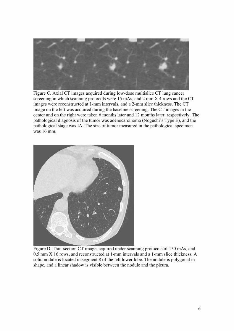
A case of Mixed GGO Adenocarcinoma (Noguchi type A)
Figure G. Screening CT
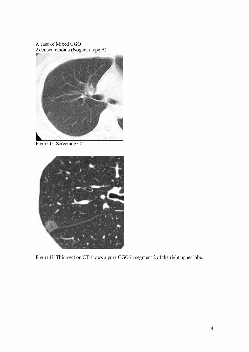
Figure H. Thin-section CT shows a pure GGO in segment 2 of the right upper lobe.

9
Footnotes for Figure 1 * Nodules that are strongly suspected of being intrapulmonary lymph nodes should be followed up even when they are larger than 10 mm in size. £ Pure GGO: nodule with pure ground-glass opacity (GGO) (nonsolid nodule) ££ Mixed GGO: nodule with mixed GGO (GGO with a solid component) (part-solid nodule) ** Thin-section CT can be performed to exclude inflammatory change, when necessary. *** Inflammatory lesions should be excluded. $ If the mixed GGO is less than 10 mm in size and is stable, further follow-up is an option. ¶ If the size of a pure GGO is in the 10 mm to 14 mm range, the management options, i.e., resection or follow-up, depend on the hospital's criteria. § If a nodule increases in size or in density, the decision between resecting the nodule and follow-up should be made according to the criteria of the hospital. # When a pure GGO increases in size or in density, the decision should be made according to the criteria of the hospital. & If a solid nodule is stable for 2 years, follow-up should be discontinued. ∞ Even pure GGOs that have been stable for more than 2 years should continue to be followed up.