Embed Size (px)
Citation preview
Localized Juvenile Spongiotic Gingival Hyperplasia: Report of Two Cases
The Journal of Clinical Pediatric Dentistry Volume 41, Number 3/2017 231
Localized Juvenile Spongiotic Gingival Hyperplasia: Report of Two Cases
Eleni-Marina Kalogirou */ Konstantina Chatzidimitriou **/ Konstantinos I. Tosios ***/ Evangelia P. Piperi ****/ Alexandra Sklavounou *****
Objective: Localized juvenile spongiotic gingival hyperplasia (LJSGH) is a painless gingival swelling that histologically exhibits hyperplasia of the non-keratinized stratified squamous epithelium, intercellular edema and spongiosis of the spinus layer, and exocytosis of inflammatory cells. LJSGH pathogenesis remains to be elucidated, while a possible origin from the gingival sulcus epithelium is nowadays proposed. Study design: We report two cases of LJSGH with immunohistochemical evaluation of cytokeratins (CKs) 18 and 19. Results: Both cases concerned 12-year-old boys, who presented with a well-circumscribed bright red pedunculated papillary swelling on the marginal gingiva of the left maxillary lateral incisor. With the provisional diagnosis of LJSGH, the lesions were excised under local anesthesia and histological examination supported the final diagnosis of LJSGH. In both cases, the lesional epithelium showed intense and mild positivity for CK19 and CK18, respectively, while the adjacent normal gingival epithelium expressed CK19, but not CK18, only in the basal cell layer. The postoperative course was uneventful in both patients and no recurrence has been reported. Conclusion: LJSGH is a recently introduced entity that is worth attention in the clinical pediatric dentistry. Clinical and histological examination is required for the final diagnosis, while immunohistochemistry has shed light to LJSGH pathogenesis.
Key words: gingiva, hyperplasia, juvenile, spongiotic, immunohistochemistry, cytokeratin 19; cytokeratin 18.
*Eleni-Marina I. Kalogirou, DDS, Postgraduate Student**Konstantina P. Chatzidimitriou, DDS, MSc, Postgraduate Student***Konstantinos I. Tosios, DDS, PhD, Assistant Professor**** Evangelia P. Piperi, DDS, MSc, PhD, Assistant Professor*****Alexandra Sklavounou, DDS, MSc, PhD, Professor.
Send all correspondence to : Konstantinos I TosiosDepartment of Oral Medicine and Pathology, Faculty of Dentistry, National and Kapodistrian University of Athens, 2 Thivon Street, 11527 Athens, Greece.Phone: +30-210-7461003E-mail: [email protected]
INTRODUCTION
Localized juvenile spongiotic gingival hyperplasia (LJSGH) is a gingival hyperplasia with unique clinicopathologic presentation1 that was initially described as juvenile spongi-
otic gingivitis (JSG) and considered to be the gingival counterpart of acute spongiotic dermatitis.2 The term LJSGH is preferred by most authors as it better delineates its features and emphasizes its hyperplastic nature. However, multifocal gingival involvement1-4 and cases in adults5 have been described; therefore the lesion may be neither localized nor juvenile.
LJSGH seems to be a rare lesion. In one study it represented 0.069% of 31.469 specimens accessioned in an oral pathology laboratory,3 and in the English language literature there are approx-imately 10 well documented publications.1-4,6-11 However, cases of LJSGH may have been misdiagnosed as pyogenic granuloma or gingivitis, in particular puberty gingivitis.1,2
LJSGH typically presents on the facial maxillary gingiva of young patients as a localized and solitary slightly mass up to 1cm in diameter, with bright red color, and papillary, granular, pebbly or velvety surface that is painless, but may bleed on teeth brushing.1-3
Its etiology remains obscure and an origin from the junctional epithelium of the gingiva is suggested.1,2,6
Only a few case reports of this unusual lesion have been published in the pediatric dentistry literature, although pediatric dentists are the health providers more likely to first examine a patient in this age group. Therefore, the clinical, microscopic and immunohistochemical features of two new cases of LJSGH are described, and the pertinent literature is reviewed.
Localized Juvenile Spongiotic Gingival Hyperplasia: Report of Two Cases
232 The Journal of Clinical Pediatric Dentistry Volume 41, Number 3/2017
Case reports
Case 1A 12-year-old boy was referred by his dentist for evaluation and
management of a painless erythematous swelling on the left side of the facial maxillary gingiva, noticed approximately 6 months before presentation. His parents reported that it “slightly shrank” following plaque debridement 5 months ago, while rinses with Povidone-io-dine oral antiseptic solution twice daily for 2 weeks were ineffec-tive. The patient’s medical and family history was noncontributory.
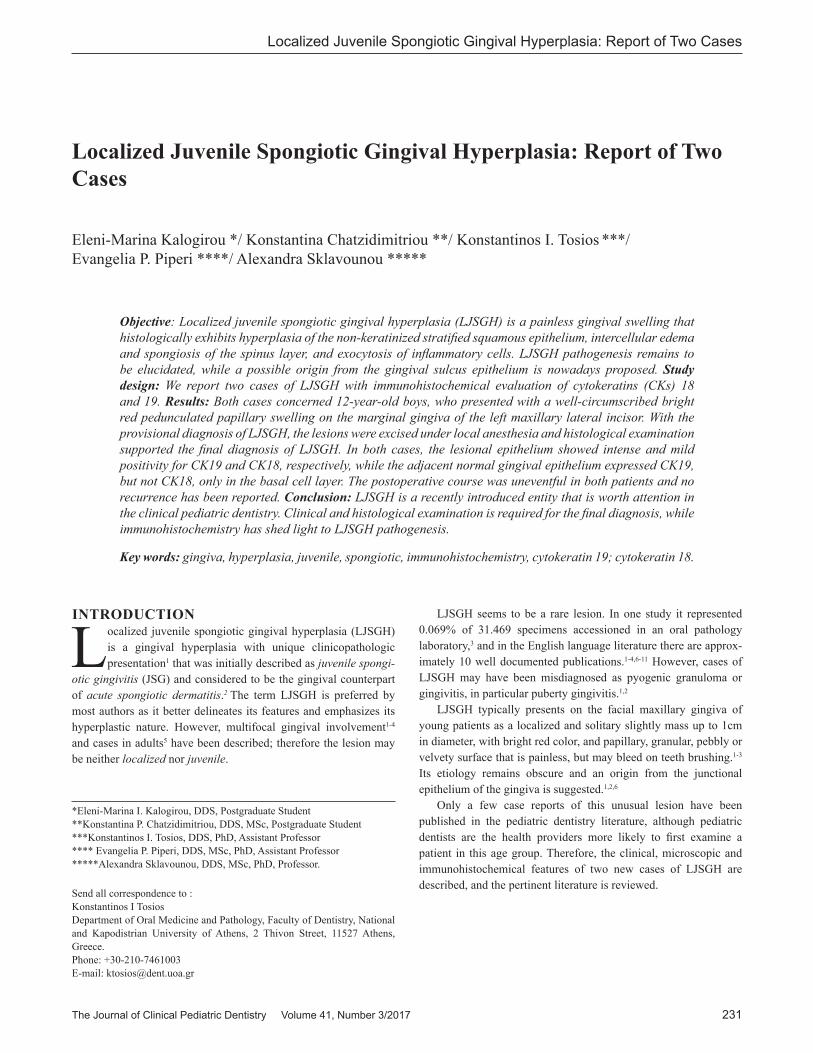
Intraoral examination revealed a well-circumscribed, bright red, pedunculated swelling with granular surface, approximately 1cm in greatest dimension, on the marginal and attached gingiva of the left maxillary lateral incisor that seemed to grow from the dentogingival junction (Fig. 1). The lesion was non-hemorrhagic on palpation and examination with a periodontal probe. The surface of the corre-sponding tooth was covered by dental plaque. No other intraoral or extraoral abnormalities were observed. A dental X-ray did not show bone involvement.
Τhe clinical diagnosis was LJSGH and the lesion was totally excised under local anesthesia.
Case 2A 12-year-old boy was referred by his pediatric dentist for diag-
nosis and management of an asymptomatic red mass on the facial anterior maxillary gingiva, of approximately 2 months duration. The patient had been diagnosed with “ulcerative colitis” a year before presentation.
Clinical examination revealed a well-defined bright red, pedun-culated mass, involving the facial marginal gingiva of the left maxil-lary lateral incisor that extended to the interproximal area between the two maxillary left incisors. It measured approximately 1cm in greatest dimension, had a granular surface and was soft, non-hemor-rhagic on palpation. The patient oral hygiene was poor, as indicated by the presence of calculus deposits in most of his teeth. The rest of oral mucosa was within normal limits. A dental X-ray did not reveal bone resorption.
The lesion was consistent with LJSGH and excised under local anesthesia.
Microscopic findingsGrossly, both lesions had solid, gray cut-surface and measured
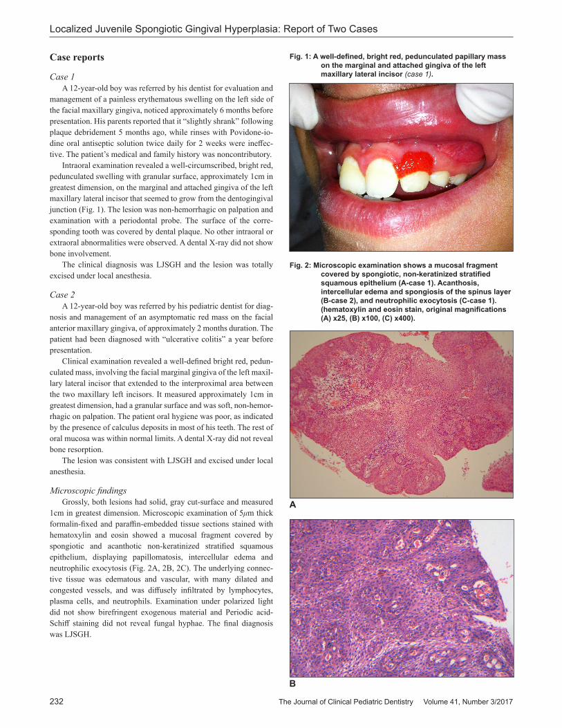
1cm in greatest dimension. Microscopic examination of 5μm thick formalin-fixed and paraffin-embedded tissue sections stained with hematoxylin and eosin showed a mucosal fragment covered by spongiotic and acanthotic non-keratinized stratified squamous epithelium, displaying papillomatosis, intercellular edema and neutrophilic exocytosis (Fig. 2A, 2B, 2C). The underlying connec-tive tissue was edematous and vascular, with many dilated and congested vessels, and was diffusely infiltrated by lymphocytes, plasma cells, and neutrophils. Examination under polarized light did not show birefringent exogenous material and Periodic acid-Schiff staining did not reveal fungal hyphae. The final diagnosis was LJSGH.
Fig. 1: A well-defined, bright red, pedunculated papillary mass on the marginal and attached gingiva of the left maxillary lateral incisor (case 1).
Fig. 2: Microscopic examination shows a mucosal fragment covered by spongiotic, non-keratinized stratified squamous epithelium (A-case 1). Acanthosis, intercellular edema and spongiosis of the spinus layer (B-case 2), and neutrophilic exocytosis (C-case 1). (hematoxylin and eosin stain, original magnifications (A) x25, (B) x100, (C) x400).
A
B
Localized Juvenile Spongiotic Gingival Hyperplasia: Report of Two Cases
The Journal of Clinical Pediatric Dentistry Volume 41, Number 3/2017 233
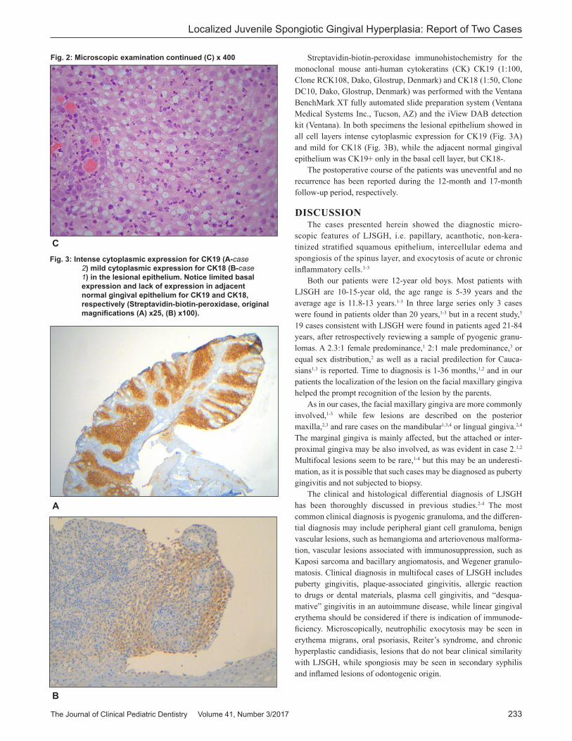
Streptavidin-biotin-peroxidase immunohistochemistry for the monoclonal mouse anti-human cytokeratins (CK) CK19 (1:100, Clone RCK108, Dako, Glostrup, Denmark) and CK18 (1:50, Clone DC10, Dako, Glostrup, Denmark) was performed with the Ventana BenchMark XT fully automated slide preparation system (Ventana Medical Systems Inc., Tucson, AZ) and the iView DAB detection kit (Ventana). In both specimens the lesional epithelium showed in all cell layers intense cytoplasmic expression for CK19 (Fig. 3A) and mild for CK18 (Fig. 3B), while the adjacent normal gingival epithelium was CK19+ only in the basal cell layer, but CK18-.
The postoperative course of the patients was uneventful and no recurrence has been reported during the 12-month and 17-month follow-up period, respectively.
DISCUSSIONThe cases presented herein showed the diagnostic micro-
scopic features of LJSGH, i.e. papillary, acanthotic, non-kera-tinized stratified squamous epithelium, intercellular edema and spongiosis of the spinus layer, and exocytosis of acute or chronic inflammatory cells.1-3
Both our patients were 12-year old boys. Most patients with LJSGH are 10-15-year old, the age range is 5-39 years and the average age is 11.8-13 years.1-3 In three large series only 3 cases were found in patients older than 20 years,1-3 but in a recent study,5 19 cases consistent with LJSGH were found in patients aged 21-84 years, after retrospectively reviewing a sample of pyogenic granu-lomas. A 2.3:1 female predominance,1 2:1 male predominance,3 or equal sex distribution,2 as well as a racial predilection for Cauca-sians1,3 is reported. Time to diagnosis is 1-36 months,1,2 and in our patients the localization of the lesion on the facial maxillary gingiva helped the prompt recognition of the lesion by the parents.
As in our cases, the facial maxillary gingiva are more commonly involved,1-3 while few lesions are described on the posterior maxilla,2,3 and rare cases on the mandibular1,3,4 or lingual gingiva.2,4 The marginal gingiva is mainly affected, but the attached or inter-proximal gingiva may be also involved, as was evident in case 2.1,2 Multifocal lesions seem to be rare,1-4 but this may be an underesti-mation, as it is possible that such cases may be diagnosed as puberty gingivitis and not subjected to biopsy.
The clinical and histological differential diagnosis of LJSGH has been thoroughly discussed in previous studies.2-4 The most common clinical diagnosis is pyogenic granuloma, and the differen-tial diagnosis may include peripheral giant cell granuloma, benign vascular lesions, such as hemangioma and arteriovenous malforma-tion, vascular lesions associated with immunosuppression, such as Kaposi sarcoma and bacillary angiomatosis, and Wegener granulo-matosis. Clinical diagnosis in multifocal cases of LJSGH includes puberty gingivitis, plaque-associated gingivitis, allergic reaction to drugs or dental materials, plasma cell gingivitis, and “desqua-mative” gingivitis in an autoimmune disease, while linear gingival erythema should be considered if there is indication of immunode-ficiency. Microscopically, neutrophilic exocytosis may be seen in erythema migrans, oral psoriasis, Reiter’s syndrome, and chronic hyperplastic candidiasis, lesions that do not bear clinical similarity with LJSGH, while spongiosis may be seen in secondary syphilis and inflamed lesions of odontogenic origin.
Fig. 3: Intense cytoplasmic expression for CK19 (A-case 2) mild cytoplasmic expression for CK18 (B-case 1) in the lesional epithelium. Notice limited basal expression and lack of expression in adjacent normal gingival epithelium for CK19 and CK18, respectively (Streptavidin-biotin-peroxidase, original magnifications (A) x25, (B) x100).
Fig. 2: Microscopic examination continued (C) x 400
C
A
B
Localized Juvenile Spongiotic Gingival Hyperplasia: Report of Two Cases
234 The Journal of Clinical Pediatric Dentistry Volume 41, Number 3/2017
The etiology of LJSGH is not clear. Dental plaque and calculus are profound etiologic factors, but LJSGH occasionally develops on the attached gingiva and is separated from the marginal gingiva with a strip of normal gingiva,2 dental plaque and calculus deposits may be absent,3 and most lesions do not respond favorably to periodontal treatment and improvement in oral hygiene measures.1-3,10,12 The predilection for younger persons is indicative of hormonal influ-ence, but no estrogen and progesterone receptors are found,2 while LJSGH may appear in prepubertal children and adults.1 Other factors considered are trauma that was documented in just one patient;3 orthodontic appliances that were present in only 8 in 521 and 1 in 213 patients with LJSGH;3 and mouth breathing that is not consistent with the mostly localized distribution of the lesion. Therefore, those factors seem to be coincidental events.1 A viral infection is suggested by epithelial hyperplasia,1 but in one study definitive positivity for HPV DNA was found by PCR in only one adult among 21 patients and was considered coincidental.1 Exogenous deposits or foreign body granulomas are not found on conventional and polarized-light microscopic examination.1,3 The localized nature of the lesion is not consistent with hypersensitivity,1 but the recognition of eosinophils and mast cells in similar lesions developing in adults suggests a hypersensitivity reaction to some undefined environmental factors.5 The positive response following plaque debridement in our case 1 could be subjective or related to minor recession of inflammation.
Darling et al2 proposed that the epithelium in LJSGH is “remark-ably similar to inflamed junctional/sulcular epithelium and peri-odontal pocket epithelium”. The similarity was supported by CK immunophenotype, as CK19 was strongly expressed throughout the LJSGH epithelium and junctional epithelium, but only in the basal layer of the adjacent normal oral epithelium, while there were no differences in the expression of CK5/6.2 In another study,6 LJSGH epithelium was CK19+ and CK8/18+ in its full thickness, but CK1/10- and CK4-. In contrast, the gingival epithelium covering fibro-epithelial hyperplasias was CK19+ and CK8/18+ only in its basal layer, but CK1/10+ and CK4+, the differences being statisti-cally significant. This CK immunophenotype in LJSGH epithelium is more consistent with normal and inflamed junctional epithe-lium13-16 and indicates that ectopic junctional epithelium is impli-cated in the pathogenesis of LJSGH.1,2,6 It is thought that junctional epithelium is “exteriorized” to the oral cavity and due to its structural features is more susceptible to the action of local irritation factors and becomes inflamed and hyperplastic.1,6 In our cases the CK19 expression pattern was consistent with LJSGH,2,6 while the intensity of CK18 expression was weaker than that of CK8/18,6 a difference that could be attributed either to the variability in the expression of CK8/18, as has been shown in normal junctional epithelium,14 or to a more intense expression of CK8 over CK18 in the previous study.6
Surgical excision is the treatment of choice, as LJSGH does not respond to conservative periodontal treatment,1-4 although there are undocumented reports of spontaneous resolution.2 Laser excision is a reliable alternative.3,11 Recurrence ranges of 6%,1 9.5%,3 and 28.6%2 are reported, and may manifest 2 months2 to 5 years after initial excision.1 Our cases did not recur following conservative surgical excision 12 and 17 months after treatment, respectively, although oral hygiene was not significantly improved.
CONCLUSIONFamiliarity with the clinicopathologic features of LJSGH by
pediatric dentists should allow the recognition of more cases of this under-diagnosed rather than rare lesion. Interestingly, although LJSGH usually affects children and adolescents, only scarce case reports exist in the pediatric literature.7 Description of more cases can help in elucidating its pathogenesis and improving its management.
RefeReNCeS1. Chang JY, Kessler HP, Wright JM. Localized juvenile spongiotic gingival
hyperplasia. Oral Surg Oral Med Oral Pathol Oral Radiol Endod 106: 411–418, 2008.
2. Darling MR, Daley TD, Wilson A, Wysocki GP. Juvenile spongiotic gingivitis. J Periodontol 78: 1235–1240, 2007.
3. Argyris PP, Nelson AC, Papanakou S, Merkourea S, Tosios KI, Koutlas IG. Localized juvenile spongiotic gingival hyperplasia featuring unusual p16INK4A labeling and negative human papillomavirus status by poly-merase chain reaction. J Oral Pathol Med 44: 37-44, 2015.
4. de Freitas Silva BS, Silva Sant’Ana SS, Watanabe S, Vêncio EF, Roriz VM, Yamamoto-Silva FP. Multifocal red bands of the marginal gingiva. Oral Surg Oral Med Oral Pathol Oral Radiol 119: 3-7, 2015.
5. Mora-Gonzalez D. Localized “Juvenile” Spongiotic Gingival Hyper-plasia in adults; investigation of possible viral etiology and comparison with Pyogenic Granuloma. The University of North Carolina at Chapel Hill, Department Dentistry (Oral and Maxillofacial Pathology), Thesis number 1557162, 2014-http://search.proquest.com/docview/1545898355
6. Allon I, Lammert KM, Iwase R, Spears R, Wright JM, Naidu A. Local-ized Juvenile Spongiotic Gingival Hyperplasia possibly originates from junctional gingival epithelium?–An immunohistochemical study. Histo-pathology 2015 Jul 7. doi: 10.1111/his.12774.
7. Solomon LW, Trahan WR, Snow JE. Localized juvenile spongiotic gingival hyperplasia: a report of 3 cases. Pediatr Dent 35: 360–363, 2013.
8. Flaitz CM, Longoria JM. Oral and maxillofacial pathology case of the month. Localized juvenile spongiotic gingival hyperplasia. Tex Dent J 127: 1312–1313, 1315–1317, 2010.
9. Damm DD. Gingival red patch. Juvenile spongiotic gingival hyperplasia. Gen Dent 57: 448, 451, 2009.
10. MacNeill SR, Rokos JR, Umaki MR, Satheesh KM, Cobb CM. Conser-vative treatment of localized juvenile spongiotic gingival hyperplasia. Clinical Advances in Periodontics 1: 199-204, 2011.
11. Rossmann JA. Reactive lesions of the gingiva: diagnosis and treatment options. The Open Pathology Journal 5: 23-32, 2011.
12. Decani S, Baruzzi E, Sutera S, Trapani A, Sardella A. A case of juvenile spongiotic gingivitis. Ann Stomatol (Roma) 4: 41, 2013.
13. Jiang Q, Yu Y, Ruan H, Luo Y, Guo X. Morphological and functional characteristics of human gingival junctional epithelium. BMC Oral Health 14: 30, 2014.- doi: 10.1186/1472-6831-14-30.
14. Pritlove-Carson S, Charlesworth S, Morgan PR, Palmer RM. Cytokeratin phenotypes at the dento-gingival junction in relative health and inflamma-tion, in smokers and nonsmokers. Oral Dis 3: 19-24, 1997.
15. Mackenzie IC, Gao Z. Patterns of cytokeratin expression in the epithelia of inflamed human gingiva and periodontal pockets. Periodontal Res 28: 49-59, 1993.
16. MacKenzie IC, Rittman G, Gao Z, Leigh I, Lane EB. Patterns of cyto-keratin expression in human gingival epithelia. J Periodontal Res 26: 468-478, 1991.


![Endometrium presentation - Dr Wright[1] · Endometrial Hyperplasia Simple hyperplasia Complex hyperplasia (adenomatous) Simple atypical hyperplasia ... Progression of Hyperplasia](https://img.dokumen.tips/doc/110x75/5b8a421e7f8b9a50388bc13d/endometrium-presentation-dr-wright1-endometrial-hyperplasia-simple-hyperplasia.jpg)