Embed Size (px)
Citation preview

87Journal of The Association of Physicians of India ■ Vol. 64 ■ August 2016
Life-Threatening Thrombo-embolic Events in a Case of Dengue Hemorrhagic FeverArun Agarwal1, Samiksha Sharma2, Mala Airun3
1Senior Consultant and Head, Department of Internal Medicine, 2Consultant and Head, Department of Pathology, 3Clinical Director, Narayana Multispeciality Hospital, Jaipur, RajasthanReceived: 26.06.2015; Revised: 08.10.2015; Accepted: 28.10.2015
AbstractIn Dengue Hemorrhagic Fever (DHF), hemorrhagic manifestations are common but thrombotic events are uncommonly reported, despite the wide range of increased procoagulant activity during Dengue Fever illness. We report a case of a 55-year-old man of Asian Indian ethnicity who developed large vein thrombotic event -Deep Vein Thrombosis (DVT) and Pulmonary Embolism (PE) in the acute phase of DHF. His condition was further complicated by associated thrombocytopenia. The etiological connections between dengue viral infection with thrombocytopenia, DVT/PE and abnormal thrombophilia profile as well as the treatment dilemmas posed in treating a patient of DF with hemorrhagic manifestations and associated DVT/PE, and the role of eltrombopag are discussed.
Introduction
The main concern in Dengue Fever (DF) has been the severe forms, i.e.
DHF and dengue shock syndrome. DVT and PTE is an uncommon complication following severe Dengue viral infection. Complications such as these could be overlooked in the overall management of DF, given that the major concern is the hemorrhagic events in DHF. Although therapeutic anticoagulation is recommended for management of DVT/PE, this treatment may be questionable when the patient has an associated bleeding diathesis due to DV infection. Awareness of these kinds of complications is necessary among all clinicians who treat patients with dengue fever.
Case Presentation
A 55 yrs male, was admitted to our institution on 01.10.2014. He had a history of fever with chills off and on for one month, black colored stools for 10-15 days, dark coloured urine and cough for 5 days, and decreased urine output for one day. He also had itching all over the body and few ecchymosis on left lower limb. He had a doubtful history of CAD 30 yrs back with no documents available. There was no history of diabetes mellitus, hypertension, dyslipedemia and venous catheter placement in lower limbs. Earlier he was admitted elsewhere for 3 days with fever with chills, nausea, vomiting, black color
stools and pain abdomen of five days and was discharged with diagnosis of DHF with bleeding manifestations and thrombocytopenia.
T h e p a t i e n t c a m e t o u s a f t e r three weeks of initial diagnosis of DF. On examination he was mildly tachypnoeic ( respiratory rate 24/ minute), hemodynamically stable and afebrile. He had swelling in left leg, skin eruptions with scratch marks on abdomen, back and both arms, few ecchymosis on left leg, right side basal crackles, and mild non tender hepatosplenomegaly. He was admitted in medical intensive care unit for further management.
During his hospital stay he had daily spike of fever up-to 100 F. Skin scrapings from skin lesion showed fungal mycelium which was diagnosed to be tinea corporis and treated with terbinafine and supportive drugs. He was further worked up and his serial hematology and biochemistry is mentioned in Table 1 and other invest igat ions in Table 2 . He had p e r s i s t e n t m o d e r a t e t o s e v e r e thrombocytopenia, mild transaminitis and posit ive serology for Dengue IgM antibody. Dengue NS1 antigen was not done as it was three weeks after the initial diagnosis of DF and anti dengue IgG was negative. Tests
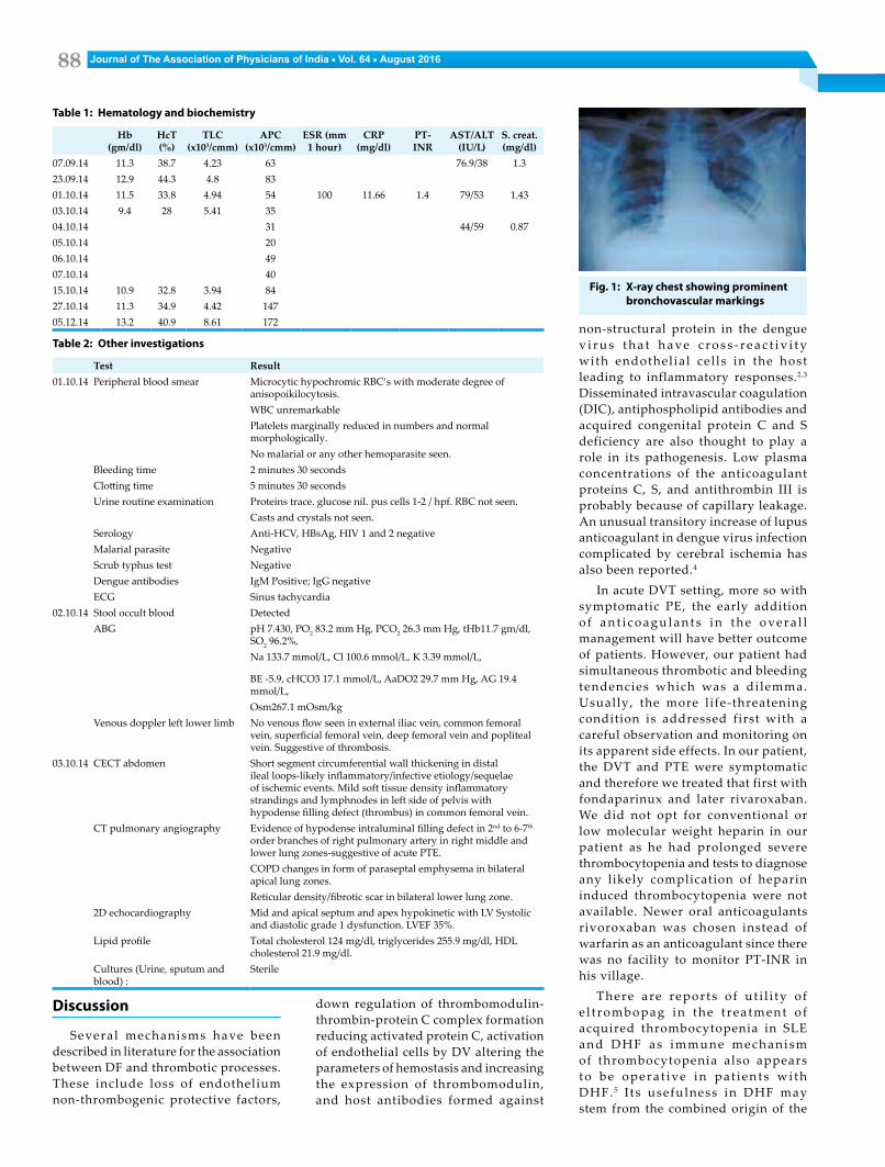
for malaria parasite, scrub typhus, body f luid cultures, serology and peripheral smear were unremarkable. In view of complaints of pain in abdomen, serum amylase was done. Cardiac markers were done to rule out myocardial dysfunction. Both were normal. His chest X-ray (Figure 1) showed cardiomegaly with prominent b r o n c h o va s c u l a r m a r k i n g s . A B G showed mi ld chronic respiratory alkalosis . He did not require any ventilator support. He was further diagnosed to have extensive thrombosis involving left common femoral vein, superficial femoral vein, deep femoral vein, external iliac vein and popliteal vein along with pulmonary thrombo-embolism (Table 2, Figure 2A and 2B). His anti-neutrophil cytoplasmic a n t i b o d y ( A N C A) , a n t i - n u c l e a r antibody (ANA) and thrombocheck panel were done. He had normal protein C, homocysteine and antithrombin levels. His protein S was low and lupus anticoagulant was present which normalized in the convalescent stage.
P a t i e n t w a s m a n a g e d w i t h antibiotics (Cefoperazone + sulbactum), fexofenadine, atorvastatin, low dose aspirin, glyceryl trinitrate, trimetazidine and supportive treatment. For DVT and PTE he was given fondaparinux 5mg subcutaneously once a day. He was la ter shi f ted on r ivoroxaban after 5 days. In view of persistent t h r o m b o c y t o p e n i a E l t r o m b o p a g (thrombopoietin receptor agonist) 50 mg once a day was given empirically for three weeks. He was eventually discharged with a diagnosis of DHF associated with severe thrombotic events and coronary artery disease. On follow up after three week, his platelet counts were in normal limits and Eltrombopag was stopped. Follow up venous Doppler showed partial recanalization in left leg. Rivoroxaban was continued for 6 months.
88 Journal of The Association of Physicians of India ■ Vol. 64 ■ August 2016
Discussion
Several mechanisms have been described in literature for the association between DF and thrombotic processes. These include loss of endothelium non-thrombogenic protective factors,
non-structural protein in the dengue v i r u s t h a t h a ve c r o s s - r e a c t i v i t y with endothel ial cel ls in the host leading to inflammatory responses.2,3 Disseminated intravascular coagulation (DIC), antiphospholipid antibodies and acquired congenital protein C and S deficiency are also thought to play a role in its pathogenesis. Low plasma concentrations of the anticoagulant proteins C, S, and antithrombin III is probably because of capillary leakage. An unusual transitory increase of lupus anticoagulant in dengue virus infection complicated by cerebral ischemia has also been reported.4
In acute DVT setting, more so with symptomatic PE, the early addition o f a n t i c o a g u l a n t s i n t h e o ve r a l l management will have better outcome of patients. However, our patient had simultaneous thrombotic and bleeding tendencies which was a dilemma. Usually, the more life-threatening condition is addressed first with a careful observation and monitoring on its apparent side effects. In our patient, the DVT and PTE were symptomatic and therefore we treated that first with fondaparinux and later rivaroxaban. We did not opt for conventional or low molecular weight heparin in our patient as he had prolonged severe thrombocytopenia and tests to diagnose any likely complication of heparin induced thrombocytopenia were not available. Newer oral anticoagulants rivoroxaban was chosen instead of warfarin as an anticoagulant since there was no facility to monitor PT-INR in his village.
There are reports of ut i l i ty of e l t rombopag in the t rea tment o f acquired thrombocytopenia in SLE and DHF as immune mechanism of thrombocytopenia also appears to be opera t ive in pat ients wi th DHF. 5 I ts usefulness in DHF may stem from the combined origin of the
Table 1: Hematology and biochemistry
Hb (gm/dl)
HcT (%)
TLC (x103/cmm)
APC (x103/cmm)
ESR (mm 1 hour)
CRP (mg/dl)
PT- INR
AST/ALT (IU/L)
S. creat. (mg/dl)
07.09.14 11.3 38.7 4.23 63 76.9/38 1.323.09.14 12.9 44.3 4.8 8301.10.14 11.5 33.8 4.94 54 100 11.66 1.4 79/53 1.4303.10.14 9.4 28 5.41 3504.10.14 31 44/59 0.8705.10.14 2006.10.14 4907.10.14 4015.10.14 10.9 32.8 3.94 8427.10.14 11.3 34.9 4.42 14705.12.14 13.2 40.9 8.61 172
Table 2: Other investigations
Test Result01.10.14 Peripheral blood smear Microcytic hypochromic RBC’s with moderate degree of
anisopoikilocytosis.WBC unremarkablePlatelets marginally reduced in numbers and normal morphologically.No malarial or any other hemoparasite seen.
Bleeding time 2 minutes 30 secondsClotting time 5 minutes 30 secondsUrine routine examination Proteins trace. glucose nil. pus cells 1-2 / hpf. RBC not seen.
Casts and crystals not seen.Serology Anti-HCV, HBsAg, HIV 1 and 2 negativeMalarial parasite NegativeScrub typhus test NegativeDengue antibodies IgM Positive; IgG negativeECG Sinus tachycardia
02.10.14 Stool occult blood DetectedABG pH 7.430, PO2 83.2 mm Hg, PCO2 26.3 mm Hg, tHb11.7 gm/dl,
SO2 96.2%,Na 133.7 mmol/L, Cl 100.6 mmol/L, K 3.39 mmol/L,
BE -5.9, cHCO3 17.1 mmol/L, AaDO2 29.7 mm Hg, AG 19.4 mmol/L,Osm267.1 mOsm/kg
Venous doppler left lower limb No venous flow seen in external iliac vein, common femoral vein, superficial femoral vein, deep femoral vein and popliteal vein. Suggestive of thrombosis.
03.10.14 CECT abdomen Short segment circumferential wall thickening in distal ileal loops-likely inflammatory/infective etiology/sequelae of ischemic events. Mild soft tissue density inflammatory strandings and lymphnodes in left side of pelvis with hypodense filling defect (thrombus) in common femoral vein.
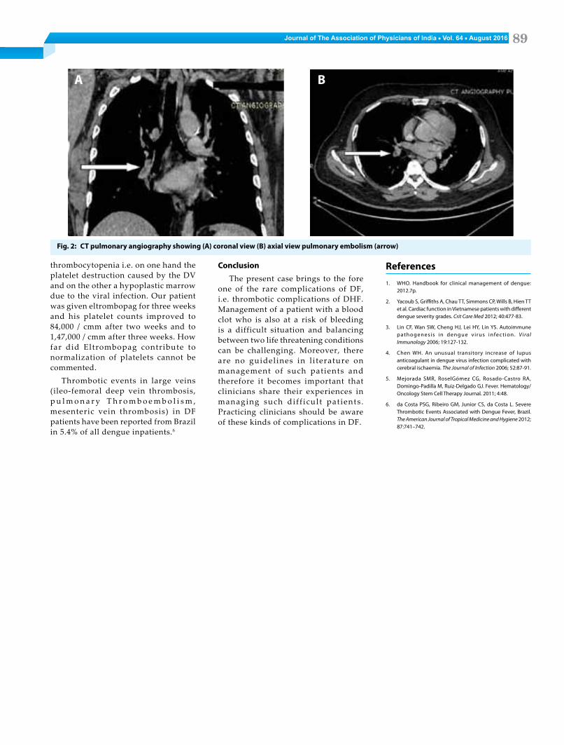
CT pulmonary angiography Evidence of hypodense intraluminal filling defect in 2nd to 6-7th order branches of right pulmonary artery in right middle and lower lung zones-suggestive of acute PTE.COPD changes in form of paraseptal emphysema in bilateral apical lung zones.Reticular density/fibrotic scar in bilateral lower lung zone.
2D echocardiography Mid and apical septum and apex hypokinetic with LV Systolic and diastolic grade 1 dysfunction. LVEF 35%.
Lipid profile Total cholesterol 124 mg/dl, triglycerides 255.9 mg/dl, HDL cholesterol 21.9 mg/dl.
Cultures (Urine, sputum and blood) :
Sterile
Fig. 1: X-ray chest showing prominent bronchovascular markings
down regulation of thrombomodulin-thrombin-protein C complex formation reducing activated protein C, activation of endothelial cells by DV altering the parameters of hemostasis and increasing the expression of thrombomodulin, and host antibodies formed against
89Journal of The Association of Physicians of India ■ Vol. 64 ■ August 2016
References1. WHO. Handbook for clinical management of dengue:
2012.7p.
2. Yacoub S, Griffiths A, Chau TT, Simmons CP, Wills B, Hien TT et al. Cardiac function in Vietnamese patients with different dengue severity grades. Crit Care Med 2012; 40:477-83.
3. Lin CF, Wan SW, Cheng HJ, Lei HY, Lin YS. Autoimmune pathogenes is in dengue v i rus infec t ion . Viral Immunology 2006; 19:127-132.
4. Chen WH. An unusual transitory increase of lupus anticoagulant in dengue virus infection complicated with cerebral ischaemia. The Journal of Infection 2006; 52:87-91.
5. Mejorada SMR, RoselGómez CG, Rosado-Castro RA, Domingo-Padilla M, Ruiz-Delgado GJ. Fever. Hematology/Oncology Stem Cell Therapy Journal. 2011; 4:48.
6. da Costa PSG, Ribeiro GM, Junior CS, da Costa L. Severe Thrombotic Events Associated with Dengue Fever, Brazil. The American Journal of Tropical Medicine and Hygiene 2012; 87:741–742.
thrombocytopenia i.e. on one hand the platelet destruction caused by the DV and on the other a hypoplastic marrow due to the viral infection. Our patient was given eltrombopag for three weeks and his platelet counts improved to 84,000 / cmm after two weeks and to 1,47,000 / cmm after three weeks. How far did Eltrombopag contribute to normalization of platelets cannot be commented.
Thrombotic events in large veins (ileo-femoral deep vein thrombosis, p u l m o n a r y T h r o m b o e m b o l i s m , mesenteric vein thrombosis) in DF patients have been reported from Brazil in 5.4% of all dengue inpatients.6
Conclusion
The present case brings to the fore one of the rare complications of DF, i.e. thrombotic complications of DHF. Management of a patient with a blood clot who is also at a risk of bleeding is a difficult situation and balancing between two life threatening conditions can be challenging. Moreover, there are no guidelines in l i terature on management of such patients and therefore it becomes important that clinicians share their experiences in managing such dif f icult pat ients . Practicing clinicians should be aware of these kinds of complications in DF.
Fig. 2: CT pulmonary angiography showing (A) coronal view (B) axial view pulmonary embolism (arrow)
A B