Embed Size (px)
Citation preview

LESSON ASSIGNMENT LESSON 1 Introduction to Human Anatomy TEXT ASSIGNMENT Paragraph 1-1 through 1-15. LESSON OBJECTIVES After completing this lesson, you should be able to: 1-1. Select correct answers to questions regarding osteology and arthrology, including general morphology of the bones and joints, bone classification, formation and growth, joint classification, and joint movement. SUGGESTION After completing the assignment, complete the exercises at the end of this lesson. These exercises will help you to achieve the lesson objectives.
MD0956 1-2

LESSON 1
INTRODUCTION TO HUMAN ANATOMY
Section I. ORIENTATION 1-1. INTRODUCTION a. The human body is a complex organism made up of various interdependent systems and components. Anatomy is the study of these components and the systems that they comprise. Put another way, anatomy is the study of the structure of the body, and the spatial relationship of its parts. Radiographic anatomy is the study of body structures that can be satisfactorily recorded as images of varying densities on x-ray film. Although the primary emphasis in radiographic anatomy will be on the skeletal system, as an X-ray technologist, you should be familiar with the structure of the whole body, and the interdependent systems of which it is comprised. b. A detailed study of human anatomy and physiology is beyond the scope of this subcourse. Fortunately, you do not require such detailed knowledge. However, a basic knowledge of bones and their joints, their locations, and their surface landmarks, is essential for proper positioning of the patient. You should also know sufficient detail of the internal organ systems, including their components, locations, and functions, to perform the various radiographic procedures. c. In the radiology department, you need to possess a thorough knowledge of medical and anatomical terminology so that you can communicate quickly and accurately with other members of the health care team. This knowledge will enable you to evaluate the situations in which you carry out your responsibilities, and to take the appropriate actions. For example, suppose that an ambulatory patient presents a request slip that shows a provisional diagnosis of arthritis with ankylosis of the right shoulder and elbow. You should immediately recognize that the patient has a fixed and inflamed right shoulder and elbow. This knowledge will influence the manner in which the exposure is made. You should do everything possible to position the patient in such a way as to cause minimum discomfort. In addition, you need to have sufficient knowledge of anatomy to interpret clinical requests correctly and to determine if the anatomical structures represented in the finished radiograph fulfill the requirements of the original request. 1-2. MEDICAL TERMINOLOGY a. General Terminology. The foundation for radiographic anatomy and allied subjects is centered primarily in medical terminology. As an X-ray technologist, you should know the meaning of the following general terms:
MD0956 1-3

(1) Science. Systematized and classified knowledge. (2) -ology (suffix). A science or branch of knowledge. (3) Regional or topographical anatomy. The study of separate parts of the body. (4) Systemic anatomy. The study of systems and associated parts. Systemic anatomy is divided into these subdivisions: (a) Osteology. The study of the bones. (b) Arthrology. The study of the articulations or joints. (b) Myology. The study of the muscular system. (c) Neurology. The study of the nervous system. (d) Angiology. The study of the vascular/lymphatic vessels. (5) Embryology. The study of the origin of the structures of the body. (6) Physiology. The study of the functions and activities of the body. (7) Pathology. The study of changes in the structures or function of the body caused by disease or trauma. (8) Radiology. That branch of medical science that deals with the use of radiant energy in the diagnosis and treatment of injuries and diseases. b. Normal Anatomical Position. To avoid misunderstanding, a standard position of the human body (figure 1-1) is arbitrarily taken to be the erect (standing) position with feet flat on the floor, heels together, upper extremities at the sides, and palms, toes, and eyes directed forward. This is the anatomical position. c. Terms Dealing with Aspects and Directions. (1) Anterior, frontal, or ventrum. The front side of the body. (2) Posterior or dorsum. The back, or dorsum, of the body. (3) Median. Pertaining to the midline of the body (figure1-1).
MD0956 1-4

Figure 1-1. Medial-lateral relationships. X is lateral to Y and Z. Y is medial to X and lateral to Z. In the example shown, the body is in the normal anatomical position.
MD0956 1-5
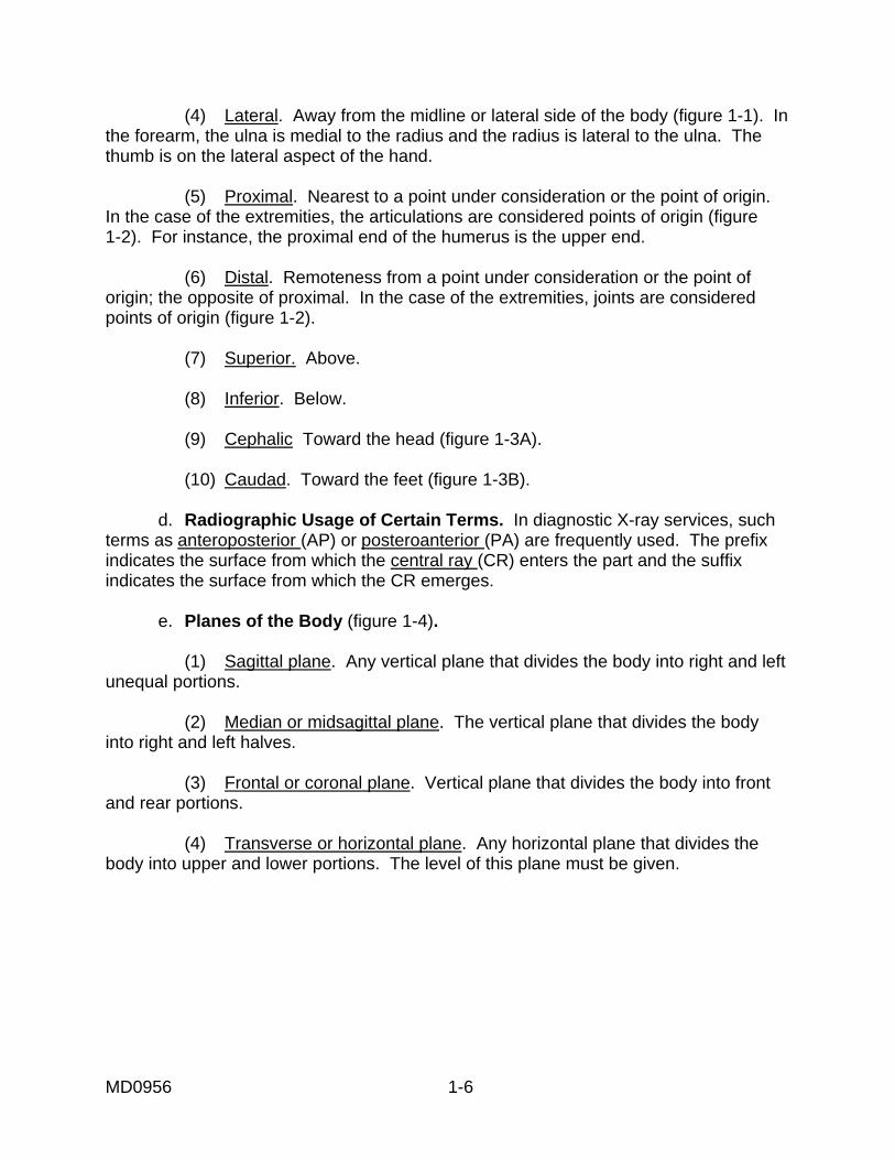
(4) Lateral. Away from the midline or lateral side of the body (figure 1-1). In the forearm, the ulna is medial to the radius and the radius is lateral to the ulna. The thumb is on the lateral aspect of the hand. (5) Proximal. Nearest to a point under consideration or the point of origin. In the case of the extremities, the articulations are considered points of origin (figure 1-2). For instance, the proximal end of the humerus is the upper end. (6) Distal. Remoteness from a point under consideration or the point of origin; the opposite of proximal. In the case of the extremities, joints are considered points of origin (figure 1-2). (7) Superior. Above. (8) Inferior. Below. (9) Cephalic Toward the head (figure 1-3A). (10) Caudad. Toward the feet (figure 1-3B). d. Radiographic Usage of Certain Terms. In diagnostic X-ray services, such terms as anteroposterior (AP) or posteroanterior (PA) are frequently used. The prefix indicates the surface from which the central ray (CR) enters the part and the suffix indicates the surface from which the CR emerges. e. Planes of the Body (figure 1-4). (1) Sagittal plane. Any vertical plane that divides the body into right and left unequal portions. (2) Median or midsagittal plane. The vertical plane that divides the body into right and left halves. (3) Frontal or coronal plane. Vertical plane that divides the body into front and rear portions. (4) Transverse or horizontal plane. Any horizontal plane that divides the body into upper and lower portions. The level of this plane must be given.
MD0956 1-6
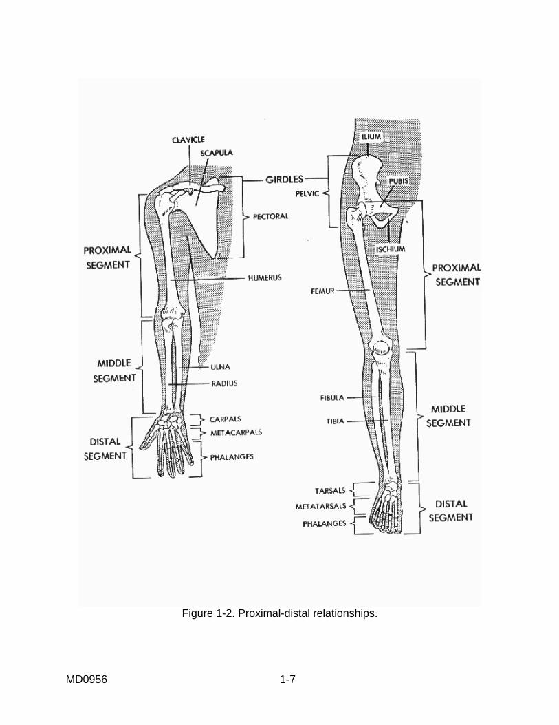
Figure 1-2. Proximal-distal relationships.
MD0956 1-7

Figure 1-3. Angulation of x-rays. (A.) Cephalic angulation. (B.) Caudal angulation.
MD0956 1-8

Figure 1-4. Planes of the body.
MD0956 1-9

f. Surfaces of the Hands and Feet. (1) Palmar surface. Anterior surface (palm) of the hand. (2) Volar surface. Anterior surface of the hand and forearm (or the sole of the foot). (3) Plantar surface. Inferior surface (sole) of the foot. (4) Dorsal surface. Top or superior surface (dorsum) of the foot. g. Terminology Relating to the Positions of the Body (figure 1-5). (1) Supine. A horizontal position of the body lying flat on the back with no rotation of the trunk. (2) Prone. A horizontal position of the body lying face and stomach down with no rotation of the trunk. (3) Lateral recumbent. A horizontal position of the body lying on either side with no rotation of the trunk. (4) Oblique. A position of the body, or any of its parts, when placed at an inclined angle to the X-ray film. (5) Erect or vertical. A position of the body either sitting or standing. h. External and Internal. These terms are used to describe locations with respect to the surface. i. Body Types. Four terms are generally used to designate the four major types of body habitus (figure 1-6). Since the position of certain organs (for example, the gallbladder) can vary as much as 6 to 8 inches between body types, it is essential that the X-ray specialist be familiar with these major body types. (1) Hypersthenic. The hypersthenic body (figure1-6A) is of massive build with a broad and deep thorax. The diaphragm is high and the stomach and gallbladder also occupy high positions. An extreme body type, the hypersthenic classification accounts for only about five percent of all people.
MD0956 1-10

Figure 1-5. Body positions. (2) Sthenic. Means active or strong. The sthenic body (figure 1-6B) is the one we usually associate with the athletic type. The body is rather heavy with large bones. The sthenic body type is the predominant type, with about half of all people falling into this classification. (3) Hyposthenic. Slender and light in weight with the stomach and gallbladder situated high in the abdomen (figure 1-6c). About 35 percent of all people fall into this classification. (4) Asthenic. Extremely slender, light build, with a narrow, shallow thorax, and the gallbladder and stomach situated low in the abdomen. An extreme type, the asthenic classification accounts for only about ten percent of all people (figure 1-6D).
MD0956 1-11
Figure 1-6. Major body types.
MD0956 1-12

j. Regions of the Abdomen. (1) The abdomen is that portion of the body that lies between the thorax and the pelvis. It consists of a large cavity, separated from the thoracic cavity by the diaphragm, bounded by muscles and fascia, and partially lined with a serous membrane called the peritoneum. (2) For purposes of description, the abdomen is divided into nine regions (figure 1-7) by means of two horizontal and two vertical lines. The upper horizontal line passes through the tenth costal cartilage inferiorly. The lower line passes through the level of the iliac tubercle. Each vertical line passes through the midpoint of a line drawn from the anterior superior iliac spine to the symphysis pubis.
Figure 1-7. Regions of the abdomen.
MD0956 1-13
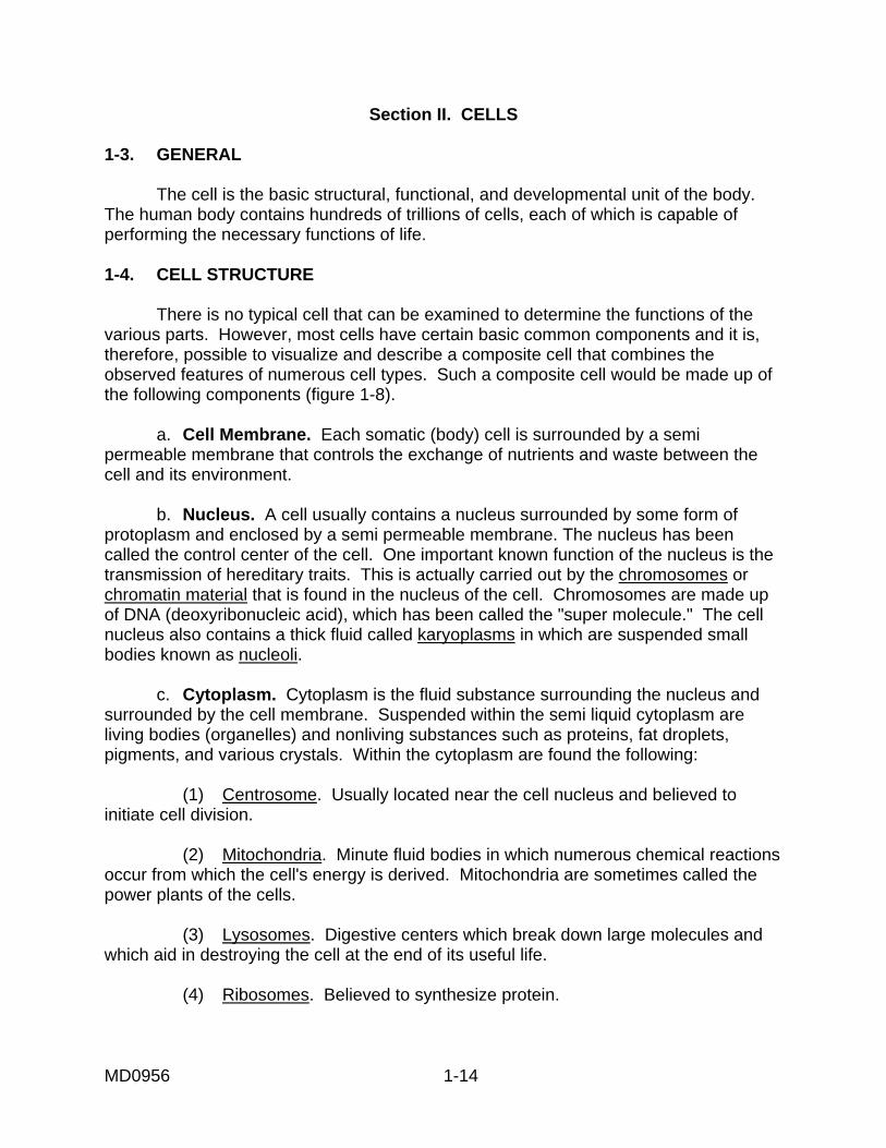
Section II. CELLS 1-3. GENERAL The cell is the basic structural, functional, and developmental unit of the body. The human body contains hundreds of trillions of cells, each of which is capable of performing the necessary functions of life. 1-4. CELL STRUCTURE There is no typical cell that can be examined to determine the functions of the various parts. However, most cells have certain basic common components and it is, therefore, possible to visualize and describe a composite cell that combines the observed features of numerous cell types. Such a composite cell would be made up of the following components (figure 1-8). a. Cell Membrane. Each somatic (body) cell is surrounded by a semi permeable membrane that controls the exchange of nutrients and waste between the cell and its environment. b. Nucleus. A cell usually contains a nucleus surrounded by some form of protoplasm and enclosed by a semi permeable membrane. The nucleus has been called the control center of the cell. One important known function of the nucleus is the transmission of hereditary traits. This is actually carried out by the chromosomes or chromatin material that is found in the nucleus of the cell. Chromosomes are made up of DNA (deoxyribonucleic acid), which has been called the "super molecule." The cell nucleus also contains a thick fluid called karyoplasms in which are suspended small bodies known as nucleoli. c. Cytoplasm. Cytoplasm is the fluid substance surrounding the nucleus and surrounded by the cell membrane. Suspended within the semi liquid cytoplasm are living bodies (organelles) and nonliving substances such as proteins, fat droplets, pigments, and various crystals. Within the cytoplasm are found the following: (1) Centrosome. Usually located near the cell nucleus and believed to initiate cell division. (2) Mitochondria. Minute fluid bodies in which numerous chemical reactions occur from which the cell's energy is derived. Mitochondria are sometimes called the power plants of the cells. (3) Lysosomes. Digestive centers which break down large molecules and which aid in destroying the cell at the end of its useful life. (4) Ribosomes. Believed to synthesize protein.
MD0956 1-14

Figure 1-8. Diagrammatic representation of a cell. (5) The endoplasmic reticulum. A network of internal membranes which form a series of small canals through the cytoplasm for the purpose of transporting substances from the cell membrane to the nuclear membrane. (6) Golgi body (or com la ex). A series of smooth membranes continuous with the endoplasmic reticulum which is believed to have something to do with regulating the movement of fluids in the cell. (7) Fibrils. Thin, protoplasmic threads in the cytoplasm that probably give the cell structural support.
MD0956 1-15

1-5. CELL DIVISION The two basic types of human cells, somatic cells and sex cells, mature and increase in number through processes known as mitosis and meiosis, respectively. a. Mitosis. Mitosis is the form of cell division that occurs in higher forms of animal life, including man. This form of cell division consists of four phases (prophase, metaphase, anaphase, and telophase) during which the chromatin material in the nucleus undergoes various changes in arrangement, leading to the ultimate division of the cell. The result of mitosis of a somatic cell is two daughter cells, each of which possesses the same number of chromosomes as the parent cell (figure1-9). In humans, each somatic cell contains 23 pairs, or 46 chromosomes.
Figure 1-9. Diagrammatic representation of mitosis.
MD0956 1-16

b. Meiosis. Sex cells mature and propagate by a different process than somatic cells. This process is called meiosis. In meiosis, the series of nuclear changes within the sex cell results in the production of new cells with half the number of chromosomes present in the original sex cell. Meiosis occurs in both female and male sex cells, resulting in the formation of ova and spermatozoa, respectively. The union of a mature spermatazoa and a mature ovum results in the formation of a new individual. As a result of meiosis, the chromosome number remains constant from one generation to the next. For this reason, meiosis is sometimes called reduction division. 1-6. TISSUE Somatic cells are usually classified as either epithelial, muscle, nerve, or connective. Cells arranged or organized to perform one or more specific functions are called a tissue. The four basic tissue types found in the human body are classified according to the type of cells that comprise them and have these specialized functions. a. Epithelial Tissue. Forms linings and coverings of various body parts and systems. b. Muscle Tissue. Contracts to cause movement and to maintain body posture. c. Nerve Tissue. Conducts messages (impulses) to and from the central nervous system. d. Connective Tissue. Serves as filler and binder substances of the body and forms the supporting framework of the body and the body organs.
Section III. BONES 1-7. BONES OF THE SKELETON a. The normal adult human body has approximately 206 bones. This includes the auditory ossicles and the patella, but excludes the small sesamoid bones. Figure 1-10 shows a human skeleton in anatomical position. b. The skeleton may be divided into the appendicular and the axial skeleton. The appendicular skeleton includes the bones of the upper and lower free extremities, the shoulder (pectoral girdle), and the pelvic girdle. The axial skeleton includes the bones of the skull, the vertebral column, the thoracic cage, the auditory ossicles, and the hyoid bone.
MD0956 1-17

Figure 1-10. The skeleton
MD0956 1-18

1-8. BONE STRUCTURE a. General. Bone is a tissue composed of living cells (osteocytes) distributed in an intercellular matrix that contains organic and inorganic substances. The organic component, largely collagenous fibers, is responsible for the strength and resilience of bone while the inorganic salts, mostly calcium phosphate, contribute to its hardness, and rigidity. The inorganic constituents make up approximately 67% of bony matter in the adult. The radiopacity of bone depends largely upon the amount of minerals present. Lack of mineral content in the young and aged alters the radiopacity and requires compensation. b. Forms of Bone Tissue. There are two forms of bone tissue, cancellous and compact. Cancellous or spongy bone consists of irregular strands of tissue, which branch and join one another, forming a loose network in which the intercommunicating spaces are filled with marrow. Compact or dense bone has a more solid, regular appearance and its intercommunicating canals are microscopic in size. The basic structure of these two types of bone is essentially the same. They differ mainly in the relative amount of solid substance and the number, size, and arrangement of the intercommunicating spaces they contain. Both cancellous and compact forms are present in most bones of the body, but the extent and distribution of each varies considerably. In adults, the exterior of all bones is compact bone while the interior is usually cancellous. c. A Typical Long Bone. In a typical long bone (figure 1-11), each end (epiphysis) is largely cancellous and is covered by a thin layer of compact bone. The reverse is true in the shaft (diaphysis), which is mostly compact bone tissue. The central medullary canal, or cavity in the shaft of a long bone, is continuous with the intercommunicating spaces in the cancellous bone located at the ends. Depending on the age of the individual and the type of bone considered, either red or yellow marrow fills these cavities. Red marrow, active in the production of blood cells, is present in all bones at birth and blood cells are produced in all locations. With advancing age, the production of blood cells decreases and red marrow is replaced by yellow marrow, which consists mostly of fat cells. In the adult, red marrow is found mainly in the skull, vertebrae, ribs, sternum, and the articular ends of some long bones. d. Long Bone Structure. Each long bone, except for its articular surface, is enclosed by a thick, fibrous sheet of membranous tissue, called the periosteum, which develops when the perichondrium, the outer covering of the embryonic skeleton, becomes permeated with blood vessels. The marrow cavity, and also the canal system, are lined by a delicate layer of reticular (netlike) tissue, called the endosteum. e. Some Other Bones. In flat bones, such as the ribs, one or more plates of compact bone surround the cancellous bone. In many irregular bones, such as the vertebrae, spongy bone is enclosed by a thin shell of compact bone.
MD0956 1-19
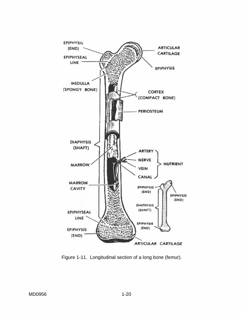
Figure 1-11. Longitudinal section of a long bone (femur).
MD0956 1-20

f. Blood Supply. The living bone cells are nourished by a system of blood vessels and capillaries. In the long bones, for example, blood vessels in the shaft supply the bone marrow. Branches of blood vessels contained in the periosteum supply the compact and cancellous bone areas. g. Microscopic Structure. When bone tissue is examined under a microscope, it is seen in layers either as a series of flat plates (for cancellous bone) or concentric cylinders (for compact bone). In compact bones, the series of concentric cylinders are formed in units called haversian systems. Here, living bone cells lie in minute cavities called lacunae. The lacunae communicate with each other, and indirectly with a central haversian canal, through a system of microscopic canals called canaliculi that contain protoplasmic extensions of bone cells. They are nourished by blood vessels from the periosteum that enter the compact bone through small pits on the surface. Branches of these blood vessels penetrate the matrix and enter the central haversian canal in each haversian system. 1-9. CLASSIFICATION OF BONES Bones may be classified according to shape. This classification is of special interest to the X-ray specialist. a. Long Bones. These are long, usually cylindrical, shafts with two expanded extremities. Long bones are radiographed lengthwise on the film. The humerus (figure 1-12A) of the arm is an example of a long bone. b. Short Bones. These bones are short, usually cylindrical shafts, with two expanded extremities. They usually occur in groups, like the metacarpals of the hand (figure 1-12B). Generally, short bones are radiographed crosswise on the film. c. Flat Bones. Flat bones consist of two plates of hard, bony substance with a layer of spongy, red marrow in between. In the adult, the red marrow is the normal site for the production of granulocytes (granular leukocytes) and erythrocytes. The scapula (figure 1-12C) is an example of a flat bone. d. Irregular Bones. These are bones whose size and shape are modified by their function and position. The vertebrae (figure 1-12D) are examples of irregular bones. e. Sesamoid Bones. Sesamoid bones are small bones embedded in tendons that pass over the joints. In addition to lessening friction, they modify pressure and help to protect ligaments and tendons. The patella (kneecap) of the knee joint (figure1-13) is an example of a sesamoid bone. Sesamoid bones are also situated within the palm of the hand and the plantar surface of the foot. f. Supernumerary Bones. These are "extra" bones of the skeletal system, such as an extra vertebra or rib, plus most sesamoid bones.
MD0956 1-21

Figure 1-12. Types of bones.
MD0956 1-22

Figure 1-13. Sesamoid bone (the patella).
1-10. BONE FORMATION AND GROWTH a. Fibrous-Membrane Formation (Intramembranous). The bones of the cranium are formed from a fibrous membrane. In various portions of the membrane, ossification centers develop. From these centers, tiny calcium spines radiate in all directions; thus, a compact network of bone is formed centrally with peripheral areas being much less compact. Between the radiating calcium columns, the osteoblasts (bone-forming cells) construct the bone. Eventually, the periosteum is formed from the membrane; the subperiosteal osteoblasts form the inner and outer tables of compact bone; and the endosteal osteoblasts form the cancellous bone. The bone marrow occupies the spaces within the cancellous bone (diploe). The bones of the cranium are not completely ossified at birth and the membranous areas are called fontanelles. See Figure 1-14 for an illustration of an infant's skull showing anterior and posterior fontanelles.
MD0956 1-23

Figure 1-14. Anterior and posterior fontanelles, infant's skull. b. Cartilaginous (Endochondral) Formation. The tones of the rest of the skeleton are preformed in cartilage. Ossification proceeds from an ossification center toward the extremities, which remain cartilaginous for some time. Subsequently, a similar process begins in one or more places in the extremities and gradually proceeds toward the center. An area of cartilaginous tissue persists after birth for various periods of time. In infants and children, this area affords growth in length. It is called the epiphyseal zone (the suffix "physeal" means "growth"). c. Cranial Growth. Growth of the cranial bones is affected in formative steps, which are modifications of intramembranous formation. Their development entails ossification of the membranous fontanelles that becomes complete when the child is approximately 2 years old. d. Growth of Other Bones. The remaining bones of the skeleton undergo changes similar to those of a long bone during growth, that is, an increase in diameter and length. The periosteum that covers the bone contains osteoblasts that progressively deposit layers of bone to form the external portion of the bone. Correlated with this growth in diameter externally, osteoclasts (bone-destroying cells) in the endosteum destroy some of the bone internally, thereby enlarging the internal (medullary) canal. Growth in length takes place in the epiphyseal zones. The shaft is called the diaphysis and the end of the bone is called the epiphysis (figure 1-11).
MD0956 1-24

e. Maturity Rates. The time required for the bones to reach full development varies for different parts of the skeleton. The skeleton matures somewhat earlier in the female than in the male. The appearance of the epiphyseal centers of ossification and their development can be followed by radiographic examination. Some appear at birth while others appear from time to time during the first 15 years of life. Later, the epiphysis and shaft of the bone unite. Ordinarily, all have united by the age of 25. 1-11. DESCRIPTIVE TERMINOLOGY By reviewing the following terms, it will help you understand the discussion of bones more easily. a. Extremity. The distal or terminal portion of a bone. An arm or leg is also referred to as an extremity. b. Diaphysis, Shaft, or Body. The long, cylindrical part or the principal portion of a bone. c. Epiphyseal Zone. The area between the shaft and the end of a bone where growth or an increase in length occurs. d. Head. The expanded portion at the end of a bone, usually rounded. e. Neck. The constricted portion of the bone next to the head. f. Base. The expanded portion at the end of a bone opposite to its head. g. Ramas. A branch of a bone. h. Projections. (1) Process. A general term for a projection. (2) Spine. A sharp projection. (3) Tubercle. A small, rounded, rough projection. (4) Tuberosity. A large eminence (prominence or projection), usually roughened, for the attachment of tendons or ligaments. (5) Styloid process. Like a stylus. (6) Trochanter. A very large, roughened process. (7) Crest. A projecting ridge of bone.
MD0956 1-25

(8) Condyle. A smooth, rounded, swelling at the articular end of a bone. (9) Epicondyle. A small eminence of bone above the condyle, usually roughened. (10) Coracoid process. A beak-like process. (11) Coronoid process. A crown-like process. (12) Malleolus. Process resembling a little hammer. i. Depressions Found on Bones. (1) Fossa. Shallow depression. (2) Facet. A smooth depression on the surface of a bone for articulation with another bone. (3) Groove or sulcus. An elongated depression on the surface of a bone; a furrow. (4) Pit. An indentation. j. Other Terms Relating to Bones. (1) Fissure. A narrow slit between two bones. (2) Foramen. A hole or opening in a bone. (3) Sinus or antrum. Terms used to designate a hollow space within a bone. (4) Meatus. An opening to a passageway. 1-12. CARTILAGE Cartilage is a living tissue that occurs in three forms: hyaline cartilage, white fibrocartilage, and yellow or elastic cartilage. a. Hyaline Cartilage. The most common of the three, it appears as a bluish-white, translucent substance and is very flexible and somewhat elastic. In the early embryo, the skeleton is composed of this type of cartilage; but during fetal development, most of this embryonic skeleton is replaced by bone. However, in the adult, hyaline cartilage persists in the smooth, articular, surfaces of joints, in the costal cartilages, in the rings of the trachea and bronchi, and in the cartilage of the nose.
MD0956 1-26

b. White Fibrocartilage. Exceptionally tough and resilient, it is found in pads or disks between the vertebrae where it provides a cushioning effect. It attaches tendons and ligaments to hyaline cartilage and is also found where limited movement occurs (for example, between the articular surfaces of the bones of the skull). c. Yellow or Elastic Cartilage. More flexible and elastic than true hyaline cartilage, it occurs where movement of cartilaginous structures is necessary. An example of this is found in the epiglottis. Elastic cartilage is also found in the larynx, external ear, and eustachian tube. 1-13. JOINTS a. General. Bones of the skeleton meet in areas called joints or articulations. According to the amount of movement they permit, joints are classified as immovable, slightly movable, and freely movable (figure 1-15).
Figure 1-15. Types of joints
MD0956 1-27

(1) Immovable joints or synarthrosis. These allow no appreciable movement and the bones are fastened together by cartilage or fibrous tissue. The bones of the skull are united by one type of immovable joint called a suture. (2) Slightly movable joints or amphiarthroses. These permit limited movement and the bony surfaces are connected by fibrocartilage, often in the form of a disk. These joints are exemplified in the intervertebral spaces and in the symphysis pubis. (3) Freely movable joints or diarthroses. The freely movable joints permit varying types of movement as discussed below. The articular surfaces of these bones are covered with hyaline (articular) cartilage and encased by an articular capsule ligament that is attached to both bones near the articulating end, holding them together. The cavity of the capsule contains synovial fluid that lubricates the joint. In some joints, an articular disk is also found between the articulating layers. Most joints of the body are freely movable (diarthrodial) and may be classified as shown below:
DIVISION MOVEMENT EXAMPLE Anhrodia (Gliding)
Gliding
Sternoclavicular
Enarthrosis Ball-and-Socket Angular, Rotation, Circumduction Hip Condyloid Angular, Circumduction Wrist GingIymus (Hinge) Angular (Single Axis) Elbow Trochoides (Pivot) Rotation Atlas-axis Saddle Angular Carpometacarpal of
the thumb b. Types of Joint Movement. Muscles are attached to bones by tendons (cords of white fibrous connective tissue). Contracting muscles provide the forces which, when transmitted to the bone, institute various movements. The different types of movement are described below. (1) Gliding movement. Limited to a sliding of articular surfaces over each other. (2) Angular movement. (a) Adduction. Movement toward the median plane of the body. (b) Abduction. Movement away from the median plane of the body. (c) Flexion. Movement to decrease the angle between adjoining parts. (d) Extension. Movement to increase the angle between adjoining parts.
MD0956 1-28

(e) Circumduction. Movement in which the bone circumscribes a pointed cone. The base of the cone is distal to the joint; the apex is the joint. (f) Rotation. The part turns about its own axis without changing position. (g) Pronation. To turn the palm of the hand (from the normal anatomical position) posteriorly. (h) Supination. To turn the palm of the hand from posterior to anterior (thus regaining the normal anatomical position). (i) Inversion. To turn the sole of the foot inward. (j) Eversion. To turn the sole of the foot outward.
Section IV. COMMON FRACTURES 1-14. GENERAL CLASSIFICATIONS A fracture is the breaking of any part, especially a bone. The abbreviation for fracture is Fx. Fractures are generally classified as simple or compound. A fracture is W (closed) if the overlying skin is intact; it is compound (open) when there is an external wound leading to the break of the bone. 1-15. SPECIFIC CLASSIFICATIONS Fractures are further classified by position, number of fragments, and direction of fracture line. A transverse fracture is usually a straight-line break at right angles to the long axis of the bone. A spiral fracture has an S-shape fracture line. The fracture line of a longitudinal fracture roughly parallels the long axis of the bone. An oblique fracture extends diagonally to the long axis of the bone. With an impacted fracture, the broken ends or fragments are jammed firmly together. In the case of a greenstick fracture, one side of the bone is broken and the other side bent. A comminuted fracture is one in which the bone is crushed or splintered into three or more fragments. A stellate fracture is a fracture with a central point of injury from which radiate numerous fissures. A buttonhole fracture is a fracture in which the bone is perforated by a missile. A compression fracture is produced by compression and usually results in a decrease of the size of the bone.
Continue with Exercises
Return to Table of Contents
MD0956 1-29

INSTRUCTIONS: Answer the following exercises by marking the lettered response that best answers the exercise, by completing the incomplete statement, or by writing the answer in the space provided at the end of the exercise. After you have completed all the exercises, turn to "Solutions to Exercises" at the end of the lesson and check your answers. For each exercise answered incorrectly, reread the material referenced with the solution. 1. A knowledge of medical terminology can help the X-ray specialist to: a. Communicate more effectively with other members of the health care team. b. Position the patient. c. Interpret clinical requests. d. All of the above. 2. Under which subdivision of systemic anatomy would information about joints and articulations be found? a. Arthrology. b. Pathology. c. Embryology. d. Myology. e. All of the above.
MD0956 1-30
EXERCISES, LESSON 1

3. The study of changes in the structure or function of the body caused by disease or trauma is called: a. Radiology. b. Physiology. c. Embryology. d. Pathology. 4. What term is used when referring to the front side of the body? a. Anterior. b. Proximal. c. Medial. d. Superior. 5. In what direction should an X-ray tube be pointed if you wish to direct the central ray in a cephalad angulation? a. Toward the wrist. b. Toward the head. c. Toward the feet. d. Perpendicular to the patient. 6. The vertical plane that divides the body into right and left halves is the __________ plane. a. Frontal. b. Sagittal. c. Median. d. Transverse.
MD0956 1-31

7. In figure 1-1 of the subcourse, the __________ surfaces of the hands and forearms are shown. a. Volar. b. Plantar. c. Dorsal. d. Lateral. 8. The body type possessed by almost half of all people is the __________ type. a. Asthenic. b. Hypersthenic. c. Hyposthenic. d Sthenic. 9. What condition found in the bones of aged patients affects X-ray technique factors? a. Low mineral content. b. Little or no fatty tissue. c. Loss of muscle tone. d. Excess muscle and fatty tissues. 10. What fibrous membrane covers the outer layer of a long bone? a. Basilar. b. Epithelial. c. Periosteum. d. Perichondrium.
MD0956 1-32
11. Under which classification of bones would the humerus be listed? a. Flat. b. Short. c. Irregular. d. Long. 12. A bone grows when there is activity in the: a. Periosteum. b. Epiphyseal zones. c. Endosteum. d. Fontanelles. 13. What is the name of the bone-forming cells? a. Osteoclasts. b. Diploe. c. Fontanelles. d. Osteoblasts. 14. In the production of bone, what is the function of the epiphyseal zones? a. Make the bone grow 1n length. b. Cause the diameter of the bone to increase. c. Destroy some of the bone internally. d. Cover the medullary cavity of a bone.
MD0956 1-33

15. Normally, at what age have the epiphysis and shaft of the bones all grown together? a. Birth. b. Five years. c. Fifteen years. d. Twenty-five years. 16. A smooth swelling at an articular end of a bone is called a(n): a. Trochanter. b. Epicondyle. c. Condyle. d. Crest. 17. What name is usually given to a hammer-like projection found on a bone? a. Coracoid process. b. Coronoid process. c. Malleolus. d. Tubercle. 18. A hole or opening in a bone is referred to as a: a. Foramen. b. Fissure. c. Meatus. d. Fossa.
MD0956 1-34

19. What structure holds one bone to another, forming the capsule of a movable joint? a. Ligament. b. Articular cartilage. c. Articular disc. d. Synovial membrane. 20. An example of a ginglymus or hinge joint is the: a. Hip. b. Elbow. c. Shoulder. d. Wrist. 21. A freely movable joint belongs in which of these classes? a. Amphiarthrodial. b. Diarthrodial. c. Synarthrodial. d. Biarthrodial. 22. The chief function of synovial fluid is to: a. Line joint capsules. b. Act as cushions between bones. c. Produce hyaline cartilage. d. Lubricate the joint.
MD0956 1-35

23. The heart is located __________ to the sternum. a. Superior. b. Posterior. c. Anterior. d. Interior. 24. What angular movement is used when a body part is moved away from the median plane? a. Abduction. b. Flexion. c. Extension. d. Adduction. 25. In supination of the hand, the palm is turned in what direction? a. From posterior to anterior. b. From flexion to extension. c. From anterior to posterior. d. From extension to flexion. 26. The bones of the skull, the vertebral column, the thoracic cage, the auditory ossicles, and the hyoid bone make up the: a. Appendicular skeleton. b. Axial skeleton. c. Cancellous bone. d. Compact bone.
Check Your Answers on Next Page
MD0956 1-36

SOLUTION TO EXERCISES: LESSON 1 1. d (para 1-1c) 2. a (para 1-2a(4)(b)) 3. d (para 1-2a(7)) 4. a (para 1-2c(1)) 5. b (para 1-2c(9); figure 1-3A) 6. c (para 1-2e(2); figure 1-4) 7. a (para 1-2f(2)) 8. d (para 1-2i(2)) 9. a (para 1-8a) 10. c (para 1-8d) 11. d (para 1-9a) 12. a (para 1-10d) 13. d. (paras 1-10a, d) 14. a (para 1-10d) 15. d (para 1-10e) 16. c (para 1-11h(8)) 17. c (para 1-11h(12)) 18. a (para 1-11j(2)) 19. a (para 1-13a(3)) 20. b (para 1-13a(3))
MD0956 1-37
21. b (para 1-13a(3)) 22. d (para 1-13a(3)) 23. c (para 1-2c, figure 1-1) 24. a (para 1-13b(2)(b)) 25. a (para 1-13b(2)(h)) 26. b (para 1-7b)
Return to Table of Contents
MD0956 1-38