Embed Size (px)
Citation preview
Chapter 10
Laryngeal Manifestations of Rheumatoid Arthritis
Stevan Stojanović and Branislav Belić
Additional information is available at the end of the chapter
http://dx.doi.org/10.5772/51730
1. Introduction
Rheumatoid arthritis is the most common inflammatory disease of joints. It is described assymmetric, persistent and destructive polyarthritis which is often followed by positive rheu‐matoid factor and/or positive results on anticyclic citrulined peptide immunoglobulins. Thelarynx is rarely considered affected, and the patients come at terminal phase of rheumatoidarthritis when the changes are so progressive and irreversible and the treatment is very dif‐ficult. The larynx is a part of upper respiratory system and an aerodigestive crossroads andis, therefore, often affected by pathological changes specific for rheumatoid arthritis. Dam‐age of its anatomical structures and physiological functions happens in the early phases ofrheumatoid arthritis, as many authors have written about it (Hart, 1966), which is manifest‐ed by different pathoanatomical and pathophysiological changes. Sequence and intensity ofthe symptoms’ appearance depend on the size, localization, spread and duration of patho‐logical changes in rheumatoid arthritis. Clinical picture of laryngeal manifestations of rheu‐matoid arthritis is characterized by numerous and various symptoms.Because of theperplexity and length of the symptoms’ manifestations of this disease on other localizationsof a human body, it is rarely thought about the laryngeal symptoms and signs when theyare in the initial phase and of weak intensity. Patients with progressive symptoms and signsof rheumatoid arthritis in the larynx do not get routine examination by otorhinolaryngolo‐gists, but this disease is usually diagnosed only when breathing and/or swallowing are verycompromised.One patient with rheumatoid arthritis in its terminal phase was treated at theORL Clinic of the Clinical Center in Kragujevac. The disease was diagnosed by techniques ofindirect laryngoscopy, microlaryngoscopy with the use of laryngoscopic claws, computeredendovideostroboscopy and multislice scanner larynx examination. Previously, the patientunderwent surgical tracheotomy because of asphyxia and very reduced breathing space.Modern diagnostics recommended by other authors include electromyography of the larynxwith the aim of differential diagnostics of cricoarytenoid joint immobility because of the pa‐
© 2013 Stojanović and Belić; licensee InTech. This is an open access article distributed under the terms of theCreative Commons Attribution License (http://creativecommons.org/licenses/by/3.0), which permitsunrestricted use, distribution, and reproduction in any medium, provided the original work is properly cited.

ralysis of nervus reccurens of other etiology.With the aim of timely diagnostics of pathologi‐cal changes in the larynx in the patients with the rheumatoid arthritis, a routine indirectlaryngoscopy is necessary to be carried out. When otorhinolaryngologists notice reducedmobility of one half of the larynx, laryngomicroscopy, electromyography of the larynx, mul‐tislice scanner neck examination are recommended in these patients. In diagnosed laryngealchanges, the therapy of intra-articular injection of corticosteroids in every affected cricoary‐tenoid joint should be considered as a possibility.
2. Larynx and rheumatoid arthritis
Rheumatoid arthritis is a generalized disease, but because of anatomical, physiological andpathoanatomial characteristics of the larynx, this disease has its manifestations in the larynx.Clinical picture, diagnostics and therapy of rheumatoid changes in the larynx have theircharacteristics to which every clinician must pay attention when treating clinical manifesta‐tions of this disease on other localizations and organs. In RA, the larynx is affected in 25% ofthe patients (Dockery, 1991). Symptoms and signs of rheumatoid arthritis in the larynx mustbe noticed and treated adequately in its earliest phase. This is necessary for preventing theprogress of the disease and manifestation of the symptoms and signs that are life threaten‐ing, while the ultimate effect would be bringing back the quality of life to a satisfying level.
2.1. Anatomy and embryology of the larynx
The larynx or voice box is located in the median line of the anterior neck. It is placed at theaerodigestive crossroads and is the beginning of lower respiratory system. It is a fibroelastictube between the hyoid bone and trachea, whose external layer is made of cartilage andmuscles, and internal layer is mucous. The larynx is tied by ligaments and muscles to thehyoid bone and, therefore, it follows its movements. The larynx extends from the third tothe fourth cervical vertebrae. From its aperture on the front wall of the inferior pharynx, thelarynx comes down through the anterior neck and continues its way through the trachea.Upper larynx border is presented by a free edge of epiglottis and aryepiglottic plicae. Loweredge of the cricoid cartilage makes the lower larynx border, Figure 1.
Topographically, hypopharynx with very mobile musculature is between the larynx and ver‐tebral column at the back, while at the front, there is a gland thyroid on both sides of the larynx.Its anterior is covered with thin infrahyoid muscles.Voice box is tied and strained to scull baseand lower jaw indirectly over the hyoid bone, suprahyoid muscles and fibroelastic connec‐tions. It follows the head and neck movements and it rises and descends while swallowing. Itsangular prominence on the anterior neck is known as Adam’s apple in men, and is more prom‐inent than in women. Structure of the larynx is such that firm part is made of cartilages con‐nected mutually as well as with other organs by fibrillar connections-membranes and joints-ligaments. Muscles move cartilages one to another. Submucous layer is made of fibroelasticmembrane and the interior is encased by mucosa with blood vessels and nerves. Epiglottisbends backwards and closes the opening of the larynx while swallowing. Cavity of the larynx
Innovative Rheumatology216

or cavum laryngis at its frontal section reminds of a sandglass or two vertical funnels connect‐ed with their narrow ends, Figure 2. Upper floor or vestibule of the larynx is vestibulum lar‐yngis that extends from the larynx aperture to upper plicae, so called false vocal cords – plicaevestibulares s. plicae vocales spuriae. Inferior mucous plicae or true vocal cords or plicae s.chordae vocales close the vocal gap or rima glottidis and it presents the entrance into the lower,subglottic floor of the larynx called cavum infraglotticum. Vocal cord is in its anterior, longerpart, membranous and that part is called pars intermembranacea s. ligamentum vocale and inits posterior, shorter part it is cartilagenous and that part is called pars intercartilaginea s. proc‐essus vocalis. Rima glottidis or just glottis consists of vocal cords and vocal extention of aryte‐noid cartilage. The median floor consists of a mucous recessus or invagination which arisesbetween the true and false vocal cords and it is larynx ventricle or sinus s. ventriculus laryngisMorgagni. That recessus between the plicae ventricularis and vocal cords has a role of resona‐tor and its length is approximately 20mm in men and 15mm in women. Mucosa of the larynxventricle external wall is full of glands or glandulae laryngis.
Figure 1. Anterior view of larynx.
Laryngeal Manifestations of Rheumatoid Arthritishttp://dx.doi.org/10.5772/51730
217

Figure 2. Coronal section of larynx.
2.1.1. Epiglottis, plicae ventricularis and glottis
The anterior side of epiglottis or pars lingulais is free and covered with weakly adhered mu‐cosa, which allows easy stretchening around edema. Its posterior side called pars laryngiscompletely belongs to the larynx and is bent above the larynx aperture. Pedicle of epiglottisor petiolus is tied along the posterior side of the thyroid angle. Petiolus makes a bump on itsmucosa, which covers the anterior commissure and, therefore, it can hardly be seen by indi‐rect laryngoscopy. Mucosa of the epiglottic laryngeal side is tightly connected with its base.Vocal cords stretch from the back side of the thyroid angle and backwards to the vocal end‐ing of the arytenoid cartilage, Figure 3.
Figure 3. Internal view of larynx.
Innovative Rheumatology218

They present triangular prismatic plicae. Their upper side is turned upwards and outsideand continues its way laterally on the base of the ventriculus Morgagni, while their inferiorside is turned downwards and inside and continues its way slopingly into the subglotticspace. When seen microscopically, they are whitish with vertical capillaries. Vocal ligamentmakes elastic skeleton of the vocal cord. Length of the vocal cord is changeable and it de‐pends on its position and tighteness. When calm, it is about 30mm in men and 20mm inwomen. Subglottic space extends from the vocal cords down to the inferior edge of the cri‐coid cartilage and it has a conical shape. Its mucosa is tightly connected to the cartilage andthey are separated only by elastic membrane.
2.1.2. Laryngeal cartilages
Skeleton of the larynx consists of 16 cartilages: 6 paired and 4 unpaired. There are four largecartilages: thyroid or cartilago thyreoidea, cricoid or anular or cartilago cricoidea, paired ar‐ytenoid or cartilago arytenoidea and epiglottic or cartilago epiglottica. The first three arehyaline and the fourth one is fibrocartilagenous. The thyroid cartilage is the gratest cartilageof the larynx. It has a shape of a shield or a shape of a book opened backwards, Figure 4.
Figure 4. Lateral view of laryngeal cartilages.
Two thyroid quandrangle plates (lamine s. alae) are connected in their anterior middle partunder the right angle in men and under 120 degrees in women. Plates become ossifiedaround the age of 25 and the process ends around the age of 65. Its posterior edge extendsupwards with the greater horn or cornu superior towards the hyoid bone and downwardswith its interior side, little horn or cornu inferior is joined with the cricoid cartilage. The epi‐glottic cartilage is located in the anterior wall of the larynx and has a shape of a rose leaf. Its
Laryngeal Manifestations of Rheumatoid Arthritishttp://dx.doi.org/10.5772/51730
219
superior, wider part is the larynx lid, and lower narrower part is pedicle or petiolus. Thereare many concaves on the epiglottic cartilage that are filled with lymph tissue. The epiglotticcartilage is elastic and never ossifies in contrast to others that are hyaline and therefore startto ossify right after the puberty. Cricoid or anular cartilage is placed in the inferior part ofthe larynx. It has a shape of a ring which is narrow in front and wider in the back. Ossifyingof this cartilage begins around the age of 65. There are two small smooth surfaces for joiningwith arytenoid cartilages on both sides on the superior edges of cricoid lamina. The anteriorcartilage is made of an arch or arcus. There are round surfaces for joining with inferior hornsof the thyroid cartilage on the joining of the arch and lamina. Arytenoid cartilages are pairedand they are placed in the posterior wall of the larynx. They have a shape of a triangularpyramid. The base of the pyramid is turned downwards and is located on the superior edgeof the anular cartilage plate. On the inferior part, near the base of this cartilage, there are twoextensions and one of them is turned medially or processus vocalis and the other one isturned laterally or processus muscularis. Musculus vocalis (m. thyroarytenoideus) attacheson vocal extension. Glottis adductors and abductors attach on the muscular extension. Themain extension of the arytenoid cartilage extends with an elastic connection (ligamentumvocale) which extends forward and ends on the interior side of the thyroid cartilage underits superior incesure. Right above the anterior joint of this connection, another connection orligamentum vestibulare begins, and it continues backwards and ends on the anterior edge ofthe arytenoid cartilage on one small protuberance or colliculus.
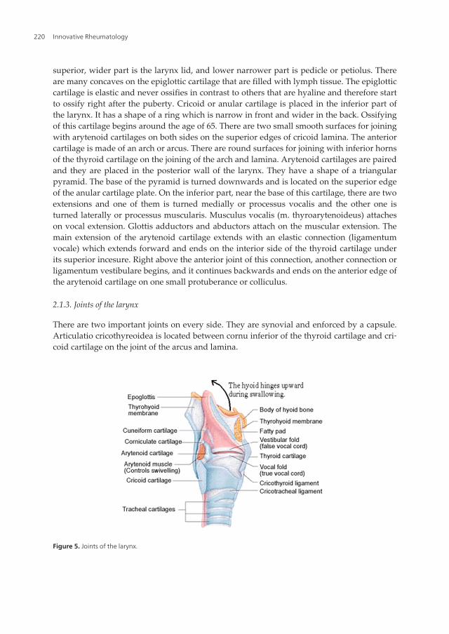
2.1.3. Joints of the larynx
There are two important joints on every side. They are synovial and enforced by a capsule.Articulatio cricothyreoidea is located between cornu inferior of the thyroid cartilage and cri‐coid cartilage on the joint of the arcus and lamina.
Figure 5. Joints of the larynx.
Innovative Rheumatology220
Movements in this joint are rotation around the horizontal arytenoid axis and very re‐stricted movements of sliding. Articulatio crycoarytenoidea is located between the aryte‐noid base and joint surfaces on superior edge of the cricoid cartilage. Movements in thejoint are rotation around the vertical arytenoid axis and movements of sliding when ary‐tenoids adduct or abduct, Figure 5.
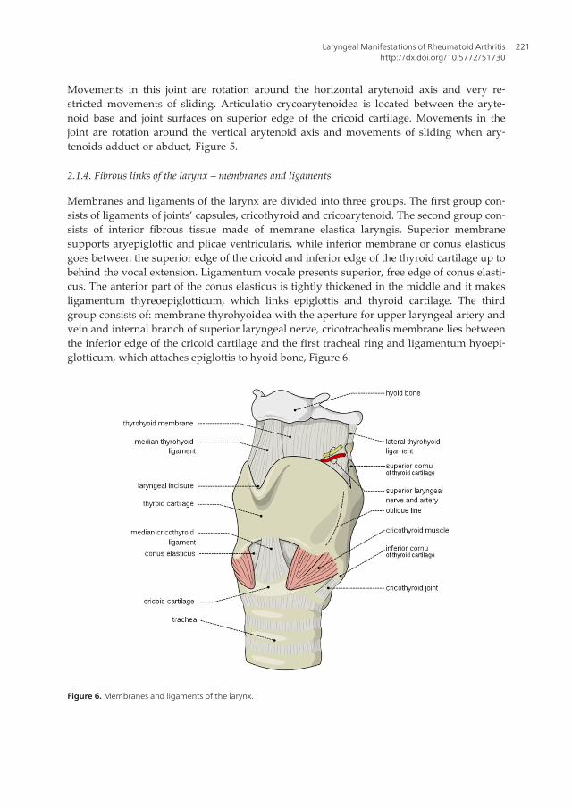
2.1.4. Fibrous links of the larynx – membranes and ligaments
Membranes and ligaments of the larynx are divided into three groups. The first group con‐sists of ligaments of joints’ capsules, cricothyroid and cricoarytenoid. The second group con‐sists of interior fibrous tissue made of memrane elastica laryngis. Superior membranesupports aryepiglottic and plicae ventricularis, while inferior membrane or conus elasticusgoes between the superior edge of the cricoid and inferior edge of the thyroid cartilage up tobehind the vocal extension. Ligamentum vocale presents superior, free edge of conus elasti‐cus. The anterior part of the conus elasticus is tightly thickened in the middle and it makesligamentum thyreoepiglotticum, which links epiglottis and thyroid cartilage. The thirdgroup consists of: membrane thyrohyoidea with the aperture for upper laryngeal artery andvein and internal branch of superior laryngeal nerve, cricotrachealis membrane lies betweenthe inferior edge of the cricoid cartilage and the first tracheal ring and ligamentum hyoepi‐glotticum, which attaches epiglottis to hyoid bone, Figure 6.
Figure 6. Membranes and ligaments of the larynx.
Laryngeal Manifestations of Rheumatoid Arthritishttp://dx.doi.org/10.5772/51730
221

2.1.5. Muscles of the larynx
Muscles of the larynx are divided into A. internal and B. external. A. Internal muscles (Fig‐ure 7) are placed between some of the cartilages and are divided into 1. abductors, 2. adduc‐tors, 3. tensors and 4. covers of the larynx lumen. 1. Laryngeal abductors are two muscles oneach side of the larynx, m. cricoarytenoideus posterior s. posticus. Its function is to abductvocal cords from the middle line and thus to open glottis. 2. Adductors adduct vocal cordsto the middle line and close glottis. These are: a) m. cricoarytenoideus lateralis s. lateralis b)m. interarytenoideus s. transversus c) m. thyroarytenoideus or pars externa s. externus. 3.Laryngeal tensors are: a) m. cricothyreoideus s. anterior is external laryngeal tensor. It ad‐ducts the thyroid cartilage to the cricoid cartilage from the anterior side, and thus tights thevocal cords intermediately. b) m. thyroarytenoideus or pars interna s. internus s. vocalis isknown as internal larynx tensor and forms a vocal cord. 4) covers of the larynx lumen: a) m.interarytenoideus – pars obliqua has a role of glottic sphincter, b) m. aryepiglotticus repre‐sents an extension of pars transversa muscles interarytenoidus into aryepiglottic plicae andhas a function of a supraglottic sphincter. B. External laryngeal muscles: a) m. sternothyroi‐deus is pulling the larynx downwards, b) m. thyrohyoideus is raising the larynx if hyoid isfixed, in other words, lowering hyoid bone if the larynx is fixed.
Figure 7. Internal laryngeal muscles.
Innovative Rheumatology222

2.1.6. Laryngeal mucosa
Laryngeal mucosa encases the whole of its cavity. It is separated from cartilage and muscles bysubmucosa and elastic membrane. On the superior larynx aperture, mucosa continues its wayforward across the superior edge and anterior side of epiglottis into the mucosa of the root ofthe tongue, laterally and backwards into the pharynx mucosa, then it encases the external sur‐face of the anterior wall of the larynx and it continues downwards into the trachea. Laryngealmucosa is tightened very loosely to submucosa and elastic aperture, except at the front on theepiglottic cartilage and at the back on Santorini cartilages and superior ends of the arytenoidcartilages. There are many tubuloalveolar glands of serous or seromucous nature. Apart fromtiny glands that can be found almost everywhere in laryngeal mucosa, three main groups ofglands are placed :a) on the top of laryngeal epiglottic side and in the root of the lingual side, b)in the wall of ventriculus Morgagni and c) in the plicae ventricularis. Otherwise, there aren’tmucous glands on the free edges of the vocal cords. Larynx mucosa epithelium can be: 1) pla‐coid-layered; 2) cylindrical-ciliary, respiratory type and 3) transitory pseudo-layered cylindri‐cal epithelium (transitory type). Placoid-layered epithelium encases the vocal cords, superiorlarynx aperture and it goes downwards into the vestibulum, free epiglottic edge, epiglottic pli‐cae, internal side of the arytenoid and interarytenoid space. This epithelium covers the wholeglottis from the anterior commissure at the front and back to processus vocalis and interior sideof arytenoid. It goes under the free edge of glottis and laterally towards the plicae ventricularisfor 6 to 8mm. There are 20 to 30 rows of cells in that region. Under normal conditions, the islesof placoid epithelium can be found scattered in the zones of cylindrical-ciliary epithelium andin the larynx vestibulum. The passage between placoid-layered and ciliar epithelium in thelevel of the vocal cords and free edges of the larynx is manifested as transformation of superfi‐cial planocells into cylindrical cells. And opposite, in the level of placoid cells isle, these varia‐tions can continue one to another without the passage. The most important of all the layers isthe superficial layer which is made of more planocells that desquamate but don’t keratinize.Under the ifluence of toxins, chronic irritation and inflammation, this epithelium can showsimilar characteristics as horny layer and epithelium extensions go into the derm so that papil‐lae become higher. Cylindrical-ciliary epithelium of respiratory type covers the remaining,largest part of the larynx surface.
In 20 % of the cases, the anterior ends of the vocal cords are characterized by a narrow bandof epithelium of transitory type. This epithelium is sometimes present in subglottis. Mucosaof the vocal cords has one macroscopic characteristic of a special importance like Reinkspaces which goes along the whole length of the vocal cords between mucosa and vocal liga‐ment. Macroscopically, there isn’t a point of joining mucosa with a vocal ligament.
2.1.7. Blood vessels, nerves and lymph vessels of the larynx
Arterial vascularization of the larynx comes from: a) a. thyreoidea superior (branch a. carotisexterna) via its branches aa. thyreoidea superior et media. These arteries go into the larynxon the posterior part of the thyrohyoid membrane, b) a. thyreoidea inferior (branch arteriasubclavia) that follows n. recurrens on its way into the larynx. Veins of the larynx follow ar‐teries of the same name. The larynx is innervated by the branch n. vagus: 1) n. laryngeus
Laryngeal Manifestations of Rheumatoid Arthritishttp://dx.doi.org/10.5772/51730
223
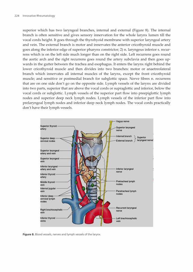
superior which has two laryngeal branches, internal and external (Figure 8). The internalbranch is often sensitive and gives sensory innervation for the whole larynx lumen till thevocal cords height. It goes through the thyrohyoid membrane with superior laryngeal arteryand vein. The external branch is motor and innervates the anterior cricothyroid muscle andgoes along the inferior edge of superior pharynx constrictor; 2) n. laryngeus inferior s. recur‐rens which is on the left side much longer than on the right side. Left recurrens goes roundthe aortic arch and the right recurrens goes round the artery subclavia and then goes up‐wards in the gutter between the trachea and esophagus. It enters the larynx right behind thelower cricothyroid muscle and then divides into two branches: motor or anaetreolateralbranch which innervates all internal muscles of the larynx, except the front cricothyroidmuscle; and sensitive or postmedial branch for subglottic space. Nerve fibres n. recurrensthat are on one side don’t go on the opposite side. Lymph vessels of the larynx are dividedinto two parts, superior that are above the vocal cords or supraglottic and inferior, below thevocal cords or subglottic. Lymph vessels of the superior part flow into preepiglottic lymphnodes and superior deep neck lymph nodes. Lymph vessels of the inferior part flow intoprelaryngeal lymph nodes and inferior deep neck lymph nodes. The vocal cords practicallydon’t have their lymph vessels.
Figure 8. Blood vessels, nerves and lymph vessels of the larynx.
Innovative Rheumatology224

2.2. Physiology of the larynx
Functions of the larynx can be primary and secondary. Primary functions are phylogeneti‐cally the oldest and they contain the following functions: respirations, protection of airways,swallowing, thorax fixation. Secondary functions are adapted to breathing and swallowingorgans, ant the most important of them is phonation.
2.2.1. Respiratory function of the larynx
Glottis opens one second before the air comes into it by lowering diaphragm. This openingis a consequence of cricoarytenoid muscle contraction which is innervated by nerve recur‐rens and it begins right before the motor activity of n. frenicus. It is led across the respiratorycentre as the activity of n. frenicus, it increases with hypercapnia and ventilatory obstruc‐tion, and it decreases with artery hyperoxygenation and hyperventilation. This activity isdeleted by tracheotomy, as a result of lowered ventilation resistence. Hemoreceptor corpus‐culs are identified in supraglottic mucosa, so their stimulation during hypercapnia decreaseslaryngeal resistence during the inspiration and expiration. Inspiratory dilation of the larynxisn’t distributed to glottis and it doesn’t depend on muscle activity. With inspiratory lower‐ing of the larynx from hyoid downwards, true and false vocal cords contract, arytenoid car‐tilages go laterally and glottis opens. Passive opening of the larynx is still intensified also bythe inspiratory phase, in other words activity of external laryngeal muscles. The result ofglottis opening size variation during respiration allows the larynx to contribute significantlyto internal air resistance during the respiration time, so abduction of the vocal cords produ‐ces glottis dilatation and reduction of opening during the inspiration time, and adduction ofthe vocal cords with glottis constriction produces greater resistence to expiratory air, whichinfluences the depth and level of respiration. These reflex changes are a consequence of reac‐tion on presoreceptors in lungs and subglottic part of the trachea and can help in mixing theair in the lungs. After vagus deafferention, neither inflation nor deflation influence respira‐tory activity in posterior cricoarytenoid. Corrections of glottis opening compensate changesin total air resistance which increased in the nose and bronchi.
2.2.2. Circulatory function of the larynx
Normal respiration, in other words normal air circulation through the larynx, allows normalfunctioning of the circulatory system, heart and blood vessels. Arrhytmia, bradycardia andperiodical cardiac arrest, can result in stimulation of the larynx. The mechanism is connectedto the nerve fibres stimulation that comes from aortal baroreceptors and goes to the centralnervous system across n. laryngeus recurrens, ramus communicans and n. laryngeus superi‐or. These fibres go through the larynx into the deep tissue near thyroid plates and they arestimulated when the larynx is dilated.
2.2.3. Protective function of the larynx
Almost at the same time with respiration, mechanism of protection of the inferior airwaysdeveloped, protecting them from entering foreign bodies, and this protection is guided by
Laryngeal Manifestations of Rheumatoid Arthritishttp://dx.doi.org/10.5772/51730
225

following mechanisms: 1. Sphincter mechanism. There are three sphincters in the larynx andthey are the vocal cords, ventriculous and aryepiglottic plicae so it comes to: adduction ofthe true vocal cords one to another, closing of the false vocal cords one to another and to thebase of epiglottis, posterior commissure of the vocal cords is closed by rotation and adduc‐tion of the arytenoid cartilages, constriction of the false vocal cords by activity of internallaryngeal muscles, lifting and moving the larynx to the front, moving the base of epiglottisbackwards and covering aditus, moving tyreoepiglottic ligamentum to the front. The previ‐ous movements, first of all the tongue base and aryepiglottic plicae, lead to direction of foodbolus to peripheral sinus and thus allows the function of the larynx while swallowing; 2. Re‐flex inhibition mechanism of respiration starts when food bolus touches the posterior wall ofthe pharynx and because of that, breathing stops immediately. Respiration ceases whileswallowing. This is a reflex which results from stimuli coming from the pharynx when foodenters, and they transfer via n. glossopharyngeus and n. vagus. Receptors are the richest inmucosa of the laryngeal side of epiglottis, aryepiglottic and plicae ventricularis and interary‐tenoid area; 3. Cough reflex is weak or it doesn’t exist in newborn children. Reflex centre isin medulla oblongata, and n. vagus is both afferent and efferent part of the reflex arch. Clos‐ing of the false vocal cords is an important moment for this reflex, because adduction of thetrue vocal cords one to another can bring by itself to preventing the air to come out of thelungs. When high subglottic pressure is reached, sphincter mechanism suddenly relaxes andthe air under the accumulated pressure comes out. In this way, matters that initiated this re‐flex also go out. 4. Phonatory function is secondary adapted to respiratory and swallowingorgans. It developed later in phylogenetic development thanks to high differentiation of thecentral nervous system. For proper accomplishment of all the activities that the larynx car‐ries out, there has to be full coordination of both synergistic and antagonistic groups of mus‐cles. During phonation, the vocal cords are in adduction near medial line by the action ofcricothyroid muscles that present the vocal cords tensors. More subtle changes are a conse‐quence of thyroarytenoid muscles action. Medial movement of the vocal cords towards thefalse ones are caused by: a) tension in the vocal cords, b) lowering of subglottic air pressurewith every vibrating aperture of glottis and c) aspirating the air that ran away which isknown as Bernuli phenomenon. The result of such repeated cycles of glottis opening andclosing is freeing of small clouds from subglottic air column which forms sound waves.
2.2.4. Function of thorax fixation
When the larynx is closed, thorax is fixed and serves as adminiculum when some activitiesconnected to the effort are held out: climbing, lifting burden, defaecation, delivery.
2.2.5. Function of emotions
Different mental conditions are expressed over the voice or they cause disorder in it.
2.2.6. Phonation function
This function of the larynx is philogenetically the youngest function which was adapted tobreathing and swallowing organs and it developed thanks to high diferentiation of the cen‐
Innovative Rheumatology226
tral nervous system. Production of voice is a very complex process and it depends on com‐pliance in the body. It presents integral function in which peripheral and central phonatoryorgans take part in. Peripheral organs are: voice activator (lungs, diaphragm), voice genera‐tor (larynx) and resonator (pharynx, mouth, nose and paranasal cavities). Central organs forvoice and speech are located in the central nervous system (cortex, lower centres, reticularsubstance, cerebellum and others). Voice and speech of the humans are under the influenceof psyche, neurovegetative system and endocrine system. One of the most important pre‐conditions for normal development of speech is preserved hearing. A system called ’’feedback’’ participates in forming and maintaining voice and speech. Besides hearing, its mainelements are eyesight and sensibility, and main activities in the sense of creating voice andspeech happen in the central nervous system. Three-dimensional analysis of movements incricoarytenoid joint shows that vocal ligaments, cricothyroid ligament and conus elasticusare the most important in the control of abduction, while posterior cricoarytenoid muscleand conus elasticus take part in restriction of adduction. Vocal ligament makes moving ofthe vocal arytenoid cartilage extension backwards impossible, while cricoarytenoid and pos‐terior capsular ligament restrict movement of vocal extension forward. Anterior capsularligament restricts slanting of the arytenoid cartilage posteriorly and moving of the arytenoidcartilage laterally across joint surface of the cricoid cartilage (Wang, 1998).
2.3. Etiology of rheumatoid arthritis and laryngeal rheumatoid arthritis
Rheumatoid arthritis (RA) is an inflammatory chronic systemic disease of unknown cause thataffects peripheral joints symmetrically and permanently and is often connected to positiverheumatoid factor and/or positive results on anticyclic citrulined peptide immunoglobulins..Annual incidence of RA in the world is 3 patients out of 1000 people, and the prevalence is from0,5 – 5%. The disease is mainly present in some groups of population like North American na‐tives, while it is less present in some other groups like black people in Carribean region. Whengender is taken into consideration, the disease is three times more frequent in women thanmen. It can start at any age but its greatest frequency is in the fourth and fifth decade and itgrows in the old age so it is the highest in people at the age of 25 to 50. Arthritis rate is from 5-6%in the Americans from Asian/Pacific islands, to 12% in Afro-Americans to 16% in white people.Etiology of rheumatoid arthritis includes more assumed theories. One of them says that obesi‐ty, weakness and morning rigidity are important in appearing of this disease. Apart fromjoints, RA can also have extra-articular localizations such as skin, heart, lungs and eyes. Anoth‐er etiological theory implies presence of infectious cause of rheumatoid arthritis (Mycoplasma,Epstein-Bar virus, parvovirus, rubella) but none of the mentioned micro-organisms has beenproved. Some of the medications from the group of medications that modify the disease alsohave antimicrobical activity and they are gold salts, antimalarial medications and minocyclin.In the joints of rheumatoid arthritis patients, bacterial DNA is found, which is also an indirectproof of the bacterial etiology. Autoimmune processes, as one more theory out of the assumedetiological theories, are tightly connected to RA but it isn’t known if they appear as a primaryor secondary process. In the RA patients, autoantibodies aren’t directed towards one immuno‐globulin G but also towards other different antigens, such as nuclear antigens (RA 33, EBNA),citrulined proteins (anti-CCP antibodies), collagen and glucose-6-isomarase phosphate. RA
Laryngeal Manifestations of Rheumatoid Arthritishttp://dx.doi.org/10.5772/51730
227
has an important genetic predisposition and that is one more theory about RA. About 60% ofpatients in the USA has a common epitope HLA-DR4 claster which consists of peptide con‐nected place of the adequate HLA-DR molecule and it is joined with RA. As women sufferfrom RA about three times more often than men, sexual hormones explain one more etiologicaltheory of RA. Complaints almost completely disappear during pregnancy, but in the postme‐nopause period, recurrences of the disease appear. RA rarely appears in women who use oralcontraceptives. It is also described that hyperprolactinemia can be a risk factor for RA. Hyper‐plasia of synovial cells and activation of endothelial cells are early occurences in the pathologi‐cal process that lead to uncontrolable inflammatory process and consequent cartilage and bonedamage. Pathological production and regulation of both pro-inflammatory and anti-inflam‐matory cytokines are found in RA. In tissue immunity, the most important are Th1 CD4 cells,mononuclear phagocytes, fibroblasts, osteoclasts and neutrophils. B lymphocytes can serve asan antigen of the presenting cell and they create autoantibodies (for example rheumatoid fac‐tor – RF). One of the therapeutic possibilities is, therefore, the elimination of B lymphocytespopulation by mononuclear antibodies (for example rituximabR which is often used in combi‐nation with methrotrexate). In RA patients, many other changed cells are found, such as nu‐merous cytokines, hemokines. Other mediators of inflammation are also described: tumornecrosis factor-alpha, interleucins 1 and 6, transforming growth factor – beta, interleucin 8, fi‐broblast growth factor, growth factor received from thrombocytes. Finally, inflammation anduncontrolable synovial proliferation lead to damage of certain tissues, mostly cartilages,bones, strings, ligaments and blood vessels. Other predisposing factors are psychologicalstress and smoking. The so far known risk factors in RA are: female gender, positive family his‐tory, older age, exposure to silicates and smoking (Kuder, 2002). Drinking more than two cupsof coffee a day, high intake of vitamin D, consuming of tea and oral contraceptives reduce riskfor RA (Mikuls, 2002; Merlino, 2004).
2.4. Pathoanatomy of rheumatoid arthritis and laryngeal rheumatoid arthritis
Damage of joints in RA is caused by proliferation of synovial macrophages and fibroblasts,probably as a response to possible autoimmune and infectious triggers. Therefore, it comes tolymphocyte proliferation of perivascular region and proliferation of endothelial cells whichcause new blood vessels to multiply and ingrow. In damaged joints, blood vessels becomeclogged by small clots or inflammatory cells. Furthermore, the progress of process leads to ir‐regular growth (Firestein, 2005; Goldring, 2000). RA in the larynx can manifest in the followingforms: 1) Arthritis of cricothyroid and/or cricoarytenoid joint (Ferdynus-Chromy, 1977; Gotze,1973; Kubiak-Socha, 1973; Woldorf, 1971; De Gandt, 1969; Copeman, 1968), 2) Rheumatoidnodules (Bridger, 1980; Bonner, 1977; Abadir, 1974), 3) Laryngeal myositis, 4) Neuropathy oflaryngeal nervus recurrens and 5) Postcricoid granulomas (Bienenstock H, 1963). Histologicalexaminations of cricoarytenoid joints in RA have shown synovitis as the earliest change thatleads to synovial proliferation, fibrinous deposit, forming of pannus on joint surfaces, erosionof the joint cartilage and finally obliteration and ankylosis of joints. Cricoid necrosis as the lastphase of pathological changes on the cricoid cartilage can cause serious pathophysiologicaldisturbances (Gatland, 1988). Neural atrophy of laryngeal muscles and degenerative changesin laryngeal nerves caused by vasculitis, can follow the degree of affection of cricoarytenoid
Innovative Rheumatology228

joint (Voulgari PV, 2005; Lofgren RH, 1962). Rheumatoid nodules of different size in the larynxare mainly found with seropositive RA. Methotrexate can raise the development of nodules(Kerstens, 1992). Microtrauma, especially a repeated one, can create predisposition for RA. Inthe largest number of cases, nodules are found subcutaneously. A few small nodules can be no‐ticed microscopically in submucous layer, and each of them consists of fibrinous necrosis focussurrounded by histiocytes arranged like palisades. There is a progressive proliferation of en‐dothelial cells and fibroblasts as well as the infiltration of plasma cells and lymphocytes in fi‐brous supporting tissue that surrounds nodules (Webb J, 1972).
2.5. Pathophysiology of rheumathoid arthritis and laryngeal rheumatoid arthritis
Factors joined with RA include the possibility of infectious trigger, genetic predispositionsand autoimmune response. CD4+T cells lead to immunological cascade reaction which caus‐es secretion of cytokines such as tumor necrosis alpha and interleucin 1. Increased formationand expression of TNF-alpha cause inflammation of synovial membranes and joint destruc‐tion. Inflammation, proliferation and degeneration are typical for affected synovial mem‐branes. Joint deformations and working inability happen because of erosion and destroyingof synovial membranes and joint surfaces. Acute obstruction of the superior airways leads toinspiratory stridor, the use of subsidiary respiratory musculature which is manifested by en‐trainment in jugulum, intercostal spaces, supraclavicular pits and in epigastrium, respirato‐ry weaknesses, peripheral cyanosis, state of shock and coma (Lehmann, 1997). Chronicobstruction of the superior airways can lead to hypoxia, hypercapnia and respiratory acido‐sis which cause pulmonary hypertension and cor pulmonale (McGeehan, 1989).
2.6. Clinical picture of rheumatoid arthritis and laryngeal rheumatoid arthritis
Clinical picture of RA can be divided into several groups of clinical manifestations of thedisease, depending on the affected organs/systems: 1) Pulmonary, 2) Cardiovascular, 3)Constitutional, 4) Manifestations from rheumatoid nodules, 5) Eye manifestations, 6) Neuro‐logical, 7) Cutaneous, 8) Hematological, 9) Renal and 10) Hepatic manifestations. 1) Pulmo‐nary manifestations of RA are pleuritic effusion, pulmonary nodules, interstitial fibrosis,pneumonitis and arteritis. 2) Cardiovascular manifestations are coronary disease, inflamma‐tory pericarditis and pericarditis with effusion, myocarditis, mitral valves disease, disorderin conducting. 3) Constitutional manifestations of RA can be high body temperature, asthe‐nia, weight loss, exhaustion and loss of appetite. 4) Rheumatoid nodules can manifest sub‐cutaneously or in pulmonary parenchyma. 5) Eye manifestations are kretoconjuctivitis,episcleritis, scleritis and conjuctivitis. 6) Neurological manifestations of RA can be neuropa‐thies such as carpal tunnel syndrome, multiple mononeuritis, cervical myelopathy, centralnervous system diseases (stroke, hemorrhage, encephalopathy, meningitis). 7) Cutaneousmanifestations in RA can appear as ulcus cruris, palmar erythema and skin vasculitis. 8)Hematological manifestations of RA appear as anemia, thrombocytosis, granulocytopenia,eosinophilia, cryglobunemia and hypertreaclines. 9) Renal manifestations can appear as glo‐merulonephritis, vasculitis and secondary amyloidosis. 10) Hepatic manifestations of RA arecharacterized by high level of liver enzymes. American association of rheumatologists has
Laryngeal Manifestations of Rheumatoid Arthritishttp://dx.doi.org/10.5772/51730
229

set up the following criteria for RA clasification: 1. Rigidity in and around joints that lasts atleast one hour before maximum improvement in the morning hours. 2. Arthritis of three ormore joint regions. At least three joint regions have soft tissue swellings or liquid which wasdiagnosed by a clinician. Fourteen possible regions include left and right superior interpha‐langeal (GIF) joint, metacarpophalangeal (MCF) joint, wrist joint, elbow joint, knee joint, an‐kle and metatarsophalangeal (MTF) joints; 3. Arthritis of wrist joints; at least one region ofcarpus, GIF and MCF are swollen; 4. Symmetric arthritis, in other words simultaneous affec‐tion of the same joint region on both sides of the body. Mutual affection of GIF, MCF andMTF without absolute proportion is also accepted; 5. Rheumatoid nodules are subcutaneousnodules that are present above the osseus bumps or extensory surfaces or surfaces aroundthe regions that are close to joints; 6. Serum RF; 7. Radiographic changes typical for RA onpostero-anterior radiographies of hand and carpus, that have to enclose erosions or dispro‐portional decalcification of bones localized in or on the rims of the most common affectedjoints. Independent osteoarthritic changes are not a criterion for RA. Presence of four out ofseven criteria are enough for diagnosis. Criteria from 1 to 4 have to be present at least 6weeks, and the physician has to establish criteria from 2 to 5. RA is often manifested withconstitutional symptoms such as myelalgia, weight loss, high body temperature, weight lossand exhaustion. Patients can have difficulties with every day activities (dressing up, gettingup, walking, personal hygiene, the use of arms). In most of the patients, RA has a perfidiousstart. It can start with systemic manifestations such as high body temperature, exhaustion,arthralgia and weakness before the appearance of swelling joints and inflammation. In thelower percentage, the patients have abrupt start with acute development of synovitis andextra-articular manifestations. Laryngeal manifestations of rheumatoid arthritis were descri‐bed for the first time in 1880. by Mackenzie M. and later, 1894. Mackenzie GH. (Mackenzie,1880; Mackenzie, 1894). Cricoarytenoid arthritis can be divided into two phases, acute andchronic, and it appears in 27-78% of RA patients (Tarnowska, 2004), and according to someauthors in 17-70% when the research is done laryngoscopically, by computered larynx to‐mography and histopathological cadaveric examinations (Voulgari, 2005). In 55% of pa‐tients, cricoarytenoid arthritis is asymptomatic (Jurik, 1984). At the beginning of the disease,symptoms are mild but usually subclinical. Acute cricoarytenoid arthritis is manifested byfeeling of a foreign body in the throat or a feeling of tension in neck or even feeling of burn‐ing , hoarseness, odynophonia, voice weakness, changes in voice tone, odynophagia or dys‐phagia, pain or the feeling of hardness that becomes worse while speaking, spreading ofpain to ear, feeling of suffocating, coughing, dyspnea or the feeling of rigidity. In the chronicphase of cricoarytenoid arthritis, patients often complain of hoarse speech, stridor that ap‐pears while making an effort, dyspnea, pain while speaking, neck swelling, hoarseness andthese symptoms appear during an infection or during a dream (Braverman, 2007). Laryngealsymptoms during RA vary in their manifestations from 31-75%, while histopathologicalchanges in the larynx are presented postmortem in 90% (Pearson, 1957; Copeman, 1957). Ac‐cording to some authors, stridor appears during exercising in 75% of the cases (Charlin,1985) and can be the result of inflammation and swelling of arytenoid and posterior com‐missure during an acute affection of joint or because of joint ankylosis in the chronic RAphase. The most frequent symptoms are the feeling of a foreign body in the throat (51%),
Innovative Rheumatology230

hoarseness (47%) and voice weakness (29%) (Amernik, 2007). Hoarseness appears only in5% of RA patients (Fisher, 2008), while some other researches have shown that it appears in30% of RA patients (Segebarth, 2007). Hard RA can be manifested by laryngeal obstructionand can lead to heart, pulmonary and fatal complications. Rheumatoid nodules of the larynxare often manifested by hoarseness and coughing.
2.7. Diagnosis of rheumatoid arthritis and laryngeal rheumatoid arthritis
Thorough anamnesis, careful examination of joints and periarticular soft tissue structures,as well as laboratory and imaging results are necessary for right diagnosis of RA. Nonelaboratory test is specific for RA, so its diagnosis is primary clinical. A clinical examina‐tion can establish that mostly small wrists and ankles are affected relatively symmetrical‐ly. The most frequently affected joints, with decreasing frequency, are MCF, wrist joint,GIF, knee joint, MTF, shoulder joint, ankle, cervical spine, hip and temporomandibularjoints. The patient whom we treated, had prominent changes on the wrists which can beclearly seen in the Figure 9.
Figure 9. Hand changes of reumatoid arthritis.
Joints show inflammation with swelling, painfulness, locally high temperature and restrictedmovement. Atrophy of interosseus muscles of hands is a typical early sign. Joints and chordaedamage can lead to deformities such as ulnar deviation, hammer like fingers and occasionallyjoint rigidity. Other muscle-skeletal manifestations that are usually found during the examina‐tion are tendosynovitis and joined chordae rupture during the ligament and chordae affection,the most often affected are chordae of the fourth and fifth finger extensor, periarticular osteo‐porosis during the localized inflammation, generalized osteoporosis during systemic chronicinflammation, changes connected to immobilization, or corticosteroid therapy and syndrom ofcarpal tunnel. Cutaneous changes in RA appear like subcutaneous nodules, often along the
Laryngeal Manifestations of Rheumatoid Arthritishttp://dx.doi.org/10.5772/51730
231

pressure points (for example olecranon), ulceration of feet cutis, rashes in vasculitis, palmar er‐ythema, gangrenous pyodermia. Vascular lesions of cutis can be manifested as palpable pur‐pura or cutis ulceration. Cardial changes in RA lead to increased cardiovascular morbidity andmortality. Myocardial infarction, myocardial disfunction and constrictive pericarditis are rare.Affection of lungs in RA can have several forms like pleural effusion, interstitial fibrosis, nod‐ules (Caplan syndrom) and bronchiolitis obliterans, in other words ogranized pneumonia. Ingastrointestinal tract, the affection of intestines is a side effect of medication action, inflamma‐tion and other diseases. Kidneys are usually intact by direct action of RA. Secondary kidneydamage often happens during medicamentous therapy (nonsteroidal anti-inflammatorydrugs, gold salt, cyclosporin), inflammation (amyloidosis) and joined diseases (Sjögren syn‐drom with kidney tubular disorders). Vascular lesions can affect any organ, but are oftenfound on cutis where they can be manifested as palpable purpura, cutis ulcerations or digitalinfarcts. Hematological disorders are often manifested by secondary anemia which is normo‐chromic-normocytic type, thrombocytosis and eosinophilia. Affection of nerves is often as innervus medianus syndrom in the carpal tunnel. Vascular lesions, mononeuritis multiplex andcervical myelopathy can cause serious neurological prolapses. RA is manifested on eyes likekeratoconjuctivitis sicca, as well as episcleritis, uveitis and nodular scleritis which can lead toscleromalacia. American college for rheumatology has determined criteria for progression, re‐mission and functional state of RA patient. A) RA progression (clinical and radiologicalstages): Stage 1 (early RA) is characterized by: a) absence of destructive changes during theroentgenography examinations and b) possible radiography presence of osteoporosis; Stage 2(advanced progression): a) radiography evidence of periarticular osteoporosis with or withoutlight subchondral destruction of bones, b) possible light destruction of cartilage, c) possible re‐striction of joint movements, without joint deformities, d) joined myatrophy, e) possible softtissue extra-articular lesions (for example nodules, tenosynovitis). Stage 3 (very advanced pro‐gression): a) radiographic evidence of cartilage and bone destruction followed by periarticularosteoporosis, b) joint deformities (for example subluxation, ulnar deviation, hyperextension)without fibrous or osseus ankylosis, c) massive myatrophy, d) possible extra-articular lesionsof soft tissue (for example nodules, tenosynovitis); Stage 4: a) fibrous or osseus ankylosis and b)criteria for stage 3. B) RA remission ( ≥ 5 below induced states that last at least two months con‐stantly): a) morning rigidity that doesn’t stop for 15 minutes, b) without weakness, c) withoutpain in joints, d) without cracking in joints or pain while moving, e) without soft tissue swel‐ling in joints or chorda wraping, f) erythrocyte sedimentation lower than 30 mm/h in women orlower than 20mm/h in men. C) Functional status of RA patients: a) Category I – completely ableto fullfil everyday activities, b) Category II – able to fullfil regular personal hygiene and activi‐ties connected to their profession but limited in other activities, c) Category III – able to fullfilthe activities of regular personal hygiene but limited in activities connected to their proffessionand other activities that aren’t connected to their profession, d) Category IV – limited to fulfillregular activities for personal hygiene, profession and activities that aren’t connected to theirprofession. American College of Rheumatologists (ACR) and European League Against Rheu‐matism (EULAR) have regulated new criteria for classification of early RA, which include jointaffection, autoantibodies status, answer to acute phase and symptoms duration (Aletaha,2010). ACR/EULAR 2010 criteria: A) Joint affection (0-5): one median to large joint (0), two to
Innovative Rheumatology232
ten median to large joints (1), one to three small joints (large joints aren’t included) (2), four toten small joints (small joints aren’t included) (3), more than ten joints (at least one small joint)(5); B) Serology (0-3): negative RF and negative cell-Purkinje antibodies (APCA) (0), Light posi‐tive RF or light positive APCA (2), high positive RF or high positive APCA (3); C) Reactants ofacute phase (0-1); normal CRP and normal percentage of erythrocyte sedimentation (ESR) (0),Abnormal CRP or abnormal ESR (1); D) Symptoms duration: a) shorter than 6 weeks (0), 6weeks and more (1). Cut point for RA is 6 weeks or more. RA can be diagnosed in patients ifthey have: a) atypical erosions or b) long lasting disease which fullfils the previous classifica‐tion criteria. Large joints are defined as: shoulder joints, elbow joints, hip joints, knee joints andankles. Small joints are defined as: MCF, PIF, from the second to fifth MTF and interphalangealthumb joints and carpus joints. Course of disease can be short and limited or progressive andhard. The following laboratory tests are necessary to be carried out: complete blood count withdifferential count, rheumatoid factor, erythrocyte sedimentation, C-reactive proteins, fibrino‐gen, haptoglobin, alpha-1-acid glycoprotein, alpha-1-trypsin, S-amyloid-A protein and hema‐tocrit (Guerra, 1992). Erythrocyte sedimentation and C-reactive protein give the bestinformation about the presence of acute phase of RA, but thrombocytosis, low level of iron inthe serum and low values of hemoglobin also point to active disease (Crassi, 1998). It is also im‐portant to examine the function of liver and kidneys because of further choice of medicaments,which shows that it is necessary to carry out a complete biochemical blood analysis. Diagnosisof rheumatoid changes in the larynx includes anamnesis, clinical examination, videolaryngo‐scopy, computered tomography and electromyography (Amernik, 2007). Indirect laryngosco‐py shows changes in the larynx in 32% of RA patients, unlike computered tomography wherethe changes are found in 54%, so indirect laryngoscopy reveals mucous and great pathoana‐tomical changes, and computered larynx tomography reveals structural lesions (Lawry, 1984).Cricoarytenoid arthritis can be asymptomatic because many RA patients have pathologicalchanges in cricoarytenoid joint, proved by computered tomography, but they don’t have lar‐yngeal difficulties (Brazeau-Lamontagne, 1986). Laryngoscopy in acute cricoarytenoid arthri‐tis shows light red medially expressed swellings in the region of arytenoid, epiglottis,cricoarytenoid arthritis and vocal cords nodules that can look normal or very edematous. Inchronic cricoarytenoid arthritis, we can find thickened mucosa in the region of arytenoid, in‐terarytenoid pachydermia, uneven rima glottidis, called “bamboo nodules” because of theirappearance that reminds of knots on the bamboo branch. During laryngoscopy of bamboonodules, subepithelial sallow mass on upper surface of glottis is noticed, often mushroomlikeshape and directed by its longer axis transversely to the vocal cords, and they are usually sur‐rounded by hyperemic mucosa (Immerman, 2007; Hilgert, 2008). Direct fiber laryngoscopy canestablish pathological changes in the larynx in 75% of RA patients (Brazeau-Lamontagne,2005). The most frequent rheumatoid changes in the larynx are hyperemia of the mucosa in ar‐ytenoid subregion in 41% of the patients and edema of the same region in 28% of the patients(Amernik, 2007). Safe diagnosis of laryngeal RA is set in direct laryngoscopy by arytenoid pal‐pation when mechanical restriction of movements in cricoarytenoid joint can be proved(Woods, 2007). Computered endovideostroboscopy allows examination of the way mucosa vi‐brates on the affected side, determination of lesion depth in mucous layer, confirming theunique characteristics of these lesions. In this way, we can diagnose disorders of adduction and
Laryngeal Manifestations of Rheumatoid Arthritishttp://dx.doi.org/10.5772/51730
233
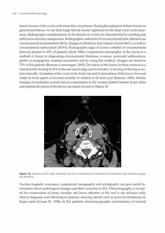
lateral torsion of the vocal cords when they are present. During the palpation of these lesions ingeneral anesthesia, we can find tough fibroid masses tightened on the deep vocal cords struc‐tures. Radiographic manifestations of the disease on wrists are characterised by swelling andmild juxta-articular osteoporosis. Radiographic indicators of cricoarytenoid joint affection arecricoarytenoid prominention (46%), changes in thickness and volume of joint (46%), as well ascricoarytenoid subluxation (39,9%). Radiographic signs of erosive arthritis of cricoarytenoidjoint are present in 45% of patients (Jurik 1984). Computered tomography of the larynx is amethod of choice in diagnosing cricoarytenoid thickness, erosions, arytenoid subluxations,glottic or aryepiglottic nodules asymmetry and by using this method, changes are found in72% of RA patients (Brazeau-Lamontagne, 2005). Deviation of the larynx in three surfaces is acharacteristic finding for RA in the advanced stage and it includes: 1) moving of the larynx an‐terio-laterally, 2) rotation of the vocal cords clockwise and 3) slantedness of the larynx forwardwhile its front aspect is lowered caudally in relation to its front part (Keenan, 1983). Similarchanges on multislice scanner larynx examination in the woman patient treated at our clinicand triploid deviation of the larynx can clearly be seen in Figure 10.
Figure 10. Sclerosis of the right arytenoid due the cricoarythenoid rheumatoid involvement. Also triplanar laryng‐eal deviation.
Nuclear magnetic resonance, computered tomography and scintigraphy can give useful in‐formation about pathological changes and their extension in RA. Ultrasonography is accept‐ed for examination of joints, chordae and bursa affection in RA and it can advance earlyclinical diagnosis and following of patients, showing details such as synovial thicknesses infinger joints (Grassi W, 1998). In RA patients, electromyographic examinations of internal
Innovative Rheumatology234
thyroarytenoid muscles show mutually normal bioelectric stimulation of thyroarytenoidmuscles during phonation, while at rest there is no denervation activity (Tarnowska, 2004).
2.8. Complications of laryngeal rheumatoid arthritis
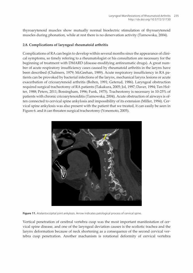
Complications of RA can begin to develop within several months since the appearance of clini‐cal symptoms, so timely refering to a rheumatologist or his consultation are necessary for thebeginning of treatment with DMARD (disease-modifying antireumatic drugs). A great num‐ber of acute respiratory insufficiency cases caused by rheumatoid arthritis in the larynx havebeen described (Chalmers, 1979; McGeehan, 1989). Acute respiratory insufficiency in RA pa‐tients can be provoked by bacterial infections of the larynx, mechanical larynx lesions or acuteexacerbation of cricoarytenoid arthritis (Bolten, 1991; Geterud, 1986). Laryngeal obstructionrequired surgical tracheotomy of RA patients (Takakura, 2005; Jol, 1997; Daver, 1994; Ten Hol‐ter, 1988; Peters, 2011; Bossingham, 1996; Funk, 1975). Tracheotomy is necessary in 10-25% ofpatients with chronic cricoarytenoiditis (Tarnowska, 2004). Acute obstruction of airways is of‐ten connected to cervical spine ankylosis and impossibility of its extension (Miller, 1994). Cer‐vical spine ankylosis was also present with the patient that we treated, it can easily be seen inFigure 6. and it can threaten surgical tracheotomy (Yonemoto, 2005).
Figure 11. Atalantoccipital joint ankylosis. Arrow indicates patological process of cervical spine.
Vertical penetration of cerebral vertebra cusp was the most important manifestation of cer‐vical spine disease, and one of the laryngeal deviation causes is the scoliotic trachea and thelarynx deformation because of neck shortening as a consequence of the second cervical ver‐tebra cusp penetration. Another machanism is rotational deformity of cervical vertebra
Laryngeal Manifestations of Rheumatoid Arthritishttp://dx.doi.org/10.5772/51730
235

caused by asymmetric osseous erosions (Keenan, 1983). Besides cricoarytenoid and cervicalvertebra ankylosis, treatment of RA patients is also difficult because of temporomandibularjoint ankylosis (Okuda, 1992; McGeehan, 1989). These pathological changes of RA patientscan make endotracheal intubation very difficult and dangerous, especially if there is a trip‐loid deviation of the larynx on computered tomography (Bamshad, 1989). Rheumatoid cri‐coarytenoid arthritis was complicated by ulcerous necrosis of cricoid esophagostenosis andtherefore the patient underwent total laryngectomy (Montgomery, 1980).
2.9. Differential diagnosis of rheumatoid arthritis and laryngeal rheumatoid arthritis
Differential diagnosis is directed to artropathies caused by infection, seronegative spondy‐loartropathies and other connective tissue diseases such as systemic lupus eritematodes(Harris, 2005; Akil, 1995; Nanke, 2001).
RA should be distinguished from a wide range of diseases that are characterized by clinical‐ly prominent synovitis, such as viral, reactive and psoriatic arthritis as well as enteroarthritisand we can come to differential diagnosis by eliminating, because there isn’t a specific testfor RA (Grassi, 1998). Arthritis of cricoarytenoid joint is also caused by gout, mumps, tuber‐culosis, syphilis, gonorrhea, Tietz syndrom, lupus eritematodes and injuries (Fried, 1991).Rheumatoid nodules of the larynx as initial signs of systemic lupus eritematodes are descri‐bed in the literature (Schwartz, 1980). Rheumatoid nodules of the larynx can be differential‐ly diagnostic problem if they are mixed up with vascular lesions (Friedman, 1975). It is oftenimportant to distinguish by differential diagnosis asthma and psychoneurosis from cricoary‐tenoid joint arthritis which is rarely manifested by acute laryngeal obstruction and colapse(Leicht 1987; Absalom 1998). Secondary amyloidosis during rheumatoid arthritis or secon‐dary Sjögren syndrom are rarely causes for laryngeal symptoms (Bayar, 2003). The case ofRA and systemic sclerosis union is also described, which led to mutual immobility of the vo‐cal cords and was manifested by dysphonia and dyspnea as main symptoms and treatmentby sphygmic doses of methylprednisolone led to slow improvement (Ingegnoli, 2007). Withother autoimmune diseases, transversal, white-yellow, striped lesions can be found, in themedian part of membranous link of the vocal cords, mostly bilateral, but they aren’t sym‐metrical (Ylitalo, 2003). Rheumatoid cricoarytenoid arthritis should be distinguished fromneurogenic disorders, traumatic changes, infections, neoplastic processes and psychosomat‐ic illnesses (Bolten, 1991; Chen, 2005). The most difficult differential diagnosis is in cricoary‐tenoid ankylosis and bilateral paresis/paralysis of recurrent laryngeal nerves. Mutualimmobility of cricoarytenoid joint and Hashimoto thyroiditis are next differentially diagnos‐tic problem which requires multidiscipline approach (Stojanovic, 2010). Then normal elec‐tromyogram of vocal muscles and fixation of cricoarytenoid joints during laryngoscopy setby application of laryngoscopic claws, confirm the diagnosis of ankylosis. Differential diag‐nosis of these two conditions is possible by using electromyography and laryngoscopy, andmutual fixation of the arytenoid cartilages is confirmed with long-term endotracheal intuba‐tion (in 68,8% of patients), short-term endotracheal intubation (in 9,4%), Wegener granulo‐matosis (in 9,4%) of rheumatoid arthritis (in 6,3%) of previous surgery in the larynx (in3,1%) and caustic ingestion (in 3,1%) (Eckel, 2003).
Innovative Rheumatology236

2.10. Treatment of rheumatoid arthritis and laryngeal rheumatoid arthritis
The aims of early prevention and early treatment of RA are to reduce pain, inflammationand inability, to prevent radiologically found damages and progression and to reduce thedevelopment of comorbidity. Joint damage in rheumatoid arthritis begins a few weeks sincethe begining of the disease symptoms and that’s why the early treatment reduces the diseaseprogression rate (Emery, 2002). Pharmacotherapy generally includes several groups of med‐ications: nonsteroidal anti-inflammatory drugs (NSAID) for pain control, oral or intra-artic‐ular glucocorticoids in low doses and start with DMARD. “Reverse pyramid” approach isrequired in RA treatment today, when DMARD are started immediately in order to slowdown the disease progression as soon as possible (Rindfleisch, 2005). This approach is ac‐cepted on the basis of several facts: joint damages start in the early phase of the disease(Emery, 2002), DMARD have a significant use when they are used in the early phase of thedisease, the uses of DMARD can be helped when the medications are used in the combina‐tion (Pincus, 1999; Lipsky, 2000; Weinblatt, 2004), a great number of medications from thisgroup with positive evidence of useful effects are accessible. RA patients of medium stageand normal radiographic results should start the treatment with hydroxychloroquine, sul‐phasalazine or minocycline, although methotrexate is also a possibility. Patients with heavi‐er stage of disease or radiographic changes should start treatment with methotrexate. If thedisease symptoms aren’t controlled well by mentioned medications, leflunomid or com‐bined therapy should be taken into consideration (methotrexate together with one medica‐tion of newer generation). For initial RA treatment for reducing pain and joint swellingtogether with the combination of the mentioned medications, NSAID, salicylate or ciclooxy‐genease-2 inhibitors should be used. These medications can’t be used independently be‐cause they don’t change clinical course of RA. Glucocorticoids, usually in the dose that isequivalent to 10 mg of prednisone a day are highly important for freeing from RA symp‐toms and they can slow down joint damage (Kirwan, 1995). Their dosing should be kept atminimum because of a great risk of unwanted effects, which include osteoporosis, cataract,hyperadrenocorticism and altered glycemia levels. American college for rheumatology hasrecommended the intake of 1500mg of calcium and 400-800 IU of vitamin D a day. The mostoften used medications for RA treatment are methotrexate, hydroxychloroquine, sulphasala‐zine, leflunomide, infliximab and etanercept. Newer DMARD are leflunomide, antagoniststumor necrosis factor (TNF) and anakinra. Pharmacotherapeutic approaches for RA are verydifferent depending on certain studies. One of such approaches is a combination of thesetwo medications from the DMARD group, mainly methotrexate and sulphasalazine or me‐thotrexate and cyclosporine (Dougados, 1999; Tugwell, 1995). Combination of methotrexate,sulphasalazine and high doses of corticosteroids has brought up to prolonged effects on ra‐diographic progression in comparison to monotherapy by sulphasalazine (Landewe, 2002).In the two-year study, 197 RA patients were chosen by coincidence to take therapeutic pro‐tocol with four medications, methotrexate, sulphasalazine, hydroxychloroquine and predni‐solone (5mg/a day) or individual medication from DMARD where it has been noticed thatgreater number of patients in remission got combined therapy, while fewer number of pa‐tients in remission were in the group of monotherapy by some other medication fromDMARD group (Korpela, 2004). In some studies, cricoarytenoid arthritis treatment in a 65-
Laryngeal Manifestations of Rheumatoid Arthritishttp://dx.doi.org/10.5772/51730
237
year-old male patient and 56-year-old female patient was carried out by local, intra-articularinjections of triamcinolone combined with prednisolone (Jol, 1997; Simpson, 1980; Habib,1977). Systemic application of corticosteroids brought up to significant mobility of the aryte‐noid cartilages in a 63-year-old patient (Jurik, 1985). Beclomethasone diproprionat in thetreatment of rheumatoid changes in the larynx was useful (Sladek, 1983). Surgical approachto immobility of the vocal cords from paramedial position, in other words rheumatoid anky‐losis of cricoarytenoid joints implies mobilizational and laterofixational techniques (Ejnell,1985). Arytenoid adduction surgery was carried out successfully in a 57-year-old female pa‐tient who didn’t have dyspnea year and a half after the surgery (Kumai, 2007). Endoscopicarytenoidectomy is usually a surgery of choice (Koufman, 2003).
3. Conclusion
Rheumatoid arthritis is a disease of unknown cause with several assumed etiological theo‐ries, but pathoanatomical and pathophysiological changes are mostly familiar. It is manifest‐ed on different organs and tissues and about 25% of all patients have clinical manifestationsin the larynx. Patients with RA manifested on more than one joint, must be sent to and ex‐amined by a rheumatologist 6 weeks since the beginning of the disease symptoms. Joint andbones swelling points to early arthritis especially if at least two joints are involved and/ormorning rigidity lasts longer than 30 minutes and/or if there is an affection of metacarpo‐phalangeal and/or metatarsophalangeal joints (Emery, 2002). Following the disease courseincludes counting of painful and swollen joints, complete cooperation between the patientand the physician, determining erythrocyte sedimentation and C - reactive protein. Diseaseactivities should be followed in the intervals from 1 to 3 months until the remission period isreached. Structural damages must be followed rardiographically every 6 to 12 months dur‐ing the first several years. Family doctor must think about the structural damage of the lar‐ynx in the patients with advanced arthritis and he must send these patients to periodicalotorhinolaryngological examinations every 1 to 3 months. At that time, it is necessary to car‐ry out indirect laryngoscopy and graphic flow/volume which is enough for the initialscreening. Forced inspiratory/expiratory relationship between the flow and volume providea simple non-invasive test for revealing stenosis in upper airways. Pathological results ofscreening cause additional examinations by fiberoptical laryngoscopy, laryngomicroscopywith the examination of the arytenoid cartilages fixation, helped by multislice scanner lar‐ynx examination every six months to a year, computered stroboscopy and electromyogra‐phy of the larynx. This is important because laryngoscopy provides better view of themucous and functional integrity preservation, and multislice scanner larynx examination of‐fers more precise visualisation of the structural changes. Periodical otorhinolaryngologicalexaminations should be routine when treating patients with rheumatoid arthritis and theyare always undertaken when family doctor and/or the rheumatologist of the clinic find thesmallest disease progression on laryngeal and/or extralaryngeal localizations. Then, togetherwith basic rheumatological therapy, intra-articular application of corticosteroid medicationsneeds to be applied into every sick cricoarytenoid joint. When conservative treatment fails,
Innovative Rheumatology238

after providing airways by tracheostomy, it is indicated to carry out endoscopic arytenoidec‐tomy which presents a surgery of choice. For patients of extremely bad state to bear laryng‐eal surgery, or with those patients where surgical procedures failed to provide adequateairway, permanent tracheostomy is the final therapeutic possibility. In RA patients where asurgery in general endotracheal anesthesia is indicated, the otorhinolaryngologists informthe anesthesiologist after every examination about every laryngeal disorder. Anesthesiologi‐cal risk is always present in the patients with rheumatoid arthritis in the larynx during en‐dotracheal intubation and immediately after the extubation (Segebarth, 2007). In thesepatients, a careful search for cricoarytenoid arthritis is the basic thing, especially in thosewith laryngeal stridor which can be inforced after general anesthesia.
Acknowledgements
I would like to give credit to my wife Tatjana, my daughter Nina and my son Luka fortheir support and immense patience during elaboration of this chapter, as well as for theirrecognition of the importance of this work in diagnosis and treatment of all rheumatoidarthritis patients.
Author details
Stevan Stojanović* and Branislav Belić
*Address all correspondence to: [email protected]
Faculty of Medical Sciences, University of Kragujevac, Republic of Serbia
References
[1] Firestein, G. S. (2005). Etiology and pathogenesis of rheumatoid arthritis. Kelley’sTextbook of rheumatology, Ruddy, S, Harris, E.D, Sledge, C.B. et al, (Ed.), 996-1042, 7th ed,W.B. Saunders, 0721601413, Philadelphia, USA.
[2] Aletaha, D., Neogi, T., Silman, A. J., Funovits, J., Felson, D. T., & Bingham, C. O.(2010). Rheumatoid arthritis classification criteria: an American College of Rheuma‐tology/European League Against Rheumatism collaborative initiative. ArthritisRheum, 62(9), 2569-81.
[3] Goldring, S. R. (2000). A 55-year-old woman with rheumatoid arthritis. JAMA, 283,524-31.
[4] Kuder, S. A., Peshimam, A. Z., & Agraharam, S. (2002). Environmental risk factorsfor rheumatoid arthritis. Rev Environ Health, 17, 307-15.
Laryngeal Manifestations of Rheumatoid Arthritishttp://dx.doi.org/10.5772/51730
239

[5] Merlino, L. A., Curtis, J., Mikuls, T. R., et al. (2004). Vitamin D intake is inversely as‐sociated with rheumatoid arthritis: results from the Iowa Women’s Health Study. Ar‐thritis Rheum, 50, 72-7.
[6] Emery, P., Breedveld, F. C., Dougados, M., et al. (2002). Early referral recommenda‐tion for newly diagnosed rheumatoid arthritis: evidence based development of aclinical guide. Ann Rheum Dis, 61, 290-7.
[7] Harris, E. D. (2005). Clinical features of rheumatoid arthritis. In: Kelley’s Textbook ofrheumatology Ruddy, S, Harris, ED, Sledge, C.B, et al, (Ed.), 1043-78, 7th ed, W.B. Saun‐ders, 0721601413, Philadelphia, USA.
[8] Akil, M., & Amos, R. S. (1995). ABC of rheumatology. Rheumatoid arthritis-I: clinicalfeatures and diagnosis. BMJ, Review, 310, 587-90, 0959-8138.
[9] Bridger, M. W., Jahn, A. F., & van Nostrand, A. W. (1980). Laryngeal rheumatoid ar‐thritis. Laryngoscope, 90(2), 296-303, 0023-852X.
[10] Lofgren, R. H., & Montgomery, W. W. (1962). Incidence of Laryngeal Involvement inRheumatoid Arthritis. N Engl J Med, 267, 193-5.
[11] Bonner, F. M. 3rd. (1977). Rheumatoid nodule. Pathological quiz case 1. Arch Otolar‐yngol, 103(2), 112-4.
[12] Ferdynus-Chromy, J., & Wagner, T. (1977). Comparative studies of synovial mem‐branes of cricoarytenoid and knee joints in rheumatoid arthritis. Reumatologia, 15(1),13-21.
[13] Grassi, W., De Angelis, R., Lamanna, G., & Cervini, C. (1998). The clinical features ofrheumatoid arthritis. Eur J Radiol, 27(1), 18-24, 0720-048X.
[14] Mackenzie, M. (1880). Diseases of the pharynx, larynx and trachea, William Wood & Co.,New York.
[15] Mackenzie, G. H. (1894). Rheumatism of the larynx. Edin Med J, 40, 507-9.
[16] Voulgari, P. V., Papazisi, D., Bai, M., et al. (2005). Laryngeal involvement in rheuma‐toid arthritis. Rheumatol Int, 25(5), 321-5.
[17] Kerstens, P. J., Boerbooms, A. M., Jeurissen, M. E., et al. (1992). Accelerated nodulosisduring low dose methotrexate therapy for rheumatoid arthritis. An analysis of tencases. J Rheumatol, 19, 867-71, 0315-162X.
[18] Fried, M.P., & Shapiro, J. (1991). Acute and chronic laryngeal infections. Otolaryngolo‐gy, edn 3, Paperella, M.M., Shumrick, D.A., Gluckman, J. I., Meyerhoff, W.L., (Ed), W.B. Saunders, Philadelphia, USA. 0721615074 9780721615073, 2245-56.
[19] Webb, J., & Payne, W. H. (1972). Rheumatoid nodules of the vocal folds. Ann RheumDis, 31, 122-5.
[20] Bienenstock, H., Ehrlich, G. E., & Breyberg, R. H. (1963). Rheumatoid arthritis of thecricoarytenoid joints: a clinicopathologic study. Arthritis Rheum, 6, 48-63.
Innovative Rheumatology240

[21] Pearson, J. E. (1957). Rheumatoid arthritis of the larynx. Br Med J, 2, 1047, 0007-1447.
[22] Copeman, W. S. (1957). Rheumatoid arthritis of the cricoarytenoid joints. Br Med J,1(5032), 1398-9.
[23] Bayar, N., Kara, S. A., Keles, I., et al. (2003). Cricoarytenoid in rheumatoid arthritis:Radiologic and clinical study. J Otolaryngol, 32(6), 373-8.
[24] Charlin, B., Brazeau-Lamontagne, L., Levesque, Ry., et al. (1985). Cricoarytenoiditisin rheumatoid arthritis: Comparision of fibrolaryngoscopic and high resolution com‐puterized tomographic findings. J Otolaryngol, 14, 381-6.
[25] Hamdan, A. L., El -Khatib, M., Dagher, W., et al. (2007). Laryngeal involvement inrheumatoid arthritis. M E J Anesth, 19(2), 335-46.
[26] Chen, J. J., Branstetter, B. F., & Myers, E. N. (2005). Cricoarytenoid rheumatoid arthri‐tis: an important consideration in aggressive lesions of the larynx. Am J Neuroradiol,26(4), 970-2.
[27] Lawry, G. V., Finerman, M. L., Hanafee, W. N., Mancuso, A. A., Fan, P. T., & Blue‐stone, R. (1984). Laryngeal involvement in rheumatoid arthritis A clinical, laryngo‐scopic, and computerized tomographic study. Arthritis Rheum, 27(8), 873-82.
[28] Jurik, A. G., & Pedersen, U. (1984). Rheumatoid arthritis of the crico-arytenoid andcrico-thyroid joints: a radiological and clinical study. Clin Radiol, 35(3), 233-6.
[29] Ingegnoli, F., Galbiati, V., Bacciu, A., Zeni, S., & Fantini, F. (2007). Bilateral vocal foldimmobility in a patient with overlap syndrome rheumatoid arthritis/systemic sclero‐sis. Clin Rheumatol, 26(10), 1765-7.
[30] Leicht, M. J., Harrington, M. H., & Davis, D. E. (1987). Cricoarytenoid arthritis: Acause of laryngeal obstruction. Annals of Emergency Medicine, 16(8), 885-8.
[31] Amernik, K. (2007). Glottis morphology and perceptive-acoustic characteristics ofvoice and speech in patients with rheumatoid arthritis. Ann Acad Med Stetin, 53(3),55-65, 1427-440X.
[32] Amernik, K., Tarnowska, C., Brzosko, I., Grzelec, H., & Burakl, M. (2007). Glottismorphology in rheumatoid arthritis. Otolaryngol Pol, 61(1), 85-90.
[33] Immerman, S., Sulica, L., & Bamboo, Nodes. (2007). Otolaryngol Head Neck Surg,137(1), 162-3.
[34] Braverman, I., Malatskey, S., & Avior, G. (2007). Bilateral vocal cord paralysis due torheumatoid arthritis. Harefuah, 146(2), 92-4.
[35] Takakura, K., Hirakawa, S., Kudo, K., Mori, M., Kitano, T., & Noguchi, T. (2005). Cri‐coarytenoid arthritis diagnosed after tracheostomy in a rheumatoid arthritis patient.Masui, 54(6), 690-3.
Laryngeal Manifestations of Rheumatoid Arthritishttp://dx.doi.org/10.5772/51730
241

[36] Yonemoto, N., Nagahata, T., Nishimura, T., Kato, H., Kitaguchi, K., & Furuya, H.(2005). Difficult airway management during emergency tracheostomy in a patientwith severe rheumatoid arthritis. Masui, 54(1), 39-41.
[37] Tarnowska, C., Amernik, K., Matyja, G., Brzosko, I., Grzelec, H., & Burak, M. (2004).Fixation of the crico-arythenoid joints in rheumatoid arthritis--preliminary report.Otolaryngol Pol, 58(4), 843-9.
[38] Ylitalo, R., Heimbürger, M., & Lindestad, P. A. (2003). Vocal fold deposits in autoim‐mune disease--an unusual cause of hoarseness. Clin Otolaryngol Allied Sci, 28(5),446-50.
[39] Eckel, H. E., Wittekindt, C., Klussmann, J. P., Schroeder, U., & Sittel, C. (2003). Man‐agement of bilateral arytenoid cartilage fixation versus recurrent laryngeal nerve pa‐ralysis. Ann Otol Rhinol Laryngol, 112(2), 103-8.
[40] Jol, J.A., van Deelen, G.W., & Dinant, H.J. (1997). Sore throat in rheumatoid arthritis:2 patients with cricoarytenoid arthritis. Ned Tijdschr Geneeskd, 141(32), 1567-70,0028-2162.
[41] Miller, F. R., Wanamaker, J. R., Hicks, D. M., & Tucker, H. M. (1994). Cricoarytenoidarthritis and ankylosing spondylitis. Arch Otolaryngol Head Neck Surg, 120(2), 214-6.
[42] Daver, L., Toussirot, E., & Acquaviva, P. C. (1994). Severe laryngeal involvement inrheumatoid arthritis requiring permanent tracheostomy. Rev Rhum Ed Fr, 61(7-8),550-3, 1169-8330.
[43] Okuda, Y., Takasugi, K., Imai, A., Hashimoto, F., Kondo, Y., Hatinota, M., et al.(1992). Cricoarytenoid joint involvement in rheumatoid arthritis. Ryumachi, 32(3),245-51.
[44] Guerra, L. G., Lau, K. Y., & Marwah, R. (1992). Upper airway obstruction as the solemanifestation of rheumatoid arthritis. J Rheumatol, 19(6), 974-6, 0315-162X.
[45] Bolten, W. (1991). The cricoarytenoid joint in chronic polyarthritis. Z Rheumatol,0340-1855, 50(1), 1-5.
[46] Dockery, K. M., Sismanis, A., & Abedi, E. (1991). Rheumatoid arthritis of the larynx:the importance of early diagnosis and corticosteroid therapy. South Med J, 84(1), 95-6.
[47] Ten, Holter. J. B., Van Buchem, F. L., & Van Beusekom, H. J. (1988). Cricoarytenoidarthritis may be a case of emergency. Clin Rheumatol, 7(2), 288-90.
[48] Gatland, D.J., Keene, M.H., & Brookes, J.D. (1988). Cricoid necrosis in laryngeal rheu‐matoid arthritis. J Laryngol Otol, 102(3), 271-5, 0022-2151.
[49] Geterud, A., Ejnell, H., Månsson, I., Sandberg, N., Bake, B., & Bjelle, A. (1986). Severeairway obstruction caused by laryngeal rheumatoid arthritis. J Rheumatol, 13(5),948-51.
Innovative Rheumatology242

[50] Ejnell, H., Bake, B., Månsson, I., Hallén, O., Sandberg, N., Geterud, A., & Bjelle, A.(1985). New mobilization and laterofixation procedure for cricoarytenoid joint anky‐losis in rheumatoid arthritis. Ann Otol Rhinol Laryngol, 94(5 Pt 1), 442-4, 0003-4894.
[51] Jurik, A. G., Pedersen, U., & Nøorgård, A. (1985). Rheumatoid arthritis of the cricoar‐ytenoid joints: a case of laryngeal obstruction due to acute and chronic joint changes.Laryngoscope, 95(7 Pt 1), 846-8, 0023-852X.
[52] Sladek, G. D., Vasey, F. B., Saraceno, C., Davis, B. J., Germain, B. F., & Espinoza, L. R.(1983). Beclomethasone dipropionate in the treatment of the rheumatoid larynx. JRheumatol, 10(3), 518-9, 0315-162X.
[53] Simpson, G. T., Javaheri, A., & Janfaza, P. (1980). Acute cricoarytenoid arthritis: localperiarticular steroid injection. Ann Otol Rhinol Laryngol, 89(6 Pt 1), 558-62, 0003-4894.
[54] Schwartz, I. S., & Grishman, E. (1980). Rheumatoid nodules of the vocal cords as theinitial manifestation of systemic lupus erythematosus. JAMA, 244(24), 2751-2.
[55] Montgomery, W. W., & Goodman, M. L. (1980). Rheumatoid cricoarytenoid arthritiscomplicated by upper esophageal ulcerations. Ann Otol Rhinol Laryngol, 89(1 Pt 1),6-8, 0003-4894.
[56] Chalmers, A., & Traynor, J. A. (1979). Cricoarytenoid arthritis as a cause of acute up‐per airway obstruction. J Rheumatol, 6(5), 541-2, 0315-162X.
[57] Friedman, B. A. (1975). Rheumatoid nodules of the larynx. Arch Otolaryngol, 101(6),361-3.
[58] Abadir, W. F., & Forster, P. M. (1974). Rheumatoid vocal cord nodules. J LaryngolOtol, 0022-2151, 88(5), 473-8.
[59] Gotze, M., Andersen, R. B., Westergaard, O., & Andersen, L. A. (1973). Rheumatoidarthritis in the crico-arythenoid joints. Ugeskr Laeger, 136(1), 39-41.
[60] Kubiak-Socha, E. (1973). Case of rheumatoid arthritis with involvement of cricoaryte‐noid articulations. Wiad Lek, 26(11), 1067-8.
[61] De Gandt, J. B. (1969). Cricoarytenoid ankylosis. Acta Otorhinolaryngol Belg, 23(6),603-11.
[62] Copeman, W. S. (1968). Rheumatoid arthritis and the crico-arytenoid joints. Br J ClinPract, 22(10), 421-2.
[63] Peters, J. E., Burke, C. J., & Morris, V. H. (2011). Three cases of rheumatoid arthritiswith laryngeal stridor. Clin Rheumatol, 30(5), 723-7.
[64] Mc Geehan, D. F., Crinnion, J. N., & Strachan, D. R. (1989). Life-threatening stridorpresenting in a patient with rheumatoid involvement of the larynx. Archives of Emer‐gency Medicine, 6, 274-6.
Laryngeal Manifestations of Rheumatoid Arthritishttp://dx.doi.org/10.5772/51730
243

[65] Fisher, B. A., Dolan, K., Hastings, L., Mc Clinton, C., & Taylor, P. C. (2008). Preva‐lence of subjective voice impairment in rheumatoid arthritis. Clin Rheumatol, 27,1441-3.
[66] Hilgert, E, Toleti, B, Kruger, K, & Nejedlo, I. (2008). Hoarseness Due to Bamboo No‐des in Patients with Autoimmune Diseases: A Review of Literature. Journal of Voice,22(3), 343-50, 0892-1997.
[67] Lehmann, T., Nef, W., Stalder, B., Thomson, D., & Gerber, N. J. (1997). Fatal postop‐erative airway obstruction in a patient with rheumatoid arthritis. Annals of the Rheu‐matic Diseases, 56, 512-3.
[68] Bossingham, D. H., & Simpson, F. G. (1996). Acute laryngeal obstruction in rheuma‐toid arthritis. BMJ, 312(7026), 295-6.
[69] Segebarth, B., & Limbird, T. J. (2007). Perioperative Acute Upper Airway ObstructionSecondary to Severe Rheumatoid Arthritis. The Journal of Arthroplasty, 22(6), 907,0883-5403.
[70] Hart, F. D. (1966). Complicated rheumatoid disease. Br Med J, 2(5506), 131-5.
[71] Kumai, Y., Murakami, D., Masuda, M., & Yumoto, E. (2007). Arytenoid adduction totreat impaired adduction of the vocal fold due to rheumatoid arthritis. Auris NasusLarynx, 34(4), 545-8.
[72] Absalom, A. R., Watts, R., & Kong, A. (1998). Airway obstruction caused by rheuma‐toid cricoarytenoid arthritis. Lancet, 351(9109), 1099-100.
[73] Keenan, M. A., Stiles, C. M., & Kaufman, R. L. (1983). Acquired laryngeal deviationassociated with cervical spine disease in erosive polyarticular arthritis. Use of the fi‐beroptic bronchoscope in rheumatoid disease. Anesthesiology, 58(5), 441-9.
[74] Bamshad, M., Rosa, U., Padda, G., & Luce, M. (1989). Acute upper airway obstruc‐tion in rheumatoid arthritis of the cricoarytenoid joints. South Med J, 82(4), 507-11.
[75] Stojanović, S. P., Zivić, Lj., Stojanović, J., & Belić, B. (2010). Total fixation of cricoary‐tenoid joint of a patient with rheumatoid arthritis and Hashimoto thyroiditis. Srp ArhCelok Lek, 138(3-4), 230-2, 0370-8179.
[76] Funk, D., & Raymon, F. (1975). Rheumatoid arthritis of the cricoarytenoid joints: anairway hazard. Anesth Analg, 54(6), 742-5.
[77] Pincus, T., O’Dell, J. R., & Kremer, J. M. (1999). Combination therapy with multipledisease-modifying antirheumatic drugs in rheumatoid arthritis: a preventive strat‐egy. Ann Intern Med, 131, 768-74.
[78] Lipsky, P. E., van der Heijde, D. M., St, Clair. E. W., Furst, D. E., Breedveld, F. C.,Kalden, J. R., et al. (2000). Infliximab and methotrexate in the treatment of rheuma‐toid arthritis. N Engl J Med, 343, 1594-602.
[79] Weinblatt, M. E., Keystone, E. C., Furst, D. E., Moreland, L. W., Weisman, M. H., Bir‐bara, C. A., et al. (2003). Adalimumab, a fully human anti-tumor necrosis factor alpha
Innovative Rheumatology244
monoclonal antibody, for the treatment of rheumatoid arthritis in patients takingconcomitant methotrexate: the ARMADA trial. Arthritis Rheum, 48, 35-45.
[80] Kirwan, J. R. (1995). Arthritis and Rheumatism Council Low-Dose GlucocorticoidStudy Group. The effect of glucocorticoids on joint destruction in rheumatoid arthri‐tis. N Engl J Med, 333, 142-6, 0028-4793.
[81] Dougados, M., Combe, B., Cantagrel, A., et al. (1999). Combination therapy in earlyrheumatoid arthritis: a randomised, controlled, double blind 52 week clinical trial ofsulphasalazine and methotrexate compared with the single components. Annals of theRheumatic Diseases, 58, 220-5.
[82] Tugwell, P., Pincus, T., Yocum, D., et al. (1995). Combination therapy with cyclospor‐ine and methotrexate in severe rheumatoid arthritis. The Methotrexate-CyclosporineCombination Study Group. N Engl J Med, 333, 137-41.
[83] Landewe, R. B., Boers, M., Verhoeven, A. C., et al. (2002). COBRA combination thera‐py in patients with early rheumatoid arthritis: long-term structural benefits of a briefintervention. Arthritis and Rheumatism, 46, 347-56.
[84] Korpela, M., Laasonen, L., Hannonen, P., et al. (2004). Retardation of joint damage inpatients with early rheumatoid arthritis by initial aggressive treatment with disease-modifying anti-rheumatic drugs: five-year experience from the FIN-RACo study. Ar‐thritis and Rheumatism, 50, 2072-81, 0300-9742.
[85] Koufman, J. A., & Belafsky, P. C. (2003). Infectious and Inflammatory Diseases of theLarynx. Ballenger’s Otorhinolaryngology Head and Neck Surgery, Snow J.B., Jr, BallengerJ.J., 1207, BC Decker Inc, 1-55009-197-2, Hamilton, Ontario.
[86] Woodson, G. E. (2007). Hoarseness and Laryngeal Paralysis. Head and Neck Manifesta‐tions of Systemic Disease, Harris, J. P., Weisman, M. H., 517-525, Informa Healthcare Inc,0-8493-4050-0, New York, USA.
[87] Brazeau-Lamontagne, L., Charlin, B., Levesque, R. Y., et al. (1986). Cricoarytenoiditis:CT assessment in rheumatoid arthritis. Radiology, 158(2), 463-6.
Laryngeal Manifestations of Rheumatoid Arthritishttp://dx.doi.org/10.5772/51730
245