Embed Size (px)
Citation preview
3645
103LAPAROSCOPIC SURGERY OFTHE KIDNEYJay T. Bishoff, MDLouis R. Kavoussi, MD
Historical Overview
Patient Evaluation and Preparation
Surgical ApproachesTransperitonealRetroperitonealHand-Assisted
Simple NephrectomyIndications and ContraindicationsPatient PositioningInsufflation and Trocar PlacementProcedurePostoperative ManagementResults
Laparoscopic Transperitoneal Donor NephrectomyPatient SelectionOperative PreparationPatient PositioningProcedureAlternative Approaches
Renal BiopsyIndicationsPatient PositioningProcedurePostoperative ConsiderationsResults
Renal Cystic DiseaseIndicationsPatient PreparationPatient Positioning
ProcedureAutosomal Dominant Polycystic Kidney DiseasePostoperative ManagementResults
NephropexyIndicationsProcedureResults
Pyelolithotomy
Calyceal DiverticulectomyIndicationsProcedureResults
Laparoscopy for Renal Malignancy
Radical NephrectomyIndications and ContraindicationsPreoperative EvaluationPositioning and Trocar PlacementProcedureResultsConclusion
Partial Nephrectomy
Laparoscopic Ablative TechniquesCryosurgeryRadiofrequency Interstitial Tissue Ablation
Complications of Laparoscopic Renal Surgery
Conclusions
HISTORICAL OVERVIEW
Since the mid-1990s, there has been an evolution insurgical practice from traditional open approaches towardminimally invasive means of treating operative lesions. Al-
though these changes have been made possible throughadvances in video technology and instrumentation design,the primary driver has been an increasingly educated pa-tient population seeking less painful means of treatment.Over a century ago, our gynecologic colleagues introduced
3646 LAPAROSCOPIC SURGERY OF THE KIDNEY
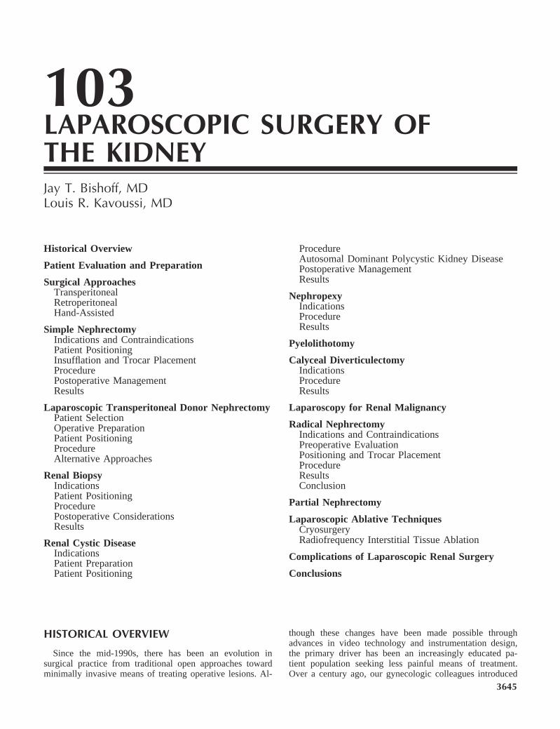
Table 103–1. LAPAROSCOPIC NEPHRECTOMY
Author Approach IndicationOR
Time (Min)LOH(days) EBL
Conver-sion(%)
Compli-cations Morcellation Intact
Rassweiler et al,1993b
TransperitonealN 24
Benign N 20Malignant N 4
239 (115–300) 6 16 16% 4/24 83% 20/24 17% 4/24
Kerbl et al, 1994 TransperitonealN 23
Benign N 20Malignant N 4
343 (100–700) 4 343 ml 4 20% 5/24 87% 21/24 13% 3/24
RetroperitonealN 1
Eraky et al, 1994 TransperitonealN 60
Benign N 60 210 (132–288) 3 10 6% 4/60
Nicol et al, 1994 TransperitonealN 5
Benign N 5 180 (120–300) 3 0 100% 5/5
Perez et al, 1994 TransperitonealN 12
Benign N 12 145 (105–360) 3 8 16% 2/12 0 100% 12/12
Parra et al, 1995 TransperitonealN 12
Benign N 12 145 (105–360) 4 140 8 16% 2/12 8% 1/12 92% 11/12
Rassweiler et al,1996
TransperitonealN 18
Benign N 18Malignant N 17
206 76
RetroperitonealN 17
Higashihara et al,1998
Benign N 51Malignant N 12
298 (168–428) 239390
168
Keeley and Tolley,1998
TransperitonealN 79
Benign N 76Malignant N 24
146 4 na 5 18% 18/100 0 100% 100/100
RetroperitonealN 23
Rassweiler et al,1998
TransperitonealN 344
Benign N 444Malignant N 38
188 (50–400) 5 910
6% 29/4826% 29/482
RetroperitonealN 138
Rozenberg et al,1999
TransperitonealN 30
Malignant N 17Benign N 13
116 13 13% 4/30
Desgrandchampset al, 1999
TransperitonealN 14
Benign N 12Malignant N 2
119 (70–180) 5 50 ml 0 7% 1/14 0 100% 14/14
EBL, estimated blood loss; LOH, length of hospitalization; na, not applicable; OR, operating room.
laparoscopic surgery, primarily as a diagnostic tool. Onlyrecently has it become a practical and acceptable alterna-tive to treat complex surgical diseases.
The development of the laparoscopic pelvic lymphade-nectomy for patients with prostate cancer inaugurated therole of laparoscopy in treating urologic lesions (Griffith etal, 1990). In June 1990, Clayman and coworkers at Wash-ington University overcame the barriers to laparoscopicsolid organ removal by performing the first laparoscopicnephrectomy (Clayman et al, 1991). In less than 7 hours,an elderly patient with a 3-cm solid renal mass underwentlaparoscopic radical nephrectomy through five trocar sites.This accomplishment represents one of the milestones inminimally invasive surgery because it provided the solutionfor removing a large solid organ without the need for anincision.
Since this report, many institutions have verified the util-ity of a laparoscopic approach to address benign and ma-lignant diseases of the kidney. Advances in instrumenttechnology and surgical techniques have also allowed com-plex renal reconstruction to be performed laparoscopically.The result for patients has been markedly less morbiditythan open flank surgery. The benefits of laparoscopic ne-phrectomy include a lower requirement for pain medi-cation, a shorter hospital stay, reduced convalescence,and a more rapid return to full activity (Table 103– 1)(Copcoat et al, 1992; Kavoussi et al, 1993; Kerbl et al1993a, 1993b; Rassweiler et al, 1993; Kerbl et al, 1994;Nicol et al, 1994; Perez et al, 1994; Parra et al, 1995;Baba et al, 1996; Doublet et al, 1996; Rassweiler et al,
1996; Rassweiler et al, 1998; Rozenberg et al, 1999; He-mal, et al 1999).
PATIENT EVALUATION ANDPREPARATION
Patient preparation for laparoscopic kidney surgery issimilar to that for comparable open procedures. Informedconsent must be obtained with a discussion of possiblecomplications including adjacent organ injury and un-recognized bowel injury (Bishoff et al, 1999). The pa-tient should be informed that conversion to open sur-gery might be necessary to safely complete the plannedprocedure.
In order for the surgeon to determine any contraindica-tions to laparoscopic surgery or conditions that may alterthe laparoscopic approach, the preoperative evaluation mustinclude a careful history and detailed physical examination.Laparoscopic kidney surgery requires a general anes-thetic. Consequently, patients who are not candidatesfor a general anesthetic should not undergo laparo-scopic surgery (Monk and Weldon, 1992). Prior abdomi-nal surgery may alter the choice between transperito-neal or retroperitoneal approaches, patient positioning,and the placement site of trocars but is not a contrain-dication to laparoscopic surgery (Chen et al, 1998; Ca-deddu et al, 1999). Severe cardiac or pulmonary diseasemay place the patient at risk for complications due to thepneumoperitoneum, which can compromise ventilation and
JAY T. BISHOFF AND LOUIS R. KAVOUSSI 3647
limit venous return (Arthure, 1970; Hodgson et al, 1970;Nunn, 1987; Lew et al, 1992). Patients with chronic ob-structive pulmonary disease may not be able to compensatefor hypercarbia induced by the pneumoperitoneum and mayrequire lower insufflation pressures or conversion to opensurgery (Monk and Weldon, 1992; Adams et al, 1995).
Laboratory and imaging studies are obtained as indi-cated. An electrocardiogram and a chest radiograph areobtained before surgery. Pulmonary function studies areperformed only in those patients with known respiratorydisease or those at high risk based on the history andphysical examination. All patients should have their bloodtyped and screened, and in advanced procedures, cross-matched blood should be available. Bowel preparation var-ies according to the procedure and the physician’s prefer-ence.
Anticoagulated patients are managed in cooperation withtheir primary physician. Patients with bleeding disordersshould have 2 to 4 units of packed red blood cells cross-matched before the start of the procedure. Fresh frozenplasma can be given if needed just before the procedure.
Patients with thrombocytopenia can receive platelets 30minutes before the incision to increase their platelet countto greater than 50,000/ml. Additional platelet transfusionshould not be necessary in the absence of symptomaticbleeding. Uremic patients may benefit from desmopressinacetate treatment to improve platelet function.
A preoperative abdominal CT scan is useful in evalu-ating the location, size, and abnormality of the kidneyto be addressed. Areas of perinephric stranding discov-ered on CT may indicate significant inflammation andadhesions, increasing the chance for conversion to anopen procedure. In addition, the scan provides data on theappearance and function of the contralateral kidney. Differ-ential function can be determined with a diethylenetri-aminepenta-acetic acid renal scan if needed. Angiographyor placement of a ureteral stent is not routinely performed.
SURGICAL APPROACHES
There are three basic laparoscopic approaches fornephrectomy: transperitoneal, retroperitoneal, andhand-assisted.
Transperitoneal
The transperitoneal route is the traditional method usedto perform laparoscopic surgery. It results in small inci-sions and gives latitude in the location of trocar placement.It also affords an optimal working space and facilitatesorientation by providing readily identifiable anatomic land-marks.
Retroperitoneal
The retroperitoneal approach mimics traditional opensurgery in that the kidney is approached without entry intothe peritoneal cavity. However, with the retroperitoneal ap-proach, adipose tissue and the limited working space can
result in difficulty with orientation, visualization, trocarspacing, and organ entrapment. Even though this providesan extraperitoneal operation, injury can occur to intra-ab-dominal organs, and a hernia can be created during balloondilatation of the extraperitoneal space (Adams et al, 1996).Compared with a transperitoneal approach, a retroperi-toneal approach results in complication rates, pain med-ication requirements, lengths of hospital stay, and timesto return to normal activity after surgery similar tothose of the retroperitoneal route (McDougall and Clay-man, 1996; Rassweiler et al, 1998a). There are patients inwhom retroperitoneal access is preferred over the transperi-toneal approach. Patients with a history of multiple abdom-inal surgical procedures or peritonitis may benefit from thisapproach. Initial retroperitoneal access can be achievedwith subsequent opening of the peritoneum as indicated(Cadeddu et al, 1999). Also, patients with abnormality onthe posterior surface of the kidney (exophytic cyst or mass)may be better served through retroperitoneal access. Con-sequently, the laparoscopic surgeon should be familiarwith both transperitoneal and retroperitoneal ap-proaches (Barreto et al, 1995; Doublet et al, 1996; Gas-man et al, 1996; Rassweiler et al, 1998b; Gill and Ras-sweiler, 1999; Hsu et al, 1999 (see Chapter 100).
Hand-Assisted
In an effort to facilitate learning of laparoscopic tech-niques, hand-assisted endoscopic techniques have been in-troduced. The hand-assisted devices offer a bridge betweenlaparoscopic and open surgery and may help surgeonswithout advanced laparoscopic training. Several devices areavailable that maintain the pneumoperitoneum while a handis introduced into the abdomen (Pneumo Sleeve, Dexterity,Inc., Blue Bell, Pennsylvania. Intromit, Medtech, Dublin,Ireland). Hand-assisted laparoscopic surgery permits tactilefeedback and allows the hand to assist with dissection,retraction, extraction, and rapid control of bleeding ifneeded. An incision large enough to allow the surgeon orassistant to insert a hand must be created, and this site canbe utilized for organ extraction. Hand-assisted dissectionmay be helpful in those patients whose pathologic con-dition makes laparoscopy more difficult, such as infec-tious processes or prior surgery. Multiple series havedocumented the utility of the hand-assisted technique insimple and radical nephrectomy as well as nephroureterec-tomy (Keeley et al, 1997; Wolf et al, 1998; Nakada et al,1997, 1999). Even if the kidney is to be extracted intact,the hand-assisted approach requires a larger incision thanthat of a complete laparoscopic approach. Moreover, one islimited in where the incision is made. Finally, currentlyavailable devices are disposable and add significant cost tothe procedure.
SIMPLE NEPHRECTOMY
Indications and Contraindications
Laparoscopic simple nephrectomy is indicated in thetreatment of most benign renal diseases in which perma-
3648 LAPAROSCOPIC SURGERY OF THE KIDNEY
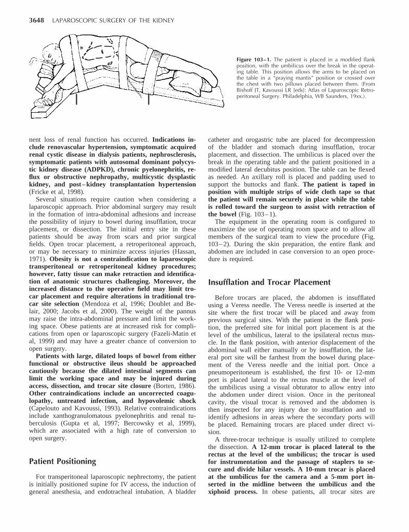
Figure 103–1. The patient is placed in a modified flankposition, with the umbilicus over the break in the operat-ing table. This position allows the arms to be placed onthe table in a “praying mantis” position or crossed overthe chest with two pillows placed between them. (FromBishoff JT, Kavoussi LR [eds]: Atlas of Laparoscopic Retro-peritoneal Surgery. Philadelphia, WB Saunders, 19xx.).
nent loss of renal function has occurred. Indications in-clude renovascular hypertension, symptomatic acquiredrenal cystic disease in dialysis patients, nephrosclerosis,symptomatic patients with autosomal dominant polycys-tic kidney disease (ADPKD), chronic pyelonephritis, re-flux or obstructive nephropathy, multicystic dysplastickidney, and post– kidney transplantation hypertension(Fricke et al, 1998).
Several situations require caution when considering alaparoscopic approach. Prior abdominal surgery may resultin the formation of intra-abdominal adhesions and increasethe possibility of injury to bowel during insufflation, trocarplacement, or dissection. The initial entry site in thesepatients should be away from scars and prior surgicalfields. Open trocar placement, a retroperitoneal approach,or may be necessary to minimize access injuries (Hassan,1971). Obesity is not a contraindication to laparoscopictransperitoneal or retroperitoneal kidney procedures;however, fatty tissue can make retraction and identifica-tion of anatomic structures challenging. Moreover, theincreased distance to the operative field may limit tro-car placement and require alterations in traditional tro-car site selection (Mendoza et al, 1996; Doublet and Be-lair, 2000; Jacobs et al, 2000). The weight of the pannusmay raise the intra-abdominal pressure and limit the work-ing space. Obese patients are at increased risk for compli-cations from open or laparoscopic surgery (Fazeli-Matin etal, 1999) and may have a greater chance of conversion toopen surgery.
Patients with large, dilated loops of bowel from eitherfunctional or obstructive ileus should be approachedcautiously because the dilated intestinal segments canlimit the working space and may be injured duringaccess, dissection, and trocar site closure (Borten, 1986).Other contraindications include an uncorrected coagu-lopathy, untreated infection, and hypovolemic shock(Capelouto and Kavoussi, 1993). Relative contraindicationsinclude xanthogranulomatous pyelonephritis and renal tu-berculosis (Gupta et al, 1997; Bercowsky et al, 1999),which are associated with a high rate of conversion toopen surgery.
Patient Positioning
For transperitoneal laparoscopic nephrectomy, the patientis initially positioned supine for IV access, the induction ofgeneral anesthesia, and endotracheal intubation. A bladder
catheter and orogastric tube are placed for decompressionof the bladder and stomach during insufflation, trocarplacement, and dissection. The umbilicus is placed over thebreak in the operating table and the patient positioned in amodified lateral decubitus position. The table can be flexedas needed. An axillary roll is placed and padding used tosupport the buttocks and flank. The patient is taped inposition with multiple strips of wide cloth tape so thatthe patient will remain securely in place while the tableis rolled toward the surgeon to assist with retraction ofthe bowel (Fig. 103– 1).
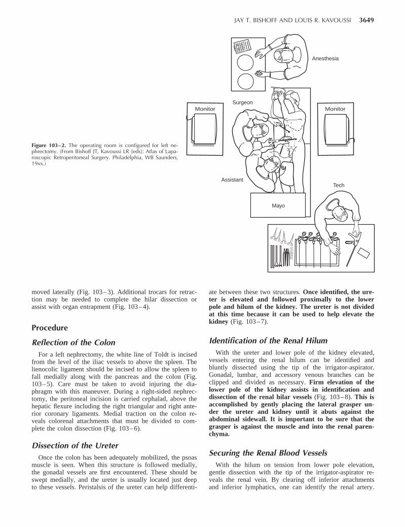
The equipment in the operating room is configured tomaximize the use of operating room space and to allow allmembers of the surgical team to view the procedure (Fig.103– 2). During the skin preparation, the entire flank andabdomen are included in case conversion to an open proce-dure is required.
Insufflation and Trocar Placement
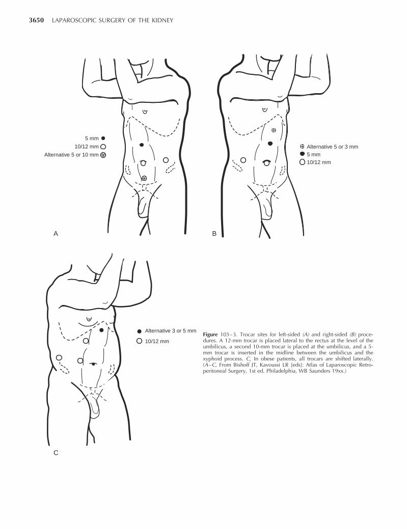
Before trocars are placed, the abdomen is insufflatedusing a Veress needle. The Veress needle is inserted at thesite where the first trocar will be placed and away fromprevious surgical sites. With the patient in the flank posi-tion, the preferred site for initial port placement is at thelevel of the umbilicus, lateral to the ipsilateral rectus mus-cle. In the flank position, with anterior displacement of theabdominal wall either manually or by insufflation, the lat-eral port site will be farthest from the bowel during place-ment of the Veress needle and the initial port. Once apneumoperitoneum is established, the first 10- or 12-mmport is placed lateral to the rectus muscle at the level ofthe umbilicus using a visual obturator to allow entry intothe abdomen under direct vision. Once in the peritonealcavity, the visual trocar is removed and the abdomen isthen inspected for any injury due to insufflation and toidentify adhesions in areas where the secondary ports willbe placed. Remaining trocars are placed under direct vi-sion.
A three-trocar technique is usually utilized to completethe dissection. A 12-mm trocar is placed lateral to therectus at the level of the umbilicus; the trocar is usedfor instrumentation and the passage of staplers to se-cure and divide hilar vessels. A 10-mm trocar is placedat the umbilicus for the camera and a 5-mm port in-serted in the midline between the umbilicus and thexiphoid process. In obese patients, all trocar sites are
JAY T. BISHOFF AND LOUIS R. KAVOUSSI 3649
Figure 103–2. The operating room is configured for left ne-phrectomy. (From Bishoff JT, Kavoussi LR [eds]: Atlas of Lapa-roscopic Retroperitoneal Surgery. Philadelphia, WB Saunders,19xx.)
Assistant
SurgeonMonitor
Mayo
Monitor
Mayo
Anesthesia
Tech
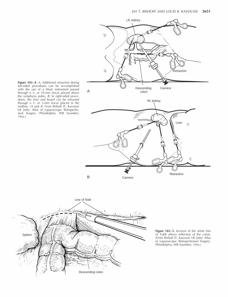
moved laterally (Fig. 103– 3). Additional trocars for retrac-tion may be needed to complete the hilar dissection orassist with organ entrapment (Fig. 103– 4).
Procedure
Reflection of the ColonFor a left nephrectomy, the white line of Toldt is incised
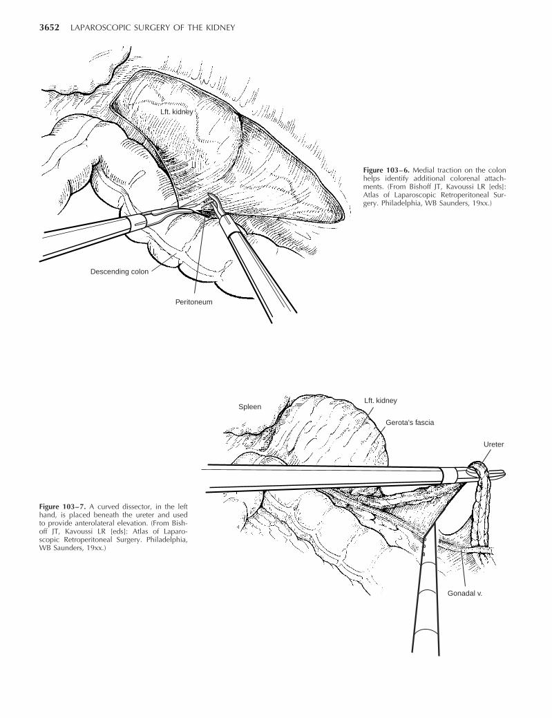
from the level of the iliac vessels to above the spleen. Thelienocolic ligament should be incised to allow the spleen tofall medially along with the pancreas and the colon (Fig.103– 5). Care must be taken to avoid injuring the dia-phragm with this maneuver. During a right-sided nephrec-tomy, the peritoneal incision is carried cephalad, above thehepatic flexure including the right triangular and right ante-rior coronary ligaments. Medial traction on the colon re-veals colorenal attachments that must be divided to com-plete the colon dissection (Fig. 103– 6).
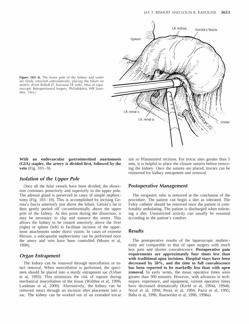
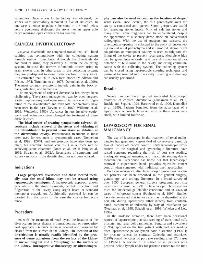
Dissection of the UreterOnce the colon has been adequately mobilized, the psoas
muscle is seen. When this structure is followed medially,the gonadal vessels are first encountered. These should beswept medially, and the ureter is usually located just deepto these vessels. Peristalsis of the ureter can help differenti-
ate between these two structures. Once identified, the ure-ter is elevated and followed proximally to the lowerpole and hilum of the kidney. The ureter is not dividedat this time because it can be used to help elevate thekidney (Fig. 103– 7).
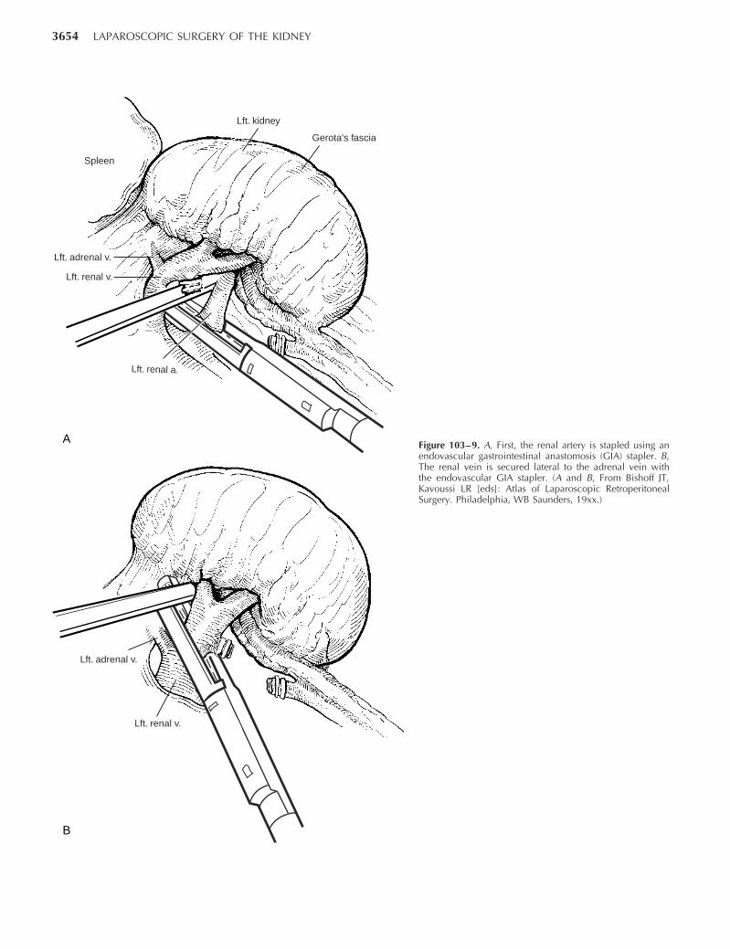
Identification of the Renal HilumWith the ureter and lower pole of the kidney elevated,
vessels entering the renal hilum can be identified andbluntly dissected using the tip of the irrigator-aspirator.Gonadal, lumbar, and accessory venous branches can beclipped and divided as necessary. Firm elevation of thelower pole of the kidney assists in identification anddissection of the renal hilar vessels (Fig. 103– 8). This isaccomplished by gently placing the lateral grasper un-der the ureter and kidney until it abuts against theabdominal sidewall. It is important to be sure that thegrasper is against the muscle and into the renal paren-chyma.
Securing the Renal Blood VesselsWith the hilum on tension from lower pole elevation,
gentle dissection with the tip of the irrigator-aspirator re-veals the renal vein. By clearing off inferior attachmentsand inferior lymphatics, one can identify the renal artery.
3650 LAPAROSCOPIC SURGERY OF THE KIDNEY
10/12 mm
5 mm
Alternative 5 or 10 mm10/12 mm
Alternative 5 or 3 mm5 mm
10/12 mm
Alternative 3 or 5 mm
A B
C
Figure 103–3. Trocar sites for left-sided (A) and right-sided (B) proce-dures. A 12-mm trocar is placed lateral to the rectus at the level of theumbilicus, a second 10-mm trocar is placed at the umbilicus, and a 5-mm trocar is inserted in the midline between the umbilicus and thexyphoid process. C, In obese patients, all trocars are shifted laterally.(A–C, From Bishoff JT, Kavoussi LR [eds]: Atlas of Laparoscopic Retro-peritoneal Surgery, 1st ed. Philadelphia, WB Saunders 19xx.)
JAY T. BISHOFF AND LOUIS R. KAVOUSSI 3651
Figure 103–4. A, Additional retraction duringleft-sided procedures can be accomplishedwith the use of a blunt instrument passedthrough a 5- or 10-mm trocar placed abovethe symphysis pubis. B, In right-sided proce-dures, the liver and bowel can be retractedthrough a 3- or 5-mm trocar placed in themidline. (A and B, From Bishoff JT, KavoussiLR [eds]: Atlas of Laparoscopic Retroperito-neal Surgery. Philadelphia, WB Saunders,19xx.)
Descending colon
Camera
Retraction
Lft. kidney
Liver
Rt. kidney
CameraRetraction
A
B
Line of Toldt
Spleen
Descending colon
Figure 103–5. Incision of the white lineof Toldt allows reflection of the colon.(From Bishoff JT, Kavoussi LR [eds]: Atlasof Laparoscopic Retroperitoneal Surgery.Philadelphia, WB Saunders, 19xx.)
3652 LAPAROSCOPIC SURGERY OF THE KIDNEY
Descending colon
Lft. kidney
Peritoneum
Figure 103–6. Medial traction on the colonhelps identify additional colorenal attach-ments. (From Bishoff JT, Kavoussi LR [eds]:Atlas of Laparoscopic Retroperitoneal Sur-gery. Philadelphia, WB Saunders, 19xx.)
Figure 103–7. A curved dissector, in the lefthand, is placed beneath the ureter and usedto provide anterolateral elevation. (From Bish-off JT, Kavoussi LR [eds]: Atlas of Laparo-scopic Retroperitoneal Surgery. Philadelphia,WB Saunders, 19xx.)
Spleen
Gerota's fascia
Lft. kidney
Ureter
Gonadal v.
JAY T. BISHOFF AND LOUIS R. KAVOUSSI 3653
Figure 103–8. The lower pole of the kidney and ureterare firmly retracted anterolaterally, placing the hilum onstretch. (From Bishoff JT, Kavoussi LR [eds]: Atlas of Lapa-roscopic Retroperitoneal Surgery. Philadelphia, WB Saun-ders, 19xx.)
Spleen
Lft. renal v.
Lft. renal a.
Gerota's fasciaLft. kidney
Ureter
With an endovascular gastrointestinal anastomosis(GIA) stapler, the artery is divided first, followed by thevein (Fig. 103– 9).
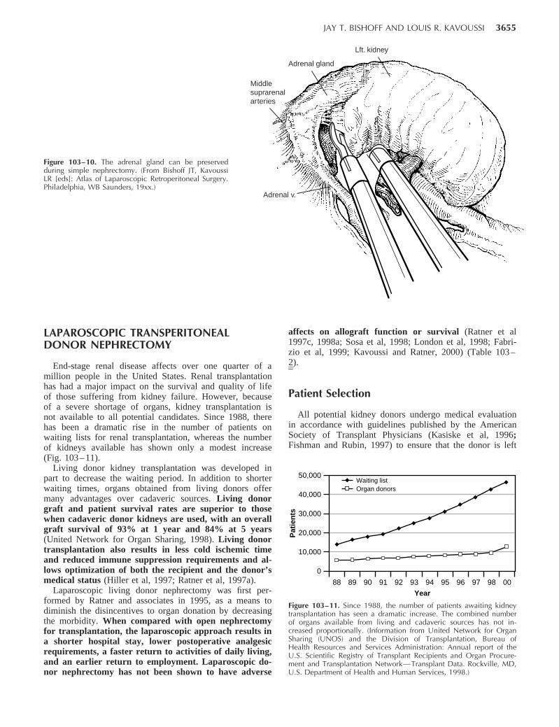
Isolation of the Upper PoleOnce all the hilar vessels have been divided, the dissec-
tion continues posteriorly and superiorly to the upper pole.The adrenal gland is preserved in cases of simple nephrec-tomy (Fig. 103– 10). This is accomplished by incising Ge-rota’s fascia anteriorly just above the hilum. Gerota’s fat isthen gently peeled off circumferentially above the upperpole of the kidney. At this point during the dissection, itmay be necessary to clip and transect the ureter. Thisallows the kidney to be rotated anteriorly above the liver(right) or spleen (left) to facilitate incision of the upper-most attachments under direct vision. In cases of extremefibrosis, a subcapsular nephrectomy can be performed oncethe artery and vein have been controlled (Moore et al,1998).
Organ EntrapmentThe kidney can be removed through morcellation or in-
tact removal. When morcellation is performed, the speci-men should be placed into a sturdy entrapment sac (Urbanet al, 1993). This minimizes the risk of rupture duringmechanical morcellation of the tissue (Walther et al, 1999;Landman et al, 2000). Alternatively, the kidney can beremoved intact through an incision after placement into asac. The kidney can be worked out of an extended trocar
site or Pfannenstiel incision. For trocar sites greater than 5mm, it is helpful to place the closure sutures before remov-ing the kidney. Once the sutures are placed, trocars can bereinserted for kidney entrapment and removal.
Postoperative Management
The orogastric tube is removed at the conclusion of theprocedure. The patient can begin a diet as tolerated. TheFoley catheter should be removed once the patient is com-fortably ambulating. The patient is discharged when tolerat-ing a diet. Unrestricted activity can usually be resumedaccording to the patient’s comfort.
Results
The postoperative results of the laparoscopic nephrec-tomy are comparable to that of open surgery with muchless pain and shorter convalescence. Postoperative painrequirements are approximately four times less thanwith traditional open incisions. Hospital stays have beendecreased by 50%, and the time to full convalescencehas been reported to be markedly less than with openremoval. In early series, the mean operative times weregreater than 300 minutes. However, with advances in tech-niques, experience, and equipment, current operative timeshave decreased dramatically (Kerbl et al, 1994a, 1994b;Nicol et al, 1994; Perez et al, 1994; Parra et al, 1995;Baba et al, 1996; Rassweiler et al, 1996, 1998a).
3654 LAPAROSCOPIC SURGERY OF THE KIDNEY
Spleen
Lft. renal v.
Gerota's fascia
Lft. kidney
Lft. adrenal v.
Lft. renal a.
Lft. renal v.
Lft. adrenal v.
A
B
Figure 103–9. A, First, the renal artery is stapled using anendovascular gastrointestinal anastomosis (GIA) stapler. B,The renal vein is secured lateral to the adrenal vein withthe endovascular GIA stapler. (A and B, From Bishoff JT,Kavoussi LR [eds]: Atlas of Laparoscopic RetroperitonealSurgery. Philadelphia, WB Saunders, 19xx.)
JAY T. BISHOFF AND LOUIS R. KAVOUSSI 3655
Figure 103–10. The adrenal gland can be preservedduring simple nephrectomy. (From Bishoff JT, KavoussiLR [eds]: Atlas of Laparoscopic Retroperitoneal Surgery.Philadelphia, WB Saunders, 19xx.)
Adrenal gland
Middle suprarenalarteries
Adrenal v.
Lft. kidney
50,000
40,000
30,000
20,000
10,000
0
Pat
ien
ts
9897969594939291908988 00
Year
Waiting listOrgan donors
Figure 103–11. Since 1988, the number of patients awaiting kidneytransplantation has seen a dramatic increase. The combined numberof organs available from living and cadaveric sources has not in-creased proportionally. (Information from United Network for OrganSharing (UNOS) and the Division of Transplantation, Bureau ofHealth Resources and Services Administration: Annual report of theU.S. Scientific Registry of Transplant Recipients and Organ Procure-ment and Transplantation Network—Transplant Data. Rockville, MD,U.S. Department of Health and Human Services, 1998.)
LAPAROSCOPIC TRANSPERITONEALDONOR NEPHRECTOMY
End-stage renal disease affects over one quarter of amillion people in the United States. Renal transplantationhas had a major impact on the survival and quality of lifeof those suffering from kidney failure. However, becauseof a severe shortage of organs, kidney transplantation isnot available to all potential candidates. Since 1988, therehas been a dramatic rise in the number of patients onwaiting lists for renal transplantation, whereas the numberof kidneys available has shown only a modest increase(Fig. 103– 11).
Living donor kidney transplantation was developed inpart to decrease the waiting period. In addition to shorterwaiting times, organs obtained from living donors offermany advantages over cadaveric sources. Living donorgraft and patient survival rates are superior to thosewhen cadaveric donor kidneys are used, with an overallgraft survival of 93% at 1 year and 84% at 5 years(United Network for Organ Sharing, 1998). Living donortransplantation also results in less cold ischemic timeand reduced immune suppression requirements and al-lows optimization of both the recipient and the donor’smedical status (Hiller et al, 1997; Ratner et al, 1997a).
Laparoscopic living donor nephrectomy was first per-formed by Ratner and associates in 1995, as a means todiminish the disincentives to organ donation by decreasingthe morbidity. When compared with open nephrectomyfor transplantation, the laparoscopic approach results ina shorter hospital stay, lower postoperative analgesicrequirements, a faster return to activities of daily living,and an earlier return to employment. Laparoscopic do-nor nephrectomy has not been shown to have adverse
affects on allograft function or survival (Ratner et al1997c, 1998a; Sosa et al, 1998; London et al, 1998; Fabri-zio et al, 1999; Kavoussi and Ratner, 2000) (Table 103–2).
Patient Selection
All potential kidney donors undergo medical evaluationin accordance with guidelines published by the AmericanSociety of Transplant Physicians (Kasiske et al, 1996;Fishman and Rubin, 1997) to ensure that the donor is left
3656 LAPAROSCOPIC SURGERY OF THE KIDNEY
Table 103–2. OPEN VERSUS LAPAROSCOPIC DONORNEPHRECTOMY
Results
Laparo-scopic NxN 110
Open NxN 48 P value
Estimated blood loss (ml) 266 174 393 335 .027Operative time (min) 232 33 183 27 .001Hospital stay (days) 3.0 0.9 5.7 1.7 .001Length of time analgesia
used (days)Oral narcotic agents* 4 12 .001Acetaminophen 3 17 .001
Resumed oral intake(days)
0.8 0.5 2.6 1.0 .001
Returned to work (wk) 4.0 2.3 6.4 3.1 .003
Nx, nephrectomy.*Milligrams of morphine equivalents.From Kavoussi LR, Ratner LE: Laparoscopic donor nephrectomy. Kidney Int
2000;57:2175–2186.
with adequate renal function after nephrectomy. In addi-tion, the donor’s motivation and emotional stability arecarefully evaluated. Laparoscopic donor nephrectomy re-quires accurate radiographic imaging to evaluate the arterialand venous anatomy. Dual-phase spiral CT with three-dimensional angiography for evaluation of the livingdonor patients has been shown to adequately depictrenal vasculature when compared with standard angi-ography (Plat et al, 1997; Dachman et al, 1998; Smith etal, 1998; Del Pizzo et al, 1999). Careful preoperativeevaluation with CT imaging is important to identify thepresence of multiple renal blood vessels preoperativelyso that they can be identified and preserved during thehilar dissection. The presence of multiple vessels is nota contraindication to laparoscopic donor nephrectomy,but preoperative identification allows the surgeon to an-ticipate their presence early in the operation (Kuo et al,1998).
Additional considerations include the patient’s bodymass index and prior abdominal surgical procedures. Lapa-roscopic donor nephrectomy has successfully been per-formed in obese patients with outcomes similar to those ofnonobese patients (Kuo et al, 2000). Patients who have hadmultiple prior abdominal surgical procedures near the in-tended operative field may be considered for donor ne-phrectomy but may be better served with a retroperitonealor an open approach.
Operative Preparation
Patients are prepared in a manner similar to that ofpersons undergoing simple nephrectomy; however, theyroutinely receive 1 to 2 L of IV fluid before insufflation ofthe abdomen. Pneumoperitoneum has been shown to de-crease renal blood flow; however, vigorous hydrationmaintains urine output (London et al, 1998, 2000). Pa-tients are routinely given a total of 5 to 6 L of crystal-loid fluid during the procedure.
Patient Positioning
To allow access to the anterior abdomen for intact organretrieval, the patient is placed in the modified flank posi-tion with the torso in a 45-degree lateral decubitus positionand the table flexed. The hips are maintained flat to facili-tate the creation of a Pfannenstiel incision. Wide tape isused to secure the hips and chest to the table, and a safetystrap is used to secure the legs. The positioning and secu-rity of the patient should be tested by maximally planingthe table to the right and left. The operating room configu-ration is identical to that used in simple nephrectomy (seeFig. 103– 2).
Procedure
Insufflation and Trocar PlacementA pneumoperitoneum is established after a Veress needle
is passed, and subsequently four transperitoneal laparo-
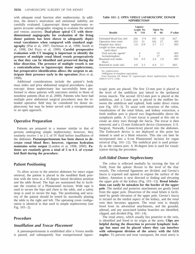
scopic ports are placed. The first 12-mm port is placed atthe level of the umbilicus just lateral to the ipsilateralrectus muscle. The second 10/12-mm port is placed at theumbilicus, and a 5-mm port is placed in the midline be-tween the umbilicus and xiphoid, both under direct vision(see Fig. 103– 3). To assist with retraction of the colon,visualization of the renal hilum, and kidney extraction, aforth midline port is placed two fingerbreadths above thesymphysis pubis. A 12-mm trocar is passed at this site tocreate an entry tract through the fascia. The trocar is thenremoved, and a 15-mm Endocatch device (Autosuture, U.S.Surgical, Norwalk, Connecticut) placed into the abdomen.The Endocatch device is not deployed at this point butinstead is used as a blunt retractor. This site can later beincorporated into a 6-cm Pfannenstiel incision for organretrieval (Fig. 103– 12). The umbilical port is used primar-ily as the camera port. A 30-degree lens is used for visual-ization during the procedure.
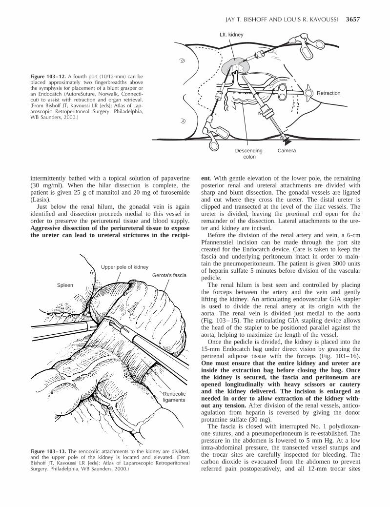
Left-Sided Donor NephrectomyThe colon is reflected medially by incising the line of
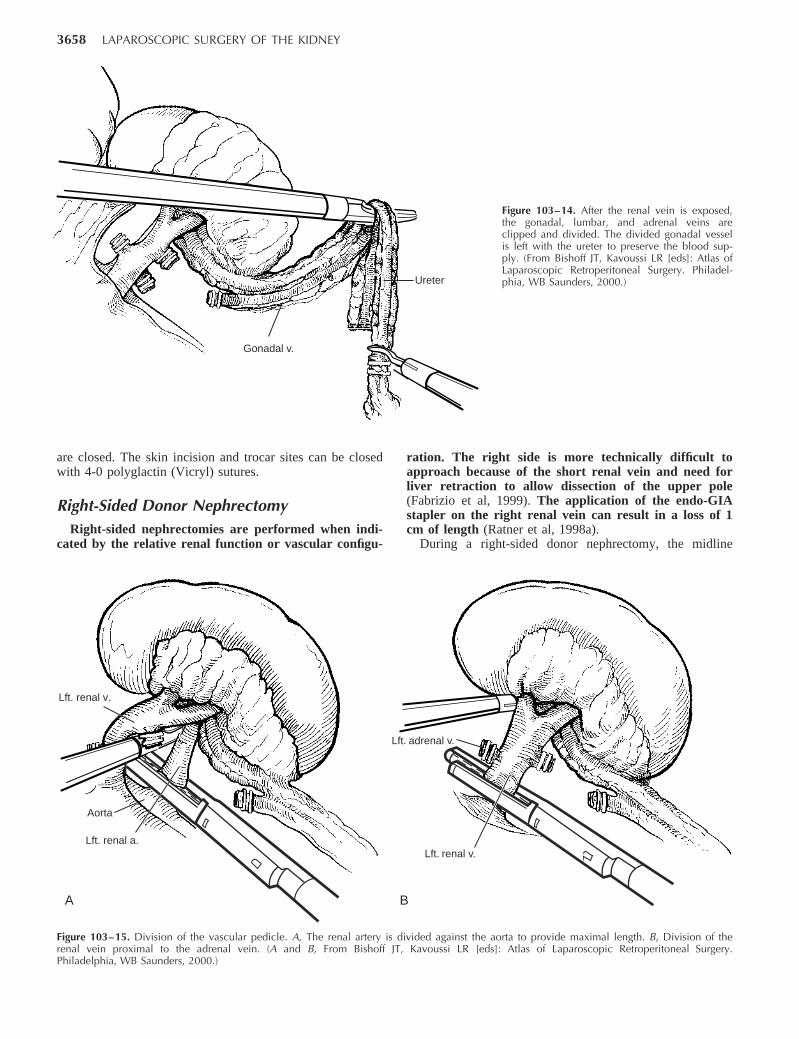
Toldt, from the splenic flexure to the level of the iliacvessels. The colorenal ligaments are divided, and Gerota’sfascia is exposed and opened to expose the surface of thekidney. Attention is next directed to finding and elevatingthe upper pole of the kidney (Fig. 103– 13). Renal lobula-tions can easily be mistaken for the border of the upperpole. The medial and posterior attachments are gently freedfrom the upper pole. Dissection of the renal hilum is facili-tated by gentle elevation of the upper pole. Gerota’s fasciais incised on the medial aspect of the kidney, and the renalvein then becomes apparent. The renal vein is sharplyfreed from its adventitial attachments, and the gonadal,adrenal, and any associated lumbar branches are identified,clipped, and divided (Fig. 103– 14).
The renal artery, which usually lies posterior to the vein,is identified and freed to its origin at the aorta. Clips arehelpful during the dissection to prevent lymphatic leak-age but must not be placed where they can interferewith subsequent division of the artery with the GIAstapler. To prevent and treat vasospasm, the renal artery is
JAY T. BISHOFF AND LOUIS R. KAVOUSSI 3657
Figure 103–12. A fourth port (10/12-mm) can beplaced approximately two fingerbreadths abovethe symphysis for placement of a blunt grasper oran Endocatch (AutoreSuture, Norwalk, Connecti-cut) to assist with retraction and organ retrieval.(From Bishoff JT, Kavoussi LR [eds]: Atlas of Lap-aroscopic Retroperitoneal Surgery. Philadelphia,WB Saunders, 2000.)
Descending colon
Camera
Retraction
Lft. kidney
Upper pole of kidney
Spleen
Gerota's fascia
Renocolicligaments
Figure 103–13. The renocolic attachments to the kidney are divided,and the upper pole of the kidney is located and elevated. (FromBishoff JT, Kavoussi LR [eds]: Atlas of Laparoscopic RetroperitonealSurgery. Philadelphia, WB Saunders, 2000.)
intermittently bathed with a topical solution of papaverine(30 mg/ml). When the hilar dissection is complete, thepatient is given 25 g of mannitol and 20 mg of furosemide(Lasix).
Just below the renal hilum, the gonadal vein is againidentified and dissection proceeds medial to this vessel inorder to preserve the periureteral tissue and blood supply.Aggressive dissection of the periureteral tissue to exposethe ureter can lead to ureteral strictures in the recipi-
ent. With gentle elevation of the lower pole, the remainingposterior renal and ureteral attachments are divided withsharp and blunt dissection. The gonadal vessels are ligatedand cut where they cross the ureter. The distal ureter isclipped and transected at the level of the iliac vessels. Theureter is divided, leaving the proximal end open for theremainder of the dissection. Lateral attachments to the ure-ter and kidney are incised.
Before the division of the renal artery and vein, a 6-cmPfannenstiel incision can be made through the port sitecreated for the Endocatch device. Care is taken to keep thefascia and underlying peritoneum intact in order to main-tain the pneumoperitoneum. The patient is given 3000 unitsof heparin sulfate 5 minutes before division of the vascularpedicle.
The renal hilum is best seen and controlled by placingthe forceps between the artery and the vein and gentlylifting the kidney. An articulating endovascular GIA stapleris used to divide the renal artery at its origin with theaorta. The renal vein is divided just medial to the aorta(Fig. 103– 15). The articulating GIA stapling device allowsthe head of the stapler to be positioned parallel against theaorta, helping to maximize the length of the vessel.
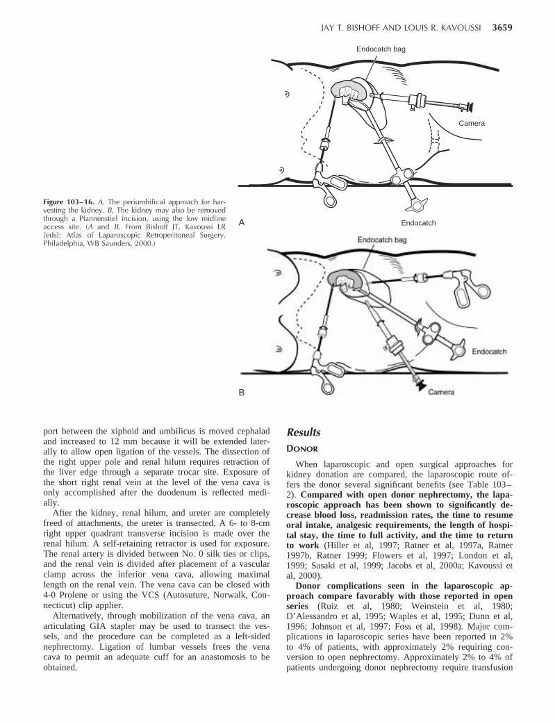
Once the pedicle is divided, the kidney is placed into the15-mm Endocatch bag under direct vision by grasping theperirenal adipose tissue with the forceps (Fig. 103– 16).One must ensure that the entire kidney and ureter areinside the extraction bag before closing the bag. Oncethe kidney is secured, the fascia and peritoneum areopened longitudinally with heavy scissors or cauteryand the kidney delivered. The incision is enlarged asneeded in order to allow extraction of the kidney with-out any tension. After division of the renal vessels, antico-agulation from heparin is reversed by giving the donorprotamine sulfate (30 mg).
The fascia is closed with interrupted No. 1 polydioxan-one sutures, and a pneumoperitoneum is re-established. Thepressure in the abdomen is lowered to 5 mm Hg. At a lowintra-abdominal pressure, the transected vessel stumps andthe trocar sites are carefully inspected for bleeding. Thecarbon dioxide is evacuated from the abdomen to preventreferred pain postoperatively, and all 12-mm trocar sites
3658 LAPAROSCOPIC SURGERY OF THE KIDNEY
Gonadal v.
Ureter
Figure 103–14. After the renal vein is exposed,the gonadal, lumbar, and adrenal veins areclipped and divided. The divided gonadal vesselis left with the ureter to preserve the blood sup-ply. (From Bishoff JT, Kavoussi LR [eds]: Atlas ofLaparoscopic Retroperitoneal Surgery. Philadel-phia, WB Saunders, 2000.)
Aorta
Lft. renal a.
A B
Lft. renal v.
Lft. renal v.
Lft. adrenal v.
Figure 103–15. Division of the vascular pedicle. A, The renal artery is divided against the aorta to provide maximal length. B, Division of therenal vein proximal to the adrenal vein. (A and B, From Bishoff JT, Kavoussi LR [eds]: Atlas of Laparoscopic Retroperitoneal Surgery.Philadelphia, WB Saunders, 2000.)
are closed. The skin incision and trocar sites can be closedwith 4-0 polyglactin (Vicryl) sutures.
Right-Sided Donor NephrectomyRight-sided nephrectomies are performed when indi-
cated by the relative renal function or vascular configu-
ration. The right side is more technically difficult toapproach because of the short renal vein and need forliver retraction to allow dissection of the upper pole(Fabrizio et al, 1999). The application of the endo-GIAstapler on the right renal vein can result in a loss of 1cm of length (Ratner et al, 1998a).
During a right-sided donor nephrectomy, the midline
JAY T. BISHOFF AND LOUIS R. KAVOUSSI 3659
Figure 103–16. A, The periumbilical approach for har-vesting the kidney. B, The kidney may also be removedthrough a Pfannenstiel incision, using the low midlineaccess site. (A and B, From Bishoff JT, Kavoussi LR[eds]: Atlas of Laparoscopic Retroperitoneal Surgery.Philadelphia, WB Saunders, 2000.)
Camera
EndocatchA
Endocatch bag
B
port between the xiphoid and umbilicus is moved cephaladand increased to 12 mm because it will be extended later-ally to allow open ligation of the vessels. The dissection ofthe right upper pole and renal hilum requires retraction ofthe liver edge through a separate trocar site. Exposure ofthe short right renal vein at the level of the vena cava isonly accomplished after the duodenum is reflected medi-ally.
After the kidney, renal hilum, and ureter are completelyfreed of attachments, the ureter is transected. A 6- to 8-cmright upper quadrant transverse incision is made over therenal hilum. A self-retaining retractor is used for exposure.The renal artery is divided between No. 0 silk ties or clips,and the renal vein is divided after placement of a vascularclamp across the inferior vena cava, allowing maximallength on the renal vein. The vena cava can be closed with4-0 Prolene or using the VCS (Autosuture, Norwalk, Con-necticut) clip applier.
Alternatively, through mobilization of the vena cava, anarticulating GIA stapler may be used to transect the ves-sels, and the procedure can be completed as a left-sidednephrectomy. Ligation of lumbar vessels frees the venacava to permit an adequate cuff for an anastomosis to beobtained.
ResultsDONOR
When laparoscopic and open surgical approaches forkidney donation are compared, the laparoscopic route of-fers the donor several significant benefits (see Table 103–2). Compared with open donor nephrectomy, the lapa-roscopic approach has been shown to significantly de-crease blood loss, readmission rates, the time to resumeoral intake, analgesic requirements, the length of hospi-tal stay, the time to full activity, and the time to returnto work (Hiller et al, 1997; Ratner et al, 1997a, Ratner1997b, Ratner 1999; Flowers et al, 1997; London et al,1999; Sasaki et al, 1999; Jacobs et al, 2000a; Kavoussi etal, 2000).
Donor complications seen in the laparoscopic ap-proach compare favorably with those reported in openseries (Ruiz et al, 1980; Weinstein et al, 1980;D’Alessandro et al, 1995; Waples et al, 1995; Dunn et al,1996; Johnson et al, 1997; Foss et al, 1998). Major com-plications in laparoscopic series have been reported in 2%to 4% of patients, with approximately 2% requiring con-version to open nephrectomy. Approximately 2% to 4% ofpatients undergoing donor nephrectomy require transfusion
3660 LAPAROSCOPIC SURGERY OF THE KIDNEY
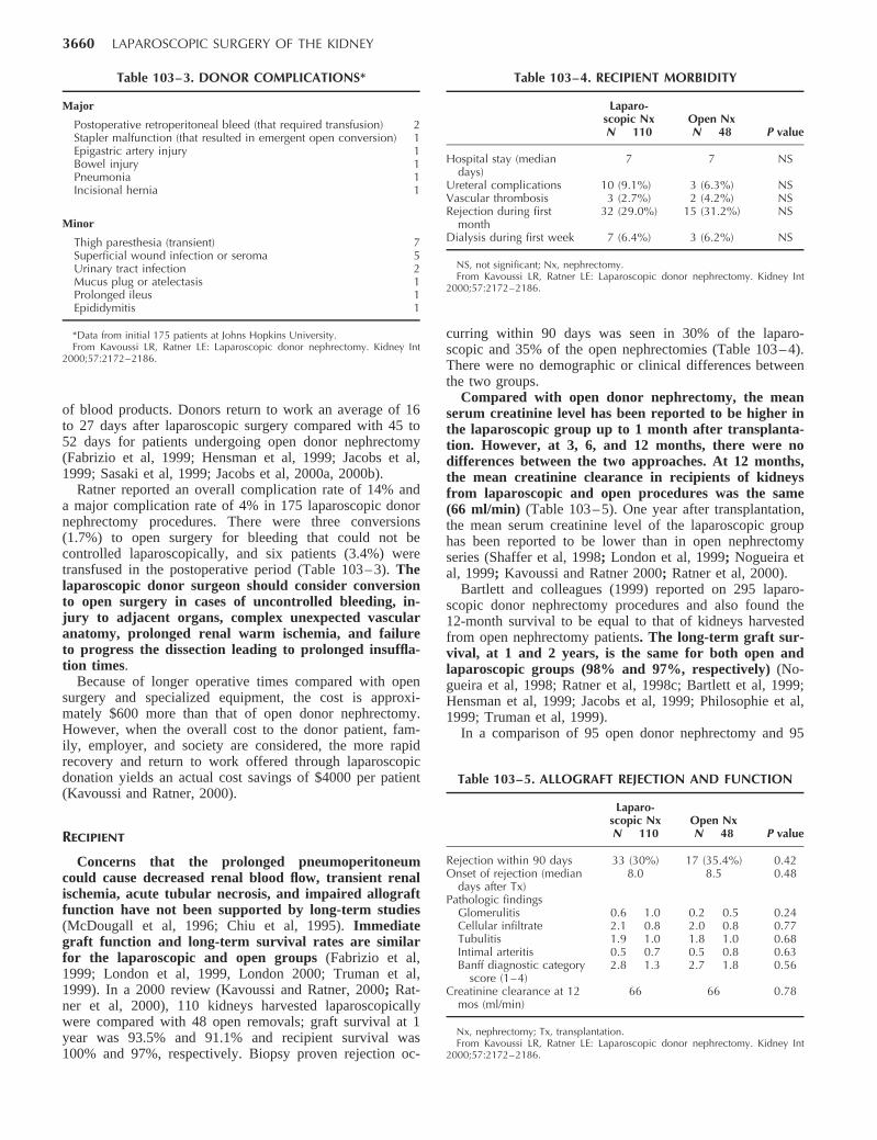
Table 103–3. DONOR COMPLICATIONS*
Major
Postoperative retroperitoneal bleed (that required transfusion) 2Stapler malfunction (that resulted in emergent open conversion) 1Epigastric artery injury 1Bowel injury 1Pneumonia 1Incisional hernia 1
Minor
Thigh paresthesia (transient) 7Superficial wound infection or seroma 5Urinary tract infection 2Mucus plug or atelectasis 1Prolonged ileus 1Epididymitis 1
*Data from initial 175 patients at Johns Hopkins University.From Kavoussi LR, Ratner LE: Laparoscopic donor nephrectomy. Kidney Int
2000;57:2172–2186.
Table 103–4. RECIPIENT MORBIDITY
Laparo-scopic NxN 110
Open NxN 48 P value
Hospital stay (mediandays)
7 7 NS
Ureteral complications 10 (9.1%) 3 (6.3%) NSVascular thrombosis 3 (2.7%) 2 (4.2%) NSRejection during firstmonth
32 (29.0%) 15 (31.2%) NS
Dialysis during first week 7 (6.4%) 3 (6.2%) NS
NS, not significant; Nx, nephrectomy.From Kavoussi LR, Ratner LE: Laparoscopic donor nephrectomy. Kidney Int
2000;57:2172–2186.
Table 103–5. ALLOGRAFT REJECTION AND FUNCTION
Laparo-scopic NxN 110
Open NxN 48 P value
Rejection within 90 days 33 (30%) 17 (35.4%) 0.42Onset of rejection (mediandays after Tx)
8.0 8.5 0.48
Pathologic findingsGlomerulitis 0.6 1.0 0.2 0.5 0.24Cellular infiltrate 2.1 0.8 2.0 0.8 0.77Tubulitis 1.9 1.0 1.8 1.0 0.68Intimal arteritis 0.5 0.7 0.5 0.8 0.63Banff diagnostic categoryscore (1–4)
2.8 1.3 2.7 1.8 0.56
Creatinine clearance at 12mos (ml/min)
66 66 0.78
Nx, nephrectomy; Tx, transplantation.From Kavoussi LR, Ratner LE: Laparoscopic donor nephrectomy. Kidney Int
2000;57:2172–2186.
of blood products. Donors return to work an average of 16to 27 days after laparoscopic surgery compared with 45 to52 days for patients undergoing open donor nephrectomy(Fabrizio et al, 1999; Hensman et al, 1999; Jacobs et al,1999; Sasaki et al, 1999; Jacobs et al, 2000a, 2000b).
Ratner reported an overall complication rate of 14% anda major complication rate of 4% in 175 laparoscopic donornephrectomy procedures. There were three conversions(1.7%) to open surgery for bleeding that could not becontrolled laparoscopically, and six patients (3.4%) weretransfused in the postoperative period (Table 103– 3). Thelaparoscopic donor surgeon should consider conversionto open surgery in cases of uncontrolled bleeding, in-jury to adjacent organs, complex unexpected vascularanatomy, prolonged renal warm ischemia, and failureto progress the dissection leading to prolonged insuffla-tion times.
Because of longer operative times compared with opensurgery and specialized equipment, the cost is approxi-mately $600 more than that of open donor nephrectomy.However, when the overall cost to the donor patient, fam-ily, employer, and society are considered, the more rapidrecovery and return to work offered through laparoscopicdonation yields an actual cost savings of $4000 per patient(Kavoussi and Ratner, 2000).
RECIPIENT
Concerns that the prolonged pneumoperitoneumcould cause decreased renal blood flow, transient renalischemia, acute tubular necrosis, and impaired allograftfunction have not been supported by long-term studies(McDougall et al, 1996; Chiu et al, 1995). Immediategraft function and long-term survival rates are similarfor the laparoscopic and open groups (Fabrizio et al,1999; London et al, 1999, London 2000; Truman et al,1999). In a 2000 review (Kavoussi and Ratner, 2000; Rat-ner et al, 2000), 110 kidneys harvested laparoscopicallywere compared with 48 open removals; graft survival at 1year was 93.5% and 91.1% and recipient survival was100% and 97%, respectively. Biopsy proven rejection oc-
curring within 90 days was seen in 30% of the laparo-scopic and 35% of the open nephrectomies (Table 103– 4).There were no demographic or clinical differences betweenthe two groups.
Compared with open donor nephrectomy, the meanserum creatinine level has been reported to be higher inthe laparoscopic group up to 1 month after transplanta-tion. However, at 3, 6, and 12 months, there were nodifferences between the two approaches. At 12 months,the mean creatinine clearance in recipients of kidneysfrom laparoscopic and open procedures was the same(66 ml/min) (Table 103– 5). One year after transplantation,the mean serum creatinine level of the laparoscopic grouphas been reported to be lower than in open nephrectomyseries (Shaffer et al, 1998; London et al, 1999; Nogueira etal, 1999; Kavoussi and Ratner 2000; Ratner et al, 2000).
Bartlett and colleagues (1999) reported on 295 laparo-scopic donor nephrectomy procedures and also found the12-month survival to be equal to that of kidneys harvestedfrom open nephrectomy patients. The long-term graft sur-vival, at 1 and 2 years, is the same for both open andlaparoscopic groups (98% and 97%, respectively) (No-gueira et al, 1998; Ratner et al, 1998c; Bartlett et al, 1999;Hensman et al, 1999; Jacobs et al, 1999; Philosophie et al,1999; Truman et al, 1999).
In a comparison of 95 open donor nephrectomy and 95
JAY T. BISHOFF AND LOUIS R. KAVOUSSI 3661
laparoscopic donor nephrectomy recipients, Edye and asso-ciates (1999) reported greater serum creatinine levels atdays 2 to 5 for the laparoscopic group. By day 6, therewas no difference between the two groups. At 3 and 12months, the serum creatinine levels were lower in the lapa-roscopic group than in the open series. Acute rejectionoccurred in 32% of kidneys from open procedures and15% of laparoscopic allografts. Three surgical graft failuresand five ureteral complications were seen in each group.
The incidence of surgical complications in recipients ap-pears to be equivalent between the two groups (Kavoussiand Ratner, 2000). Early reports of laparoscopic donornephrectomy cited ureteral complications ranging from9% to 15%. Disruption of the ureteral blood supplythrough aggressive dissection and exposure of the ure-ter was the cause in most cases. Occasionally, the ureterhad been trapped outside the bag retrieval device dur-ing extraction. Changes in ureteral dissection, in whichthe gonadal vein is divided at the renal vein and thendissected laterally with the ureter, have preserved theperiureteral blood supply and have resulted in a de-crease in ureteral complication rates to less than 3%(Kuo et al, 1998, Ratner et al, 1999; Dunkin et al, 2000;Jacobs et al, 2000; Kavoussi and Ratner, 2000).
Alternative Approaches
RetroperitonealThe retroperitoneal approach has been used for the donor
procedures (Rassweiler et al, 1998; Ishikawa et al, 1998).Compared with the transabdominal working space,there is less room in the retroperitoneum for organmanipulation and entrapment. The kidney is removedthrough a 6- to 8-cm flank incision.
Hand-AssistedThe success of laparoscopic living donor nephrectomy in
experienced hands has encouraged many institutions to of-fer this technique. One limitation is the need for advancedlaparoscopic training and a case volume sufficient to main-tain expertise in performing renal surgery. Hand-assistedlaparoscopic donor nephrectomy has been an alternativedeveloped to bridge the gap between laparoscopy and opensurgery. The warm ischemic time and operative timeshave been shown to be improved over the laparoscopicapproach, and there is no difference to the donor interms of the length of hospital stay and the time to fullrecovery (Nakada, 1997; Slakey et al, 1999).
Wolf and coworkers (2000) compared 10 hand-assisted,left-sided laparoscopic donor nephrectomies with 40 opendonor surgeries. They demonstrated a longer operative timewith the laparoscopic hand-assisted approach comparedwith open surgery (mean 215 vs. 95 min, respectively) anda shorter hospital stay for the laparoscopic group (mean 1.8vs. 2.9 days). The mean warm ischemic time was 2.9minutes in the laparoscopic group and not reported in theopen series. There were no differences between groups interms of donor complications, allograft function, and ure-teral problems. The mean hospital costs were 23% greater
in the laparoscopic group (P .005), with the hand-as-sisted device accounting for 11% of the operative costs.The cost savings from slightly shorter operating roomtimes were offset by the costs of disposable items.
RENAL BIOPSY
Renal biopsy is an important component in the assess-ment of proteinuria or unexplained medical disease of thekidney. Histologic information is often pivotal in makingtreatment decisions and providing prognostic information(Morel-Maroger, 1982; Gault and Muehrcke, 1983; Manoli-god et al, 1985).
Ultrasound-guided percutaneous needle biopsy, underlocal anesthesia, is the current standard for obtainingrenal tissue. Unfortunately, there is a 5% rate of signifi-cant hemorrhagic complications, and as many as 5% to20% of cases yield inadequate tissue for accurate diag-nosis (Wickre and Golper, 1982). When percutaneous bi-opsy fails or is considered to have a high risk, patients arereferred for surgical renal biopsy. This allows hemostasisto be achieved under direct vision and provides the pathol-ogist with adequate tissue to make a diagnosis. Laparo-scopic renal biopsy offers the advantages of open biopsywith the decreased morbidity of a two-port outpatient pro-cedure.
Indications
Renal biopsy under direct vision is indicated in threeprimary categories of patients: failed percutaneous nee-dle biopsy, anatomic variations, and a high risk ofbleeding complication. Factors that may make a patientunsuitable for percutaneous biopsy include morbid obe-sity, multiple bilateral cysts, a body habitus that makeslocalization impossible, and a solitary functioning kid-ney. Laparoscopic renal biopsy is contraindicated in thosepatients with an uncorrectable coagulopathy, uncontrolledhypertension, or an inability to tolerate general anesthesia.Renal imaging with CT or ultrasonography is performed todetermine any abnormality, such as renal cysts or solitarykidney, that may alter the choice of kidney for biopsy.
Patient Positioning
The patient is placed in the full flank position with theumbilicus over the table break. The table is fully flexed tohelp increase the distance between the ribs and the iliaccrest. Grounding pads for electrocautery and the argonbeam coagulator are placed on the exposed upper thigh.
Procedure
Retroperitoneal AccessA 10-mm transverse incision is made in the skin midway
between the iliac crest and the tip of the 12th rib in theposterior axillary line. A 0-degree lens and visual obturator
JAY T. BISHOFF AND LOUIS R. KAVOUSSI 3663
obturator is removed, leaving behind the 10-mm trocar.Carbon dioxide insufflation is begun at a pressure of 20mm Hg. Blunt dissection, using only the laparoscope, isinitially used to create a retroperitoneal working space.Anteriorly, the peritoneum is swept medially, exposing theunderside of the transversalis fascia (Fig. 103– 17). Onceanterior dissection has mobilized the peritoneum medially,a 5-mm port is placed in the anterior axillary line at thesame level as the first port (Fig. 103– 18).
Kidney Exposure and BiopsyThe lower pole of the kidney is located and Gerota’s
fascia opened using blunt and sharp dissection. In anatom-ically challenging patients such as those who are mor-bidly obese, preoperative transcutaneous or intraopera-tive ultrasonography may be valuable in localizing thekidney (Chen et al, 1997). Once Gerota’s fascia is incised,the perirenal fat is swept aside to expose an approximatelythe lower pole. A 5-mm biopsy forceps is used to takeseveral cortical samples (Fig. 103– 19).
Hemostasis and ClosureHemostasis is obtained with the argon beam coagula-
tor. During activation of the argon beam, it is impor-tant to open an insufflation port, because the flow ofargon gas can markedly increase the intra-abdominalpressure. Once adequate hemostasis is believed to beachieved, the insufflation pressure is lowered to 5 mm Hgfor at least 5 minutes and the entire retroperitoneum in-spected for hemostasis. Persistent bleeding from the biopsysite is treated with repeated argon beam coagulation. Oncehemostasis has been confirmed under low pressure, oxi-dized cellulose (Surgicel) is packed into the biopsy site anddirect pressure is applied. The skin incisions are irrigated,inspected for hemostasis, and closed with a 4-0 absorbablesubcuticular suture.
Postoperative Considerations
The length of hospital stay depends on the patient’shealth status. Specific attention is given to blood pressurecontrol. Care must be taken in patients requiring post-operative resumption of anticoagulation. Usually, pa-tients can resume their usual oral warfarin dose 24 to48 hours after surgery. Patients who require IV heparinmust be followed very closely to ensure than they donot become supratherapeutic.
Most nonhospitalized patients can be discharged thesame day as the biopsy. They are given oxycodone-aceta-minophen for pain control and can resume activity as toler-ated.
Results
A report of 32 consecutive patients who underwent lapa-roscopic renal biopsy showed 100% success in obtainingadequate tissue for histopathologic diagnosis (Gimenez etal, 1998). The mean blood loss was 26 ml, the operative
time was 1.5 hours, and the hospital stay was 1.7 days.Sixteen patients (50%) were treated as outpatients. Compli-cations included one inadvertent biopsy of the spleen with-out consequence and one 300-ml hematoma that resolvedwithout a need for intervention. The overall complicationrate was 6% (2 of 32). One patient on high-dose steroidtherapy developed a perforated peptic ulcer and died sevendays after surgery. Hemorrhage is the most common ma-jor complication associated with laparoscopic renal bi-opsy. Careful resumption of anticoagulation is manda-tory. The cause of a persistent decline in hematocrit orsymptoms of hypovolemia should be evaluated by CT.
In another series, of 17 patients, a balloon was insertedinto the retroperitoneum to create the working space (Gauret al, 1994). Adequate renal tissue for diagnosis was ob-tained in each case. The mean operative time was 35 min-utes, excluding anesthesia time, with a range of 20 to 45minutes. Fifteen patients were discharged within 24 hours,and two patients remained hospitalized for 4 days. Compli-cations were seen in 11% of the patients (2 of 17), includ-ing severe bleeding requiring conversion to an open proce-dure and one patient with marked gross hematuria days.
RENAL CYSTIC DISEASE
Renal cysts are present in approximately one third ofpatients older than 50 years, and few require surgicalintervention (Hoenig et al, 1997; Wolf, 1998). Patientswith pain, infection, or obstruction may need cyst exci-sion. Moreover, with the advent of CT and ultrasonogra-phy, the detection of indeterminate renal masses and com-plex renal cysts has become a frequent occurrence.Classification schemes have been devised based on CTcriteria to help surgeons determine whether further diag-nostic or therapeutic maneuvers are necessary (Table 103–6) (Bosniak and Morton, 1986). Unfortunately, imagingstudies are not always diagnostic, and surgical removalmay be needed to exclude the possibility of malignancy.
In addition, approximately 600,000 individuals sufferfrom ADPKD. The clinical course of ADPKD is usuallydominated by symptoms of abdominal fullness or pain andmay require surgical treatment for symptomatic relief. End-stage ADPKD may occasionally require bilateral or unilat-eral nephrectomy for the treatment of cyst complications,bleeding, infection, or pain.
Indications
Needle aspiration with sclerosis of symptomatic renalcysts is usually the first line of therapy. However, it isnot always effective and can pose a risk of fibrosiswhen perihilar cysts are approached (Wehle and Grab-stald, 1986; Hulbert et al, 1988; Lifson et al, 1998; Mc-Dougall, 1998; Santiago et al, 1998). Renal cysts may alsoobstruct the collecting system, compress renal parenchyma,or spontaneously hemorrhage, inducing pain and hematuria.In addition, they may become infected, cause obstructiveuropathy, and cause hypertension. The laparoscopic ap-proach offers a minimally invasive modality to decompresscysts under direct vision. Complex cysts can also be ex-
JAY T. BISHOFF AND LOUIS R. KAVOUSSI 3665
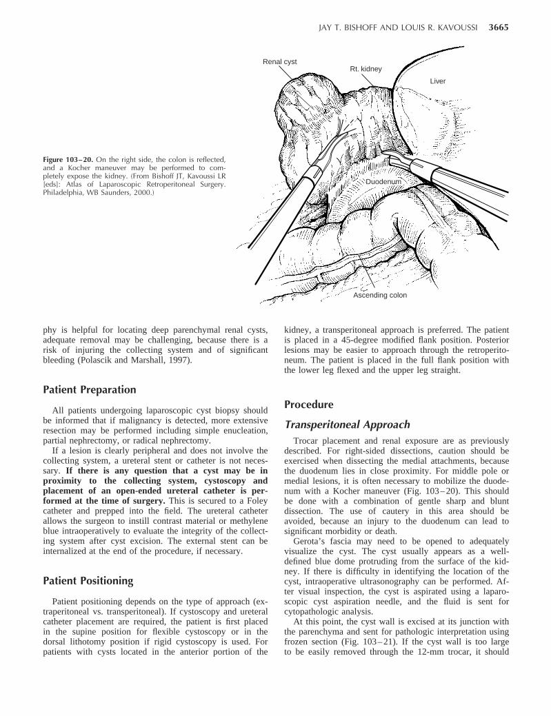
Figure 103–20. On the right side, the colon is reflected,and a Kocher maneuver may be performed to com-pletely expose the kidney. (From Bishoff JT, Kavoussi LR[eds]: Atlas of Laparoscopic Retroperitoneal Surgery.Philadelphia, WB Saunders, 2000.)
Rt. kidneyRenal cyst
Ascending colon
Duodenum
Liver
phy is helpful for locating deep parenchymal renal cysts,adequate removal may be challenging, because there is arisk of injuring the collecting system and of significantbleeding (Polascik and Marshall, 1997).
Patient Preparation
All patients undergoing laparoscopic cyst biopsy shouldbe informed that if malignancy is detected, more extensiveresection may be performed including simple enucleation,partial nephrectomy, or radical nephrectomy.
If a lesion is clearly peripheral and does not involve thecollecting system, a ureteral stent or catheter is not neces-sary. If there is any question that a cyst may be inproximity to the collecting system, cystoscopy andplacement of an open-ended ureteral catheter is per-formed at the time of surgery. This is secured to a Foleycatheter and prepped into the field. The ureteral catheterallows the surgeon to instill contrast material or methyleneblue intraoperatively to evaluate the integrity of the collect-ing system after cyst excision. The external stent can beinternalized at the end of the procedure, if necessary.
Patient Positioning
Patient positioning depends on the type of approach (ex-traperitoneal vs. transperitoneal). If cystoscopy and ureteralcatheter placement are required, the patient is first placedin the supine position for flexible cystoscopy or in thedorsal lithotomy position if rigid cystoscopy is used. Forpatients with cysts located in the anterior portion of the
kidney, a transperitoneal approach is preferred. The patientis placed in a 45-degree modified flank position. Posteriorlesions may be easier to approach through the retroperito-neum. The patient is placed in the full flank position withthe lower leg flexed and the upper leg straight.
Procedure
Transperitoneal ApproachTrocar placement and renal exposure are as previously
described. For right-sided dissections, caution should beexercised when dissecting the medial attachments, becausethe duodenum lies in close proximity. For middle pole ormedial lesions, it is often necessary to mobilize the duode-num with a Kocher maneuver (Fig. 103– 20). This shouldbe done with a combination of gentle sharp and bluntdissection. The use of cautery in this area should beavoided, because an injury to the duodenum can lead tosignificant morbidity or death.
Gerota’s fascia may need to be opened to adequatelyvisualize the cyst. The cyst usually appears as a well-defined blue dome protruding from the surface of the kid-ney. If there is difficulty in identifying the location of thecyst, intraoperative ultrasonography can be performed. Af-ter visual inspection, the cyst is aspirated using a laparo-scopic cyst aspiration needle, and the fluid is sent forcytopathologic analysis.
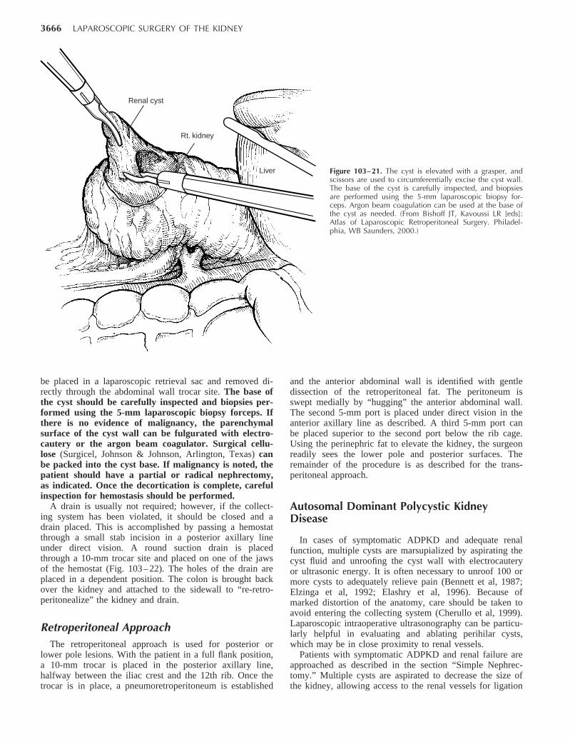
At this point, the cyst wall is excised at its junction withthe parenchyma and sent for pathologic interpretation usingfrozen section (Fig. 103– 21). If the cyst wall is too largeto be easily removed through the 12-mm trocar, it should
3666 LAPAROSCOPIC SURGERY OF THE KIDNEY
Liver
Renal cyst
Rt. kidney
Figure 103–21. The cyst is elevated with a grasper, andscissors are used to circumferentially excise the cyst wall.The base of the cyst is carefully inspected, and biopsiesare performed using the 5-mm laparoscopic biopsy for-ceps. Argon beam coagulation can be used at the base ofthe cyst as needed. (From Bishoff JT, Kavoussi LR [eds]:Atlas of Laparoscopic Retroperitoneal Surgery. Philadel-phia, WB Saunders, 2000.)
be placed in a laparoscopic retrieval sac and removed di-rectly through the abdominal wall trocar site. The base ofthe cyst should be carefully inspected and biopsies per-formed using the 5-mm laparoscopic biopsy forceps. Ifthere is no evidence of malignancy, the parenchymalsurface of the cyst wall can be fulgurated with electro-cautery or the argon beam coagulator. Surgical cellu-lose (Surgicel, Johnson & Johnson, Arlington, Texas) canbe packed into the cyst base. If malignancy is noted, thepatient should have a partial or radical nephrectomy,as indicated. Once the decortication is complete, carefulinspection for hemostasis should be performed.
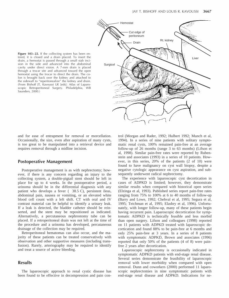
A drain is usually not required; however, if the collect-ing system has been violated, it should be closed and adrain placed. This is accomplished by passing a hemostatthrough a small stab incision in a posterior axillary lineunder direct vision. A round suction drain is placedthrough a 10-mm trocar site and placed on one of the jawsof the hemostat (Fig. 103– 22). The holes of the drain areplaced in a dependent position. The colon is brought backover the kidney and attached to the sidewall to “re-retro-peritonealize” the kidney and drain.
Retroperitoneal ApproachThe retroperitoneal approach is used for posterior or
lower pole lesions. With the patient in a full flank position,a 10-mm trocar is placed in the posterior axillary line,halfway between the iliac crest and the 12th rib. Once thetrocar is in place, a pneumoretroperitoneum is established
and the anterior abdominal wall is identified with gentledissection of the retroperitoneal fat. The peritoneum isswept medially by “hugging” the anterior abdominal wall.The second 5-mm port is placed under direct vision in theanterior axillary line as described. A third 5-mm port canbe placed superior to the second port below the rib cage.Using the perinephric fat to elevate the kidney, the surgeonreadily sees the lower pole and posterior surfaces. Theremainder of the procedure is as described for the trans-peritoneal approach.
Autosomal Dominant Polycystic KidneyDisease
In cases of symptomatic ADPKD and adequate renalfunction, multiple cysts are marsupialized by aspirating thecyst fluid and unroofing the cyst wall with electrocauteryor ultrasonic energy. It is often necessary to unroof 100 ormore cysts to adequately relieve pain (Bennett et al, 1987;Elzinga et al, 1992; Elashry et al, 1996). Because ofmarked distortion of the anatomy, care should be taken toavoid entering the collecting system (Cherullo et al, 1999).Laparoscopic intraoperative ultrasonography can be particu-larly helpful in evaluating and ablating perihilar cysts,which may be in close proximity to renal vessels.
Patients with symptomatic ADPKD and renal failure areapproached as described in the section “Simple Nephrec-tomy.” Multiple cysts are aspirated to decrease the size ofthe kidney, allowing access to the renal vessels for ligation
JAY T. BISHOFF AND LOUIS R. KAVOUSSI 3667
Figure 103–22. If the collecting system has been en-tered, it is closed and a drain placed. To insert thedrain, a hemostat is passed through a small stab inci-sion in the side and advanced into the abdominalcavity under direct vision. A 7-mm drain is placedthrough a trocar site and advanced toward the openhemostat using the trocar to direct the drain. The co-lon is brought back over the kidney and attached tothe sidewall to “reperitonealize” the kidney and drain.(From Bishoff JT, Kavoussi LR [eds]: Atlas of Laparo-scopic Retroperitoneal Surgery. Philadelphia, WBSaunders, 2000.)
Surgicel
Drain
Cut edge of peritoneum
Hemostat
Liver
Rt. kidney
and for ease of entrapment for removal or morcellation.Occasionally, the size, even after aspiration of many cysts,is too great to be manipulated into a retrieval device andrequires removal through a midline incision.
Postoperative Management
Postoperative management is as with nephrectomy; how-ever, if there is any concern regarding an injury to thecollecting system, a double-pigtail stent should be left inplace for up to 4 weeks. In the postoperative period, aurinoma should be in the differential diagnosis with anypatient who develops a fever ( 38.5 C), persistent ileus,abdominal pain, nausea or vomiting, or an elevated whiteblood cell count with a left shift. CT with oral and IVcontrast material can be helpful to identify a urinary leak.If a leak is detected, the bladder catheter should be rein-serted, and the stent may be repositioned as indicated.Alternatively, a percutaneous nephrostomy tube can beplaced. If a retroperitoneal drain was not left at the time ofthe procedure and a urinoma has developed, percutaneousdrainage of the collection may be required.
Retroperitoneal hematomas can also occur, and the ma-jority of these patients can be treated conservatively withobservation and other supportive measures (including trans-fusion). Rarely, arteriography may be required to identifyand treat a source of active bleeding.
Results
The laparoscopic approach to renal cystic disease hasbeen found to be effective in decompression and pain con-
trol (Morgan and Rader, 1992; Hulbert 1992; Munch et al,1994). In a series of nine patients with solitary sympto-matic renal cysts, 100% remained pain-free at an averagefollow-up of 26 months (range 3 to 63 months) (Lifson etal, 1998). Similar pain-free rates were reported by Ruben-stein and associates (1993) in a series of 10 patients. How-ever, in this series, 20% of the patients (2 of 10) werefound to have malignancy on cyst wall biopsy, despite anegative cytologic appearance on cyst aspiration, and sub-sequently underwent radical nephrectomy.
The experience with laparoscopic cyst decortication incases of ADPKD is limited; however, they demonstratesimilar results when compared with historical open series(Elzinga et al, 1993). Published series report pain-free ratesranging from 75% to 100% at 6 to 40 months of follow-up(Barry and Lowe, 1992; Chehval et al, 1995; Segura et al,1995; Teichman et al, 1995; Elashry et al, 1996). Unfortu-nately, with longer follow-up, many of these patients beginhaving recurrent pain. Laparoscopic decortication for symp-tomatic ADPKD is technically feasible and less morbidthan open surgery. Lifson and colleagues (1998) reportedon 11 patients with ADPKD treated with laparoscopic de-cortication and found 88% to be pain-free at 6 months andonly 25% pain-free at 3 years. In a series of 8 patientswith symptomatic ADPKD, Brown and associates (1996)reported that only 50% of the patients (4 of 8) were pain-free 2 years after decortication.
Laparoscopic nephrectomy is occasionally indicated insymptomatic ADPKD patients with end-stage renal disease.Several series demonstrate the feasibility of laparoscopicremoval with lower morbidity when compared with openremoval. Dunn and coworkers (2000) performed 11 laparo-scopic nephrectomies in nine symptomatic patients withend-stage renal disease and ADPKD. Indications for ne-
3668 LAPAROSCOPIC SURGERY OF THE KIDNEY
Rt. kidney
Liver
Ascending colon
Peritoneum
Figure 103–23. The kidney is stripped of overlying Gerota’sfascia down to the kidney capsule. (From Bishoff JT, Ka-voussi LR [eds]: Atlas of Laparoscopic Retroperitoneal Sur-gery. Philadelphia, WB Saunders, 2000.)
phrectomy included pain, hypertension, recurrent urinarytract infections, and gross hematuria. The mean operativetime was 6 hours, with a mean estimated blood loss of 153ml. The majority of kidneys were morcellated. The meanhospital stay was 3 days, and the mean time to normalactivity was 5 weeks.
Kaouk and associates (2000) compared a series of 9bilateral synchronous laparoscopic nephrectomies per-formed for symptomatic ADPKD to 14 open proceduresperformed for the same indication. None of the laparo-scopic procedures required conversion to open surgery.There was no difference in operating time, specimenweight, or blood loss between the two groups. However,there was a significant difference in the length of the hos-pital stay (2.4 vs. 8.1 days, P .001) and narcotic use (30mg vs. 177 mg, P .001) favoring the laparoscopic ap-proach. Two patients in the laparoscopic group had a retro-peritoneal hematoma that did not require surgical interven-tion.
NEPHROPEXY
Nephroptosis has been recognized and treated surgicallysince the 1800s, when nephropexy was described in Berlinby Hahn (Harrison, 1969). In one of the early urologytexts, Hugh Hampton Young defined nephroptosis as theinferior displacement of the kidney by more than 5 cmwhen the patient moves from a supine to an erect position(Young and Davis, 1926). William Mayo diagnosed float-ing kidneys in 20% of random patients, and William Osler(1892) had recorded over 700 cases. The ptotic kidney hasalso been blamed for abnormality and symptoms in otherorgans including stomach, bowel, and pancreas. It has beenestimated that over 170 innovative procedures have beendescribed to adhere the kidney to the retroperitoneum toprevent descent while standing (Deming, 1930; Moss,1997).
Unfortunately, nephroptosis was frequently cited as thecause of a variety of symptoms, and surgical repair was
greatly overutilized. Because repair did not afford patientsrelief of symptoms, this diagnosis was questioned to thepoint at which it disappeared from the urologic literature.However, there are patients in whom positional changesresult in obstruction of the collecting system, and suchpatients could benefit from surgical repair. Laparoscopicnephropexy represents the latest surgical approach to treat-ing this entity.
Indications
Nephroptosis is characterized by a significant downwarddisplacement ( 5 cm) of the kidney as the patient movesfrom the supine to the erect position, causing pain in theabdomen or flank. The upper pole of the kidney can usu-ally be palpated on deep inspiration. The most severe man-ifestation causes Dietl’s crisis heralded by severe colickyflank pain, nausea, chills, tachycardia, oliguria, and tran-sient hematuria or proteinuria (Irwin, 1948). The exactcause of pain is not known but is likely to be from tran-sient renal ischemia or obstruction (Moss, 1997). Patientswho are symptomatic are generally young thin females,and pain in the erect position is the primary symptom.
Either erect and supine IV urograms or renal scansdocumenting obstruction are the best diagnostic studiesfor nephroptosis. Descent of the symptomatic kidney bytwo vertebral bodies and obstruction or diminished flowto the symptomatic side should be documented beforesurgical repair.
Procedure
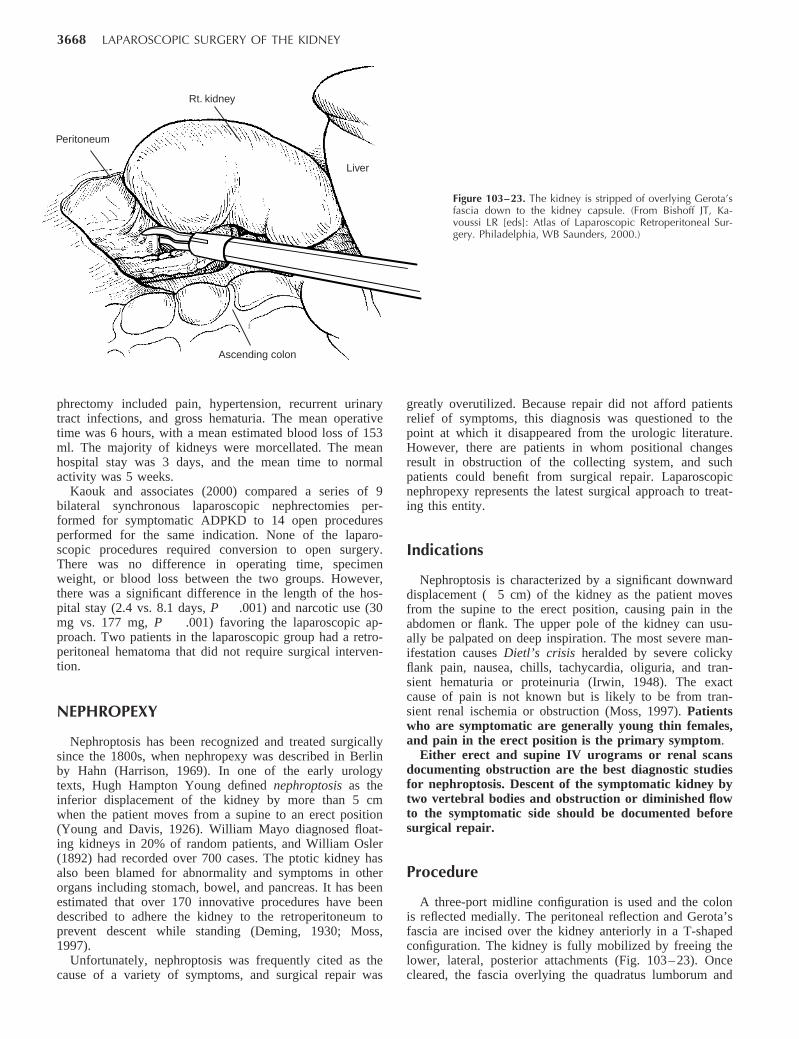
A three-port midline configuration is used and the colonis reflected medially. The peritoneal reflection and Gerota’sfascia are incised over the kidney anteriorly in a T-shapedconfiguration. The kidney is fully mobilized by freeing thelower, lateral, posterior attachments (Fig. 103– 23). Oncecleared, the fascia overlying the quadratus lumborum and
JAY T. BISHOFF AND LOUIS R. KAVOUSSI 3669
Figure 103–24. Once the kidney is free of lat-eral and posterior attachments, multiple 2-0 su-tures are placed into the capsule and the lateraledge of the fascia overlying the abdominalwall. (From Bishoff JT, Kavoussi LR [eds]: Atlasof Laparoscopic Retroperitoneal Surgery. Phila-delphia, WB Saunders, 2000.)
Rt. kidney
Peritoneum
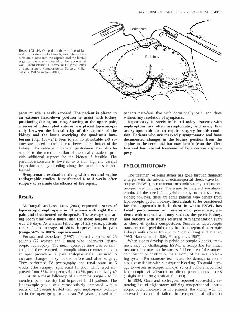
psoas muscle is easily exposed. The patient is placed inan extreme head-down position to assist with kidneypositioning during suturing. Starting at the upper pole,a series of interrupted sutures are placed laparoscopi-cally between the lateral edge of the capsule of thekidney and the fascia overlying the quadratus lum-borum (Fig. 103– 24). Four to six nonabsorbable 2-0 su-tures are placed in the upper to lower lateral border of thekidney. The subhepatic parietal peritoneum may also besutured to the anterior portion of the renal capsule to pro-vide additional support for the kidney if feasible. Thepneumoperitoneum is lowered to 5 mm Hg, and carefulinspection for any bleeding along the suture lines is per-formed.
Symptomatic evaluation, along with erect and supineradiographic studies, is performed 6 to 8 weeks aftersurgery to evaluate the efficacy of the repair.
Results
McDougall and associates (2000) reported a series oflaparoscopic nephropexy in 14 women with right flankpain and documented nephroptosis. The average operat-ing room time was 4 hours, and the mean hospital staywas 2.6 days. At a mean follow-up of 3.3 years, patientsreported an average of 80% improvement in pain(range 56% to 100% improvement).
Fornara and associates (1997) reported a series of 23patients (22 women and 1 man) who underwent laparo-scopic nephropexy. The mean operative time was 60 min-utes, and they reported no complications or conversions toan open procedure. A pain analogue scale was used tomeasure changes in symptoms before and after surgery.They performed IV pyelography and renal scans at 6weeks after surgery. Split renal function while erect im-proved from 38% preoperatively to 47% postoperatively (P
.03). At a mean follow-up of 13 months (range 2 to 37months), pain intensity had improved in 21 patients. Thelaparoscopic group was retrospectively compared with aseries of 12 patients treated with open nephropexy. Follow-up in the open group at a mean 7.6 years showed four
patients pain-free, five with occasionally pain, and threewithout any resolution of symptoms.
Nephropexy is rarely indicated today. Patients withnephroptosis are often asymptomatic, and many thatare symptomatic do not require surgery for this condi-tion. Patients who are markedly symptomatic and havedocumented changes in the kidney position from thesupine to the erect position may benefit from the effec-tive and less morbid treatment of laparoscopic nephro-pexy.
PYELOLITHOTOMY
The treatment of renal stones has gone through dramaticchanges with the advent of extracorporeal shock wave lith-otripsy (ESWL), percutaneous nephrolithotomy, and ureter-oscopic laser lithotripsy. These new techniques have almosteliminated the need for pyelolithotomy to remove renalstones; however, there are some patients who benefit fromlaparoscopic pyelolithotomy. Individuals to be consideredfor this approach include those in whom ESWL hasfailed, percutaneous or ureteroscopic procedures, pa-tients with unusual anatomy such as the pelvic kidney,and patients with stones resistant to fragmentation suchas those of cystine composition. Successful laparoscopictransperitoneal pyelolithotomy has been reported in ectopickidneys with stones from 2 to 4 cm (Chang and Dretler,1996; Harmon et al, 1996; Hoenig et al, 1997).
When stones develop in pelvic or ectopic kidneys, treat-ment may be challenging. ESWL is acceptable for initialtreatment but may not be successful because of the stones’composition or position or the anatomy of the renal collect-ing system. Percutaneous techniques risk damage to anom-alous vasculature with subsequent bleeding. To avoid dam-age to vessels in ectopic kidneys, several authors have usedlaparoscopic visualization to direct percutaneous access(Eshghi et al, 1985; Toth et al, 1993).
In 1994, Gaur and colleagues reported successfully re-moving five of eight stones utilizing retroperitoneal laparo-scopic pyelolithotomy. In two patients, the kidney was notaccessed because of failure in retroperitoneal dilatation
3670 LAPAROSCOPIC SURGERY OF THE KIDNEY
techniques. Once access to the kidney was obtained, thestones were successfully removed in five of six cases. Inone case, attempts to palpate the stone in the renal pelvisbefore pyelotomy dislodged the stone into an upper polecalyx requiring open conversion for removal.
CALYCEAL DIVERTICULECTOMY
Calyceal diverticula are congenital transitional cell– linedcavities that communicate with the collecting systemthrough narrow infundibula. Although the diverticula donot produce urine, they passively fill from the collectingsystem. Because the narrow infundibula causes urine todrain slower than in other areas in the collecting system,they are predisposed to stone formation from urinary stasis.It is estimated that 9% to 35% form stones (Middleton andPfister, 1974; Timmons et al, 1975; Donnellan et al, 1999).The most common symptoms include pain in the back orflank, infection, and hematuria.
The management of calyceal diverticula has always beenchallenging. The classic management was by open surgery.Partial nephrectomy and open marsupialization with fulgu-ration of the diverticulum and even total nephrectomy havebeen used in the past (Devine et al, 1969; Williams et al,1969; Wulfsohn, 1980). Advances in less invasive equip-ment and techniques have changed the treatment of thesedifficult cases.
The ideal means of treating symptomatic calyceal di-verticula include removal of the stones and widening ofthe infundibulum to prevent urine stasis or ablation ofthe diverticular cavity. Percutaneous treatment is mostoften used for treatment in symptomatic cases (Donnellanet al, 1999). ESWL and ureteroscopy have also been ap-plied, but anatomic factors can result in a lower rate ofachieving stone clearance (Jones et al, 1991; Pang et al,1992; Stream et al, 1992;). Recurrence of symptoms andstones can occur if the diverticulum has not been ablated.
Indications
Large peripheral diverticula and those located medi-ally near the renal hilum may best be treated usinglaparoscopic techniques. A laparoscopic approach allowsevacuation of the stone fragments, careful inspection, andfulguration of the cavity using argon beam or standardmonopolar coagulation. Additionally, perirenal fat can beinserted into the cavity to decrease the chance for recur-rence.
Procedure
As with the treatment of renal cysts, the location of thediverticulum helps dictate a transabdominal or retroperito-neal approach. Gerota’s fascia is opened and perirenal fatcleared from the surface of the kidney. The location of thediverticulum is usually readily identified by the pres-ence of dense adhesions from the surface of the kidneyto surrounding fat and a “dimpling” on the surface ofthe kidney. Intraoperative fluoroscopy or ultrasonogra-
phy can also be used to confirm the location of deeperrenal cysts. Once located, the thin parenchyma over thecavity is cauterized and opened. Spoon graspers are usefulfor removing stones intact. Milk of calcium stones andmany small stone fragments can be encountered, despitethe appearance of a solitary dense stone on conventionalradiographs. With the use of graspers and scissors, thediverticulum opening is enlarged to the point of encounter-ing normal renal parenchyma and is unroofed. Argon beamcoagulation or monopolar cautery is used to fulgurate thelining of the cavity to prevent recurrence. Methylene bluecan be given intravenously, and careful inspection allowsdetection of blue urine in the cavity, indicating communi-cation with the collecting system. The collecting systemcan be closed using laparoscopic suturing techniques andperirenal fat inserted into the cavity. Stenting and drainageare usually performed.
Results
Several authors have reported successful laparoscopictreatment of calyceal diverticula (Gluckman et al, 1993,Ruckle and Segura, 1994, Harewood et al, 1996, Donnellanet al, 1999). Patients benefited from the advantages of alaparoscopic approach; however, most of these series weresmall, with limited follow-up.
LAPAROSCOPY FOR RENALMALIGNANCY
The use of laparoscopy in the treatment of renal malig-nancies has generated a great deal of controversy based onfear of inadequate cancer control. Early laparoscopic expe-riences in the surgical and gynecologic literature haveraised concerns regarding the risk of port site seeding,inadequate surgical margins, and inaccurate staging due tomorcellation. Experience has borne out that laparoscopicremoval in experienced hands provides equivalent cancercontrol when compared with traditional open approaches.
Port site recurrence after laparoscopic procedures in can-cer patients has been described in the general surgery,gynecology, and urology literature. In a broad survey ofover 1050 European general surgery programs, port siterecurrence occurred in 17% of laparoscopic cholecystecto-mies for incidental gallbladder carcinoma and in 4.6% ofcases of colorectal cancer (Paolucci et al, 1999). Studieshave demonstrated that tumor cells may be deposited at theport site during laparoscopy either directly from contami-nated instruments or indirectly by way of insufflation gas(Neuhaus et al, 1998; Schaeff et al, 1998; Whelan and Lee,1999).
In the urologic literature, there have been occasionalreports of laparoscopic port site seeding of transitional cell,prostate, and renal cell carcinomas. Bangma and coworkers(1995) reported on the first patient with port site seedingafter laparoscopic pelvic lymph node dissection (LPLND)for prostate cancer. In contrast, Cadeddu and associates(1996) found no cases of port site seeding after 372 casesof LPLND. A review of a subset of 40 patients withpositive pelvic lymph nodes for prostate cancer (at the time
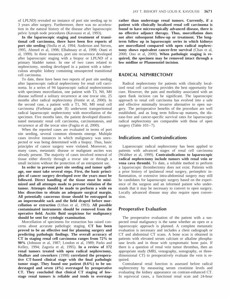
JAY T. BISHOFF AND LOUIS R. KAVOUSSI 3671
of LPLND) revealed no instance of port site seeding up to3 years after surgery. Furthermore, there was no accelera-tion in the natural history of the disease after laparoscopicpelvic lymph node procedures (Kavoussi et al, 1993).
In the laparoscopic staging and treatment of transi-tional cell carcinoma, there have been five reports ofport site seeding (Stolla et al, 1994; Anderson and Steven,1995, Ahmed et al, 1998; Elbahnasy et al, 1998; Otani etal, 1999). In three instances, port site recurrence developedafter laparoscopic staging with a biopsy or LPLND of aprimary bladder tumor. In one of two cases related tonephrectomy, seeding developed in a patient with a tuber-culous atrophic kidney containing unsuspected transitionalcell carcinoma.
To date, there have been two reports of port site seedingafter laparoscopic radical nephrectomy for renal cell carci-noma. In a series of 94 laparoscopic radical nephrectomieswith specimen morcellation, one patient with T3, N0, M0disease suffered a solitary recurrence at one trocar site, 25months after radical nephrectomy (Fentie et al, 2000). Inthe second case, a patient with a T1, N0, M0 renal cellcarcinoma (Furhman grade II) underwent transperitonealradical laparoscopic nephrectomy with morcellation of thespecimen. Five months later, the patient developed dissemi-nated metastatic renal cell carcinoma, carcinomatosis, andrecurrence at all the trocar sites (Fugita et al, 2000).
When the reported cases are evaluated in terms of portsite seeding, several common elements emerge. Multiplecases involve instances in which malignancy was unex-pected or was being determined with a biopsy. Thus, basicprinciples of cancer surgery were violated. Moreover, inmany cases, metastatic disease or malignant ascites wasalready present. The majority of cases involved removal oftissue either directly through a trocar site or through asmall incision without the protection of an entrapment sac.
In order to prevent port site seeding and tumor spill-age, one must take several steps. First, the basic princi-ples of cancer surgery developed over the years must befollowed. Direct handling of the tissue must be mini-mized and all attempts made to prevent violation of thetumor. Attempts should be made to perform a wide enbloc dissection to obtain an adequate surgical margin.All potentially cancerous tissue should be entrapped inan impermeable sack and the field draped before mor-cellation or extraction (Urban et al, 1993). All possiblecontaminated instruments should be removed from theoperative field. Ascitic fluid suspicious for malignancyshould be sent for cytologic examination.
Morcellation of specimens for extraction has raised con-cerns about accurate pathologic staging. CT has beenproved to be an effective tool for planning surgery andpredicting pathologic findings. The overall accuracy ofCT in staging renal cell carcinoma ranges from 72% to90% (Johnson et al, 1987; London et al, 1989; Parks andKelley, 1994; Zagoria et al, 1995). In a review of 172renal tumors treated with open radical nephrectomy,Shalhav and coworkers (1998) correlated the preopera-tive CT-based clinical stage with the final pathologictumor stage. They found one patient (0.6%) to be un-derstaged and seven (4%) overstaged by preoperativeCT. They concluded that clinical CT staging of low-stage renal tumors is reliable and tends to overstage
rather than understage renal tumors. Currently, if apatient with clinically localized renal cell carcinoma isfound to have microscopically advanced disease, there isno effective adjunct therapy. Thus, morcellation doesnot alter subsequent follow-up or treatment. The long-term follow up in laparoscopic series in which kidneysare morcellated compared with open radical nephrec-tomy shows equivalent cancer-free survival (Chan et al,2000; Ono et al, 2000). When pathologic staging is re-quired, the specimen may be removed intact through alow midline or Pfannenstiel incision.
RADICAL NEPHRECTOMY
Radical nephrectomy for patients with clinically local-ized renal cell carcinoma provides the best opportunity forcure. However, the pain and morbidity associated with anopen flank incision can be significant. The laparoscopicapproach to renal cell carcinoma has evolved into a safeand effective minimally invasive alternative to open sur-gery. The perioperative benefits of the procedure are wellestablished, and as long term follow-up matures, the dis-ease-free and cancer-specific survival rates for laparoscopicradical nephrectomy are comparable with those of opensurgery (Table 103– 7).
Indications and Contraindications
Laparoscopic radical nephrectomy has been applied topatients with advanced stages of renal cell carcinoma(Walther et al, 1999). Contraindications to laparoscopicradical nephrectomy include tumors with renal vein orvena cava thrombi. To date, a reliable method to performa laparoscopic thrombectomy does not exist. Patients witha prior history of ipsilateral renal surgery, perinephric in-flammation, or extensive intra-abdominal surgery may stillbe candidates for laparoscopic surgery based on the experi-ence of the surgeon and an informed patient who under-stands that it may be necessary to convert to open surgery.Locally advanced disease may also require open conver-sion.
Preoperative Evaluation
The preoperative evaluation of the patient with a sus-pected renal malignancy is the same whether an open or alaparoscopic approach is planned. A complete metastaticevaluation is necessary and includes a chest radiograph orCT and abdominal CT scans. A bone scan is obtained inpatients with elevated serum calcium or alkaline phospha-tase levels and in those with symptomatic bone pain. Ifthere is a question of renal vein tumor thrombus, then anappropriate study (MRI, venography, sonography, or three-dimensional CT) to preoperatively evaluate the vein is re-quired.
Contralateral renal function is assessed before radicalnephrectomy by measuring serum creatinine levels andevaluating the kidney appearance on contrast-enhanced CT.In equivocal cases, a functional renal scan and 24-hour

3672 LAPAROSCOPIC SURGERY OF THE KIDNEY
Table 103–7. LAPAROSCOPIC RADICAL NEPHRECTOMY SERIES (MEAN VALUES PRESENTED FOR EACH CATEGORY)
Author Approach Indication
ORTime(Min)
LOH(Days)
EBL(ml)
Conver-sion (%)
Compli-cations
(%)
Morcel-lation(%)
Intact(%)
Follow-up (mo)
LocalRecur-rence(%)
Metas-tatic
Disease(%)
McDougall et al,1996
TransperitonealN 17
T1–T2 414 4 105 6 18 29 71 14 0 0
Cadeddu et al,1998
TransperitonealN 139
T1, T2 na na na 4 10 90 10 19 1 3
RetroperitonealN 18
Abbou et al,1999
RetroperitonealN 29
T1 145 5 100 0 8 0 100 3 0
Ono et al, 1999 TransperitonealN 34
T1, T2 300 11 225 2 60 0 3
RetroperitonealN 15
Barrett and Fentie,1999
TransperitonealN 94
T1–T4 173 4.6 10 9 89 11 36 4 2
Gill et al, 2000 RetroperitonealN 53
T1, T2 180 2 128 4 17 0 100 13 0 4
Jeschke et al,2000
TransperitonealN 51
T1, T2 125 7 0 4 0 100 8 0 0
Janetschek et al,2000
TransperitonealN 100
T1, T2 137 7 168 0 10 0 100 14 0 0
Dunn et al,2000
TransperitonealN 55
T1, T2, T3 330 3.4 172 1 3* 65 35 25 0 8
RetroperitonealN 6
Chan et al,2001
TransperitonealN 67
T1, T2 256 3.8 289 1 15 60 40 21 0 3
*Major complicationsEBL, estimated blood loss; LOH, length of hospitalization; na, not applicable; OR, operating room.
urinary creatinine clearance studies can be obtained. Thepresence of renal insufficiency should prompt the surgeonto consider partial nephrectomy.
Positioning and Trocar Placement
Patient positioning and trocar placement are similar tothose described for the simple nephrectomy (see Fig. 103–3). Three ports are generally sufficient to complete theprocedure, although a fourth trocar may be necessary fororgan entrapment and liver retraction during right-sidednephrectomy.
Procedure
Reflection of the ColonThe colon is reflected to provide adequate visualization
of the anterior surface of Gerota’s fascia. On the right, theduodenum is mobilized medially, using the Kocher maneu-ver, until the vena cava is clearly visualized.
Dissection of the UreterThe midureter is located in the retroperitoneal fat medial
to the psoas muscle. During proximal mobilization, thegonadal vein is usually first encountered and should beswept medially. The ureter is located just posterior to this
structure (Fig. 103– 25). Once located, the ureter is ele-vated, revealing the psoas muscle and traced proximally toidentify the renal hilum.
Mobilization of the Lower PoleAs opposed to a simple nephrectomy, the radical ne-
phrectomy preserves Gerota’s fascia so that the renal pa-renchyma and mass are not visualized during this opera-tion. Once the ureter is mobilized up to the ureteropelvicjunction, forceps are inserted beneath Gerota’s fascia andlower pole along the psoas fascia. The specimen is liftedsuperolaterally, and, with the use of the suction-irrigatorand electrosurgical scissors, the inferior and posterior side-wall attachments are divided. The inferior cone of Gerota’sfascia lateral to the ureter is also divided. To facilitate thisdissection and assist with lateral specimen retraction duringthe hilar dissection, the fourth port may be necessary asoutlined earlier.
Securing the Renal Blood VesselsDuring right-sided radical nephrectomies, retraction of
the liver to improve visualization of the renal hilum andupper pole can be accomplished by passing a 3- or 5-mminstrument through a fourth trocar placed below the ribs inthe anterior axillary line. The renal artery and vein areindividually dissected and divided. The surgeon places therenal hilum on gentle tension by lifting the lower pole
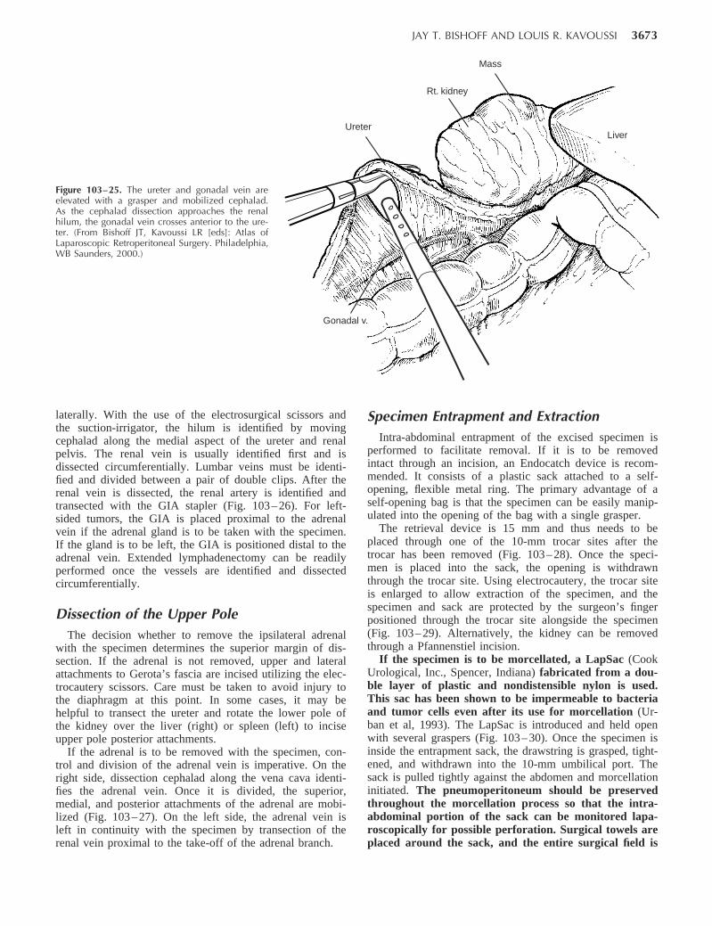
JAY T. BISHOFF AND LOUIS R. KAVOUSSI 3673
Figure 103–25. The ureter and gonadal vein areelevated with a grasper and mobilized cephalad.As the cephalad dissection approaches the renalhilum, the gonadal vein crosses anterior to the ure-ter. (From Bishoff JT, Kavoussi LR [eds]: Atlas ofLaparoscopic Retroperitoneal Surgery. Philadelphia,WB Saunders, 2000.)
Ureter
Rt. kidney
Mass
Liver
Gonadal v.
laterally. With the use of the electrosurgical scissors andthe suction-irrigator, the hilum is identified by movingcephalad along the medial aspect of the ureter and renalpelvis. The renal vein is usually identified first and isdissected circumferentially. Lumbar veins must be identi-fied and divided between a pair of double clips. After therenal vein is dissected, the renal artery is identified andtransected with the GIA stapler (Fig. 103– 26). For left-sided tumors, the GIA is placed proximal to the adrenalvein if the adrenal gland is to be taken with the specimen.If the gland is to be left, the GIA is positioned distal to theadrenal vein. Extended lymphadenectomy can be readilyperformed once the vessels are identified and dissectedcircumferentially.
Dissection of the Upper PoleThe decision whether to remove the ipsilateral adrenal
with the specimen determines the superior margin of dis-section. If the adrenal is not removed, upper and lateralattachments to Gerota’s fascia are incised utilizing the elec-trocautery scissors. Care must be taken to avoid injury tothe diaphragm at this point. In some cases, it may behelpful to transect the ureter and rotate the lower pole ofthe kidney over the liver (right) or spleen (left) to inciseupper pole posterior attachments.
If the adrenal is to be removed with the specimen, con-trol and division of the adrenal vein is imperative. On theright side, dissection cephalad along the vena cava identi-fies the adrenal vein. Once it is divided, the superior,medial, and posterior attachments of the adrenal are mobi-lized (Fig. 103– 27). On the left side, the adrenal vein isleft in continuity with the specimen by transection of therenal vein proximal to the take-off of the adrenal branch.
Specimen Entrapment and ExtractionIntra-abdominal entrapment of the excised specimen is
performed to facilitate removal. If it is to be removedintact through an incision, an Endocatch device is recom-mended. It consists of a plastic sack attached to a self-opening, flexible metal ring. The primary advantage of aself-opening bag is that the specimen can be easily manip-ulated into the opening of the bag with a single grasper.
The retrieval device is 15 mm and thus needs to beplaced through one of the 10-mm trocar sites after thetrocar has been removed (Fig. 103– 28). Once the speci-men is placed into the sack, the opening is withdrawnthrough the trocar site. Using electrocautery, the trocar siteis enlarged to allow extraction of the specimen, and thespecimen and sack are protected by the surgeon’s fingerpositioned through the trocar site alongside the specimen(Fig. 103– 29). Alternatively, the kidney can be removedthrough a Pfannenstiel incision.
If the specimen is to be morcellated, a LapSac (CookUrological, Inc., Spencer, Indiana) fabricated from a dou-ble layer of plastic and nondistensible nylon is used.This sac has been shown to be impermeable to bacteriaand tumor cells even after its use for morcellation (Ur-ban et al, 1993). The LapSac is introduced and held openwith several graspers (Fig. 103– 30). Once the specimen isinside the entrapment sack, the drawstring is grasped, tight-ened, and withdrawn into the 10-mm umbilical port. Thesack is pulled tightly against the abdomen and morcellationinitiated. The pneumoperitoneum should be preservedthroughout the morcellation process so that the intra-abdominal portion of the sack can be monitored lapa-roscopically for possible perforation. Surgical towels areplaced around the sack, and the entire surgical field is
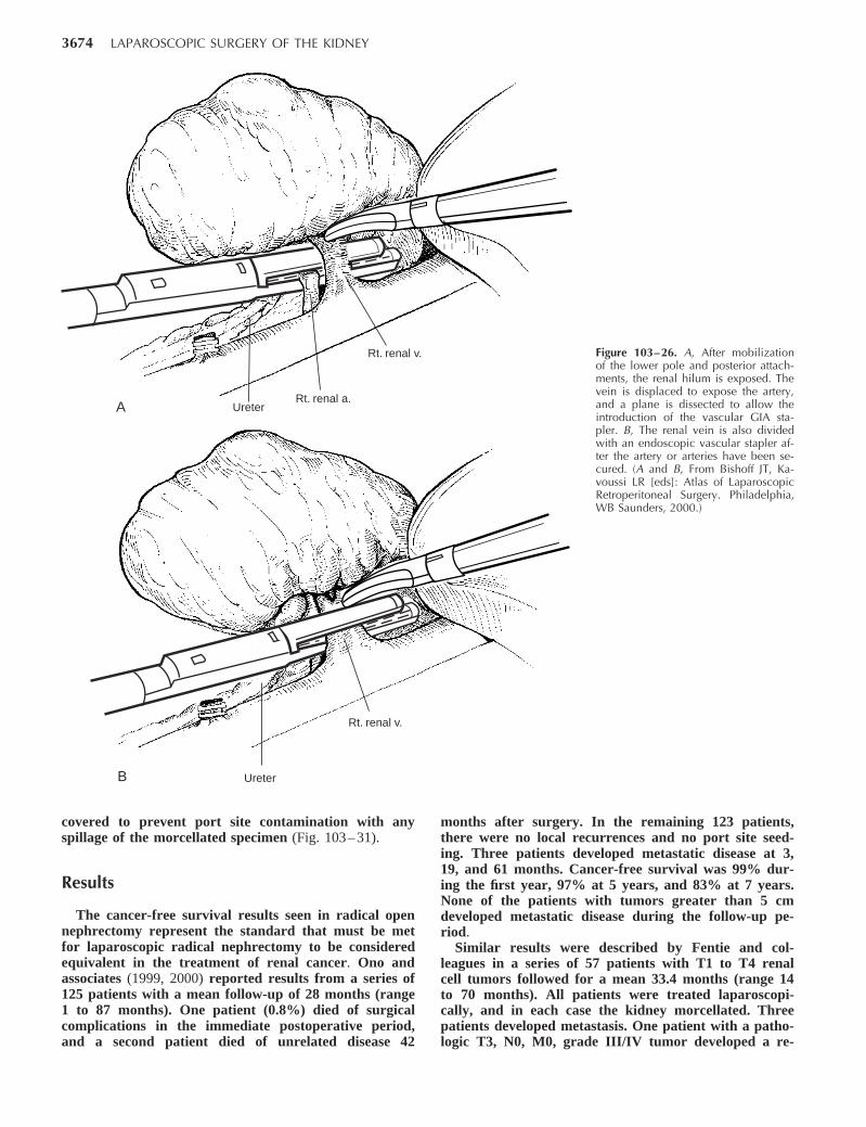
3674 LAPAROSCOPIC SURGERY OF THE KIDNEY
Rt. renal v.
Rt. renal a.UreterA
B
Rt. renal v.
Ureter
Figure 103–26. A, After mobilizationof the lower pole and posterior attach-ments, the renal hilum is exposed. Thevein is displaced to expose the artery,and a plane is dissected to allow theintroduction of the vascular GIA sta-pler. B, The renal vein is also dividedwith an endoscopic vascular stapler af-ter the artery or arteries have been se-cured. (A and B, From Bishoff JT, Ka-voussi LR [eds]: Atlas of LaparoscopicRetroperitoneal Surgery. Philadelphia,WB Saunders, 2000.)
covered to prevent port site contamination with anyspillage of the morcellated specimen (Fig. 103– 31).
Results
The cancer-free survival results seen in radical opennephrectomy represent the standard that must be metfor laparoscopic radical nephrectomy to be consideredequivalent in the treatment of renal cancer. Ono andassociates (1999, 2000) reported results from a series of125 patients with a mean follow-up of 28 months (range1 to 87 months). One patient (0.8%) died of surgicalcomplications in the immediate postoperative period,and a second patient died of unrelated disease 42
months after surgery. In the remaining 123 patients,there were no local recurrences and no port site seed-ing. Three patients developed metastatic disease at 3,19, and 61 months. Cancer-free survival was 99% dur-ing the first year, 97% at 5 years, and 83% at 7 years.None of the patients with tumors greater than 5 cmdeveloped metastatic disease during the follow-up pe-riod.
Similar results were described by Fentie and col-leagues in a series of 57 patients with T1 to T4 renalcell tumors followed for a mean 33.4 months (range 14to 70 months). All patients were treated laparoscopi-cally, and in each case the kidney morcellated. Threepatients developed metastasis. One patient with a patho-logic T3, N0, M0, grade III/IV tumor developed a re-
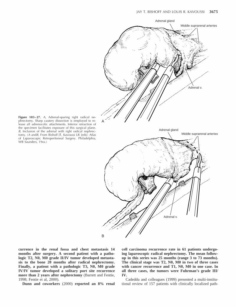
JAY T. BISHOFF AND LOUIS R. KAVOUSSI 3675
Figure 103–27. A, Adrenal-sparing right radical ne-phrectomy. Sharp cautery dissection is employed to re-lease all adrenocolic attachments. Inferior retraction ofthe specimen facilitates exposure of this surgical plane.B, Inclusion of the adrenal with right radical nephrec-tomy. (A andB, From Bishoff JT, Kavoussi LR [eds]: Atlasof Laparoscopic Retroperitoneal Surgery. Philadelphia,WB Saunders, 19xx.)
A
B
Adrenal glandMiddle suprarenal arteries
Adrenal v.
Adrenal gland
Middle suprarenal arteries
Adrenal v.
currence in the renal fossa and chest metastasis 14months after surgery. A second patient with a patho-logic T2, N0, M0 grade II/IV tumor developed metasta-sis to the bone 20 months after radical nephrectomy.Finally, a patient with a pathologic T3, N0, M0 gradeIV/IV tumor developed a solitary port site recurrencemore than 2 years after nephrectomy (Barrett and Fentie,1998; Fentie et al, 2000).
Dunn and coworkers (2000) reported an 8% renal
cell carcinoma recurrence rate in 61 patients undergo-ing laparoscopic radical nephrectomy. The mean follow-up in this series was 25 months (range 3 to 73 months).The clinical stage was T2, N0, M0 in two of three caseswith cancer recurrence and T1, N0, M0 in one case. Inall three cases, the tumors were Fuhrman’s grade III/IV.
Cadeddu and colleagues (1999) presented a multi-institu-tional review of 157 patients with clinically localized path-
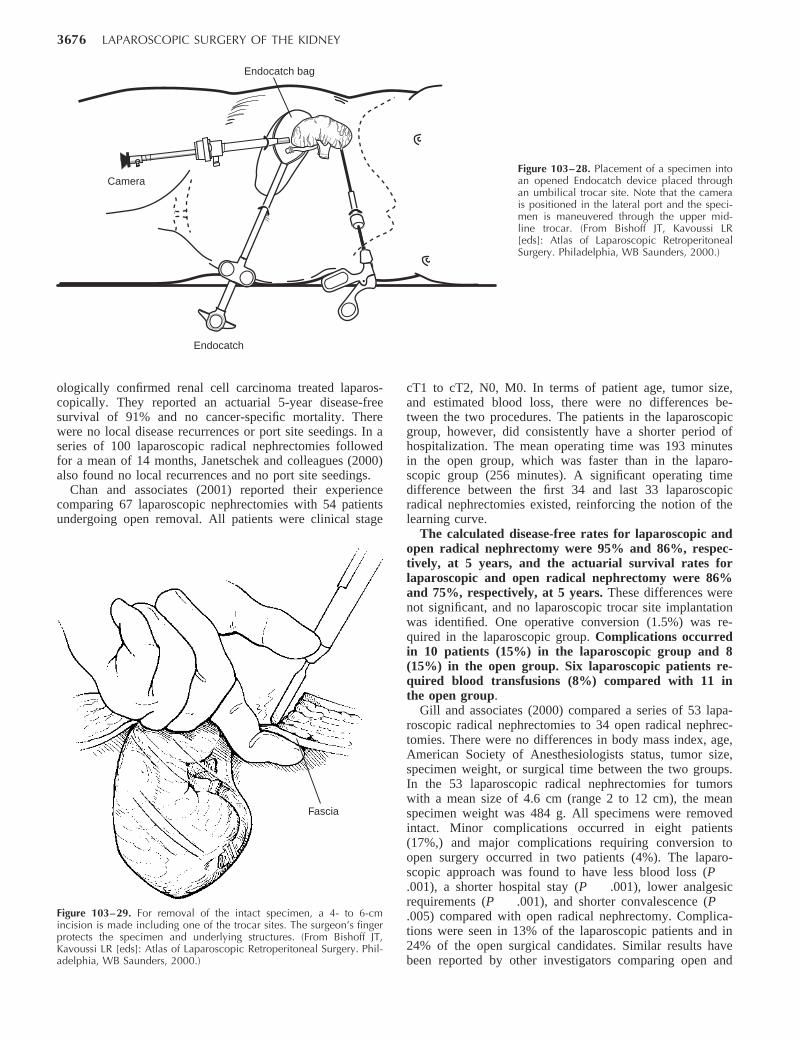
3676 LAPAROSCOPIC SURGERY OF THE KIDNEY
Camera
Endocatch
Endocatch bag
Figure 103–28. Placement of a specimen intoan opened Endocatch device placed throughan umbilical trocar site. Note that the camerais positioned in the lateral port and the speci-men is maneuvered through the upper mid-line trocar. (From Bishoff JT, Kavoussi LR[eds]: Atlas of Laparoscopic RetroperitonealSurgery. Philadelphia, WB Saunders, 2000.)
Fascia
Figure 103–29. For removal of the intact specimen, a 4- to 6-cmincision is made including one of the trocar sites. The surgeon’s fingerprotects the specimen and underlying structures. (From Bishoff JT,Kavoussi LR [eds]: Atlas of Laparoscopic Retroperitoneal Surgery. Phil-adelphia, WB Saunders, 2000.)
ologically confirmed renal cell carcinoma treated laparos-copically. They reported an actuarial 5-year disease-freesurvival of 91% and no cancer-specific mortality. Therewere no local disease recurrences or port site seedings. In aseries of 100 laparoscopic radical nephrectomies followedfor a mean of 14 months, Janetschek and colleagues (2000)also found no local recurrences and no port site seedings.
Chan and associates (2001) reported their experiencecomparing 67 laparoscopic nephrectomies with 54 patientsundergoing open removal. All patients were clinical stage
cT1 to cT2, N0, M0. In terms of patient age, tumor size,and estimated blood loss, there were no differences be-tween the two procedures. The patients in the laparoscopicgroup, however, did consistently have a shorter period ofhospitalization. The mean operating time was 193 minutesin the open group, which was faster than in the laparo-scopic group (256 minutes). A significant operating timedifference between the first 34 and last 33 laparoscopicradical nephrectomies existed, reinforcing the notion of thelearning curve.
The calculated disease-free rates for laparoscopic andopen radical nephrectomy were 95% and 86%, respec-tively, at 5 years, and the actuarial survival rates forlaparoscopic and open radical nephrectomy were 86%and 75%, respectively, at 5 years. These differences werenot significant, and no laparoscopic trocar site implantationwas identified. One operative conversion (1.5%) was re-quired in the laparoscopic group. Complications occurredin 10 patients (15%) in the laparoscopic group and 8(15%) in the open group. Six laparoscopic patients re-quired blood transfusions (8%) compared with 11 inthe open group.
Gill and associates (2000) compared a series of 53 lapa-roscopic radical nephrectomies to 34 open radical nephrec-tomies. There were no differences in body mass index, age,American Society of Anesthesiologists status, tumor size,specimen weight, or surgical time between the two groups.In the 53 laparoscopic radical nephrectomies for tumorswith a mean size of 4.6 cm (range 2 to 12 cm), the meanspecimen weight was 484 g. All specimens were removedintact. Minor complications occurred in eight patients(17%,) and major complications requiring conversion toopen surgery occurred in two patients (4%). The laparo-scopic approach was found to have less blood loss (P.001), a shorter hospital stay (P .001), lower analgesicrequirements (P .001), and shorter convalescence (P.005) compared with open radical nephrectomy. Complica-tions were seen in 13% of the laparoscopic patients and in24% of the open surgical candidates. Similar results havebeen reported by other investigators comparing open and
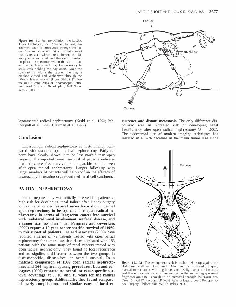
JAY T. BISHOFF AND LOUIS R. KAVOUSSI 3677
Figure 103–30. For morcellation, the LapSac(Cook Urological, Inc., Spencer, Indiana) en-trapment sack is introduced through the lat-eral 10-mm trocar site. After the entrapmentsack is released within the abdomen, the 10-mm port is replaced and the sack unfurled.To place the specimen within the sack, a lat-eral 5- or 3-mm port may be necessary toassist with holding the bag open. Once thespecimen is within the Lapsac, the bag iscinched closed and withdrawn through the10-mm lateral trocar. (From Bishoff JT, Ka-voussi LR [eds]: Atlas of Laparoscopic Retro-peritoneal Surgery. Philadelphia, WB Saun-ders, 2000.)
Rt. kidney
Camera
LapSac
Forceps
Figure 103–31. The entrapment sack is pulled tightly up against theabdominal wall with two hands. After the site is carefully draped,manual morcellation with ring forceps or a Kelly clamp can be used,and the entrapment sack is removed once the remaining specimenfragments are small enough to be extracted through the trocar site.(From Bishoff JT, Kavoussi LR [eds]: Atlas of Laparoscopic Retroperito-neal Surgery. Philadelphia, WB Saunders, 2000.)
laparoscopic radical nephrectomy (Kerbl et al, 1994; Mc-Dougall et al, 1996; Clayman et al, 1997)
Conclusion
Laparoscopic radical nephrectomy is in its infancy com-pared with standard open radical nephrectomy. Early re-ports have clearly shown it to be less morbid than opensurgery. The reported 5-year survival of patients indicatesthat the cancer-free survival is comparable to that seenafter open radical nephrectomy. Longer follow-up withlarger numbers of patients will help confirm the efficacy oflaparoscopy in treating organ-confined renal cell carcinoma.
PARTIAL NEPHRECTOMY
Partial nephrectomy was initially reserved for patients athigh risk for developing renal failure after kidney surgeryto treat renal cancer. Several series have shown partialopen nephrectomy to be equivalent to open radical ne-phrectomy in terms of long-term cancer-free survivalwith unilateral renal involvement, unifocal disease, anda tumor size less than 4 cm. Fergnany and coworkers(2000) report a 10-year cancer-specific survival of 100%in this subset of patients. Lee and associates (2000) havereported a series of 79 patients treated with open partialnephrectomy for tumors less than 4 cm compared with 183patients with the same stage of renal cancers treated withopen radical nephrectomy. They found no local recurrenceand no significant difference between the two groups indisease-specific, disease-free, or overall survival. In amatched comparison of 1566 open radical nephrecto-mies and 164 nephron-sparing procedures, Lau and col-leagues (2000) reported no overall or cause-specific sur-vival advantage at 5, 10, and 15 years for the radicalnephrectomy group. Additionally, they found compara-ble early complications and similar rates of local re-
currence and distant metastasis. The only difference dis-covered was an increased risk of developing renalinsufficiency after open radical nephrectomy (P .002).The widespread use of modern imaging techniques hasresulted in a 32% decrease in the mean tumor size since
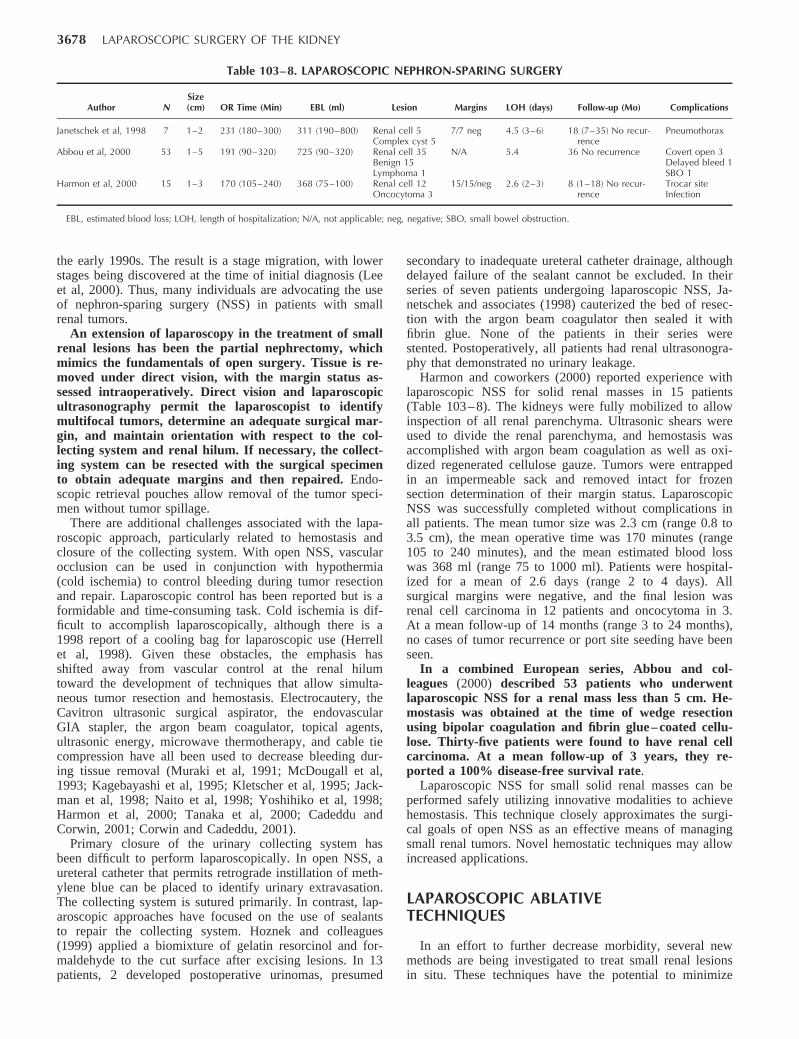
3678 LAPAROSCOPIC SURGERY OF THE KIDNEY
Table 103–8. LAPAROSCOPIC NEPHRON-SPARING SURGERY
Author NSize(cm) OR Time (Min) EBL (ml) Lesion Margins LOH (days) Follow-up (Mo) Complications
Janetschek et al, 1998 7 1–2 231 (180–300) 311 (190–800) Renal cell 5Complex cyst 5
7/7 neg 4.5 (3–6) 18 (7–35) No recur-rence
Pneumothorax
Abbou et al, 2000 53 1–5 191 (90–320) 725 (90–320) Renal cell 35Benign 15Lymphoma 1
N/A 5.4 36 No recurrence Covert open 3Delayed bleed 1SBO 1
Harmon et al, 2000 15 1–3 170 (105–240) 368 (75–100) Renal cell 12Oncocytoma 3
15/15/neg 2.6 (2–3) 8 (1–18) No recur-rence
Trocar siteInfection
EBL, estimated blood loss; LOH, length of hospitalization; N/A, not applicable; neg, negative; SBO, small bowel obstruction.
the early 1990s. The result is a stage migration, with lowerstages being discovered at the time of initial diagnosis (Leeet al, 2000). Thus, many individuals are advocating the useof nephron-sparing surgery (NSS) in patients with smallrenal tumors.
An extension of laparoscopy in the treatment of smallrenal lesions has been the partial nephrectomy, whichmimics the fundamentals of open surgery. Tissue is re-moved under direct vision, with the margin status as-sessed intraoperatively. Direct vision and laparoscopicultrasonography permit the laparoscopist to identifymultifocal tumors, determine an adequate surgical mar-gin, and maintain orientation with respect to the col-lecting system and renal hilum. If necessary, the collect-ing system can be resected with the surgical specimento obtain adequate margins and then repaired. Endo-scopic retrieval pouches allow removal of the tumor speci-men without tumor spillage.
There are additional challenges associated with the lapa-roscopic approach, particularly related to hemostasis andclosure of the collecting system. With open NSS, vascularocclusion can be used in conjunction with hypothermia(cold ischemia) to control bleeding during tumor resectionand repair. Laparoscopic control has been reported but is aformidable and time-consuming task. Cold ischemia is dif-ficult to accomplish laparoscopically, although there is a1998 report of a cooling bag for laparoscopic use (Herrellet al, 1998). Given these obstacles, the emphasis hasshifted away from vascular control at the renal hilumtoward the development of techniques that allow simulta-neous tumor resection and hemostasis. Electrocautery, theCavitron ultrasonic surgical aspirator, the endovascularGIA stapler, the argon beam coagulator, topical agents,ultrasonic energy, microwave thermotherapy, and cable tiecompression have all been used to decrease bleeding dur-ing tissue removal (Muraki et al, 1991; McDougall et al,1993; Kagebayashi et al, 1995; Kletscher et al, 1995; Jack-man et al, 1998; Naito et al, 1998; Yoshihiko et al, 1998;Harmon et al, 2000; Tanaka et al, 2000; Cadeddu andCorwin, 2001; Corwin and Cadeddu, 2001).
Primary closure of the urinary collecting system hasbeen difficult to perform laparoscopically. In open NSS, aureteral catheter that permits retrograde instillation of meth-ylene blue can be placed to identify urinary extravasation.The collecting system is sutured primarily. In contrast, lap-aroscopic approaches have focused on the use of sealantsto repair the collecting system. Hoznek and colleagues(1999) applied a biomixture of gelatin resorcinol and for-maldehyde to the cut surface after excising lesions. In 13patients, 2 developed postoperative urinomas, presumed
secondary to inadequate ureteral catheter drainage, althoughdelayed failure of the sealant cannot be excluded. In theirseries of seven patients undergoing laparoscopic NSS, Ja-netschek and associates (1998) cauterized the bed of resec-tion with the argon beam coagulator then sealed it withfibrin glue. None of the patients in their series werestented. Postoperatively, all patients had renal ultrasonogra-phy that demonstrated no urinary leakage.
Harmon and coworkers (2000) reported experience withlaparoscopic NSS for solid renal masses in 15 patients(Table 103– 8). The kidneys were fully mobilized to allowinspection of all renal parenchyma. Ultrasonic shears wereused to divide the renal parenchyma, and hemostasis wasaccomplished with argon beam coagulation as well as oxi-dized regenerated cellulose gauze. Tumors were entrappedin an impermeable sack and removed intact for frozensection determination of their margin status. LaparoscopicNSS was successfully completed without complications inall patients. The mean tumor size was 2.3 cm (range 0.8 to3.5 cm), the mean operative time was 170 minutes (range105 to 240 minutes), and the mean estimated blood losswas 368 ml (range 75 to 1000 ml). Patients were hospital-ized for a mean of 2.6 days (range 2 to 4 days). Allsurgical margins were negative, and the final lesion wasrenal cell carcinoma in 12 patients and oncocytoma in 3.At a mean follow-up of 14 months (range 3 to 24 months),no cases of tumor recurrence or port site seeding have beenseen.
In a combined European series, Abbou and col-leagues (2000) described 53 patients who underwentlaparoscopic NSS for a renal mass less than 5 cm. He-mostasis was obtained at the time of wedge resectionusing bipolar coagulation and fibrin glue– coated cellu-lose. Thirty-five patients were found to have renal cellcarcinoma. At a mean follow-up of 3 years, they re-ported a 100% disease-free survival rate.
Laparoscopic NSS for small solid renal masses can beperformed safely utilizing innovative modalities to achievehemostasis. This technique closely approximates the surgi-cal goals of open NSS as an effective means of managingsmall renal tumors. Novel hemostatic techniques may allowincreased applications.
LAPAROSCOPIC ABLATIVETECHNIQUES
In an effort to further decrease morbidity, several newmethods are being investigated to treat small renal lesionsin situ. These techniques have the potential to minimize
JAY T. BISHOFF AND LOUIS R. KAVOUSSI 3679
blood loss, dissection, and complications when comparedwith extirpative approaches. Moreover, these modalitieshave the potential to be used in an image-guided manner toeliminate the need for surgical intervention. Cryotherapy,radiofrequency (RF) ablation, high-intensity focused ultra-sonography, microwave therapy, and high-intensity radia-tion have all been investigated as tools to ablate renaltumors.
Cryosurgery
Cryotherapy has been one of the most studied abla-tive techniques. Multiple investigators have demon-strated the ability of temperatures of 20 C to inducetissue necrosis (Chosy et al, 1996; Campbell et al, 1998;Bishoff et al, 1999; Gill and Novick, 1999). Rapid freezingcauses crystal formation in the microvasculature and extra-cellular spaces and within the cells. The latter results inuncoupling of oxidative phosphorylation and rupturing ofplasma cell membranes with subsequent solute shifts. Pro-gressive failure of the microvasculature results in endothe-lial cell damage, edema, and platelet aggregation withthrombosis and vascular occlusion (Bishoff et al, 1999).
Percutaneous, open, and laparoscopic approacheshave all been utilized in cryoablation. Adequate cry-odestruction requires intraoperative monitoring of theresultant “ice-ball.” To date, CT and MRI have notproved to be reliable modalities for detecting the progres-sion of the ice-ball. On ultrasonography, however, thecryoablated region appears hypoechoic and demonstrates aloss of normal flow characteristics on color-flow Dopplerthat closely approximates the size and location of the ice-ball (Delworth et al, 1996; Campbell et al, 1998; Bishoff etal, 1999; Gill and Novick, 1999). Laparoscopy permitsthe use of intraoperative laparoscopic ultrasonographyto monitor the progression of the ice-ball. Campbelland associates (1998) demonstrated that the ice-ball vi-sualized on intraoperative laparoscopic ultrasonographymust extend at least 3.1 mm beyond the outer edge ofthe renal lesion of interest in order to ensure that atemperature of 20 C is reached in the tissues farthestfrom the cryoprobe. Larger lesions would therefore re-quire multiple passes with multiple probes to achieve ade-quate ablation.
Microscopic examination of the ablation site immedi-ately after the procedure demonstrates extensive hemor-rhage into the glomerular and tubular urinary spaces.One week after cryosurgery, the lesion reflects four dis-tinct histological zones from the center: (1) completenecrosis, (2) inflammatory infiltrate, (3) hemorrhage,and (4) fibrosis and regeneration. By 13 weeks, thesezones are replaced by fibrous connective tissue withoccasional necrotic cellular debris (Bishoff et al, 1999).The cryotherapy literature lacks definitive evidence thatthe size of the ice-ball visualized during cryoablationcorresponds precisely with the histopathologic changesdescribed.
Potential complications of cryosurgery include uri-nary fistula formation, post-treatment hemorrhage, andinjury to adjacent structures to include the collectingsystem, bowel, and liver. Given that even momentarycontact of the active cryoprobe can lead to necrosis and
fibrosis, disastrous results could soon follow (Campbellet al, 1998). Bishoff and coworkers (1999) in a porcinemodel noted severe adhesions between cryoablated kid-ney and overlying bowel in non-retroperitonealized kid-neys. There was no evidence of bowel injury or fistulas.Gill and colleagues (1999) reported the complication ofsmall bowel obstruction when the cryoprobe inadver-tently contacted a loop of small bowel during porcinelaparoscopic renal cryoablation. Campbell and associates(1998) described an obstructive stricture of the ureteropel-vic junction.
To date, there are only limited short-term clinical trialsassessing the efficacy of cryoablation in select patients.Rodriguez and colleagues (2000) performed the procedurein eight patients with small (mean 2-cm) renal tumors. At9 months of follow-up, they identified one patient with anenhancing lesion in the area of cryoablation that was abiopsy-proven renal cell recurrence. Levin and associates(2000), who performed laparoscopic cryoablation in 39 pa-tients, reported one patient with a recurrence at 9 months.Shingleton and coworkers (2000) performed percutaneousMRI-guided cryoablation in 17 patients with 18 renal tu-mors; one tumor required retreatment due to an enhancingportion of the tumor on 1-month CT scans. These reportsof short-term local recurrences mandate further investiga-tion to predict cryotherapy’s ultimate role in the treatmentof renal tumors.
Radiofrequency Interstitial TissueAblation
RF ablation has been utilized in a number of differentclinical applications, including ablation of cardiac dysrhyth-mias and treatment of lesions arising from the liver, ner-vous system, and bone (Zlotta et al, 1997; Dupuy, 1999;McGovern et al, 1999; Nakada, 1999; Crowley and Bish-off, 2000). Urologists have used this technology to treatbenign prostatic hypertrophy through a transurethral ap-proach.
The RF energy can be introduced percutaneously,laparoscopically, or through an open approach underultrasonographic, fluoroscopic, CT, or MRI guidance.The RF energy returns to the RF generator by way of agrounding pad that completes the electric circuit. Theprobe carries an alternating current of high-frequencyradiowaves that causes the local ions to vibrate, and theresistance in the tissue creates heat to the point of des-iccation (thermal coagulation). Microscopic examinationreveals intense stromal and epithelial edema withmarked hypereosinophilia and pyknosis immediately af-ter RF treatment (Zlotta et al, 1997). This is replaced ina matter of days to weeks by coagulative necrosis withconcentric zones of inflammatory infiltrate, hemorrhage,and fibrosis (Crowley et al, 2000).
Precise control of the size of the lesion can be achievedby temperature-based or impedance-based monitoring.Temperature-based systems depend on thermocouples im-bedded within the tips of the electrodes. Sufficient amountsof RF energy (10 to 90 W) are applied to raise the tissuetemperature to the minimal 60 C required to induce coagu-lative necrosis (Crowley et al, 2000). However, the obser-vation that temperatures recorded at the limit of ablation
3680 LAPAROSCOPIC SURGERY OF THE KIDNEY
have been found to be 20 C to 30 C cooler than tempera-tures recorded at the thermocouples makes sufficient tissuedesiccation uncertain. For this reason, some authors favoran impedance-based system, which applies RF energythrough the electric circuit until the tissue becomes suffi-ciently desiccated to become an insulator. In this manner,the flow of RF energy back to the return pad is blocked. Agradual decrease in tissue impedance suggests that the ther-mal lesion is continuing to evolve; a rise in impedance (toapproximately 200 ) suggests that the tissue is desiccatedand that further thermal lesion growth is unlikely (Lewin etal, 1998).
Although ultrasonography, fluoroscopy, CT, and MRIhave all been used for positioning of the patient and forpercutaneous placement of the RF probe, none of thesemodalities has proved reliable for the intraoperative moni-toring of the RF lesion. On ultrasound imaging, there is noimmediate change in the echotexture in the area of RFablation, and color and power Doppler ultrasonography areof no added benefit because of variable and inconsistentfindings (Crowley et al, 2000). Moreover, RF treatment cansometimes disturb ultrasound imaging (Zlotta et al, 1997).
Crowley and colleagues (2000) found CT to be excellentfor positioning of the patient and probe and for immediatepost-treatment imaging, but it was not utilized for intraop-erative monitoring. Lewin and associates (1998) used MRIto monitor real-time tissue destruction with RF ablation.This group demonstrated a zone of decreased signal sur-rounded by a rim of hyperintensity on T2-weighted andSTIR (turbo short inversion-time inversion recovery) im-ages. However, confirmatory images obtained after conclu-sion of the ablation session revealed a propensity for theseT2-weighted images to underestimate the size of the RFlesion. Thus, insofar as intraoperative monitoring for RFintensity is concerned, there is no imaging modality thatcan in real time ensure a sufficient extent of tissue ablationwhile avoiding injury to normal adjacent parenchyma.
Walther and coworkers (2000) explored 14 tumors lessthan 5 cm in four patients with multiple renal lesions andperformed RF ablation just before surgical excision. Acomplete immediate treatment effect was noted in 10 of 11patients; in the final case, only 35% of the tumor wasablated. Excluding any technical inconsistencies, as withthe early results of cryoablation, this early treatment failureneeds to be put into the context of long-term, prospectiveclinical trials.
COMPLICATIONS OFLAPAROSCOPIC RENAL SURGERY
Complications are an unavoidable consequence of surgi-cal practice. Even when the surgery is in the most experi-enced hands, factors related to the patient, operating roomenvironment, and chaotic forces can lead to an untowardevent. Thus, efforts at prevention and the patients’ under-standing should be maximized. Moreover, if complicationsoccur, the consequences can often be minimized throughearly recognition and appropriate intervention.
Laparoscopic renal surgery shares several potential riskswith traditional open approaches; however, there are differ-ences in the type and presentation of these complications.
It must be kept in mind that all situations are individual,and unique problems may arise requiring innovative ac-tions.
Complications can arise during each step of the laparo-scopic nephrectomy. Access-related problems such as solidorgan injury, bowel injury, abdominal wall hematoma, andepigastric vessel injuries have been reported. Bleedingcomplications can be minimized by carefully inspectingthe abdomen at the conclusion of surgery. Examinationafter lowering the intra-abdominal pressure can revealvenous bleeders tamponaded by the pneumoperitoneum.One also needs to carefully inspect the area of surgicaldissection and the trocar sites for adequate hemostasis.Common areas of postoperative intra-abdominal bleed-ing include the adrenal gland, mesentery, gonadal ves-sels, and ureteral stump.
Another potential problem in patients undergoing laparo-scopic renal surgery is volume overload. In the laparo-scopic approach, patients have less insensible fluid losscompared with their open surgical counterparts. There isalso a vascular-mediated oliguria that should not be aggres-sively treated because diuresis is seen after release of thepneumoperitoneum. Volume overload in patients with di-minished cardiac reserve can result in congestive heart fail-ure manifested by oliguria. Poor urine output or hemody-namic instability in the postoperative period should initiatean evaluation to rule out bleeding; if the work-up is nega-tive, diuresis can be induced if clinically indicated.
Additionally reported common complications include in-cisional hernia after intact specimen removal, transientthigh numbness, prolonged ileus, pulmonary embolus,pneumonia, brachial nerve injury, and unrecognized bowelinjury (Copcoat et al, 1992; Kavoussi et al, 1993; Kerbl etal 1993a, 1993b, 1994a, 1994b; Rassweiler et al, 1993,1996; Eraky et al, 1994; Nicol et al, 1994; Perez et al,1994; Parra et al, 1995; Keeley and Tolley, 1998; Rozen-berg et al, 1999).
Unrecognized bowel injury is a potential complication ofany transabdominal or retroperitoneal surgical procedure.During open surgery, bowel injuries are usually recognizedand treated at the time of the procedure. One of the mostdevastating complications occurring as a result of lapa-roscopic surgery is unrecognized bowel injury. Becauseonly 10% of the laparoscopic instrument is in the visualfield, these injuries can occur out of the surgeon’s fieldof view. The combined incidence of bowel injury in theliterature is 1.3 in 1000 cases (Bishoff et al, 1999). Mostinjuries (69%) are not recognized at the time of sur-gery. Small bowel segments account for 58% of injuriesfollowed by colon (32%) and stomach (7%). Fifty per-cent of bowel injuries are caused by electrocautery, and32% occur during Veress needle or trocar insertion.
The presentation of bowel injuries in the laparoscopicgroup differs from that described with open postopera-tive injury with peritonitis. Patients with unrecognizedbowel injury after laparoscopy typically present withpersistent and increased trocar site pain at the site clos-est to the bowel injury. Later signs and symptoms in-clude nausea, diarrhea, anorexia, low-grade fever, per-sistent bowel sounds, and a low or normal white bloodcell count. The patient’s condition can rapidly deterio-rate to hemodynamic instability and death if the injury
JAY T. BISHOFF AND LOUIS R. KAVOUSSI 3681
is not quickly recognized and treated. CT is the initialdiagnostic modality of choice (Cadeddu et al, 1997), andopen exploration is usually required to evacuate bowelspillage and perform the necessary repair.
Complication rates have been reported in several largemulti-institutional reviews of laparoscopic simple nephrec-tomy. In a series of 185 patients, Gill and coworkers(1995) reported an overall complication rate of 12% forbenign disease, with 5% of patients requiring conversion toopen surgery. In their series, the incidence of complicationsdecreased markedly with increasing experience. In fact,70% of the complications occurred during the first 20cases at each institution. A learning curve of approxi-mately 20 laparoscopic nephrectomy cases is also sup-ported by other reports (Keeley and Tolley, 1998; Ras-sweiler et al 1998; Fahlenkamp et al, 1999).
Rassweiler and colleagues (1998) published the resultsof a series of 482 laparoscopic nephrectomies (444 proce-dures for benign disease) performed by 20 surgeons at 14different European medical centers. They reported a 6%overall complication rate, with 10% of the cases convertedto open surgery. The majority of patients converted toopen surgery had infectious causes of renal abnormalityas the leading indication for kidney removal. Bleedingwas the most common cause of open conversion, fol-lowed by the surgeon’s inability to visualize the renalhilum for dissection.
Gupta and associates (1999) presented a series of 126laparoscopic nephrectomies performed for benign disease.In their series, 12% (15 of 126) of the patients requiredconversion to an open surgical procedure. The overall inci-dence of major and minor complications was 17% (22 of126).
In 1998, Cadeddu and colleagues reported a minor andmajor complication rate of 9.6% in a series of 157 laparo-scopic radical nephrectomies. In a contemporary compar-ison of laparoscopic and open radical nephrectomy,Chan and associates (2000) found no difference in thenumber and severity of complications.
CONCLUSIONS
Laparoscopy has emerged as a powerful minimallyinvasive surgical tool to treat renal lesions. In a widevariety of applications, patients benefit from decreasedmorbidity without sacrificing therapeutic outcomes. Un-fortunately, the availability of laparoscopic renal surgery islimited.
Laparoscopic skill requires intensive training and fre-quent practice to maintain proficiency. Unlike the situationin the fields of general surgery and gynecology, there is nosimple, frequently performed urologic procedure that al-lows most urologic surgeons the opportunity to sustainlaparoscopic skills. Weekend courses and occasional casesare not sufficient to impart the necessary expertise to sur-geons.
For laparoscopy to become an integral part of main-stream urology, changes in education and advances in tech-nology are needed. Large community groups and academicpractices need to identify select urologic surgeons to trainin laparoscopic approaches. These individuals need to be
supported through case referrals to maintain operative pro-ficiency. Novel methods of mentoring and monitoring pro-gress through the use of telecommunication technologiesneed to be developed. Continued advances in suturing, sta-pling, and robotic and imaging modalities will help shortenthe learning curve associated with this new skill and in-crease patients’ access to the benefits offered by laparo-scopic surgery.
Laparoscopic surgery represents a significant advance inthe surgical treatment of renal abnormalities; however, thisaccess technique is but a stepping stone in the evolution ofsurgery. Early results with ablative techniques hold promisefor eventual nonoperative management of most renal le-sions.
REFERENCES
Historical Overview
Baba S, Nakagawa K, Nakamura, et al: Experience of 143 cases oflaparoscopic surgery in urology—clinical outcome in comparison toopen surgery. Jpn J Urol 1996;87:842–850.
Clayman RV, Kavoussi LR, Soper NJ, et al: Laparoscopic nephrectomy:Initial case report. J Urol 1991;146:278–282.
Copcoat MJ, Joyce AJ, Popert R, et al: Laparoscopic nephrectomy—theKing’s experience. Minim Invasive Ther 1992;1(Suppl):67.
Doublet JD, Barreto HS, Degromont AC, et al: Retroperitoneal nephrec-tomy: Comparison of laparoscopy with open surgery. World J Surg1996;20:713–716.
Griffith DO, Schussler WW, Vancaille TH: Laparoscopic lymphadenec-tomy: A low morbidity alternative for staging pelvic malignancies. JEndourol 1990;4:S84.
Hemal AK, Talwar M, Wadhwa SN, Gupta NP: Retroperitoneoscopicnephrectomy for benign diseases of the kidney: Prospective nonrandom-ized comparison with open surgical nephrectomy. J Endourol 1999;13:425–431.
Kavoussi LR, Kerbl K, Capelouto CC, et al: Laparoscopic nephrectomyfor renal neoplasms. Urology 1993;42:603–609.
Kerbl K, Clayman RV, McDougall EM, Kavoussi LR: Laparoscopic ne-phrectomy: Current status. Arch Esp Urol 1993a;46:581–584.
Kerbl K, Clayman RV, McDougall EM, Kavoussi LR: Laparoscopic ne-phrectomy. BMJ 1993b;307:1488–1489.
Kerbl K, Clayman RV, McDougall EM, et al: Transperitoneal nephrec-tomy for benign disease of the kidney: A comparison of laparoscopicand open surgical techniques. Urol 1994;43:607–613.
Nicol DL, Winkle DC, Nathanson LK, Smithers B: Laparoscopic nephrec-tomy for benign disease. Br J Urol 1994;73:237–241.
Perez MG, Parra RO, Boullier JA, et al: Comparison between standardflank versus laparoscopic nephrectomy for benign renal disease. J Urol1994;2:151 667a.
Rassweiler JJ, Henkel TO, Stock C, et al: Transperitoneal and retroperito-neal laparoscopic nephrectomy: Indications and results. J Endourol1993;7:S175.
Rassweiler JJ, Frede T, Henkel TO, et al: Nephrectomy: A comparativestudy between the transperitoneal and retroperitoneal laparoscopic ver-sus the open approach. Eur Urol 1998;33:489–496.
Rassweiler JJ, Stock C, Frede T, et al: Transperitoneal and retroperitoneallaparoscopic nephrectomy in comparison with conventional nephrec-tomy. Urologe A 1996;35:215–222.
Rozenberg H, Bruyere F, Abdelkader T, et al: Transperitoneal laparo-scopic nephrectomy. Prog Urol 1999;9:1034–1038.
Patient Evaluation and Preparation
Adams JB, Marco AD, Averch TD, et al: Physiologic changes duringlaparoscopy utilizing 20 mm Hg intra-abdominal pressure. J Endourol1995;9:S152.
Arthure H: Laparoscopy hazard. BMJ 1970;4:492.Bishoff JT, Allaf ME, Kirkels W, et al: Laparoscopic bowel injury:
Incidence and clinical presentation. J Urol 1999;3:887–891.Cadeddu JA, Chan DY, Hedican, et al: Retroperitoneal access for trans-