Embed Size (px)
Citation preview

1
LacR mutations are frequently observed in Streptococcus intermedius 1
and are responsible for increased intermedilysin production and 2
virulence 3
4
Running title: LacR is negative regulator of ily expression 5
6
Toshifumi Tomoyasu*1,2, Hidenori Imaki*1, Sachiko Masuda1, Ayumi Okamoto1, 7
Hye-Jin Kim1, Richard D. Waite3, Robert A. Whiley4, Ken Kikuchi5, Keiichi Hiramatsu5, 8
Atsushi Tabata1, and Hideaki Nagamune#1 9
10
1Department of Biological Science and Technology, Institute of Technology and Science, 11
The University of Tokushima Graduate School, Minami-josanjima-cho, Tokushima 12
770-8506, Japan; 2Department of Resource Circulation Engineering, Center for Frontier 13
Research of Engineering, The University of Tokushima Graduate School, 14
Minami-josanjima-cho, Tokushima 770-8506, Japan; 3Centre for Immunology and 15
Infectious Disease, Blizard Institute, and 4Department of Clinical and Diagnostic Oral 16
Sciences, Institute of Dentistry, Bart’s and The London School of Medicine and 17
Copyright © 2013, American Society for Microbiology. All Rights Reserved.Infect. Immun. doi:10.1128/IAI.00638-13 IAI Accepts, published online ahead of print on 24 June 2013
on February 2, 2018 by guest
http://iai.asm.org/
Dow
nloaded from

2
Dentistry, Queen Mary University of London, 4 Newark Street, London E1 2AT, United 18
Kingdom; 5Department of Infection Control Science and Department of Bacteriology, 19
Faculty of Medicine, Juntendo University, 2-1-1 Hongo, Bunkyo-ku, Tokyo 113-8421, 20
Japan. 21
22
#Corresponding author. 23
Mailing address: Department of Biological Science and Technology, Institute of 24
Technology and Science, The University of Tokushima Graduate School, 25
Minamijosanjima-cho, Tokushima 770-8506, Japan 26
Phone and Fax: (81) 88-656-7525; E-mail: [email protected]. 27
28
*These authors contributed equally to this work 29
30
Keywords: Streptococcus intermedius, ILY, LacR, Sugar, Lactose, Galactose 31
32
on February 2, 2018 by guest
http://iai.asm.org/
Dow
nloaded from
3
ABSTRACT 33
Streptococcus intermedius secretes a human-specific cytolysin, intermedilysin (ILY) 34
which is considered to be the major virulence factor of this pathogen. We screened for 35
a repressor of ily expression by using random gene disruption in an ILY low-producing 36
strain (PC574). Three independent ILY high-producing colonies were isolated which 37
had plasmid insertions within a gene that has high homology to lacR. Validation of 38
these observations was achieved through disruption of lacR in PC574 with an 39
erythromycin cassette, which also led to higher hemolytic activity, increased 40
transcription of ily and higher cytotoxicity against HepG2 cells, when compared to the 41
parental strain. The direct binding of LacR within the ily promoter region was shown 42
by a biotinylated DNA probe pull-down assay and the amount of ILY secreted into the 43
culture supernatant by PC574 cells was increased by adding lactose or galactose to the 44
medium as a carbon source. Furthermore, we examined lacR nucleotide sequences 45
and the hemolytic activity of 50 strains isolated from clinical infections and 7 strains 46
isolated from dental plaque. Of the 50 strains isolated from infections, 13 showed high 47
ILY production; 11 of these 13 strains had one or more point mutations and/or an 48
insertion mutation in LacR, and almost all mutations were associated with a marked 49
on February 2, 2018 by guest
http://iai.asm.org/
Dow
nloaded from

4
decline in LacR function. These results strongly suggest that mutation in lacR is 50
required for the overproduction of ILY, which is associated with an increase in 51
pathogenicity of S. intermedius. 52
53
on February 2, 2018 by guest
http://iai.asm.org/
Dow
nloaded from

5
INTRODUCTION 54
Streptococcus intermedius is a facultatively anaerobic member of the normal flora of the 55
human oral cavity and the upper respiratory, gastrointestinal, and female urogenital 56
tracts. S. intermedius belongs to the Anginosus group of streptococci (AGS), which 57
also includes Streptococcus anginosus and Streptococcus constellatus (1, 2). AGS 58
tend to form local suppurative infections, and these organisms are the most common 59
pathogens associated with bacterial intracerebral abscesses (1–6). S. intermedius is the 60
most pathogenic species of AGS and a leading cause of deep-seated, serious purulent 61
infections, including brain and liver abscesses (1, 2). This pathogen secretes a 62
human-specific cytolysin, intermedilysin (ILY), which was originally identified in 63
studies using S. intermedius strain, UNS46, isolated from a human liver abscess (7). 64
ILY is a member of the cholesterol-dependent cytolysin (CDC) family and is considered 65
the major virulence factor for infectivity and cytotoxicity towards human cells by S. 66
intermedius (8–11). Therefore, investigation of the mechanisms that regulate ily 67
expression could help elucidate how S. intermedius mediates its pathogenicity by 68
controlling the amount of ILY secreted. To date two factors have been reported to 69
control the expression of ily. The first is AI-2 (a LuxS product used by several bacteria 70
on February 2, 2018 by guest
http://iai.asm.org/
Dow
nloaded from

6
in quorum-sensing signaling), which is reported to be an exponential growth 71
phase-specific activator of ily transcription (12). In addition, we recently revealed that 72
ily expression and the growth rate of the bacteria are modulated through catabolite 73
control protein A (CcpA), which is a LacI/GalR-type repressor that monitors the 74
extracellular glucose/utilizable carbohydrate concentration (13). 75
Oral bacteria can metabolize several sugars found in foods and drinks regularly 76
consumed by humans. Lactose, a disaccharide formed from galactose and glucose, is 77
most notably found in milk and other dairy products. This sugar plays an important 78
role in oral microbial ecology and can contribute to the development of dental caries in 79
both adults and young children. The metabolism of lactose and galactose in 80
Gram-positive bacteria has been well characterized using Gram-positive cocci as 81
models (14–17). It has been reported that these sugars are rapidly fermented by both 82
the tagatose-6-phosphate (lac) and Leloir (gal) pathways in Streptococcus mutans strain 83
UA159 (17). The tagatose-6-phosphate pathway, known to be the most efficient route 84
for lactose and galactose fermentation, is found almost exclusively in Gram-positive 85
bacteria. Lactose and galactose fermentation can occur through these pathways. 86
Lactose is first internalized by the phosphoenolpyruvate (PEP)-dependent 87
on February 2, 2018 by guest
http://iai.asm.org/
Dow
nloaded from

7
lactose-phosphotransferase system (lactose-PTS permease, LacFE), yielding 88
lactose-6-phosphate (Lac-6-P). Lac-6-P is then hydrolyzed to glucose and Gal-6-P by 89
a cytoplasmic phospho-β-galactosidase (LacG). Galactose is internalized by the 90
glucose- and lactose-PTS permeases, yielding Gal-6-P. The Gal-6-P generated from 91
these sugars can then be catabolized to glycerone phosphate and 92
D-glyceraldehyde-3-phosphate by enzymes in the tagatose-6-phosphate pathway 93
(LacA−LacD). It has been reported that these enzymes are encoded by the lac operon 94
in some Gram-positive cocci (14–17). The lactose phosphotransferase system 95
repressor (LacR) is a member of the GntR family of transcriptional regulators (18). It 96
has been shown that LacR can repress transcription of the lac operon by binding the 97
LacR recognition element, which is direct repeats of the sequence TGTTTNWTTT (N = 98
any base and W = A or T), on the lac promoter under lactose- or galactose-limited 99
conditions (18, 19). It is believed that tagatose-6-phospate, a catabolite of galactose, 100
can bind LacR and inhibit the interaction between LacR and the lac promoter. This 101
allows RNA polymerase to bind to the promoter and initiate transcription of the lac 102
operon in lactose- or galactose-abundant conditions (17, 18). 103
AI-2 and CcpA have been reported to regulate ily expression. However, the action 104
on February 2, 2018 by guest
http://iai.asm.org/
Dow
nloaded from

8
of these two factors cannot explain the difference between strains with constitutively 105
high production of ILY, which seem to be more highly pathogenic and strains with low 106
production of ILY. Therefore, we screened for additional factors that could regulate ily 107
expression by employing random gene disruption in an ILY low-producing strain from 108
human dental plaque. 109
110
on February 2, 2018 by guest
http://iai.asm.org/
Dow
nloaded from
9
MATERIALS AND METHODS 111
Bacterial strains, plasmids, and growth conditions. The bacterial strains and 112
plasmids used in this study are listed in Table 1 and 2. Streptococcus intermedius was 113
cultured at 37°C or 42°C under anaerobic conditions. Brain-heart infusion (BHI) broth 114
(Becton-Dickinson, Palo Alto, CA, USA) was used as the culture medium. 115
Accumulation of lactate acidifies the culture medium and causes a loss of ILY activity 116
in the culture supernatant (13). Therefore, we used 3-(N-morpholino)propanesulfonic 117
acid (MOPS)-buffered BHI (MOPS-BHI) medium for culture to monitor the amount of 118
ILY secreted. The MOPS-BHI medium contained 100 mM MOPS buffer (pH 7.4) and 119
either 18.5 g/L BHI broth or 17.5 g/L BHI broth without dextrose (United States 120
Biological, Swampscott, MA, USA). MOPS-BHI medium was supplemented with 121
glucose or other sugars at specified concentrations. Escherichia coli cells were grown 122
in Luria-Bertani (LB) medium at 37°C under aerobic conditions. Antibiotics were 123
added at the following concentrations: ampicillin, 100 µg/mL for E. coli; 124
chloramphenicol (Cm), 20 µg/mL for E. coli and 2 µg/mL for S. intermedius; and 125
erythromycin (Em), 100 µg/mL for E. coli and 1 µg/mL for S. intermedius. 126
127
on February 2, 2018 by guest
http://iai.asm.org/
Dow
nloaded from

10
Random gene disruption of ILY low-producing strain PC574. pGh9:ISS1 (Table 128
1) was transformed into competence-stimulating peptide (CSP: 129
DSRIRMGFDFSKLFGK)-treated PC574 cells, which were then cultured on BHI agar 130
with Em for plasmid selection at 42°C. Around 5,000 colonies were transferred with 131
toothpicks on to human erythrocyte agar. Three independent ILY high-producing 132
strains (PC574 ISS1 1–3), which could generate larger β-hemolysis zones than PC574 133
on human erythrocyte agar, were used for plasmid rescue experiments. 134
135
Plasmid-rescue method. Sequences flanking the pGh9:ISS1 insertion site were 136
obtained using a sequence-rescue strategy, as described previously (21). Briefly, the 137
chromosomal DNA of PC574 ISS1 1 was purified and digested with EcoRI. The 138
digested DNA was self-ligated and then introduced into E. coli TG1 (der) cells. The 139
recombinant plasmids were purified, and the chromosomal DNA regions corresponding 140
to these plasmids were amplified by PCR and sequenced using the primers pGh+9#02 141
and 5′ISS1(rev) (21). Alignment of the PCR product sequences bridging the 142
transposition site and the S. intermedius NCDO2227 genome sequence (GenBank Acc. 143
No. YP_006468907) helped identify the chromosomal sequence flanking the 144
on February 2, 2018 by guest
http://iai.asm.org/
Dow
nloaded from
11
transposition site. 145
146
Databases and sequence alignment. Nucleotide and protein sequences were 147
obtained from the Microbes genomic BLAST databases by an Entrez cross-database 148
search at the National Center for Biotechnology Information (National Institutes of 149
Health, USA). The degree of homology between the lac operon from S. intermedius 150
NCDO2227 and the consensus sequences of the LacR recognition element was 151
determined using the software GENETYX-MAC ver. 17. Sequence alignments 152
between LacR sequences from the type strain NCDO2227 and the strains isolated from 153
clinical specimens or dental plaques were performed using the NCBI BLAST 154
Needleman-Wunsch Global Sequence Alignment Tool. 155
156
Generation of lacR knockout mutant in strain PC574. A lacR knockout mutant 157
(ΔlacR) was produced by homologous recombination. Briefly, the 5′ region of the 158
lacR DNA fragment (533 bp) was amplified using lacR F and internal primer lacR 159
BamHI R (Table 3), and then digested with BamHI. The 3′ region of the latter (560 160
bp) DNA fragment was amplified using the internal primers lacR SalI F and lacR R 161
on February 2, 2018 by guest
http://iai.asm.org/
Dow
nloaded from

12
(Table 3), and then digested with SalI. The Em resistance cassette was amplified from 162
the genomic DNA of ily knockout mutant UNS38 B3 (11) using primers erm BamHI F 163
and erm SalI R (Table 3). The BamHI- and SalI-digested erythromycin cassette was 164
ligated to the BamHI-digested 5′ region and SalI-digested 3′ region, and the ligated 165
fragment was then amplified by PCR with primers lacR F and lacR R (Table 3). The 166
amplified fragment was used to construct the ΔlacR mutant. The ΔlacR mutant was 167
produced by transformation of CSP-treated PC574 cells with the PCR amplicon. 168
Colonies were selected on BHI agar containing 1 µg/mL Em. Disruption of lacR was 169
confirmed by PCR, as well as by immunoblotting using anti-LacR rabbit antiserum (Fig. 170
1B). 171
172
Complementation of S. intermedius PC574 ΔlacR strain. Streptococcus-E. coli 173
shuttle vector pSETN1 (13, 22) was used for complementation of the PC574 ΔlacR 174
mutant. lacR fragments containing the putative native promoter were amplified by 175
PCR using the primers lacR EcoRI F and lacR PstI R (Table 3) from S. intermedius type 176
strain NCDO2227 and genomic DNA from the clinically isolated strains A4676a, 177
UNS46, UNS38, UNS35, UNS32, UNS45, JICC 33616, HW7, and P22 (Table 2). 178
on February 2, 2018 by guest
http://iai.asm.org/
Dow
nloaded from

13
The amplified fragments were digested with EcoRI and PstI, cloned into the 179
corresponding sites in pSETN1, and transformed into E. coli DH5αZ1 (Table 1). Each 180
resultant plasmid (Table 1) was transformed into a CSP-treated PC574 ΔlacR mutant. 181
Transformants were selected and isolated on to BHI agar containing 2 µg/mL Cm, and 182
then confirmed by immunoblotting using anti-LacR rabbit antiserum, PCR, and 183
reverse-transcription (RT)-PCR (data not shown). Hemolysis assays were used to 184
monitor the ability of these plasmids to complement the ΔlacR mutant. 185
186
qRT-PCR analysis. S. intermedius cells were grown in the MOPS–BHI medium at 187
37°C for 16 h under anaerobic conditions, and the cells were subsequently separated by 188
centrifugation (5,000 × g). Isolation of total RNA from cells and quantitative RT-PCR 189
(qRT-PCR) analysis was performed as previously described (13). Real-time PCR was 190
performed in 96-well plates using an ABI PRISM 7900HT instrument with Power 191
SYBER Green Master Mix (Applied Biosystems, Warrington, UK). The primer set of 192
qRT-ily F and qRT-ily R (13) was used for quantification of ily mRNA. The primer set 193
of qRT-gyrB F and qRT-gyrB R (13) was used as an internal control to normalize the 194
amount of total RNA in each sample. To plot calibration curves for the primer set, 195
on February 2, 2018 by guest
http://iai.asm.org/
Dow
nloaded from

14
cDNA from the PC574 ΔlacR mutant was used as template in a 5-step dilution process 196
(corresponding to 100, 50, 25, 12.5, and 6.25 ng of input RNA). Thermal cycling 197
conditions were as follows: initial denaturation at 95°C for 10 min; followed by 40 198
cycles of 95°C for 15 s and 60°C for 1 min. The amounts of target RNAs were 199
calculated from the calibration curves. 200
201
Nucleotide sequences of lacR from S. intermedius clinical isolates. lacR fragments 202
containing the putative native promoter were amplified by PCR and sequenced using 203
either primer set: lacR seq. F and lacR seq. R, or lacR seq. F1 and lacR seq. R2. DNA 204
sequencing was performed by an industrial sequence commission (Hokkaido System 205
Science, Sapporo, Japan). 206
207
Infection assay. S. intermedius cells were grown in BHI broth at 37°C for 20 h under 208
anaerobic conditions. The infection assays were performed as previously described, 209
with minor modifications (11, 24). HepG2 cells in 350 µL of DMEM containing 10% 210
fetal bovine serum (FBS) without antibiotics were dispensed into 48-multiwell tissue 211
culture plates (1 × 10⁵ cells/well) and cultured overnight at 37°C in the presence of 5% 212
on February 2, 2018 by guest
http://iai.asm.org/
Dow
nloaded from

15
CO2. For cell infection, bacterial cultures were centrifuged at 13,000 × g for 1 min, 213
and the cells were resuspended at a density of 1 × 10⁶ cells in 350 µL of DMEM in the 214
absence of antibiotics containing 5% FBS and 0.1% heat-inactivated human plasma 215
from a healthy Japanese volunteer. The bacterial suspension was added to the HepG2 216
cells, and infection was allowed to proceed for 3 h in the 48-multiwell tissue culture 217
plates. The supernatant was then completely removed, and cells were washed 3 times 218
with PBS. Infected cells were cultured in 350 µL of fresh medium containing 5% FBS 219
and 0.1% human plasma. A portion of the culture medium (200 µL) was replaced with 220
fresh medium every 12 h to avoid accumulation of ILY. The viability of infected cells 221
was determined using the neutral red (NR) method (25). After infection, the medium 222
was removed at the indicated time point, and the cells were incubated with 350 µL of 223
NR solution (50 µg/mL) in DMEM for 3 h at 37°C. The cells were subsequently 224
washed 3 times with PBS, and then fixed with 200 µL formaldehyde solution (1.0%, 225
v/v) containing 1 mM HEPES-KOH (pH 7.3), 0.85% NaCl, and 1.0% CaCl2. To 226
extract the dye taken into viable cells, the fixed cells were lysed with 1% acetic acid in 227
50% (v/v) ethanol. The absorbance was then measured at 540 nm (A540 nm). The 228
control for 0% viability consisted of cells exposed to 1.0 M HCl, while the control for 229
on February 2, 2018 by guest
http://iai.asm.org/
Dow
nloaded from

16
100% viability consisted of cells incubated in bacterium-free DMEM. The level of 230
cytotoxicity was calculated as follows: Viability (%) = [(A540 nm of the extract from 231
infected cells – A540 nm of the extract from the control for 0% viability)/(A540 nm of the 232
extract from the control with 100% viability – A540 nm of the extract from the control for 233
0% viability)] × 100. 234
235
Human erythrocyte agar plating. Hemolysis induced by the bacterial cells was 236
examined on human erythrocyte agar incubated at 37°C for 1 day under anaerobic 237
conditions. Human blood was obtained from healthy Japanese volunteers and stored 238
in an equal volume of sterilized Alsever’s solution at 4°C. Before use the human 239
blood cells (5 mL) in Alsever’s solution (5 mL) were washed 3 times with PBS 240
followed by centrifugation (1,000 × g), and resuspended in 5 mL of PBS. 241
PBS-suspended human erythrocytes were added to BHI medium containing 1% (w/v) 242
agar at a final concentration of 10% (v/v). 243
244
Hemolysis assay. S. intermedius cells were grown in MOPS-BHI medium containing 245
1% (w/v) glucose, galactose, or lactose at 37°C for 48 h under anaerobic conditions. 246
on February 2, 2018 by guest
http://iai.asm.org/
Dow
nloaded from

17
The culture supernatant was obtained by centrifugation (5,000 × g) and standardized by 247
dilution with PBS for an OD600 nm = 0.25–0.5 for the assay. Hemolysis was assayed as 248
previously described (7), with minor modifications. Human erythrocytes stored in 249
sterilized Alsever’s solution were washed 3 times with PBS at 4°C by centrifugation 250
(1,000 × g) before use. Chilled PBS containing 5 × 10⁷ erythrocytes/mL and the 251
dilution series (25–1,600-fold) of the culture supernatant with PBS were mixed in 252
micro-centrifuge tubes (total volume of 0.5 mL). Incubation was at 37°C for 1 h. 253
After the reaction, non-lysed erythrocytes were removed by centrifugation (1,000 × g) 254
at 4°C for 5 min. The A540 nm of 200 µL of the supernatant was measured in a 255
Microplate Reader Model 550 (Bio-Rad, Hercules, CA, USA). The percent hemolysis 256
and the relative hemolytic activity was calculated as follows: hemolysis (%) = [(A540 nm 257
of the supernatant from the sample containing diluted culture supernatant − A540 nm of 258
the supernatant from the sample containing no diluted culture supernatant)/(A540 nm of 259
the supernatant from the sample completely hemolyzed by hypotonic processing − A540 260
nm of the supernatant from the sample containing no diluted culture supernatant)] × 100. 261
Relative hemolytic activity (%) = (dilution rate of culture supernatant sample giving 262
50% of hemolysis/dilution rate of culture supernatant of UNS38 or PC574 ΔlacR giving 263
on February 2, 2018 by guest
http://iai.asm.org/
Dow
nloaded from

18
50% of hemolysis) × 100. 264
265
Preparation of His-tagged recombinant LacR. lacR was amplified from the 266
chromosomal DNA of S. intermedius type strain NCDO2227 by using the primers lacR 267
BamHI F and lacR PstI R (Table 3). The amplified fragment was digested with 268
BamHI and PstI, and cloned into pUHE212-1 (26). The resultant plasmid (pN-his 269
lacR) was transformed into E. coli DH5αZ1. Hyper-expression of the recombinant 270
protein was induced by adding 1 mM isopropyl-β-D-thiogalactopyranoside to E. coli 271
cells in the mid-log phase and by continuing incubation at 37°C for 2 h. The cells 272
were then harvested by centrifugation (5,000 × g) and resuspended in buffer A (20 mM 273
Tris-HCl buffer (pH 8.0) containing 300 mM NaCl, 20 mM imidazole, and 6 M urea). 274
The suspension was sonicated using an Astrason Ultrasonic Processor (model XL2020; 275
MISONIX Inc., Farmingdale, NY, USA), and then incubated at 30°C for 1 h to denature 276
the proteins. The resultant cell extract was centrifuged at 10,000 × g for 20 min to 277
remove unbroken cells. The supernatant was loaded onto a Ni-NTA agarose column 278
(Qiagen, Valencia, CA, USA) and the column washed with buffer A. Proteins bound 279
to the column were eluted with a linear gradient of 20–500 mM imidazole in 20 mM 280
on February 2, 2018 by guest
http://iai.asm.org/
Dow
nloaded from
19
Tris-HCl (pH 8.0) containing 300 mM NaCl and 6 M urea. Peak fractions were 281
dialysed with 20 mM Tris-HCl buffer (pH 8.0) containing 100 mM NaCl, 1 mM EDTA, 282
and 10% glycerol. The renatured and precipitated LacR was frozen at -80°C until use. 283
284
Anti-LacR rabbit antiserum. To obtain anti-LacR rabbit antiserum, 150 µg of 285
purified N-his LacR in 1.5 mL of PBS was emulsified with an equal volume of Freund’s 286
complete adjuvant and administered to rabbits (intramuscular injection). Three booster 287
shots of 150 µg of the antigen were administered using Freund’s incomplete adjuvant 288
(subcutaneous injection) at 3-week intervals. Ten milliliters of blood was drawn 2 289
weeks after the final booster, and antiserum was collected for immunoblotting. 290
291
Biotinylated DNA probe pull-downs. Biotinylated DNA probe pull-down assay was 292
performed as previously described with minor modifications (27). Biotinylated DNA 293
fragments were generated by PCR using 5′ biotinylated primers (Eurofins MWG 294
Operon, Huntsville, AL, USA) listed in Table 3 and NCDO2227 genomic DNA. The 295
ily promoter region (213 bp) was amplified using the primers Bio-Pily F and Pily R; the 296
lacD promoter region (168 bp) using Bio-PlacD F and PlacD R; and the lacA promoter 297
on February 2, 2018 by guest
http://iai.asm.org/
Dow
nloaded from

20
region (164 bp) using Bio-PlacA F and PlacA R, respectively. A nonspecific DNA 298
fragment (181 bp) with no LacR recognition element was amplified using the primers 299
Bio-lacF F and lacF R. Unincorporated primers were removed using a QIAquick PCR 300
Purification Kit (Qiagen, Valencia, CA, USA). A 100 µL aliquot of a solution of 301
NeutrAvidin (deglycosylated avidin with far less nonspecific binding than biotin) 302
Agarose Resins (Thermo Scientific, Rockford, IL, USA) were then coated with 1 µg of 303
biotinylated DNA as per the manufacturer’s instructions. Whole soluble protein from 304
the cell extract was produced as follows: PC574 were grown for 16 h in 40 ml of BHI 305
medium. Cells were harvested by centrifugation (6,000 × g, 5 min, 4°C), and the cell 306
pellet was washed twice with 5 mL of lysis buffer containing 10 mM Tris-HCl (pH 7.5) 307
and 50 mM NaCl and then resuspended in 1 mL of lysis buffer containing Protease 308
Inhibitor Cocktail (EDTA free) (Nacalai tesque, Tokyo, Japan). The resuspended cells 309
were then disrupted using lysing matrix B (Qbiogene Inc., Carlsbad, CA, USA) tubes in 310
a FastPrep cell disruptor (Savant Instruments, Holbrook, NY, USA) at a setting of 6.0 311
for 3 × 20 s, with cooling. Debris and undisrupted cells were removed by 312
centrifugation (14,000 × g, 5 min, 4°C), and the total protein concentration of the 313
cleared supernatant was determined using Bradford assay reagent (Bio-Rad, Hercules, 314
on February 2, 2018 by guest
http://iai.asm.org/
Dow
nloaded from

21
CA, USA). The protein concentration was adjusted to 2.0 mg/mL, glycerol was added 315
to a final concentration of 20%, and the solution was stored at –80°C until use. 316
For the pull-down, an aliquot of DNA-coated resins was mixed with the protein 317
extract (1 mg), and binding buffer containing 10 mM Tris-HCl (pH 8.0), 100 mM KCl, 318
3 mM MgCl2, 20 mM EDTA, 5% glycerol, 40 µg/mL sonicated salmon sperm DNA, 10 319
µg/mL bovine serum albumin was added to make a total volume 1 mL. Following a 320
30 min incubation at room temperature with gentle mixing, the resin was collected by 321
centrifugation (500 × g, 1 min, 4°C) and washed 4 times with 500 µL of binding buffer 322
and then suspended in 50 µL of sodium dodecyl sulfate (SDS) sample buffer for a 323
sodium dodecyl sulfate-polyacrylamide gel electrophoresis (SDS-PAGE). A similar 324
method was used for the pull-down assay using the His-tagged recombinant LacR (5 325
µg) instead of the whole soluble protein, except for addition of 0.2% Nonidet P-40 in 326
binding buffer to reduce non-specific binding of recombinant LacR. LacR precipitated 327
by the resins was visualized by Coomassie brilliant blue staining or by immunoblotting 328
analysis using anti-LacR rabbit serum. 329
330
Gel electrophoresis and immunoblotting. S. intermedius cells were grown in BHI or 331
on February 2, 2018 by guest
http://iai.asm.org/
Dow
nloaded from

22
MOPS-BHI medium at 37°C under anaerobic conditions. The culture supernatant and 332
cells were separated by centrifugation (5,000 × g). The cells were washed three times 333
with PBS and resuspended in 0.5 mL of 20 mM Tris-HCl buffer (pH 8.0) containing 334
100 mM NaCl, 1 mM EDTA, and 10% glycerol. Samples were then added to lysing 335
matrix B (Qbiogene Inc., Carlsbad, CA, USA) tubes and lysed in a FastPrep cell 336
disruptor (Savant Instruments, Holbrook, NY, USA). To obtain the soluble protein 337
fraction, samples were centrifuged at 17,400 × g for 30 min, and the supernatant was 338
retained. Total protein (5 or 10 µg) and LacR, which precipitated along with the 339
biotinylated DNA probe subjected to 12.0% SDS-PAGE according to the method 340
described by Laemmli (28). For immunoblotting analysis, the gel-resolved proteins 341
were transferred to a poly(vinylidene difluoride) membrane (Millipore, Bedford, MA, 342
USA). Blots were incubated with anti-LacR or anti-ILY rabbit serum (9) developed 343
with 5-bromo-4-chloro-3'-indolylphosphate (BCIP)/nitro-blue tetrazolium chloride 344
(NBT) by using alkaline phosphatase-conjugated anti-rabbit or anti-mouse 345
immunoglobulin G as the secondary antibody. 346
347
Statistics. Data are presented as the mean ± standard deviation (SD) values. 348
on February 2, 2018 by guest
http://iai.asm.org/
Dow
nloaded from

23
349
RESULTS 350
Identification of a factor that represses ily expression. 351
It has been reported that AI-2 and CcpA can regulate expression of ily. AI-2 is 352
synthesized by LuxS and is reported to be an exponential growth phase-specific 353
activator of ily, while CcpA regulates ily expression through carbon catabolite 354
repression (CCR). However, our previous results showed that these control factors 355
could not account for the difference between constitutively ILY high-producing strains, 356
which seem to be highly pathogenic, and ILY low-producing strains (data not shown). 357
Therefore, we screened for another factor regulating ily expression by performing 358
random gene disruption of an ILY low-producing strain (PC574) from human dental 359
plaque using plasmid pGh9:ISS1 with a thermo-sensitive replicon and transposable 360
element (20). By culturing plasmid-transformed cells at 42°C the plasmid integrated 361
into the chromosome and disrupted the gene at random locations. Three independent 362
high ILY-producing colonies were identified after observing the degree of hemolysis 363
produced by approximately 5,000 colonies on human erythrocyte agar. Using a 364
plasmid-rescue method, the position of integration of the plasmid in one ILY 365
on February 2, 2018 by guest
http://iai.asm.org/
Dow
nloaded from
24
high-producing strain was determined to be at nucleotide 588 of a 747-bp open reading 366
frame sharing the highest homology with lacR of Streptococcus anginosus. In addition, 367
we confirmed the localization of plasmid pGh9:ISS1 in the other two ILY 368
high-producing strains by PCR amplification of lacR. The size of the PCR product for 369
these strains was approximately 4,600 bp larger than that expected for the PCR product 370
from the lacR gene and corresponded to the presence of pGh9:ISS1. Thus, in all three 371
high ILY-producing strains, lacR was disrupted by pGh9:ISS1 integration strongly 372
indicating that LacR can repress ily expression i.e., that LacR is a negative regulator of 373
ily expression. 374
375
Construction and characterization of ΔlacR and its complementation strain. 376
In order to confirm that a lacR mutation is responsible for the ILY high-producing 377
phenotype, a lacR knockout mutation was introduced into the PC574 genome through 378
insertion of an Em cassette (Fig. 1A). PC574 and lacR-knockout strain (ΔlacR) had a 379
similar colony shape and growth rate in our culture conditions and hence a lacR 380
mutation has no observable effect on fitness (data not shown). To exclude the 381
possibility that the mutant phenotypes result from other mutations in the chromosome, 382
on February 2, 2018 by guest
http://iai.asm.org/
Dow
nloaded from

25
ΔlacR was complemented in trans with a recombinant plasmid carrying lacR and its 383
putative native promoter (placR). Immunoblotting analysis using anti-LacR rabbit 384
antiserum was conducted to confirm the deletion of lacR and complementation by placR 385
(Fig. 1B). The results showed a band corresponding to the molecular weight of LacR 386
(27.7 kDa) from the PC574 cell extract that was not present in the ΔlacR cell extract. 387
Recovery of LacR was observed in the cell extract of the ΔlacR complementation strain. 388
The level of LacR in the ΔlacR complementation strain was virtually that observable in 389
the wild-type cells. 390
After construction of ΔlacR and the complemented strain the hemolytic activities of 391
these strains were examined on human erythrocyte agar (Fig. 2A). ΔlacR strain 392
formed a larger β-hemolysis zone than the wild-type cells. Only a small zone of 393
β-hemolysis, similar in extent to the wild-type cells, was observed around the 394
lacR-complementation cells. Therefore, we further examined the amount of ILY 395
secreted in the culture supernatant by hemolysis assays for PC574, ΔlacR, and the 396
complemented strain (Fig. 2B). As expected, ΔlacR cells secreted higher levels of ILY 397
than the wild-type cells, and lacR-complementation reduced ILY secretion to the level 398
of the wild-type cells (Fig 2B). The higher level of ILY secretion by ΔlacR into the 399
on February 2, 2018 by guest
http://iai.asm.org/
Dow
nloaded from

26
culture medium was also confirmed by immunoblotting using anti-ILY antibody (data 400
not shown). 401
We also compared the level of ily mRNA in PC574, ΔlacR, and the 402
complemented strain by qRT-PCR and measured the relative amounts of ily (ily/gyrB) in 403
these strains (Fig. 2C). The expression level of ily in ΔlacR cells was 80.7-fold greater 404
than that in PC574 and was reduced to a similar level as in PC574 by placR 405
complementation. These results clearly indicate that LacR can repress ily expression 406
either directly or indirectly. 407
408
LacR binds the ily promoter. 409
We performed the biotinylated DNA probe pull-down assay using whole-cell extracts 410
from PC574 to determine the possibility of direct interaction between LacR and the ily 411
promoter region. In all, 4 different biotinylated DNA fragments were used for this 412
assay: Pily, the ily promoter region; PlacD, the lacD promoter region; PlacA, the lacA 413
promoter region; and a nonspecific DNA probe. Homology search results showed that 414
both PlacD and PlacA contain sequences that are very similar to the LacR recognition 415
element in the lac operon (Fig. 3A), which differed by only 2 nucleotides when 416
on February 2, 2018 by guest
http://iai.asm.org/
Dow
nloaded from

27
compared to the consensus sequence (19). Pily and the nonspecific DNA fragment did 417
not show any obvious homology with this element (data not shown). Results from the 418
pull-down assay showed that LacR from the whole cell extracts co-precipitated with 419
PlacD and PlacA but did not co-precipitate with the nonspecific DNA probe, as 420
expected (Fig. 3B). Interestingly, LacR co-precipitated with Pily at the same 421
efficiency as with PlacD and PlacA, despite the fact that the ily promoter does not have 422
any homology to the LacR recognition element. In addition, similar results were 423
obtained using recombinant LacR (rLacR) which also co-precipitated with Pily, PlacD, 424
and PlacA but not with the nonspecific DNA probe (Fig. 3C). These data strongly 425
suggest that the direct interaction of LacR within Pily is a cause of ily repression. 426
427
Effect of sugars on the ily expression. 428
The ability of LacR to repress the transcription of the lac operon is believed to derive 429
from its ability to interact with the lac promoter. This interaction, and hence function, 430
can be blocked by LacR binding to tagatose-6-phospate, which is a catabolite of lactose 431
and galactose. Since our data strongly suggest that LacR represses ily expression, 432
derepression may occur in the presence of lactose or galactose in the culture medium. 433
on February 2, 2018 by guest
http://iai.asm.org/
Dow
nloaded from
28
To investigate this possibility, PC574 cells were cultured in MOPS-BHI medium 434
supplied with 0.1% glucose, lactose, or galactose (Fig. 4). The amount of ILY secreted 435
into the culture supernatant increased with the addition of lactose or galactose as a 436
carbon source for PC574 cells. The cells cultured with galactose-supplemented 437
medium secreted higher amounts of ILY than those cultured with lactose. It is possible 438
that the glucose produced by lactose digestion can repress ily expression by catabolite 439
control repression with CcpA, and this might account for the difference in ILY secretion 440
between the lactose- and galactose-supplemented cells. We also confirmed these 441
results by immunoblotting using anti-ILY antibody (data not shown). These data 442
clearly showed that ily expression was regulated by LacR monitoring of extracellular 443
galactose-containing sugars in the growth environment. 444
445
Cytotoxicity of ΔlacR mutant on human liver HepG2 cells. 446
The average level of ILY produced from isolates found in deep-seated abscesses is 447
significantly higher than that produced from the strains found in normal habitats in 448
contrast to the expression levels of other potential virulence factors such as 449
hyaluronidase and sialidase where no significant difference is observed (9). Moreover, 450
on February 2, 2018 by guest
http://iai.asm.org/
Dow
nloaded from

29
knockout of ily or inactivation of ILY in a strain producing high levels of ILY (UNS38) 451
using an anti-ILY antibody showed greatly decreased adherence, invasion, and 452
cytotoxicity of HepG2 cells (11). Therefore, ILY is considered to be the major 453
virulence factor of S. intermedius, which is essential for invasion of and cytotoxicity to 454
human cells. It was observed that ΔlacR cells secreted higher amounts of ILY when 455
compared with the wild-type cells, suggesting that this mutation may result in increased 456
cytotoxicity to human cells. Therefore, we examined the cytotoxicity of ΔlacR and the 457
complemented strain on the human hepatoma cell line HepG2 (Fig. 5). With ΔlacR, 458
viability of the HepG2 cells was markedly reduced after infection, and almost all 459
HepG2 cells were killed after 2 days. However, by comparison PC574 cells and the 460
complemented strain showed only slight cytotoxicity toward HepG2 cells with 461
approximately 60% of the HepG2 cells surviving 3 days after infection. These data 462
clearly show that disruption of lacR from S. intermedius causes an increase in 463
cytotoxicity, compared to the parental strain, through increased ILY production. 464
465
Analysis of the correlation between ILY production and mutation of LacR in 466
clinical isolates. 467
on February 2, 2018 by guest
http://iai.asm.org/
Dow
nloaded from

30
The results shown thus far strongly suggest that hyper-production and secretion of ILY 468
in ΔlacR should lead to increased pathogenicity in S. intermedius. Therefore, we 469
investigated the hemolytic activity and nucleotide sequences of lacR from 50 strains 470
isolated from clinical specimens, 7 strains from dental plaques and the type strain 471
NCDO2227, to determine the possible correlations between ILY production and 472
mutations in LacR (Table 2). We classified 13 strains from the 50 strains isolated 473
from clinical specimens as ILY high-producing strains and determined that these could 474
produce >30% ILY compared to ILY high-producing strain UNS38 (Table 2). Almost 475
all of the ILY high-producing strains were from serious, deep-seated abscesses. 476
Among 57 strains, nine ILY high-producing strains (A4676a, UNS46, UNS38, UNS35, 477
NMH2, JICC 1063, UNS45, 40138-2, and JICC 33616) had a point mutation or an 478
insertion mutation in the DeoR-type helix-turn-helix domain predicted by the sequence 479
motif search; this domain also appears to be important for DNA binding of LacR (Table 480
2). Two strains had a point mutation at serine 117 of LacR (UNS32, HW7) and 12 481
strains were mutated at cysteine 135 of LacR. High ILY-production was not found in 482
the strains isolated from dental plaques, and only 1 strain (AC800) had a C135Y 483
mutation in LacR (Table 2). In addition, two ILY high-producing strains (UNS40, 484
on February 2, 2018 by guest
http://iai.asm.org/
Dow
nloaded from

31
F600) did not have mutations in the amino acid sequence of LacR or in the lacR 485
promoter region and could produce LacR at the wild-type levels (data not shown) 486
indicating that an additional factor(s) besides LacR might also play an important role in 487
regulating ily expression. 488
489
Complementation of ΔlacR mutant by the mutated lacR. 490
We further examined whether 9 different mutations (R37L, L48F, V2D, R50W, S117I 491
V30A, 42Q_44Ldup, S117N, and C135Y) could affect the function of LacR. Each 492
mutated lacR was cloned into pSETN1 and transformed into the ΔlacR mutant. The 493
ability of the nine lacR mutations to complement the ΔlacR mutant was analyzed by 494
examining the relative activities of ILY in the culture supernatant by the hemolysis 495
assay (Fig. 6). Transformation with the mutated lacR in the helix-turn-helix domain 496
(R37L, L48F, V2D, R50W, V30A, or 42Q_44Ldup) was not able to complement or only 497
partially complemented the ILY-overproducing phenotype, indicating that this domain is 498
important for LacR function. C135Y was the most frequently observed mutation in 499
LacR and the twelve strains analyzed possessed this mutation (Table 2). Because 500
transformation by the LacR C135Y-expressing plasmid resulted in a decrease in the 501
on February 2, 2018 by guest
http://iai.asm.org/
Dow
nloaded from

32
level of hemolytic activity to that observed in wild-type lacR-transformed cells, LacR 502
C135Y was therefore considered to be functional. However, an ILY high-producing 503
strain, JICC 33405 produced LacR C135Y at the levels observed with the wild-type 504
(data not shown). These data suggest that an additional factor(s) to LacR may be 505
involved in the regulation of ily expression in strain JICC 33405 as with strains UNS40 506
and F600. Two LacR mutations of S117I and S117N caused partial reduction in 507
activity; the plasmids expressing these LacR mutations could not suppress the ΔlacR 508
phenotype completely. Strain HW7 has S117N and C135Y mutations in LacR; 509
nevertheless, this strain did not show an ILY high-producing phenotype and secreted 510
only 6.3% ILY compared to UNS38 (Table 2). The amount of ILY secreted by HW7 511
was lower than the amount expected after the complementation experiment (Fig. 6). 512
Therefore, this strain may well carry an additional mutation that reduces either the 513
production or the secretion of ILY.514
on February 2, 2018 by guest
http://iai.asm.org/
Dow
nloaded from

33
DISCUSSION 515
It had been reported that the genes involved in basic metabolic processes, including the 516
catabolism of complex carbohydrates, are crucial to the pathogenicity of many 517
streptococci (29–32). It is known that CcpA is a major regulator of the expression of 518
carbohydrate catabolism genes and in addition can control the expression of many 519
streptococcal virulence factors (e.g., ILY of S. intermedius, streptolysin S, the multiple 520
virulence gene regulator of group A streptococci [GAS], and fructan hydrolase of 521
Streptococcus mutans) by CCR (13, 31–35). Therefore, transcriptional control of 522
carbohydrate catabolism genes by CcpA is thought to have an important role in 523
regulating the pathogenicity of streptococci. In this study we demonstrated by random 524
insertional mutagenesis that another negative transcriptional regulator, LacR could also 525
control ily expression, observed by measuring the enlargement of the zone of hemolysis 526
on human erythrocyte agar as an index. Subsequently, a biotinylated DNA probe 527
pull-down assay showed that LacR could interact with Pily even in the absence of any 528
region of homology with the LacR recognition element (Fig. 3B, C). These 529
unprecedented results suggest that S. intermedius LacR might recognize not only this 530
reported consensus sequence but also another unidentified sequence that is localized in 531
on February 2, 2018 by guest
http://iai.asm.org/
Dow
nloaded from

34
Pily. Further studies are required to identify this new recognition sequence, which will 532
further our understanding of how LacR controls the expression of ily and other genes in 533
S. intermedius. 534
It is well known that cdc genes are found in many Gram-positive pathogens. 535
Nevertheless, CcpA or LacR regulation of cdc genes such as ily has not been reported to 536
date and the mechanisms regulating ily expression seem to have evolved specifically in 537
S. intermedius. This poses the question as to why this pathogen has evolved this 538
regulation mechanism? The specific binding of ILY to the 539
glycosyl-phosphatidylinositol-linked membrane protein, human CD59 (huCD59), a 540
regulator of the terminal pathway of complement in man (36), suggests that S. 541
intermedius is primarily adapted to be a human pathogen. It follows then that this 542
bacterium requires horizontal and vertical (mother to child) human transmission for 543
successful proliferation within the host population. Our results suggest that S. 544
intermedius with a functional LacR has two modes: a less virulent (ILY low-producing) 545
mode under glucose-abundant conditions (13) and a highly virulent (ILY 546
high-producing) mode under conditions of galactose excess (Fig. 4). In the presence 547
of lactose-abundant foods such as milk or its derived foods, this bacterium might 548
on February 2, 2018 by guest
http://iai.asm.org/
Dow
nloaded from

35
transiently increase its pathogenicity thereby increasing the chances of successful 549
transmission/colonization as a result of horizontal transmission. Maternal milk 550
contains high amounts of lactose which, in this context might help to promote vertical 551
transmission of this bacterium. 552
We found that ILY high-producing strains isolated from severe clinical cases 553
have an amino acid(s) substitution and/or an insertion mutation in LacR (Table 2). 554
However, the levels of ILY secreted from 50 clinically isolated strains covered a wide 555
range (Table 2), and 3 ILY high-producing strains (JICC 33405, UNS40, and F600) had 556
functional LacR (Table 3, Fig. 6), indicating that ily is also regulated by a factor other 557
than LacR. It has been reported that compared to other AGS, infection with S. 558
intermedius can cause brain or liver abscesses with high frequency (1, 2). Indeed, 21 559
of the 50 clinical isolates were derived from these abscesses. We found that 7 strains 560
isolated from liver abscesses secreted elevated levels of ILY, ranging from 15.3% 561
(UNS27s) to 187.0% (UNS46) relative to the ILY high-producing strain UNS38 (Table 562
2). Therefore, the increase in ILY production induced by mutation of LacR or some 563
other factor seems to be important for abscess development. However, the levels of 564
ILY secreted from 14 strains derived from brain abscesses were more widely distributed, 565
on February 2, 2018 by guest
http://iai.asm.org/
Dow
nloaded from

36
ranging from <0.1% (2Q) to 329.9% (A4676a) relative to strain UNS38. The 566
processes in the development of a liver abscess by S. intermedius (invasion of the 567
human host, survival in neutrophils, and migration into the liver) might require 568
constitutive and higher induction of ILY than that required for the development of brain 569
abscesses. Although, at present, it is unknown whether wild-type strains can benefit 570
from the enhanced production of intermedilysin and resultant increased cell damage by 571
lacR mutants, our data showing that PC574 and ΔlacR strain have a similar growth rate 572
indicate that both could coexist in the same niche. Therefore in order to further our 573
knowledge of S. intermedius pathogenicity, it is important to investigate possible 574
synergistic partnerships between wild-type and lacR mutant strain populations in the 575
human oral cavity (e.g. during tissue invasion). However, as ILY is human-specific, 576
animal models of S. intermedius infection are precluded and alternative strategies will 577
be required such as the development of human CD59-transgenic mice in order to study 578
cooperation between these strains. 579
It has been shown that mutations in the cov(csr)R/S, which encodes a 580
two-component regulatory system, are important in the transition of M1T1 serotype 581
strains from the noninvasive to the invasive phenotype of Streptococcus pyogenes (37). 582
on February 2, 2018 by guest
http://iai.asm.org/
Dow
nloaded from

37
These mutations result in the transcriptional up-regulation of multiple 583
virulence-associated genes, including the NAD-glycohydrolase operon for synthesis of 584
the hyaluronic acid capsule and streptolysin O (SLO), streptococcal inhibitor of 585
complement (SIC), and down-regulation of the streptococcal pyogenic exotoxin B 586
(SpeB). It was recently reported that 57.3% of S. pyogenes strains isolated from Group 587
A streptococcal toxic shock syndrome (STSS) contained mutations in cov(csr)R/S 588
and/or rgg (ropB) (38). Rgg is also a known repressor of the virulence-associated 589
NAD-glycohydrolase operon, including the gene encoding SLO, and mutation of rgg 590
results in the transcriptional up-regulation of this operon and down-regulation of SpeB, 591
as with covR/S mutations (39, 40). SLO is also a member of the CDC family and a 592
known major virulence factor for S. pyogenes. Previous studies using a mouse model 593
showed that strains with up-regulated SLO induced by covR/S mutation could induce 594
necrosis of neutrophils and prompt the escape of mutated strains, resulting in increased 595
lethality (41). Thus, in addition to the up-regulation of ily expression and ILY 596
secretion, mutations in LacR could also affect the regulation of other genes/operons 597
associated with virulence of S. intermedius. Further studies on the transcriptional 598
control mechanism for ily will help us to understand further the mechanisms underlying 599
on February 2, 2018 by guest
http://iai.asm.org/
Dow
nloaded from

38
gene expression and pathogenic phenotype in S. intermedius. It had been believed that 600
deep abscesses caused by AGS including S. intermedius are uncommon in healthy 601
individuals without any identifiable risk factors such as immunocompromised states 602
caused by diabetes, cirrhosis, and cancer (42–44). However, some reports have shown 603
that S. intermedius can form deep-seated abscesses in the brain, lung, and spleen in 604
healthy humans (45–47). It is important to analyze whether clinical isolates from such 605
cases show ILY high-producing phenotypes associated with lacR mutation. 606
on February 2, 2018 by guest
http://iai.asm.org/
Dow
nloaded from

39
ACKNOWLEDGMENTS 607
The authors would like to thank Prof. A. Gurss for the plasmid pGh9:ISS1 and Mr. 608
F. Ohdake, Ms. M. Hashimoto, T. Hori and Y. Shidahara for technical assistance. This 609
work was supported by KAKENHI (Grants-in-Aid for Scientific Research (C) 610
23590510) from the Ministry of Education, Culture, Sports, Science, and Technology of 611
the Japanese Government. 612
613
on February 2, 2018 by guest
http://iai.asm.org/
Dow
nloaded from

40
REFERENCES 614
1. Whiley RA, Beighton D, Winstanley TG, Fraser HY, Hardie JM. 1992. 615
Streptococcus intermedius, Streptococcus constellatus, and Streptococcus anginosus 616
(the Streptococcus milleri group): association with different body sites and clinical 617
infections. J. Clin. Microbiol. 30:243–244. 618
2. Whiley RA, Fraser H, Hardie JM, Beighton D. 1990. Phenotypic differentiation of 619
Streptococcus intermedius, Streptococcus constellatus, and Streptococcus anginosus 620
strains within the "Streptococcus milleri group". J. Clin. Microbiol. 28:1497–1501. 621
3. Claridge JE III, Attorri S, Musher DM, Hebert J, Dunbar S. 2001. Streptococcus 622
intermedius, Streptococcus constellatus, and Streptococcus anginosus 623
("Streptococcus milleri group") are of different clinical importance and are not 624
equally associated with abscess. Clin. Infect. Dis. 15:1511–1515. 625
4. Jacobs JA, Pietersen HG, Stobberingh EE, Soeters PB. 1995. Streptococcus 626
anginosus, Streptococcus constellatus and Streptococcus intermedius. Clinical 627
relevance, hemolytic and serologic characteristics. Am. J. Clin. Pathol. 104:547–553. 628
5. Jerng JS, Hsueh PR, Teng LJ, Lee LN, Yang PC, Luh KT. 1997. Empyema 629
thoracis and lung abscess caused by viridans streptococci. Am. J. Respir. Crit. Care. 630
on February 2, 2018 by guest
http://iai.asm.org/
Dow
nloaded from

41
Med. 156:1508–1514. 631
6. Ruoff KL. 1988. Streptococcus anginosus ("Streptococcus milleri"): the 632
unrecognized pathogen. Clin. Microbiol. Rev. 1:102–108. 633
7. Nagamune H, Ohnishi C, Katsuura A, Fushitani K, Whiley RA, Tsuji A, 634
Matsuda Y. 1996. Intermedilysin, a novel cytotoxin specific for human cells 635
secreted by Streptococcus intermedius UNS46 isolated from a human liver abscess. 636
Infect. Immun. 64:3093–3100. 637
8. Billington SJ, Jost BH, Songer JG. 2000. Thiol-activated cytolysins: structure, 638
function and role in pathogenesis. FEMS Microbiol. Lett. 182:197–205. 639
9. Nagamune H, Whiley RA, Goto T, Inai Y, Maeda T, Hardie JM, Kourai H. 2000. 640
Distribution of the intermedilysin gene among the anginosus group streptococci and 641
correlation between intermedilysin production and deep-seated infection with 642
Streptococcus intermedius. J. Clin. Microbiol. 38:220–226. 643
10. Palmer M. 2001. The family of thiol-activated, cholesterol-binding cytolysins. 644
Toxicon. 39:1681–1689. 645
11. Sukeno A, Nagamune H, Whiley RA, Jafar SI, Aduse-Opoku J, Ohkura K, 646
Maeda T, Hirota K, Miyake Y, Kourai H. 2005. Intermedilysin is essential for the 647
on February 2, 2018 by guest
http://iai.asm.org/
Dow
nloaded from
42
invasion of hepatoma HepG2 cells by Streptococcus intermedius. Microbiol. 648
Immunol. 49:681–694. 649
12. Pecharki D, Petersen FC, Scheie AA. 2008. LuxS and expression of virulence 650
factors in Streptococcus intermedius. Oral. Microbiol. Immunol. 23:79–83. 651
13. Tomoyasu T, Tabata A, Hiroshima R, Imaki H, Masuda S, Whiley RA, 652
Aduse-Opoku J, Kikuchi K, Hiramatsu K, Nagamune H. 2010. Role of catabolite 653
control protein A in the regulation of intermedilysin production by Streptococcus 654
intermedius. Infect. Immun. 78: 4012–4021. 655
14. Loughman JA, Caparon MG. 2007. Comparative functional analysis of the lac 656
operons in Streptococcus pyogenes. Mol. Microbiol. 64:269–280. 657
15. Rosey EL, Oskouian B, Stewart GC. 1991. Lactose metabolism by 658
Staphylococcus aureus: characterization of lacABCD, the structural genes of the 659
tagatose 6-phosphate pathway. J. Bacteriol. 173:5992–5998. 660
16. van Rooijen RJ, van Schalkwijk S, de Vos WM. 1991. Molecular cloning, 661
characterization, and nucleotide sequence of the tagatose 6-phosphate pathway gene 662
cluster of the lactose operon of Lactococcus lactis. J. Biol. Chem. 266:7176–7181. 663
17. Zeng L, Das S, Burne RA. 2010. Utilization of lactose and galactose by 664
on February 2, 2018 by guest
http://iai.asm.org/
Dow
nloaded from

43
Streptococcus mutans: transport, toxicity, and carbon catabolite repression. J. 665
Bacteriol. 192:2434–2444. 666
18. van Rooijen RJ, Gasson MJ, de Vos WM. 1992. Characterization of the 667
Lactococcus lactis lactose operon promoter: contribution of flanking sequences and 668
LacR repressor to promoter activity. J. Bacteriol. 174:2273–2280. 669
19. Barrière C, Veiga-da-Cunha M, Pons N, Guédon E, van Hijum SA, Kok J, 670
Kuipers OP, Ehrlich DS, Renault P. 2005. Fructose utilization in Lactococcus 671
lactis as a model for low-GC gram-positive bacteria: its regulator, signal, and 672
DNA-binding site. J Bacteriol. 187:3752–3761. 673
20. Maguin E, Prévost H, Ehrlich SD, Gruss A. 1996. Efficient insertional 674
mutagenesis in lactococci and other gram-positive bacteria. J. Bacteriol. 675
178:931–935. 676
21. Lun S, Willson PJ. 1992. Putative mannose-specific phosphotransferase system 677
component IID represses expression of suilysin in serotype 2 Streptococcus suis. Vet. 678
Microbiol. 105:169–180. 679
22. Takamatsu D, Osaki M, Sekizaki T. 2001. Construction and characterization of 680
Streptococcus suis-Escherichia coli shuttle cloning vectors. Plasmid. 45:101–113. 681
on February 2, 2018 by guest
http://iai.asm.org/
Dow
nloaded from

44
23. Lutz R, Bujard H. 1997. Independent and tight regulation of transcriptional units 682
in Escherichia coli via the LacR/O, the TetR/O and AraC/I1-I2 regulatory elements. 683
Nucleic. Acids Res. 25:1203–1210. 684
24. Tomoyasu T, Tabata A, Imaki H, Tsuruno K, Miyazaki A, Sonomoto K, Whiley 685
RA, Nagamune H. 2012. Role of Streptococcus intermedius DnaK chaperone 686
system in stress tolerance and pathogenicity. Cell Stress Chaperones. 17: 41–55. 687
25. Borenfreund E, Puerner JA. 1985. Toxicity determined in vitro by morphological 688
alterations and neutral red absorption. Toxicol. Lett. 24:119–124. 689
26. Gamer J, Bujard H, Bukau B. 1992. Physical interaction between heat shock 690
proteins DnaK, DnaJ, and GrpE and the bacterial heat shock transcription factor σ32. 691
Cell. 69: 833–842. 692
27. Kietzman CC, Caparon MG. 2010. CcpA and LacD.1 affect temporal regulation of 693
Streptococcus pyogenes virulence genes. Infect. Immun. 78: 241-252 694
28. Laemmli UK. 1970. Cleavage of structural proteins during the assembly of the head 695
of bacterophage T4. Nature. 227:680–685. 696
29. Graham MR, Virtaneva K, Porcella SF, Gardner DJ, Long RD, Welty DM, 697
Barry WT, Johnson CA, Parkins LD, Wright FA, Musser JM. 2006. Analysis of 698
on February 2, 2018 by guest
http://iai.asm.org/
Dow
nloaded from

45
the transcriptome of group A Streptococcus in mouse soft tissue infection. Am. J. 699
Pathol. 169:927–942. 700
30. Orihuela CJ, Radin JN, Sublett JE, Gao G, Kaushal D, Tuomanen EI. 2004. 701
Microarray analysis of pneumococcal gene expression during invasive disease. Infect. 702
Immun. 72:5582–5596. 703
31. Shelburne SA, Davenport MT, Keith DB, Musser JM. 2008a. The role of 704
complex carbohydrate catabolism in the pathogenesis of invasive streptococci. 705
Trends Microbiol. 16:318–325. 706
32. Shelburne SA, Keith D, Horstmann N, Sumby P, Davenport MT, Graviss EA, 707
Brennan RG, Musser JM. 2008b. A direct link between carbohydrate utilization and 708
virulence in the major human pathogen group A Streptococcus. Proc. Natl. Acad. Sci. 709
U S A. 105:1698–1703. 710
33. Abranches J, Nascimento MM, Zeng L, Browngardt CM, Wen ZT, Rivera MF, 711
Burne RA. 2008. CcpA regulates central metabolism and virulence gene expression 712
in Streptococcus mutans. J. Bacteriol. 190:2340–2349. 713
34. Almengor AC, Kinkel TL, Day SJ, McIver KS. 2007. The catabolite control 714
protein CcpA binds to Pmga and influences expression of the virulence regulator 715
on February 2, 2018 by guest
http://iai.asm.org/
Dow
nloaded from

46
Mga in the Group A streptococcus. J. Bacteriol. 189:8405–8416. 716
35. Kinkel TL, McIver KS. 2008. CcpA-mediated repression of streptolysin S 717
expression and virulence in the group A streptococcus. Infect. Immun. 718
76:3451–3463. 719
36. Giddings KS, Zhao J, Sims PJ, Tweten RK. 2004. Human CD59 is a receptor for 720
the cholesterol-dependent cytolysin intermedilysin. Nat. Struct. Mol. Biol. 721
11:1173–1178. 722
37. Cole JN, Barnett TC, Nizet V, Walker MJ. 2011. Molecular insight into invasive 723
group A streptococcal disease. Nat. Rev. Microbiol. 9:729–736. 724
38. Ikebe T, Ato M, Matsumura T, Hasegawa H, Sata T, Kobayashi, Watanabe H. 725
2010. Highly frequent mutations in negative regulators of multiple virulence genes in 726
group A streptococcal toxic shock syndrome isolates. PLoS Pathog. 6:e1000832. 727
39. Dmitriev AV, McDowell EJ, Kappeler KV, Chaussee MA, Rieck LD, Chaussee 728
MS. 2006. The Rgg regulator of Streptococcus pyogenes influences utilization of 729
nonglucose carbohydrates, prophage induction, and expression of the 730
NAD-glycohydrolase virulence operon. J. Bacteriol. 188:7230–7241. 731
40. Kappeler KV, Anbalagan S, Dmitriev AV, McDowell EJ, Neely MN, Chaussee 732
on February 2, 2018 by guest
http://iai.asm.org/
Dow
nloaded from

47
MS. 2009. A naturally occurring Rgg variant in serotype M3 Streptococcus pyogenes 733
does not activate speB expression due to altered specificity of DNA binding. Infect. 734
Immun. 77:5411–5417. 735
41. Ato M, Ikebe T, Kawabata H, Takemori T, Watanabe H. 2008. Incompetence of 736
neutrophils to invasive group A streptococcus is attributed to induction of plural 737
virulence factors by dysfunction of a regulator. PLoS One. 3:e3455. 738
42. Bert F, Bariou-Lancelin M, Lambert-Zechovsky N. 1998. Clinical significance of 739
bacteremia involving the “Streptococcus milleri” group: 51 cases and review. Clin 740
Infect Dis 27:385-387 741
43. Jacobs JA, Pietersen HG, Stobberingh EE, Soeters PB. 1994. Bacteremia 742
involving the “Streptococcus milleri” group: analysis of 19 cases. Clin Infect Dis 743
19:704-713. 744
44. Murray HW, Gross KC, Masur H, Roberts RB. 1978. Serious infections caused 745
by Streptococcus milleri. Am J Med. 64:759-764. 746
45. Maliyil J, Caire W, Nair R, Bridges D. 2011. Splenic abscess and multiple brain 747
abscesses caused by Streptococcus intermedius in a young healthy man. Proc. (Bayl. 748
Univ. Med. Cent.) 24:195–199. 749
on February 2, 2018 by guest
http://iai.asm.org/
Dow
nloaded from

48
46. Saito N, Hida A, Koide Y, Ooka T, Ichikawa Y, Shimizu J, Mukasa A, Nakatomi 750
H, Hatakeyama S, Hayashi T, Tsuji S. 2012. Culture-negative brain abscess with 751
Streptococcus intermedius infection with diagnosis established by direct nucleotide 752
sequence analysis of the 16s ribosomal RNA gene. Intern. Med. 51:211-216. 753
47. Tran MP, Caldwell-McMillan M, Khalife W, Young VB. 2008. Streptococcus 754
intermedius causing infective endocarditis and abscesses: a report of three cases and 755
review of the literature. BMC Infect. Dis. 8:154. 756
757
on February 2, 2018 by guest
http://iai.asm.org/
Dow
nloaded from
49
TABLES 758
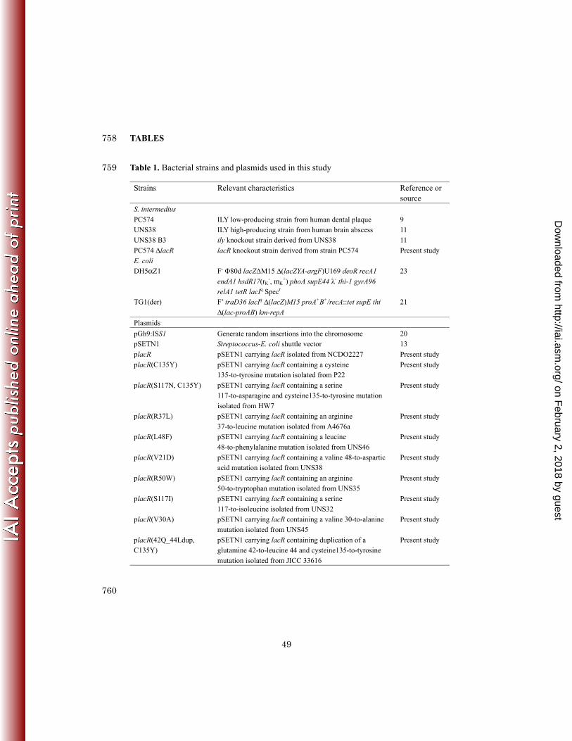
Table 1. Bacterial strains and plasmids used in this study 759
Strains Relevant characteristics Reference or source
S. intermedius
PC574 ILY low-producing strain from human dental plaque 9
UNS38 ILY high-producing strain from human brain abscess 11
UNS38 B3 ily knockout strain derived from UNS38 11
PC574 ΔlacR lacR knockout strain derived from strain PC574 Present study
E. coli
DH5αZ1 F- Φ80d lacZΔM15 Δ(lacZYA-argF)U169 deoR recA1
endA1 hsdR17(rK-, mK
+) phoA supE44 λ- thi-1 gyrA96
relA1 tetR lacIq Specr
23
TG1(der) F’ traD36 lacIq Δ(lacZ)M15 proA+B+/recA::tet supE thi
Δ(lac-proAB) km-repA
21
Plasmids
pGh9:ISS1 Generate random insertions into the chromosome 20
pSETN1 Streptococcus-E. coli shuttle vector 13
placR pSETN1 carrying lacR isolated from NCDO2227 Present study
placR(C135Y) pSETN1 carrying lacR containing a cysteine
135-to-tyrosine mutation isolated from P22
Present study
placR(S117N, C135Y) pSETN1 carrying lacR containing a serine
117-to-asparagine and cysteine135-to-tyrosine mutation
isolated from HW7
Present study
placR(R37L) pSETN1 carrying lacR containing an arginine
37-to-leucine mutation isolated from A4676a
Present study
placR(L48F) pSETN1 carrying lacR containing a leucine
48-to-phenylalanine mutation isolated from UNS46
Present study
placR(V21D) pSETN1 carrying lacR containing a valine 48-to-aspartic
acid mutation isolated from UNS38
Present study
placR(R50W) pSETN1 carrying lacR containing an arginine
50-to-tryptophan mutation isolated from UNS35
Present study
placR(S117I) pSETN1 carrying lacR containing a serine
117-to-isoleucine isolated from UNS32
Present study
placR(V30A) pSETN1 carrying lacR containing a valine 30-to-alanine
mutation isolated from UNS45
Present study
placR(42Q_44Ldup,
C135Y)
pSETN1 carrying lacR containing duplication of a
glutamine 42-to-leucine 44 and cysteine135-to-tyrosine
mutation isolated from JICC 33616
Present study
760
on February 2, 2018 by guest
http://iai.asm.org/
Dow
nloaded from

50
Table 2. S. intermedius strains used for sequencing of lacR and measurement of 761
relative hemolytic activity compared with strain UNS38 762
Strains a Isolation source Mutation in LacR Relative hemolytic
activity b (%)
Reference or
source
A4676a Brain abscess R37L 329.9 ± 13.3 9
UNS46 Liver abscess L48F 187.0 ± 18.2 7
JICC 33405 Empyema,
mediastinitis
C135Y 113.2 ± 3.3 This study
UNS38 Brain abscess V21D 100 9
UNS35 Brain abscess R50W 91.9 ± 0.9 9
UNS40 Liver abscess − 82.4 ± 26.5 9
NMH2 Brain abscess V21D 61.3 ± 2.3 9
UNS32 Liver abscess S117I 54.5 ± 3.7 9
JICC 1063 Liver abscess V30A, C135Y 53.0 ± 7.5 This study
UNS45 Liver abscess V30A 46.1 ± 8.6 9
JICC 40138-2 Infective endocarditis 42Q_44Ldup,
C135Y
42.0 ± 2.7 This study
F600 Abdominal abscess − 48.2 ± 0.5 9
JICC 33616 Brain abscess 42Q_44Ldup,
C135Y
34.6 ± 5.8 This study
JICC 32157 Empyema,
mediastinitis
C135Y 28.2 ± 0.6 This study
UNS42 Liver abscess − 27.6 ± 2.2 9
HW13 Abdominal Umbilical − 22.3 ± 0.5 9
UNS27s Liver abscess − 15.3 ± 0.9 9
JICC 674 Septicemia (not
infective endocarditis)
− 14.4 ± 0.3 This study
HW58 Brain abscess C135Y 12.9 ± 0.2 9
JICC 32100 Septicemia (not
infective endocarditis)
− 12.6 ± 0.7 This study
P58 Gingivitis − 11.2 ± 0.8 This study
on February 2, 2018 by guest
http://iai.asm.org/
Dow
nloaded from

51
JICC 33404 Pelvic abscess − 10.6 ± 0.3 This study
JICC 32138 mediastinitis − 10.4 ± 0.6 This study
JICC 32132 Brain abscess − 10.2 ± 0.3 This study
JICC 32122 Brain abscess − 9.9 ± 0.8 This study
P68 Gingivitis − 9.4 ± 0.7 This study
CDC415/87 Brain abscess − 7.8 ± 0.4 9
JICC 33620 Brain abscess − 7.8 ± 0.2 This study
P22 Gingivitis C135Y 7.3 ± 0.3 This study
JICC33412 Subcutaneous abscess − 6.6 ± 0.2 This study
GN472 Dental plaque − 6.3 ± 0.7 9
HW7 Brain abscess S117N C135Y 6.3 ± 0.3 9
JICC 689 Infective endocarditis − 6.3 ± 1.8 This study
JICC 32151 Empyema,
mediastinitis
− 5.0 ± 0.3 This study
NMH8 Unknown
(wound swab)
C135Y 3.6 ± 0.8 9
E691 Eye − 3.4 ± 0.1 9
DP101 Dental abscess − 3.2 ± 0.3 9
HW69 Brain abscess C135Y 3.2 ± 0.3 9
JICC 32135 Empyema,
mediastinitis
C135Y 2.8 ± 0.6 This study
JICC 33425 Subcutaneous abscess − 2.7 ± 2.4 This study
F458s Abdominal mass − 2.4 ± 1.0 9
P101 Gingivitis − 2.3 ± 0.4 This study
WS100s Bite wound, hand − 2.3 ± 0.3 9
JICC 33494 Brain abscess − 2.2 ± 0.5 This study
PC574 Dental plaque − 1.8 ± 1.1 9
AC800 Dental plaque C135Y 1.6 ± 1.1 9
JICC 53299 Suppurative arthritis − 0.6 ± 0.4 This study
AC5803 Dental plaque − 0.5 ± 0.1 9
P88 Gingivitis − 0.3 ± 0.3 This study
F44R Arm abscess − 0.1 ± 0.1 9
PC7466 Dental plaque − < 0.1 9
on February 2, 2018 by guest
http://iai.asm.org/
Dow
nloaded from

52
2Q Brain abscess − < 0.1 9
HARDY-DAVID
T1
Acute sinusitis − < 0.1 9
DP102 Dental plaque − < 0.1 This study
AC4720 Dental plaque − < 0.1 9
P16 Gingivitis − < 0.1 This study
NCDO2227 Unknown (Type strain) − < 0.1 9
JICC 253 Septicemia (not
infective endocarditis)
− < 0.1 This study
aILY high-producing strains were indicated by bold letters. 763
bRelative hemolytic activity (see Materials and Methods) showed ILY hemolytic activity 764
in the culture supernatant of UNS38 set as 1. The data represent the mean values ± 765
standard deviation of 3 replicates each. bNo amino acid substitution was observed in 766
the amino acid sequence of LacR. 767
768
on February 2, 2018 by guest
http://iai.asm.org/
Dow
nloaded from

53
769
Table 3. Oligonucleotides used in this study 770
Purpose Name Sequence (5’-3’)
Disruption of lacR lacR F GAGGCGTTGAACTGATACATTTTCGAC
lacR BamHI R TGCGGATCCAGTTCTTGAAGAATAACTC
lacR SalI F AATGTCGACCGTATACGCGTGTGTTATAG
lacR R GATTTTCATCGTACTCATTACCCAATC
erm BamHI F AATGGATCCCCCGATAGCTTCCGCTATTG
erm SalI R CAGTAGTCGACCTAATAATTTATCTAC
Complementation of
ΔlacR mutant and
6his-tagged lacR
lacR EcoRI F CAAGAATTCGGCGTAAAGCTCCACGTTGG
lacR BamHI F GAGGATCCATGAAAGAAGGACGACATAGAG
lacR PstI R AAAATCACCTGCAGCTTCACGAACAGGTG
Nucleotide sequences
of lacR
lacR seq. F CTTGTTTTGTTGTCATTCCCAGACTCC
lacR seq. R CAGGCTCAATCTAACATAGATGAGACCTG
lacR seq. F1 GGAATCTAATTATATGATTAGAAAGGAG
lacR seq. R2 GTCAATCTTTCTTCAAAAAAATCACCTGC
Biotinylated DNA
probe pull-down
assay
Bio-Pily F Bio-TAGCCGCTTTATCCATCTAACTCTTATCCC
Pily R AAATTAGCCTCCTTTTGCTAAATTGCTAAC
Bio-PlacD F Bio-TTTGTCTCCTTTCTAATCATATAATTAG
PlacD R TCCCAGACTCCTTTTATTTTATATGATTTC
Bio-PlacA F Bio-ATCCTCTCCTTCTGTTTATTTGTGTTG
PlacA R TGTTATACCTCCTTTTTCTTTTACAACAAC
Bio-lacF F Bio-GAATAGGGAAGAAACAACATTACTTGG
lacF R CAGTAAATCAGTCTGTGCACGATGCGCGTC
771
772
773
on February 2, 2018 by guest
http://iai.asm.org/
Dow
nloaded from

54
FIGURE LEGENDS 774
FIG. 1. Schematic illustration of the strategy for producing the ΔlacR strain by allelic 775
exchange mutagenesis; ermF-ermAM: Em-resistant genes for an Em cassette (A). 776
LacR immunoblotting analysis for confirmation of the disruption of lacR and its 777
plasmid complementation (B). PC574 and PC574 ΔlacR strain containing control 778
vector pSETN1, and PC574 ΔlacR strain transformed with placR were cultured in BHI 779
medium for 24 h. Whole-cell extracts (10 µg) were separated by SDS-PAGE. 780
Immunodetection was carried out with anti-LacR rabbit serum. Lane 1, PC574 781
pSETN1; lane 2, PC574 ΔlacR pSETN1; lane 3, PC574 ΔlacR placR. 782
783
FIG. 2. Hemolysis and ily transcriptional activity of ΔlacR strain 784
Hemolytic activity on human erythrocyte agar (A). PC574 and PC574 ΔlacR strain 785
transformed with pSETN1 or placR (WT) were inoculated onto human erythrocyte agar, 786
and then incubated at 37°C for 1 day. Hemolytic activity of the culture supernatant (B). 787
Cells were grown for 48 h at 37°C in MOPS-BHI medium containing 0.1% glucose. 788
Culture supernatant standardized at OD600 nm was diluted from 25- to 800-fold by 2-fold 789
serial dilutions, and the cytolytic activity of ILY in the diluted culture supernatant was 790
on February 2, 2018 by guest
http://iai.asm.org/
Dow
nloaded from
55
estimated by hemolysis assay. The results are plotted on a logarithmic scale in the 791
horizontal axis. Symbols: solid circle, PC574 pSETN1; open triangle, PC574 ΔlacR 792
pSETN1; open square, PC574 ΔlacR placR. Relative expression levels of ily (C). 793
The strains were grown for 16 h at 37°C in MOPS–BHI medium. The ily expression 794
levels in PC574+pSETN1, ΔlacR+pSETN1 (PC574 ΔlacR pSETN1), and ΔlacR+placR 795
(PC574 ΔlacR placR) are indicated relative to the gyrB expression level. The results 796
are plotted on a logarithmic scale in the vertical axis. The data represent the mean 797
values ± standard deviation of 6 replicates each. 798
799
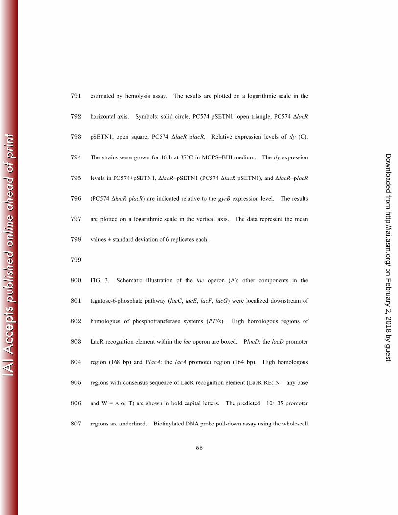
FIG. 3. Schematic illustration of the lac operon (A); other components in the 800
tagatose-6-phosphate pathway (lacC, lacE, lacF, lacG) were localized downstream of 801
homologues of phosphotransferase systems (PTSs). High homologous regions of 802
LacR recognition element within the lac operon are boxed. PlacD: the lacD promoter 803
region (168 bp) and PlacA: the lacA promoter region (164 bp). High homologous 804
regions with consensus sequence of LacR recognition element (LacR RE: N = any base 805
and W = A or T) are shown in bold capital letters. The predicted −10/−35 promoter 806
regions are underlined. Biotinylated DNA probe pull-down assay using the whole-cell 807
on February 2, 2018 by guest
http://iai.asm.org/
Dow
nloaded from

56
extracts from PC574 (B); 4 different biotinylated DNA fragments were used for this 808
assay: the ily promoter region (Pily) consisted of the 213 bp region upstream of the ily 809
gene translation start site, PlacD, PlacA, and a nonspecific DNA probe (181 bp). 810
Co-precipitated LacR protein from 5 µg whole-cell extracts with biotinylated DNA 811
probe was detected by immunoblotting analysis using anti-LacR rabbit serum. Lane 1, 812
non-specific DNA probe; lane 2, Pily; lane 3, PlacD; lane 4, PlacA; lane 5, LacR exists 813
in 5 µg whole-cell extracts as the standard marker. Biotinylated DNA probe pull-down 814
assay using the recombinant purified LacR (C); 5 µg recombinant LacR (rLacR) was 815
used for the pull-down assay. Co-precipitated LacR protein with biotinylated DNA 816
probe was detected by Coomassie brilliant blue staining. Lane 1, non-specific DNA 817
probe; lane 2, Pily; lane 3, PlacD; lane 4, PlacA; lane 5, rLacR (0.2 µg). 818
819
FIG. 4. Effect of sugars on ILY secretion 820
PC574 was grown for 48 h at 37°C in MOPS-BHI medium containing 0.1% glucose, 821
lactose, or galactose. Culture supernatant standardized at OD600 nm was diluted from 822
25- to 1,600-fold by 2-fold serial dilutions, and then, the hemolytic activity was 823
measured. The results are plotted on a logarithmic scale in the horizontal axis. 824
on February 2, 2018 by guest
http://iai.asm.org/
Dow
nloaded from

57
Symbols: solid circle, PC574 cultured with glucose; open circle, lactose; open square, 825
galactose. The data represent the mean ± SD of 4 replicates each. 826
827
FIG. 5. Cytotoxic effect of ΔlacR and its complemented strain on HepG2 cells 828
Cytotoxic effects were observed over 3 days post bacterial infection. Symbols: solid 829
circle, PC574 pSETN1; open triangle, PC574 ΔlacR pSETN1; open square, PC574 830
ΔlacR placR. The data in the graph are the mean ± SD of 5 replicates of independent 831
experiments. 832
833
FIG. 6. Complementation of ΔlacR mutant by the mutated lacR 834
PC574 ΔlacR transformed with plasmids carrying each mutated lacR was grown for 48 835
h at 37°C in MOPS-BHI medium containing 0.1% glucose. Culture supernatant 836
standardized at OD600 nm was diluted from 25- to 800-fold by 2-fold serial dilutions, and 837
the hemolytic activity was measured. Relative hemolytic activity (see Materials and 838
Methods) showed ILY hemolytic activity in the culture supernatant of PC574 ΔlacR set 839
as 1. R37L: PC574 ΔlacR transformed with placR(R37L), L48F: with placR(L48F), 840
V21D: with placR(V21D), R50W: with placR(R50W), S117I: with placR(S117I), 841
on February 2, 2018 by guest
http://iai.asm.org/
Dow
nloaded from

58
V30A: with placR(V30A), 42Q_44Ldup C135Y: with placR(42Q_44Ldup, C135Y), 842
S117N C135Y: with placR(S117N C135Y), C135Y: with placR(C135Y), and WT: 843
PC574 ΔlacR placR. The data in the graph are the mean ± SD of 6 replicates of 844
independent experiments. 845
on February 2, 2018 by guest
http://iai.asm.org/
Dow
nloaded from

5/22/2013
1
(B)
(kDa)1 2 3
(A)
ermF-ermAM
BamHI SalI
(kDa)
LacR
120
85
50
35
25
CSP
PC574 lacR
ermF-ermAM
Tomoyasu et al. FIG. 1
25
20ΔlacR ermF-ermAM
(A) (C)(B)pSETN1
PC574
placR
ΔlacR
0
20
40
60
80
100
Hem
olys
is (
%)
0 01
0.1
1
Rel
ativ
e am
oun
t (i
ly/g
yrB
)
0 01
0.1
1
0
20
40
60
80
100
0 10 100 1000
Dilution factor
PC574+pSETN1
ΔlacR+pSETN1
0.01ΔlacR
+placR
Tomoyasu et al. FIG. 2
0.01010 100 1000
on February 2, 2018 by guest
http://iai.asm.org/
Dow
nloaded from
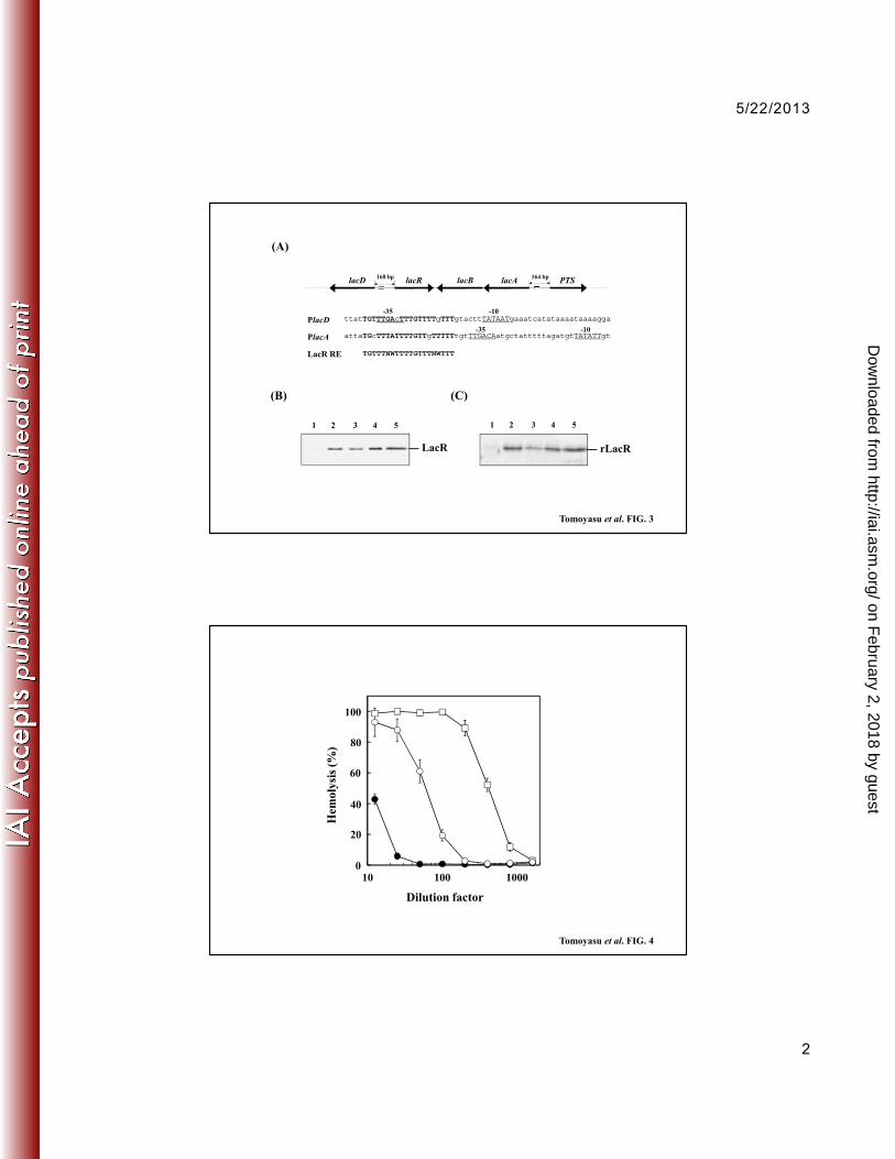
5/22/2013
2
lacD lacR lacB lacA PTS
-35 -10
(A)
168 bp 164 bp
1 2 43 5
PlacD
PlacA attaTGcTTTATTTTGTTgTTTTTtgtTTGACAatgctatttttagatgtTATATTgt
ttatTGTTTGAcTTTGTTTTgTTTgtacttTATAATgaaatcatataaaataaaagga
LacR RE
35 10
-35 -10
(B) (C)
1 2 43 5
TGTTTNWTTTTGTTTNWTTT
Tomoyasu et al. FIG. 3
LacR
1 2 43 5
rLacR
1 2 43 5
100100
20
40
60
80
Hem
olys
is (
%)
20
40
60
80
010 100 1000
Tomoyasu et al. FIG. 4
Dilution factor
10 100 10000
on February 2, 2018 by guest
http://iai.asm.org/
Dow
nloaded from

5/22/2013
3
100100
20
40
60
80S
urv
ival
(%
)
20
40
60
80
00 1 2 3
Incubation time (d)
Tomoyasu et al. FIG. 5
00 1 2 3
0.8
1
acti
vity
0.8
1
0.2
0.4
0.6
Rel
ativ
e h
emol
ytic
a
0.2
0.4
0.6
0
Mutation in LacR
Tomoyasu et al. FIG. 6
0
on February 2, 2018 by guest
http://iai.asm.org/
Dow
nloaded from


![LACR IP Site System Integration[1] MOTORBO.pdf](https://img.dokumen.tips/doc/110x75/577cc0241a28aba7118f02fd/lacr-ip-site-system-integration1-motorbopdf.jpg)