Embed Size (px)
Citation preview
6
Laboratorial Diagnosis of Syphilis
Neuza Satomi Sato Center of Immunology, Institute Adolfo Lutz
São Paulo, SP Brazil
1. Introduction
Syphilis is a curable sexually transmitted infection caused by the bacterium Treponema pallidum; the infection can also be passed from mother to her fetus during pregnancy. Diagnosis of syphilis is based on clinical evaluation, detection of the causative organism, and confirmation of the disease by serodiagnosis. T. pallidum cannot be cultured in the laboratory, but can be identified in lesions using dark-field or fluorescence microscopy or by molecular techniques. Most infected individuals have no symptoms or have transient lesions and therefore a serological test must be used to screen for infection.
2. Serological tests
Serology is still the most reliable method for laboratory diagnosis of syphilis, regardless of the stage of infection. Serologic test are divided into nontreponemal and treponemal tests, neither alone is sufficient for diagnosis. Conventional serologic diagnosis used a two step approach, of first screening with nontreponemal method, and then using a confirmatory test that uses treponemal antigens based methods to confirm a positive screening test result. Nontreponemal test are usefull also to monitoring treatment response. The first serologic test for syphilis was the Wassermann test developed in 1906. It was a complement fixation test and the antigen used was an extract of liver from newborn who had died of congenital syphilis. Landsteiner demonstrated that other tissues, such as beef heart extracted in alcohol, could be used equally well as antigens. Cholesterol and lecithin were added to increase the sensitivity of antigens. In 1922, Kahn introduced a flocculation test without complement that could be read macroscopically in a few hours. In 1941, Pangborn isolated from the beef heart the active antigenic component cardiolipin. The pure phospholipid cardiolipin combined with lecithin and cholesterol could be standardized chemically and serologically, ensuring greater reproducibility of test results both within and between laboratories. In 1946, Harris, Rosenberg and Riedel developed the Veneral Disease Research Laboratory (VDRL) and the Rapid Plasma regain (RPR) was developed in 1957, both are still in use currently. The addition of choline chloride and EDTA to the VDRL antigen enhanced the reactivity of the test and stabilized the antigen suspension (Larsen et al., 1995). The T. pallidum was identified in 1905, and the first test identifying treponemal antibodies was developed in 1949 by Nelson and Meyer. The T. pallidum immobilization test (TPI) uses T.pallidum (Nichols strain) grown in rabbit testes as the antigen and is based on the ability of
www.intechopen.com

Syphilis – Recognition, Description and Diagnosis
88
patient’s antibody and complement to immobilize living treponemes, as observed by dark-field microscopy. The fluorescent treponemal antibody (FTA) test was developed in 1957, which was later improved by the absorption procedure (FTA-ABS) in 1964 (Larsen et al., 1995).
2.1 Non-treponemal tests
The most common nontreponemal screening tests include the Veneral Disease Research Laboratory (VDRL) and the Rapid Plasma Reagin (RPR) which detect IgM and IgG antibodies against cardiolipin that is present in the sera of patients with syphilis. The VDRL test is a slide microfloculation test. The antigen, which is an alcohol solution containing 0.03% cardiolipin, 0.21% lecithin, and 0.9% cholesterol, is suspended in a buffered saline solution. When combined with antibodies, it forms flocculates that are visible using microscope´s low magnification (Larsen et al., 1995). The RPR is a modification of VDRL test, the antigen for RPR contain choline chloride (to eliminate the inactivation of tested serum), ethylenediaminotetraacetic acid – EDTA (to enhance the stability of the suspension), and charcoal particle for visualization of the suspension. This macroscopic flocculation test is done on plastic cards having multiple 18-mm circles onto which serum and modified VDRL antigen are placed and gently rotated. In the presence of antibodies a flocculation reaction takes place, and the charcoal particles are entrapped in the antigen-antibody aggregates, causing visible agglutination. The mean sensitivities of the VDRL during primary syphilis, secondary, latent and late latent are 78%, 100%, 95% and 71%, respectively; while sensitivities of RPR are 86%, 100%, 98% and 73%. The mean specificities of both tests are 98% (Larsen et al., 1995). These tests are widely available, relatively inexpensive and important for monitoring treatment. Only VDRL is the test of choice for examination of cerebrospinal fluid (CSF) in suspected neurosyphilis. Limitation of the nontreponemal serologic tests include: lack of sensitivity in early primary and late latent syphilis, the possibility of prozone reaction or false positive results (Larsen et al., 1995). A prozone reaction occurs when antibody is in excess and it is occasionally demonstrated in the nontreponemal serologic tests. Prozone reactions occur in 1 to 2% of patients with secondary syphilis (Jurado et al., 1993). False positive reactions are associated with increased age, pregnancy, drug addition, malignancy, and auto-imune diseases, such as lupus erythematosus or rheumatoid arthritis, as well as with viral (hepatitis, infectious mononucleosis, viral pneumonia, measles and others), protozoal (malaria) or mycoplasma infection (Hook & Marra, 1992; Larsen et al., 1995). Nontreponemal test results must be interpreted according to the stage of syphilis disease. Also, the interpretation of these results depends on the population being tested. The predicitive value of the nontreponemal tests is increased when combined with a reactive treponemal tests (Larsen et al., 1995).
2.2 Treponemal tests
Treponemal tests which are based in antigens derived from T.pallidum, allow detection of specific anti-treponemal antibodies. These tests have higher sensitivity and specificity than nontreponemal and were used as confirmatory tests for syphilis after a reactive nontreponemal on screening.
www.intechopen.com
Laboratorial Diagnosis of Syphilis
89
Treponemal tests include the fluorescent treponemal antibody-absorbed test (FTA-ABS), Treponema pallidum hemaglutination assay (TPHA), Treponema pallidum particle agglutination (TPPA) and enzyme immunoassay (EIA). Treponemal tests may remain reactive for years with or without treatment. Therefore, these tests should not be used to evaluate response to therapy, relapse or re-infection in previously treated patients. Also, it do not differentiate veneral syphilis from endemic syphilis (yaw and pinta). However, one treponemal IgM test, the Captia Syphilis-M EIA showed high sensitivity in primary syphilis(Lefevre et al., 1990) and also useful in monitoring treatment response of early syphilis (McMillan & Young, 2008).
2.2.1 FTA-ABS
The FTA-ABS test is an indirect fluorescent-antibody technique, the antigen used is T.pallidum subsp. pallidum (Nichols strain). The patient’s serum is diluted 1:5 in sorbent (an extract from cultures of the nonpathogenic Reiter treponeme) to remove group treponemal antibodies that are produced in some person in response to nonpathogenic treponemes. The absorbed serum is layered on a microscope slide to which T.pallidum has been fixed. If the patient’s serum contains antibody, it coats the treponeme. FICT-labeled anti-human immunoglobulin is added and combines with the patient’s antibodies resulting in FICT-stained spirochetes that are visible when examined by a fluorescence microscope. The mean sensitivities of the FTA-ABS during primary syphilis and late latent are 84% and 96%, respectively; while the sensitivities during secondary and recent latent syphilis are 100%. The mean specificities are 97%(Larsen et al., 1995). Until recently, FTA-ABS was considered the “gold standard” serological test for laboratorial diagnosis of syphilis.
2.2.2 TPHA and TPPA
Hemagglutination test was developed in the same era as the FTA-ABS test. It is technically simpler test and detects reactive antibody that agglutinates red blood cells sensitized with T.pallidum antigen (Rathlev, 1967; Rudolph, 1976; Tomizawa, 1966). A formalinized, tanned sheep (or turkey) erythrocytes are sensitized with ultrasonicated antigen from T.pallidum, Nichols strain. The patient’s serum is first mixed with absorbing diluents made from nonpathogenic Reiter treponemes and other absorbents and stabilizers. The reaction is performed in a microtiter plate. Serum containing antibodies reacts with these cells to form a smooth mat of agglutinated cells in the microtiter plate. Unsensitized cells are used as a control for nonspecific reactivity. The mean sensitivities of the TPHA during primary syphilis, latent and late latent are 76%, 97-100% and 94%, respectively; while the sensitivities during secondary is 100%. The mean specificities are 99% (Larsen et al., 1995). The T.pallidum particle agglutination assay (TPPA) uses biologically inert colored gel particles in place of red blood cells and has fewer equivocal reactions than the hemagglutination test, because heterophile reactions are eliminated (Deguchi et al., 1994). The mean sensitivities of the TPPA during primary syphilis, secondary and latent syphilis are 88%, 100% and 98%, respectively, while the mean specificities are 95% (Pope et al., 2000).
2.2.3 EIA
Veldkamp and Visser recognized the potencial for na automated T.pallidum enzyme-linked immunosorbent assay in the 1970s (Veldkamp & Visser, 1975). Since then, several EIAs
www.intechopen.com
Syphilis – Recognition, Description and Diagnosis
90
using either native or recombinant T.pallidum antigens have been developed and numerous are commercially available. EIA reported in the literature have used different approach to determine sensitivities and specificities. Some studies had used a panel of anti-treponemal positive specimens from patients whose disease stage and treatment status was known and a negative serum from health blood donors. Other studies had evaluated the performance of new test by comparing with results of conventional laboratorial tests used for diagnosis of syphilis. In general, EIA presents higher sensitivity and specificity. Captia Syphilis G (Trinity Biotech, former Centocor) is an indirect test for detection of treponemal antibodies. This test uses microtitration plates or strips coated with sonicated T.pallidum antigen. The reacting human IgG treponemal antibodies are detected by anti-human IgG monoclonal antibodies labeled with biotin and horse radish peroxidase (HRP) labeled streptavidin and revealed by tetramethylbenzidine (TMB) substrate. The sensitivity has ranged from 92.4% to 100% and specificity from 98.2% to 99.3%(Halling et al., 1999; Silletti, 1995; Young et al., 1989; Young et al., 1998). The newer Captia select Syph-G EIA (Trinity Biotech) using anti-human IgG monoclonal antibodies labeled with HRP as a conjugate instead of biotin-streptavidin system had sensitivity and specificity of 99.0% and 98.0%, respectively (Woznicova & Valisova, 2007). Enzygnost Syphilis (Dade Behring) is a one step competitive EIA with T.pallidum Nichols strain detergent extract antigen. T.pallidum specific antibodies, IgG and/or IgM contained in the sample and the conjugate (HRP labeled anti-Tp antibodies) compete for the binding sites of T.pallidum antigen coated onto wells of microtitration plates. The reaction is revealed with substrate TMB. The intensity of the resultant color is inversely proportional to the concentration of anti-treponemal antibodies in the sample. Enzygnost Syphilis showed sensitivities varying from 98.2% to 100% and specificities from 96.8% to 100% (Cole et al., 2007; Gutierrez et al., 2000; Maidment et al., 1998; Marangoni Antonella et al., 2009; Viriyataveekul et al., 2006). Bioelisa Syphilis (Biokit) is a competitive assay using T.pallidum whole antigen to coat the well of a plate. The treponemal antibodies in the test serum compete with HRP labeled human anti-treponemal antibodies. In this assay, the binding of the conjugate to the specific antigen, determined by measuring the intensity of substrate (TMB) color, is inversely proportional to the amount of specific antibodies in the test sample. Compared with FTA-ABS and TPHA, this assay had sensitivity of 99.5% and specificity of 99.4% (Ebel et al., 1998). Another version of this assay, Bioelisa Syphilis 3.0 (Biokit) is a two step recombinant EIA using recombinant antigen (TpN15 and TpN17) to coat the solid phase and HRP conjugated recombinant antigen for detection of anti-treponemal IgG and IgM. This assay showed sensitivity of 97.4%, and specificity of 100%. However, lower detection rate was observed in samples from patients with untreated primary syphilis (Cole et al., 2007). ICE Syphilis (Murex) is a two step recombinant sandwich EIA using three T. pallidum recombinant antigen (TpN15, TpN17 and TpN47) coated onto the wells of microtiter plate strips; the wells are also coated with anti-human immunoglobulin G (IgG) and M (IgM). If the antibodies to T.pallidum are present in the specimens (serum or plasma) they are captured by the antigen on the plate. In addition, a proportion of a total IgG and IgM of tested specimens are captured by the anti-human antibodies. The anti-treponemal components of the captured antibodies is detected by recombinant antigens (TpN15, TpN17 and TpN47) labeled with HRP. The intensity of the enzyme substrate TMB color is proportional to the concentration of antibodies reacting with recombinant T.pallidum antigens. The range of sensitivity and specificity for the ICE Syphilis assay were 98.2% to 100% and 99.2% to 100%, respectively (Cole
www.intechopen.com
Laboratorial Diagnosis of Syphilis
91
et al., 2007; Lam et al., 2010; Viriyataveekul et al., 2006; Young et al., 1998). Other studies for evaluation of ICE Syphilis (Murex) as a screening test for syphilis, the sensitivity in primary syphilis were 84% (48/50) (Manavi & McMillan, 2007) and 77.2% (61/79)(Young et al., 2009). Trep-Chek (Phoenix) is an indirect test for detection of anti-treponemal antibodies. The microplates wells is coated with specific recombinant treponemal antigens. Anti-treponemal antibodies present in the serum samples binds to the immobilized antigens. Anti-human IgG antibodies labeled with HRP and substrate TMB are used to detect specific anti-treponemal antibodies present in the patient´s samples. when compared with results of convencional serological tests, the sensitivity and specificity for Trep-chek was 85.3% and 95.6%, respectively (Tsang et al., 2007); other study found sensitivity of 98.9% and specificity of 95.6% in comparison with results of FTA-ABS (Binnicker et al., 2011). Trep-Sure (Phoenix) is a two step recombinant sandwich EIA for detection of anti-treponemal antibodies IgG and IgM. This assay uses specific recombinant treponemal antigens immobilized on the microplate wells. Anti-treponemal antibodies from patient´s samples bind to the immobilized antigen, which is detected with HRP conjugated treponemal antigens and substrate TMB. Trep-Sure had sensitivity and specificity of 98.9% and 94.3%, respectively (Binnicker et al., 2011). Captia Syphilis M (Trinity, former Centocor) is a capture ELISA using a microtitration plates coated with anti-human µ chain specific antibodies, which bind IgM present in serum. A tracer complex is used to detect anti-treponemal specific IgM antibody captured on the plate. The tracer complex consisted of T. pallidum antigens, a biotinylated antiaxial filament monoclonal IgM antibody, and streptavidin conjugated with horseradish peroxidase. The enzyme substrate TMB yields a colored product and the intensity of the color is proporcional to the concentration of antibodies (Lefevre et al., 1990); newer version of this assay employ a HRP conjugated recombinant antigen instead of the tracer complex (Rotty et al., 2010). This EIA was specifically designed for diagnosis of congenital syphilis, but may be applied for detection of primary infection. The sensitivity was 94% for primary syphilis, 85% for secondary, and 82% for early latent syphilis (Lefevre et al., 1990). This IgM capture EIA is also useful for monitoring treatment response in early syphilis (McMillan & Young, 2008; Rotty et al., 2010). Schmidt et al performed a comparative evaluation of different EIAs for determination of antibodies against T.pallidum in patients with primary syphilis by testing 52 sera, all negative in TPHA. The sensitivity for Captia Syphilis M was 86.5% (45/52) and other assays for detection of IgG and IgM, such as ICE Syphilis (Murex), Enzygnost Syphilis (Berhing) and Bioelisa Syphilis (Biokit) showed sensitivities of 75.0% (39/52), 69.2% (36/52) and 67.3 (24/41), respectively (Schmidt et al., 2000).
2.2.4 Immunochemoluminescence assay (CIA) and multiplex flow immunoassay (MFI)
In addition to EIA, new treponemal assay based on CIA or MFI technology are available with the advantage of automation facilities, higher testing throughput and the objective interpretation. The LIAISON Treponema screen (Diasorin) is a one-step sandwich chemiluminescence immunoassay (LIAISON CLIA) that measures total anti-treponemal antibody (IgG and IgM). A recombinant treponemal antigen (TpN17) is coated onto paramagnetic microparticles and patient’s serum (or plasma) are added along with an isoluminol-antigen conjugate. The same antigen TpN17 is component of conjugate. Specific anti-treponemal antibodies present in the specimen will be captured to the antigen on the paramagnetic particle and bind to the antigen conjugate in a sandwich manner. After incubation, unbound
www.intechopen.com

Syphilis – Recognition, Description and Diagnosis
92
material is removed with a wash cycle. Starter reagents are added and a light signal is produced from a flash chemiluminescence reaction if anti-treponemal antibodies are present. The light signal is measured by a photomultipler as Relative Light Units (RLU), and the results are reported as an index value. A retrospective study performed by Marangoni et al analyzed a panel of 2,494 blood donor sera, 131 clinically and serologically characterized syphilitic sera and 96 samples obtained from subjects with potentially interfering diseases or conditions (including Lyme disease, mononucleosis, rheumatoid factor positive and T.denticola positive). Also, 1,800 unselected samples submitted for routine screening for syphilis was included in this study. LIAISON sensitivity was 99.2%, which was higher than EIA (95.4%) or TPHA (94.7%) especially when primary syphilis samples were tested. Only Western Blot had 100% sensitivity. In this study LIAISON missed detection of a sample from treated latent syphilis patient. Specificity was 99.9% for all treponemal tests. From 96 sera obtained from patient suffering from potentially cross-reactive conditions, no serum gave a positive result by LIAISON (Marangoni et al., 2005). However, another study for evaluation of LIAISON test found lower sensitivity of 94.1% (48/51), in primary and secondary syphilis (Knight et al., 2007). The Abbott ARCHITECT Syphilis TP assay is a two-step sandwich chemiluminescent microparticle immunoassay (CMIA) for the qualitative detection of antibody to Treponema pallidum in human serum or plasma based on recombinant antigens TpN15, TpN17 and TpN47. Antibody present in the sample binds to T.pallidum recombinant antigen coated paramagnetic particles. After wash step, murine anti-human-IgG/anti-human-IgM acridinium-labelled conjugate is added. Following a further wash step, pre-trigger solution (hydrogen peroxide) and trigger solution (sodium hydroxide) are added. The resulting chemiluminescent reaction is measured in relative light units (RLUs) which are directly proportional to the amount of anti-T.pallidum present in the sample. The Architect Syphilis was highly sensitive in detecting primary syphilis(97.5%) and had specificity of 99.1% (Young et al., 2009). In another evaluation, this assay presented 100% sensitivity (121/121) and specificity (500/500) and good performance on reproducibility, with intra-assay coefficient variation lower than 4% (Yoshioka et al., 2007). Recently, Wellinghausen & Dietenberger evaluated two automated CIA, the LIAISON Treponema Screen and the ARCHITECT Syphilis TP in comparison to the TPPA test for laboratory diagnosis of syphilis. A prospective study was performed using 577 sera submitted for diagnosis of syphilis and 42 stored sera from patients with syphilis infection diagnosed by clinic and serology. Sensitivity was 100% for all three tests, and specificity were 100%, 99,8% and 99,6% respectively for LIAISON, ARCHTECT and TPPA (Wellinghausen & Dietenberger, 2011). Bio-Rad BioPlex 2200 Syphilis Multiplex Flow Immunoassay (MFI) was evaluated for the detection of anti-treponemal antibodies, IgM and IgG. The principle of MFI technology is based on cytometric beads array. The BioPlex Syphilis IgG kit uses three different populations of microspheres coated with recombinant proteins from T.pallidum (15 kDa, 17 kDa and 47 kDa). The patient’s specimens is added to a reaction vessel containing bead reagent and sample diluents and incubated at 37oC. Antibody present in the sample binds to T.pallidum recombinant antigen coated beads. After wash step, a phycoerytrin-conjugated reporter antibody is added. After second incubation and washing step, the beads are read by a flow-based detector which quantitates each analyte and compares it to a pre-established calibration curve. The data are initially calculated as relative fluorescence intensity and then converted to a fluorescence ratio (FR)
www.intechopen.com
Laboratorial Diagnosis of Syphilis
93
using an internal standard bead. The FR is compared to an assay specific calibration curve to determine the analyte concentration in antibody index (AI) units, where AI is higher than 1.0 for positive samples and lower than 1.0 for negative one. The kits for IgM uses two different beads sets individually coated with recombinant proteins associated with T.pallidum (17 and 47 kDa). In a prospective analysis of 1008 serum samples submitted for serologic testing for syphilis, the BioPlex IgG MFI assay demonstrated sensitivity of 98.7% (77/78) and specificity of 99.4% (916/930) in comparison with EIA assay. For anti-treponemal IgM antibody detection, BioPlex IgM MFI assay showed 80% (4/5) sensitivity and 97.9% (652/666) specificity, when compared to the Trep-Chek IgM EIA (Phoenix-Biotech). BioPlex syphilis MFI assay allow for a fully automated random-accesses platform that provides fast (1.7h for 100 samples) and high throughput (800 samples per 9 hours) analysis of the syphilis serologic response (Gomez et al., 2010).
2.2.5 Westen Blot (WB) and Immuno-blot
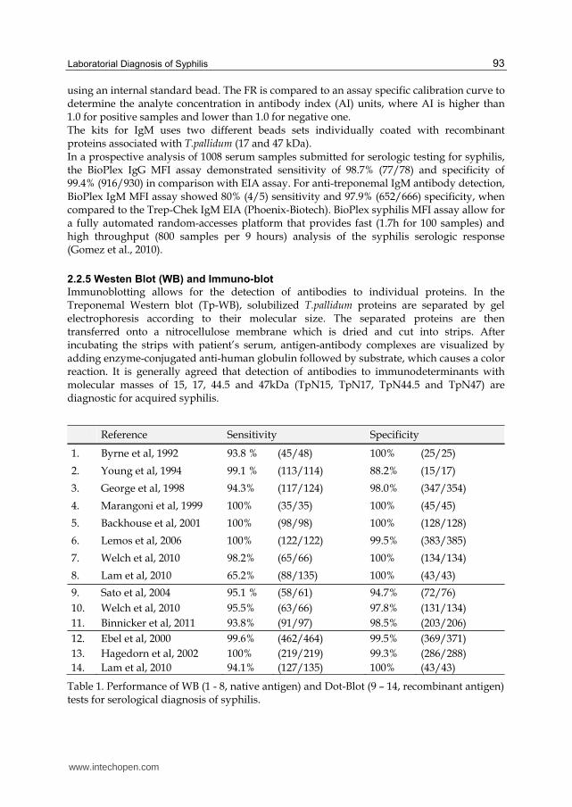
Immunoblotting allows for the detection of antibodies to individual proteins. In the Treponemal Western blot (Tp-WB), solubilized T.pallidum proteins are separated by gel electrophoresis according to their molecular size. The separated proteins are then transferred onto a nitrocellulose membrane which is dried and cut into strips. After incubating the strips with patient’s serum, antigen-antibody complexes are visualized by adding enzyme-conjugated anti-human globulin followed by substrate, which causes a color reaction. It is generally agreed that detection of antibodies to immunodeterminants with molecular masses of 15, 17, 44.5 and 47kDa (TpN15, TpN17, TpN44.5 and TpN47) are diagnostic for acquired syphilis.
Reference Sensitivity Specificity
1. Byrne et al, 1992 93.8 % (45/48) 100% (25/25)
2. Young et al, 1994 99.1 % (113/114) 88.2% (15/17)
3. George et al, 1998 94.3% (117/124) 98.0% (347/354)
4. Marangoni et al, 1999 100% (35/35) 100% (45/45)
5. Backhouse et al, 2001 100% (98/98) 100% (128/128)
6. Lemos et al, 2006 100% (122/122) 99.5% (383/385)
7. Welch et al, 2010 98.2% (65/66) 100% (134/134)
8. Lam et al, 2010 65.2% (88/135) 100% (43/43)
9. Sato et al, 2004 95.1 % (58/61) 94.7% (72/76) 10. Welch et al, 2010 95.5% (63/66) 97.8% (131/134) 11. Binnicker et al, 2011 93.8% (91/97) 98.5% (203/206)
12. Ebel et al, 2000 99.6% (462/464) 99.5% (369/371) 13. Hagedorn et al, 2002 100% (219/219) 99.3% (286/288) 14. Lam et al, 2010 94.1% (127/135) 100% (43/43)
Table 1. Performance of WB (1 - 8, native antigen) and Dot-Blot (9 – 14, recombinant antigen) tests for serological diagnosis of syphilis.
www.intechopen.com
Syphilis – Recognition, Description and Diagnosis
94
The Tp-WB showed a high sensitivity in both, treated and untreated infection (Young et al., 1994). One study found that the 17 kDa antigen have the best combined attributes of sensitivity and specificity for diagnostic of syphilis (George et al., 1998). Another study found that a more sensitive and specific criterion for the WB would be the reactivity with the antigen of 15kDa and other two of the three major antigens, TpN47, TpN44.5 and TpN17. The criterion was based on the analysis of reactivity for individual antigenic determinant where only the antigen of TpN15 had 100% sensitivity and specificity. For other three antigens the sensitivities were 100%, 100%, 96% and the specificities were 20%, 96%, 100%, respectively for of TpN47, TpN44.5 and TpN17 (Backhouse & Nesteroff, 2001). Lemos et al (2006) evaluated the association of clinical phases of syphilis with the reactivity of individual antigenic determinants. The reaction with TpN47 was present in all phases of syphilis, with higher intensity in primary and early syphilis than in late latent and tertiary syphilis. The reaction to TpN17 was observed in samples from patients with early syphilis (primary, secondary and early latent). Except to patients with primary syphilis, samples from patients with any of the other clinical forms of syphilis showed reactivity against TpN15. In tertiary syphilis, the reactivity of TpN15 showed higher intensity than TpN47 (de Lemos et al., 2007). Recently, a commercially available treponemal WB kit has been evaluated for its use as confirmatory test for the serological diagnosis of syphilis (Lam et al., 2010; Welch & Litwin, 2010). The TWB (MarDx Diagnostics, CA, USA) is a Western Blot assay and uses three native antigens (15.5, 17 and 47 kDa) to detect IgG antitreponemal antibody. One study was carried with 200 serum samples collected for routine laboratorial diagnosis of syphilis, which were separated according to the results of classical syphilis tests, RPR and FTA-ABS. The TWB – MarDx assay showed a sensitivity of 98.2%, specificity of 100% and overall agreement of 99.4% (Lam et al., 2010; Welch & Litwin, 2010). Another study was performed with 173 serum samples from patients in different clinical stage, including primary syphilis (39), secondary (20), early latent (18), latent of unknown duration (58) and normal health subjets (43). The overall sensitivity was as low as 65.2%. The highest sensitivity of 90% was found in secondary stage and the lowest was 50% in the latent stage of unknown duration, whereas in primary and early latent stages the sensitivity was around 72%. This assay presented 38 samples (28.1%) with indeterminate results (Lam et al., 2010; Welch & Litwin, 2010). Based on published data, Tp-WB showed specificity from 88 – 100% and sensitivity ranging from 94-100%, except for one study that found sensitivity of 65.2% for TWB kit. Overall, these native antigen based Tp-WB assay is a useful additional confirmatory test for syphilis. A blot assay using recombinant antigens instead of fractionates native proteins had been described (Sato et al., 2004) and some are also commercially available, including Treponema Virablot IgG (Viramed Biotech) (Binnicker et al., 2011; Welch & Litwin, 2010) and INNO-LIA (Immunogenetics, Belgium) (Ebel et al., 2000). The immune-slot-blot assay is an “in house” technique prepared with three recombinant antigen rTp47, rTp17 and rTp15 immobilized on nitrocellulose strip. It was analyzed 137 serum samples from patients with a clinical and laboratory diagnosis of syphilis (61), from healthy blood donors (50), individuals with sexually transmitted disease other than syphilis (3), and from individuals with other spirochetal diseases such as Lyme disease (20) and leptospirosis (3). The sensitivity was 95.1% (58/61) and specificity was 94.7% (72/76) (Sato et al., 2004).
www.intechopen.com
Laboratorial Diagnosis of Syphilis
95
The Treponema Virablot IgG (Viramed Biotech) is composed with Tp47, Tp44.5, Tp17 and Tp15 and it was evaluated with serum samples previously analyzed with classical serological tests for diagnosis of syphilis. In comparison with results of RPR, FTA-ABS and TPPA, the sensibility was 95.5% and the specificity was 97.8% (Welch, 2010). Similar results was found by Binnicker et al, the sensibility and specificity were 93.8% and 98.5%, respectively, when compared with FTA-ABS (Binnicker et al., 2011). The INNO-LIA Syphilis test (Innogenetics) is a line immunoassay utilizing three recombinant antigens of T.pallidum (TpN 47, TpN17 and TpN15) and one synthetic peptide derived from TpN44.5 (TmpA), which coated as discrete lines on nylon. This assay was initially validated by using a large number of sera (835) from a clinical laboratory and the overall sensitivity and specificity were calculated with reference to consensus diagnostic assay results. The INNO-LIA Syphilis test had sensitivity of 99.6% and specificity of 99.5% (Ebel et al., 2000). These results were confirmed by another evaluation of the assay using 507 serum samples, the sensitivity and specificity were 100% and 99.3%, respectively (Hagedorn et al., 2002). Recently, the test was analyzed with 135 serum samples from patients in different clinical stages of syphilis. A lower sensitivity of 94.1% was determined in this group. The sensitivity were 92.3%, 100%, 94.4% and 94.1%, respectively for primary, secondary, early latent and latent of unknown duration clinical stages of syphilis (Lam et al., 2010). The INNO-LIA syphilis test provides highly reliable results and can be considered to be a valid alternative confirmatory for serological tests for syphilis.
2.2.6 Treponema specific rapid diagnostic test (point of care - POC)
A number of simple, point-of-care (POC) treponema specific rapid diagnostic tests are also commercially available. Most rapid tests detect IgM, IgG and IgA antibodies and involve immunochromatographic strips in which one or multiple T.pallidum recombinant antigens are applied to nitrocellulose strips as a capture reagent. Antibodies in the specimen bound at antigen site on the strip and are revealed with dye bound anti-immunoglobulin or dye bound antigen and a positive reaction appears as a colored line. Most of these tests can be used with whole blood from a finger prick as well as serum or plasma. The results can be read visually in less than 30 minutes, these rapid tests are simple to perform, require minimal equipment and training. It is suitable for use in primary healthcare settings, and also it can be performed in the field at point of care. Overall, rapid tests are highly sensitive and specific. The World Health Organization compared the performance of 8 rapid tests to a combined reference standart TPHA/TPPA, reporting sensitivities of 84.5%-97.7% and specificities of 92.8% - 98% (Herring et al., 2006). A lower sensitivity was found for a whole blood in comparison with serum or plasma. Additionally, the sensitivity was lower in the field conditions than in assay performed in the laboratory (Benzaken et al., 2008; Mabey et al., 2006a; Mishra et al., 2010). Determine Syphilis TP (Abbott) has recombinant antigen TpN47 immobilized on nitrocellulose strips and use 50uL of sample, either whole blood, serum or plasma and results can be read in 5 to 20 minutes. For serum or plasma specimens, sensitivity range were 93.7 to 99.2% and specificity 92.4 to 100% (Diaz et al., 2004; Herring et al., 2006; Mabey et al., 2006a; Mabey et al., 2006b; Oshiro et al., 1999; Sato et al., 2003). For whole blood sample, the sensitivity ranged from 85,9% to 95.0%, and specificity was higher than 97.7% (Gianino et al., 2007; Mabey et al., 2006a; Siedner et al., 2004; Tinajeros et al., 2006) when
www.intechopen.com
Syphilis – Recognition, Description and Diagnosis
96
performed in the reference laboratory. Lower sensitivity of 75.6% was found when the assay was performed at local clinic (Li et al., 2009; Mabey et al., 2006a). SD BioLine Syphilis 3.0 ICS (Standard Diagnostics, Korea) use three recombinant antigens TpN15, TpN17 and TpN47 to capture specific antibodies. The assay is performed with 20 uL of whole blood or 10 uL of serum or plasma, and results can be read in 5 to 20 minutes. For serum samples, overall sensitivities were 94.2 -95.0% and specificities 94.9% - 97.8%(Herring et al., 2006; Mabey et al., 2006a). However, for whole blood samples, this rapid test showed better sensitivities for assay performed in the laboratory when compared with local clinic testing. These differences were observed in the results obtained at three countries (Tanzania, Brazil and China), according to the study published by Mabey et al. Sensitivities ranged from 90.2% to 95.5% and 85.7% to 88.2%, respectively for laboratory and local clinic testing. Specifities were 95.5% to 99.4% and 98.8% to 99.6%, respectively (Mabey et al., 2006a). Montoya et al found similar results; sensitivities were 96.3% and 86.0%, respectively for laboratory and healthy facility testing, while specificity showed no difference, 96.8% and 96.4% (Herring et al., 2006; Mabey et al., 2006a; Montoya et al., 2006). Visitec Syphilis (Omega Diagnostics, Scotland UK) uses two recombinant antigens TpN17 and TpN47 to capture specific antibodies. The assay is performed with 50 uL of whole blood or 25 uL of serum or plasma, and results can be read in 15 minutes. For serum samples, sensitivities were 84.2% to 98.2% and specificities 98.0% to 99.1% (Herring et al., 2006; Mabey et al., 2006a). For whole blood samples, sensitivities were 77.9% to 98.2% and 72.7% to 96.1% respectively for laboratory and local clinic testing, while overall specificities were around 99% for both settings (Herring et al., 2006; Mabey et al., 2006a). A finger prick blood samples were tested with Visitec Syphilis to assess the performance of this rapid test to o detect syphilis in field evaluation of high risk populations. Visitec Syphilis test had identified 79% (30/38) of active syphilis cases (Benzaken et al., 2008). Qualpro Syphicheck-WB rapid syphilis test (Qualpro Diagnostic, India) use two recombinant antigens TpN17 and TpN47 to capture specific antibodies. The assay is performed with 50 uL of whole blood or 25 uL of serum or plasma, and results can be read in 15 minutes. For serum samples, sensitivities were 84.5% to 95.3% and specificities 93.7 to 98.9% (Herring et al., 2006; Mabey et al., 2006a). For whole blood samples, sensitivities were 70.8% to 97.6% and 64.0% to 84.3% respectively for laboratory and local clinic testing, while overall specificities were around 99% for both settings (Herring et al., 2006; Mabey et al., 2006a). The Syphcheck-WB test using finger prick whole blood was evaluated for detection of active syphilis among female sex workers in Bangalore, India. Compared with the reference RPR and TPHA, the sensitivity and specificity of the POC syphilis were 70.8% and 97.8%, respectively (Mishra et al., 2010). Besides its lower sensitivity, this study revealed that the use of POC screening conferred an advantage over offsite RPR testing among hard-to-reach populations, who may not return for their test results and follow-up treatment. The proportion of women with active syphilis who were appropriately treated rose from 44.8% to 68.3% (p=0.003) with the use of POC syphilis screening (Mishra et al., 2010). The limitation of rapid syphilis tests is that they cannot distinguish active and treated syphilis; positive results need confirmation with quantitative nontreponemal testing to determine recent infection and response to therapy. New approach based POC tests has been described and it may be promissory to overcome the limitations of rapid tests currently available.
www.intechopen.com
Laboratorial Diagnosis of Syphilis
97
Recently, a novel dual POC syphilis test had been developed for simultaneous detection of nontreponemal and treponemal antibodies in patients with syphilis. Compared with RPR, the concordance of the dual non-treponemal line was 98.4% and the nonreactive concordance was 98.6%. Compared to the TPPA assay, the reactive and nonreactive concordances of the treponemal line were 96.5% and 95.5%, respectively (Castro et al., 2010). The test for detection of T. pallidum specific IgM antibody named colloidal gold immunochromatography assay (GICA) had been described by Lin et al. GICA has monoclonal antibody to the human µ chain-specific IgM immobilized on nitrocellulose strips to capture the patient treponemal-specific IgM which is revealed by the colloidal gold conjugated recombinant antigens, TpN17 and TpN47. The sensitivity and specificity were 98.2% and 99.0% when compared with FTA-IgM (Lin et al., 2011). These new POC test can be helpful for diagnosing active syphilis disease.
3. Direct detection of Treponema pallidum
There are number of methods available for direct detection of intact organisms or T.pallidum DNA.
3.1 Darkfield microscopy
The oldest method still remains one of the simplest and most reliable for the direct detection of T. pallidum. An experienced microscopist can identify T.pallidum from lesions based on the characteristic morphology and motility of the spirochete. This method is suitable when the lesions are moist, and the examination can be done immediately after specimen collection. Exudates and fluids from lesions are examined as a wet mount using dark-field microscopy. During the primary stage, serous fluid from the lesion contains numerous treponemes and, therefore, this approach is particularly useful in patients with immunodeficiency or in early syphilis when antibodies are not yet detectable. Success is dependent on a number of factors, including too little or too much fluid on the slide, the presence of refractile elements in the specimen, improper thickness of the slide or cover slip, etc. Treatment with antibiotics may result in a false-negative finding. Therefore, although the demonstration of T pallidum is the definitive method of diagnosis, dark-field microscopy has limited sensitivity, and failure to detect T pallidum by this test does not rule out syphilis (Larsen S.A. et al., 1990).
3.2 Direct fluorescent antibody test for T.pallidum (DFA-TP) in the blood fluids
The direct fluorescent antibody test for T.pallidum is easier to perform than dark-field microscopy, it detects antigens and does not require the presence of motile treponemes. DFA-TP test use fluorescein isothiocyanate-labelled anti-T.pallidum antibody to identify the organism. However, this test does not differentiate between T.pallidum and other pathogenic treponemes causing yaws, endemic syphilis and pinta specific to pathogenic treponemes. The number organisms in the fluid or tissue that can be detected by fluorescent antibody tests is similar to that for darkfiled micoscopy. The sensitivity of these methods is only stightly better than darkfiled microscopy (Larsen S.A. et al., 1990).
3.3 Direct test for T.pallidum in tissue sections
Direct fluorescent antibody test for T.pallidum using fluorescein isothiocyanate-labelled anti-T.pallidum antibody can also be used in tissue section to identify the organism (DFAT-T).
www.intechopen.com
Syphilis – Recognition, Description and Diagnosis
98
Immunohistochemical (IHC) detection of T.pallidum use an unlabeled treponemal antibody and the detection complex based on secondary antibody, enzyme conjugate and insoluble enzyme substrate system for IHC stain. This method can be used in combination with histological staining; the advantage of hematoxylin counterstain is to examine tissue structure simultaneously. Silver staining, while useful, is nonspecific. Silver nitrate will impregnate a number of different organisms and allows only for identification of the morphology of the organism. tissue artifacts are a potential hazard for misidentification (Larsen S.A. et al., 1990).
3.4 Rabbit Infectivity Test (RIT)
RIT is probably the most sensitive methods for detecting infectious treponeme. Any source of specimen can be used for RIT as long as the material is less than 1 hour old or was flash-frozen immediately after collection and maintained in liquid nitrogen or at temperature of -78oC or bellow. The RIT remains a research tool of academic interest for detection of virulent organism in clinical specimens because of the need for animal, the very long incubation time after infection (several weeks to months), and variation in rabbit susceptibility to infection (Larsen S.A. et al., 1990). This technique was also used as a gold standard for measuring the sensitivity of methods such as PCR. The RIT, using susceptible rabbit, has a sensitivity of 10 to 50 organisms, similar to that of DNA PCR (Grimprel et al., 1991; Sanchez et al., 1993).
3.5 Polymerase Chain Reaction (PCR)
A number of PCR based methods have been developed for the detection of T.pallidum DNA or RNA in clinical specimens. These assays are based on the detection of various target genes including tmpA (a 45kDa membrane protein Hay, 1990a); bmp, 39-kDa basic membrane protein (Noordhoek et al., 1991); tpp47, a 47kDa membrane immunogen (Burstain et al., 1991); polA, DNA polymerase I (Liu et al., 2001); tmpC, a 35kDa membrane protein (Flasarova et al., 2006) and 16SrRNA (Centurion-Lara et al., 1997). The first application PCR in clinical samples from patients with syphilis was reported by Hay et al in 1990. They used primers derived from the gene sequence of TmpA and the 4D antigen (and oligomeric protein with multiple forms). The detection limit was 65 organisms. When PCR was used to detect T.pallidum DNA in cerebrospinal fluid (CSF) of patients with and without syphilis, sensitivity was 47% and specificity 93% (Hay et al., 1990a; Hay et al., 1990b). The bmp gene was used as target for nested-PCR described by Noordhoek et al. Initially, a 617-bp (171-788) portion of bmp gene was amplified and used for second PCR to generate the 500 bp (256-762) products, which was detected with 32P-labeled probe of 19 nt (500-519). The detection limit was 1 fg DNA (1 organism). PCR was applied to detect T.pallidum DNA in CSF from neurosyphilis patients before and after antibiotic treatment. Prior to treatment, PCR was positive in 71% (5/7) patients with acute symptomatic and 12% (2/16) asymptomatic neurosyphilis. However, they found treponemal DNA in CSF up to 3 year after treatment (Noordhoek et al., 1991). Brustain et al had used this same bmp gene as target in a multiplex PCR for simultaneous detection of T.pallidum and Haemophilus ducrey DNA in genital ulcer samples. The nested PCR product was analyzed on a 10% polyacrylamide gel by using ethidium bromide
www.intechopen.com
Laboratorial Diagnosis of Syphilis
99
staining. Multiplex nested PCR showed higher sensitivity then serological methods for diagnosis of syphilis in the population studied, from outpatient clinic for sexually transmitted diseases (STDs) in Amsterdan. Sensitivity and specificity were 75% (12/16) and 100% (348/348), respectively (Bruisten et al., 2001). The ttp47 gene PCR to detect T.pallidum DNA in clinical samples was developed by Burstain et al. A 658-bp (648-1305) portion of tpp47 gene was amplified and the PCR products were probed by DNA-DNA hybridization with a 496-bp (713-1208) fragment internal to the amplified DNA. The assay detected approximately 0.01 pg of purified T. pallidum DNA, and it was able to detect as low as one to 10 organisms per specimen with high sensitivity. T. pallidum DNA was detected in serum, CSF and amniotic fluids from syphilis patients but not in nonsyphilitic controls (Bruisten et al., 2001). The same technique was applied in several clinical materials in order to investigate the potential of PCR in diagnosis of congenital syphilis. The PCR was 100% specific for T. pallidum compared with the sensitive rabbit infectivity test (RIT) for all clinical materials tested. Sensitivities for amniotic fluids, CSF and serum were 100%, 60% and 67% respectively (Grimprel et al., 1991). Kouznetsov et al used nested-PCR to amplify a different portion of ttp47 gene. The 379 bp (357-735) PCR product was used to amplify the 194 bp (391-584) products by nested-PCR; detection was performed by 32P-labeled probe of 19 bp (481-499), and the detection limit was 20 organism (Kouznetsov & Prinz, 2002). The ttp47 gene was detected in 80% (12/15) of peripheral blood mononuclear cells (PBMCs) from patients with syphilis; when serum samples were analyzed the PCR was positive in 83% (5/6) patients with secondary syphilis and in 50% (2/4) patients with early latent syphilis (Kouznetsov et al., 2005). A smaller fragment of 260 bp (537-776) of tpp47 was targeted in multiplex-PCR (M-PCR). This M-PCR assay with colorimetric detection was devised for the simultaneous amplification of DNA targets from H. ducreyi, T. pallidum, and herpes simplex virus (HSV) types 1 and 2. The assay detected from 1 to 10 organisms. In analysis of 298 genital ulcer swabs, M-PCR showed higher sensitivity (91%) when compared with darkfield microscopy (81%), the specificity was 99%(Orle et al., 1996). The same target (260 bp) was used by Palmer et al to evaluated PCR for diagnosis of early syphilis. DNA was extracted from swabs of ano-genital or oral ulcers, and PCR product was analyzed by electrophoresis using 2% agarose. The detection limit of PCR was 1 pg T. pallidum DNA (~800 organisms). Sensitivities were 94.7% and 80.0% for primary and secondary syphilis, respectively, while specificity was 98.6% for both clinical stages (Palmer et al., 2003). Further, this 260 bp target was used in PCR for detection of T.pallidum DNA in latent syphilis by testing whole blood, serum or plasma samples. Ear lobe scraping presented the highest positivity of 51.7% (16/28) followed by plasma, whole blood and serum, with 44.9% (31/69), 39.1% (27/69) and 21.6% (18/69), respectively (Castro et al., 2007). The PCR based on two unique features of the DNA polymerase I gene (polA) of T. pallidum was developed by Liu et al. The first distinctive characteristic is that the region codes for a high cysteine content and has low homology with similar regions of DNA polymerase I gene from known microorganisms. The second unique feature is the presence of four insertions in the gene. PCR tests using primers designed on the basis these regions reacted with various pathogenic T.pallidum subspecies but did not react with nonpathogenic treponemal species or other spirochetes. Two sets of primers were used to amplify a fragment of 377 bp (1759-2135) and 395 bp (1539-1933). The detection limit was about 10 to 25 organisms when analyzed on gel, and a single organism when the ABI 310 Prism Genetic
www.intechopen.com
Syphilis – Recognition, Description and Diagnosis
100
Analyzer was used to detect fluorescence-labeled amplicons. The 112 genital ulcer specimens were tested by polA PCR; sensitivity and specificity was 95.8% and 95.7%, respectively (Liu et al., 2001). The polA gene of T.pallidum was detected in a whole blood from person with syphilis; PCR was positive in 43% (3/7) patients with incubating disease and 62% (8/13) with latent syphilis (Marfin et al., 2001). When used as a screening test for T. pallidum DNA detection in lesion samples from patients with clinical diagnosis of syphilis, polA PCR was found positive in 36% (15/42) and 75% (9/12) of samples from patients with primary and secondary syphilis, respectively (Pope et al., 2005). Martin at al used PCR to amplify the three T. pallidum genes: tpp47(Burstain et al., 1991) bmp (Noordhoek et al., 1991) and polA (Liu et al., 2001). PCR was positive in 36%(19/53) specimens, three treponemal gene PCR assays gave concordant results in all specimens collected from syphilis patients, regardless of the specimen types (blood or swab) or the stages of disease (primary or secondary) (Martin et al., 2009). Another study found positivity ratio of 39.1% for ttp47 PCR (260 bp) and 31.1% for polA PCR (378 bp), in blood samples from patients with latent syphilis (Castro et al., 2007). The real-time PCR had been developed for detection of T.pallidum DNA in clinical samples, they targeted polA gene (Heymans et al., 2010; Koek et al., 2006; Leslie et al., 2007) or tpp47 gene (Gayet-Ageron et al., 2009). The first test was validated using an analytical panel (n = 140) and a clinical panel of genital samples (n = 112) from patients attending a sexually transmitted infections clinic. High sensitivities and specificities of 94-100% were achieved using two real-time PCR platforms, the Rotor-Gene and the iCycler (Koek et al., 2006). Leslie et al developed real-time PCR which analytical sensitivity was estimated to be 1.75 target copies per reaction. Real-time PCR was performed in genital lesion specimens, but not in serum. When compared with serology results, polA gene real-time PCR presented the sensitivity of 80.39% and the specificity of 98.40%(Leslie et al., 2007). Another study found sensitivity of 72.8% and specificity of 95.5% in patients with clinical diagnosis of primary syphilis, the detection of secondary syphilis was low, and the sensitivity was 43.0% (Heymans et al., 2010). Gayet-Ageron et al evaluated a real-time PCR assay for the detection of tpp47 gene of T.pallidum in various biological specimens from patients with primary and secondary syphilis. They found global sensitivity of 65% during primary, 53% during secondary, and null during latent syphilis. Among primary syphilis, real-time PCR positivity was 80% in lesion swabs and 55% in serum, while among secondary syphilis it was 100% in plasma samples (Gayet-Ageron et al., 2009). In general, PCR presents high sensitivity to detect treponemal DNA in ulcer lesion samples from patients with primary syphilis. T.pallidum DNA can be detected in blood samples from patients with latent syphilis, also.
4. Conclusion
The ideal test for syphilis should have both high sensitivity and specifitity, be suitable for monitoring response to treatment, give a negative result after adequate therapy and also give a clear indication of reinfection. Unfortunately, such test does not exist. PCR methods had been shown high sensitivity for detection of T.pallidum DNA in ulcer lesion samples, but lower performance was observed performance in secondary or latent syphilis, probably due to low number of circulating treponemes in these stages.
www.intechopen.com
Laboratorial Diagnosis of Syphilis
101
The new recombinant antigen based automated syphilis assays have the advantage of their high sensitivities in early syphilis and their high throughputs; however as treponemal tests, they cannot distinguish among recent, remote, and previously treated infections. The detection of specific IgM antibodies may be helpful in this issue as it could be detected in active infection including re-infection. Some studies reported a good association between the detection of treponemal specific IgM antibodies and nontreponemal antibodies (VDRL or RPR) in serum samples from patients with untreated syphilis, and also in follow-up of treatment. POC tests may prove to be effective tools in the control of syphilis and for screening pregnant women to prevent congenital syphilis in primary health care settings, because of its the simplicity and low cost. However, currently available POC tests do not distinguish active and treated syphilis. New POC tests for simultaneous detection of non-treponemal antibodies or for detection of specific IgM antibody developed recently are promising to overcome these limitations. A great improvement has been achieved for syphilis laboratory diagnosis and it is constantly evolving, but to date, no test is ideal for all stages of syphilis.
5. References
Backhouse, J. L. & Nesteroff, S. I. (2001). Treponema pallidum western blot: Comparison with the FTA-ABS test as a confirmatory test for syphilis. Diagnostic Microbiology
and Infectious Disease Vol.39, No.1, pp. 9-14, ISSN 0732-8893 Benzaken, A. S., Sabido, M., Galban, E. G., Pedroza, V., Vasquez, F., Araujo, A., Peeling, R.
W. & Mayaud, P. (2008). Field evaluation of the performance and testing costs of a rapid point-of-care test for syphilis in a red-light district of Manaus, Brazil. Sexually
Transmitted Infection Vol.84, No.4, (Aug), pp. 297-302, ISSN 1472-3263 Binnicker, M. J., Jespersen, D. J. & Rollins, L. O. (2011). Treponema-specific tests for
serodiagnosis of syphilis: comparative evaluation of seven assays. Journal of Clinical
Microbiology Vol.49, No.4, (Apr), pp. 1313-1317, ISSN 1098-660X Bruisten, S. M., Cairo, I., Fennema, H., Pijl, A., Buimer, M., Peerbooms, P. G., Van Dyck, E.,
Meijer, A., Ossewaarde, J. M. & van Doornum, G. J. (2001). Diagnosing genital ulcer disease in a clinic for sexually transmitted diseases in Amsterdam, The Netherlands. Journal of Clinical Microbiology Vol.39, No.2, (Feb), pp. 601-605, ISSN 0095-1137
Burstain, J. M., Grimprel, E., Lukehart, S. A., Norgard, M. V. & Radolf, J. D. (1991). Sensitive detection of Treponema pallidum by using the polymerase chain reaction. Journal of
Clinical Microbiology Vol.29, No.1, (January 1, 1991), pp. 62-69, ISSN 1098-660X Castro, A. R., Mody, H. C., Parab, S. Y., Patel, M. T., Kikkert, S. E., Park, M. M. & Ballard, R.
C. (2010). An immunofiltration device for the simultaneous detection of non-treponemal and treponemal antibodies in patients with syphilis. Sexually
Transmitted Infections Vol.86, No.7, (Dec), pp. 532-536, ISSN 1472-3263 Castro, R., Prieto, E., Aguas, M. J., Manata, M. J., Botas, J., Santo, I., Azevedo, J. & Pereira, F.
L. H. (2007). Detection of Treponema pallidum sp pallidum DNA in latent syphilis. International Journal of STD & AIDS Vol.18, pp. 842-845, ISSN 0956-4624
www.intechopen.com
Syphilis – Recognition, Description and Diagnosis
102
Centurion-Lara, A., Castro, C., Shaffer, J. M., Van Voorhis, W. C., Marra, C. M. & Lukehart, S. A. (1997). Detection of Treponema pallidum by a sensitive reverse transcriptase PCR. Journal of Clinical Microbiology, No.6, (June), pp. 1348-1352, ISSN 1098-660X
Cole, M. J., Perry, K. R. & Parry, J. V. (2007). Comparative evaluation of 15 serological assays for the detection of syphilis infection. European Journal of Clinical Microbiology and
Infectious Disease Vol.26, No.10, (Oct), pp. 705-713, ISSN 0934-9723 de Lemos, E. A., Belem, Z. R., Santos, A. n. & Ferreira, A. n. W. (2007). Characterization of
the Western blotting IgG reactivity patterns in the clinical phases of acquired syphilis. Diagnostic Microbiology and Infectious Disease Vol.58, No.2, pp. 177-183, ISSN 1879-0070
Deguchi, M., Hosotsubo, H., Yamashita, N., Ohmine, T. & Asari, S. (1994). Evaluation of gelatin particle agglutination method for detection of Treponema pallidum antibody. Kansenshogaku Zasshi Vol.68, No.10, (Oct), pp. 1271-1277, ISSN 0387-5911
Diaz, T., Almeida, M. G., Georg, I., Maia, S. C., De Souza, R. V. & Markowitz, L. E. (2004). Evaluation of the Determine Rapid Syphilis TP assay using sera. Clinical and
Diagnostic Laboratory Immunology Vol.11, No.1, (Jan), pp. 98-101, ISSN 1071-412X Ebel, A., Bachelart, L. & Alonso, J. M. (1998). Evaluation of a new competitive immunoassay
(BioElisa Syphilis) for screening for Treponema pallidum antibodies at various stages of syphilis. Journal of Clinical Microbiology Vol.36, No.2, (Feb), pp. 358-361, ISSN 0095-1137
Ebel, A., Vanneste, L., Cardinaels, M., Sablon, E., Samson, I., De Bosschere, K., Hulstaert, F. & Zrein, M. (2000). Validation of the INNO-LIA syphilis kit as a confirmatory assay for Treponema pallidum antibodies. Journal of Clinical Microbiology Vol.38, No.1, (Jan), pp. 215-219, ISSN 0095-1137
Flasarova, M., Smajs, D., Matejkova, P., Woznicova, V., Heroldova-Dvorakova, M. & Votava, M. (2006). Molecular detection and subtyping of Treponema pallidum subsp. pallidum in clinical specimens. Epidemiologie, Mikrobiologie, Imunologie Vol.55, No.3, (Aug), pp. 105-111, ISSN 1210-7913
Gayet-Ageron, A., Ninet, B., Toutous-Trellu, L., Lautenschlager, S., Furrer, H., Piguet, V., Schrenzel, J. & Hirschel, B. (2009). Assessment of a real-time PCR test to diagnose syphilis from diverse biological samples. Sexually Transmitted Infections, Vol.85, No.4 (Jan), pp.264-269, ISSN 1472-3263
George, R., Pope, V., Fears, M., Morrill, B. & Larsen, S. (1998). An analysis of the value of some antigen-antibody interactions used as diagnostic indicators in a treponemal Western blot (TWB) test for syphilis. Journal of Clinical and Laboratory Immunology Vol.50, No.1, pp. 27-44, ISSN 0141-2760
Gianino, M. M., Dal Conte, I., Sciole, K., Galzerano, M., Castelli, L., Zerbi, R., Arnaudo, I., Di Perri, G. & Renga, G. (2007). Performance and costs of a rapid syphilis test in an urban population at high risk for sexually transmitted infections. Journal of
Preventive Medicine and Hygiene Vol.48, No.4, (Dec), pp. 118-122, ISSN 1121-2233 Gomez, E., Jespersen, D. J., Harring, J. A. & Binnicker, M. J. (2010). Evaluation of the Bio-Rad
BioPlex 2200 Syphilis Multiplex Flow Immunoassay for the Detection of IgM- and IgG-Class Antitreponemal Antibodies. Clinical and Vaccine Immunology Vol.17, No.6, (Jun), pp. 966-968, ISSN 1556-6811
www.intechopen.com
Laboratorial Diagnosis of Syphilis
103
Grimprel, E., Sanchez, P. J., Wendel, G. D., Burstain, J. M., McCracken, G. H., Jr., Radolf, J. D. & Norgard, M. V. (1991). Use of polymerase chain reaction and rabbit infectivity testing to detect Treponema pallidum in amniotic fluid, fetal and neonatal sera, and cerebrospinal fluid. Journal of Clinical Microbiology Vol.29, No.8, (Aug), pp. 1711-1718, ISSN 0095-1137
Gutierrez, J., Vergara, M. J., Soto, M. J., Piedrola, G. & Maroto, M. (2000). Clinical utility of a competitive ELISA to detect antibodies against Treponema pallidum. Journal of
Clinical Laboratory Analysis Vol.14, No.2, (Feb), pp. 83-86, ISSN 0887-8013 Hagedorn, H. J., Kraminer-Hagedorn, A., De Bosschere, K., Hulstaert, F., Pottel, H. & Zrein,
M. (2002). Evaluation of INNO-LIA syphilis assay as a confirmatory test for syphilis. Journal of Clinical Microbiology Vol.40, No.3, (Mar), pp. 973-978, ISSN 0095-1137
Halling, V. W., Jones, M. F., Bestrom, J. E., Wold, A. D., Rosenblatt, J. E., Smith, T. F. & Cockerill, F. R., 3rd (1999). Clinical comparison of the Treponema pallidum CAPTIA syphilis-G enzyme immunoassay with the fluorescent treponemal antibody absorption immunoglobulin G assay for syphilis testing. Journal of Clinical
Microbiology Vol.37, No.10, (Oct), pp. 3233-3234, ISSN 0095-1137 Hay, P. E., Clarke, J. R., Strugnell, R. A., Taylor-Robinson, D. & Goldmeier, D. (1990a). Use
of the polymerase chain reaction to detect DNA sequences specific to pathogenic treponemes in cerebrospinal fluid. FEMS Microbiology Letters. Vol.56, No.3, (Mar), pp. 233-238, ISSN 0378-1097
Hay, P. E., Clarke, J. R., Taylor-Robinson, D. & Goldmeier, D. (1990b). Detection of treponemal DNA in the CSF of patients with syphilis and HIV infection using the polymerase chain reaction. Genitourinary Medicine Vol.66, No.6, (Dec), pp. 428-432, ISSN 0266-4348
Herring, A. J., Ballard, R. C., Pope, V., Adegbola, R. A., Changalucha, J., Fitzgerald, D. W., Hook, E. W., Kubanova, A., Mananwatte, S., Pape, J. W., Sturm, A. W., West, B., Yin, Y. P. & Peeling, R. W. (2006). A multi-centre evaluation of nine rapid, point-of-care syphilis tests using archived sera. Sexually Transmitted Infections Vol.82, No.suppl 5, (Dec), pp. 7-12, ISSN 1472-3263
Heymans, R., van der Helm, J. J., de Vries, H. J. C., Fennema, H. S. A., Coutinho, R. A. & Bruisten, S. M. (2010). Clinical Value of Treponema pallidum Real-Time PCR for Diagnosis of Syphilis. Journal of Clinical Microbiology Vol.48, No.2, (Feb), pp. 497-502, ISSN 0095-1137
Hook, E. W. & Marra, C. M. (1992). Acquired Syphilis in Adults. New England Journal of
Medicine Vol.326, No.16, (Apr), pp. 1060-1069, ISSN 0028-4793 Jurado, R. L., Campbell, J. & Martin, P. D. (1993). Prozone Phenomenon in Secondary
Syphilis: Has Its Time Arrived? Archives of Internal Medicine Vol.153, No.21, (Nov), pp. 2496-2498, ISSN 0003-9926
Knight, C. S., Crum, M. A. & Hardy, R. W. (2007). Evaluation of the LIAISON chemiluminescence immunoassay for diagnosis of syphilis. Clinical and Vaccine
Immunology Vol.14, No.6, (Jun), pp. 710-713, ISSN 1556-679X Koek, A. G., Bruisten, S. M., Dierdorp, M., van Dam, A. P. & Templeton, K. (2006). Specific
and sensitive diagnosis of syphilis using a real-time PCR for Treponema pallidum.
www.intechopen.com
Syphilis – Recognition, Description and Diagnosis
104
Clinical Microbiology and Infection Vol.12, No.12, (Dec), pp. 1233-1236, ISSN 1469-0691
Kouznetsov, A. V. & Prinz, J. C. (2002). Molecular diagnosis of syphilis: the Schaudinn-Hoffmann lymph-node biopsy. The Lancet Vol.360, No.9330, (Aug), pp. 388-389, ISSN 0140-6736
Kouznetsov, A. V., Weisenseel, P., Trommler, P., Multhaup, S. & Prinz, J. C. (2005). Detection of the 47-kilodalton membrane immunogen gene of Treponema pallidum in various tissue sources of patients with syphilis. Diagnostic Microbiology and
Infectious Disease Vol.51, No.2, (Feb), pp. 143-145, ISSN 0732-8893 Lam, T. K., Lau, H. Y., Lee, Y. P., Fung, S. M., Leung, W. L. & Kam, K. M. (2010).
Comparative evaluation of the INNO-LIA syphilis score and the MarDx Treponema pallidum immunoglobulin G Marblot test assays for the serological diagnosis of syphilis. International Journal of STD & AIDS Vol.21, No.2, (Feb), pp. 110-113, ISSN 0956-4624
Larsen S.A., Hunter E.F. & Kraus S.J., (1990). A Manual of tests for syphilis, (8th Ed.) Washington, DC : American Public Health Association, pp.191 ISBN 0875531741
Larsen, S. A., Steiner, B. M. & Rudolph, A. H. (1995). Laboratory diagnosis and interpretation of tests for syphilis. Clinical Microbiology Reviews. Vol.8, No.1, (Jan), pp. 1-21, ISSN 0893-8512
Lefevre, J. C., Bertrand, M. A. & Bauriaud, R. (1990). Evaluation of the Captia enzyme immunoassays for detection of immunoglobulins G and M to Treponema pallidum in syphilis. Journal of Clinical Microbiology Vol.28, No.8, (Aug), pp. 1704-1707, ISSN 0095-1137
Leslie, D. E., Azzato, F., Karapanagiotidis, T., Leydon, J. & Fyfe, J. (2007). Development of a Real-Time PCR Assay To Detect Treponema pallidum in Clinical Specimens and Assessment of the Assay's Performance by Comparison with Serological Testing. Journal of Clinical Microbiology Vol.45, No.1, (Jan), pp. 93-96, ISSN 1098-660X
Li, J., Zheng, H. Y., Wang, L. N., Liu, Y. X., Wang, X. F. & Liu, X. R. (2009). Clinical evaluation of four recombinant Treponema pallidum antigen-based rapid diagnostic tests for syphilis. Journal of the European Academy of Dermatology and
Venereology Vol.23, No.6, (Jun), pp. 648-650, ISSN 1468-3083 Lin, L.-R., Tong, M.-L., Fu, Z.-G., Dan, B., Zheng, W.-H., Zhang, C.-G., Yang, T.-C. & Zhang,
Z.-Y. (2011). Evaluation of a colloidal gold immunochromatography assay in the detection of Treponema pallidum specific IgM antibody in syphilis serofast reaction patients: a serologic marker for the relapse and infection of syphilis. Diagnostic Microbiology and Infectious Disease Vol.70, No.1, (May) pp. 10-16, ISSN 1879-0070
Liu, H., Rodes, B., Chen, C. Y. & Steiner, B. (2001). New Tests for Syphilis: Rational Design of a PCR Method for Detection of Treponema pallidum in Clinical Specimens Using Unique Regions of the DNA Polymerase I Gene. Journal of Clinical Microbiology
Vol.39, No.5, (May), pp. 1941-1946, ISSN 1098-660X Mabey, D., Peeling, R. W., Ballard, R., Benzaken, A. S., Galbán, E., Changalucha, J., Everett,
D., Balira, R., Fitzgerald, D., Joseph, P., Nerette, S., Li, J. & Zheng, H. (2006a). Prospective, multi-centre clinic-based evaluation of four rapid diagnostic tests for
www.intechopen.com
Laboratorial Diagnosis of Syphilis
105
syphilis. Sexually Transmitted Infections Vol.82, No.suppl 5, (Dec), pp. v13-v16, ISSN 1368-4973
Maidment, C., Woods, A. & Chan, R. (1998). An evaluation of the Behring Diagnostics Enzygnost Syphilis enzyme immunoassay. Pathology Vol.30, No.2, (May), pp. 177-178, ISSN 0031-3025
Manavi, K. & McMillan, A. (2007). The outcome of treatment of early latent syphilis and syphilis with undetermined duration in HIV-infected and HIV-uninfected patients. International Journal of Std & Aids Vol.18, No.12, (Dec), pp. 814-818, ISSN 0956-4624
Marangoni, A., Sambri, V., Accardo, S., Cavrini, F., D'Antuono, A., Moroni, A., Storni, E. & Cevenini, R. (2005). Evaluation of LIAISON Treponema Screen, a Novel Recombinant Antigen-Based Chemiluminescence Immunoassay for Laboratory Diagnosis of Syphilis. Clinical and Diagnostic Laboratory Immunology. Vol.12, No.10, (Oct), pp. 1231-1234, ISSN 1098-6588
Marangoni A., Moroni A., Accardo, S. & Cevenini, R. (2009). Laboratory diagnosis of syphilis with automated immunoassays. Journal of Clinical Laboratory Analysis Vol.23, No.1, (Jan), pp. 1-6, ISSN 1098-2825
Marfin, A. A., Liu, H., Sutton, M. Y., Steiner, B., Pillay, A. & Markowitz, L. E. (2001). Amplification of the DNA polymerase I gene of Treponema pallidum from whole blood of persons with syphilis. Diagnostic Microbiology and Infectious Disease Vol.40, No.4, (Aug) pp. 163-166, ISSN 0732-8893
Martin, I. E., Tsang, R. S. W., Sutherland, K., Tilley, P., Read, R., Anderson, B., Roy, C. & Singh, A. E. (2009). Molecular Characterization of Syphilis in Patients in Canada: Azithromycin Resistance and Detection of Treponema pallidum DNA in Whole-Blood Samples versus Ulcerative Swabs. Journal of Clinical Microbiology Vol.47, No.6, (Jun), pp. 1668-1673, ISSN 1098-660X
McMillan, A. & Young, H. (2008). Reactivity in the Venereal Diseases Research Laboratory test and the Mercia(R) IgM enzyme immunoassay after treatment of early syphilis. International Journal of Std & Aids Vol.19, No.10, (Oct), pp. 689-693, ISSN 0956-4624
Mishra, S., Naik, B., Venugopal, B., Kudur, P., Washington, R., Becker, M., Kenneth, J., Jayanna, K., Ramesh, B. M., Isac, S., Boily, M. C., Blanchard, J. F. & Moses, S. (2010). Syphilis screening among female sex workers in Bangalore, India: comparison of point-of-care testing and traditional serological approaches. Sexually Transmitted
Infections Vol.86, No.3, (Jun), pp. 193-198, ISSN 1472-3263 Montoya, P. J., Lukehart, S. A., Brentlinger, P. E., Blanco, A. J., Floriano, F., Sairosse, J. &
Gloyd, S. (2006). Comparison of the diagnostic accuracy of a rapid immunochromatographic test and the rapid plasma reagin test for antenatal syphilis screening in Mozambique. Bulletin of World Health Organization Vol.84, No.2, (Feb), pp. 97-104, ISSN 0042-9686
Noordhoek, G. T., Wolters, E. C., de Jonge, M. E. & van Embden, J. D. (1991). Detection by polymerase chain reaction of Treponema pallidum DNA in cerebrospinal fluid from neurosyphilis patients before and after antibiotic treatment. Journal of Clinical
Microbiology. Vol.29, No.9, (Sep), pp. 1976-1984, ISSN 0095-1137 Orle, K. A., Gates, C. A., Martin, D. H., Body, B. A. & Weiss, J. B. (1996). Simultaneous PCR
detection of Haemophilus ducreyi, Treponema pallidum, and herpes simplex virus
www.intechopen.com
Syphilis – Recognition, Description and Diagnosis
106
types 1 and 2 from genital ulcers. Journal of Clinical Microbiology. Vol.34, No.1, (Jan), pp. 49-54, ISSN 1098-660X
Oshiro, M., Taira, R., Kyan, T. & Yamane, N. (1999). Laboratory-based evaluation of DainaScreen TPAb to detect specific antibodies against Treponema pallidum. Rinsho Biseibutshu Jinsoku Shindan Kenkyukai Shi Vol.10, No.1, pp. 27-32, ISSN 0915-1753
Palmer, H. M., Higgins, S. P., Herring, A. J. & Kingston, M. A. (2003). Use of PCR in the diagnosis of early syphilis in the United Kingdom. Sexually Transmitted Infectious Vol.79, No.6, (Dec), pp. 479-483, ISSN 1368-4973
Pope, V., Fears, M. B., Morrill, W. E., Castro, A. & Kikkert, S. E. (2000). Comparison of the Serodia Treponema pallidum particle agglutination, Captia Syphilis-G, and SpiroTek Reagin II tests with standard test techniques for diagnosis of syphilis. Journal of Clinical MicrobiologyVol.38, No.7, (Jul), pp. 2543-2545, ISSN 0095-1137
Pope, V., Fox, K., Liu, H., Marfin, A. A., Leone, P., Sena, A. C., Chapin, J., Fears, M. B. & Markowitz, L. (2005). Molecular Subtyping of Treponema pallidum from North and South Carolina. Journal of Clinical Microbiology Vol.43, No.8, (Aug), pp. 3743-3746, ISSN 1098-660X
Rathlev, T. (1967). Haemagglutination test utilizing pathogenic Treponema pallidum for the sero-diagnosis of syphilis. The British Journal of Venereal DiseasesVol.43, No.3, (Sep), pp. 181-185, ISSN 0007-134X
Rotty, J., Anderson, D., Garcia, M., Diaz, J., Van de Waarsenburg, S., Howard, T., Dennison, A., Lewin, S. R., Elliott, J. H. & Hoy, J. (2010). Preliminary assessment of Treponema pallidum-specific IgM antibody detection and a new rapid point-of-care assay for the diagnosis of syphilis in human immunodeficiency virus-1-infected patients. International Journal of Std & Aids Vol.21, No.11, (Nov), pp. 758-764, ISSN 1758-1052
Rudolph, A. H. (1976). The microhemagglutination assay for Treponema pallidum antibodies (MHA-TP), a new treponemal test for syphilis: where does it fit? Journal
of the American Venereal Disease Association Vol.3, No.1, (Sep), pp. 3-8, ISSN 0095-148X
Sanchez, P. J., Wendel, G. D., Jr., Grimprel, E., Goldberg, M., Hall, M., Arencibia-Mireles, O., Radolf, J. D. & Norgard, M. V. (1993). Evaluation of molecular methodologies and rabbit infectivity testing for the diagnosis of congenital syphilis and neonatal central nervous system invasion by Treponema pallidum. The Journal of Infectious
Diseases Vol.167, No.1, (Jan), pp. 148-157, ISSN 0022-1899 Sato, N. S., de Melo, C. S., Zerbini, L. C., Silveira, E. P., Fagundes, L. J. & Ueda, M. (2003).
Assessment of the rapid test based on an immunochromatography technique for detecting anti-Treponema pallidum antibodies. Revista do Instituto de Medicina
Tropical de Sao Paulo Vol.45, No.6, (Nov-Dec), pp. 319-322, ISSN 0036-4665 Sato, N. S., Suzuki, T., Ueda, T., Watanabe, K., Hirata, R. D. & Hirata, M. H. (2004). Recombinant antigen-based immuno-slot blot method for serodiagnosis of syphilis. Brazilian Journal Medical Biology Research Vol.37, No.7, (Jul), pp. 949-955, ISSN 0100-879X
Schmidt, B. L., Edjlalipour, M. & Luger, A. (2000). Comparative evaluation of nine different enzyme-linked immunosorbent assays for determination of antibodies against
www.intechopen.com
Laboratorial Diagnosis of Syphilis
107
Treponema pallidum in patients with primary syphilis. Journal of Clinical
MicrobiologyVol.38, No.3, (Mar), pp. 1279-1282, ISSN 0095-1137 Siedner, M., Zapitz, V., Ishida, M., De La Roca, R. & Klausner, J. D. (2004). Performance of
rapid syphilis tests in venous and fingerstick whole blood specimens. Sexually
Transmitted Diseases Vol.31, No.9, (Sep), pp. 557-560, ISSN 0148-5717 Silletti, R. P. (1995). Comparison of CAPTIA syphilis G enzyme immunoassay with rapid
plasma reagin test for detection of syphilis. Journal of Clinical Microbiology Vol.33, No.7, (Jul), pp. 1829-1831, ISSN 0095-1137
Tinajeros, F., Grossman, D., Richmond, K., Steele, M., Garcia, S. G., Zegarra, L. & Revollo, R. (2006). Diagnostic accuracy of a point-of-care syphilis test when used among pregnant women in Bolivia. Sexually Transmitted Infections Vol.82 Suppl 5, (Dec), pp. v17-21, ISSN 1368-4973
Tomizawa, T. (1966). Hemagglutination tests for diagnosis of syphilis. A preliminary report. Japanese Journal of Medical Science & Biology. Vol.19, No.6, (Dec), pp. 305-308, ISSN 0021-5112
Tsang, R. S., Martin, I. E., Lau, A. & Sawatzky, P. (2007). Serological diagnosis of syphilis: comparison of the Trep-Chek IgG enzyme immunoassay with other screening and confirmatory tests. FEMS Immunology and Medical Microbiology Vol.51, No.1, (Oct), pp. 118-124, 0928-8244
Veldkamp, J. & Visser, A. M. (1975). Application of the enzyme-linked immunosorbent assay (ELISA) in the serodiagnosis of syphilis. The British Journal of Venereal Diseases Vol.51, No.4, (Aug), pp. 227-231, ISSN 0007-134X
Viriyataveekul, R., Laodee, N., Potprasat, S. & Piyophirapong, S. (2006). Comparative evaluation of three different treponemal enzyme immunoassays for syphilis. Journal of the Medical Association of Thailand Vol.89, No.6, (Jun), pp. 773-779, ISSN 0125-2208
Welch, R. J. & Litwin, C. M. (2010). Evaluation of two immunoblot assays and a Western blot assay for the detection of antisyphilis immunoglobulin g antibodies. Clinical and
Vaccine Immunology : Vol.17, No.1, (Jan), pp. 183-184, ISSN 1556-679X Wellinghausen, N. & Dietenberger, H. (2011). Evaluation of two automated
chemiluminescence immunoassays, the LIAISON Treponema Screen and the ARCHITECT Syphilis TP, and the Treponema pallidum particle agglutination test for laboratory diagnosis of syphilis. Clinical Chemistry and Laboratory Medicine Vol.49, No.8, (Aug), pp. 1375-1377, ISSN 1434-6621
Woznicova, V. & Valisova, Z. (2007). Performance of CAPTIA SelectSyph-G enzyme-linked immunosorbent assay in syphilis testing of a high-risk population: analysis of discordant results. Journal of Clinical Microbiology Vol.45, No.6, (Jun), pp. 1794-1797, ISSN 0095-1137
Yoshioka, N., Deguchi, M., Kagita, M., Kita, M., Watanabe, M., Asari, S. & Iwatani, Y. (2007). Evaluation of a chemiluminescent microparticle immunoassay for determination of Treponema pallidum antibodies. Clinical Laboratory Vol.53, No.9-12, (Dec), pp. 597-603, ISSN 1433-6510
Young, H., Moyes, A., McMillan, A. & Robertson, D. H. (1989). Screening for treponemal infection by a new enzyme immunoassay. Genitourinary Medicine Vol.65, No.2, (Apr), pp. 72-78, ISSN 0266-4348
www.intechopen.com

Syphilis – Recognition, Description and Diagnosis
108
Young, H., Moyes, A., Seagar, L. & McMillan, A. (1998). Novel recombinant-antigen enzyme immunoassay for serological diagnosis of syphilis. Journal of Clinical Microbiology Vol.36, No.4, (Apr), pp. 913-917, ISSN 0095-1137
Young, H., Pryde, J., Duncan, L. & Dave, J. (2009). The Architect Syphilis assay for antibodies to Treponema pallidum: an automated screening assay with high sensitivity in primary syphilis. Sexually Transmitted Infections Vol.85, No.1, (Feb), pp. 19-23, ISSN 1368-4973
Young, H., Walker, P. J., Merry, D. & Mifsud, A. (1994). A preliminary evaluation of a prototype western blot confirmatory test kit for syphilis. International Journal of STD
& AIDS Vol.5, No.6, (Nov-Dec), pp. 409-414, ISSN 0956-4624
www.intechopen.com
Syphilis - Recognition, Description and DiagnosisEdited by Dr. Neuza Satomi Sato
ISBN 978-953-307-554-9Hard cover, 130 pagesPublisher InTechPublished online 21, November, 2011Published in print edition November, 2011
InTech EuropeUniversity Campus STeP Ri Slavka Krautzeka 83/A 51000 Rijeka, Croatia Phone: +385 (51) 770 447 Fax: +385 (51) 686 166www.intechopen.com
InTech ChinaUnit 405, Office Block, Hotel Equatorial Shanghai No.65, Yan An Road (West), Shanghai, 200040, China
Phone: +86-21-62489820 Fax: +86-21-62489821
Syphilis, a sexually transmitted disease was first described in 15th century, is caused by Treponema pallidumsubsp. pallidum and occurs worldwide. This book is a collection of chapters presenting the novel knowledgeabout the T. pallidum and some historical and up to date information about venereal disease and syphilis. Thecollection of articles includes: immunological aspects recognition of T. pallidum by the pattern recognitionreceptors of the innate immune; the whole genome analysis of treponemes and new targets for its moleculardiagnosis; some historical aspects of venereal diseases treatment; natural history of syphilis including clinicalmanifestation and epidemiology; a clinical aspects dealing with psychiatric manifestations of neurosyphilis;spatial and temporal patterns of primary syphilis and secondary syphilis described by the spatial and space-time scan statistics; a commonly used methods for laboratorial diagnosis, the serological response totreatment of syphilis and safety in blood transfusion. I hope this book will be useful for students and researchfellows as well for the wide audience.
How to referenceIn order to correctly reference this scholarly work, feel free to copy and paste the following:
Neuza Satomi Sato (2011). Laboratorial Diagnosis of Syphilis, Syphilis - Recognition, Description andDiagnosis, Dr. Neuza Satomi Sato (Ed.), ISBN: 978-953-307-554-9, InTech, Available from:http://www.intechopen.com/books/syphilis-recognition-description-and-diagnosis/laboratorial-diagnosis-of-syphilis