Embed Size (px)
Citation preview

10.1261/rna.032284.112Access the most recent version at doi: published online July 31, 2012RNA
Nadya Morozova, Andrei Zinovyev, Nora Nonne, et al. Kinetic signatures of microRNA modes of action
MaterialSupplemental http://rnajournal.cshlp.org/content/suppl/2012/07/11/rna.032284.112.DC1.html
P<P Published online July 31, 2012 in advance of the print journal.
serviceEmail alerting
click heretop right corner of the article orReceive free email alerts when new articles cite this article - sign up in the box at the
object identifier (DOIs) and date of initial publication. by PubMed from initial publication. Citations to Advance online articles must include the digital publication). Advance online articles are citable and establish publication priority; they are indexedappeared in the paper journal (edited, typeset versions may be posted when available prior to final Advance online articles have been peer reviewed and accepted for publication but have not yet
http://rnajournal.cshlp.org/subscriptions go to: RNATo subscribe to
Copyright © 2012 RNA Society
Cold Spring Harbor Laboratory Press on July 31, 2012 - Published by rnajournal.cshlp.orgDownloaded from

Kinetic signatures of microRNA modes of action
NADYA MOROZOVA,1,2,7 ANDREI ZINOVYEV,3,4,5,7 NORA NONNE,1,2 LINDA-LOUISE PRITCHARD,1,2
ALEXANDER N. GORBAN,6 and ANNICK HAREL-BELLAN1,2,8
1CNRS FRE 3377, CEA Saclay, and 2Universite Paris-Sud, F-91191, Gif-sur-Yvette, France3Institut Curie, Service Bioinformatique, F-75248 Paris, France4Ecole des Mines ParisTech, F-77300 Fontainebleau, France5INSERM, U900, Paris, F-75248, France6University of Leicester, Centre for Mathematical Modelling, Leicester, LE1 7RH, United Kingdom
ABSTRACT
MicroRNAs (miRNAs) are key regulators of all important biological processes, including development, differentiation, andcancer. Although remarkable progress has been made in deciphering the mechanisms used by miRNAs to regulate translation,many contradictory findings have been published that stimulate active debate in this field. Here we contribute to this discussionin three ways. First, based on a comprehensive analysis of the existing literature, we hypothesize a model in which all proposedmechanisms of microRNA action coexist, and where the apparent mechanism that is detected in a given experiment isdetermined by the relative values of the intrinsic characteristics of the target mRNAs and associated biological processes.Among several coexisting miRNA mechanisms, the one that will effectively be measurable is that which acts on or changes thesensitive parameters of the translation process. Second, we have created a mathematical model that combines nine knownmechanisms of miRNA action and estimated the model parameters from the literature. Third, based on the mathematicalmodeling, we have developed a computational tool for discriminating among different possible individual mechanisms ofmiRNA action based on translation kinetics data that can be experimentally measured (kinetic signatures). To confirm thediscriminatory power of these kinetic signatures and to test our hypothesis, we have performed several computationalexperiments with the model in which we simulated the coexistence of several miRNA action mechanisms in the context ofvariable parameter values of the translation.
Keywords: microRNA; kinetic rates; mathematical modeling; dominant systems
INTRODUCTION
MicroRNA (miRNAs) are short (21–23-nt-long) noncod-ing RNAs that negatively regulate gene expression. MiRNAsare currently considered to be modulators of a wide varietyof biological pathways, including development, differenti-ation, and oncogenesis. Mature miRNAs are processedfrom long transcripts and are incorporated into the RISCcomplex, whose key component is an Argonaute protein.MiRNAs regulate gene expression by guiding the RISCcomplex toward specific target mRNAs. The exact mode ofaction of the RISC complex is still a matter of debate. Severaldistinct mechanisms have been reported in many studies(Esquela-Kerscher and Slack 2006; Kloosterman and Plasterk
2006; Jackson and Standart 2007; Pillai et al. 2007; Eulalioet al. 2008a; Filipowicz et al. 2008; Bartel 2009; Carthewand Sontheimer 2009; Chekulaeva and Filipowicz 2009;Ghildiyal and Zamore 2009; Moazed 2009; and others)(summarized in Table 1; Fig. 1), the experimental dataand subsequent conclusions of which are highly contro-versial. Several attempts to resolve the contradictions havebeen made, but they do not provide satisfactory explana-tions for all published observations (Valencia-Sanchez et al.2006; Jackson and Standart 2007; Eulalio et al. 2008a; Faraziet al. 2008; Filipowicz et al. 2008; Kozak 2008; Iwasaki et al.2009).
We will outline the proposed mechanisms and contra-dictory experimental results from different published re-ports. Our analysis begins with the arguable premise thatmultiple miRNA mechanisms of action can coexist.
Firstly, the level at which the microRNA acts (transcrip-tional, translational, etc.) is still debated. Gene repressionby microRNAs at the level of mRNA translation is the mostfrequently reported mechanism and includes repression of
7These authors contributed equally to this work.8Corresponding authorE-mail [email protected] published online ahead of print. Article and publication date are
at http://www.rnajournal.org/cgi/doi/10.1261/rna.032284.112.
RNA (2012), 18:00–00. Published by Cold Spring Harbor Laboratory Press. Copyright � 2012 RNA Society. 1
Cold Spring Harbor Laboratory Press on July 31, 2012 - Published by rnajournal.cshlp.orgDownloaded from

TABLE 1. Main experimental data supporting the proposed mechanisms of microRNA action
Proposed mechanismMain experimental data
supporting given mechanism ReferencesAdditionalcomments
M1. Cap Inhibition(Inhibition of translation
initiation via cap-40Sassociation)
1. IRES-driven or A-capped mRNAsare refractory to microRNAinhibition
2. Shift toward the light fraction inthe polysomal gradient
3.GW182 involvement in thesuppression of initiation viacap-40S association
Pillai et al. 2005Humphreys et al. 2005Kiriakidou et al. 2007Thermann and Hentze 2007Filipowicz et al. 2008Eulalio et al. 2008bZipprich et al. 2009Hendrickson et al. 2009
Postulated: initiationinhibition upstream ofeIF4G recruitment by eIF4E,suppressing the recognitionof the cap by eIF4E
M2. 60S Joining Inhibition(Inhibition of translation
initiation via40S-AUG-60Sassociation)
1. A lower amount of 60S relativeto 40S on inhibited mRNAs
2. Toe-printing experimentsshow that 40S is positionedon the AUG.
Chendrimada et al. 2007Wang et al. 2008
It is important to point out that,strictly speaking, there existsno proof of the effect on theAUG scanning in this work,although some authors(Nissan and Parker 2008)interpret this data as aninhibition of scanning.
M3. Inhibition ofelongation
1. Normal polysomal distributionof the inhibited mRNA
2. Sensitivity to EDTA andpuromycinindicating functional,translating polysomes
3. Some mRNAs can be repressedby a microRNA even whentheir translation is capindependent (IRES orA-capped mRNAs).
4. Ribosome ‘‘stay’’ longer onthe inhibited mRNA.
5. Decrease in the number ofassociated ribosomes
Olsen and Ambros 1999Landthaler et al. 2008Maroney et al. 2006Petersen et al. 2006Lytle et al. 2007Gu et al. 2009Baillat and Shiekhattar 2009Karaa et al. 2009
It is important to note thatit is really difficult todiscriminate experimentallybetween differentpost-initiation mechanisms(elongation inhibition, ribosomedrop-off, or normal elongationwith nascent polypeptidedegradation). Possibly thepolysomal profile should beslightly different, showing anormal profile in the caseof nascent protein degradation,fewer ribosomes per mRNAin the case of elongation arrest,and the smallestribosome number per mRNAin the case of drop-off.
M4. Ribosome drop-off(premature termination)
1. No difference in polysomalprofile in presence of miRNA
2. Addition of puromycin showsactively transcribing polysomes
3. Any nascent peptide wasdetected.
4. The read-throughcodon-stop and morerapid loss of polyribosomeupon initiation block.
5. Decrease in the numberof associated ribosomes
Petersen et al. 2006Wang et al. 2006Hendrickson et al. 2009
M5. Cotranslationalprotein degradation
1. Sedimentation of the mRNAtogether with miRNA–RISCcomplexes in activelytranslating (puromycin-sensitive) polysomes
2. Polysomal profile,suggesting that therepressed mRNA isactively transcribed
Nottrott et al. 2006Petersen et al. 2006Pillai et al. 2005Wang et al. 2006Maroney et al. 2006Gu et al. 2009
1. No nascent peptide hasever been experimentallydemonstrated. Possibly,its degradation occursextremely rapidly afterthe synthesis.
2. This degradation, if it exists,was shown to be proteasomeindependent, but no otherspecific protease orcomplex involved init has ever been identified.
(continued )
Morozova et al.
2 RNA, Vol. 18, No. 9
Cold Spring Harbor Laboratory Press on July 31, 2012 - Published by rnajournal.cshlp.orgDownloaded from

initiation and/or elongation, ribosome drop-off, and nascentpolypeptide degradation. Gene repression by microRNAs atthe level of mRNA stability (before translation) includesmicroRNA-mediated mRNA decay, sequestration of target
mRNAs in P-bodies (cytoplasmic structures in which themRNA degradation machinery is enriched), and rarely inanimals, but frequently in plants, target mRNA cleavage.Moreover, some observations suggest that miRNAs can also
TABLE 1. Continued
Proposed mechanismMain experimental data
supporting given mechanism ReferencesAdditionalcomments
M6. Sequestrationin P-bodies
1. In situ hybridization revealedlocalization of miRNA, mRNA,and RISC complex insidecytoplasmic structures calledP-bodies.
2. In P-bodies, translationalmachinery is absent anddegradation machinery isenriched (local concentrationof all needed enzymes).
Pillai et al. 2005Sen and Blau 2005Jakymiw et al. 2005Liu et al. 2005a,bBhattacharyya et al. 2006Leung et al. 2006Pauley et al. 2006Eulalio et al. 2007a
1. There are two different propositionsabout the P-bodies’ function: ‘(a) sequestration of targeted mRNA
apart from translational machinery;(b) a kinetics advantage for mRNA
decay.2. The main concept today is that P-bodies
are not required for but rathera consequence of microRNA-driventranslational inhibition.
3. Only a small portion of miRNA, mRNA,and RISC complex is localizedinside P-bodies.
M7. mRNA decay(degradation,destabilization)
1. Decay of targeted mRNAoccurs without directcleavage at the binding site.
2. Only a slight proteindecrease can be obtainedby translational inhibitionalone. When the proteinlevel decreases by >33%,mRNA decay is the majorcomponent ofmicroRNA-driven silencing.
3. Different details of decaymechanism have been shown:decay by mRNAdeadenylation,decapping, or 59 to 39degradation of the mRNA.
Coller and Parker 2004Lim et al. 2005Bagga et al. 2005Jing et al. 2005Behm-Ansmant et al. 2006Wu et al. 2006Eulalio et al. 2007bWakiyama et al. 2007Filipowicz et al. 2008Baek et al. 2008Selbach et al. 2008Hendrickson et al. 2009Guo et al. 2010
1. Degradation mechanism is usuallycoupled with translationalinhibition.
2. In some studies the translationalinhibition had the same efficiencywith or without mRNA degradation.
3. Depending on the mRNA,two different cases for mRNAdegradation via microRNAare possible: Ongoingtranslation is required for thedecay, or else decay occursin the absence of active translation(Eulalio et al. 2007b).
M8. mRNA cleavage 1. Full complementaritybetween microRNAand its mRNA target
2. RNA fragments diagnosticof directed targetmRNA cleavage
3. Down-regulation ofcorresponding targetmRNA
Rhoades et al. 2002Llave et al. 2002Hutvagner and
Zamore 2002Yekta et al. 2004Bagga et al. 2005Valencia-Sanchez
et al. 2006Aleman et al. 2007
1. mRNA cleavage occurs onlyif microRNA is fully or near-fullycomplementary to its target.
2. It is similar to siRNA-mediatedmRNA cleavage mechanism.
3. mRNA cleavage was provedto be very common for plants,and much rarer in animals.
M9. TranscriptionalInhibition(miRNA-mediatedchromatin reorganizationfollowing by genesilencing)
1. Complementaritybetween some microRNAsand promoter sequencesof target genes
2. microRNA increasesmethylation of the targetedmRNA promoters.
3. Evidence for direct nuclearmicroRNA import
4. The levels of target RNAtranscripts are stronglyreduced, while no mRNAdecay is detected.
Kim et al. 2008Khraiwesh et al. 2010
1. siRNA-mediated transcriptionalrepression was shown by Morriset al. (2004) and Weinberget al. (2006).
2. The possibility of miRNA-mediatedtranscriptional activation wasalso shown (Place et al. 2008).
Kinetic signatures of microRNA modes of action
www.rnajournal.org 3
Cold Spring Harbor Laboratory Press on July 31, 2012 - Published by rnajournal.cshlp.orgDownloaded from
act at the transcriptional level by mediating chromatinreorganization, which involves mechanisms strikingly dif-ferent from the repression modes already mentioned.Finally, transcriptional (Kim et al. 2008; Place et al. 2008)and translational (Vasudevan and Steitz 2007; Orom et al.2008) activation by microRNAs have also been reported.However, currently the best-documented mechanisms arethe action of microRNAs at the level of initiation of trans-lation and target mRNA decay (degradation).
Second, the issue of miRNA mode(s) of action is contro-versial due to the fact that the ground on which some of theconclusions are based is shaky. Here we list some of the moststriking illustrations of this:
1. Though inhibition of translation initiation is the mostfrequently proposed mode of microRNA action, the ex-perimental data supporting this mechanism are discor-dant. For example, it has been proposed that repressionof initiation may occur through the binding of AGOproteins to the mRNA cap sequence, which would physi-cally exclude the initiation factor eIF4E (Kiriakidou et al.2007). However, this interpretation has been weakened byrecent findings indicating that the eIF4E-like domain inAGO proteins cannot be involved in cap binding, because itis occupied by an AGO partner protein, GW182; moreover,crystallographic analysis has suggested that the foldingof AGO proteins precludes an interaction with the capsequence (Eulalio et al. 2008b; Kinch and Grishin 2009).
The observations that mRNAs with an IRES (internalribosome entry site) or A-caps are insensitive to micro-RNAs, which provided strong support for this model,
have also been challenged, as some mRNAs were shownto be repressed by a microRNA even though theirtranslation is cap independent (Petersen et al. 2006;Lytle et al. 2007; Baillat and Shiekhattar 2009; Karaaet al. 2009). The issue looks especially controversial inthe case of VEGF protein, which is endogenouslyregulated by miR-16. VEGF protein was shown to betranslated from two possible IRES, but only one ofthese IRES allows inhibition by miR-16, whereas theother does not (Karaa et al. 2009).Furthermore, Kozak (2008) is very critical about theresults of IRES experiments and any conclusionsarising from them, instead proposing that differentIRES might affect mRNA translation at the upstreamlevel of splicing, rather than by providing alternativeinitiation sites.
2. It was shown that eIF6, an inhibitor of 60S joining(a later step of translation initiation), is required formicroRNA action (Chendrimada et al. 2007), thus sup-porting a mechanism of microRNAs action at the level of60S subunit joining, but this conclusion has been calledinto question by other studies (Eulalio et al. 2008b).
3. Kong et al. (2008) reported that the same mRNA tar-geted by a given microRNA was found to be regulatedeither at the initiation or at the elongation level, depend-ing on the promoter from which the mRNA was tran-scribed. However, other investigators (Gu et al. 2009)later concluded that repression occurs at the level ofelongation with the ‘‘initiation’’ promoter used by Kongand colleagues.
4. In Lytle et al. (2007), different mechanisms were foundto be implicated when different transfection methodswere used.
5. According to Olsen and Ambros (1999), analysis of thepolysomal distribution of target mRNAs supports a si-lencing mechanism operating during the elongation stepof translation. Based on the same analysis, degradation ofnascent polypeptide has also been proposed (Nottrottet al. 2006). However, the polysomal profiles in these twostudies were very similar.
6. Several groups have reported that degradation andtranslational arrest can be coupled in various exper-imental systems (Pillai et al. 2005; Wu et al. 2006;Eulalio et al. 2007b, 2008b, 2009), but the details of thiscombination are not completely understood: SomemRNA are repressed mostly at the translational level,others mostly at the stability level (with or withouta requirement for concurrent translation inhibition),and some at both levels (Aleman et al. 2007). However,mRNA decay might be a consequence of translationalrepression, or alternatively, the two mechanisms mightfunction in parallel (Eulalio et al. 2008a,b; Zipprich et al.2009). Eulalio et al. (2008b) have concluded that therelative contributions of translational repression anddecay depend on the length of the poly(A) tail. Whether
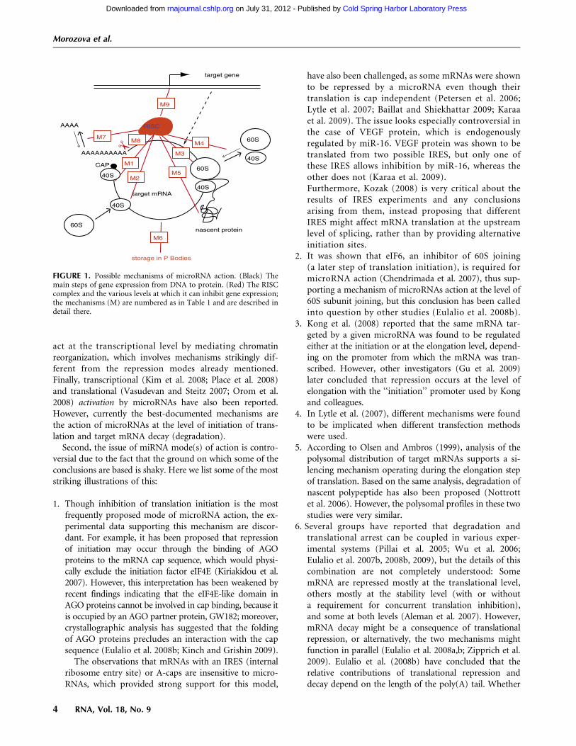
FIGURE 1. Possible mechanisms of microRNA action. (Black) Themain steps of gene expression from DNA to protein. (Red) The RISCcomplex and the various levels at which it can inhibit gene expression;the mechanisms (M) are numbered as in Table 1 and are described indetail there.
Morozova et al.
4 RNA, Vol. 18, No. 9
Cold Spring Harbor Laboratory Press on July 31, 2012 - Published by rnajournal.cshlp.orgDownloaded from
deadenylation is the cause or consequence of silencing isanother unresolved issue (Standart and Jackson 2007).
The experimental data and summarizing conclusionsabout the mechanisms of microRNA action thus may beconsidered to be highly controversial. We believe thatmathematical modeling can help to systematize the avail-able information and to suggest a computational biologytool for discriminating among all proposed mechanisms ofmiRNA action and their combinations. Using this tool,some of the contradictory experimental findings may beexplained in the framework of the unifying model ofmicroRNA action, whereas others may be discarded asbeing highly unlikely.
We suggest that ‘‘kinetic signatures’’ (i.e., characteristictime-course patterns to attain protein and mRNA steady-state levels, as well as the number of ribosomes per mRNAmolecule after microRNA application) computed for eachproposed mechanism could be a good indicator to dis-criminate between alternative mechanisms. This tool couldbe useful for proving the existence of the mechanismclaimed in a given system or for selecting between severalalternative suggested mechanisms.
Repression of protein translation by miRNA has alreadybeen the subject of mathematical modeling (Nissan andParker 2008; Zinovyev et al. 2010). However, a computa-tional tool for discriminating among all proposed mecha-nisms of miRNA action remains to be developed.
In this work we provide kinetic signatures of microRNAaction for each proposed mechanism and also for com-binations of mechanisms. To generate these kineticsignatures, we created a complete mathematical modelof microRNA action, taking into account all previouslydescribed mechanisms, and analyzed this model using themethodology developed in our previous work (Zinovyevet al. 2010), where a rigorous mathematical analysis ofdynamical behavior of the systems involving microRNAaction was performed. This analysis led us, in particular, toformulate a new hypothesis for microRNA action thatunderlies the unifying model of microRNA action presentedin this work. This new hypothesis, if validated, could rec-oncile the contradictory interpretations of existing data.
RESULTS
Complete Model based on unifying hypothesisof microRNA action
In our previous work (Zinovyev et al. 2010) we suggestedan analytical approach to study important characteristics ofmiRNA action based on a chemical kinetics mathematicalmodel considering the most important reactions involvedin protein translation. In that work, using the simplestmodel system involving only three possible mechanisms ofmicroRNA action, we worked out methods for analyzing
the system of equations corresponding to complex net-works of such reactions. As a result, we showed that byusing this method we can discriminate between threedifferent mechanisms if we have the experimental datareflecting mRNA and protein dynamics in time-courseexperiments. Additionally, we found that a given mech-anism of microRNA action can be detected experimentallyonly if it targets parameters of the dominant system (ageneralization of the concept of a rate-limiting step tocomplex networks) of the translation mechanism. This ledus to hypothesize that what appears experimentally as asingle mechanism may in fact be the result of multiplemechanisms running at the same time, but with only oneof these mechanisms eventually affecting the proteintranslation because it affects the rate-limiting step(s). Asa further development of this hypothesis, we suggest herea model in which all mechanisms of microRNA actioncoexist in a cell, while the apparent mechanism X, i.e.,that which will be detected experimentally, depends onthe kinetic rate constants of the different steps of mRNAtranslation, mRNA or protein degradation, and others, bothinternal and external to microRNA pathways. We think thatthis postulate can be considered as representing a unifyinghypothesis of microRNA action, because, if confirmed, itgives us the possibility to reconcile the apparently contra-dictory interpretations of previous results in the frameworkof a single model of microRNA action. In the Discussionsection we revisit the controversial studies cited in the Intro-duction section in the framework of our model. This analysisprovides an explanation for most of the discrepancies in theliterature, and we propose an experimental approach forvalidating the model.
The interplay between the kinetic rates of different stepsof mRNA translation and degradation depends on a setof intrinsic characteristics of the given experimental system,such as the content of target mRNA, the set of mRNA-binding proteins, the mRNA degradation rate, and otherdetails of the translational machinery. Differences in theseparameters between various experimental systems or chang-ing some of these parameters within a given experimentalsystem will result in implementation of the dominant sys-tems corresponding to different mechanisms.
Indeed, many studies underscore the importance of theintrinsic characteristics of mRNAs for the final outcome ofmiRNA action. For example, RNA-binding proteins notrelated to the miRNA pathway have been shown to have astrong influence on the regulation by miRNA (Yang et al.2003; Moore 2005; Sandberg et al. 2008; Mayr and Bartel2009).
Another argument in favor of the unifying hypothesis isthat the possibility of coexistence of two or more mecha-nisms has already been discussed and proven in the literature(Pillai et al. 2005; Valencia-Sanchez et al. 2006; Wu et al.2006; Eulalio et al. 2007b, 2008a; Leung and Sharp 2007;Filipowicz et al. 2008; Zipprich et al. 2009). At the biological
Kinetic signatures of microRNA modes of action
www.rnajournal.org 5
Cold Spring Harbor Laboratory Press on July 31, 2012 - Published by rnajournal.cshlp.orgDownloaded from

level, the molecular mechanisms enabling miRNAs to interactwith a variety of distinct proteins in order to affect varioussteps of protein synthesis remain to be explored. However, theRISC complex appears to be formed by a number of proteins,and it is conceivable that each of these proteins itself interactswith other proteins belonging to different regulatory pathways.
In the present work we apply our analytical approach to theComplete Model containing all (or most) of the proposedmechanisms of microRNA action, with the main goal toobtain a tool for discriminating between all of these mecha-nisms on the basis of the experimental kinetic data. Namely,we introduce here the idea of kinetic signatures of mechanismsof microRNA action as a tool for discriminating betweenthem. The kinetic signature of each mechanism is a chart rep-resenting experimental data on the dynamics of three measur-able biological parameters: (1) the amount of the targetmRNA, (2) the amount of the corresponding protein, and (3)the average number of ribosomes on the target mRNA (whichcould be determined from the polysomal profile). Followingour results, in those cases when only one given mechanism isresponsible for the observed result of microRNA application,these charts will be unique for each mechanism.
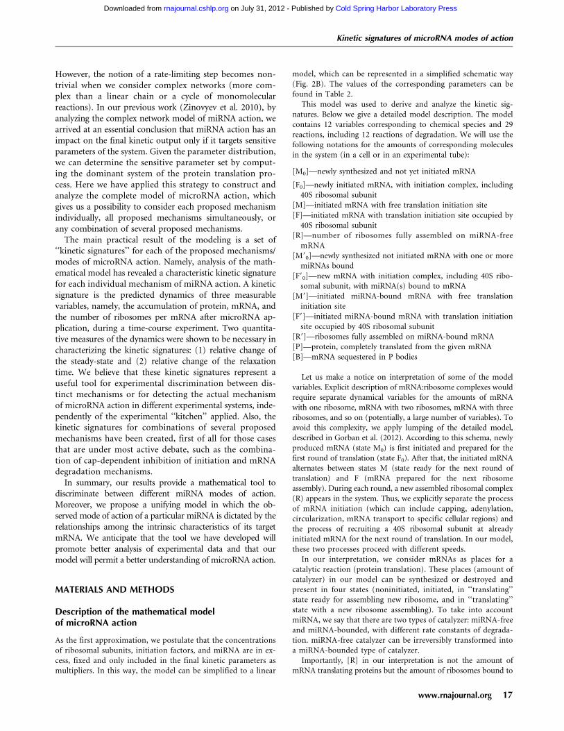
We based our Complete Model on nine distinct mech-anisms of microRNA action that have been described in theliterature; the main experimental data supporting eachproposed mechanism are summarized in Table 1, and theschematic illustration of the mechanisms are presented inFigure 1. Following our unifying hypothesis, the Com-plete Model is created as a network of reactions involvedin protein production, with nine possible mechanisms ofmicroRNA action (Fig. 2) that can potentially coexist.In the framework of this model we can model the situa-tion where any combination of proposed mechanisms ofmicroRNA action acts simultaneously. We can also studyindividual mathematical models for each proposed mech-anism, which can thus be considered as particular cases ofthe Complete Model, where only the kinetic rate constant(or several constants) corresponding to a given mechanismis changed as a result of microRNA application. For ouranalysis, we assumed that the initiation factors and ribo-somal subunits are always available in excess, and that theirconcentration is constant. This allowed us to simplify themodel to 12 chemical species and 29 reactions, thedetailed descriptions of which are given in the Materialsand Methods section. All steps of the mathematical analysisand the process of creating the charts for kinetic signaturesare also described in the Materials and Methods section.The corresponding MATLAB code for the model is pro-vided in the Supplemental File.
Determination of the kinetic rate constantsfor dynamical modeling
For simulations, we needed the numerical values of 18kinetic coefficients, which were estimated from published
reports, recapitulated in Tables 2 and 3. Although it is ob-vious that the values of the rate constants can vary consi-derably for different mRNAs, experimental data miningallowed us to make plausible assumptions for most of thekinetic rate constants used in the model. For example,mRNA half-lives vary from several minutes to >24 h, witha mean value at 10 h (Yang et al. 2003), which we selectedas the corresponding rate. It is nevertheless possible thathighly regulated mRNAs, such as most miRNA targets,have shorter half-lives. The same reasoning also applies toprotein half-lives.
Similarly, we estimated the elongation time for mRNAtranslation as 1–2 min (Hunt et al. 1969; Scornik 1974;Bergmann and Lodish 1979), even though it depends onthe mRNA length: at 10–15 aa/sec (Gilchrist and Wagner2006); 1–2 min corresponds to a mean length of 1.8–3.6 kb(Hartl and Jones 2005, page 410). Likewise, the numbers ofribosomes per mRNA molecule are highly variable, fromfour to five to more than 10 (Bergmann and Lodish 1979;Maroney et al. 2006; A Polesskaya, pers. comm.). We con-sidered six ribosomes per mRNA as being a reasonable as-sumption representing normal translation conditions. Wetherefore postulated that six initiation events occur duringa cycle of elongation, which leads to an estimate of sixinitiations/minute, and is of the same order of magnitude aswhat has been proposed previously (Bergmann and Lodish1979). All information concerning the kinetic coefficientsthat we used for our modeling is summarized in Table 2,and the corresponding values of kinetic coefficients for theMATLAB program is given in Table 3.
In addition, we provide a qualitative analytical solutionof the model, which is valid for any distribution of para-meters and can be used as a guide in analysis.
Kinetic signatures for nine proposed mechanisms(modes) of microRNA action
After introducing the kinetic parameters into the mathe-matical model and running the numerical simulations, spe-cific kinetic signatures were obtained (Fig. 3), which providea practical recipe for distinguishing between nine differentmechanisms of miRNA action by studying the dynamicalbehavior of three measurable biological parameters: theamount of target mRNA, the amount of the correspondingprotein, and the average number of ribosomes on the targetmRNA. The time courses of these three measures arequantified by the relative change of their steady state (SS)and relaxation times (RT). The relaxation time is the timebetween microRNA application to the system and stabiliza-tion of the system, meaning reaching new steady-state levelsof mRNA and protein. This parameter can be determinedfrom the kinetic signature plots (for a more detailed expla-nation, see Zinovyev et al. 2010) and the experimental data,and then compared.
Morozova et al.
6 RNA, Vol. 18, No. 9
Cold Spring Harbor Laboratory Press on July 31, 2012 - Published by rnajournal.cshlp.orgDownloaded from

Figure 3 presents kinetic signatures of particularmiRNA action mechanisms, obtained for those cases whenonly one individual mechanism is active in the biologicalsystem investigated, and at maximum, i.e., 100% effi-ciency (which means, for example, a complete block ofmRNA elongation in the presence of miRNA, for theElongation Inhibition mechanism). These signatures arealso equivalent to the situation where the effect of oneparticular mechanism is dominant over all of the others(in other words, the inhibiting effect of miRNA on therate of the target reaction is at least an order of magnitudegreater than its effect on any other reaction). All kineticparameters corresponding to the process of translation inthe absence of miRNA, as well as general kinetic parameters
corresponding to microRNA action, were taken frompublications (Table 2) and considered to be the samefor all simulations. An exception was the kinetic coeffi-cient of microRNA–mRNA binding, for which we studiedthree cases: weak, medium, and strong miRNA-bindingstrengths (Fig. 3). The time points on the x axis are givenin 1/kd units, where kd is the equilibrium dissociationconstant characterizing the degradation rate of the mRNA.The figure shows that the proper time intervals for obtainingthe kinetic signatures of a particular mRNA of interest mustbe chosen taking into account its specific half-life. Thenumbers on the y axis for mRNA and protein levels arearbitrary and depend on their steady-state levels withoutmicroRNA action.
FIGURE 2. The unifying mathematical model taking into account all nine mechanisms of miRNA action. (A) Created in Systems BiologyGraphical Notation (SBGN) standard using CellDesigner 4.1 software (Funahashi et al. 2003). (B) Simplified schematic model presentation in theassumption that ribosomal subunits, initiation factors and miRNA are present in excess and their concentrations are fixed. The description of thereaction graph is given in the Materials and Methods section.
Kinetic signatures of microRNA modes of action
www.rnajournal.org 7
Cold Spring Harbor Laboratory Press on July 31, 2012 - Published by rnajournal.cshlp.orgDownloaded from

Each kinetic signature plot on Figure 3 is supplementedwith arrow diagrams, visualizing the six numbers charac-terizing the corresponding simulation. These numbers arethe relative changes of steady state (SS) and relaxation time(RT) of the three main measurable parameters: amount ofmRNA (mRNA), amount of protein (Protein), and numberof ribosomes per mRNA (RB). One can see that the kineticsignatures of the nine mechanisms are qualitatively differ-ent, which means that alternative or controversial mecha-nisms can be reliably distinguished by using this modeling
approach. For example, the difference between microRNA-driven cap inhibition and 60S joining (both influencing theprocess of translation initiation) or between Elongation in-hibition, Ribosome drop-off, and Nascent protein degrada-tion (all influencing the post-initiation steps of translation)can be distinguished using the kinetic signatures (Fig. 3). Itis important to note that not all mechanisms can be dis-tinguished based solely on the steady-state value analysis.Some of the relaxation time relative changes should be mea-sured as well in order to distinguish, for example, Ribosome
TABLE 2. Parameters of translation and miRNA action
StepValue in absence
of microRNAValue when the step isinhibited by microRNA Explanation and references
Initiation-40S recruitment � 6 initiations/min � 1 initiation/min Six ribosomes/mRNA under normal conditions(Bergmann and Lodish 1979; Maroney et al. 2006)and one ribosome/mRNA when inhibited bymicroRNA (Pillai et al. 2005; Huang et al. 2007;Kiriakidou et al. 2007)
2 3 10�4/mole2/sec 2 3 10�5/mole2/sec
Initiation-60S recruitment <sec A few minutes Usually considered as ‘‘rapid’’ (Nissan andParker 2008). When inhibited by microRNA,it is slowed down enough for the 40S to bedetectable on the AUG (Wang et al. 2008).
1/mole2/sec 1 3 10�3/mole2/sec
Elongation and termination 1–2 min 2 or 3 times slower A mRNA is translated in 1 or 2 min (Hunt et al.1969; Scornik 1974; Bergmann and Lodish 1979).When elongation is inhibited, mRNA are still inpolysomes (Maroney et al. 2006; Nottrott et al.2006; Petersen et al. 2006).
1 3 10�2/sec 3 3 10�3/sec
Ribosome drop-off Rare 1 ribosome of each 2 Read-through rate is divided by two when miRNAis inhibiting mRNA, so one can assume thatonly half or the ribosomes arrives to the stopcodon (Petersen et al. 2006).
0 1 3 10�2/sec
Degradation (decay) of mRNA Half-life: �10 h Half-life: �2 h In the absence of miRNA (Yang et al. 2003)3 3 10�5/sec 15 3 10�5/sec In the presence of miRNA (Wu et al. 2006;
Mathonnet et al. 2007)Degradation (decay) of
translated mRNARare 1.5 3 10�5/sec Usually translated mRNAs are considered as
protected from degradation. No data fordegradation rate of translated mRNA targetedby microRNA are available, so we just assumethat this degradation is possible but occursslower than for naked mRNA.
0
mRNA cleavage — 10�3/sec As mRNA cleavage are supposed to be very quick,we assume it to be 100 times quicker thanmRNA degradation.
Degradation of nascent polypeptide Half-life: �24 h Half-life: <1 sec In presence of microRNA, nascent polypeptidecannot been experimentally observed (Pillaiet al. 2005; Nottrott et al. 2006; Petersenet al. 2006), thus it has to be very quick.
1 3 10�5/sec 100 sec
Sequestration in P-bodies — 5 3 10�2/sec The rate of influx of target mRNAs into P-bodies, witha reverse rate constant assumed to be around fivetimes lower. We also consider that mRNA canbe degraded in P-bodies (with the degradationrate for translated mRNA) (Pillai et al. 2005;Leung et al. 2006).
Transcription ;1–5 nt/sec 0 For an average transcription rate in theabsence of microRNA (Hartland Jones 2005)
10�3/sec
mRNA–microRNA binding — �10 min In vitro data (Mathonnet et al. 2007)2 3 10�3/mole/sec
Morozova et al.
8 RNA, Vol. 18, No. 9
Cold Spring Harbor Laboratory Press on July 31, 2012 - Published by rnajournal.cshlp.orgDownloaded from

Drop-Off from 60S Unit Joining Inhibition. Finally, one canobserve that some of the signature components stronglydepend in a quantitative fashion on the order of magnitudeof the miRNA-binding constant, and some are completelyinsensitive. This suggests a further computational exper-iment in which several sequences of miRNA would beutilized having different (weak, medium, tight) affinities tothe target mRNA-binding site. Observing how the dynamicsof the observable quantities vary with the binding affinity,one can distinguish the mechanisms more reliably. For
example, in the case of Cap Inhibition, the protein profileshould be more sensitive to changing miRNA affinity thanis Cotranslational protein degradation. Our modeling showsthat, given the rate of recycling of target mRNAs in P-bodies(Pillai et al. 2005; Leung et al. 2006), the influence of thissequestration on total protein level is very small and will notbe detected if we take into account sequestration alone, i.e.,without mRNA degradation within P-bodies. The influenceof this sequestration on total mRNA levels also will not bedetected due to the experimental procedure for total mRNA
TABLE 3. Reference set of parameters of the Complete Model
Kinetic rateconstant
Reference valueor interval Comment
Parameters of transcription and translation in the absence of miRNA actionkt 10�3 Transcription kinetic rate. If Transcriptional Inhibition mechanism is active then this constant
is proportionally reduced from kt (0% efficiency of the mechanism) to zero(100% efficiency of the mechanism).
k01 2 3 10�4 mRNA early initiation rate in the absence of miRNAk1 1 Rate of 40S recruitement at already translated mRNA, considered to be fast and not
rate-limitingk2 6 3 10�2 60S unit joining and assembly of the full ribosome on mRNA rate in
the absence of miRNAk3 10�2 Rate including elongation and termination of translation in the absence of miRNA. In all
simulations of translation without miRNA, we assume that k3 = k3/6, which givessix ribosomes sitting on one translated mRNA in average.
kd 10�5 mRNA degradation rate in the absence of miRNA. In all simulations of translation withoutmiRNA, we assume that kd << k1, k2, k3. Otherwise mRNA will be degraded much fasterthan it will be initiated and translated.
krd 0 Rate of ribosome drop-off. We neglect the ribosome drop-off in the absence of miRNA.kp 5 3 10�6 Rate of protein degradation in the absence of miRNA
Parameters of various mechanisms of miRNA actionkb 10�3 (strong) Rate of miRNA binging to mRNA. This rate depends on many factors
including the complementarity of miRNA sequence to the sequence of the bindingsite. We assume that depending on these factors, the rate can vary in the rangeof from several orders of magnitude. When kb << min (k1, k2, k3), we considerthe binding as weak, because it does not considerably influence the rate of translation.
10�4 (medium)10�5 (weak)
k019 [0; k01] mRNA initiation rate with miRNA. If Cap Inhibition mechanism is active then this constantcan be proportionally reduced from k1 to zero.
k19 k01 40S recruitment at already translated miRNA-bound mRNA, we do not consider thecorresponding hypothetical mechanism in the model.
k29 [0; k2] 60S unit joining and assembly of the full ribosome on mRNA rate with miRNA. If 60S UnitJoining Inhibition mechanism is active then this constant can be proportionally reducedfrom k2 to zero.
k39 [0; k3] Rate including elongation and termination of translation with miRNA. If Elongation Inhibitionmechanism is active then this constant can be proportionally reduced from k3 to zero.
kd9 [kd ; 102 3 kd] Rate of mRNA degradation with miRNA. If Decay mechanism is active then this constantcan increase 10-fold at 100% mechanism efficiency. If Cleavage mechanism is activethen this constant can increase by 100-fold.
k6s [0; 5 3 10�2] Rate of reversible capturing of mRNA to P-bodies. If the mechanism of sequestrationin P-bodies is active, this constant can be proportionally increased from zeroto k+s. The reverse rate constant k�s is assumed to be k�s = 5 3 k+s.We assume that mRNA can be degraded in P-bodies with the rate kd9.
k9rd [0; 5 3 k39] Rate of ribosome drop-off. If Ribosome Drop-Off mechanism is active,then this constant is proportionally increased from 0 to 5�k39.
kr [0; 5 3 10�5] Rate of cotranslational protein degradation catalysis. If Cotranslational Protein Degradationmechanism is active, then this constant is proportionally increased from 0 to 5 3 10�5,and the protein degradation rate is increased as kp
miRNA = kp + kr�R9.
Kinetic signatures of microRNA modes of action
www.rnajournal.org 9
Cold Spring Harbor Laboratory Press on July 31, 2012 - Published by rnajournal.cshlp.orgDownloaded from
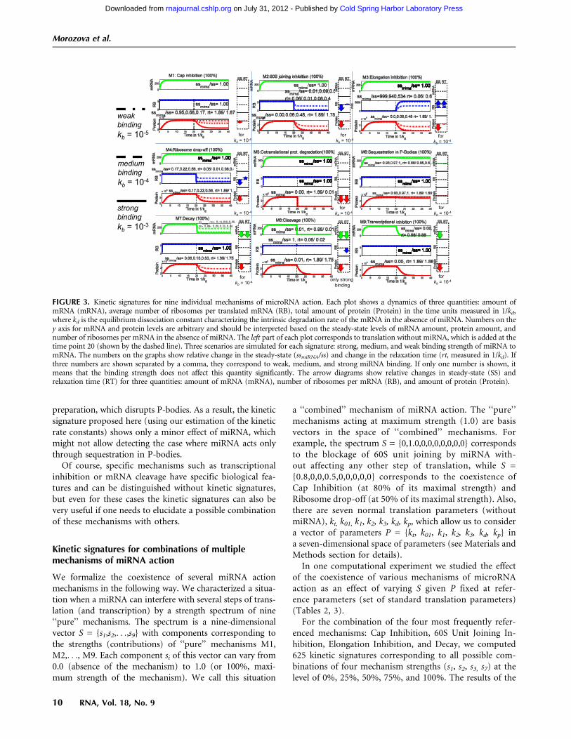
preparation, which disrupts P-bodies. As a result, the kineticsignature proposed here (using our estimation of the kineticrate constants) shows only a minor effect of miRNA, whichmight not allow detecting the case where miRNA acts onlythrough sequestration in P-bodies.
Of course, specific mechanisms such as transcriptionalinhibition or mRNA cleavage have specific biological fea-tures and can be distinguished without kinetic signatures,but even for these cases the kinetic signatures can also bevery useful if one needs to elucidate a possible combinationof these mechanisms with others.
Kinetic signatures for combinations of multiplemechanisms of miRNA action
We formalize the coexistence of several miRNA actionmechanisms in the following way. We characterized a situa-tion when a miRNA can interfere with several steps of trans-lation (and transcription) by a strength spectrum of nine‘‘pure’’ mechanisms. The spectrum is a nine-dimensionalvector S = {s1,s2,. . .,s9} with components corresponding tothe strengths (contributions) of ‘‘pure’’ mechanisms M1,M2,. . ., M9. Each component si of this vector can vary from0.0 (absence of the mechanism) to 1.0 (or 100%, maxi-mum strength of the mechanism). We call this situation
a ‘‘combined’’ mechanism of miRNA action. The ‘‘pure’’mechanisms acting at maximum strength (1.0) are basisvectors in the space of ‘‘combined’’ mechanisms. Forexample, the spectrum S = {0,1.0,0,0,0,0,0,0,0} correspondsto the blockage of 60S unit joining by miRNA with-out affecting any other step of translation, while S ={0.8,0,0,0.5,0,0,0,0,0} corresponds to the coexistence ofCap Inhibition (at 80% of its maximal strength) andRibosome drop-off (at 50% of its maximal strength). Also,there are seven normal translation parameters (withoutmiRNA), kt, k01, k1, k2, k3, kd, kp, which allow us to considera vector of parameters P = {kt, k01, k1, k2, k3, kd, kp} ina seven-dimensional space of parameters (see Materials andMethods section for details).
In one computational experiment we studied the effectof the coexistence of various mechanisms of microRNAaction as an effect of varying S given P fixed at refer-ence parameters (set of standard translation parameters)(Tables 2, 3).
For the combination of the four most frequently refer-enced mechanisms: Cap Inhibition, 60S Unit Joining In-hibition, Elongation Inhibition, and Decay, we computed625 kinetic signatures corresponding to all possible com-binations of four mechanism strengths (s1, s2, s3, s7) at thelevel of 0%, 25%, 50%, 75%, and 100%. The results of the
FIGURE 3. Kinetic signatures for nine individual mechanisms of microRNA action. Each plot shows a dynamics of three quantities: amount ofmRNA (mRNA), average number of ribosomes per translated mRNA (RB), total amount of protein (Protein) in the time units measured in 1/kd,where kd is the equilibrium dissociation constant characterizing the intrinsic degradation rate of the mRNA in the absence of miRNA. Numbers on they axis for mRNA and protein levels are arbitrary and should be interpreted based on the steady-state levels of mRNA amount, protein amount, andnumber of ribosomes per mRNA in the absence of miRNA. The left part of each plot corresponds to translation without miRNA, which is added at thetime point 20 (shown by the dashed line). Three scenarios are simulated for each signature: strong, medium, and weak binding strength of miRNA tomRNA. The numbers on the graphs show relative change in the steady-state (ssmiRNA/ss) and change in the relaxation time (rt, measured in 1/kd). Ifthree numbers are shown separated by a comma, they correspond to weak, medium, and strong miRNA binding. If only one number is shown, itmeans that the binding strength does not affect this quantity significantly. The arrow diagrams show relative changes in steady-state (SS) andrelaxation time (RT) for three quantities: amount of mRNA (mRNA), number of ribosomes per mRNA (RB), and amount of protein (Protein).
Morozova et al.
10 RNA, Vol. 18, No. 9
Cold Spring Harbor Laboratory Press on July 31, 2012 - Published by rnajournal.cshlp.orgDownloaded from
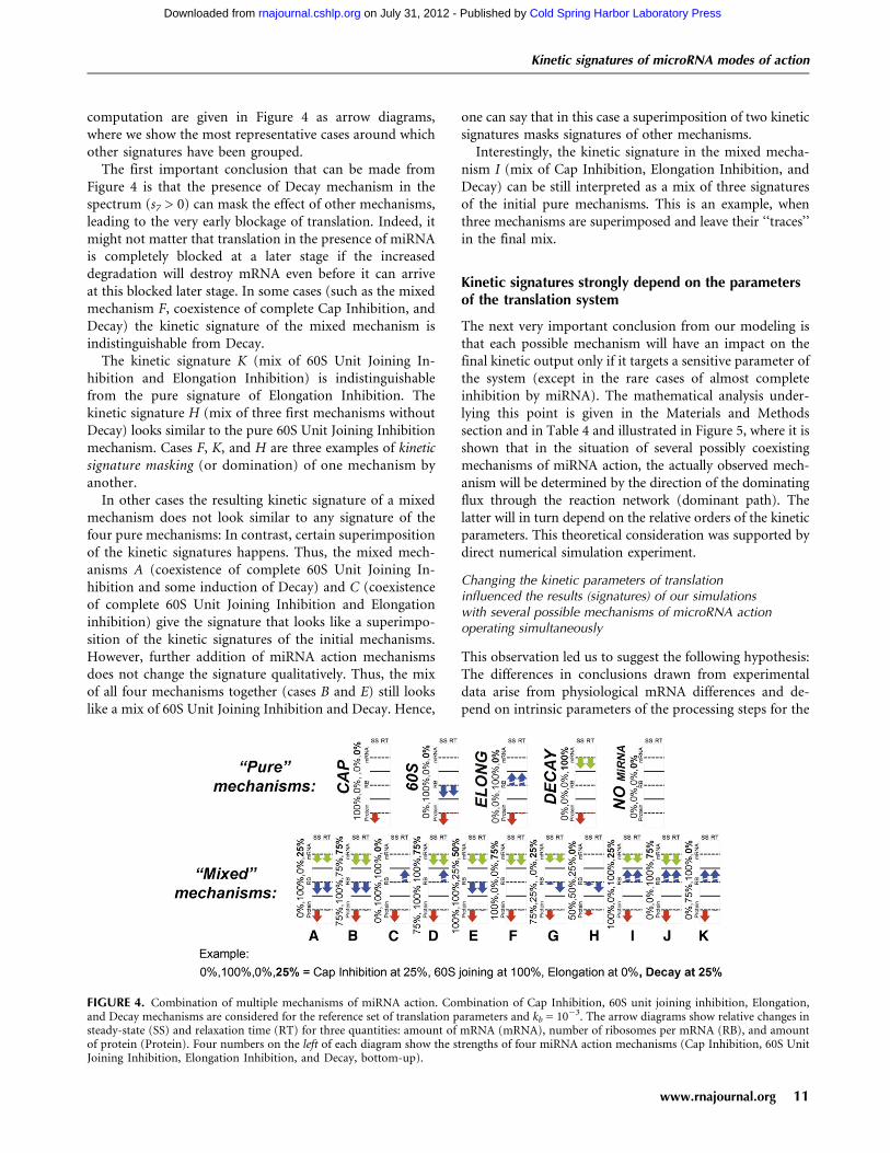
computation are given in Figure 4 as arrow diagrams,where we show the most representative cases around whichother signatures have been grouped.
The first important conclusion that can be made fromFigure 4 is that the presence of Decay mechanism in thespectrum (s7 > 0) can mask the effect of other mechanisms,leading to the very early blockage of translation. Indeed, itmight not matter that translation in the presence of miRNAis completely blocked at a later stage if the increaseddegradation will destroy mRNA even before it can arriveat this blocked later stage. In some cases (such as the mixedmechanism F, coexistence of complete Cap Inhibition, andDecay) the kinetic signature of the mixed mechanism isindistinguishable from Decay.
The kinetic signature K (mix of 60S Unit Joining In-hibition and Elongation Inhibition) is indistinguishablefrom the pure signature of Elongation Inhibition. Thekinetic signature H (mix of three first mechanisms withoutDecay) looks similar to the pure 60S Unit Joining Inhibitionmechanism. Cases F, K, and H are three examples of kineticsignature masking (or domination) of one mechanism byanother.
In other cases the resulting kinetic signature of a mixedmechanism does not look similar to any signature of thefour pure mechanisms: In contrast, certain superimpositionof the kinetic signatures happens. Thus, the mixed mech-anisms A (coexistence of complete 60S Unit Joining In-hibition and some induction of Decay) and C (coexistenceof complete 60S Unit Joining Inhibition and Elongationinhibition) give the signature that looks like a superimpo-sition of the kinetic signatures of the initial mechanisms.However, further addition of miRNA action mechanismsdoes not change the signature qualitatively. Thus, the mixof all four mechanisms together (cases B and E) still lookslike a mix of 60S Unit Joining Inhibition and Decay. Hence,
one can say that in this case a superimposition of two kineticsignatures masks signatures of other mechanisms.
Interestingly, the kinetic signature in the mixed mecha-nism I (mix of Cap Inhibition, Elongation Inhibition, andDecay) can be still interpreted as a mix of three signaturesof the initial pure mechanisms. This is an example, whenthree mechanisms are superimposed and leave their ‘‘traces’’in the final mix.
Kinetic signatures strongly depend on the parametersof the translation system
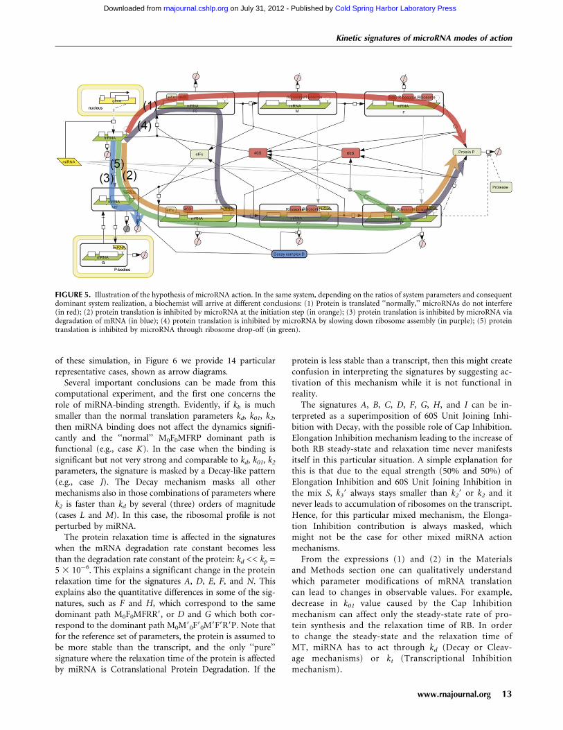
The next very important conclusion from our modeling isthat each possible mechanism will have an impact on thefinal kinetic output only if it targets a sensitive parameter ofthe system (except in the rare cases of almost completeinhibition by miRNA). The mathematical analysis under-lying this point is given in the Materials and Methodssection and in Table 4 and illustrated in Figure 5, where it isshown that in the situation of several possibly coexistingmechanisms of miRNA action, the actually observed mech-anism will be determined by the direction of the dominatingflux through the reaction network (dominant path). Thelatter will in turn depend on the relative orders of the kineticparameters. This theoretical consideration was supported bydirect numerical simulation experiment.
Changing the kinetic parameters of translationinfluenced the results (signatures) of our simulationswith several possible mechanisms of microRNA actionoperating simultaneously
This observation led us to suggest the following hypothesis:The differences in conclusions drawn from experimentaldata arise from physiological mRNA differences and de-pend on intrinsic parameters of the processing steps for the
FIGURE 4. Combination of multiple mechanisms of miRNA action. Combination of Cap Inhibition, 60S unit joining inhibition, Elongation,and Decay mechanisms are considered for the reference set of translation parameters and kb = 10�3. The arrow diagrams show relative changes insteady-state (SS) and relaxation time (RT) for three quantities: amount of mRNA (mRNA), number of ribosomes per mRNA (RB), and amountof protein (Protein). Four numbers on the left of each diagram show the strengths of four miRNA action mechanisms (Cap Inhibition, 60S UnitJoining Inhibition, Elongation Inhibition, and Decay, bottom-up).
Kinetic signatures of microRNA modes of action
www.rnajournal.org 11
Cold Spring Harbor Laboratory Press on July 31, 2012 - Published by rnajournal.cshlp.orgDownloaded from

target mRNA in question. In other words, we propose thatthe relative abundance and activity of factors not directlyrelated to the miRNA pathways, but rather to the process-ing of the specific mRNA, can be responsible for theapparent inhibition mechanism(s) detected. The effect willbe measurable only on the limiting, sensitive steps of targetmRNA processing. If two or more steps are sensitive, thentwo or more mechanisms of microRNA action will beapparent experimentally.
To illustrate this idea, we performed the second com-putational experiment, where we considered a combinationof four mechanisms (Cap Inhibition, 60S Unit Joining,Elongation Inhibition, and Decay) acting simultaneously,with the fixed strengths 50% of each one, i.e., we considerthe combined mechanism of miRNA action characterizedby the spectrum S = {0.5, 0.5, 0, 0.5, 0.5, 0, 0, 0, 0, 0}. We
study the resulting kinetic signatures of this mixed mecha-nism when the kinetic parameters of the normal translationare varied in very large intervals (five orders of magnitude),or in other words, we study the effect of varying P given S.We varied four kinetic rate constants kd, kb, k01, k2, leavingkt and kp fixed at the reference values and putting k3 = k2/6to provide a constant average number of about six ribo-somes sitting on one mRNA. The parameters took thefollowing range of values: kd2{10�3,10�4,10�5,10�6,10�7},kb,k01,k22{10�1,10�2,10�3,10�4,10�5} in all possible com-binations. From these combinations those were excludedthat violated the condition of efficient translation (not do-minated by degradation) kd << k01, k2, k3. As a result, wehave tried 440 different simulations for which we createdkinetic signatures, characterized by six numbers, as for theprevious computational experiment. To illustrate the results
TABLE 4. Dominant paths of the simplified model of microRNA action mechanisms
Dominant path Biological interpretationCorresponding miRNA-mediated
translation repression mechanism(s)
M0F0MFRP NoneNormal translation with negligible
effect of miRNA
M0M90 M1: Cap inhibitionThe dominant effect is degradation
of mRNA by miRNA.M7: DecayM8: Cleavage
M0M90B M6: Sequestration of mRNA in P-BodiesmRNA is captured in P-bodies.
M0M90F90 M2: 60S subunit joining inhibitionmRNA translation is stuck after initiation,
before the assembly of the ribosome.
M0M90F90M9F9R9 M3: Elongation inhibitionmRNA is stuck with ribosomes on it
and destroyed, or mRNA translationis prematurely aborted.
M4: Ribosome drop-off
M0M90F90M9F9R9P M1: Cap inhibitionProtein synthesis in the presence of
miRNA with low mRNA degradationM2: 60S subunit joining inhibitionM3: Elongation inhibitionM5: Cotranslational protein degradation
mechanisms
Morozova et al.
12 RNA, Vol. 18, No. 9
Cold Spring Harbor Laboratory Press on July 31, 2012 - Published by rnajournal.cshlp.orgDownloaded from
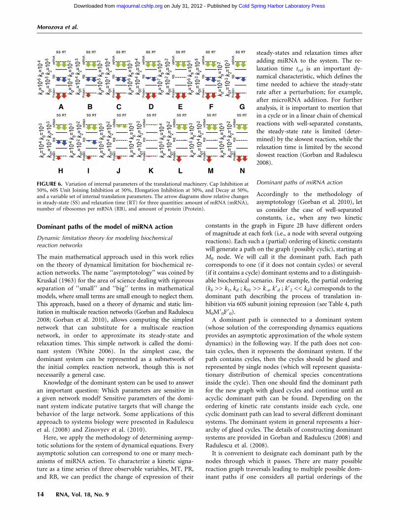
of these simulation, in Figure 6 we provide 14 particularrepresentative cases, shown as arrow diagrams.
Several important conclusions can be made from thiscomputational experiment, and the first one concerns therole of miRNA-binding strength. Evidently, if kb is muchsmaller than the normal translation parameters kd, k01, k2,then miRNA binding does not affect the dynamics signifi-cantly and the ‘‘normal’’ M0F0MFRP dominant path isfunctional (e.g., case K). In the case when the binding issignificant but not very strong and comparable to kd, k01, k2
parameters, the signature is masked by a Decay-like pattern(e.g., case J). The Decay mechanism masks all othermechanisms also in those combinations of parameters wherek2 is faster than kd by several (three) orders of magnitude(cases L and M). In this case, the ribosomal profile is notperturbed by miRNA.
The protein relaxation time is affected in the signatureswhen the mRNA degradation rate constant becomes lessthan the degradation rate constant of the protein: kd << kp =5 3 10�6. This explains a significant change in the proteinrelaxation time for the signatures A, D, E, F, and N. Thisexplains also the quantitative differences in some of the sig-natures, such as F and H, which correspond to the samedominant path M0F0MFRR9, or D and G which both cor-respond to the dominant path M0M90F90M9F9R9P. Note thatfor the reference set of parameters, the protein is assumed tobe more stable than the transcript, and the only ‘‘pure’’signature where the relaxation time of the protein is affectedby miRNA is Cotranslational Protein Degradation. If the
protein is less stable than a transcript, then this might createconfusion in interpreting the signatures by suggesting ac-tivation of this mechanism while it is not functional inreality.
The signatures A, B, C, D, F, G, H, and I can be in-terpreted as a superimposition of 60S Unit Joining Inhi-bition with Decay, with the possible role of Cap Inhibition.Elongation Inhibition mechanism leading to the increase ofboth RB steady-state and relaxation time never manifestsitself in this particular situation. A simple explanation forthis is that due to the equal strength (50% and 50%) ofElongation Inhibition and 60S Unit Joining Inhibition inthe mix S, k39 always stays smaller than k29 or k2 and itnever leads to accumulation of ribosomes on the transcript.Hence, for this particular mixed mechanism, the Elonga-tion Inhibition contribution is always masked, whichmight not be the case for other mixed miRNA actionmechanisms.
From the expressions (1) and (2) in the Materialsand Methods section one can qualitatively understandwhich parameter modifications of mRNA translationcan lead to changes in observable values. For example,decrease in k01 value caused by the Cap Inhibitionmechanism can affect only the steady-state rate of pro-tein synthesis and the relaxation time of RB. In orderto change the steady-state and the relaxation time ofMT, miRNA has to act through kd (Decay or Cleav-age mechanisms) or kt (Transcriptional Inhibitionmechanism).
FIGURE 5. Illustration of the hypothesis of microRNA action. In the same system, depending on the ratios of system parameters and consequentdominant system realization, a biochemist will arrive at different conclusions: (1) Protein is translated ‘‘normally,’’ microRNAs do not interfere(in red); (2) protein translation is inhibited by microRNA at the initiation step (in orange); (3) protein translation is inhibited by microRNA viadegradation of mRNA (in blue); (4) protein translation is inhibited by microRNA by slowing down ribosome assembly (in purple); (5) proteintranslation is inhibited by microRNA through ribosome drop-off (in green).
Kinetic signatures of microRNA modes of action
www.rnajournal.org 13
Cold Spring Harbor Laboratory Press on July 31, 2012 - Published by rnajournal.cshlp.orgDownloaded from
Dominant paths of the model of miRNA action
Dynamic limitation theory for modeling biochemicalreaction networks
The main mathematical approach used in this work relieson the theory of dynamical limitation for biochemical re-action networks. The name ‘‘asymptotology’’ was coined byKruskal (1963) for the area of science dealing with rigorousseparation of ‘‘small’’ and ‘‘big’’ terms in mathematicalmodels, where small terms are small enough to neglect them.This approach, based on a theory of dynamic and static lim-itation in multiscale reaction networks (Gorban and Radulescu2008; Gorban et al. 2010), allows computing the simplestnetwork that can substitute for a multiscale reactionnetwork, in order to approximate its steady-state andrelaxation times. This simple network is called the domi-nant system (White 2006). In the simplest case, thedominant system can be represented as a subnetwork ofthe initial complex reaction network, though this is notnecessarily a general case.
Knowledge of the dominant system can be used to answeran important question: Which parameters are sensitive ina given network model? Sensitive parameters of the domi-nant system indicate putative targets that will change thebehavior of the large network. Some applications of thisapproach to systems biology were presented in Radulescuet al. (2008) and Zinovyev et al. (2010).
Here, we apply the methodology of determining asymp-totic solutions for the system of dynamical equations. Everyasymptotic solution can correspond to one or many mech-anisms of miRNA action. To characterize a kinetic signa-ture as a time series of three observable variables, MT, PR,and RB, we can predict the change of expression of their
steady-states and relaxation times afteradding miRNA to the system. The re-laxation time trel is an important dy-namical characteristic, which defines thetime needed to achieve the steady-staterate after a perturbation; for example,after microRNA addition. For furtheranalysis, it is important to mention thatin a cycle or in a linear chain of chemicalreactions with well-separated constants,the steady-state rate is limited (deter-mined) by the slowest reaction, while therelaxation time is limited by the secondslowest reaction (Gorban and Radulescu2008).
Dominant paths of miRNA action
Accordingly to the methodology ofasymptotology (Gorban et al. 2010), letus consider the case of well-separatedconstants, i.e., when any two kinetic
constants in the graph in Figure 2B have different ordersof magnitude at each fork (i.e., a node with several outgoingreactions). Each such a (partial) ordering of kinetic constantswill generate a path on the graph (possibly cyclic), starting atM0 node. We will call it the dominant path. Each pathcorresponds to one (if it does not contain cycles) or several(if it contains a cycle) dominant systems and to a distinguish-able biochemical scenario. For example, the partial ordering(kb >> k1, kd ; k01 >> k-s, k9d ; k92 << kd) corresponds to thedominant path describing the process of translation in-hibition via 60S subunit joining repression (see Table 4, pathM0M90F90).
A dominant path is connected to a dominant system(whose solution of the corresponding dynamics equationsprovides an asymptotic approximation of the whole systemdynamics) in the following way. If the path does not con-tain cycles, then it represents the dominant system. If thepath contains cycles, then the cycles should be glued andrepresented by single nodes (which will represent quasista-tionary distribution of chemical species concentrationsinside the cycle). Then one should find the dominant pathfor the new graph with glued cycles and continue until anacyclic dominant path can be found. Depending on theordering of kinetic rate constants inside each cycle, onecyclic dominant path can lead to several different dominantsystems. The dominant system in general represents a hier-archy of glued cycles. The details of constructing dominantsystems are provided in Gorban and Radulescu (2008) andRadulescu et al. (2008).
It is convenient to designate each dominant path by thenodes through which it passes. There are many possiblereaction graph traversals leading to multiple possible dom-inant paths if one considers all partial orderings of the
FIGURE 6. Variation of internal parameters of the translational machinery. Cap Inhibition at50%, 60S Unit Joining Inhibition at 50%, Elongation Inhibition at 50%, and Decay at 50%,and a variable set of internal translation parameters. The arrow diagrams show relative changesin steady-state (SS) and relaxation time (RT) for three quantities: amount of mRNA (mRNA),number of ribosomes per mRNA (RB), and amount of protein (Protein).
Morozova et al.
14 RNA, Vol. 18, No. 9
Cold Spring Harbor Laboratory Press on July 31, 2012 - Published by rnajournal.cshlp.orgDownloaded from
constants in the reaction forks. However, some of them arebiologically nonrelevant. For example, the ordering kd >>k01 (dominant path M0) will not lead to any translation(the mRNA will be degraded before it will be initiated). Inthe same way, kd >> k2 (dominant path M0F0) will ter-minate the normal translation prematurely. Thus, we pos-tulate kd << k01, k2, k3. Also, for simplicity we assume thatbinding of miRNA to mRNA is more rapid than normalinitiation, i.e., kb >> k01, k2, k3 if there is miRNA in thesystem, and kb = 0, if not. Also we assume that k01 << k1,because k1 corresponds to recruiting 40S subunit on alreadyinitiated and translated mRNA (which we assume never berate-limiting), while k01 includes both mRNA initiation and40S subunit recruiting. This leads to six biologically rel-evant dominant paths, all of which are listed in Table 4.
Table 4 shows that the types of dynamical behavior (dom-inant paths) can be mapped onto the biologically charac-terized mechanisms of miRNA action, but this mappingis not one-to-one: Several biological mechanisms can cor-respond to one dynamical type (for example, M0M90 dom-inant path corresponds to M1, M7, and M8 biologicalmechanisms and, conversely, biological mechanism M1 cancorrespond to M0M90 or M0M90F90M9F9R9P dominantpaths).
DISCUSSION
The mode/mechanisms of microRNA action have generatedconsiderable discussion, and remain a controversial issue. Inthis work we (1) propose a unifying model of microRNAmechanisms stating that all known mechanisms can poten-tially coexist and work simultaneously; (2) create a completemathematical model of microRNA action based on thishypothesis; (3) suggest a set of ‘‘kinetic signatures’’ for in-dividual mechanisms to be used as a tool for discriminatingbetween different mechanisms. The signatures are obtainedfrom analysis of this mathematical model in the particularcase where each mechanism is considered as the only oneacting in the system. It is important to note that, thoughtightly connected, these three parts of the study have in-dependent meaning, significance, and novelty.
It is of utmost significance that the proposed unifyinghypothesis of microRNA action explains how apparentlycontradictory and/or controversial conclusions about themechanism(s) of microRNA action in different experimentalsystems/approaches can be reconciled. Below, we will dem-onstrate several examples of such reconciliation of contro-versial interpretations in the light of this hypothesis, usingpublications mentioned in the Introduction section.
The proposed unifying hypothesis of miRNA modes ofaction postulates that all (or many) possible modes ofmicroRNA action may coexist in the cell and operate simul-taneously, and that the observed mode of miRNA action in aparticular biological system depends on a set of intrinsicparameters of this system that influence the interplay be-
tween the kinetic rates of the different steps of mRNAtranslation, mRNA or protein degradation, and others.These parameters/factors are not necessarily related tomiRNA pathways, but, in the first place, intrinsically de-termine the sensitive parameters of the system and can de-pend on the characteristics of the target mRNA under study(e.g., mRNA content, mRNA-binding proteins, specialmarks of mRNA processing, mRNA degradation rate) andthe details of its translation.
The following examples show how changes in the kineticrates of different steps of mRNA translation may lead todifferences in the final interpretation of the data, resultingin different conclusions about the microRNA mode of ac-tion, and how this depends on the relative abundance and/oractivity of a set of factors intrinsic to any given biologicalexperimental system.
First of all, it can be noted that in most of the studiesshowing initiation inhibition, in vitro-transcribed mRNAs(transfected into cells or studied directly in vitro) were used.In contrast, almost all data supporting elongation inhibitionwere obtained in living cells (in vivo) (Humphreys et al.2005; Pillai et al. 2005; Kiriakidou et al. 2007; Mathonnetet al. 2007; Thermann and Hentze 2007; Wang et al. 2008),with only one very specific exception (Lytle et al. 2007).
In the framework of our model, the first possibleexplanation of this experimental bias could be, for example,the influence of splicing marks that are attached to mRNAin vivo during its processing in the nucleus (‘‘mRNA nuclearhistory’’). The process of mRNA splicing leaves proteinmarks on mRNA, which promotes the first round of trans-lation at the initiation step (Le Hir and Seraphin 2008;Moore and Proudfoot 2009). Though these marks are dis-sociated during the first round of translation, speeding upthe initial initiations during the first round of translationcan lead, in the ‘‘closed-loop’’ model, to increasing effi-ciency of reinitiation (Kapp and Lorsch 2004). This couldexplain higher initiation rates on intron-containing mRNAin vivo, when elongation becomes a critical step. Incontrast, in vitro-transcribed mRNAs lack splicing marks,resulting in a decreased initiation rate, which becomeslimiting.
Another very important point explaining the contradic-tory findings is that the elongation efficiency depends oncodon usage. In particular, we observe that microRNAhas been reported to act on initiation steps when codonusage is optimized for human translation (Pillai et al. 2004;Kiriakidou et al. 2007), whereas with nonoptimized codons,microRNA was found to act on elongation (Nottrott et al.2006, Petersen et al. 2006; Lytle et al. 2007; Gu et al. 2009).This means that characteristics of mRNAs that decreaseelongation rates cause elongation to become the limiting stepin translation, so that interference with elongation becomesthe mode of microRNA action that is detected.
Finally, the most trivial explanation for discrepanciesbetween in vitro and in vivo studies is that under in vitro
Kinetic signatures of microRNA modes of action
www.rnajournal.org 15
Cold Spring Harbor Laboratory Press on July 31, 2012 - Published by rnajournal.cshlp.orgDownloaded from
conditions, initiation is highly dependent on the concen-tration of initiation factors, thus making initiation thelimiting step, with the result that it is detected as the ap-parent mode of microRNA action in in vitro studies.
Next, reviewing the publications reporting a mechanismof translational inhibition via the 40S-cap, one can notethat most of the experiments proving this mechanism weredone in systems with a very slow, critical initiation rate(A-capped mRNA or mRNA with IRES). For this reason,the correct interpretation of results showing that cap mod-ifications are refractory to miRNA action may be that capmodifications (A-capped mRNA or mRNA with IRES) leadto such a strong, critical, decrease of the initiation rate, thatthe system becomes insensitive to any additional decreasecaused by miRNA action.
As all IRES are different, some of them can have initiationefficiencies similar to ‘‘canonical’’ initiation, whereas othersmay be less efficient. This could explain, in the frameworkof our model, the data concerning two IRES in the samegene (VEGF), one of which allows inhibition of this geneby miR16, whereas the other does not (Karaa et al. 2009).Additionally, most of the studies showing IRES-drivenmRNAs to be refractory to microRNAs were carried outeither in vitro (Mathonnet et al. 2007) or using in vitro-transcribed mRNAs transfected into cells (Humphreys et al.2005; Pillai et al. 2005; Kiriakidou et al. 2007), whereas thestudies showing IRES-driven mRNAs to be repressed bymiRNAs were carried out with mRNAs transcribed in situ,i.e., inside cells (Petersen et al. 2006; Karaa et al. 2009). Thus,the difference might come from the status of the targetmRNA, or from the absence in in vitro systems of the spe-cific factors influencing (increasing or decreasing) theefficiency of cap initiation or other steps, thus conditioningthe final outcome.
The proposed unifying hypothesis also explains the ob-servations that the same microRNA apparently uses dis-tinct mechanisms on different targets (e.g., for let7, Pillaiet al. 2005; Maroney et al. 2006; Chendrimada et al. 2007;Mathonet et al. 2007; Wakiyama et al. 2007; for CXCR4microRNA, Humphreys et al. 2005; Petersen et al. 2006;Wang et al. 2006, 2008; for miR16, Huang et al. 2007; Karaaet al. 2009; for miR122, Jopling et al. 2008); and that thestatus of the cell affects the final observable mode ofmiRNA action (Bhattacharyya et al. 2006; Leung et al.2006; Valencia-Sanchez et al. 2006).
Finally, our hypothesis predicts that the different exper-imental procedures used might lead to ‘‘pre-existing’’ dif-ferences in the rates of translation, degradation, and otherprocesses essential for microRNA action. This would im-ply different parameter sensitivities and, hence, different‘‘observed’’ mechanisms. This analysis is supported by thearticle by Kong et al. (2008), who showed that the samemicroRNA can act on the initiation or the elongation stepaccording to the promoter used for its transcription. Thisphenomenon, not explained in the original article, could
be explained by our model. For a reporter mRNA withintron(s) and nonoptimized codons, different promotersmay ‘‘promote’’ the accumulation of different ‘‘mRNA nu-clear history’’ marks (e.g., splicing marks, or possibly othermodifications), or lead to different amounts of mRNA beingtranscribed, thus changing the whole set of critical param-eters of the system in such a way that the critical step appearsto be initiation in one case, and elongation in the other.
Also, the report about the dependence of microRNAmode of action on the experimental procedure used for thetransfection (Lytle et al. 2007) becomes understandable inthe framework of the unifying model. The transfectionprocedure is likely to influence the association of the tar-get mRNA with mRNA-binding proteins, which, in turn,changes the sensitive parameters of the system and, hence,the final outcome of microRNA action.
For experimental validation of the unifying modelhypothesis, we propose the design of an experiment forstudying the influence of codon frequency on detection ofthe mechanism of microRNA action. It is well known thatelongation efficiency is dependent on the codons used:Some tRNAs are more frequent than others in the cell, andtherefore the corresponding codons will be translatedfaster. mRNAs enriched in ‘‘frequent codons’’ will conse-quently be translated faster than mRNAs enriched in ‘‘rarecodons.’’ We therefore propose the construction of twomRNA reporters with identical microRNA-binding sites,promoters, introns, 59UTR, 39UTR, and protein products.The only difference between the two reporters would bethe codon usage: one would be enriched in rare codons,whereas the other would be optimized for human trans-lation. We predict that in this case, elongation inhibitionwill be observed for the ‘‘optimized codons’’ mRNA, andinitiation inhibition for the ‘‘rare codons’’ mRNA. Accord-ing to our model, in those systems for which the initiationinhibition mechanism has already been proved, the de-creased elongation efficiency (in the case of ‘‘rare codons’’)should convert the initially observed initiation inhibitionmode to an elongation inhibition mode. Inversely, any in-crease in elongation efficiency (in the case of ‘‘optimizedcodons’’) should favor detection of an initiation inhibitionmechanism.
The mathematical model developed and analyzed in thiswork presents a set of new approaches for analysis of com-plex systems. The mathematical approach we have developedhere for analysis of the complete model of microRNA ac-tion is based on the general theory of dynamical limitation(Gorban and Radulescu 2008) and uses the notion ofa dominant dynamical system, itself a generalization of therate-limiting step concept to complex networks (Gorban andRadulescu 2008; Gorban et al. 2010; Zinovyev et al. 2010).The idea that microRNA action will not have a visibleimpact on inhibition of translational initiation unless this isthe rate-limiting step in protein translation has already beenmentioned in the work by Nissan and Parker (2008).
Morozova et al.
16 RNA, Vol. 18, No. 9
Cold Spring Harbor Laboratory Press on July 31, 2012 - Published by rnajournal.cshlp.orgDownloaded from
However, the notion of a rate-limiting step becomes non-trivial when we consider complex networks (more com-plex than a linear chain or a cycle of monomolecularreactions). In our previous work (Zinovyev et al. 2010), byanalyzing the complex network model of miRNA action, wearrived at an essential conclusion that miRNA action has animpact on the final kinetic output only if it targets sensitiveparameters of the system. Given the parameter distribution,we can determine the sensitive parameter set by comput-ing the dominant system of the protein translation pro-cess. Here we have applied this strategy to construct andanalyze the complete model of microRNA action, whichgives us a possibility to consider each proposed mechanismindividually, all proposed mechanisms simultaneously, orany combination of several proposed mechanisms.
The main practical result of the modeling is a set of‘‘kinetic signatures’’ for each of the proposed mechanisms/modes of microRNA action. Namely, analysis of the math-ematical model has revealed a characteristic kinetic signaturefor each individual mechanism of miRNA action. A kineticsignature is the predicted dynamics of three measurablevariables, namely, the accumulation of protein, mRNA, andthe number of ribosomes per mRNA after microRNA ap-plication, during a time-course experiment. Two quantita-tive measures of the dynamics were shown to be necessary incharacterizing the kinetic signatures: (1) relative change ofthe steady-state and (2) relative change of the relaxationtime. We believe that these kinetic signatures represent auseful tool for experimental discrimination between dis-tinct mechanisms or for detecting the actual mechanismof microRNA action in different experimental systems, inde-pendently of the experimental ‘‘kitchen’’ applied. Also, thekinetic signatures for combinations of several proposedmechanisms have been created, first of all for those casesthat are under most active debate, such as the combina-tion of cap-dependent inhibition of initiation and mRNAdegradation mechanisms.
In summary, our results provide a mathematical tool todiscriminate between different miRNA modes of action.Moreover, we propose a unifying model in which the ob-served mode of action of a particular miRNA is dictated by therelationships among the intrinsic characteristics of its targetmRNA. We anticipate that the tool we have developed willpromote better analysis of experimental data and that ourmodel will permit a better understanding of microRNA action.
MATERIALS AND METHODS
Description of the mathematical modelof microRNA action
As the first approximation, we postulate that the concentrationsof ribosomal subunits, initiation factors, and miRNA are in ex-cess, fixed and only included in the final kinetic parameters asmultipliers. In this way, the model can be simplified to a linear
model, which can be represented in a simplified schematic way(Fig. 2B). The values of the corresponding parameters can befound in Table 2.
This model was used to derive and analyze the kinetic sig-natures. Below we give a detailed model description. The modelcontains 12 variables corresponding to chemical species and 29reactions, including 12 reactions of degradation. We will use thefollowing notations for the amounts of corresponding moleculesin the system (in a cell or in an experimental tube):
[M0]—newly synthesized and not yet initiated mRNA
[F0]—newly initiated mRNA, with initiation complex, including40S ribosomal subunit
[M]—initiated mRNA with free translation initiation site[F]—initiated mRNA with translation initiation site occupied by
40S ribosomal subunit[R]—number of ribosomes fully assembled on miRNA-free
mRNA[M90]—newly synthesized not initiated mRNA with one or more
miRNAs bound[F90]—new mRNA with initiation complex, including 40S ribo-
somal subunit, with miRNA(s) bound to mRNA[M9]—initiated miRNA-bound mRNA with free translation
initiation site[F9]—initiated miRNA-bound mRNA with translation initiation
site occupied by 40S ribosomal subunit[R9]—ribosomes fully assembled on miRNA-bound mRNA[P]—protein, completely translated from the given mRNA[B]—mRNA sequestered in P bodies
Let us make a notice on interpretation of some of the modelvariables. Explicit description of mRNA:ribosome complexes wouldrequire separate dynamical variables for the amounts of mRNAwith one ribosome, mRNA with two ribosomes, mRNA with threeribosomes, and so on (potentially, a large number of variables). Toavoid this complexity, we apply lumping of the detailed model,described in Gorban et al. (2012). According to this schema, newlyproduced mRNA (state M0) is first initiated and prepared for thefirst round of translation (state F0). After that, the initiated mRNAalternates between states M (state ready for the next round oftranslation) and F (mRNA prepared for the next ribosomeassembly). During each round, a new assembled ribosomal complex(R) appears in the system. Thus, we explicitly separate the processof mRNA initiation (which can include capping, adenylation,circularization, mRNA transport to specific cellular regions) andthe process of recruiting a 40S ribosomal subunit at alreadyinitiated mRNA for the next round of translation. In our model,these two processes proceed with different speeds.
In our interpretation, we consider mRNAs as places for acatalytic reaction (protein translation). These places (amount ofcatalyzer) in our model can be synthesized or destroyed andpresent in four states (noninitiated, initiated, in ‘‘translating’’state ready for assembling new ribosome, and in ‘‘translating’’state with a new ribosome assembling). To take into accountmiRNA, we say that there are two types of catalyzer: miRNA-freeand miRNA-bounded, with different rate constants of degrada-tion. miRNA-free catalyzer can be irreversibly transformed intoa miRNA-bounded type of catalyzer.
Importantly, [R] in our interpretation is not the amount ofmRNA translating proteins but the amount of ribosomes bound to
Kinetic signatures of microRNA modes of action
www.rnajournal.org 17
Cold Spring Harbor Laboratory Press on July 31, 2012 - Published by rnajournal.cshlp.orgDownloaded from

mRNA and translating proteins, i.e., the number of sites where thecatalysis takes place. Dividing the number of these sites by theamount of the catalyzer in the initiated state [M] + [F] givesthe average number of ribosomes per mRNA, which we denoteas [RB].
The definition of the kinetic rate constants used further in thestudy is as follows:1. null / M0, the free mRNA is transcribed in the system with
the rate constant kt
2. M0 / F0, assembly of initiation complex and 40S ribosomalsubunit with mRNA occurs with the rate constant k01
3. F0 / M+R, assembly of the first ribosome on the initiationsite with the rate constant k2
4. M / F, initiation of the translation (recruitment of 40Ssubunit) on already translated mRNA, with the rate con-stant k1
5. F / M + R, assembly of full ribosome (S80) on mRNA occurswith the rate constant k2
6. R / P, translation of the protein with consequent release ofribosomes occurs with the rate constant k3
7. R / null, degradation of mRNA leads to release of ribo-somes with the rate constant kd, same reaction describespremature ribosome drop-off from mRNA with the rateconstant krd
We will assume that the process of microRNA binding tomRNA can occur at various stages of translation and that its ratekb will be the same in each of the following reactions:
8. M0 / M09
9. F0 / F09
10. M / M9
11. F / F9
12. R / R9
The same way we will assume that the rate of degradation ofmRNA not driven by microRNA action (kd) can be considered asthe same one at all stages of translation:
13. M0 / null14. F0 / null15. M / null16. F / null
The degradation rate of mRNA bound to microRNA couldoccur with or without direct action of microRNA on itsdegradation. For the beginning we will assume that this rateconstant (kd9) is different from the free mRNA degradation and isthe same one for all stages of translation:
17. M90 / null18. F90 / null19. M9 / null20. F9 / null21. R9 / null
Next, we assume that the reaction corresponding to theassembly of initiation complex and 40S ribosomal subunit withmRNA in the presence of miRNA (M90 / F90) will occur with therate constant k019.
22. M90 / F90
Recruitment of 40S subunit on already translated miRNA-bound mRNA occurs with the rate constant k19:
23. M9 / F9
Reactions of assembly of the full ribosome (S80) on mRNA inthe presence of microRNA occur with the rate constant k29:
24. F90 / M9+R25. F9 / M9+R
The rate of protein production in the case of microRNA actionis described by the following reaction:
26. R9 / P, with the rate constant k39
27. R9 / null, describes possible mechanism of ribosomal drop-off (without protein production), with the rate constant krd9,and mRNA degradation with ribosome release, with the rateconstant kd9.
Reactions 26 and 27 describe the reverse process of mRNAsequestration in P-bodies, with rates k+s and k�s correspondingly:
28. M09 / B,29. B / M09
The mRNA in P-bodies is degraded with specific rate kbd9:
30. B / null31. P+R9 / null, the rate of protein degradation by microRNA-
independent mechanisms is kp, while it can be increased in thepresence of miRNA by kr 3 R9.
The system of equations dx/dt = K0 + Kx (where x is the vectorof 12 dynamic variables, K is the kinetic matrix, and K0 is the vectorof production with only one non-zero component correspondingto the transcriptional synthesis of mRNA) has the following form:
d½M0�dt
= kt � ðkd + k01 + kbÞ½M0�
d½F0�dt
= k01½M0� � ðkd + k2 + kbÞ½F0�
d½M�dt
= k2ð½F0�+ ½F�Þ � ðkd + k1 + kbÞ½M�
d½F�dt
= k1½M� � ðkd + k2 + kbÞ½F�
d½R�dt
= k2ð½F0�+ ½F�Þ � ðkd + krd + k3 + kbÞ½R�
d½M90�dt
= kb½M0� � ðk9d + k901Þ½M90� � ðk + s½M� � k�s½B�Þ
d½F90�dt
= kb½F0�+ k901½M90� � ðk9d + k92Þ½F90�
d½M9�dt
= kb½M�+ k92ð½F90�+ ½F9�Þ � ðk9d + k91Þ½M9�
d½F0�dt
= kb½F�+ k91½M� � ðk9d + k92Þ½F�
d½R9�dt
= kb½R�+ k92ð½F0�+ ½F�Þ � ðk9d + k9rd + k93Þ½R�
d½P�dt
= k3½R�+ k93½R0� � ðkp + kr½R9�Þ½P�
d½B�dt
= k + s½M9� � k�s½B� � kbd½B�
8>>>>>>>>>>>>>>>>>>>>>>>>>>>>>>>>>>>>>>>>>>>>><>>>>>>>>>>>>>>>>>>>>>>>>>>>>>>>>>>>>>>>>>>>>>:
Morozova et al.
18 RNA, Vol. 18, No. 9
Cold Spring Harbor Laboratory Press on July 31, 2012 - Published by rnajournal.cshlp.orgDownloaded from
For this simplified linear model we need to define theproper values for 18 coefficients corresponding to the rates ofreactions.
The dynamical variables that can be observed and measured inthe experiment are
Total amount of mRNA : MT = M0 + F0 + M + F + M00 + F0
0
+ M0 + F0 + B
Total amount of protein : PR = P
Average number of ribosomes; translating one mRNA :
RB = ðR + R0Þ=ðM + M0 + F + F0Þ
These expressions were used to define the kinetic signatures.First, the solution of model equations and expression for [MT],
[PR], [RB] for the trivial case without miRNA in the system wereobtained. This can be modeled by putting to zero the bindingconstant kb = 0.
For this case the steady-state values for the measurable quan-tities are
MTSS =kt
kd; RBSS =
k2 + kd
k3 + kd + krd;
PTSS =k3
kp
kt
kd
k01k2
ðk01 + kdÞðk3 + kdÞð1Þ
and the relaxation times are
MTRT =1
kd; RBRT =
1
minðk01 + kd; k2 + kd; k3 + kd + krdÞ;
PTRT =1
minðkd; kpÞ; ð2Þ
where we have assumed that k1 >> k01, k2, k3.These formulas allow qualitative understanding of the effect of
miRNA on various steps of translation and the correspondingkinetic signatures. They can also help to decipher experimentallyobserved kinetic signatures when multiple mechanisms arepresent simultaneously and the translation parameters are notknown. The exact recipe for doing this will be a subject of ourfuture work.
Simulating the kinetic signatures
The mathematical model of miRNA-mediated translation regula-tion is encoded in MATLAB’s SBTOOLBOX and provided as aSupplemental File. The corresponding values of kinetic coefficientsfor the MATLAB program are given in Table 3. The MATLAB codeused to draw and classify kinetic signatures is available on demandfrom the investigators.
The simulation for the kinetic signatures was performed in thefollowing way:
(1) First, the system was simulated from zero initial conditionswithout the presence of miRNA (kb = 0) in the time interval
[0; 20/kd]. The steady-state and relaxation time values for MT,RB, and PR values were estimated from the simulation.
(2) The miRNA-binding constant was changed to the correspond-ing value and the simulation was continued from the steady-state obtained before in the time interval [20/kd; 40/kd]. Newsteady-state and relaxation time values were estimated fromthe simulation.
The model includes a vector of parameters P = {kt, k01, k1, k2,k3, kd, kp} and of a strength spectrum S = {s1,s2,. . .,s9}, which canvary. Each computational experiment is defined by the corre-sponding vectors P and S, binding constant for miRNA (kb), andthe rest of the model parameters are computed using thefollowing formulas:
M1 Cap Inhibitionð Þ: k901: = 1�s1ð Þ � k01;
M2 60S Unit Joining Inhibitionð Þ: k92: = 1�s2ð Þ � k2;
M3 Elongation Inhibitionð Þ: k93: = 1�s3ð Þ � k3;
M4 Ribosome Drop�Offð Þ: krd : = 5 � s4 � k3;
M5ðCo� translational protein
degradationÞ: kr : = s5 � k refð Þr :
M6 Sequestration in P� bodiesð Þ: k + s: = 5 � s6 � k refð Þs ;
k�s = s6 � k refð Þs ;
M7 Decay of mRNAð Þ: k9d: = 1 + 9 � s7ð Þ � kd;
M8 Cleavage of mRNAð Þ: k9d: = 1 + 99 � s8ð Þ � kd;
kb: = 1 + 99 � s8ð Þ � kb
M9ðTranscriptional InhibitionÞ: kt : = 1�s9ð Þ � kt ;
The result of the simulation is a kinetic signature for a mixedmechanism of miRNA action, characterized by six numbers:relative changes of the steady states MTSS =
MTssmiRNA
MTssno miRNA
,
RBSS =RBss
miRNA
RBssno miRNA
, PRSS =PRss
miRNA
PRssno miRNA
and relative changes of relaxation
times MTRT =MTRT
miRNA
MTRTno miRNA
, RBRT =RBRT
miRNA
RBRTno miRNA
, PRRT =PRRT
miRNA
PRRTno miRNA
.
SUPPLEMENTAL MATERIAL
Supplemental material is available for this article.
ACKNOWLEDGMENTS
This work was supported by a grant from the European Commis-sion Sixth Framework Programme (Integrated Project SIROCCO,contract number LSHG-CT-2006-037900) to A.H.B., and from theAgence Nationale de la Recherche (project ANR-08-SYSC-003CALAMAR) and the Projet Incitatif Collaboratif ‘‘Bioinfor-matics and Biostatistics of Cancer’’ to Institut Curie. A.Z. isa member of the team ‘‘Systems Biology of Cancer,’’ team labeledby the Ligue Nationale Contre le Cancer.
Received January 6, 2012; accepted June 4, 2012.
REFERENCES
Aleman LM, Doench J, Sharp PA. 2007. Comparison of siRNA-induced off-target RNA and protein effects. RNA 13: 385–395.
Kinetic signatures of microRNA modes of action
www.rnajournal.org 19
Cold Spring Harbor Laboratory Press on July 31, 2012 - Published by rnajournal.cshlp.orgDownloaded from

Baek D, Villen J, Shin C, Camargo FD, Gygi SP, Bartel DP. 2008. Theimpact of microRNAs on protein output. Nature 455: 64–71.
Bagga S, Bracht J, Hunter S, Massirer K, Holtz J, Eachus R, PasquinelliAE. 2005. Regulation by let-7 and lin-4 miRNAs results in targetmRNA degradation. Cell 122: 553–563.
Baillat D, Shiekhattar R. 2009. Functional dissection of the humanTNRC6 (GW182-related) family of proteins. Mol Cell Biol 29:4144–4155.
Bartel DP. 2009. MicroRNAs: Target recognition and regulatoryfunctions. Cell 136: 215–233.
Behm-Ansmant I, Rehwinkel J, Doerks T, Stark A, Bork P, IzaurraldeE. 2006. mRNA degradation by miRNAs and GW182 requiresboth CCR4:NOT deadenylase and DCP1:DCP2 decapping com-plexes. Genes Dev 20: 1885–1898.
Bergmann JE, Lodish HF. 1979. A kinetic model of protein synthesis.Application to hemoglobin synthesis and translational control. JBiol Chem 254: 11927–11937.
Bhattacharyya SN, Habermacher R, Martine U, Closs EI, FilipowiczW. 2006. Relief of microRNA-mediated translational repression inhuman cells subjected to stress. Cell 125: 1111–1124.
Carthew RW, Sontheimer EJ. 2009. Origins and mechanisms ofmiRNAs and siRNAs. Cell 136: 642–655.
Chekulaeva M, Filipowicz W. 2009. Mechanisms of miRNA-mediatedpost-transcriptional regulation in animal cells. Curr Opin Cell Biol21: 452–460.
Chendrimada TP, Finn KJ, Ji X, Baillat D, Gregory RI, Liebhaber SA,Pasquinelli AE, Shiekhattar R. 2007. MicroRNA silencing throughRISC recruitment of eIF6. Nature 447: 823–828.
Coller J, Parker R. 2004. Eukaryotic mRNA decapping. Annu RevBiochem 73: 861–890.
Esquela-Kerscher A, Slack FJ. 2006. Oncomirs—microRNAs witha role in cancer. Nat Rev Cancer 6: 259–269.
Eulalio A, Behm-Ansmant I, Schweizer D, Izaurralde E. 2007a. P-bodyformation is a consequence, not the cause, of RNA-mediated genesilencing. Mol Cell Biol 27: 3970–3981.
Eulalio A, Rehwinkel J, Stricker M, Huntzinger E, Yang SF, Doerks T,Dorner S, Bork P, Boutros M, Izaurralde E. 2007b. Target-specificrequirements for enhancers of decapping in miRNA-mediatedgene silencing. Genes Dev 21: 2558–2570.
Eulalio A, Huntzinger E, Izaurralde E. 2008a. Getting to the root ofmiRNA-mediated gene silencing. Cell 132: 9–14.
Eulalio A, Huntzinger E, Izaurralde E. 2008b. GW182 interaction withArgonaute is essential for miRNA-mediated translational repres-sion and mRNA decay. Nat Struct Mol Biol 15: 346–353.
Eulalio A, Huntzinger E, Nishihara T, Rehwinkel J, Fauser M,Izaurralde E. 2009. Deadenylation is a widespread effect of miRNAregulation. RNA 15: 21–32.
Farazi TA, Juranek SA, Tuschl T. 2008. The growing catalog of smallRNAs and their association with distinct Argonaute/Piwi familymembers. Development 135: 1201–1214.
Filipowicz W, Bhattacharyya SN, Sonenberg N. 2008. Mechanisms ofpost-transcriptional regulation by microRNAs: Are the answers insight? Nat Rev Genet 9: 102–114.
Funahashi A, Tanimura N, Morohashi M, Kitano H. 2003. CellDe-signer: A process diagram editor for gene-regulatory and bio-chemical networks. BIOSILICO 1: 159–162.
Ghildiyal M, Zamore PD. 2009. Small silencing RNAs: An expandinguniverse. Nat Rev Genet 10: 94–108.
Gilchrist MA, Wagner A. 2006. A model of protein translationincluding codon bias, nonsense errors, and ribosome recycling.J Theor Biol 239: 417–434.
Gorban AN, Radulescu O. 2008. Dynamic and static limitation inmultiscale reaction networks, revisited. Adv. Chem. Eng. 34: 103–173.
Gorban AN, Radulescu O, Zinovyev A. 2010. Asymptotology ofchemical reaction networks. Chem. Eng. Sci. 65: 2310–2324.
Gorban AN, Zinovyev A, Morozova N, Harel-Bellan A. 2012.Modeling coupled transcription, translation and degradation andmiRNA-based regulation of this process. E-print arXiv:1204.5941[q-bio.MN].
Gu S, Jin L, Zhang F, Sarnow P, Kay MA. 2009. Biological basis forrestriction of microRNA targets to the 39 untranslated region inmammalian mRNAs. Nat Struct Mol Biol 16: 144–150.
Guo H, Ingolia NT, Weissman JS, Bartel DP. 2010. MammalianmicroRNAs predominantly act to decrease target mRNA levels.Nature 466: 835–840.
Hartl DL, Jones EW. 2005. Genetics: Analysis of genes and genomes, 6thed. Jones & Bartlett Publishers, Burlington, MA.
Hendrickson DG, Hogan DJ, McCullough HL, Myers JW, HerschlagD, Ferrell JE, Brown PO. 2009. Concordant regulation of trans-lation and mRNA abundance for hundreds of targets of a humanmicroRNA. PLoS Biol 7: e1000238. doi: 10.1371/journal.pbio.1000238.
Huang J, Liang Z, Yang B, Tian H, Ma J, Zhang H. 2007. Derepression ofmicroRNA-mediated protein translation inhibition by apolipoproteinB mRNA-editing enzyme catalytic polypeptide-like 3G (APOBEC3G)and its family members. J Biol Chem 282: 33632–33640.
Humphreys DT, Westman BJ, Martin DI, Preiss T. 2005. MicroRNAscontrol translation initiation by inhibiting eukaryotic initiationfactor 4E/cap and poly(A) tail function. Proc Natl Acad Sci 102:16961–16966.
Hunt T, Hunter T, Munro A. 1969. Control of haemoglobin synthesis:Rate of translation of the messenger RNA for the a and b chains.J Mol Biol 43: 123–133.
Hutvagner G, Zamore PD. 2002. A microRNA in a multiple-turnoverRNAi enzyme complex. Science 297: 2056–2060.
Iwasaki S, Kawamata T, Tomari Y. 2009. Drosophila Argonaute1 andArgonaute2 employ distinct mechanisms for translational repres-sion. Mol Cell 34: 58–67.
Jackson RJ, Standart N. 2007. How do microRNAs regulate geneexpression? Sci STKE 2007: re1. doi: 10.1126/stke.3672007re1.
Jakymiw A, Lian S, Eystathioy T, Li S, Satoh M, Hamel JC, Fritzler MJ,Chan EK. 2005. Disruption of GW bodies impairs mammalianRNA interference. Nat Cell Biol 7: 1267–1274.
Jing Q, Huang S, Guth S, Zarubin T, Motoyama A, Chen J, Di Padova F,Lin SC, Gram H, Han J. 2005. Involvement of microRNA in AU-richelement-mediated mRNA instability. Cell 120: 623–634.
Jopling CL, Schutz S, Sarnow P. 2008. Position-dependent functionfor a tandem microRNA miR-122-binding site located in theHepatitis C virus RNA genome. Cell Host Microbe 4: 77–85.
Kapp LD, Lorsch JR. 2004. The molecular mechanics of eukaryotictranslation. Annu Rev Biochem 73: 657–704.
Karaa ZS, Iacovoni JS, Bastide A, Lacazette E, Touriol C, Prats H.2009. The VEGF IRESes are differentially susceptible to translationinhibition by miR-16. RNA 15: 249–254.
Khraiwesh B, Arif MA, Seumel GI, Ossowski S, Weigel D, Reski R,Frank W. 2010. Transcriptional control of gene expression bymicroRNAs. Cell 140: 111–122.
Kim DH, Saetrom P, Snove O Jr, Rossi JJ. 2008. MicroRNA-directedtranscriptional gene silencing in mammalian cells. Proc Natl AcadSci 105: 16230–16235.
Kinch LN, Grishin NV. 2009. The human Ago2 MC region does notcontain an eIF4E-like mRNA cap binding motif. Biol Direct 4: 2.doi: 10.1186/1745-6150-4-2.
Kiriakidou M, Tan GS, Lamprinaki S, De Planell-Saguer M, NelsonPT, Mourelatos Z. 2007. An mRNA m7G cap binding-like motifwithin human Ago2 represses translation. Cell 129: 1141–1151.
Kloosterman WP, Plasterk RH. 2006. The diverse functions of microRNAsin animal development and disease. Dev Cell 11: 441–450.
Kong YW, Cannell IG, de Moor CH, Hill K, Garside PG, HamiltonTL, Meijer HA, Dobbyn HC, Stoneley M, Spriggs KA, et al. 2008.The mechanism of micro-RNA-mediated translation repression isdetermined by the promoter of the target gene. Proc Natl Acad Sci105: 8866–8871.
Kozak M. 2008. Faulty old ideas about translational regulation pavedthe way for current confusion about how microRNAs function.Gene 423: 108–115.
Kruskal M. 1963. Asymptotology. In Proceedings of Conference onMathematical Models on Physical Sciences, pp. 17–48. Prentice-Hall, Englewood Cliffs, NJ.
Morozova et al.
20 RNA, Vol. 18, No. 9
Cold Spring Harbor Laboratory Press on July 31, 2012 - Published by rnajournal.cshlp.orgDownloaded from

Landthaler M, Gaidatzis D, Rothballer A, Chen PY, Soll SJ, Dinic L, OjoT, Hafner M, Zavolan M, Tuschl T. 2008. Molecular characteriza-tion of human Argonaute-containing ribonucleoprotein complexesand their bound target mRNAs. RNA 14: 2580–2596.
Le Hir H, Seraphin B. 2008. EJCs at the heart of translational control.Cell 133: 213–216.
Leung AK, Sharp PA. 2007. microRNAs: A safeguard against turmoil?Cell 130: 581–585.
Leung AK, Calabrese JM, Sharp PA. 2006. Quantitative analysis ofArgonaute protein reveals microRNA-dependent localization tostress granules. Proc Natl Acad Sci 103: 18125–18130.
Lim LP, Lau NC, Garrett-Engele P, Grimson A, Schelter JM, Castle J,Bartel DP, Linsley PS, Johnson JM. 2005. Microarray analysisshows that some microRNAs downregulate large numbers oftarget mRNAs. Nature 433: 769–773.
Liu J, Rivas FV, Wohlschlegel J, Yates JR 3rd, Parker R, Hannon GJ.2005a. A role for the P-body component GW182 in microRNAfunction. Nat Cell Biol 7: 1261–1266.
Liu J, Valencia-Sanchez MA, Hannon GJ, Parker R. 2005b. MicroRNA-dependent localization of targeted mRNAs to mammalian P-bodies.Nat Cell Biol 7: 719–723.
Llave C, Xie Z, Kasschau KD, Carrington JC. 2002. Cleavage ofScarecrow-like mRNA targets directed by a class of ArabidopsismiRNA. Science 297: 2053–2056.
Lytle JR, Yario TA, Steitz JA. 2007. Target mRNAs are repressed asefficiently by microRNA-binding sites in the 59 UTR as in the 39UTR. Proc Natl Acad Sci 104: 9667–9672.
Maroney PA, Yu Y, Fisher J, Nilsen TW. 2006. Evidence thatmicroRNAs are associated with translating messenger RNAs inhuman cells. Nat Struct Mol Biol 13: 1102–1107.
Mathonnet G, Fabian MR, Svitkin YV, Parsyan A, Huck L, Murata T,Biffo S, Merrick WC, Darzynkiewicz E, Pillai RS, et al. 2007.MicroRNA inhibition of translation initiation in vitro by targetingthe cap-binding complex eIF4F. Science 317: 1764–1767.
Mayr C, Bartel DP. 2009. Widespread shortening of 39UTRs byalternative cleavage and polyadenylation activates oncogenes incancer cells. Cell 138: 673–684.
Moazed D. 2009. Small RNAs in transcriptional gene silencing andgenome defence. Nature 457: 413–420.
Moore MJ. 2005. From birth to death: The complex lives of eukaryoticmRNAs. Science 309: 1514–1518.
Moore MJ, Proudfoot NJ. 2009. Pre-mRNA processing reaches backto transcription and ahead to translation. Cell 136: 688–700.
Morris KV, Chan SW, Jacobsen SE, Looney DJ. 2004. Small interferingRNA-induced transcriptional gene silencing in human cells.Science 305: 1289–1292.
Nissan T, Parker R. 2008. Computational analysis of miRNA-mediated repression of translation: Implications for models oftranslation initiation inhibition. RNA 14: 1480–1491.
Nottrott S, Simard MJ, Richter JD. 2006. Human let-7a miRNAblocks protein production on actively translating polyribosomes.Nat Struct Mol Biol 13: 1108–1114.
Olsen PH, Ambros V. 1999. The lin-4 regulatory RNA controlsdevelopmental timing in Caenorhabditis elegans by blockingLIN-14 protein synthesis after the initiation of translation. DevBiol 216: 671–680.
Orom UA, Nielsen FC, Lund AH. 2008. MicroRNA-10a binds the59UTR of ribosomal protein mRNAs and enhances their trans-lation. Mol Cell 30: 460–471.
Pauley KM, Eystathioy T, Jakymiw A, Hamel JC, Fritzler MJ, Chan EK2006. Formation of GW bodies is a consequence of microRNAgenesis. EMBO Rep 7: 904–910.
Petersen CP, Bordeleau ME, Pelletier J, Sharp PA. 2006. Short RNAsrepress translation after initiation in mammalian cells. Mol Cell 21:533–542.
Pillai RS, Artus CG, Filipowicz W. 2004. Tethering of human Agoproteins to mRNA mimics the miRNA-mediated repression ofprotein synthesis. RNA 10: 1518–1525.
Pillai RS, Bhattacharyya SN, Artus CG, Zoller T, Cougot N, Basyuk E,Bertrand E, Filipowicz W. 2005. Inhibition of translationalinitiation by let-7 MicroRNA in human cells. Science 309: 1573–1576.
Pillai RS, Bhattacharyya SN, Filipowicz W. 2007. Repression ofprotein synthesis by miRNAs: How many mechanisms? TrendsCell Biol 17: 118–126.
Place RF, Li LC, Pookot D, Noonan EJ, Dahiya R. 2008. MicroRNA-373 induces expression of genes with complementary promotersequences. Proc Natl Acad Sci 105: 1608–1613.
Radulescu O, Gorban AN, Zinovyev A, Lilienbaum A. 2008. Robustsimplifications of multiscale biochemical networks. BMC Syst Biol2: 86. doi: 10.1186/1752-0509-2-86.
Rhoades MW, Reinhart BJ, Lim LP, Burge CB, Bartel B, Bartel DP.2002. Prediction of plant microRNA targets. Cell 110: 513–520.
Sandberg R, Neilson JR, Sarma A, Sharp PA, Burge CB. 2008.Proliferating cells express mRNAs with shortened 39 untranslatedregions and fewer microRNA target sites. Science 320: 1643–1647.
Scornik OA. 1974. In vivo rate of translation by ribosomes of normaland regenerating liver. J Biol Chem 249: 3876–3883.
Selbach M, Schwanhausser B, Thierfelder N, Fang Z, Khanin R,Rajewsky N. 2008. Widespread changes in protein synthesisinduced by microRNAs. Nature 455: 58–63.
Sen GL, Blau HM. 2005. Argonaute 2/RISC resides in sites ofmammalian mRNA decay known as cytoplasmic bodies. Nat CellBiol 7: 633–636.
Standart N, Jackson RJ. 2007. MicroRNAs repress translation ofm7Gppp-capped target mRNAs in vitro by inhibiting initiationand promoting deadenylation. Genes Dev 21: 1975–1982.
Thermann R, Hentze MW. 2007. Drosophila miR2 induces pseudo-polysomes and inhibits translation initiation. Nature 447: 875–878.
Valencia-Sanchez MA, Liu J, Hannon GJ, Parker R. 2006. Control oftranslation and mRNA degradation by miRNAs and siRNAs.Genes Dev 20: 515–524.
Vasudevan S, Steitz JA. 2007. AU-rich-element-mediated upregula-tion of translation by FXR1 and Argonaute 2. Cell 128: 1105–1118.
Wakiyama M, Takimoto K, Ohara O, Yokoyama S. 2007. Let-7microRNA-mediated mRNA deadenylation and translational re-pression in a mammalian cell-free system. Genes Dev 21: 1857–1862.
Wang B, Love TM, Call ME, Doench JG, Novina CD. 2006. Re-capitulation of short RNA-directed translational gene silencing invitro. Mol Cell 22: 553–560.
Wang B, Yanez A, Novina CD. 2008. MicroRNA-repressed mRNAscontain 40S but not 60S components. Proc Natl Acad Sci 105:5343–5348.
Weinberg MS, Villeneuve LM, Ehsani A, Amarzguioui M, Aagaard L,Chen ZX, Riggs AD, Rossi JJ, Morris KV. 2006. The antisensestrand of small interfering RNAs directs histone methylation andtranscriptional gene silencing in human cells. RNA 12: 256–262.
White RB. 2006. Asymptotic analysis of differential equations. ImperialCollege Press & World Scientific, London, UK.
Wu L, Fan J, Belasco JG. 2006. MicroRNAs direct rapid deadenylationof mRNA. Proc Natl Acad Sci 103: 4034–4039.
Yang E, van Nimwegen E, Zavolan M, Rajewsky N, Schroeder M,Magnasco M, Darnell JE Jr. 2003. Decay rates of human mRNAs:Correlation with functional characteristics and sequence attri-butes. Genome Res 13: 1863–1872.
Yekta S, Shih IH, Bartel DP. 2004. MicroRNA-directed cleavage ofHOXB8 mRNA. Science 304: 594–596.
Zinovyev A, Morozova N, Nonne N, Barillot E, Harel-Bellan A,Gorban AN. 2010. Dynamical modeling of microRNA action onthe protein translation process. BMC Syst Biol 4: 13. doi: 10.1186/1752-0509-4-13.
Zipprich JT, Bhattacharyya S, Mathys H, Filipowicz W. 2009.Importance of the C-terminal domain of the human GW182protein TNRC6C for translational repression. RNA 15: 781–793.
Kinetic signatures of microRNA modes of action
www.rnajournal.org 21
Cold Spring Harbor Laboratory Press on July 31, 2012 - Published by rnajournal.cshlp.orgDownloaded from