Embed Size (px)
Citation preview
THE OPERABILITY OF INOPERABLE CASES
Macular pucker and massive periretinal proliferation (MPP) are relatively infrequent complications of aphakic retinal detachment surgery: each is associated with a postoperative incidence of approximately 3%. However, these complications are the most frequent causes of permanent visual loss after such surgery. Although many retinal surgeons still consider macular pucker and MPP to be inoperable, cases can often be successfully treated using recent advances in vitreous surgery.
Macular pucker should be suspected whenever an aphakic (or pseudophakic) retinal detachment patient has initial visual recovery and then experiences metamorphopsia and visual decrease. Macular pucker classically appears one to three months after successful repair of retinal detachment. The thin membranes which form on the macular surface can be difficult to recognize, particularly in pseudophakic eyes. Because these membranes uniformly cause nonrhegmatogenous macular elevation, their removal using vitreous surgery techniques is associated with a 97% rate of visual improvement. 1 Cystoid macular edema can be secondary to the retinal elevation and therefore is not a contraindication for surgery. Chronic macular pucker cases of three or four years duration can also be surgically treated using the author's method of scissors delamination of the epiretinal membrane.
Massive periretinal proliferation is the newer term for massive preretinal retraction (MPR) or organization (MPRO). It is also called massive vitreous retraction (MVR), although the vitreous plays little role. Macheme~ shows that the pathogenic mechanism involves migration and proliferation of glial and/or retinal pigment epithelial cells on the anterior and posterior surfaces Qf the retina. Resection and delamination of periretinal membranes with the scissors method mentioned above, combined with hydraulic reattachment3 and very broad encircling exoplant tires, have greatly improved the success rate over membrane peeling techniques and sequential fluid-gas exchange. Data from the author's series of 185 cases l show a visual success rate of 45% and a permanent anatomic success rate of 65% for cases inoperable by standard retinal detachment surgery.
The primary physician, that is the cataract surgeon, must take the responsibility for identification of these cases to permit appropriate referral and thereby reduce permanent visual loss from macular pucker or MPP in a significant number of previously "inoperable" cases.
Steve Charles, M. D. Vitreo-Retinal Research Foundation Memphis, TN
REFERENCES 1. Charles S: Clinical application of vitreo-retinal surgery - the
Ocutome system. Highlights of Ophthalmol, in press
2. Machemer R: Pathogenesis and classification of massive periretinal proliferation. BrJ Ophthalmo162:737, 1978
3. Charles S: Hydraulic reattachment of the retina. Presented at the American Academy of Ophthalmology annual meeting, San Francisco, CA, Nov 6, 1979
KERATOPROSTHESIS: AN ALTERNATIVE IN ANTERIOR SEGMENT RECONSTRUCTION
Blindness due to corneal causes has long been a challenge to anterior segment surgeons, and keratoplasty with or without anterior segment reconstruction is not universally successful. Fortunately, recent technologic alloplastic advances in the field of keratoprosthetic research have improved the success rate for anterior segment reconstruction.
The idea of replacing the scarred cornea with artificial material was first conceived by Pellier de Quengsy, who suggested intracorneal implantation of glass. In 1853, Nussbaum l reported implantation of glass in the rabbit cornea. Later investigators unsuccessfully implanted glass, celluloid and quartz in human corneas. 2 Shortly after World War II, it was found that the cornea tolerated imbedded acrylic plastic (Plexiglas) exceptionally well. This observation stimulated research into both corneal and lens alloplastic implants.
Stone3 developed the conceptual framework for keratoprosthetic design; his polymethylmethacrylate keratoprosthetic device consisted of a fenestrated sup-
126 AM INTRA-OCULAR IMPLANT SOC J-VOL. 6, APRIL 1980
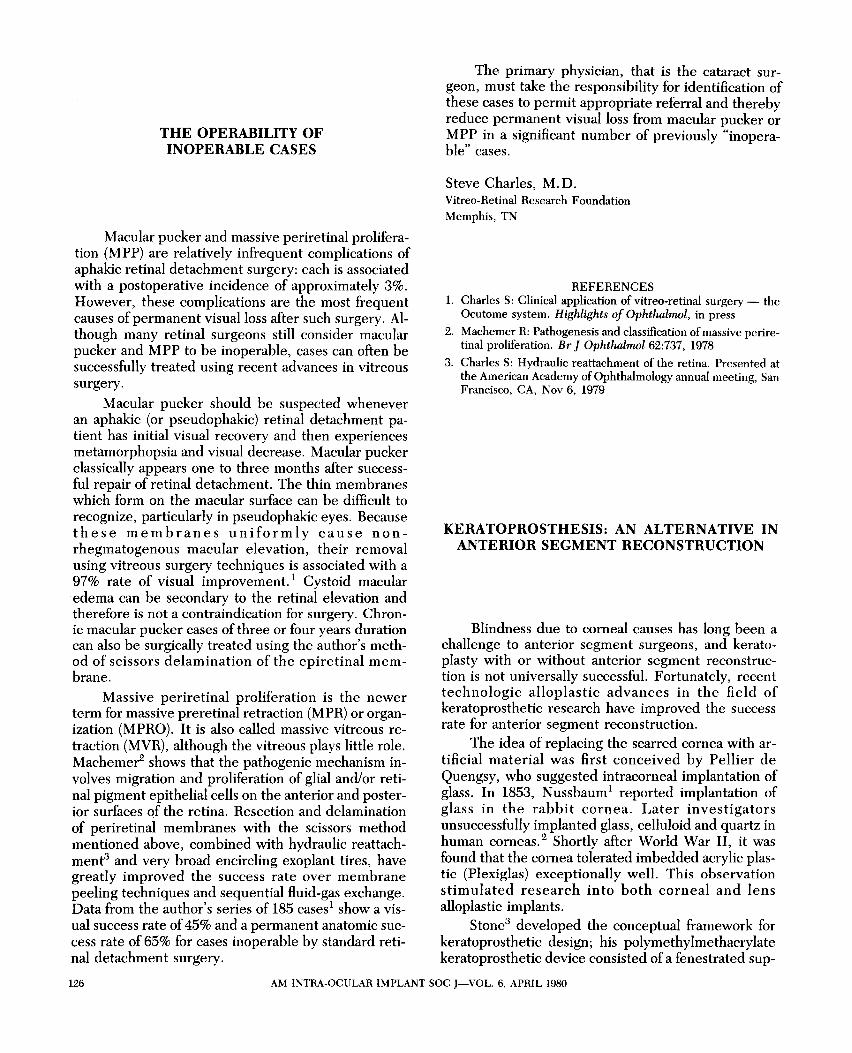
porting plate with a threaded removable optical cylinder (Fig. 1). The fenestrations permit ingrowth of connective tissue and improve nutrition of anterior corneal layers. The supporting plate accepts either a perforating or a nonperforating optical cylinder (Fig. 2),4 allowing the surgeon to postpone perforation until the supporting plate is fixed on the host cornea.
Fig. l(Bath). Stone keratoprosthetic device.
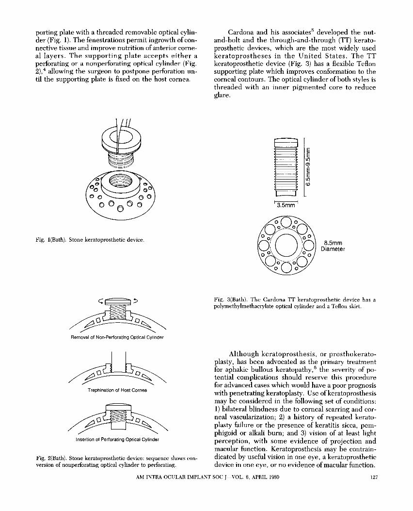
Removal of Non-Perforating Optical Cylinder
Trephination of Host Cornea
Insertion of Perforating Optical Cylinder
Fig. 2(Bath). Stone keratoprosthetic device: sequence shows conversion of nonperforating optical cylinder to perforating.
Cardona and his associates5 developed the nutand-bolt and the through-and-through (IT) keratoprosthetic devices, which are the most widely used keratoprostheses in the United States. The TT keratoprosthetic device (Fig. 3) has a flexible Teflon supporting plate which improves conformation to the corneal contours. The optical cylinder of both styles is threaded with an inner pigmented core to reduce glare.
13.Smm I
E E It)
~ E It)
<ri
8.Smm Diameter
Fig. 3(Bath). The Cardona TT keratoprosthetic device has a polymethylmethacrylate optical cylinder and a Teflon skirt.
Although keratoprosthesis, or prosthokeratoplasty, has been advocated as the primary treatment for aphakic bullous keratopathy, 6 the severity of potential complications should reserve this procedure for advanced cases which would have a poor prognosis with penetrating keratoplasty. Use ofkeratoprosthesis may be considered in the following set of conditions: 1) bilateral blindness due to corneal scarring and corneal vascularization; 2) a history of repeated keratoplasty failure or the presence of keratitis sicca, pemphigoid or alkali burn; and 3) vision of at least light perception, with some evidence of projection and macular function. Keratoprosthesis may be contraindicated by useful vision in one eye, a keratoprosthetic device in one eye, or no evidence of macular function.
AM INTRA-OCULAR IMPLANT SOC J-VOL. 6, APRIL 1980 127
Reported rates of implant extrusion,'5-9 the most serious potential complication of keratoprosthesis, are not comparable with respect to preoperative diagnosis and follow-up times. However, the most recently reported data, from Rao's series9 of 21 cases receiving Cardona IT keratoprosthetic devices implanted with Cardona's techniques,5 show an encouraging absence of both extrusion and endophthalmitis over a maximum follow-up period of 3V2 years.
The complication of endophthalmitis is usually observed in conjunction with erosion and extrusion of the keratoprosthetic device; however it may also occur independently as a result of aqueous leakage around the optical cylinder and concomitant intraocular invasion by pathogens. Aqueous leakage may also be associated with ingrowth of epithelial tissue along the optical cylinder, resulting in epithelial downgrowth and retroprosthetic membranes. 7-9
The extensive anterior segment pathology in eyes eligible for keratoprosthesis makes glaucoma a frequent problem either preoperatively or postoperatively. Diagnosis and management of glaucoma in a keratoprosthetic eye is complex because there is no accurate way to measure intraocular pressure and standard perimetric measurements are altered by the optics of the prosthesis. Serial observations of the optic nerve head coupled with tactile tonometry are clinically rational and useful methods of glaucoma assessment; however better methods of glaucoma surveillance are needed for these cases.
Extrusion of implanted keratoprosthetic devices has spurred on-going investigations of autologous tissues which may offer better bioadhesion than the polymethylmethacrylate, Teflon and Dacron synthetics. Strampelli10 describes a technique of osteoodontoprosthokeratoplasty which uses a patient's tooth as the supporting structure for the acrylic optical cylinder. Using this technique, he has rarely observed extrusion and never encountered the complication of conjunctival overgrowth. Other investigators are exploring the feasibility of using human ungual tissue (onychotransplantation). 11,12 Promising alloplastic research is being done by Frank M. Polack, M.D., using a ceramic keratoprosthetic device (unpublished data, 1979). The ceramic material (aluminum oxidemagnesium oxide) offers improved bioadhesion,13 and it is available with a larger optical cylinder to give a wider visual field. 14
Modern prosthokeratoplasty is an appropriate surgical alternative for anterior segment reconstruction in selected cases. Continuing advances in keratoprosthetic design and technology will undoubtedly increase the indications for this procedure in the future.
Patricia E. Bath, M. D. Jules Stein Eye Institute Los Angeles, CA
REFERENCES l. Nussbaum IN: Cornea Artificialis. Munich, Carl Robert
Schurich Press, 1853 2. Cardona HA: Keratoprosthesis. Am J Ophthalmol 54:284,
1962 3. Stone W, Siderman M: The plastic artificial cornea (an
18-year study): basic principles, in Rycroft PV (ed): Corneo Plastic Surgery. Oxford, Pergamon Press, 1967, pp 375-379
4. Stone W, Yasuda H, Refojo ~ID: A 15-year study of the plastic artificial cornea - basic principles, in King JH, McTigue JW (eds): The Cornea World Congress, Washington, DC, ButteIWorths, 1965, pp 654-671
5. Cardona H, DeVoe AG: Prosthokeratoplasty. Trans Am Acad Ophthalmol Otolaryngol 83:271, 1977
6. Donn A: Aphakic bullous keratopathy treated with prosthokeratoplasty. Arch Ophthalmol94:270, 1976
7. Choyce DP: The Choyce two-piece perforating keratoprosthesis, 70 cases, 1967-1973. Trans Ophthalmol Soc UK 93:333, 1973
8. Girard LJ, Hawkins RS, Nieves R et al: Keratoprosthesis: a 12-year follow-up. Trans Am Acad Ophthalmol Otolaryngol 83:252, 1977
9. Rao GM, Blatt HL, AquavellaJV: Results ofkeratoprosthesis. Am J Ophthalmol 88:190, 1979
10. Strampelli B: Osteo-odontoceratoprostesi. Ann Otta189:1039, 1963
11. Mazhdrakova I: Implantation of ungual particles in the eye tissue of rabbit. Buloftalmologiya (Sofia) 25:127, 1977
12. Schimmelpfennig B: Study on the tissue compatibility of intracorneal horn lenses in experimental animals. Klin Monatsbl Augenheilkd 172:464, 1978
13. Blencke A, Hagen P, Bromer H, Deutscher K: Study of the use of glass ceramics in osteo-odontokeratoplasty. Ophthalmologica 176:105, 1978
14. Hoffman F, Harnisch JP, Strunz V et al: Osteo-keramokeratoprosthesis. Klin Monatsbl Augenheilkd 173:747, 1978
128 AM INTRA-OCULAR IMPLANT SOC J-VOL. 6, APRIL 19S0