Embed Size (px)
Citation preview
Karyotype Curiosities Determining Different Karyotypes
When geneticists want to look at a person’s chromosomes, they make a karyotype. Usually, this is done by a genetic counselor trying to determine if the person has a genetic abnormality. A karyotype is a picture of chromosomes that have been organized in homologous pairs from largest to smallest, with the sex chromosomes placed last. The organization is based on the length of the chromosome, the position of the centromere, and the bands.
This procedure begins by obtaining a sample of blood from the person or by removing amniotic fluid surrounding a fetus. The white blood cells are then removed from the blood or amniotic fluid and cultured in a medium in which they will undergo mitosis. Mitosis is chemically stopped at the metaphase stage to take advantage of the spread-out nature of this stage of mitosis. The metaphase cells are then placed on a slide and viewed under a microscope. A stain is added that stains some segments darker than others, creating unique banding patterns for each chromosome.
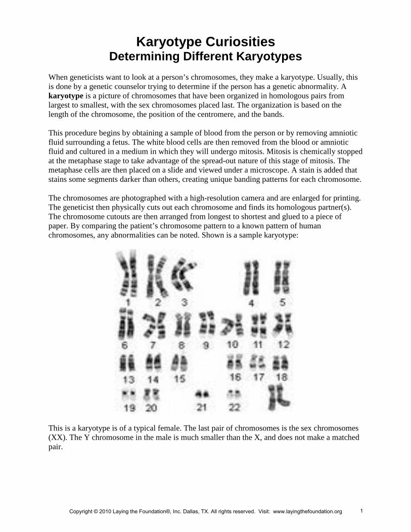
The chromosomes are photographed with a high-resolution camera and are enlarged for printing. The geneticist then physically cuts out each chromosome and finds its homologous partner(s). The chromosome cutouts are then arranged from longest to shortest and glued to a piece of paper. By comparing the patient’s chromosome pattern to a known pattern of human chromosomes, any abnormalities can be noted. Shown is a sample karyotype:
This is a karyotype is of a typical female. The last pair of chromosomes is the sex chromosomes (XX). The Y chromosome in the male is much smaller than the X, and does not make a matched pair.
Copyright © 2010 Laying the Foundation®, Inc. Dallas, TX. All rights reserved. Visit: www.layingthefoundation.org 1
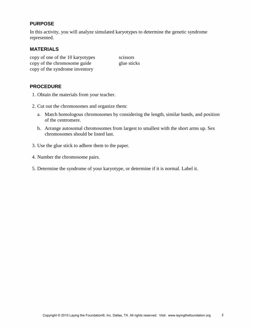
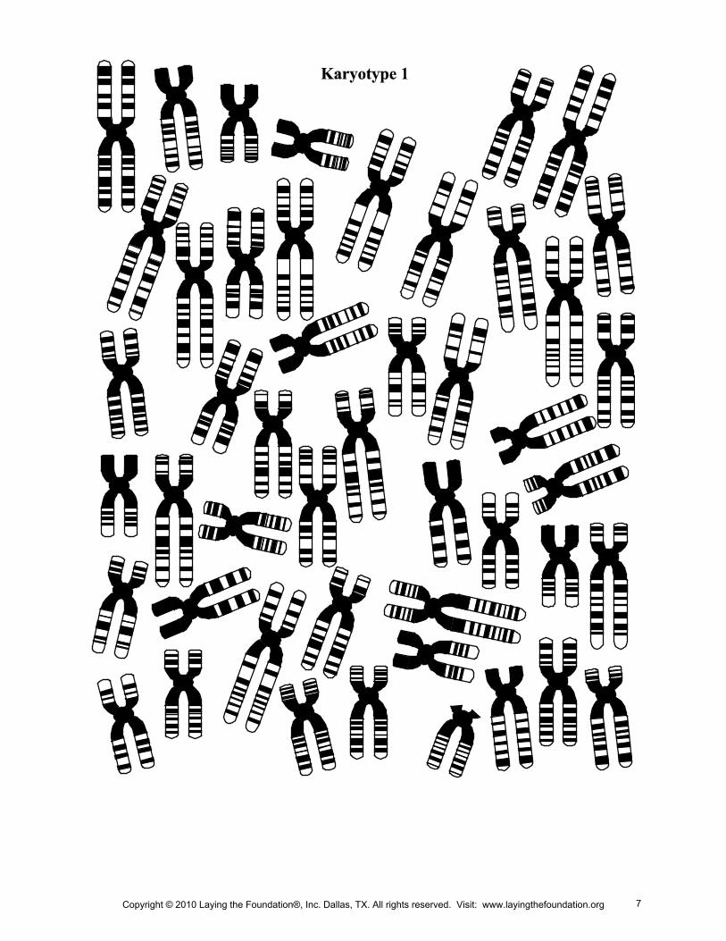
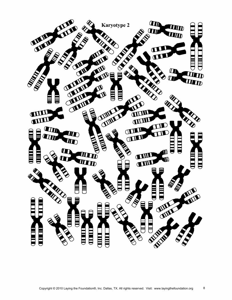
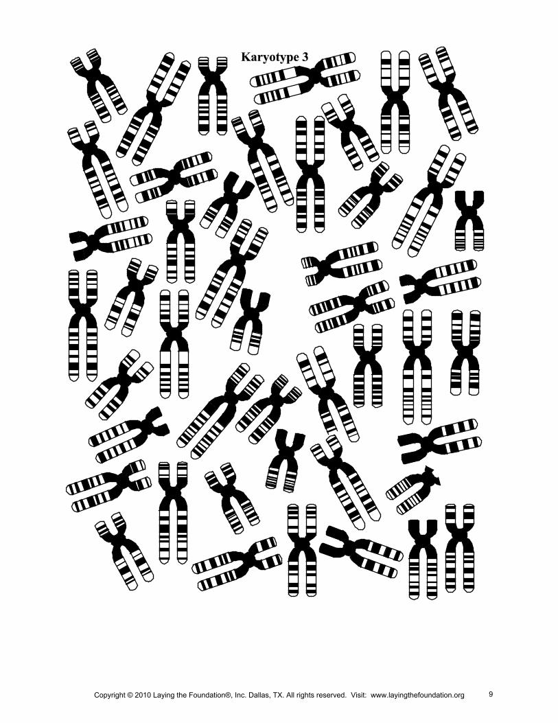
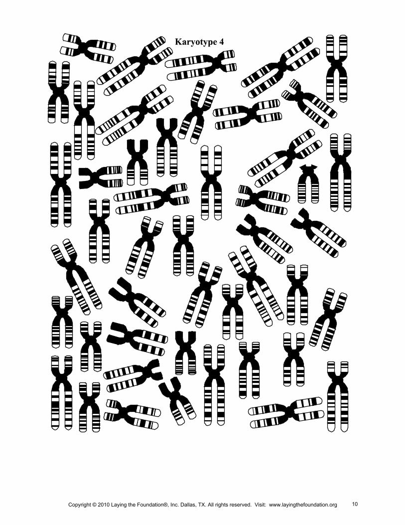
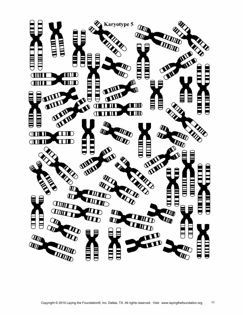
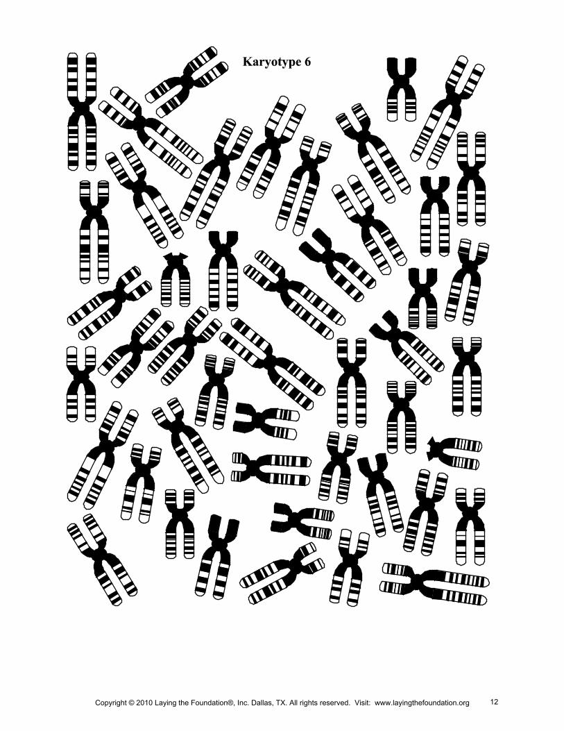
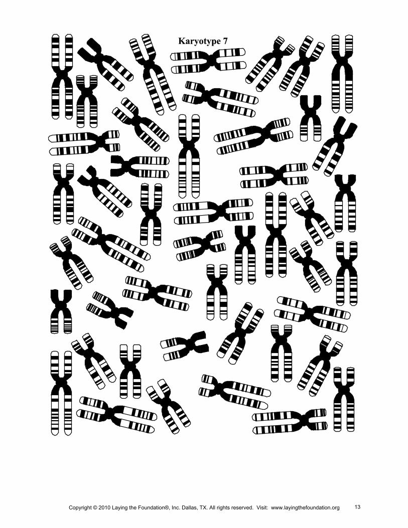
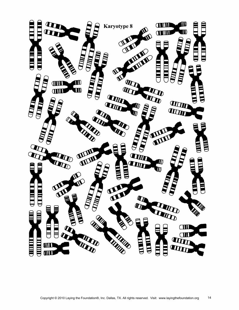
PURPOSE In this activity, you will analyze simulated karyotypes to determine the genetic syndrome represented.
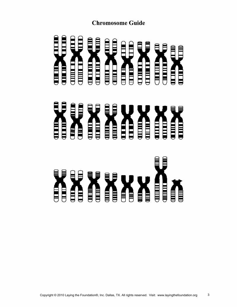
MATERIALS copy of one of the 10 karyotypes scissors copy of the chromosome guide glue sticks copy of the syndrome inventory
PROCEDURE 1. Obtain the materials from your teacher.
2. Cut out the chromosomes and organize them:
a. Match homologous chromosomes by considering the length, similar bands, and position of the centromere.
b. Arrange autosomal chromosomes from largest to smallest with the short arms up. Sex chromosomes should be listed last.
3. Use the glue stick to adhere them to the paper.
4. Number the chromosome pairs.
5. Determine the syndrome of your karyotype, or determine if it is normal. Label it.
Copyright © 2010 Laying the Foundation®, Inc. Dallas, TX. All rights reserved. Visit: www.layingthefoundation.org 2
Copyright © 2010 Laying the Foundation®, Inc. Dallas, TX. All rights reserved. Visit: www.layingthefoundation.org 3
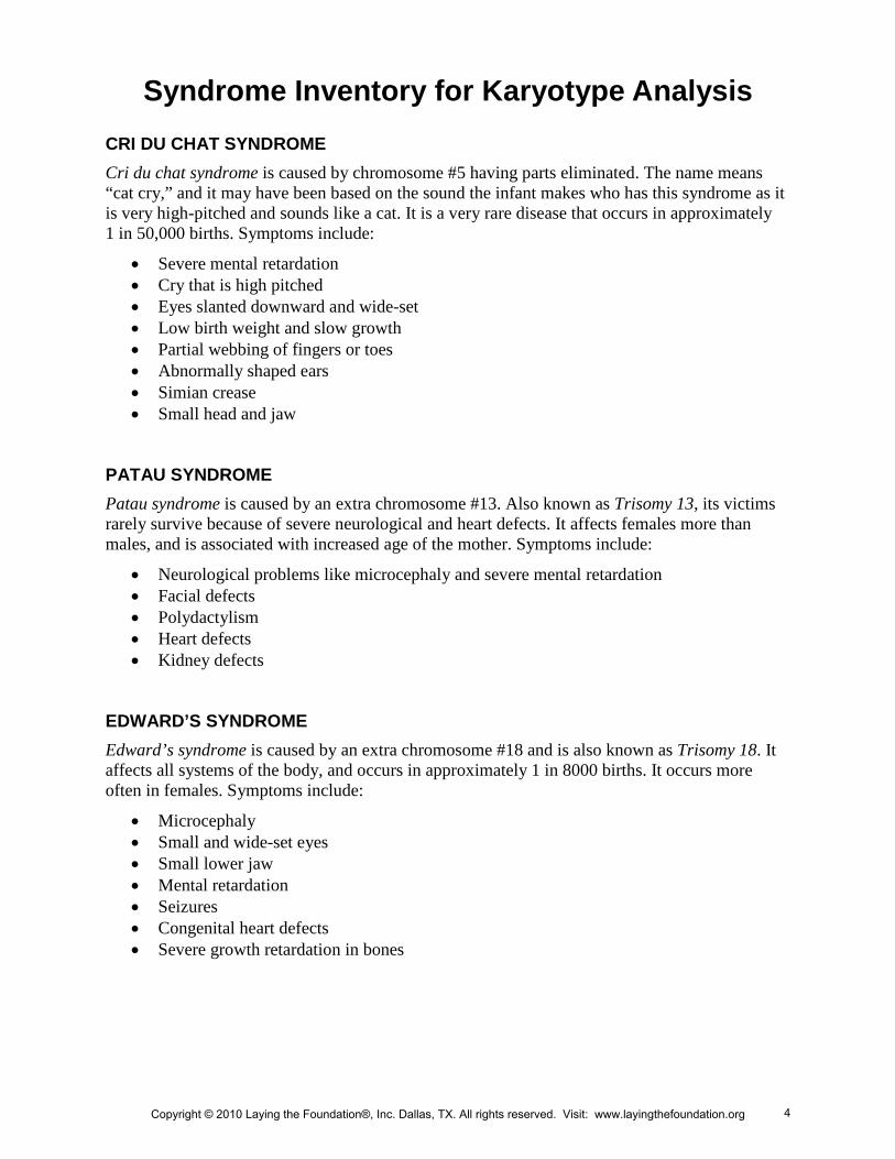
Syndrome Inventory for Karyotype Analysis CRI DU CHAT SYNDROME Cri du chat syndrome is caused by chromosome #5 having parts eliminated. The name means “cat cry,” and it may have been based on the sound the infant makes who has this syndrome as it is very high-pitched and sounds like a cat. It is a very rare disease that occurs in approximately 1 in 50,000 births. Symptoms include:
• Severe mental retardation • Cry that is high pitched • Eyes slanted downward and wide-set • Low birth weight and slow growth • Partial webbing of fingers or toes • Abnormally shaped ears • Simian crease • Small head and jaw
PATAU SYNDROME Patau syndrome is caused by an extra chromosome #13. Also known as Trisomy 13, its victims rarely survive because of severe neurological and heart defects. It affects females more than males, and is associated with increased age of the mother. Symptoms include:
• Neurological problems like microcephaly and severe mental retardation • Facial defects • Polydactylism • Heart defects • Kidney defects
EDWARD’S SYNDROME Edward’s syndrome is caused by an extra chromosome #18 and is also known as Trisomy 18. It affects all systems of the body, and occurs in approximately 1 in 8000 births. It occurs more often in females. Symptoms include:
• Microcephaly • Small and wide-set eyes • Small lower jaw • Mental retardation • Seizures • Congenital heart defects • Severe growth retardation in bones
Copyright © 2010 Laying the Foundation®, Inc. Dallas, TX. All rights reserved. Visit: www.layingthefoundation.org 4
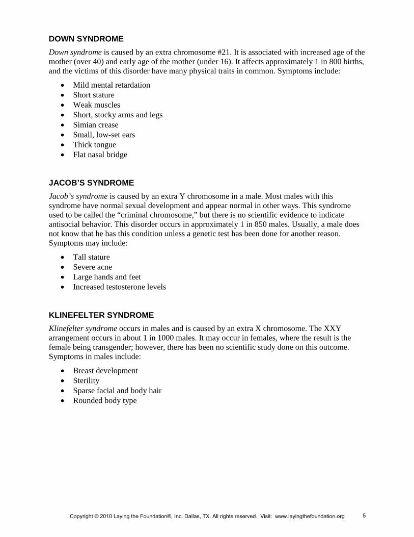
DOWN SYNDROME Down syndrome is caused by an extra chromosome #21. It is associated with increased age of the mother (over 40) and early age of the mother (under 16). It affects approximately 1 in 800 births, and the victims of this disorder have many physical traits in common. Symptoms include:
• Mild mental retardation • Short stature • Weak muscles • Short, stocky arms and legs • Simian crease • Small, low-set ears • Thick tongue • Flat nasal bridge
JACOB’S SYNDROME Jacob’s syndrome is caused by an extra Y chromosome in a male. Most males with this syndrome have normal sexual development and appear normal in other ways. This syndrome used to be called the “criminal chromosome,” but there is no scientific evidence to indicate antisocial behavior. This disorder occurs in approximately 1 in 850 males. Usually, a male does not know that he has this condition unless a genetic test has been done for another reason. Symptoms may include:
• Tall stature • Severe acne • Large hands and feet • Increased testosterone levels
KLINEFELTER SYNDROME Klinefelter syndrome occurs in males and is caused by an extra X chromosome. The XXY arrangement occurs in about 1 in 1000 males. It may occur in females, where the result is the female being transgender; however, there has been no scientific study done on this outcome. Symptoms in males include:
• Breast development • Sterility • Sparse facial and body hair • Rounded body type
Copyright © 2010 Laying the Foundation®, Inc. Dallas, TX. All rights reserved. Visit: www.layingthefoundation.org 5
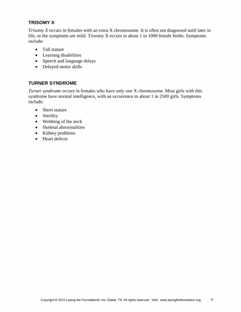
TRISOMY X Trisomy X occurs in females with an extra X chromosome. It is often not diagnosed until later in life, as the symptoms are mild. Trisomy X occurs in about 1 in 1000 female births. Symptoms include:
• Tall stature • Learning disabilities • Speech and language delays • Delayed motor skills
TURNER SYNDROME Turner syndrome occurs in females who have only one X chromosome. Most girls with this syndrome have normal intelligence, with an occurrence in about 1 in 2500 girls. Symptoms include:
• Short stature • Sterility • Webbing of the neck • Skeletal abnormalities • Kidney problems • Heart defects
Copyright © 2010 Laying the Foundation®, Inc. Dallas, TX. All rights reserved. Visit: www.layingthefoundation.org 6
Copyright © 2010 Laying the Foundation®, Inc. Dallas, TX. All rights reserved. Visit: www.layingthefoundation.org 7
Copyright © 2010 Laying the Foundation®, Inc. Dallas, TX. All rights reserved. Visit: www.layingthefoundation.org 8
Copyright © 2010 Laying the Foundation®, Inc. Dallas, TX. All rights reserved. Visit: www.layingthefoundation.org 9
Copyright © 2010 Laying the Foundation®, Inc. Dallas, TX. All rights reserved. Visit: www.layingthefoundation.org 10
Copyright © 2010 Laying the Foundation®, Inc. Dallas, TX. All rights reserved. Visit: www.layingthefoundation.org 11
Copyright © 2010 Laying the Foundation®, Inc. Dallas, TX. All rights reserved. Visit: www.layingthefoundation.org 12
Copyright © 2010 Laying the Foundation®, Inc. Dallas, TX. All rights reserved. Visit: www.layingthefoundation.org 13
Copyright © 2010 Laying the Foundation®, Inc. Dallas, TX. All rights reserved. Visit: www.layingthefoundation.org 14
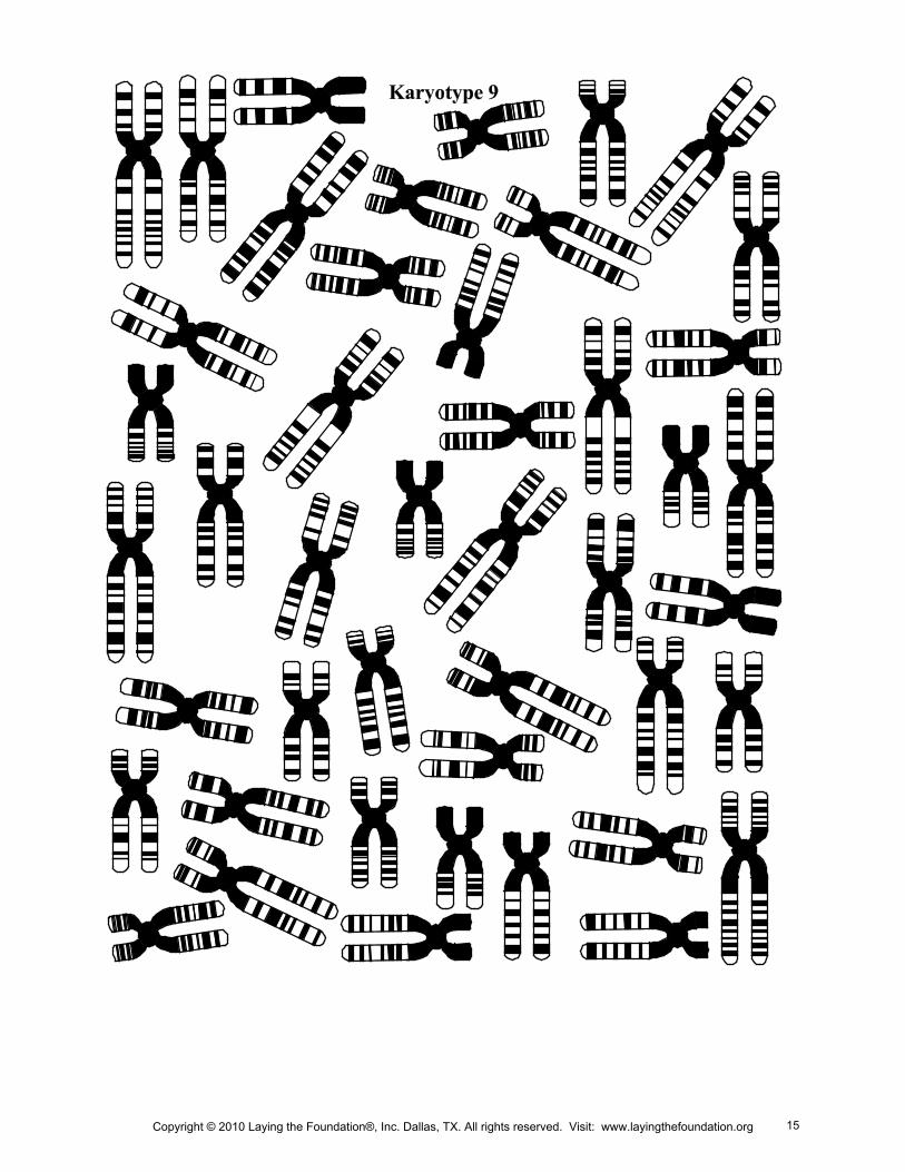
Copyright © 2010 Laying the Foundation®, Inc. Dallas, TX. All rights reserved. Visit: www.layingthefoundation.org 15
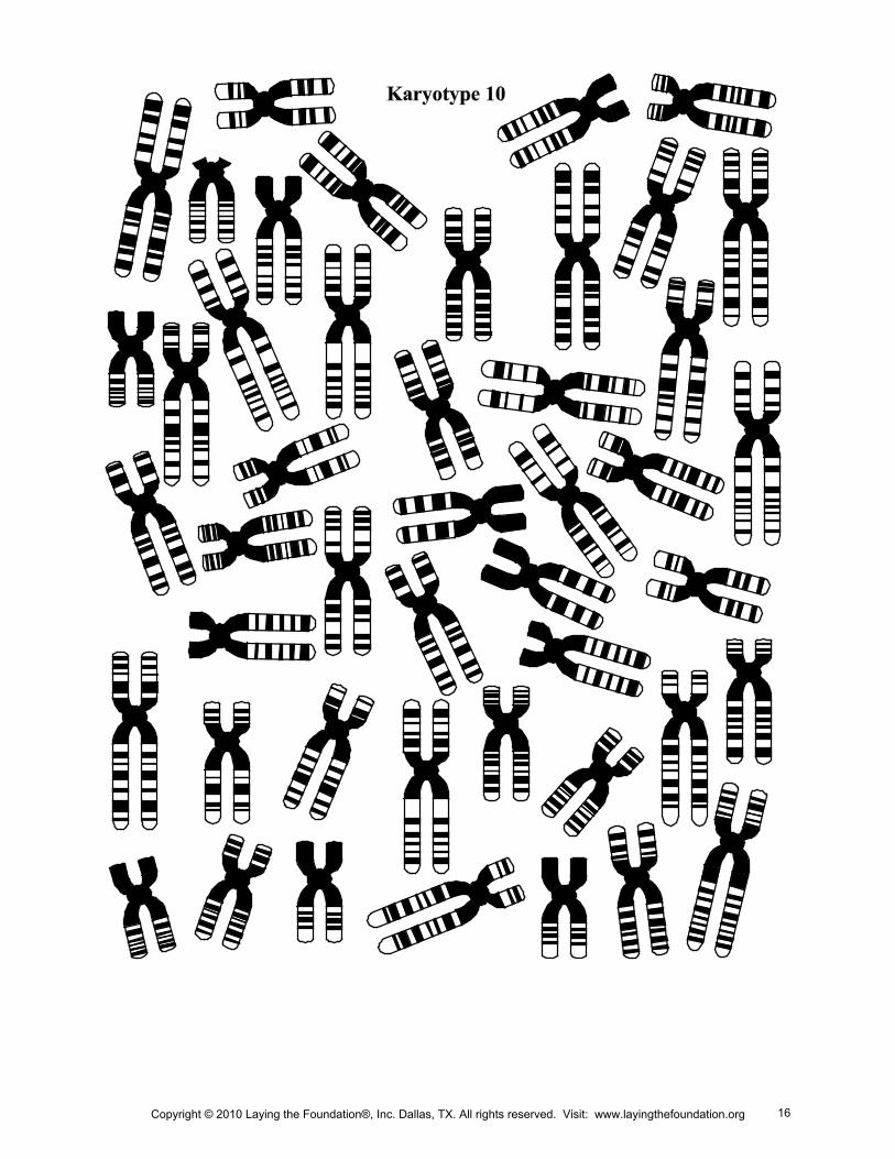
Copyright © 2010 Laying the Foundation®, Inc. Dallas, TX. All rights reserved. Visit: www.layingthefoundation.org 16
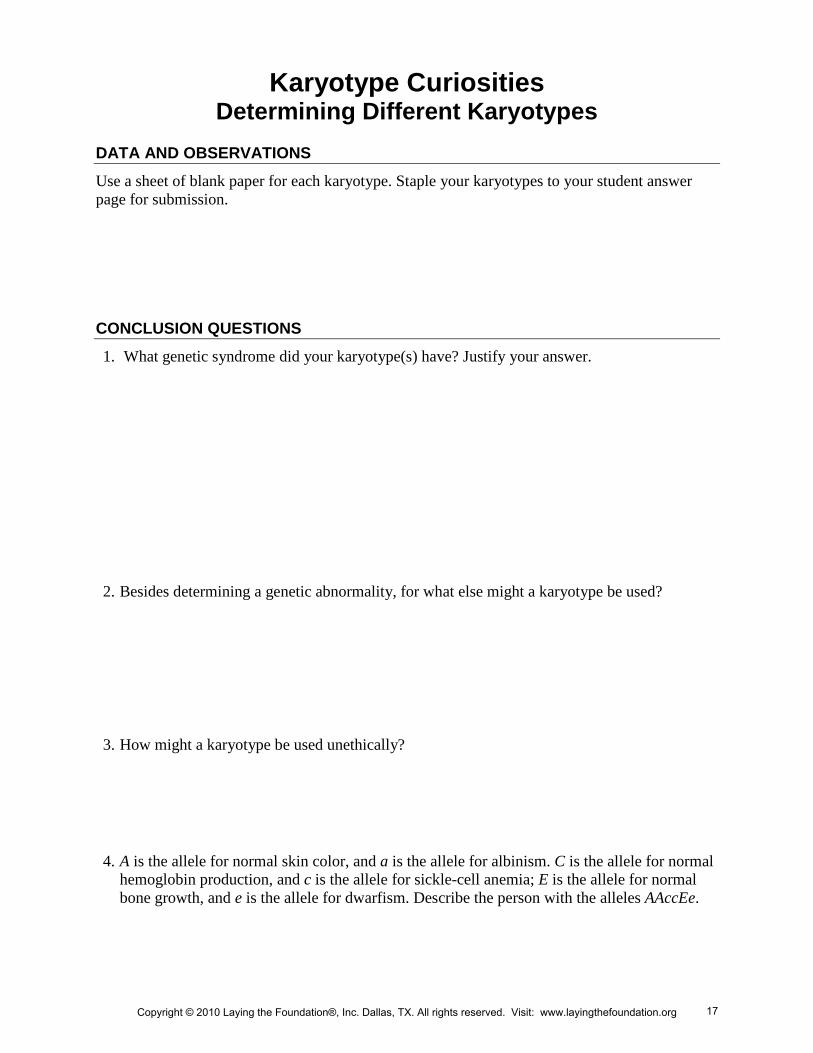
Karyotype Curiosities Determining Different Karyotypes
DATA AND OBSERVATIONS
Use a sheet of blank paper for each karyotype. Staple your karyotypes to your student answer page for submission.
CONCLUSION QUESTIONS
1. What genetic syndrome did your karyotype(s) have? Justify your answer.
2. Besides determining a genetic abnormality, for what else might a karyotype be used?
3. How might a karyotype be used unethically?
4. A is the allele for normal skin color, and a is the allele for albinism. C is the allele for normal hemoglobin production, and c is the allele for sickle-cell anemia; E is the allele for normal bone growth, and e is the allele for dwarfism. Describe the person with the alleles AAccEe.
Copyright © 2010 Laying the Foundation®, Inc. Dallas, TX. All rights reserved. Visit: www.layingthefoundation.org 17