Embed Size (px)
Citation preview
Journal of the
Egyptian Society of
Cardio-thoracic SurgeryEDITOR-IN-CHIEF
Yasser M. W. Hegazy, MD, FRCS
PAST EDITORSHassouna M. El-sabea, FRCS (1995-1996)
Mohamed S. El-fiky, MD (1997-2004)Ezzeldin A Mostafa, MD (2004 -2008)
CO-EDITORAhmed M Deebis, MD
Ahmed M Hassouna, MDAhmed El Kerdani, MDMohamed A Nasser, MD
ETHICS EDITORM. Anwar Balbaa, MD
ASSOCIATE (SECTION) EDITORSAhmed El Nouri, MD
Ashraf El Bassiony, FRCSMamdouh Sharawi, MDSamir A. Hassan, MDSamir A. Keshk, MD
Submit Manuscripts: Editorial office Journal of the Egyptian Society of Cardio-Thoracic Surgery
330 El Sudan Street, Embaba , EgyptTel. (+ 202) 3303 8054
Website: www.arabmedics.com/jescts.htmlEmail : [email protected]


A3The Journal of Egyptian Society of Cardiothoracic Surgery ● Volume 16, Number (1-2)
Abdel Rahman A Fahmy , Cairo , EgyptAbdel Fattah A. Abid ,Tunis , TunisiaAmal Ayoub, Cairo, EgyptAhmed M. Amin, Cairo, EgyptAhmed M. Ali, Banha, EgyptAhmed R. Nasr, Cairo, EgyptA. Samir El-Kosheiry , Cairo , EgyptAli S. Maklad , Cairo , EgyptM. Ayman A Soieb, Cairo, EgyptMamdoud A. Sharawi,Zagazig,EgyptAhmed El-Kerdani, Cairo, EgyptAlradi Kamal, Zagazig, EgyptBabulal Sethia, London, EnglandBertrand M. Goudot, Paris, FranceB Ben-Ismail , Tunis , TunisiaB M Fabri , Liverpool , EnglandBryn T Williams, Weybridge, EnglandDaniel G. Guilmet, Paris, FranceDavid J. Wheatley, Glasgow, EnglandEl Nouri Ahmed , Cairo , EgyptEl Hussieiny Gamil , Cairo , EgyptFawzi Estefanos , Cleveland , USAFouad Z Abdalla , Cairo , EgyptGerard Block, Paris, FranceGamal O. Abou Senna , Cairo , EgyptGraham E. Venn, London, EnglandHasan Alzahrani, Mekka, Saudi ArabiaHussein A. Gaafar, Cairo, EgyptHamdy M. El-Sayed, Cairo , EgyptHassan Ezzeldin Attia, Cairo , EgyptHamed M. Al Akshar , Tanta , EgyptHisham A. Sawki, Cairo , EgyptIsmail A. Sallam , Cairo , EgyptIbrahim Haggag, Cairo , EgyptJames J. Pollock, Glasgow, England
Jean E. Bachet, Paris, FranceJean-Paul F. Bessou, Rouen, FranceJohn R. Pepper , London , EnglandLotfi Eissa, Cairo , Egypt Mohamed A. Hamed, Cairo , EgyptMohamed Abou El-Ezz, Cairo , EgyptMostafa Agha, Alexandria, EgyptMohamed F. Bassiouni , Cairo , EgyptMarc de Leval , London , EnglandM El-Fakih , Riadh , Saudi ArabiaMamdouh Gamal , Einthoven, HollandM. Ezzeldin Abdel Raouf ,Cairo,EgyptMaher Fourati, Tunis, TunisiaMagdi Gomaa , Cairo , EgyptMohamed S El-Fiky, Cairo, EgyptMarco Pozzi, Liverpool, EnglandM S Ammar, Tunis, TunisiaMaher Shoier, Cairo, EgyptMogazy A. Tantawy, Cairo, EgyptMedhat A. El-Gamal, Cairo , EgyptMostafa M. Radwan , Cairo , EgyptNahed Attia , Assiout , EgyptPierre Michel Roux, Metz, FranceRobert M. Soyer, Rouen, FranceSherif Abdel Hady , Cairo , EgyptShaaban Abu Elelaa , Mansoura , EgyptSamieh A Amer , Cairo , EgyptSami S. Kabbani , Damascus , SyriaSamir Mahmoudi , Cairo , EgyptSteven Tsui , Cambridge , EnglandTarek Z. Shallaby Cairo , EgyptWadih R. Dimitri, Birmingham, EnglandWahid Osman , Cairo , EgyptZohair Al-Halees, Riyadh, Saudi ArabiaZohni M. Farrag , London , England
EDITORIAL BOARD
Journal Secretary Ahmed Ali Kalifa

A5The Journal of Egyptian Society of Cardiothoracic Surgery ● Volume 16, Number (1-2)
The Society Board of Directors2006-2008
THE Egyptian Society of
CARDIO-THORACIC SURGERYPresident
Magdy Mostafa Ali, MD
Vice President Mounir Zeerban, MD
General SecretarySamir A Hassan, MD
TreasurerLotfi M. Eissa, MD
Immediate Past PresidentSamieh A Amer, MD
BoardAhmed Dokhan, MD
Ahmed M .Elkerdani, MDAhmed M. Deebis, MD
Ezzeldin A. Mostafa, MDEzzeldin Abdel Raoof, MD
M. MamdouhA. Sharawi, MDM. Mostafa A. Agha, MDMohamed A. Nasser, MD
Magdi Gomaa, MDSamir A Keshk, MD
Yasser M. Hegazy, MD


A7The Journal of Egyptian Society of Cardiothoracic Surgery ● Volume 16, Number (1-2)
CONTENTS
ANNOUCEMENT A9 Guidelines for authorsA13 Condition for publication form A15 Guidelines for reviewersA17 Events of interests
EDITORIAL1 Editorial Letter Yasser Hegazy, MD , FRCS
CARDIOVASCULAR
2 OFF-PUMP VERSUS ON-PUMP FOR MULTI VESSEL CORONARY ARTERY BYPASS GRAFTING: COMPARATIVE STUDY OF OP ERATIVE AND SHORT-TERM OUTCOMES Mohamed Essa MD, Ahmed Deebis MD,Mamdouh Sharawy MD,Khalid abdelbariy MD, Ehab Yehia
8 THE USE OF OFF–PUMP CARDIOPULMO NARY BYPASS IN EMERGENCY MYOCAR DIAL REVASCULARIZATION Ahmad Darwazah MD,FRCSRaed. Abu Shama M.D , Ismail Isleem PhD, MRCP ,Basel Hanbali² MRCP, Bashar Jaber M.D
15 RISK FACTORS OF CEREBRO-VASCULAR ACCIDENTS AFTER ONPUMP ISOLATED CORONARY ARTERY SURGERY Ibrahim M. Yassin, MD,Yousry A. Shaheen ,MD, Bedir M. Ibrahim, MD,Michele Di Mauro, MD, Antonio Maria Calafiore, MD.
23 OUTCOMES OF REDO CORONARY ARTERY SURGERY Tarek Nosseir MD.,Adel Ragheb MD.,Mohamed Adel MD.,Alaa Farouk MD.,Hesham Zayed MD., Nashat Abdelhamid MD.
28 ROUTINE USE OF TRANEXAMIC ACID PR--EOPERATIVELY IN CORONARY ARTERY BYPASS SURGERY
Mohamed Abul-dahab M.D.,Tamer Farouk M.D., Amr Rushdi M.D.,Tarek S. Abdalah M.D., Moham- ed Sweilam M.D.
Journal of The Egyptian Society of Cardio-Thoracic Surgery
Volume 16 Mar-Jun 2008 Number 1,2 ISSN 1110-578X
33 CAN LOCAL APPLICATION OF TRANEXAM--IC ACID REDUCE POST- CABG BLOOD LOSS?
Elsayed Elmistekawy MD, Abdelsalam Elhenawy MD, Hosam Fawzy MD, 39 A RANDOMIZED TRIAL OF APROTININ ON
BLEEDING, BLOOD PRODUCTS REQUIRE--MENT AND MYOCARDIAL INFRACTION IN PATIENTS TREATED WITH CLOPIDOG--REL BEFORE CORONARY ARTERY BYPASS GRA
Ghada Ali M.D.,Ahmed abdel-razek M.D. ,Heba el- serwey M.D.,Ashraf El-Sebaieft M.D.,
48 MODERATE ISCHEMIC MITRAL REGUR--GITATION: EVALUATION OF FACTORS AF--FECTING ITS DEGREE AFTER ISOLATED CORONARY ARTERY BYPASS GRAFTING
Tamer Farouk MD.
55 PCOMBINED ATRIAL FIBRILLATION (AF) ABLATION WITH MITRAL VALVE SUR--GERY: THE PREDICTORS OF SUCCESS
MKhaled Samir MD,Ayman Ammar MD ,Tamer El Gho--bary MD,Ashraf A. ELSebaie MD.
59 FEFFECT OF VALVE PROSTHESIS – PA--TIENT MISMATCH ON SHORT TERM OUT--COME AFTER AORTIC VALVE REPLAC--MENT
IAmr Badr MD,Hosam Fawzy MD,
66 RELATION OF LEFT VENTRICULAR MASS TO VOLUME AND ITS INFLUENCE ON THE OUTCOME OF AORTIC VALVE REPLACE--MENT.
Adel Ragheb MD.,Tarek Nosseir MD.,Ahmed Amin Ms., Mohammed Adel MD.,Nashat Abdelhamid MD.,Maher
Mousa MD..
70 CRETROGRADE VERSUS ANTEGRADE BLOOD CARDIOPLEGIA FOR CABG PA--TIENTS: DOES IT AFFECT MYOCARDIAL PRESERVATION?
Heba B. El-Serwi,Ahmed A-Razek Hasan,Ghada Ali, Ashraf Abdalla El-Sebaie MD,

A8 The Journal of Egyptian Society of Cardiothoracic Surgery ● Mar-Jun 2008
75 IMPACT OF HIGH THORACIC EPIDURAL ANALGESIA ON INCIDENCE OF PERIOP--ERATIVE STRESS RESPONSE IN OFF PUMP CABG
Hala Elsheikh MD,Saeed M.R. Elassy MD.
THORACIC83 ACQUIRED NON- OESOPHAGEAL
EXTRATHORACIC BRONCHIAL FISTULAS
Journal of The Egyptian Society of Cardio-Thoracic Surgery
Volume 16 Mar-Jun 2008 Number 1,2 ISSN 1110-578X
Uvie Onakpoya MD,Ahmed Saleh MD, Bassem Ramadan MD,Abd el Meguid Ramadan MD,Munir Zeerban MD,
Mohamed Elhofie MD,Sahar Morad MD,Amr Saleh MD.
88 BILATERAL BULLECTOMY THROUGH ME--DIAN STERNOTOMY
Mohamed Abul-dahab M.D, Ahmed El-Agaty MD.
A9The Journal of Egyptian Society of Cardiothoracic Surgery ● Volume 16, Number (1-2)
Editorial Office Please address all correspondence to:Ezzeldin A. Mostafa, MD, Editor, In-chiefJournal of the Egyptian Society of Cardio-thoracic Surgery 330 El-Sudan St., Imbaba, Cairo, Egypt.Telephone: (+202) 303 6634Fax: (+202) 303 8054E-Mail: [email protected]
The Journal of the Egyptian Society of Cardio-Thoracic Surgery [ISSN 1110-578 X] is the official publication of the Egyptian Society of Cardio-thoracic Surgery. The journal is published every three months .
General Instructions
Every submission must include: Cover letter, indicating the category of article , the Complete manuscript, including title page, abstract, text, tables, ac--knowledgments ,references and illustrations .
Required Disclosures;
A. Conditions for Publication Form which includes dis--closures regarding freedom of investigation and conflicts of interest, signed by all authors. In single Author publication an additional Senior Consultant Signature is required.B. Written permission from the publisher (copyright holder) is required to reproduce any previously published table(s), illustration(s) or photograph(s) in both print and electronic media. C. Written permission from unmasked patients appearing in photographs is also required.
Revised_Manuscripts:Revised manuscripts must be submitted in three parts as Microsoft word-processing files : (1) cover letter with responses to reviewers’ comments (2) revised, marked manuscript showing additions and deletions; (3) revised, un--marked manuscript.
General Information Three copies of the Manuscripts should be sent preferably
prepared in Microsoft Word , typed double-spaced through--out (including title page, abstract, text, references, tables and legends) with one (1) inch (2.5 cm) margins all around. Place Author name and page number in the upper right corner of each page. Manuscripts written in 12 point Arial or Times New Roman fonts are preferred (Note: Do not submit your manuscript in PDF format it causes problems in processing your submis--sion.)Arrange manuscript as follows: (1) title page, (2) abstract, (3) text, (4) acknowledgments, (5) disclosures if required, (6) references, (7) tables and (8) legends. Number pages consecu--tively, beginning with the title page as page 1 and ending with the legend page.If your manuscript contains illustrations, in addition to submit--ting them online, you must send two sets of original illustra--tions to the editorial office labeled with manuscript number, first author, and figure number on back. Tables and figures should be provided separate from the text while there position in the text should be marked on the manu--script.
Word Limits by Category of Manuscript
Original articles should not exceed 4500 words including title page, abstract of 150-200 words, text, figure legends and refer--ences. The combined total of illustrations and tables should not exceed 10 and the number of references should not exceed 40.
Case reports and “The way I do it” articles are limited to a total of 1500 words including title page, abstract, text, refer--ences and figure legends. For each illustration subtract 100 words and for each table subtract 300 words from the word limit. References are limited to eight. A “how to do it” article should be a description of a useful surgical technique and con--tain descriptive, illustrative material.
Images in cardiothoracic surgery are limited to 350 words including title and text and to two, possibly three figures. The entire contribution must fit on one printed page .
Review articles are limited to 6500 words including title page, abstract, text, figure legends and all references. The to--tal number of references should not exceed 80. Subtract 100
Guidelines for Authors
Journal of The Egyptian Society of Cardio-Thoracic Surgery (J. Egypt. Soc. Cardiothorac. Surg.)
A10 The Journal of Egyptian Society of Cardiothoracic Surgery ● Mar-Jun 2008
words for each illustration and 300 words for each table.
Our surgical heritage articles are limited to 2500 words in--cluding title page, abstract, text, figure legends and references. Subtract 100 words for each illustration and 300 words for each table.
Correspondence (Letters to the Editor) and commentaries are limited to 500 words. Subtract 100 words for each illustration and 300 words for each table.
Editorials are limited to 2500 words including references. Subtract 100 words for each illustration and 300 words for each table.
Manuscript Preparation
Title Page (first page)
The title is limited to 100 characters and spaces for original manuscripts and to 80 characters and spaces for all other cat--egories of manuscripts. The title may not contain acronyms or abbreviations. All submissions, must have a title.
Running Head. Supply a short title of 40 characters and spac--es.
Authors. List all authors by first name, all initials, family name and highest academic degree using “MD, PhD” for holders of both degrees ( if more then 7 Authors justifie).
Institution and Affiliations. List the name and full address of all institutions where the work was done. List departmental affiliations of each author affiliated with that institution after each institutional address.
Meeting Presentation. If the paper has been or is to be pre--sented at the annual meeting of The Society, provide the name, location and dates of the meeting.
Keywords. Provide up to 5 keywords selected from the ap--pended list to describe the manuscript. Do not use any key--words that are not on the list.
Word Count. Provide the electronic total word count of the entire manuscript including title page, abstract,text,figure leg--ends and entire reference list.
Corresponding Author. Provide the name, exact postal ad--dress with postal code, telephone number, fax number and e-mail address of the author to whom communications, proofs and requests for reprints should be sent.
Abstract Page (Second page)
Original articlesProvide a structured Abstract, no longer than 250 words, di--vided into four sections: Background or Objective, Methods, Results, Conclusions. Avoid abbreviations and acronyms. In--
dicate the abstract word count below the abstract. Case reports, “the way i do it” articles, review articles and our surgical heritage articles. Provide an unstructured abstract of 100 words. Images, correspondence, commentaries, editorials and up--dates. No abstract is required.
Text Text should be organized as follows: Introduction, Mate--rial (or Patients) and Methods, Results, and Comment.Cite references,illustrations and tables in numeric order by order of mention in the text.
Avoid abbreviations. Consult the American Medical Associa--tion Manual of Style, 9th edition, for recommended abbrevia--tions. Define abbreviations at first appearance in the text. If 8 or more abbreviations or acronyms are used, provide a separate table of abbreviations and acronyms.
Measurements and weights should be given in standard metric units.Statistical nomenclature and data analysis. Fol--low the “Guidelines for Data Reporting and Nomenclature” published in The Annals of Thoracic Surgery (1988;46:260-1). Footnotes. Type footnotes at the bottom of the manuscript page on which they are cited. Suppliers of drugs, equipment and other brand mentioned in the article within parentheses , giving company name, city and country .
AcknowledgmentsGrants, financial support and technical or other assistance must be acknowledged at the end of the text before the references.
ReferencesIdentify references in the text using Arabic numerals in brack--ets on the line.Type references double-spaced after text or acknowl--edgments beginning on a separate sheet. Number con--secutively in the order in which they appear in the text. Journal references should provide inclusive page num--bers; book references should cite specific page numbers. Journal abbreviations should conform to those used in Index Medicus. follow the formats outlined below: Journal ArticleJones DR, Stiles BM, Denlinger CE, Antie P . Pulmonary segmentectomy: results and complications. Ann Thorac Surg 2000;76:343-9.(List all authors if 6 or fewer; otherwise list first 3 and add “et al.”)
Chapter in Book12. Vinten-Johansen J, Zhao Z-Q, Guyton RA. Cardiac surgi--cal physiology. In: Cohn LH, Edmunds LH Jr, eds. Cardiac Surgery in the Adult. 2nd ed. New York, NY: McGraw-Hill; 2003:53-84.
A11The Journal of Egyptian Society of Cardiothoracic Surgery ● Volume 16, Number (1-2)
Internet Address3. 1996 NRC Guide for the Care and Use of Laboratory Ani--mals. Available at: http://www.nap.edu/readingroom/books/labrats/contents.html. Accessed October 20, 2003.
Tables :Tables should be typewritten double-spaced on separate sheets (one to each page). Do not use vertical lines. Each table should be numbered (Arabic) and have a title above. Legends and explanatory notes should be placed below the table. Abbrevia--tions used in the table follow the legend in alphabetic order. Lower case letter superscripts beginning with “a” and follow--ing in alphabetic order are used for notations of within-group and between-group statistical probabilities. FigureLegends :Figure Legends should be numbered (Arabic) and typed double-spaced in order of appearance beginning on a sepa--rate sheet. Identify (in alphabetical order) all abbreviations appearing in the illustrations at the end of each legend. Cite the source of previously published material in the legend and indicate permission has been obtained. Proof of permis--sion must be surface mailed or faxed to the editor .
Illustrations :You must send two sets of original illustrations to the editorial office labeled with manuscript number, first author, and figure number on back.
Images or figures are submitted online as one or more separate files that may contain one or more images. Within each file containing images, use the figure number (eg, Figure 1A) as the image filename. The system accepts Powerpoint (.ppt) files Most illustrations will be reproduced at a width of one column (8.25 cm; 3 1/4 inches). Black, white and widely crosshatched bars are preferable; do not use stippling, gray fill or thin lines.
Instructions :Identify print proofs of figures on the back with figure number and name of the first author; when necessary, indicate the top with an up arrow For figures submitted in electronic format, all images should be at least 5 inches wide. Graphics software such as Photoshop and Illustrator, should be used to create art. Color images need to be at least 300 dpi.Gray scale images should be at least 300 dpi .Line art should be at least 1200 DPI .
Cover letter :Include with the manuscript a cover letter that provides 1) the category of manuscript (e.g., original research, Brief Commu--nication, Letter to the Editor); 2) statement that the material
has not been previously published or submitted elsewhere for publication; 3) information about any personal conflicts of in--terest of any of the authors; and 4) names of sources of out--side support for research, including funding, equipment, and drugs .You may also submit the name of one reviewer of your choice. You should include that individual’s mailing address, telephone, fax and e-mail address. Editorial Policies Scientific Responsibility StatementBefore publication of an accepted manuscript, each author is required to certify by signing the Conditions for Publication Form that he or she has participated sufficiently in the work and approved the final version of the manuscript to be pub--lished. Exclusive Publication StatementEach author must certify that none of the material in this manuscript has been published previously in either print or electronic form, and that none of this material is currently under consideration for publication elsewhere. This includes symposia and preliminary publications of any kind except an abstract of 400 words or fewer.
Conflict of Interest :Authors should disclose any conflict of interests. Authors who have a financial relationship with one or more companies whose products are featured in an article will disclose the ex--istence of this relationship in a box at the bottom of the first page of the published article.
Consultant Statistician and Statistical Methods : All manuscripts with statistical analysis are required to undergo biostatistical review .The most appropriate way is to involve a biostatistician consultant or coauthor from the investigators’ home institution . Manuscripts may undergo further biostatistical review by the Journal after submission. Additional information on statistical methods can be found in “Uniform Requirements for Manuscripts Submitted to Biomedical Journals”(www.acponline.org/journals/resource/unifreqr.htm).
Copyright :Authors of articles submitted to The J. Egypt. Soc. Cardiotho--rac. Surg. must transfer copyright to The Egyptian Society of Cardio-Thoracic Surgery by signing the “Conditions for Publi--cation Form.” This transfer becomes binding upon acceptance of the article for publication. No part of the published material may be reproduced elsewhere without written permission.Date of Receipt: The “received for publication” date is the date when the editorial office receives the manuscript, the cover let--ter, and the Copyright Transfer and Author Declaration State--ment, signed by all authors. For Date of acceptance : letter is provided from the editor.
A12 The Journal of Egyptian Society of Cardiothoracic Surgery ● Mar-Jun 2008
Checklist
A] Cover Letter □ Letter to the Editor □ Manuscript category designation .□ Single-journal submission affirmation .□ Conflict of interest statement (if appropriate). □ Sources of outside funding. □ Signed Statistical Collaboration .
B] Complete Manuscript□ Title page .□ Title of article□ Full name(s), academic degrees, and affiliation(s) of authors.□ Corresponding author .□ Telephones, fax, and e-mail address□ Abstract (250 words; double-spaced) .□ Ultramini-abstract (50 words) .□ Text (double-spaced). □ References (double-spaced; separate pages). □ Tables (double-spaced; separate pages). □ Figures (separate files; on hardcopy; properly identified), □ Figure legends (double-spaced; separate pages) .□ Word count.
C] Required Disclosures □ Conditions for Publication Form signed by all authors. Which transfers copyright to The
Egyptian Society of Cardio-Thoracic Surgery□ Written permission from the publisher to reproduce any previously published material .□ Written permission from unmasked patients .
A13The Journal of Egyptian Society of Cardiothoracic Surgery ● Volume 16, Number (1-2)
This form MUST be completed, signed by ALL authors, and returned to the Editorial Office before your manuscript can be accepted for publication.
Scientific Responsibility Statement:Each author must sign this form to certify that he or she
has participated sufficiently in the work to take responsibility for a meaningful share of the content of the manuscript, and that this participation included: (a) conception or design of the experiment(s), or collection and analysis or interpretation of data; (b) drafting the manuscript or revising its intellectual content; and (c) approval of the final version of the manuscript to be published. In addition, each author must indicate whether or not he or she has had full freedom of investigation; defined as freedom from outside interests in controlling the design of the study, collection, analysis, and interpretation of data, and having freedom to full disclose all results.
Exclusive Publication Statement:Each author must sign this form to certify that none of the
material in this manuscript has been published previously in either print or electronic form, and that none of this material is currently under consideration for publication elsewhere. This includes symposia, transactions, books, articles published by invitation and preliminary publications of any kind except an abstract of 400 words or fewer.
Copyright Transfer Agreement:Each author must sign this form to certify that, if the manu--
script is accepted for publication in the Journal of the Egyptian Society of Cardio-Thoracic Surgery ( JESCTS), copyright (including the right to obtain copyright registration, whether separately or as part of a journal issue .) in and to the above article transfers throughout the world and for the full term and all extensions and renewals thereof to: THE EGYPTIAN SO--CIETY OF CARDIO-THORACIC SURGERY
This transfer includes the right to adapt the article for use in conjunction with computer systems and programs, includ--ing reproductions or publication and incorporation in retrieval systems.
Rights of authors:The ESCTS hereby licenses the following rights back to
the author(s): A. Patent and trademark rights to any process or procedure
described in the article. B. The right to photocopy or make single electronic copies of
the article for their own personal use, including for their
Conditions for Publication Form
own classroom use, or for the personal use of colleagues, provided the copies are not offered for sale .
C.The right, subsequent to publication, to use the article or any part thereof free of charge in a printed compilation of works of their own, such as collected writings or lecture notes.
Note: All copies, paper or electronic, or other use of the informa--
tion must include an indication of The ESCTS copyright and a full citation of the journal source.
Authorship: If copyright is held by the employer, the employer or an
authorized representative of the employer must sign in addi--tion to the author(s).
Warranties: The author(s) warrant that the article is the author’s origi--
nal work and has not been published before. The author(s) war--rant that the article does not infringe on the rights of others. If excerpts from copyrighted works are included, the author(s) has (have) obtained written permission from the copyright owners and will credit the sources in the article.
Preprints: The author(s) warrant(s) that if a prior version of this work
(normally a preprint) has been posted to an electronic server, such version was accessible to only a small group of individu--als and the author(s) will cause its prompt removal from such server.
Conflict of Interest Disclosure Statements: Each author must indicate below that either (a) no financial
conflict of interest exists with any commercial entity whose products are described, reviewed, evaluated or compared in the manuscript, except for that disclosed under “Acknowledgements” or (b) a potential conflict of interest exists with one or more commercial entities whose products are described, reviewed, evaluated or compared in the manuscript through the existence of one or more of the following relationships: the author is a full or part-time employee of a company; has an existing or optional equity interest in a company; owns or partly owns patents licensed to a company; has an ongoing retainer relationship (consultantship, speaker, etc.) with a company for which he/she receives financial remuneration; or has received financial compensation for this publication. If Yes is checked, a box on the first page of the published article will read: ?Dr. X discloses that he/she has a financial relationship with company Y.?
A14 The Journal of Egyptian Society of Cardiothoracic Surgery ● Mar-Jun 2008
Author: Manuscript Title:
I agree with the preceding conditions and provide the appropriate signatures and information below accordingly:
A 16
If there are additional authors on the article, please photocopy this form and attach additional sheets as need be with appropri-ate information and signatures affixed .
Author:Manuscript Title:
I agree with the preceding conditions and provide the appropriate signatures and information below accordingly:
Author’s Name:_____________________________________________________________________________________Signature:______________________________________________ Date:_______________________________________Author’s employer’s signature, if appropriate: ___________________________________________________________Conflict of interest: Yes ___ No ___ If yes, with which entity: _______________________________________________Did you have freedom of investigation in all aspects of this work?: Yes ___ No ___
Author’s Name:_____________________________________________________________________________________Signature:______________________________________________ Date:_______________________________________Author’s employer’s signature, if appropriate: ___________________________________________________________Conflict of interest: Yes ___ No ___ If yes, with which entity: _______________________________________________Did you have freedom of investigation in all aspects of this work?: Yes ___ No ___
Author’s Name:_____________________________________________________________________________________Signature:______________________________________________ Date:_______________________________________Author’s employer’s signature, if appropriate: ___________________________________________________________Conflict of interest: Yes ___ No ___ If yes, with which entity: _______________________________________________Did you have freedom of investigation in all aspects of this work?: Yes ___ No ___
Author’s Name:_____________________________________________________________________________________Signature:______________________________________________ Date:_______________________________________Author’s employer’s signature, if appropriate: ___________________________________________________________Conflict of interest: Yes ___ No ___ If yes, with which entity: _______________________________________________Did you have freedom of investigation in all aspects of this work?: Yes ___ No ___
Author’s Name:_____________________________________________________________________________________Signature:______________________________________________ Date:_______________________________________Author’s employer’s signature, if appropriate: ___________________________________________________________Conflict of interest: Yes ___ No ___ If yes, with which entity: _______________________________________________Did you have freedom of investigation in all aspects of this work?: Yes ___ No ___
Author’s Name:_____________________________________________________________________________________Signature:______________________________________________ Date:_______________________________________Author’s employer’s signature, if appropriate: ___________________________________________________________Conflict of interest: Yes ___ No ___ If yes, with which entity: _______________________________________________Did you have freedom of investigation in all aspects of this work?: Yes ___ No ___
Author’s Name:_____________________________________________________________________________________Signature:______________________________________________ Date:_______________________________________Author’s employer’s signature, if appropriate: ___________________________________________________________Conflict of interest: Yes ___ No ___ If yes, with which entity: _______________________________________________Did you have freedom of investigation in all aspects of this work?: Yes ___ No ___
The Journal of Egyptian Society of Cardiothoracic Surgery ● Sep-Dec 2006
Author’s Name:_____________________________________________________________________________________Signature:______________________________________________ Date:_______________________________________Author’s employer’s signature, if appropriate: ___________________________________________________________Conflict of interest: Yes ___ No ___ If yes, with which entity: _______________________________________________Did you have freedom of investigation in all aspects of this work?: Yes ___ No ___
Author’s Name:_____________________________________________________________________________________Signature:______________________________________________ Date:_______________________________________Author’s employer’s signature, if appropriate: ___________________________________________________________Conflict of interest: Yes ___ No ___ If yes, with which entity: _______________________________________________Did you have freedom of investigation in all aspects of this work?: Yes ___ No ___
Author’s Name:_____________________________________________________________________________________Signature:______________________________________________ Date:_______________________________________Author’s employer’s signature, if appropriate: ___________________________________________________________Conflict of interest: Yes ___ No ___ If yes, with which entity: _______________________________________________Did you have freedom of investigation in all aspects of this work?: Yes ___ No ___
Author’s Name:_____________________________________________________________________________________Signature:______________________________________________ Date:_______________________________________Author’s employer’s signature, if appropriate: ___________________________________________________________Conflict of interest: Yes ___ No ___ If yes, with which entity: _______________________________________________Did you have freedom of investigation in all aspects of this work?: Yes ___ No ___
Author’s Name:_____________________________________________________________________________________Signature:______________________________________________ Date:_______________________________________Author’s employer’s signature, if appropriate: ___________________________________________________________Conflict of interest: Yes ___ No ___ If yes, with which entity: _______________________________________________Did you have freedom of investigation in all aspects of this work?: Yes ___ No ___
Author’s Name:_____________________________________________________________________________________Signature:______________________________________________ Date:_______________________________________Author’s employer’s signature, if appropriate: ___________________________________________________________Conflict of interest: Yes ___ No ___ If yes, with which entity: _______________________________________________Did you have freedom of investigation in all aspects of this work?: Yes ___ No ___
Author’s Name:_____________________________________________________________________________________Signature:______________________________________________ Date:_______________________________________Author’s employer’s signature, if appropriate: ___________________________________________________________Conflict of interest: Yes ___ No ___ If yes, with which entity: _______________________________________________Did you have freedom of investigation in all aspects of this work?: Yes ___ No ___
If there are additional authors on the article, please photocopy this form and attach additional sheets as need be with appropriate information and signatures affixed .
A 16
If there are additional authors on the article, please photocopy this form and attach additional sheets as need be with appropri-ate information and signatures affixed .
Author:Manuscript Title:
I agree with the preceding conditions and provide the appropriate signatures and information below accordingly:
Author’s Name:_____________________________________________________________________________________Signature:______________________________________________ Date:_______________________________________Author’s employer’s signature, if appropriate: ___________________________________________________________Conflict of interest: Yes ___ No ___ If yes, with which entity: _______________________________________________Did you have freedom of investigation in all aspects of this work?: Yes ___ No ___
Author’s Name:_____________________________________________________________________________________Signature:______________________________________________ Date:_______________________________________Author’s employer’s signature, if appropriate: ___________________________________________________________Conflict of interest: Yes ___ No ___ If yes, with which entity: _______________________________________________Did you have freedom of investigation in all aspects of this work?: Yes ___ No ___
Author’s Name:_____________________________________________________________________________________Signature:______________________________________________ Date:_______________________________________Author’s employer’s signature, if appropriate: ___________________________________________________________Conflict of interest: Yes ___ No ___ If yes, with which entity: _______________________________________________Did you have freedom of investigation in all aspects of this work?: Yes ___ No ___
Author’s Name:_____________________________________________________________________________________Signature:______________________________________________ Date:_______________________________________Author’s employer’s signature, if appropriate: ___________________________________________________________Conflict of interest: Yes ___ No ___ If yes, with which entity: _______________________________________________Did you have freedom of investigation in all aspects of this work?: Yes ___ No ___
Author’s Name:_____________________________________________________________________________________Signature:______________________________________________ Date:_______________________________________Author’s employer’s signature, if appropriate: ___________________________________________________________Conflict of interest: Yes ___ No ___ If yes, with which entity: _______________________________________________Did you have freedom of investigation in all aspects of this work?: Yes ___ No ___
Author’s Name:_____________________________________________________________________________________Signature:______________________________________________ Date:_______________________________________Author’s employer’s signature, if appropriate: ___________________________________________________________Conflict of interest: Yes ___ No ___ If yes, with which entity: _______________________________________________Did you have freedom of investigation in all aspects of this work?: Yes ___ No ___
Author’s Name:_____________________________________________________________________________________Signature:______________________________________________ Date:_______________________________________Author’s employer’s signature, if appropriate: ___________________________________________________________Conflict of interest: Yes ___ No ___ If yes, with which entity: _______________________________________________Did you have freedom of investigation in all aspects of this work?: Yes ___ No ___
The Journal of Egyptian Society of Cardiothoracic Surgery ● Sep-Dec 2006
A15The Journal of Egyptian Society of Cardiothoracic Surgery ● Volume 16, Number (1-2)
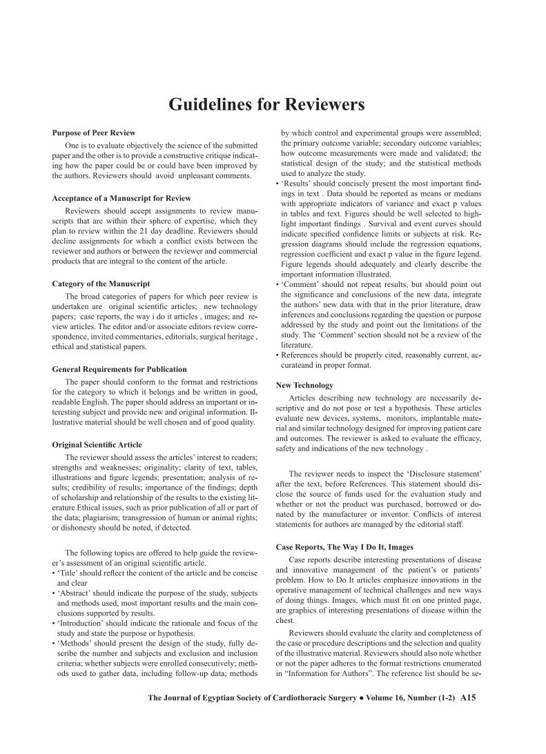
Purpose of Peer ReviewOne is to evaluate objectively the science of the submitted
paper and the other is to provide a constructive critique indicat--ing how the paper could be or could have been improved by the authors. Reviewers should avoid unpleasant comments.
Acceptance of a Manuscript for ReviewReviewers should accept assignments to review manu--
scripts that are within their sphere of expertise, which they plan to review within the 21 day deadline. Reviewers should decline assignments for which a conflict exists between the reviewer and authors or between the reviewer and commercial products that are integral to the content of the article.
Category of the Manuscript The broad categories of papers for which peer review is
undertaken are original scientific articles; new technology papers; case reports, the way i do it articles , images; and re--view articles. The editor and/or associate editors review corre--spondence, invited commentaries, editorials, surgical heritage , ethical and statistical papers.
General Requirements for PublicationThe paper should conform to the format and restrictions
for the category to which it belongs and be written in good, readable English. The paper should address an important or in--teresting subject and provide new and original information. Il--lustrative material should be well chosen and of good quality.
Original Scientific ArticleThe reviewer should assess the articles’ interest to readers;
strengths and weaknesses; originality; clarity of text, tables, illustrations and figure legends; presentation; analysis of re--sults; credibility of results; importance of the findings; depth of scholarship and relationship of the results to the existing lit--erature Ethical issues, such as prior publication of all or part of the data; plagiarism; transgression of human or animal rights; or dishonesty should be noted, if detected.
The following topics are offered to help guide the review--
er’s assessment of an original scientific article. • ‘Title’ should reflect the content of the article and be concise
and clear• ‘Abstract’ should indicate the purpose of the study, subjects
and methods used, most important results and the main con--clusions supported by results.
• ‘Introduction’ should indicate the rationale and focus of the study and state the purpose or hypothesis.
• ‘Methods’ should present the design of the study, fully de--scribe the number and subjects and exclusion and inclusion criteria; whether subjects were enrolled consecutively; meth--ods used to gather data, including follow-up data; methods
Guidelines for Reviewersby which control and experimental groups were assembled; the primary outcome variable; secondary outcome variables; how outcome measurements were made and validated; the statistical design of the study; and the statistical methods used to analyze the study.
• ‘Results’ should concisely present the most important find--ings in text . Data should be reported as means or medians with appropriate indicators of variance and exact p values in tables and text. Figures should be well selected to high--light important findings . Survival and event curves should indicate specified confidence limits or subjects at risk. Re--gression diagrams should include the regression equations, regression coefficient and exact p value in the figure legend. Figure legends should adequately and clearly describe the important information illustrated.
• ‘Comment’ should not repeat results, but should point out the significance and conclusions of the new data, integrate the authors’ new data with that in the prior literature, draw inferences and conclusions regarding the question or purpose addressed by the study and point out the limitations of the study. The ‘Comment’ section should not be a review of the literature.
• References should be properly cited, reasonably current, ac--curateand in proper format.
New TechnologyArticles describing new technology are necessarily de--
scriptive and do not pose or test a hypothesis. These articles evaluate new devices, systems, monitors, implantable mate--rial and similar technology designed for improving patient care and outcomes. The reviewer is asked to evaluate the efficacy, safety and indications of the new technology .
The reviewer needs to inspect the ‘Disclosure statement’ after the text, before References. This statement should dis--close the source of funds used for the evaluation study and whether or not the product was purchased, borrowed or do--nated by the manufacturer or inventor. Conflicts of interest statements for authors are managed by the editorial staff.
Case Reports, The Way I Do It, ImagesCase reports describe interesting presentations of disease
and innovative management of the patient’s or patients’ problem. How to Do It articles emphasize innovations in the operative management of technical challenges and new ways of doing things. Images, which must fit on one printed page, are graphics of interesting presentations of disease within the chest.
Reviewers should evaluate the clarity and completeness of the case or procedure descriptions and the selection and quality of the illustrative material. Reviewers should also note whether or not the paper adheres to the format restrictions enumerated in “Information for Authors”. The reference list should be se--
A16 The Journal of Egyptian Society of Cardiothoracic Surgery ● Mar-Jun 2008
lective rather than inclusive.
Review ArticleReviewers should assess the importance of the subject
matter, need for the review and probable interest to readers. Reviews of very rare and unusual diseases are discouraged . Reviewers should note if authors have respected the format and restrictions of this category as stated in “Information for Authors”.
The ‘Introduction’ should provide the rationale for re--viewing the subject matter and provide the outlines of what is included and not included in the review. In the ‘Methods’ section reviewers should assess the methods used to search for articles, including search words and databases probed. The body of the review should be well organized with well chosen topical headings arranged in logical order. Within each topi--
cal heading the material should be presented in an integrated, comprehensive, objective manner. Statements should be refer--enced accurately. Reviewers should look for a “summing up” of the topical content .
The review should provide a general overview of the sub--ject matter assessing progress, pointing out deficiencies in present management and indicating opportunities and direc--tions of future work. The reviewer should also assess the selec--tion of references .
FootnoteThe reviewer remains anonymous . The reviewer should
direct his or her critique to the authors in the style and format that suits them best. The recommendation to the editor is made separately with or without additio

A17The Journal of Egyptian Society of Cardiothoracic Surgery ● Volume 16, Number (1-2)
Events of InterestThe 15th Annual Conference of the Egyptian Society of Cardiothoracic Surgery Cairo - Egypt
Timing : .........................................................................12 - 14 March 2008 Location: ........................................................................Cairo Sheraton Hotel Email : ......................................................................... [email protected]
■ 15th Annual Echocardiographic Work--shop on 2-D and Doppler Echocardiography-Vail, • Colorado—March 9-13, 2008 For more information on this meet ing, contact Mayo Clinic, 200 First St, SW/GO-06-138SW, Rochester, MN 55905.
■ 5th Annual Course on Exrracorporeal Mem--brane Oxygenation-Jeddah, Saudi Arabia—March 10-12, 2008For more information on this meet ing, contact Faiz Almalki, Rawdah Street (Khalediyah District), PO Box 40047 (MBC-J 16), Jeddah 21499, Saudi Aiabia; telephone: (966-2) 667-7777, ext 2166; e-mail: fiaz_winner<§1 gmailxom or faizwin--ner@awalnet. net-sa; website: www.kfshrcj.org.
■ 15th Annual Mayo Clinic Arrhythmias and the Heart-Kauai, Hawaii—March 10-13, 2008For more information on this meet ing, contact Mayo Clinic, 200 First St, SW/GO-06-138SW, Rochester, MN 55905; tel--ephone: (507) 266-0677; e-mail: [email protected].
■ 16th Annual Meeting of the Asian Society for Cardiovascular Surgery-Singapore, Singa--pore—March 13-16, 2008For more information on this meet ing, contact ASCVS Con--gress Secre tariat, c/o The Meeting Lab Pte Ltd, 176 B Joo Chiat Rd, Singapore 427447, Singapore; telephone: +65 63464402; fax: -65 63464403; e-mail: [email protected]; website: www. ascvs2008.com.
■ CTS Critical Care 2008-Washington, DC—March 27-29, 2008For more information on this meet ing, contact Alexander T. Taft III, Foundation for the Advancement of CTS Care, 616 E St NW, Suite 316, Washington, DC 20004; telephone: (202) 536-4822; fax: (202) 478-1669;e-mail: [email protected]; web site: http://ctscriticalcare.ws.
■ American College of Cardiology 57th Annual Scientific Session-Chicago, Illinois—March 29-April 1,2008For more information on this meet ing, contact American
College of Cardiology Foundation, Heart House, 2400 N St NW, Washington, DC 20037; telephone: (202) 375-6000; fax: (202) 375-7000; website: www. acc.org.
■ Houston Aortic Symposium: Frontiers in Cardiovascular Diseases-Houston, Tex--as— April 4-6, 2008For more information on this meet ing, contact Promedica International CME, 2333 State St, Suite 203, Carls bad, CA 92008; telephone: (760) 720-2263; fax: (760) 720-6263; e-mail: [email protected].
■ International Society for Heart and Lung Transplantation 28th Annual Meet--ing and Scientific Sessions-Boston, Massa--chusetts—April 9-12, 2008For more information on this meet ing, contact International Society for Heart and Lung Transplantation, 14673 Midway Rd, Suite 200, Addi-son, TX 75001, telephone: (972) 490-9495; fax: (972) 490-9499; e-mail: [email protected]; website: www.ishit, org.
■ Vascular and Endovascular Consensus Update-London, United Kingdom—April 12-15, 2008For more information on this meet ing, contact Chris Tim--mins, BIBA Medical Ltd, 44 Burlington Rd, Lon don SW6 4NX, UK; telephone: 44 (0) 20 7736 8788; fax: 44 (0) 20 7736 8283; e-mail: [email protected]; website: www.cxsymposium.com.
■ American Surgical Association 128th Annual Meeting-New York, New York—April 24-26, 2008For more information on this meet ing, contact American Surgical As sociation, 900 Cummings Center, Suite 221-U, Beverly, MA 01915; e-mail: [email protected]; website: www. americansurgical.info.
■ 57th International Congress of The Eu--ropean Society for Cardiovascular Sur--
A18 The Journal of Egyptian Society of Cardiothoracic Surgery ● Mar-Jun 2008
gery-Barcelona, Spain—April 24-27, 2008For more information on this meet ing, contact Professor Claudio Mu-neretto, ESCVS Secretary General, UDA Car--diochirurgia, Spedali Civili PJe Spedali Civili, 25123 Brescia, Italy; telephone; +39 030 3996401; fax: +39 030 3996096; e-mail: [email protected]; website: www .escvs.org.
■ 18th World Society of Cardiothoracic Surgeons World Congress-Kos Island, Greece— April 30-May 3, 2008For more information on this meet ing, contact 18th WSCTS Congress Secretariat, 29 Sinopis Str 11527, Athens, Greece; telephone: +30 210 7799261; fax: +30 210 7711768; e-mail: [email protected]; website: www .wscts2008.com.
■ Aortic Symposium 2008-New York, New York—May 8-9, 2008For more information on this meet ing, contact American As--sociation for Thoracic Surgery, 900 Cummings Center, Suite 221-U, Beverly, MA 01915; telephone: (978) 927-8330; fax: (978) 524-8890; e-mail: aats@prri. com; website: www.aats.org.
■ 88th Annual Meeting of the American Association for Thoracic Surgery-San Di--ego, California—May 10-14, 2008For more information on this meet ing, contact 900 Cummings Center, Suite 221-U, Beverly, MA 01915; telephone: (978) 927-8330; website: www. aats.org/annualmeeting.
■ 16th World Congress of Cardiology 2008-Buenos Aires, Argentina—May 18-21, 2008For more information on this meet ing, contact telephone: +54 11 4812-3444; e-mail: wcc2008@congresosint. com.ar; web--site: www.worldheart.org.
■ 4th International Conference on Pe--diatric Mechanical Circulatory Support Systems, Pediatric Heart Transplanta--tion, and Pediatric Cardiopulmonary Perf usion-Portland, Oregon— May 22-24, 2008For more information on this meeting, contact Perm State College of Medi cine CME Department, PO Box 851, Here--hey, PA 17033; telephone: (717) 531-6483; fax: (717) 531-5604; e-mail: [email protected]; website: www. hmc. psu.edu/ce/pe diatrics.
■ Innovations in Treatment of Cardiac Structural Disease: The Mediterranean Meeting-Palermo, Sicily, Italy—June 6-7, 2008For more information on this meet ing, contact University of Pittsburgh Medical Center, Division of Car diac Surgery, The
Heart, Lung, and Esophageal Surgery Institute, UPMC Pres--byterian, Suite C-700, 200 Lothrop sC Pittsburgh, PA 15213; telephone: (412) 802-6591; fax: (412) 648-6358; e-mail: [email protected]; website: http://ccehs. upmc.edu.
■ Western Thoracic Surgical Association 34th Annual Meeting-Kona, Hawaii—June25-28, 2008For more information on this meet ing, contact Western Thoracic Surgi cal Association, 900 Cummings Cen ter, Suite 221-U, Beverly, MA 01915; telephone: (978) 927-8330; fax: (978) 524-8890; e-mail: [email protected]; website: www.west--ernthoracic.org.
■ 14th World Congress on Heart Disease: International Academy of Cardiology An--nual Scientific Sessions 2008-Toronto, On--tario, Canada-July 26-29, 2008For more information on this meet ing, contact Asher Kimchi, MD, In ternational Academy of Cardiology, PO Box 17659, Beverly Hills, CA 90209; telephone: (310) 657-8777; fax: (310) 659-4781; e-mail: klimedco@uda. edu; website: www.cardiologyonline, com.
■ 22nd Annual Meeting of the European Association for Cardio-Thoracic Surgery-Lisbon, Portugal—September 13-17, 2008For more information on this meet ing, contact EACTS Exec--utive Secre tariat, 3 Park St, Berkshire, SL4 1LU; telephone: +44 1753 832166; fax: +44 1753 620407; e-mail: [email protected].
■ American College of Surgeons Annual Meeting-San Francisco, California—Octo--ber 12-16, 2008For more information on this meet ing, contact website: www.facs.org/ index.html.
■ 6th Triennial Brigham CardiaValve Symposium-Boston, Massachusetts-Octo--ber 23-24, 2008For more information on this meet ing, contact R. Morton Bolman III, MD, Brigham and Women’s Hospi tal, 75 Francis St, Boston, MA 02115; telephone: (617) 732-6964; fax: (617) 732-6559; e-mail: [email protected]; website: www.brigha--mandwomens. org/ ca rdi acsurge ry / e vents.aspx.
■ Chest 2008-Philadelphia, Pennsylvania october 25-30,2008For more information on this meet ing, contact American College of Chest Physicians, 3300 Dundee Rd, Northbrook, IL 60062-2348; tele phone: (847) 498-1400; website: www. chestnet.org.

EditorialEditorial
1The Journal of Egyptian Society of Cardiothoracic Surgery ● Volume 16, Number (1-2)
As winds of change are still blowing our journal chief in editor is leaving us being assigned to a more important post director of the new cardiothoracic center at Ain Shams university , Hoda Talaat Harb center .
Professer Ezz Eldin Mostafa has been always the leading drive in every position and responsibility he has under taken .
I was fourtunate to be his faithful student and follower throughout my carrer trying to find the right path with his continuous helps and assistance.
His abilites and skills as a professor of cardiothoracic surgery were all the time as excellent as it could be really wraped in a humble nice and always smiley attitude.he never turned anybody who seeked his help down and that despite his ultra busy schedule.
Professor Dr Ezz is the man of the job all the time very precise very simple doing any job as it must be done.
We as the board of the journal owe him a lot and would like to express our sincere gratitude and thanks to this greatman and professor who led us during the past four years.
We wish him all the suceess in his present post and tell him that he still our chief and consltant.
I would like as well to welcome to the board some distirguished professors and surgeous who will be sharing the responsibilities and carrying together the heavy tasks .
Dr Ahmed Deebis , Dr Ahmed Hassoura,Dr Ahmed El Kerdani and Dr Mohamed Nasr as coeditors.
This in addition to our new associate section editors Dr Ahmed el Nour,Dr Ashraf Bassiony,Dr Samir Keshk,Dr Samir Hossom and Dr Momdouh Sharawi.
Wishing them all the best Yasser Hegazy MD,FRCS
DISTIRGUISHED MAN AND KNOWLEDGEABLE PROFESSOR
Editorial
Cardiovascular
2 The Journal of Egyptian Society of Cardiothoracic Surgery ● Mar-Jun 2008
OFF-PUMP VERSUS ON-PUMP FOR MULTIVESSEL CORONARY ARTERY BYPASS GRAFTING: COMPARATIVE
STUDY OF OPERATIVE AND SHORT-TERM OUTCOMESBackground: patients undergoing multivessel coronary artery bypass grafting (CABG) are at increased risk of death, stroke, or myocardial infarction. Our objective was to compare the operative and short-term outcomes of multivessel CABG on the beating heart (OPCAP) versus con--ventional surgery with cardiopulmonary bypass (ONCAB) to determine which technique has the better early benefits and outcomes.Methods: This prospective study included 150 consecutive patients who underwent elective isolated multivessel CABG which done either by OP--CAB (68 patients) or ONCAB (82 patients) and followed up over a period of 40 months. All preoperative, intraoperative and postoperative data of all patients were collected, analyzed and compaired between OPCAB and ONCAB groups.Results: The total operating time was significantly less in the OPCAB group (p <0.01). Postoperatively, ventilation time, blood loss and blood and blood product transfusion requirements were significantly less in OPCAB group patients (p <0.01). The major postoperative complications acknowl--edged as indicators for benefits did not result in any significant difference between the two groups. There was no significant statistical difference in overall mortality rate between the two groups. A significant early benefit in OPCAB surgery were the dramatic decrease in the length of intensive care unit (ICU) and hospital stay and the better postoperative biochemical profile of OPCAB group patients (p <0.05). At follow-up, both groups had similar frequency for readmission and similar quality of life. Conclusions: Both OPCAB and ONCAB is a safe and effective for patients with multivessel coronary artery disease with equal operative and short-term outcomes. There are overall early benefits in OPCAB surgery; pa--tients have a better post-operative biochemical profile, dramatic decrease in the length of stay in the ICU and hospital with quicker mobilization of patients.
The development of cardiac surgery in the last 30 years is directly related to the improvement of the techniques of cardiopulmonary bypass (CPB). However, CPB utilization constitutes one of the primary causes of perioperative complications [1]. These compli--cations relate to hypoperfusion, systemic inflammatory response,
cardioplegia administration and interruption of normal blood flow to the aorta with potential complications to the brain and the kidneys [1-4]. In an attempt to avoid the above mentioned complications without jeopardizing the benefits, there was recently a renewal of interest in the performance of coronary ar--tery bypass grafting (CABG) without using CPB (OPCAB). The technical im--provements in OPCAB surgery such as the development of several stabilizers,
Department of cardiothoracic surgery, Zagazig University, Egypt.Accepted for publication october 1,2007 Address reprint request toDr/ Mohamed EssaPostal address: Department of cardio--thoracic surgery , Zagazig University- Zagazig- Egypt.Mobile: 0102246761E-mail: [email protected] : 04/cord/64/0710
Mohamed Essa MD, Ahmed Deebis MD,Mamdouh Sharawy MD,Khalid abdelbariy MD,Ehab Yehia MD.

Essa et al Cardiovascular
3
Car
diov
ascu
lar
The Journal of Egyptian Society of Cardiothoracic Surgery ● Volume 16, Number (1-2)
positioning devices and coronary occluder shunts have made OPCAB a more popular alternative to convention--al surgery with CPB (ONCAB) for treating multivessel coronary artery disease [5-14].
Up to now, the access to deep areas that have to be re--vascularized remains problematic in OPCAB technique , as to bypass the circumflex system and the posterolateral branches of the right system the surgeon must expose the posterolateral aspect of the heart by displacing the heart vertically, which may compromise the hemodynamic stability and may induce dramatic hypoperfused state that may contribute to an increase in the incidence of complications or mortality[15].
Comparing multivessel CABG by OPCAB tech--nique versus ONCAB technique was not well docu--mented in literature [5]. For this purpose, our objective was to compare the operative and short-term outcomes of multivessel coronary revascularization by OPCAP versus ONCAB to determine which technique has the better early benefits and outcomes.
Patients and methodsThis prospective study included all planned 150 con--
secutive patients who underwent elective isolated mul--tivessel CABG for multivessel coronary artery disease which done either by OPCAB or ONCAB techniques and followed up between the periods of October 2004 to January 2008 (40 months) in Zagazig University Hos--pital. All procedures were performed by experienced surgeons and anaesthesia was exclusively managed by experienced anaesthesiologists.
The decision for a patient to undergo OPCAB or ONCAB techniques was made by the operating surgeon. All surgeons in our department perform OPCAB and ONCAB procedures with nearly equal distribution. Ex--clusion criteria included: 1) The presence of ≥ grad II mitral valve insufficiency, 2) Coronary endarterctomy, 3) Incomplete revascularization of the multivessel coro--nary disease due to severe calcification of the coronar--ies and 4) Redo surgery. Also, patients whose coronary revascularization was initially attempted without car--diopulmonary bypass and who required conversion to cardiopulmonary bypass (typically due to hemodynamic instability) were also excluded form the study. Such pa--tients’ potentially increased incidence of complications may disadvantage one method for revascularization in comparison with the other [1, 14].
Method of Anesthesia and hemodynamic moni--toring:
The anesthesia protocol applied was the same for all patients. All patients received the same general anesthet--
ic technique with standard hemodynamic monitoring including mean arterial and central venous pres--sures and electrocardiography. Oxygen saturation was continuously monitored with a pulse oximeter. After harvesting of conduits, Heparin was injected in a dose of 2 mg/kg in patients who underwent operation with--out CPB and 3 mg/kg in patients with CPB. Activated clotting time was measured initially and then every 30 minutes; it was maintained for more than 300 seconds in patients who underwent surgery off pump and more than 400 seconds in those who had their surgery on pump. Protamine sulfate was used in 1:1 ratio to reverse the heparin effect after the procedure.
Surgical technique for off-pump coronary ar--tery bypass surgery
All patients underwent operation through a median sternotomy. The left internal mammary artery (LIMA) was harvested by standard technique using hemoclips. The other conduits (saphenous veins and radial artery) were harvested simultaneously.
Distal anastomoses were always constructed before proximal anastomoses. In most cases, the left anterior descending coronary artery was the first coronary artery to be grafted. The right coronary artery was always the second artery to be grafted. The vessels on the lateral and posterior wall were usually grafted last. However, the sequence of grafting was individualized for a partic--ular patient, depending on the severity of the lesions in different coronary arteries and patient’s hemodynamics.
Deep retracting sutures, the placement of a warm moist laparotomy sponge in the posterolateral aspect of the pericardial sac, Trendelenburg position, and right tilt were used to facilitate exposure of the lateral and poste--rior vessels of the heart. Octopus tissue suction stabi--lizer (Medtronic, Inc., Minneapolis, MN, USA) was used to stabilize the myocardium. During the construc--tion of all anastomosis, target vessel hemostasis was ob--tained with proximal and distal silicone rubber (Silastic; Dow Corning, Midland, Mich) vessel loops as coronary snares and intracoronary shunts (Anastafloa, Research Medical, Midvale, UT, USA) were used for most of the anastomosis. The anastomoses were constructed with a single running stitch of 7-0 or 8-0 polypropylene (Prole--neTM, Johnson & Johnson, New Brunswick, NJ, USA) and visualization was improved with use of a continuous air/saline blower.
Proximal anastomosis was performed with partial clamping of the ascending aorta using standard tech--niques. The proximal anastomosis was performed using a single running stitch of 6-0 Prolene suture.
Cardiovascular Essa et al
4
Car
diov
ascu
lar
The Journal of Egyptian Society of Cardiothoracic Surgery ● Mar-Jun 2008
Surgical technique for on-pump coronary artery bypass surgery
Conventional coronary artery bypass procedures were performed through a median sternotomy using standard CPB, which was established after full hepa--rinization (at a dose of 3 mg/kg),using ascending aortic and two-stage venous cannulation with the use of mem--brane oxygenator and a roller pump. Arterial flow was adjusted to 2.2-2.4L/min/m-2, and blood pressure was maintained between 50 and 70mmHg. Haematocrit level was kept at >22%. The patient was not actively cooled but temperature was allowed to drift was maintained at 34ºC. Intermittent Antegrade warm blood cardioplegia was used for myocardial protection. Cardioplegia was repeated after every distal anastomosis.
In both Surgical techniques, OPCAB and ONCAB, the total operative time was defined as time from skin incision to closure of the skin.
Postoperative clinical and biochemical param--eters
To evaluate the in-hospital morbidity of both tech--niques, several clinical parameters were analyzed (e.g. the intubation time, the administration of inotropic drugs, the amount of blood transfusions, the incidence of perioperative myocardial infraction (MI), stroke (CVA), transient ischemic attack (TIA), atrial fibrillation (AF) and renal failure (RF), period of stay in intensive care unit (ICU) and hospital).
Blood loss was defined as total chest tube drainage until chest tubes were removed. Prolonged ventilation was defined as ventilation for more than 48 hours. The perioperative diagnosis of MI was based on the presence of two out of three standard criteria: a) development of a new pathological Q-wave or loss of R wave progression, new left bundle branch block, or new ST and T wave changes on postoperative electrocardiography, and b) in--crease of the creatine kinase (CK) level more than 40 U/L or increase of CK-MB value by more than 30% of the total value of CK. The development of a local or atypi--cal precordium pain was not considered an MI criterion. Mediastinisis was defined as mediastinal collection with positive culture. Acute RF was defined as requirement of peritoneal or hemodialysis.
The biochemical markers that were included in the analysis were a) creatinine kinase myocardial fraction band (CK-MB) value, and b) serum creatinine value (Cr).Follow-up:
At 6 months post-procedure, follow-up was done by
a standardized questionnaire that focused on quality of life, lifestyle activities, recurrent angina, frequency and intensity of angina, and reporting rehospitalization for specific cardiac causes and follow-up mortality. Echo--cardiography was done for all patients at 6 months.
To compare the operative and short-term outcomes of multivessel coronary revascularization by OPCAP versus ONCAB, all preoperative, intraoperative and postoperative data of all patients were collected, ana--lyzed and compaired between both groups.
Statistical analysisThe Values of variables data were presented as a
mean value ± standard deviation (mean ± SD). Preop--erative, intraoperative, and postoperative variables were analyzed and compared between these two groups us--ing Student’s t test and univariate analysis (chi-square test (χ2), Fisher’s exact test). Statistical significance was defined as a p value of less than 0.05(p<0.05). Statistical analyses were performed using SPSS for Windows ver--sion 11.5 statistical package (SPSS, Inc, Chicago, Ill).
ResultsThe preoperative variables of the all patients of the
two groups are shown in Table 1. There were no differ--ences between the two groups as regards gender, age, severity of the coronary artery disease, ejection fraction and preoperative co-morbidities and risk factors, except that there were significantly more patients with hyper--cholesterolemia and history of CVA or TIA in the OP--CAB Group (p <0.05) (table 1).
*Table (1)
There were more patients with aortic arteriosclerosis in the OPCAB Group, which was statistically significant (p <0.05). The total number of grafts used in OPCAB group and ONCAB group were 2.6 ± 0.4 and 2.7 ± 0.5 respectively; hence such differences were not statistical--ly significant. Venous grafts were used in all patients in both groups. LIMA was used in 96% and 95% patients in the OPCAB and ONCAB groups, respectively. Ra--dial arterial grafts were use infrequently in both groups. Differences were not statistically significant between both groups as regard the types of grafts used. Intraop--eratively, there were significantly more patients in the OPCAB group required inotropic drugs than those in the ONCAB group (p <0.01). The total operating time was significantly less in the OPCAB group (p <0.01) (table 2).
*Table (2)
Essa et al Cardiovascular
5
Car
diov
ascu
lar
The Journal of Egyptian Society of Cardiothoracic Surgery ● Volume 16, Number (1-2)
Table 3 summarizes the results regarding the postop--erative outcome data for the two groups. During the stay in ICU, ventilation time, blood loss and blood and blood product transfusion requirements were significantly less in OPCAB group patients (p <0.01). The major postop--erative complications acknowledged as indicators for benefits did not result in any significant difference be--tween the two groups (Table 3). The incidence of post--operative MI, stroke, renal dysfunction, pulmonary in--fection, new AF and sternal infection were comparable between the two groups (p = NS). Significant differences were observed in the biochemical markers of the study; OPCAB group patients have a better post-operative bio--chemical profile (p <0.05) (Table 3). A significant early benefit in OPCAB surgery was the dramatic decrease in the length of stay in the ICU and hospital with quicker mobilization of OPCAB group patients (p <0.05) (Table 3). There was no significant statistical difference in to--tal 30-day postoperative mortality rate between the two groups (p = NS).
*Table (3)
At the time of 6-months follow-up, both groups had improvement in LVEF by about 2-5% but with no sig--nificant statistical difference between both groups. Both OPCAB and ONCAB groups had nearly the same fre--quency for readmission to the hospital since discharge (4.4% vs 4.9% respectively; p = NS). Reasons for re--admission focused on specific cardiac causes indica--tions: recurrent angina (1.5% vs 2.4%), congestive heart failure (1.5% vs 1.2%), arrhythmias (1.5% vs 1.2%) for OPCAB Group and ONCAB Group, respectively; and all were non-significant (Table 4). Additionally, patient-reported quality of life and lifestyle activities were simi--lar in both groups. There was no significant statistical difference in follow-up mortality rate between the two groups (p = NS).
The overall mortality rate for OPCAB Group and ONCAB Group were 2.9% and 3.7 % respectively, which was non statistically significant (p = NS).
*Table (4)
DiscussionMultivessel cardiac disease patients present more
frequently with multifactorial disease processes such as renal, pulmonary, neurologic, and peripheral vascular disease [5]. Accordingly, patients undergoing multives--sel coronary artery bypass surgery are at increased risk of death, stroke, or MI compared with patients who need
fewer diseased vessels [14]. ONCAB offers excellent symptomatic relief, carries a low operative mortality, and provides excellent long-term survival benefit. De--spite these excellent results, the adverse effects of extra--corporeal circulation and cardioplegic arrest may aggra--vate preexisting complications and disorders. CPB and cardioplegic arrest can cause myocardial dysfunction, negative central nervous system effects, neuropsychiat--ric phenomena, severe systemic inflammatory response, and coagulopathy associated with end-organ injury [5]. Accordingly, OPCAB has recently gained renewed in--terest as an alternative to ONCAB for multivessel revas--cularization to avoid CPB related complications [5- 14]. However, the role of OPCAB is still vaguely defined and is being critically evaluated. The main explanation for this difficulty in defining the role of off-pump proce--dures appears to be the lack of conclusive data and con--troversy with respect to the potential benefit (mortality, morbidity, graft patency and long-term outcome) of this approach compared with ONCAB [16].
Concerns regarding higher incidence of incomplete revascularization, recurrent angina, and early graft oc--clusion have prevented OPCAB from being universally adopted [5, 16]. There is no doubt that completion of the graft to coronary artery anastomosis is more difficult in the OPCAB procedure due to the cardiac motion and lack of a blood-free operative field, compared to con--ventional coronary artery bypass grafting on an arrested, flaccid heart [16]. Despite the availability of cardiac sta--bilizers and positioning devices, up to now, one of the main disadvantages of the technique is the difficulty of its application in bypassing the lateral and inferior wall vessels in triple vessel disease patients as the anastomo--sis is often performed in a vertical position [5, 15, 16]. This creates a parallax view with poor depth of vision for judging distance between stitches and contributes to the theoretical possibility of poor quality of the anastomosis and less anastomotic accuracy [16]. The coronaries un--dergo significant spasm when manipulated on a hemo--dynamically active heart. Also, the vertical displacement may compromise the hemodynamic stability and may induce dramatic hypoperfused state that may contribute to an increase in the incidence of complications or mor--tality [15, 16]. A recent meta-analysis of graft patency by Takagi et al. demonstrated a 27% increase in overall graft occlusion with OPCAB, especially a 28% increase in venous graft occlusion [17].
Current prospective data suggest that both techniques OPCAB and ONCAB have similar rates of mortality in patients with multivessel coronary artery disease [5- 14]. Our results showed that, the total 30-day post-operative mortality rate and the overall mortality rate did not differ
Cardiovascular Essa et al
6
Car
diov
ascu
lar
The Journal of Egyptian Society of Cardiothoracic Surgery ● Mar-Jun 2008
significantly between the two techniques (2.9% vs 2.4%, and 2.9% vs 3.7%, respectively).
As regard the morbidity in the postoperative period, perioperative bleeding and transfusion-related compli--cations are among the major risks associated with open heart surgery. Avoidance of CPB has been reported to be beneficial in reducing blood product use [9, 18, 19]. In the present study, postoperative bleeding and transfusion requirements were significantly less with OPCAB tech--nique (p <0.01). Also, OPCAB group patients clearly needed significantly less ventilation time after the op--eration (p <0.01), a finding that is consistent with that of other researchers [5, 9, 18, 19].
However, in this study, the differences in postop--erative major complications acknowledged as indica--tors for benefits were non significant between the two groups which is consistent with that of other studies [5, 9, 18, 19]. The postoperative stroke rate in our study was comparable in the two groups, though there were more patients in the OPCAB group had arteriosclerosis of the aorta than those in the ONCAB group. After refinements in CPB techniques, especially use of membrane oxygen--ators and arterial line microfilters, a lowered incidence of postoperative neurologic dysfunction have been dem--onstrated [5]. Some studies [11, 14] suggest a decrease in stroke rates for OPCAB patients, but according to the most recent meta-analysis by Takagi and associates, randomized clinical trials of OPCAB versus ONCAB, postoperative 30-day stroke was not statistically signifi--cantly reduced in the OPCAB patients [20].
Also, in this study, we observed that incidence of postoperative TIA, MI, new RF, and AF were similar and statistically non-significant between the two groups, a finding that recent studies on multivessel coronary re--vascularization showed the same pattern [5, 9, 10, 14]. Also, the recent meta-analysis study by Cheng and as--sociates [19] showed no significant differences in MI, stroke, renal dysfunction, intra-aortic balloon pump (IABP) support and wound infection between OPCAB and ONCAB techniques, meanwhile, OPCAB signifi--cantly decreased AF. However, results of neurocognitive function were inconclusive in this meta-analysis.
As regards the biochemical parameters, the pattern of perioperative enzyme release after either procedure is still contradictory [5, 12, 15, 18]. In this study, we observed significantly lower CK-MB values for patients operated by OPCAB technique (p <0.01). Although this finding is directly related to the number of periopera--tive MI, it seems that the cardioplegia administered to patients on whom CPB is used, greatly affects the post-operative increase of myocardial enzymes. Also, the difference in post-operative creatinine values of the pa--
tients of two groups was significant. Patients operated by OPCAB technique had significantly lower creatinine values (p <0.05). The renal function is a parameter that has been recently studied and the data to-date show a clear superiority of off-pump surgery on the beating heart versus CPB surgery with respect to maintenance of normal creatinine clearance values [2].
Nevertheless, both the mean stay in ICU and hos--pital were significantly less in OPCAB group patients (p <0.05) and this was important factor in quicker mo--bilization of patients in the postoperative period with early benefit in OPCAB patients. These findings are in accordance with the studies of Kshettry et al. [5] and Maharwal et al. [9] and the meta-analysis by Cheng and associates [19].
At follow up, the results presented here seem to in--dicate that both techniques provide similar rates of im--provement of LVEF, quality of life, readmission rate and freedom from surgical reintervention. Selvanayagam et al. [21] used cine magnetic resonance imaging to as--sess left ventricular function and newly occurring irre--versible myocardial damage in a consecutive series of 60 patients. Postoperative left ventricular function was significantly better in the OPCABG group. Interestingly, new irreversible myocardial damage does not differ be--tween the two techniques. Kshettry et al. [5] showed that the rate of readmission, quality of life and freedom from surgical re-intervention were similar and statistically non significant between OPCAB and ONCAB patients.
OPCAB remains an emerging and developing surgi--cal technique, and continued reevaluation is warranted. What remains to be confirmed are the quality of the anastomoses and the long-term patency of the grafts [5, 9, 15]. In conclusion, both OPCAB and ONCAB are safe and effective for patients with multivessel coronary artery disease with equal operative and short-term out--comes. Elimination of CPB did not significantly reduce postoperative morbidity. There are overall early benefits in OPCAB surgery; patients have a better post-opera--tive biochemical profile, and dramatic decrease in the length of stay in the ICU and hospital with quicker mo--bilization of patients. However, prospective randomized studies with long-term results comparing these two tech--niques are required.
References
1. Calafiore AM, Di Mauro M, Canosa C, Di Giammarco G, Lorena Iacò A, Contini M. Myocardial revascularization with and without cardiopulmonary bypass: advantages, disadvantages and similarities. Eur. J. Cardiothorac. Surg. 2003; 24:953—960.
Essa et al Cardiovascular
7
Car
diov
ascu
lar
The Journal of Egyptian Society of Cardiothoracic Surgery ● Volume 16, Number (1-2)
2. Weerasinghe A, Athanasiou T, Al-Ruzzeh S, Casula R, Tek--kis PR, Amrani M, Punjabi P, Taylor K, Stanbridge R, Glen--ville B. Functional renal outcome in on-pump and off-pump coronary revascularization: a propensity-based analysis. Ann. Thorac. Surg. 2005; 79:1577—1583.
3. Bucerius J, Gummert JF, Borger MA, Walther T, Doll N, Onnasch FJ, Metz S, Falk V, Mohr WF. Stroke after cardiac surgery: a risk factor analysis of 16,184 consecutive adult patients. Ann. Thorac. Surg. 2003; 75:472—478.
4. Staton GW, Williams WH, Mahoney EM, Hu J, Chu H, Duke GP, Puskas DJ. Pulmonary outcomes of off-pump vs on-pump coronary artery bypass surgery in a randomized trial. Chest 2005; 127:892—901.
5. Kshettry RV, Flavin FT, Emery WR, Nicoloff MD, Arom VK, Petersen JR. Does multivessel, off-pump coronary ar--tery bypass reduce postoperative morbidity? Ann. Thorac. Surg. 2000; 69: 1725 - 1730.
6. Cartier R, Brann S, Dagenais F, Martineau R, Couturier A. Systematic off-pump coronary artery reasculiriztion in multivessel disease: Experience of three hundred cases. J. Thorac. Cardiovasc. Surg. 2000; 119: 221 - 229.
7. Demers P and Cartier R. Multivessel off-pump coronary artery bypass surgery in the elderly. Eur. J. Cardiothorac. Surg. 2001; 20: 908 - 912.
8. Magee JM, Jablonski AK, Stamou CS, Pfister JA, Dewey MT, Dullum KCM, Edgerton RJ, Prince LS, Acuff ET, Cor--so JP, Mack JM. Elimination of cardiopulmonary bypass improves early survival for multivessel coronary artery by--pass patients. Ann. Thorac. Surg. 2002; 73: 1196 - 1203.
9. Meharwal ZS, Mishra KY, Kohli V, Bapna R, Singh S, Tre--han N. Off-pump multivessel coronary artery surgery in high-risk patients. Ann. Thorac. Surg. 2002; 74: S1353 - S1357.
10. Al-Ruzzeh S, Athanasiou T, George S, Glenville EB, DeSouza CA, Pepper RJ, Amrani M. Is the use of cardio--pulmonary bypass for multivessel coronary artery bypass surgery an independent predictor of operative mortality in patients with ischemic left ventricular dysfunction? Ann. Thorac. Surg. 2003; 76: 444 - 451.
11. Mack JM, Pfister A, Bachand D, Emery R, Magee JM, Connolly M, Subramanian V. Comparison of coronary by--pass surgery with and without cardiopulmonary bypass in patients with multivessel disease. J. Thorac. Cardiovasc. Surg. 2004; 127: 167 - 173.
12. Khan NE, De Souza A, Mister R, Flather M, Clague J, Davies S, Collins P, Wang D, Sigwart U, Pepper J. A ran--domized comparison of off-pump and on-pump multives--sel coronary-artery bypass surgery. N. Engl. J. Med. 2004; 350:21—28.
13. Taggart PD. Coronary artery bypass grafting is still the best treatment for multivessel and left main disease, But Patients Need to Know. Ann. Thorac. Surg. 2006; 82: 1966 - 1975.
14. Lattouf MO, Puskas DJ, Thourani HV, Noora J, Kilgo DP, Guyton AR. Does the Number of Grafts Influence Surgeon Choice and Patient Benefit of Off-Pump Over Conventional On-Pump Coronary Artery Revascularization in Multives--sel Coronary Artery Disease? Ann. Thorac. Surg. 2007; 84: 1485 - 1495.
15. Arom VK, Flavin FT, Emery WR, Kshettry RV, Petersen JR, Janey AP. Is low ejection fraction safe for off-pump coronary bypass operation? Ann. Thorac. Surg. 2000; 70: 1021 - 1025.
16. Raja GS, Dreyfus DG. Impact of Off-Pump Coronary Ar--tery Bypass Surgery on Graft Patency: Current Best Avail--able Evidence. J. Card. Surg. 2007; 22:165-169.
17. Takagi H, Tanabashi T, Kawai N, Kato T, Umemoto T. Off-pump coronary artery bypass sacrifices graft patency: meta-analysis of randomized trials. J. Thorac. Cardiovasc. Surg. 2007; 133:e2–3.
18. Puskas JD, Williams WH, Duke PG, Staples JR, Glas KE, Marshall JJ, Leimbach M, Huber P, Garas S, Sammons BH, McCall SA, Petersen RJ, Bailey DE, Chu H, Mahoney EM, Weintraub WS, Guyton RA. Off-pump coronary ar--tery bypass grafting provides complete revascularization with reduced myocardial injury, transfusion requirements, and length of stay: a prospective randomized comparison of two hundred unselected patients undergoing off-pump ver--sus conventional coronary artery bypass grafting. J. Thorac. Cardiovasc. Surg. 2003; 125:797—808.
19. Chen DC, Bainbridge D, Martin JE, Novick RJ. Evidence- Based Perioperative Clinical Outcomes Research Group: does off-pump coronary artery bypass reduce mortality, morbidity, and resource utilization when compared with conventional coronary artery bypass? A meta-analysis of randomized trials. Anesthesiology 2005; 102:188–203.
20. Takagi H, Tanabashi T, Kawai N, Umemoto T. Off-pump surgery does not reduce stroke, compared with results of on-pump coronary artery bypass grafting: A meta-analysis of randomized clinical trials. J. Thorac. Cardiovasc. Surg. 2007; 134:1059-1060.
21. Selvanayagam JB, Petersen SE, Francis JM, Robson DM, Kardos A, Neubauer S, Taggart PD. Effects of off-pump versus on-pump coronary surgery on reversible and irre--versible myocardial injury: a randomized trial using car--diovascular magnetic resonance imaging and biochemical markers. Circulation 2004; 109:345—350.
Cardiovascular Ahmad Darwazah et al
8
Car
diov
ascu
lar
The Journal of Egyptian Society of Cardiothoracic Surgery ● Mar-Jun 2008
THE USE OF OFF–PUMP CARDIOPULMONARY BYPASS IN EMERGENCY MYOCARDIAL REVASCULARIZATION
Background: Emergency coronary artery bypass grafting is associated with increased operative mortality. The use of off- pump coronary artery bypass is a well established technique for elective cases and is associated with low mortality rate even among high risk patients. The objective of this study is to evaluate the efficiency and safety of the use of off-pump surgery in emergency CABG and to compare the outcome with that of conventional CABG.Methods: Seventy nine patients who underwent emergency isolated CABG between April 1999 to April 2005 were included in the study. The criteria for selection of these patients for emergency bypass were based on the guidelines of the Society of Thoracic Surgeons (1). 45 of these patients un--derwent off-pump cardiopulmonary bypass (OPCAB), while the rest (34 patients) had conventional cardiopulmonary bypass (CCAB). Their data was reviewed and evaluated. Patients were followed up to find midterm survival and control of symptoms.Results: Patients undergoing OPCAB had a higher incidence of associated preoperative comorbidities. Their mean ejection fraction was 28±9 vs. 39 ± 10 in CCAB (P<0.001). Cardiogenic shock, acute myocardial infarction < 24 hours and post PTCA complications were seen more frequently in OPCAB patients, but the difference was not significant. Main stem disease was significantly more common in CCAB (P= 0.002). The mean number of grafts per patient was 1.8 ± 0.7 in OPCAB and 3.2 ± 0.8 in CCAB group (P <0.001). Subsequently, completeness of revascularization was greater in CCAB patients (79% vs. 51%; P = 0.01). Overall operative mortality was 11.4%. Patients who underwent CCAB had a higher mortality rate 14.7% vs. 8.9%; P=0.42. Major morbidity was more common among OPCAB patients, with a higher incidence of myocardial infarction, mediastinitis and reopening for extra grafting. Late follow up (15-82.8 months) was achieved in 96% of cases. Late mortality was similar among both groups.Patients operated on with CPB had lower rates of recurrent angina (16 vs. 34%; p = 0.08) and symptoms of congestive heart failure (20% vs. 51%; p = 0.04).The incidence of rehospitalization was more common among OPCAB patients. However, the percentage of cardiac reintervention was similar among both groups (8.6% vs. 8%; p = 0.94). Five – year survival (Kaplan Meier) of patients operated upon without CPB was better (90% vs. 83%), however the difference was not statistically significant (p = 0.94).Conclusions: Patients undergoing emergency CABG are at high risk. The use of either off-pump or conventional CABG is associated with high op--erative and late mortality. Off-pump coronary artery bypass can be per--formed safely among emergency cases. This technique did not show any significant superiority over conventional CABG except among patients with myocardial infarction less than 24 hours.
Accepted for publication october 10 ,
2007 Address reprint request to
Dr: Ahmad K Darwazah Division of
Thoracic and Cardiovascular Surgery ,
Makassed Hospital, mount of olives Po
box 19482
E-mail:[email protected]
codex : 04/cord/65/0710
Ahmad Darwazah MD,FRCSRaed. Abu Shama M.DIsmail Isleem PhD, MRCPBasel Hanbali² MRCPBashar Jaber M.D

Ahmad Darwazah et al Cardiovascular
9
Car
diov
ascu
lar
The Journal of Egyptian Society of Cardiothoracic Surgery ● Volume 16, Number (1-2)
Off-Pump coronary artery bypass (OPCAB) is a well established technique used in myocardial revasculariza--tion. It has been shown to reduce both the mortality and morbidity which are associated with extra corporal cir--culation [2].
Initially, it was used in highly selected cases espe--cially in young patients with single or double coronary artery disease. Indications have gradually expanded, especially with the advanced development of cardiac stabilizers. It is now routinely used in multi-vessel coro--nary disease. Recent studies have shown the importance of using off-pump technique among high risk patients with multiple comorbidities [3-5], and proved its superi--ority over conventional coronary artery bypass (CCAB) surgery [5, 6].
Mortality of patients undergoing emergency coronary revascularization whether after acute myocardial infarc--tion (MI) or post angioplasty complications remains high [7, 8]. Avoiding the inflammatory reaction and ischemic injury caused by cardiopulmonary bypass (CPB) in these patients may be of great benefit. The objective of this study is to evaluate the efficiency and safety of the use of OPCAB in emergency coronary revascularization and to compare the outcome with CCAB.
Patients and MethodsSeventy nine patients who underwent emergency
isolated coronary artery bypass surgery between April 1999 to April 2005 were included in this study. Those with combined procedures or with beating heart on pump were excluded. Patients were divided into two groups according to the technique used. Group I (OP--CAB) included 45 patients who underwent off-pump op--erations and group II (CCAB) 34 patients who had con--ventional bypass. The emergency status of these patients was based on the guidelines of the Society of Thoracic Surgeons [1] which includes ongoing ischemia inspite of maximum medical therapy, acute evolving myocardial infarction and cardiogenic shock with or without sup--port. The criteria used to select either technique were based on the hemodynamic status of the patients, the anatomy of the coronary vessels and associated risk factors. Patients with diffuse multi-vessel disease, poor quality vessels, evidence of coronary calcification and hemodynamic instability were considered for CCAB by--pass. Those patients with associated co-morbidities, low ejection fraction (EF), suitable coronary anatomy (i.e. accessible vessels, no evidence of calcification and good vessel size), and acceptable hemodynamic state were considered candidates for OPCAB surgery.
Surgical techniqueHemodynamic stabilization was maintained either by
using inotropic drugs or by an intra aortic balloon pump (IABP). Exposure of the heart was done using standard median sternotomy in all patients. After harvesting sa--phenous vein and left internal mammary artery, full heparinization was done keeping the activated clotting time more than 400 seconds in both groups. Standard cannulation of the aorta and right atrium was used in CCAB group. Myocardial protection was achieved by intermittent blood cardioplegia given antegradely and systemic hypothermia at 32oC.
In the OPCAB group, anterior vessels were simply exposed by elevation of the heart. Deep pericardial re--traction sutures were used to expose circumflex and right coronary artery. The Starfish device (Medtronic, Inc, Minneapolis, MN) was used to expose the circumflex artery since early 2005. Stabilization of target vessels was accomplished by either pressure U-shaped foot sta--bilizer (Guidant, Indianapolis, IN) or suction stabilizer (Medtronic Octopus II and III). Intra coronary shunts (Medtronic Inc.) were used in constructing bypass grafts in all cases.
Different preoperative and postoperative variables were collected and analyzed. These variables are pre--sented as mean ± standard deviation using student’s t test. The data were expressed as percentage and com--parison was made using the Fisher exact test. Postop--erative survival was expressed by the Kaplan – Meier Method. Statistical significance was assumed if p value < 0.05 (2-tailed). Analyses were performed using SPSS 13 software (Chicago, IL).
ResultsPreoperative patients’ demography and risk factors
among both groups are listed in (Table 1). The majority of patients who underwent OPCAB
had either single or double vessel disease (Table 2). The mean number of grafts per patient was 1.8 ± 0.7 and 3.2 ± 0.8 among OPCAB and CCAB respectively (p < 0.001). Grafting of obtuse marginal and posterior descending artery was much higher among CCAB (P < 0.001). Left internal mammary arteries was used in 71% of CCAB patients and in 67% of OPCAB (p = 0.71). Completeness of revascularization was achieved in 79 % of CCAB patients in comparison to 51% in OPCAB group (p = 0.01).
Operative mortality among both groups was seen in 9 patients (11.4 %), (Table 3). It was lower among the OPCAB group 4 patients (8.9%) than CCAB 5 patients (14.7%) p = 0.42. Patients who died among OPCAB group had a mean EF of 19 ± 6 vs. 33 ±13 in the CCAB
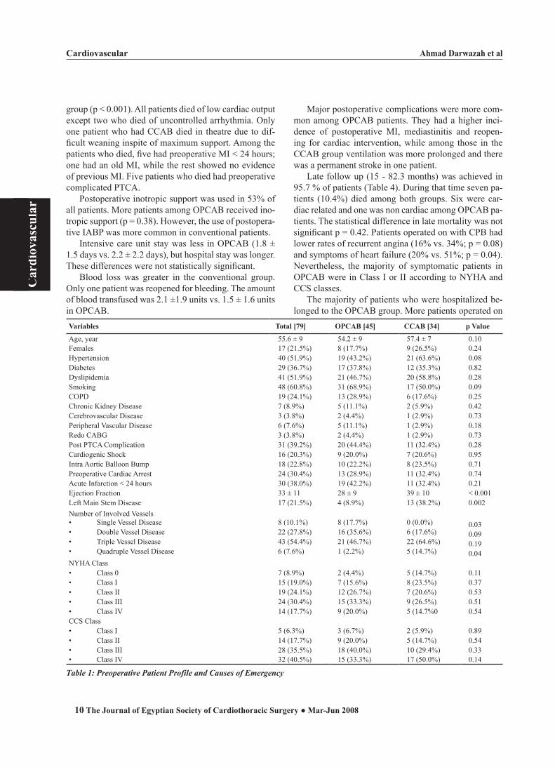
Cardiovascular Ahmad Darwazah et al
10
Car
diov
ascu
lar
The Journal of Egyptian Society of Cardiothoracic Surgery ● Mar-Jun 2008
group (p < 0.001). All patients died of low cardiac output except two who died of uncontrolled arrhythmia. Only one patient who had CCAB died in theatre due to dif--ficult weaning inspite of maximum support. Among the patients who died, five had preoperative MI < 24 hours; one had an old MI, while the rest showed no evidence of previous MI. Five patients who died had preoperative complicated PTCA.
Postoperative inotropic support was used in 53% of all patients. More patients among OPCAB received ino--tropic support (p = 0.38). However, the use of postopera--tive IABP was more common in conventional patients.
Intensive care unit stay was less in OPCAB (1.8 ± 1.5 days vs. 2.2 ± 2.2 days), but hospital stay was longer. These differences were not statistically significant.
Blood loss was greater in the conventional group. Only one patient was reopened for bleeding. The amount of blood transfused was 2.1 ±1.9 units vs. 1.5 ± 1.6 units in OPCAB.
Major postoperative complications were more com--mon among OPCAB patients. They had a higher inci--dence of postoperative MI, mediastinitis and reopen--ing for cardiac intervention, while among those in the CCAB group ventilation was more prolonged and there was a permanent stroke in one patient.
Late follow up (15 - 82.3 months) was achieved in 95.7 % of patients (Table 4). During that time seven pa--tients (10.4%) died among both groups. Six were car--diac related and one was non cardiac among OPCAB pa--tients. The statistical difference in late mortality was not significant p = 0.42. Patients operated on with CPB had lower rates of recurrent angina (16% vs. 34%; p = 0.08) and symptoms of heart failure (20% vs. 51%; p = 0.04). Nevertheless, the majority of symptomatic patients in OPCAB were in Class I or II according to NYHA and CCS classes.
The majority of patients who were hospitalized be--longed to the OPCAB group. More patients operated on
Variables Total [79] OPCAB [45] CCAB [34] p Value
Age, year 55.6 ± 9 54.2 ± 9 57.4 ± 7 0.10Females 17 (21.5%) 8 (17.7%) 9 (26.5%) 0.24Hypertension 40 (51.9%) 19 (43.2%) 21 (63.6%) 0.08Diabetes 29 (36.7%) 17 (37.8%) 12 (35.3%) 0.82Dyslipidemia 41 (51.9%) 21 (46.7%) 20 (58.8%) 0.28Smoking 48 (60.8%) 31 (68.9%) 17 (50.0%) 0.09COPD 19 (24.1%) 13 (28.9%) 6 (17.6%) 0.25Chronic Kidney Disease 7 (8.9%) 5 (11.1%) 2 (5.9%) 0.42Cerebrovascular Disease 3 (3.8%) 2 (4.4%) 1 (2.9%) 0.73Peripheral Vascular Disease 6 (7.6%) 5 (11.1%) 1 (2.9%) 0.18Redo CABG 3 (3.8%) 2 (4.4%) 1 (2.9%) 0.73Post PTCA Complication 31 (39.2%) 20 (44.4%) 11 (32.4%) 0.28Cardiogenic Shock 16 (20.3%) 9 (20.0%) 7 (20.6%) 0.95Intra Aortic Balloon Bump 18 (22.8%) 10 (22.2%) 8 (23.5%) 0.71Preoperative Cardiac Arrest 24 (30.4%) 13 (28.9%) 11 (32.4%) 0.74Acute Infarction < 24 hours 30 (38.0%) 19 (42.2%) 11 (32.4%) 0.21Ejection Fraction 33 ± 11 28 ± 9 39 ± 10 < 0.001Left Main Stem Disease 17 (21.5%) 4 (8.9%) 13 (38.2%) 0.002Number of Involved Vessels• Single Vessel Disease 8 (10.1%) 8 (17.7%) 0 (0.0%) 0.03• Double Vessel Disease 22 (27.8%) 16 (35.6%) 6 (17.6%) 0.09• Triple Vessel Disease 43 (54.4%) 21 (46.7%) 22 (64.6%) 0.19• Quadruple Vessel Disease 6 (7.6%) 1 (2.2%) 5 (14.7%) 0.04NYHA Class• Class 0 7 (8.9%) 2 (4.4%) 5 (14.7%) 0.11• Class I 15 (19.0%) 7 (15.6%) 8 (23.5%) 0.37• Class II 19 (24.1%) 12 (26.7%) 7 (20.6%) 0.53• Class III 24 (30.4%) 15 (33.3%) 9 (26.5%) 0.51• Class IV 14 (17.7%) 9 (20.0%) 5 (14.7%0 0.54CCS Class• Class I 5 (6.3%) 3 (6.7%) 2 (5.9%) 0.89• Class II 14 (17.7%) 9 (20.0%) 5 (14.7%) 0.54• Class III 28 (35.5%) 18 (40.0%) 10 (29.4%) 0.33• Class IV 32 (40.5%) 15 (33.3%) 17 (50.0%) 0.14
Table 1: Preoperative Patient Profile and Causes of Emergency
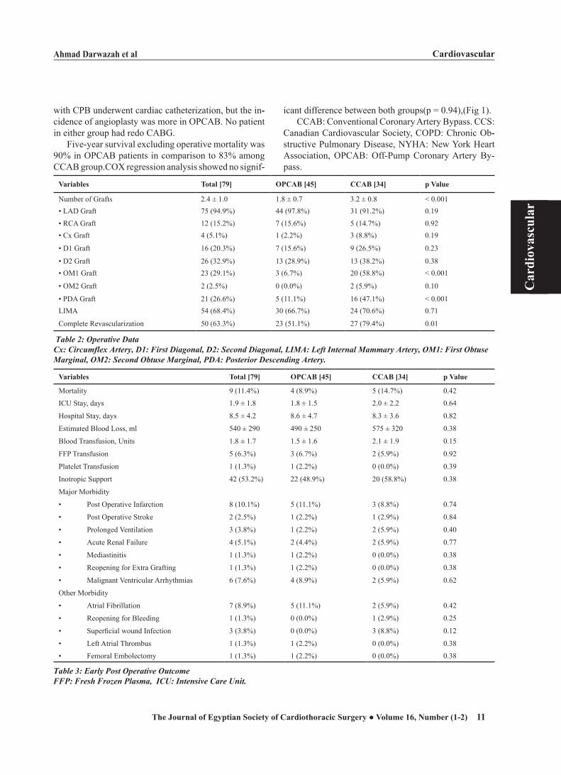
Ahmad Darwazah et al Cardiovascular
11
Car
diov
ascu
lar
The Journal of Egyptian Society of Cardiothoracic Surgery ● Volume 16, Number (1-2)
with CPB underwent cardiac catheterization, but the in--cidence of angioplasty was more in OPCAB. No patient in either group had redo CABG.
Five-year survival excluding operative mortality was 90% in OPCAB patients in comparison to 83% among CCAB group.COX regression analysis showed no signif--
icant difference between both groups(p = 0.94),(Fig 1).CCAB: Conventional Coronary Artery Bypass. CCS:
Canadian Cardiovascular Society, COPD: Chronic Ob--structive Pulmonary Disease, NYHA: New York Heart Association, OPCAB: Off-Pump Coronary Artery By--pass.
Variables Total [79] OPCAB [45] CCAB [34] p Value
Number of Grafts 2.4 ± 1.0 1.8 ± 0.7 3.2 ± 0.8 < 0.001• LAD Graft 75 (94.9%) 44 (97.8%) 31 (91.2%) 0.19• RCA Graft 12 (15.2%) 7 (15.6%) 5 (14.7%) 0.92• Cx Graft 4 (5.1%) 1 (2.2%) 3 (8.8%) 0.19
• D1 Graft 16 (20.3%) 7 (15.6%) 9 (26.5%) 0.23
• D2 Graft 26 (32.9%) 13 (28.9%) 13 (38.2%) 0.38• OM1 Graft 23 (29.1%) 3 (6.7%) 20 (58.8%) < 0.001
• OM2 Graft 2 (2.5%) 0 (0.0%) 2 (5.9%) 0.10
• PDA Graft 21 (26.6%) 5 (11.1%) 16 (47.1%) < 0.001LIMA 54 (68.4%) 30 (66.7%) 24 (70.6%) 0.71
Complete Revascularization 50 (63.3%) 23 (51.1%) 27 (79.4%) 0.01
Table 2: Operative DataCx: Circumflex Artery, D1: First Diagonal, D2: Second Diagonal, LIMA: Left Internal Mammary Artery, OM1: First Obtuse Marginal, OM2: Second Obtuse Marginal, PDA: Posterior Descending Artery.
Variables Total [79] OPCAB [45] CCAB [34] p Value
Mortality 9 (11.4%) 4 (8.9%) 5 (14.7%) 0.42
ICU Stay, days 1.9 ± 1.8 1.8 ± 1.5 2.0 ± 2.2 0.64
Hospital Stay, days 8.5 ± 4.2 8.6 ± 4.7 8.3 ± 3.6 0.82
Estimated Blood Loss, ml 540 ± 290 490 ± 250 575 ± 320 0.38
Blood Transfusion, Units 1.8 ± 1.7 1.5 ± 1.6 2.1 ± 1.9 0.15
FFP Transfusion 5 (6.3%) 3 (6.7%) 2 (5.9%) 0.92
Platelet Transfusion 1 (1.3%) 1 (2.2%) 0 (0.0%) 0.39
Inotropic Support 42 (53.2%) 22 (48.9%) 20 (58.8%) 0.38
Major Morbidity
• Post Operative Infarction 8 (10.1%) 5 (11.1%) 3 (8.8%) 0.74
• Post Operative Stroke 2 (2.5%) 1 (2.2%) 1 (2.9%) 0.84
• Prolonged Ventilation 3 (3.8%) 1 (2.2%) 2 (5.9%) 0.40
• Acute Renal Failure 4 (5.1%) 2 (4.4%) 2 (5.9%) 0.77
• Mediastinitis 1 (1.3%) 1 (2.2%) 0 (0.0%) 0.38
• Reopening for Extra Grafting 1 (1.3%) 1 (2.2%) 0 (0.0%) 0.38
• Malignant Ventricular Arrhythmias 6 (7.6%) 4 (8.9%) 2 (5.9%) 0.62
Other Morbidity
• Atrial Fibrillation 7 (8.9%) 5 (11.1%) 2 (5.9%) 0.42
• Reopening for Bleeding 1 (1.3%) 0 (0.0%) 1 (2.9%) 0.25
• Superficial wound Infection 3 (3.8%) 0 (0.0%) 3 (8.8%) 0.12
• Left Atrial Thrombus 1 (1.3%) 1 (2.2%) 0 (0.0%) 0.38
• Femoral Embolectomy 1 (1.3%) 1 (2.2%) 0 (0.0%) 0.38
Table 3: Early Post Operative OutcomeFFP: Fresh Frozen Plasma, ICU: Intensive Care Unit.
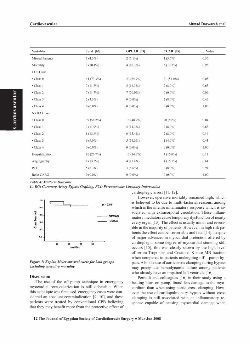
Cardiovascular Ahmad Darwazah et al
12
Car
diov
ascu
lar
The Journal of Egyptian Society of Cardiothoracic Surgery ● Mar-Jun 2008
Figure 1: Kaplan Meier survival curve for both groupsexcluding operative mortality.
DiscussionThe use of the off-pump technique in emergency
myocardial revascularization is still debatable. When this technique was first used, emergency cases were con--sidered an absolute contraindication [9, 10], and these patients were treated by conventional CPB believing that they may benefit more from the protective effect of
cardioplegic arrest [11, 12].However, operative mortality remained high, which
is believed to be due to multi-factorial reasons, among which is the intense inflammatory response which is as--sociated with extracorporal circulation. These inflam--matory mediators cause temporary dysfunction of nearly every organ [13]. The effect is usually minor and revers--ible in the majority of patients. However, in high risk pa--tients the effect can be irreversible and fatal [14]. In spite of major advances in myocardial protection offered by cardioplegia, some degree of myocardial stunning still occurs [15], this was clearly shown by the high level of serum Troponins and Creatine Kinase MB fraction when compared to patients undergoing off – pump by--pass. Also the use of aortic cross clamping during bypass may precipitate hemodynamic failure among patients who already have an impaired left ventricle [16].
Perrault and colleagues [16] in their study using a beating heart on pump, found less damage to the myo--cardium than when using aortic cross clamping. How--ever the use of cardiopulmonary bypass without cross clamping is still associated with an inflammatory re--sponse capable of causing myocardial damage when
Variables Total [67] OPCAB [39] CCAB [28] p Value
Missed Patients 3 (4.3%) 2 (5.1%) 1 (3.6%) 0.56
Mortality 7 (10.4%) 4 (10.3%) 3 (10.7%) 0.95
CCS Class
• Class 0 44 (73.3%) 23 (65.7%) 21 (84.0%) 0.08
• Class 1 7 (11.7%) 5 (14.3%) 2 (8.0%) 0.63
• Class 2 7 (11.7%) 7 (20.0%) 0 (0.0%) 0.09
• Class 3 2 (3.3%) 0 (0.0%) 2 (8.0%) 0.46
• Class 4 0 (0.0%) 0 (0.0%) 0 (0.0%) 1.00
NYHA Class
• Class 0 39 (58.2%) 19 (48.7%) 20 (80%) 0.04
• Class 1 7 (11.9%) 5 (14.3%) 2 (8.0%) 0.63
• Class 2 8 (13.6%) 6 (15.4%) 2 (8.0%) 0.14
• Class 3 6 (9.0%) 5 (14.3%) 1 (4.0%) 0.43
• Class 4 0 (0.0%) 0 (0.0%) 0 (0.0%) 1.00
Hospitalization 16 (26.7%) 12 (34.3%) 4 (16.0%) 0.11
Angiography 8 (13.3%) 4 (11.4%) 4 (16.1%) 0.61
PCI 5 (8.3%) 3 (8.6%) 2 (8.0%) 0.94
Redo CABG 0 (0.0%) 0 (0.0%) 0 (0.0%) 1.00
Table 4: Midterm OutcomeCABG: Coronary Artery Bypass Grafting, PCI: Percutaneous Coronary Intervention

Ahmad Darwazah et al Cardiovascular
13
Car
diov
ascu
lar
The Journal of Egyptian Society of Cardiothoracic Surgery ● Volume 16, Number (1-2)
compared with off-pump technique [17].Clinical applications of off-pump bypass in emer--
gency CABG is limited [7, 8, 10, 18 – 21]. Most of the studies showed that off-pump surgery can be applied effectively with low risk inspite of the compromised hemodynamic state of these patients. Operative mortal--ity varies from 0 to 5 % [19, 20], depending on various factors. Worse prognosis is associated with preoperative use of IABP, non use of LIMA to LAD and less than three grafts used during operation, also patients under--going cardiopulmonary resuscitation on their way to theater have a higher operative risk [7,19].
Operative mortality among our off-pump patients was 8.9%. The majority of these patients had MI less than 24 hours prior to surgery, low EF and complica--tion of angioplasty. Similar to the observation of Locker and colleagues [7], we found that 60% of these patients had no LIMA used, and less than three grafts have been applied. These factors contributed to the high early mor--tality.
The mortality rate among our patients is in accor--dance with the results found by Locker and colleagues [20], but was higher than the recent study by Kerendi and coworkers [19], who had a 0% early mortality. This is directly attributed to the preoperative hemodynamic state of their patients. In the Kerendi study, the mean EF was 47 ± 2.3 % in comparison to 28 ± 9 % among our patients. Similarly, the incidence of cardiogenic shock and MI was much lower than in our patients.
Late mortality among both of our groups was the same. Patients in the OPCAB group died early after op--eration, within 1-2 years, in contrast to conventional pa--tients who died after three years. Control of symptoms whether angina or heart failure was better among CCAB patients, subsequently hospitalization and reintervention was less frequent. These results were related to com--pleteness of myocardial revascularization and the preop--erative hemodynamic condition among both groups.
Previous studies [7, 20] had a higher late mortality among their patients who were subjected to OPCAB. It varied from 19 % [7], to 23% [20]. Similarly, control of symptoms was less among their patients.
The difference in mortality from our study is due to the fact that all their patients had myocardial infarc--tion within one week before surgery. It seems that the timing of performing surgery is crucial and affects both early and late mortality. In Locker and colleagues’ study [20], their patients had infarction within 48 hours; sub--sequently they had a higher early and late mortality in comparison to their more recent study [7] whom their patients had infarction within 7 days.
The timing and technique of surgery used among pa--tients with acute myocardial infarction remains contro--versial. Previous studies [22, 23] showed that mortality is related to the time of intervention. Patients operated upon after 48 hours of infarction had a better result than those who were operated on earlier [22].
A similar observation was noticed in our recent work. Fifty percent of our patients who died either early or late had acute MI less than 24 hours before surgery. Beside the timing of intervention, the technique used whether off-pump or conventional is also important. The appli--cation of off-pump among patients with recent MI has been studied [7, 19 – 21] and confirmed the superiority of off-pump surgery in comparison to conventional by--pass in these patients
Evaluating early and late mortality among our pa--tients who had infarction less than 24 hours before surgery, we noticed that the majority of these patients underwent conventional bypass. This emphasized the importance of using off-pump among patients with early acute myocardial infarction.
The relationship between preoperative hemodynamic state, completeness of myocardial revascularization and late control of symptoms was clearly seen in previous studies [7, 20], and our recent work. We noticed that the control of symptoms whether angina or heart failure was better among patients who underwent conventional by--pass. They had a better preoperative hemodynamic state in relation to EF, cardiogenic shock and angioplasty complications. Also the number of grafts used per pa--tient was significantly higher among these patients.
Similarly, in the early work by Locker and colleagues (20), the control of symptoms among their off-pump pa--tients during follow up was less than conventional pa--tients. More of their patients referred to off-pump had PTCA complications. This reflects the hemodynamic in--stability among their off-pump patients. Also, the mean number of grafts used was less than the conventional by--pass group. In their recent study [7], their off-pump pa--tients had better results whether late mortality or control of symptoms. This is due to the fact that preoperative he--modynamic state of their patients was much better than their previous study.
In conclusion, we believe that the use of either off-pump or conventional emergency CABG is associated with high operative and late mortality. Patients associ--ated with preoperative cardiogenic shock, low EF, acute MI less than 24 hours and angioplasty complication carry the highest risk. Off-pump bypass can be used in emergency myocardial revascularization .Although, it is associated with lower rate of early and late mortality, it did not show any significant superiority over conven--
Cardiovascular Ahmad Darwazah et al
14
Car
diov
ascu
lar
The Journal of Egyptian Society of Cardiothoracic Surgery ● Mar-Jun 2008
tional CABG except for patients with myocardial infarc--tion less than 24 hours.
Limitations of the StudyOur study has some limitations. It is an observational
retrospective trial. It contained a relatively small number of patients with preoperative inequality of left ventricle function which may represents a potential source of bias of choosing the surgical approach. But, the surgeon’s preference and the general agreement that OPCAB sur--gery represent a safe approach for patients of severely compromised left ventricle may ameliorate this bias. Of course, the nature of the study precluded the investiga--tors from being blinded to the procedure.
References
1. Society of Thoracic Surgeons Adult Cardiac Surgery Data--base, WWW.StS.org. Last accessed on 04.12.2007.
2. Boyd WD, Desai ND, Del Rizzo DF, Novick RJ, McKenzie FN, Menkis AH. Off- Pump surgery decreases postopera--tive complications and resource utilization in the elderly. Ann Thorac Surg 1999; 68: 1490-3.
3. Chamberlain MH, Ascione R, Reeves BC, Angelini GD. Evaluation of the effectiveness of off –pump coronary ar--tery bypass grafting in high-risk patients: an observational study. Ann Thorac surg 2002; 73: 1866-73
4. Puskas JD, Sharoni E, Petersen T, et al. Angiographic graft patency and clinical outcomes among diabetic patients after off-pump versus conventional coronary artery bypass graft--ing: results of a prospective raudomized trial. Heart Surgery Forum 2003; 6: S27.
5. Darwazah AK, Abu Sham’a RA, Hussein E, Hawari MH, Ismail H. Myocardial Revascularization in Patients with low Ejection Fraction ≤ 35%: Effect of Pump Technique on Early Morbidity and Mortality . J Card Surg 2006; 21: 22-27.
6. Yokoyama T, Baumgartner FJ, Gheissari A, Capouya ER, Panagiotides GP, Declusin RJ. Off-Pump versus on-pump coronary artery Bypass in High Risk Subgroups . Ann Tho--rac Surg 2000; 70: 1546-50.
7. Locker C, Mohr R, Paz Y, Kramer A, Lev-Ran O, Pevni D, Shapira I. Myocardial Revascularization for acute Myocar--dial Infraction: Benefits and Drawbacks of Avoiding Car--diopulmonary Bypass. Ann Thorac Surg 2003; 76: 771-7.
8. Yang EH, Gumina RJ, lennon RJ, Holmes DR, Rihal CS, Singh H Emergency Coronary Artery Bypass Surgery for Percutaneous Coronary interventions. J Am Coll Cardial 2005 ; 46: 2004-9.
9. Yacoub M.H. Off- Pump Coronary Bypass Surgery . In Search of an Identity. Circulation 2001; 104: 1743-1745.
10. Varghese D, Yacoub MH, Trimlett R, et al. Outcome of non-elective coronary artery bypass grafting without car--dio-pulmonary bypass. Eur J Cardiothorac Surg. 2001;19: 245-248.
11. Vinten- Johansen J, Thourani VH. Myocardial protection: an overview . J Extra Corpor Technol 2000 ; 32: 38-48.
12. Beyersdorf F. Protection of evolving myocardial infarction and failed PTCA. Ann Thorac Surg 1995; 60: 833-8.
13. Edmunds LH Jr. Advances in the heart – lung machine af--ter John and Mary Gibbon. Ann Thorac Surg 2003 ; 76: S2220-3.
14. Kirklin JW, Barratt-Boyes BG. Cardiac Surgery. 2nd edi--tion. New York: Churchill Livingston; 1993.p.83-97, 210-17, 337.
15. Misare BD, Krukenkamp BD, Lazer ZP, Levitsky SL. Re--covery of post ischemic contractile function is depressed by antegrade warm continuous blood cardioplegia. J Thorac Cardiovasc Surg 1993; 105: 37-44.
16. Perrault LP, Menasche’ P, Peynet J, Faris B etal. On – Pump, Beating – Heart coronary Artery Operations in High – Risk Patients: An Acceptable Trade – off?. Ann Thorac Surg 1997; 64: 1368-73.
17. Wan IYP, Arifi AA, Wan S, etal. Beating heart revascular--ization with or without Cardiopulmonary bypass: evalua--tion of inflammatory response in a prospective randomized study. J Thorac Cardiovasc Surg 2004; 127: 1624-31.
18. Capdeville M, Lee JH. Emergency Off-pump Coronary Ar--tery Bypass Grafting for Acute left Main coronary Artery Dissection .Tex Heart Inst J 2001 ; 28:208-11.
19. Kerendi F, Puskas JD, Craver JM, Cooper WA, Jones EL, Lattouf OH, Vega JD, Guyton RA. Emergency Coronary Artery Bypass Grafting Can Be Performed Safely Without Cardiopulmonary Bypass in Selected patients. Ann Thorac Surg 2005; 79: 801-6.
20. Locker C, Shapira I, Paz Y, Kramer A, Gurevitch J, Matsa M, Pevni D, Mohr R. Emergency Myocardial revascular--ization for acute myocardial infarction: Survival benefits of avoiding cardiopulmonary bypass. Eur J Cardiothorac Surg. 2000; 17: 234- 238.
21. Mohr R, Moshkovitch Y, Shapira I, Amir G, Hod H, Gur--evitch J Coronary Artery Bypass without Cardiopulmonary Bypass For Patients With Acute Myocardial infarction . J Thorac Cardiovasc Surg 1999 : 118: 50-6
22. Nunley DL, Grunkemeier GL, Teply JF et al. Coronary bypass operation following acute complicated myocardial infarction. J Thorac Cardiovasc Surg 1983 ; 85: 485-91.
23. Creswell LL, Moulton MJ, Cox AL, Rosenbloom M. Re--vascularization after acute Myocardial infarction. Ann Tho--rac Surg 1995; 60: 19-26.
Ibrahim Yassin et al Cardiovascular
15
Car
diov
ascu
lar
The Journal of Egyptian Society of Cardiothoracic Surgery ● Volume 16, Number (1-2)
RISK FACTORS OF CEREBRO-VASCULAR ACCIDENTS
AFTER ONPUMP ISOLATED CORONARY ARTERY SURGERY
Background: Cerebrovascular accidents (CVAs) after surgical myocar--dial revascularization is one of the most hazardous complication. Its -related mortality remains high and patients who experienc*e this event have lower long-term survival than patients who do not . Many reports focus on the relationship between the degree of atherosclerotic affection of the ascending aorta ; degree of manipulation and postoperative CVAs . Different surgical strategies for myocardial revascularization with the least manipulation to the aorta of these patients were described in the literatures. Other independent predictors of cerebrovascular accidents ( CVAs ) following onpump isolated CABG(s) include Extracoronary Vas--culopathy ,Low output syndrome ,Low EF. and others Identification of other risk factors allows preoperative risk stratification and may facilitate improved patient selection and contribute to reduce the risk of a stroke by providing an opportunity for adequate medical and surgical intervention. Objective: Identification of the independent predictors of cerebrovascu--lar accidents ( CVAs ) following onpump isolated CABG(s).Methods: A retrospective study on a total of ( 1192 ) isolated coronary patients whom were operated upon ONPUMP at the Department of Car--diology and Cardiac Surgery, “G. D’Annunzio” University, Chieti, Italy , From January 1991 to December 2001. The patients who survived less than 24 hours and who had aortic cannulation without cross-clamping were excluded.Univariate and multivariate analyses were applied to iden--tify independent predictors of higher incidence of CVAs. Results: Our study demonstrated that (23) patients out of the total num--ber ( 1192 ) patients had experienced CVAs with an incidence of ( 1.93% ) . Univariate analysis of risk factors in patients with and without a CVAs showed that there are seven independent factors: Low Output Syndrome ( LOS ) ( p .000 ) & Simultaneous (CEA) (p .000 ) & LV dysfunction ( EF<0.35 )(p .003 ) & Previous CVAs (p.034 ) ECV(Extra Coronary Vas--culopathy )(p .024) & CVD ( Carotid Vascular Disease ) (p.042) & COPD (p.047). In all of them p<.05. Stepwise Logistic Regression analysis for CVAs confirmed five of them ( p<0.05) Low Output Syndrome ( LOS ) ( p .0002 ) & LV dysfunction ( EF<0.35 ) ( p .0096 ) & Simultaneous carotid endarterectomy (CEA ) ( p .0063 ) & Extra Coronary Vasculopathy( ECV ) (p .0315) & COPD ( p.0489 ) In all of them p<.05Conclusions: Aortic manipulation must be avoided in patients with ath--erosclerotic changes of the ascending aorta. Maintenance of a good he--modynamic status is crucial for any patient to reduce CVAs incidence. Patients with low EF. ; Simultaneous carotid endarterectomy ; extracoro--nary vasculopathy or COPD are at higher risk, and a correct surgical strategy should be tailored for each case.
Accepted for publication Aug 1 , 2007
Address reprint request to Dr: Ibrahim
hassin
E-mail:[email protected]
codex : 04/cord/66/0710
Ibrahim M. Yassin, MD,Yousry A. Shaheen ,MD,Bedir M. Ibrahim, MD,Michele Di Mauro, MD,Antonio Maria Calafiore, MD.
Cardiovascular Ibrahim Yassin et al
16
Car
diov
ascu
lar
The Journal of Egyptian Society of Cardiothoracic Surgery ● Mar-Jun 2008
Cerebrovascular accidents (CVAs) after surgical myocardial revascularization remain a complication that, despite the increased quality of treatment, still has an incidence that ranges from ( 1.0% to 3.1% ) . Cere--brovascular accident-related mortality remains high. The outcome of patients with a postoperative CVA is still poor, both in the early and in the late period. Its in--cidence is not high, but it is relatively stable and patients who experience this event have lower long-term survival than patients who do not. ( 1- 11)
The pathophysiology of this severe complication is complex and multifactorial, Different authors found dif--ferent risk factors, most of it focused upon arterioscle--rosis of the ascending aorta .2 , 3 , 7 , 8, 12 , 13 , 14 , previous CVA 2 , 5 , 7 ,8 , 9, 10 , 15, 16, 17 , peripheral vascular disease 3 , 5 , 8 , ,10 , 17 , Carotid occlusion was a risk factor for some authors 3, but not for others 11 . Unstable angina 2 , low ejection fraction 10 and LOS 7, are logic factors and were also reported but to a less extent.
Atrial fibrillation was a risk factor in some reports 7, 21 , 22 and CPB time in others 4, 7 , 8 , 15 , 17 Cala--fiore et al 23 found no correlation between postop--erative atrial fibrillation and CVAs incidence was found (7 CVAs in 526 patients who experienced postoperative atrial fibrillation, 1.2%, and 43 in 4,297 patients who did not, 1.0%). None of the 68 patients with preoperative atrial fibrillation had any CVA.
Other risk factors, such as age, were constantly pres--ent in the great majority of the studies 2, 3 , 4 , 5 ,8, 9 , 10 , 15 , 16 , 17, , Diabetes 2 , 5 , 8 , 10 , 16 , 17 , hypertension 2 , female sex 7, smoking 8, chronic renal failure 4, 5 , 8
Normothermic perfusion, identified as a risk factor by some studies 17, 18 but excluded by others 19 , was considered by Gaudino and associates 6 to be responsi--ble for a superior extension of cerebral damage, but this finding was not confirmed by other reports 20. Cala--fiore et al 23 concluded that hypothermic perfusion was related to a higher CVA rate (2.4% versus 1.2%; p = 0.023); multivariate analysis confirmed this finding.
In the last 13 years different surgical strategies for myocardial revascularization were used in our institu--tion for patients with atherosclerotic changes. In almost all of these procedures the ascending aorta was either not touched or touched only to a limited extent and the role became revascularization with the least manipula--tion to the aorta of these patients.
Material and methodsPatient population
From January 1991 to December 2001, 1,192 pa--
tients underwent isolated onpump coronary artery by--pass grafting (CABG). Two hundred seventy five (275 ) were females( 23%) and nine hundred seventeen (917) were males ( 77% ) .All the patients who died in the operating room and in the first 24 hours after operation were excluded.
Preoperative dataPatient data were taken from our database, which
was assembled from the medical records. Prior CVA was documented on the basis of previous medical re--cords and reviewing the computed tomographic scan or nuclear magnetic resonance images related to the epi--sode. All variables included in the study are listed in the Appendix.
Perioperative data
After median sternotomy,and pericardiotomy, The quality of the aorta was assessed by the surgeon using his fingers. Disease of the ascending aorta was suspect--ed preoperatively from the angiography or the echocar--diogram, and, in selected cases, confirmed by computed tomographic scan. Intraoperatively, no routine epicardi--al scanning was performed. The operative technique has been described in detail elsewhere with nothing special in the technique,3mg \kg of heparin was administered to the patients,after preparing the conduits, Myocardial protection was achieved with Intermittent Antegrade Warm Blood Cardioplegia; Blood is taken directly from the oxygenator by means of ¼ inch tubing and a roller pump and is infused in the aortic root. The tubing is connected to a Syringe pump that deliver potassium(K) In the concentration of 2 mEq./ ml. After the first infu--sion of cardioplegia (600 ml of blood and 10 mEq. K In 2 min ) , Reinfusions are administered every 15 min. of ischaemia( 400 ml of blood and 4 mEq. K in 2 min. The body temprature is maintained at 37°c . The distal and proximal anastomoses were performed during aortic crossclamoing ( Single Clamp Technique “ SCT ” ). The left system vascularization was routinely with arte--rial conduits, mostly Y grafting to the anterior and lateral walls , and Vein graft to the inferior wall. Anastomosis was fashioned with 8/0 polypropylene . At the end of the grafting procedure,Transit time Flowmetry (Butter--fly Flowmeter Version 111) evaluation to all grafts was done as a routine. Finally ,protamine was injected to re--verse the effect of heparin.
Postoperative dataCerebrovascular accident was defined as global or
focal neurologic deficit, lasting less (transient ischemic

Ibrahim Yassin et al Cardiovascular
17
Car
diov
ascu
lar
The Journal of Egyptian Society of Cardiothoracic Surgery ● Volume 16, Number (1-2)
attack) or more (stroke) than 24 hours, that could be evident after emergence from anesthesia (early CVA) or after first awaking without any neurologic deficits (de--layed CVA). Cerebrovascular accident was diagnosed by a neurologist and confirmed by a brain computed to--mographic scan or nuclear magnetic resonance image. Stroke was defined as a focal or global cerebral dysfunc--tion of presumed vascular origin lasting more than 24 hours. Transient ischemic attack was defined as a focal cerebral dysfunction of presumed vascular origin that resolved completely within 24 hours.
Statistical analysisResults are expressed as mean value ± standard de--
viation unless otherwise indicated. Statistical analysis
comparing two groups was performed with unpaired two-tailed Student’s t test for the means or x2 test for categorical variables. All variables included in the study are listed in the Appendix. Univariate analysis of risk factors in patients with and without a CVA was per--formed. Stepwise logistic regression was used to select the independent variables that could predict postopera--tive CVAs,. For any independent predictor odds ratio (OR), 95% confidence limit when necessary, and prob--ability value are indicated. All the multivariate analyses included univariate variables with p < 0.2; the final sig--nificance level for univariate and multivariate analyses was at p less than 0.05. SPSS software (Chicago, IL) was used for all the analyses in this study.
Results
Risk factors CVA (n=23) Non-CVA (n=1169) P-value
Age (years) 70.5±3.7 70.8±4.4 ns
Female sex 5 (21.7%) 270 (23.1%) ns
Diabetes mellitus 7 (30.4%) 285 (24.4%) ns
Hypertension 14 (60.9%) 557 (47.6) ns
Dyslipidemia 6 (26.1%) 372 (31.8%) ns
Smoking 8 (34.8%) 364 (31.2%) ns
COPD 4 (17.4%) 64 (5.5%) .047
ECV(Extra Coronary Vasculopathy ) 12 (52.2%) 332 (28.4%) .024
PVD ( Peripheral Vascular Disease ) 4 (17.4%) 139 (11.9%) ns
CVD ( Carotid Vascular Disease ) 8 (34.8%) 193 (16.5%) .042
Left Main Coronary Artery Disease 7 (30.4%) 192 (16.4%) .088
Previous CVAs 4 (17.4%) 62 (5.3%) .034
Non-elective surgery 8 (34.8%) 234 (28.6%) ns
Unstable angina 11 (47.8%) 414 (35.4%) ns
Intraaortic Ballon Pump( IABP ) 0 4 (0.3%) ns
LV dysfunction ( EF<0.35 ) 6 (26.1%) 75 (6.4%) .003
Simultaneous (CEA) 4 (17.4%) 33 (2.9%) .000
Mean CPB time (min) 65.6±30.2 65.6±23.8 ns
Mean CXC time (min) 47.3±15.2 54.2±20.7 ns
Need for Inotropic Support 1 (4.3%) 6 (0.5%) ns
Low Output Syndrome ( LOS ) 6 (26.1%) 43 (3.7%) .000
Postoperative Atrial Fibrillation ( AF ) 0 28(2.4%) nsTable 1. Univariate analysis of risk factors in patients with and without a CVAs:
CVA, cerebrovascular accident; CEA, Carotid Endarterectomy; COPD, chronic obstructive pulmonary disease; LV, left ventttricle; EF, ejection fraction; CPB, cardiopulmonary bypass.
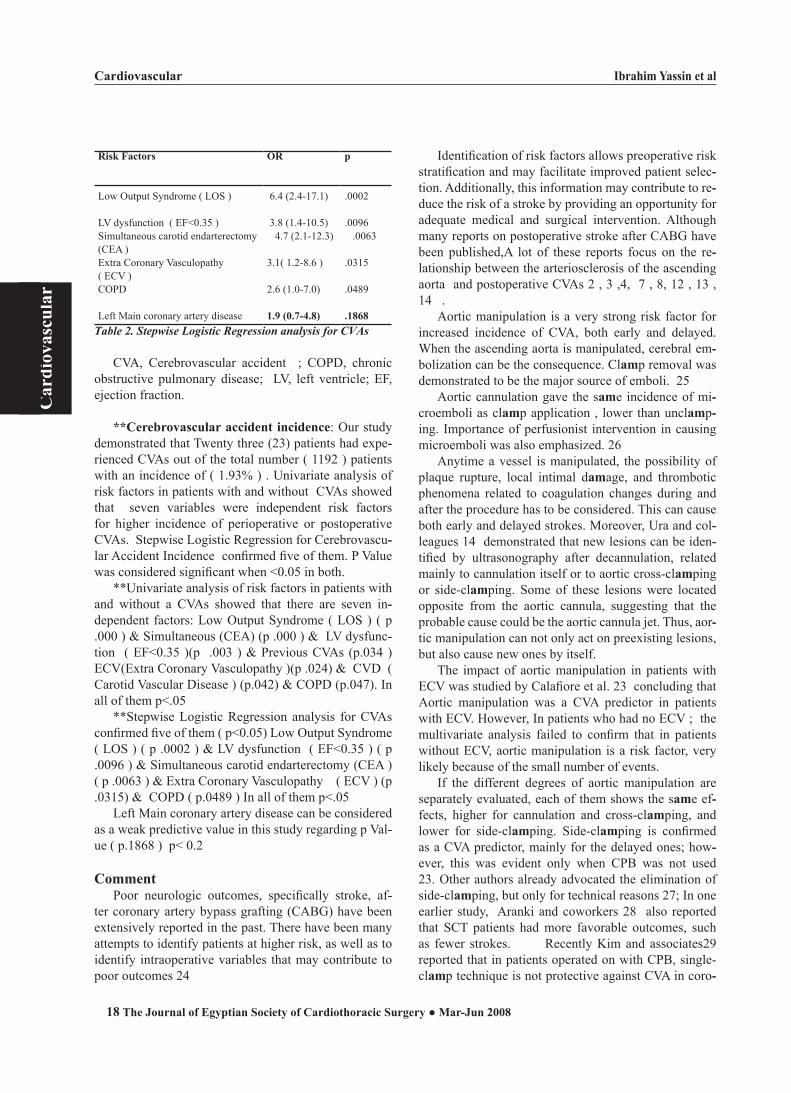
Cardiovascular Ibrahim Yassin et al
18
Car
diov
ascu
lar
The Journal of Egyptian Society of Cardiothoracic Surgery ● Mar-Jun 2008
Risk Factors OR p
Low Output Syndrome ( LOS ) 6.4 (2.4-17.1) .0002
LV dysfunction ( EF<0.35 ) 3.8 (1.4-10.5) .0096Simultaneous carotid endarterectomy(CEA )
4.7 (2.1-12.3) .0063
Extra Coronary Vasculopathy( ECV )
3.1( 1.2-8.6 ) .0315
COPD 2.6 (1.0-7.0) .0489
Left Main coronary artery disease 1.9 (0.7-4.8) .1868Table 2. Stepwise Logistic Regression analysis for CVAs
CVA, Cerebrovascular accident ; COPD, chronic obstructive pulmonary disease; LV, left ventricle; EF, ejection fraction.
**Cerebrovascular accident incidence: Our study demonstrated that Twenty three (23) patients had expe--rienced CVAs out of the total number ( 1192 ) patients with an incidence of ( 1.93% ) . Univariate analysis of risk factors in patients with and without CVAs showed that seven variables were independent risk factors for higher incidence of perioperative or postoperative CVAs. Stepwise Logistic Regression for Cerebrovascu--lar Accident Incidence confirmed five of them. P Value was considered significant when <0.05 in both.
**Univariate analysis of risk factors in patients with and without a CVAs showed that there are seven in--dependent factors: Low Output Syndrome ( LOS ) ( p .000 ) & Simultaneous (CEA) (p .000 ) & LV dysfunc--tion ( EF<0.35 )(p .003 ) & Previous CVAs (p.034 ) ECV(Extra Coronary Vasculopathy )(p .024) & CVD ( Carotid Vascular Disease ) (p.042) & COPD (p.047). In all of them p<.05
**Stepwise Logistic Regression analysis for CVAs confirmed five of them ( p<0.05) Low Output Syndrome ( LOS ) ( p .0002 ) & LV dysfunction ( EF<0.35 ) ( p .0096 ) & Simultaneous carotid endarterectomy (CEA ) ( p .0063 ) & Extra Coronary Vasculopathy ( ECV ) (p .0315) & COPD ( p.0489 ) In all of them p<.05
Left Main coronary artery disease can be considered as a weak predictive value in this study regarding p Val--ue ( p.1868 ) p< 0.2
CommentPoor neurologic outcomes, specifically stroke, af--
ter coronary artery bypass grafting (CABG) have been extensively reported in the past. There have been many attempts to identify patients at higher risk, as well as to identify intraoperative variables that may contribute to poor outcomes 24
Identification of risk factors allows preoperative risk stratification and may facilitate improved patient selec--tion. Additionally, this information may contribute to re--duce the risk of a stroke by providing an opportunity for adequate medical and surgical intervention. Although many reports on postoperative stroke after CABG have been published,A lot of these reports focus on the re--lationship between the arteriosclerosis of the ascending aorta and postoperative CVAs 2 , 3 ,4, 7 , 8, 12 , 13 , 14 .
Aortic manipulation is a very strong risk factor for increased incidence of CVA, both early and delayed. When the ascending aorta is manipulated, cerebral em--bolization can be the consequence. Clamp removal was demonstrated to be the major source of emboli. 25
Aortic cannulation gave the same incidence of mi--croemboli as clamp application , lower than unclamp--ing. Importance of perfusionist intervention in causing microemboli was also emphasized. 26
Anytime a vessel is manipulated, the possibility of plaque rupture, local intimal damage, and thrombotic phenomena related to coagulation changes during and after the procedure has to be considered. This can cause both early and delayed strokes. Moreover, Ura and col--leagues 14 demonstrated that new lesions can be iden--tified by ultrasonography after decannulation, related mainly to cannulation itself or to aortic cross-clamping or side-clamping. Some of these lesions were located opposite from the aortic cannula, suggesting that the probable cause could be the aortic cannula jet. Thus, aor--tic manipulation can not only act on preexisting lesions, but also cause new ones by itself.
The impact of aortic manipulation in patients with ECV was studied by Calafiore et al. 23 concluding that Aortic manipulation was a CVA predictor in patients with ECV. However, In patients who had no ECV ; the multivariate analysis failed to confirm that in patients without ECV, aortic manipulation is a risk factor, very likely because of the small number of events.
If the different degrees of aortic manipulation are separately evaluated, each of them shows the same ef--fects, higher for cannulation and cross-clamping, and lower for side-clamping. Side-clamping is confirmed as a CVA predictor, mainly for the delayed ones; how--ever, this was evident only when CPB was not used 23. Other authors already advocated the elimination of side-clamping, but only for technical reasons 27; In one earlier study, Aranki and coworkers 28 also reported that SCT patients had more favorable outcomes, such as fewer strokes. Recently Kim and associates29 reported that in patients operated on with CPB, single-clamp technique is not protective against CVA in coro--

Ibrahim Yassin et al Cardiovascular
19
Car
diov
ascu
lar
The Journal of Egyptian Society of Cardiothoracic Surgery ● Volume 16, Number (1-2)
nary operation, demonstrating that the topic is still con--troversial. Calafiore et al. 23 support these conclusions, as in patients operated on with CPB, side-clamping did not add any further risk to CVA incidence. It was an in--dependent predictor only when CPB was not used. This means that CPB, per se, is not related to higher incidence of CVA, but the maneuvers necessary to institute it are. Conversely, myocardial revascularization without CPB does not reduce CVA incidence per se, as previously demonstrated 30, but only when any aortic manipulation is avoided.
In the last 13 years different surgical strategies for myocardial revascularization were used in our institu--tion. In some of these procedures the ascending aorta was either not touched or touched only to a limited ex--tent. Disease of the ascending aorta can, by itself, be an important risk factor 2 ,3 ,7 ,8 , but its detection forces the surgeon to consider possible strategies to avoid aortic manipulation 12 ,13 ,23 such as operation without CPB or arterial cannulation in peripheral sites (eg, femoral or subclavian artery) without cross-clamping. Use of arte--rial revascularization or proximal anastomosis of veins in nonaortic sites limits the effects of aortic disease on early outcome.
In patients whom operated upon using CPB; “SCT” Single Clamp Technique was the routine of our work aiming for minimization of the degree of manipulation of the aorta although not proved to be a risk factor in this group of patients. Safety of the cardioplegic regi--men used in our center in myocardial protection was dis--cussed in previous literatures31 and reflected on the rou--tine “SCT” Single Clamp Technique in these patients
Our study specifically addresses the other inepenent risk factors other than dealing with the atheromatous aorta with its different degees.
Stepwise Logistic Regression analysis for CVAs confirmed five of the independent risk factors shown by the univariate analysis ( p<0.05)
Extra Coronary Vasculopathy ( ECV ) showed to be a significant inepenent risk factor for CVAs in the univari--ate analysis ( p .024 ) and was confimed with the multi--variate analysis (p .0315) . Although It is the sumption of both the PVD and CVD , the PVD showed insignifi--cant values in both. So, we can say that the risk become more if the Vasculopathy is Cerebral.It was shown to be significant in the univariate analysis (p .042 ) but not confimed in the multivariate regression analysis.
The presence of left main coronary artery disease could be considered as a risk factor as shown in the re--sults of Stepwise Logistic Regression analysis for CVAs if P Value was considered significant when < 0.2( not in our study ) . Left main coronary artery disease is related
to higher incidence of ECV. It is likely that even if ECV is not diagnosed, the presence of left main coronary artery disease can be an indirect sign of some degree of vasculopathy not clinically evident, at the basis of a CVA in particular situations such as during myocardial revascularization.
A history of prior stroke or ITA is a strong predictor of recurrent stroke, and this variable is present in the great majority of the risk models . Such a history denotes the existence of pathologic conditions within the cere--brovascular system. 32 ,33
Univariate analysis showed that the pesence of Pre--vious CVAs (p.034 )is an inepenent risk factor .A history of neurologic disease suggests existing pathologic cere--brovascular conditions, such as impaired cerebral blood flow and autoregulation or inadequate collateral vessels, which may predispose patients to a cerebral complica--tion after CABG surgery 34
Multivariate regression analysis in our stuy showed that Simultaneous carotid endarterectomy (CEA ) is a stong inepenent factor ( p .0063 ) for CVAs.
In our study Single Carotid endarterectomy (CEA) was performed simultaneously with CABG if the patient had unstable angina or was considered at risk for a car--diac-related event, such as left main coronary artery dis--ease; it was performed before CABG if the patient was stable. If the patient needed bilateral CEA, the carotid artery with the most dangerous lesion was operated on before CABG and the second lesion at the same opera--tion as CABG.
Calafiore et al. 23 analyzed more deeply the risk factor carotid disease, which was divided into carotid disease without and with CEA. The latter was the stron--gest one; however, the presence of carotid disease, even if without any necessity of operation, remains related to higher incidence of CVA. Whereas carotid disease can be a sign of diffuse cerebral vasculopathy, CEA of this artery can cause, both immediately and hours after op--eration, local clotting (perioperative and postoperative changes from hypocoagulation to hypercoagulation can be a predisposing cause) followed by cerebral emboliza--tion. These results were also shown by others 35
However, other authors 11 , in a group of patients smaller than ours, found that extracranial carotid ste--nosis was a risk factor for postoperative CVA, but con--temporary CEA was protective. These authors advised prophylactic CEA also in patients with asymptomatic carotid stenosis of at least 75%.
Multivarient regression analysis confirmed that Low Output Syndrome ( LOS ) was a powerful risk factor ( p .0002 ) At the end of the procedure,it was identified as a need for intraaortic balloon pump or inotropic agents

Cardiovascular Ibrahim Yassin et al
20
Car
diov
ascu
lar
The Journal of Egyptian Society of Cardiothoracic Surgery ● Mar-Jun 2008
(epinephrine or norepinephrine, any dose; dopamine > 10 µg · kg-1 · min-1; or dobutamine > 5 µg · kg-1 · min-1).
Critical cerebral perfusion enhances the possibility of focal ischemia and can be at the basis of a postoperative CVA. However, a significant number of these patients had ECV, a direct expression of diffuse vascular disease, with an incidence higher than in the remaining patients. Multivariate analysis in patients with LOS showed that presence of ECV was the only risk factor for CVA. In--deed, maintenance of a satisfying hemodynamic status is crucial for brain integrity. The impact of LOS is constant in total, early, and delayed CVAs, but is higher in early CVAs, likely because of the direct effects of reduced ce--rebral perfusion. Other more general risk factors, such as peripheral vasculopathy or carotid disease without need of CEA, acted more on early CVA because of expression of a diffuse vasculopathy, more sensitive to any periop--erative hypotension or early hypercoagulability.
Multivarient regression analysis confirmed that LV dysfunction ( EF<0.35 ) was a powerful risk factor in t( p .0096 ) Several other studies have identified the same risk factors for stroke during CABG. Most probably, the mechanism related to the higher rate of CVA in patients with LV dysfunction observed in this series is related to the increased incidence of left side intracardiac thrombi as a direct cause or indirect cause through LOS affecting the brain blood supply. 32, 33, 36, 37
Surprisingly, a well-known risk factor, age, was not found to be an independent CVA predictor. Mean age was 70.5±3.7 years in patients with and 70.8±4.4 years in patients without CVA (ns).
A less common inepenent factors like Chronic Obsstructive Pulmonary Disease COPD was found sig--nificant in univariate analysis( p.047 ) and confirmed to be significant ( p.0489 ) with the multivarient regres--sion analysis. Pulmonary disease (emphysema, chronic bronchitis, restrictive lung disease, or asthma), was pre--viously reported as a risk factor in CVAs. 34, 38
Patients with pulmonary disease probably retained carbon dioxide (thus affecting cerebral vasoreactivity) or required prolonged mechanical ventilation (thus af--fecting the degree of cerebral perfusion and oxygen--ation). 39, 40, 41
ConclusionPostoperative CVAs are a dangerous event after
myocardial revascularization, related to higher early and late mortality. Its prevention is a critical feature of coro--nary operation. Our study reports that preventing LOS is crucial in reducing CVA incidence in any patient, with or without ECV.
Avoiding any aortic manipulation is another goal, not always easy to fulfill, but it is valid mainly for patients with ECV. In patients with ECV any of the general risk factors increases CVA incidence. Maintaining hemody--namic stability and avoiding any aortic manipulation is crucial in this cohort of patients at high risk for postop--erative CVA.
Limitations of the studyis that it is a retrospective study that considers 11
years of activity. During this time, surgical strategies changed and could influence directly CVA incidence. No preoperative screening of disease of the ascending aorta was made. Even if some patients had transesophageal echocardiography, epicardial scanning was never used. However, this reflects the reality of the great majority of the operating theaters and, for this reason, the conclu--sions of this study can be used by the great majority of surgical teams.
References
1. Gardner T.J., Horneffer P.J., Manolio T.A., et al. Stroke fol--lowing coronary artery bypass grafting: a ten-year study. Ann Thorac Surg 1985;40:574-581.
2. Roach G.W., Kanchuger M., Mangano C.M., et al. Adverse cerebral outcome after coronary bypass surgery. N Engl J Med 1996;335:1857-1863.
3. Mickleborough L.L., Walker P.M., Takagi Y., Ohashi M., Ivanov J., Tamariz M. Risk factors for stroke in patients undergoing coronary artery bypass surgery. J Thorac Car--diovasc Surg 1996;112:1250-1259.
4. Almassi G.H., Sommers T., Moritz T.E., et al. Stroke in cardiac surgical patients: determinants and outcome. Ann Thorac Surg 1999;68:391-398.
5. Engelman D.T., Cohn L.H., Rizzo R.J. Incidence and pre--dictors of TIAs and strokes following coronary artery by--pass grafting: report and collective review. Heart Surg Fo--rum 1999;2:242-245.
6. Gaudino M., Martinelli L., Di Lella G., et al. Superior ex--tension of intraoperative brain damage in case of normo--thermic systemic perfusion during coronary bypass opera--tions. J Thorac Cardiovasc Surg 1999;118:432-437.
7. Hogue C.W., Murphy S.F., Schechtman K.B., Davila-Ro--man V.G. Risk factors for early and delayed stroke after cardiac surgery. Circulation 1999;100:642-647.
8. John R., Choudhri A.F., Weinberg A.D., et al. Multicenter review of preoperative risk factors for stroke after coronary artery bypass surgery. Ann Thorac Surg 2000;69:30-36.
9. Puskas J.D., Winston D., Wright C.E., et al. Stroke after coronary artery operation: incidence, correlates, outcome and cost. Ann Thorac Surg 2000;69:1053-1056.
10. Borger M.A., Ivanov J., Weisel R.D., Rao V., Peniston C.M. Stroke during coronary by pass surgery: principal role of macroemboli. Eur J Cardiothorac Surg 2001;19:627-632.

Ibrahim Yassin et al Cardiovascular
21
Car
diov
ascu
lar
The Journal of Egyptian Society of Cardiothoracic Surgery ● Volume 16, Number (1-2)
11. Hirotani T., Kameda T., Kumamoto T., Shirota S., Yamano M. Stroke after coronary bypass grafting in patients with cerebrovascular disease. Ann Thorac Surg 2000;70:1571-1576.
12. Wareing T.H., Davila-Roman V.G., Barzilai B., Murphy S.F., Kouchoukos N.T. Management of the severely athero--sclerotic ascending aorta during cardiac operations. J Tho--rac Cardiovasc Surg 1992;103:453-462.
13. Gaudino M., Glieca F., Alessandrini F., et al. Individual--ized surgical strategy for the reduction of stroke in patients undergoing coronary artery bypass grafting. Ann Thorac Surg 1999;67:1246-1253.
14. Ura M., Sakata R., Nakayama Y., Goto T. Ultrasonograph--ic demonstration of manipulation-related aortic injury after cardiac surgery. J Am Coll Cardiol 2000;35:1303-1310.
15. Tuman K.J., McCarthy R.J., Najafi H., Ivankowich A.D. Differential effects of advanced age on neurologic and car--diac risk of coronary operations. J Thorac Cardiovasc Surg 1992;104:1510-1517.
16. McKhann G.M., Goldsborough M.A., Borowicz L.M., Jr, et al. Predictors of stroke risk in coronary artery bypass pa--tients. Ann Thorac Surg 1997;63:516-521.
17. Rao V., Christakis G.T., Weisel R.D., et al. Risk factors for stroke following coronary bypass surgery. J Card Surg 1995;10:468-474.
18. Martin T.D., Craver J.M., Gott J.P., et al. A prospective randomized trial of retrograde warm blood cardioplegia: myocardial benefit and neurologic threat. Ann Thorac Surg 1994;57:298-304.
19. Singh A.K., Bert A.A., Feng W.C., Rotenberg F.A. Stroke during coronary artery bypass grafting using hypother--mic versus normothermic perfusion. Ann Thorac Surg 1995;59:84-89.
20. Engelman R.M., Pleet A.B., Hicks R., et al. Is there a re--lationship between systemic perfusion temperature during coronary artery bypass surgery and extent of intraoperative ischemic central nervous system injury?. J Thorac Cardio--vasc Surg 2000;119:230-232.
21. Odell J.A., Blackshear J.L., Hodge D., Bailey K.R. Stroke after coronary bypass grafting: are we forgetting atrial fi--brillation? [Letter]. Ann Thorac Surg 2001;71:400-401.
22. Creswell L.L., Schuessler R.B., Rosenbloom M., Cox J.L. Hazards of postoperative atrial arrhythmias. Ann Thorac Surg 1993;56:539-549.
23. Calafiore A.M., Di Mauro M. , Teodori G. , Di Giammarco G. , Cirmeni S. , Contini M.,Iacò A.L., Pano M. Impact of aortic manipulation on incidence of cerebrovascular ac--cidents after surgical myocardial revascularization . Ann Thorac Surg 2002;73:1387-1393
24. Grega M.A., Borowicz L.M., andBaumgartner W.A. Im--pact of single clamp versus double clamp technique on neu--rologic outcome . Ann Thorac Surg 2003;75:1387-1391
25. Barbut D., Hinton R.B., Szatrowski T.P., et al. Cerebral emboli detected during bypass surgery are associated with clamp removal. Stroke 1994;25:2398-2402.
26. Taylor R.L., Borger M.A., Weisel R.D., Fedorko L., Fein--del C.M. Cerebral microemboli during cardiopulmonary bypass: in-creased emboli during perfusionist intervention.
Ann Thorac Surg 1999;68:89-93.27. Salerno T. Single aortic cross-clamping for distal and
proximal anastomoses in coronary surgery: an alternative to conventional techniques. Ann Thorac Surg 1982;33:518-520.[
28. Aranki S.F., Rizzo R.J., Adams D.H., et al. Single-clamp technique: an important adjunct to myocardial and cere--bral protection in coronary operations. Ann Thorac Surg 1994;58:296-303.
29. Kim R.W., Mariconda D.C., Tellides G., et al. Single-clamp technique does not protect against cerebrovascular accident in coronary artery bypass grafting. Eur J Cardio--thorac Surg 2001;20:127-132.
30. Calafiore A.M., Di Mauro M., Contini M., et al. Myocardi--al revascularization with and without cardiopulmonary by--pass in multivessel disease: impact of the strategy on early outcome. Ann Thorac Surg 2001;72:456-463.
31. Calafiore AM, Lapenna D, Deslauriers R , Tian G , Saler--no T A , Verna A M , Giovanni Bo sco G , Pierdomenico S G, Caccurullo F. Intermittent antegrade warm cardiople--gia reduces oxidative stress and improves metabolism of the ischemic reperfused human myocardium. J Thorac Car--diovasc Surg 1995;109:787-95).
32- Borger M., Ivanov J., Weisel R.D., Rao V., Peniston C.M. Stroke during coronary bypass surgery: principal role of cerebral macroemboli. Eur J Cardiothorac Surg 2001;19:627-632.
33 . Stamou S.C., Hill P.C., Dangas G., Pfister A.J., Boyce S.W., Dullum M.K., Bafi A.S., Corso P.J. Stroke after coro--nary artery bypass. Stroke 2001;32:1508-1517.
34. Roach G.W. , Kanchuger M., Mangano C.M. , Newman M., Nussmeier N., Wolman R.,Aggarwal A. , Marschall K. , Graham S.H., Ley C., Ozanne G. , Mangano .T.,Herskowitz A.,Katseva V., Sears R. , Adverse Cerebral Outcomes after Coronary Bypass Surgery.The New Englan J. Of Meicne 1996, vol. 335:1857-1864; N25
35. Vermeulen F.E.E., Hamerlijick P.H.M., Defauw J.J.A.M., Ernst S.M.P.G. Synchronous operation for ischemic cardiac and cerebrovascular disease: early results and long-term follow-up. Ann Thorac Surg 1992;53:381-390.
36. Mickleborough L.L., Walker P.M., Takagi Y., Ohashi M., Ivanov J., Tamariz M. Risk factors for stroke in patients undergoing coronary artery bypass grafting. J Thorac Car--diovasc Surg 1996;112:1250-1259.
37. John R., Choudhri A.F., Weinberg A.D., Ting W., Rose E.A., Smith C.R., Oz M.C. Multicenter review of preop--erative risk factors for stroke after coronary bypass surgery. Ann Thorac Surg 2000;69:30-35.
38. Newman M.F., Wolman R., Kanchuger M., et al. Multi--center preoperative stroke risk index for patients un--dergoing coronary artery bypass graft surgery. Multicenter study of Peri-operative Ischemia (McSPI) Research Group. Circulation 1996;94(Suppl):II74-II80.
39. Mangano DT. Cardiovascular morbidity and CABG sur--gery -- a perspective: epidemiology, costs, and potential therapeutic solutions. J Card Surg 1995;10:Suppl:366-368.
40. Mora CT, Henson MB, Weintraub WS, et al. The effect of temperature management during cardiopulmonary bypass

Cardiovascular Ibrahim Yassin et al
22
Car
diov
ascu
lar
The Journal of Egyptian Society of Cardiothoracic Surgery ● Mar-Jun 2008
on neurologic and neurophysiologic outcomes in patients undergoing coronary revascularization. J Thorac Cardio--vasc Surg 1996;112:514-522.
41. Gold JP, Charlson ME, Williams-Russo P, et al. Improve--
ment of outcomes after coronary artery bypass: a random--ized trial comparing intraoperative high versus low mean arterial pressure. J Thorac Cardiovasc Surg 1995;110:1302-1314.
Tarek Nosseir et al Cardiovascular
23
Car
diov
ascu
lar
The Journal of Egyptian Society of Cardiothoracic Surgery ● Volume 16, Number (1-2)
OUTCOMES OF REDO CORONARY ARTERY SURGERY
Objective: To assess early and midterm results of redo CABG procedures performed .Methods: Between 2004 and 2007 twenty five patients underwent redo--CAGB in Cairo University hospitals and National Heart institute. History of myocardial infarction (MI) was present in 19 (76 %) patients, reduced (<50%) left ventricular ejection fraction in 10 ( 40 % ) patients.. Coro--nary angiography revealed left main disease in 4 (16%) patients, three-vessel disease in 17 (68%) patients, and graft occlusion in 21 (84%) pa--tients. Results: The redoCABG procedures included 25 operations with the use of cardiopulmonary bypass, performed through median sternotomy. One intra-operative death occurred. In the postoperative period 5 (20%) pa--tients developed peri-operative MI, 3 (12%) - renal failure, and 7 (28%) - heart failure. Four (16%) patients required prolonged intubation. There were three deaths. Thus, in hospital mortality rate was 12% (3 patients). Twelve patients remained free from anginal symptoms, 8 were in CCS class I, 2 - in CCS class II, and there were no patients in CCS class III or IV. Conclusions: RedoCABG can be performed successfully in most cases but in-hospital mortality and morbidity are higher compared with primary CABG. Patients are older had longer time between bypass operations dur--ing which underwent more MI . survival is good with improved quality of life and reduction of anginal symptoms.
Between 2004 and 2007 twenty five patients underwent redo--CABG.
Fifteen (62%) patients had a history of myocardial infarction (MI). Ten (40%) patients had reduced (<50%) left ventricular ejection fraction (LVEF). Clinical data are presented in Table I.
Accepted for publication sep 1 , 2007
Address reprint request to Dr: Tarek
Nosseir
E-mail:[email protected]
codex : 04/cord/67/0710
Tarek Nosseir MD.,Adel Ragheb MD.,Mohamed Adel MD.,Alaa Farouk MD.,Hesham Zayed MD.,Nashat Abdelhamid MD.
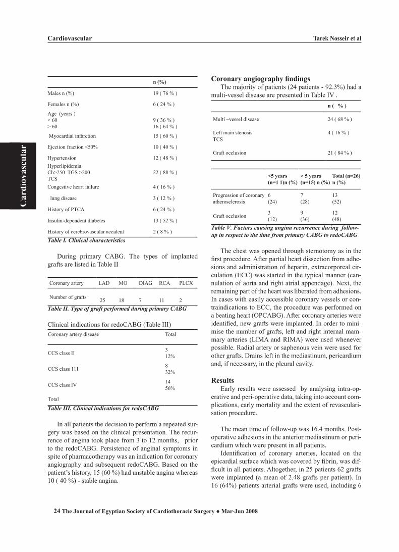
Cardiovascular Tarek Nosseir et al
24
Car
diov
ascu
lar
The Journal of Egyptian Society of Cardiothoracic Surgery ● Mar-Jun 2008
n (%)
Males n (%) 19 ( 76 % )
Females n (%) 6 ( 24 % )
Age (years )< 60> 60
9 ( 36 % )16 ( 64 % )
Myocardial infarction 15 ( 60 % )
Ejection fraction <50% 10 ( 40 % )
Hypertension 12 ( 48 % )HyperlipidemiaCh>250 TGS >200TCS
22 ( 88 % )
Congestive heart failure 4 ( 16 % )
lung disease 3 ( 12 % )
History of PTCA 6 ( 24 % )
Insulin-dependent diabetes 13 ( 52 % )
History of cerebrovascular accident 2 ( 8 % )
Table I. Clinical characteristics
During primary CABG. The types of implanted grafts are listed in Table II
Coronary artery LAD MO DIAG RCA PLCX
Number of grafts 25 18 7 11 2Table II. Type of graft performed during primary CABG
Clinical indications for redoCABG (Table III) Coronary artery disease Total
CCS class II 312%
CCS class 111 832%
CCS class IV 1456%
Total
Table III. Clinical indications for redoCABG
In all patients the decision to perform a repeated sur--gery was based on the clinical presentation. The recur--rence of angina took place from 3 to 12 months, prior to the redoCABG. Persistence of anginal symptoms in spite of pharmacotherapy was an indication for coronary angiography and subsequent redoCABG. Based on the patient’s history, 15 (60 %) had unstable angina whereas 10 ( 40 %) - stable angina.
Coronary angiography findingsThe majority of patients (24 patients - 92.3%) had a
multi-vessel disease are presented in Table IV . n ( % )
Multi –vessel disease 24 ( 68 % )
Left main stenosisTCS
4 ( 16 % )
Graft occlusion 21 ( 84 % )
<5 years (n=1 1)n (%)
> 5 years (n=15) n (%)
Total (n=26) n (%)
Progression of coronaryatherosclerosis
6(24)
7(28)
13(52)
Graft occlusion 3(12)
9(36)
12(48)
Table V. Factors causing angina recurrence during followtup in respect to the time from primary CABG to redoCABG
The chest was opened through sternotomy as in the
first procedure. After partial heart dissection from adhe--sions and administration of heparin, extracorporeal cir--culation (ECC) was started in the typical manner (can--nulation of aorta and right atrial appendage). Next, the remaining part of the heart was liberated from adhesions. In cases with easily accessible coronary vessels or con--traindications to ECC, the procedure was performed on a beating heart (OPCABG). After coronary arteries were identified, new grafts were implanted. In order to mini--mise the number of grafts, left and right internal mam--mary arteries (LIMA and RIMA) were used whenever possible. Radial artery or saphenous vein were used for other grafts. Drains left in the mediastinum, pericardium and, if necessary, in the pleural cavity.
ResultsEarly results were assessed by analysing intra-op--
erative and peri-operative data, taking into account com--plications, early mortality and the extent of revasculari--sation procedure.
The mean time of follow-up was 16.4 months. Post-operative adhesions in the anterior mediastinum or peri--cardium which were present in all patients.
Identification of coronary arteries, located on the epicardial surface which was covered by fibrin, was dif--ficult in all patients. Altogether, in 25 patients 62 grafts were implanted (a mean of 2.48 grafts per patient). In 16 (64%) patients arterial grafts were used, including 6
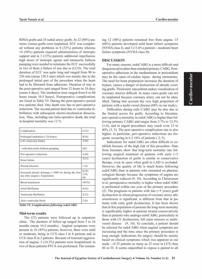
Tarek Nosseir et al Cardiovascular
25
Car
diov
ascu
lar
The Journal of Egyptian Society of Cardiothoracic Surgery ● Volume 16, Number (1-2)
RIMA grafts and 10 radial artery grafts. In 22 (88%) pa--tients venous grafts were implanted. ECC was complet--ed without any problems in 8 (32%) patients whereas 14 (56%) patients required administration of inotropic support and in 3 (12%) patients additional reperfusion, high doses of inotropic agents and intra aortic balloon pumping were needed to terminate the ECC successfully in two of them a failure of one due to (LCO). The total duration of ECC was quite long and ranged from 90 to 256 min (mean 120.3 min) which was mainly due to the prolonged initial part of the procedure when the heart had to be liberated from adhesions. Duration of stay in the post-operative unit ranged from 12 hours to 10 days (mean 4 days). The intubation time ranged from 8 to 60 hours (mean 10.5 hours). Post operative complications are listed in Table VI. During the post-operative period two patients died. One death was due to peri-operative infarction. The second patient died due to ventricular fi--brillation with subsequent electro-mechanical dissocia--tion. Thus, including one intra-operative death, the total in-hospital mortality was 12 %.
Complication n (%)
Prolonged intubation (>24 hours) 4(16)Left ventricular failure 7(28)
- with intra-aortic balloon pumping 2(8)
Peri-operative infarction 5(20)
Renal failure 3(12)
Wound infection 1 (4)
Increased pleural drainage (>1000 ml during the first day after surgery) Exploration
6(24)2 (8)(4)
Blood transfusion 19(76)
Atrial fibrillation 4(16)
Ventricular fibrillation 3(12)
Atrio-ventricular block 2 (8)
Table VI. Complications following redoCABG
Mid-term resultsThe (22) patients were followed up in outpatient
clinic. The duration of follow-up ranged from 3 to 24 months (mean 16.4 months). Anginal symptoms were present in 10 (45%) patients, however, there were mild or moderate, being in CCS class I in 8 patients and in CCS class II in 2 patients. Because of transient aggrava--tion of angina, 3 (14.3%) patients were hospitalised; in two of these patients PTCA was performed. The remain--
ing 12 (48%) patients remained free from angina. 15 (68%) patients developed mild heart failure symptoms (NYHA class I), and 3 (13.6%) patients - moderate heart failure symptoms (NYHA class II).
DISCUSSIONFor many reasons, redoCABG is a more difficult and
dangerous procedure than standard primary CABG. Post-operative adhesions in the mediastinum or pericardium may be the cause of cardiac injury during sternotomy. The need for heart preparation increases the duration of bypass, causes a danger of destruction of already exist--ing grafts. Thickened epicardium makes visualisation of coronary arteries difficult. In many cases grafts can not be implanted because coronary artery can not be iden--tified. Taking into account the very high proportion of patients with a multi-vessel disease (68% in our study)..
Difficulties during redo CABG may be also due to the limited access for grafts. According to literature, peri-operative mortality in redoCABG is higher than fol--lowing primary CABG and ranges from 3.7% to 12.5% (1-6), and in urgent procedures may reach even 13 to 40% (3, 5). The post-operative complication rate is also higher; in particular, peri-operative infarctions are fre--quent, occurring in 6.3-16% of patients ( 2, 5).
Indications for redoCABG are often difficult to es--tablish because of the high risk of this procedure. Data from literature show that long-term mortality rate fol--lowing surgical treatment of patients with early (<5 years) dysfunction of grafts is similar to conservative therapy, even in cases when graft to LAD is occluded. However, the quality of life is much better following redoCABG than in patients who remained on pharma--cological therapy because the symptoms of angina are significantly reduced (9, 10). According to Christenson et al. peri operative mortality is higher when redoCABG is performed within one year of the primary procedure (2). The prognosis in patients with late (>5 years) graft dysfunction in whom progression of coronary artery ath--erosclerosis is significant, is different from that in pa--tients with early graft dysfunction. It has been shown that in this population of patients the long-term mortality is significantly higher in patients treated conservatively than in patients who undergo redoCABG, particularly in those with LV dysfunction, left main stenosis or multi-vessel disease (9, 10). To conclude, a patient should be selected for redoCABG when anginal symptoms are increasing and the time since the primary procedure is long enough. Indications for surgery should be mainly based on clinical symptoms which was apparent in our study - of 25 patients as many as 22 were in CCS class III or IV. It seems unjustified to expose a patient to all
Cardiovascular Tarek Nosseir et al
26
Car
diov
ascu
lar
The Journal of Egyptian Society of Cardiothoracic Surgery ● Mar-Jun 2008
the risks of redoCABG only on the basis of the results of coronary angiography. Overall, the 5-year survival rate following redoCABG is satisfactory and according to literature ranges between 75 and 90.1% (2, 4, 7).
Dysfunction of grafts and progression of atheroscle--rosis in coronary arteries, are the main causes of angina recurrence. According to our findings, the contribution of these factors in symptom recurrence depends on the time passed from the primary CABG. During the first five years, problems with progression of coronary ath--erosclerosis are dominant, whereas in the later stages of follow-up graft patency becomes more important. Various factors cause graft malfunction. Histological changes in vein grafts may be observed as early as 2 to 3 months following surgery. Narrow microlesions with thrombus may be seen in the endothelium (11-13). Early thrombosis in vein graft, occurring up to 4-5 years after surgery, is usually caused by an inadequate blood flow through the vessel due to graft occlusion following tech--nical surgical error or low blood uptake in case of poor distal run off. The effects of risk factors as smoking have also to be taken into account (11-13). Intimal pro--liferation which is the first step of atherosclerosis, are observed in vein grafts shortly after surgery (within a few months). These extensive, concentric and progres--sive changes cause graft narrowing and finally, closure. Graft dysfunction due to atheromatous changes may be enhanced by thrombus formation which is a frequent finding in this place (11, 12). Intra-graft thrombus may be also a source of thrombosis of distal coronary arteries which is an important issue during redoCABG because of the possibility of mechanical dislodgement of throm--bi. After 10 years, the proportion of closed vein grafts is high, reaching 30%, whereas a further 30% of these grafts show significant narrowing (13).
Atherosclerotic changes in arterial grafts develop much slower compared with vein grafts. They remain patent for many years. It has been shown that the first and most frequently performed anastomosis - LIMA to LAD, shows a high patency rate even 20 years after sur--gery (13). Because LAD is the most important vessel for a systolic function of the LV, arterial grafting of this vessel decreases mortality and the need for redoCABG during long-term follow-up. These results prompted the use of other arteries for grafting such as radial artery. However, the duration of follow-up is not as long as that concerning the use of mammary arteries.
The course of atherosclerotic changes in arterial grafts is different than in native coronary arteries. The changes mainly develop in segments proximal to anas--tomosis and adversely effect graft function. This cause obstruction of the graft and may diminish blood uptake
from the graft with subsequent graft thrombosis when they develop in a distal part of the coronary artery.
CONCLUSIONSOur data suggest that surgery can be performed and
give expectation for success in early results. Progression of atherosclerosis in coronary arteries and graft dysfunc--tion are the two main causes of angina recurrences fol--lowing CABG. Reoperative coronary grafting is current--ly being performed in patients with greater incremental risk. Indications for redoCABG are mainly based on clinical presentation (CCS class III or IV), particularly when the time from primary procedure is long. Peri-op--erative mortality and peri-operative infarction rate are higher after redoCABG than primary procedure which is in line with data from literature.
REFERENCES
1. Yau TM, Borger MA, Weiset RD, Ivanov J. The Chang--ing Pattern Reoperative coronary surgery: trends in 1230 consecutive reoperations J Thorac Cardiovasc Surg 2000; 120:156-163 .
2. Christenson J. T., Schmuziger M., Simonet F.: Reoperative coronary artery bypass procedures: risk factors for early mortality and late survival. Eur. J. Cardio-Thorac. Surg. 1997, 11, 129-133.
3. He G. W., Acuff T. E., Ryan W. H. et al.: Determinants of operative mortality in reoperative coronary artery bypasss grafting. J. Thorac. Cardiovasc. Surg. 1995, 110, 971-978.
4. Weintraub WS, Jones EL, Craver JM, Grosswald R, Guyton RA. In-hospital and long-term outcome after reoperative coronary artery bypass graft surgery. Circulation 1995;92 (Suppl II):II-50 –II-57.
5. Akins C. W., Buckley M. J., Daggett W. M. et al.: Reopera--tive coronary grafting: changing patient profiles, operative indications, techniques and results. Ann Thorac Surg. 1994, 58, 359-364.
6. Lytle B. W., Mc Erloy D., Mc Carthy P. et al.: Influence of arterial coronary bypass grafts on the mortality in coronary reoperations. J. Thorac. Cardiovasc. Surg. 1994, 107, 675-682.
7. Shapira I., Isakov A., Heller I. et al.: Long-term follow-up after coronary artery bypass grafting reoperation. Chest. 1999, 115, 1593-1597.
8. Eck van FM, Noyez L, Verheugt FWA, Brouwer RMHJ. Changing profile of patients undergoing redo-coronary ar--tery surgery. Eur J Cardiothorac Surg 2001;21:205-211.
9. Lytle B. W., Loop F. D., Tylor P. C. et al.: Vein graft disease: The clinical impact of stenoses in saphenous vein bypass grafts to coronary arteries. J. Thorac. Cardiovasc. Surg. 1992, 103, 831.
10. Lytle B. W., Loop F. D., Tylor P. C. et al.: The effects of coronary reoperations on the survival of patients with ste--noses in saphenous vein to coronary bypass grafts. J. Tho--

Tarek Nosseir et al Cardiovascular
27
Car
diov
ascu
lar
The Journal of Egyptian Society of Cardiothoracic Surgery ● Volume 16, Number (1-2)
rac. Cardiovasc. Surg. 1993, 105, 605.11. Solymoss B. C., Leung T. K., Pelletier L. C. et al.: Patho--
logic changes in coronary artery saphenous vein grafts and related etiologic factors. Cardiovasc. Clin. 1995, 21, 45.
12. Bourassa M. G., Campeau L., Lesperance J.: Changes in grafts and in coronary arteries after coronary bypass sur--
gery. Cardiovasc. Clin. 1991, 21, 83.
13. Lytle B. W., Loop F. D., Cosgrove D. M. et al.: Long term
(5-12 years) serial studies of internal mammary artery and
saphenous vein coronary bypass grafts. J. Thorac. Cardio--
vasc. Surg. 1985, 89, 248.
Cardiovascular Abul-dahab et al
28
Car
diov
ascu
lar
The Journal of Egyptian Society of Cardiothoracic Surgery ● Mar-Jun 2008
ROUTINE USE OF TRANEXAMIC ACID PREOPERATIVELY IN
CORONARY ARTERY BYPASS SURGERY
Background: Our objective in this study was to study the effect of low-dose tranexamic acid on postoperative bleeding after coronary artery bypass operation done on-pump.Methods: 200 patients undergoing primary coronary artery bypass were divided into two groups. One group composed of 100 patients received 2g tranexamic acid with induction of anesthesia. The other group also composed of 100 patients received placebo. Data measured included blood loss, blood transfusion or its products and reoperation. Results: All demographic data were the same between the two groups. Postoperative bleeding was less in the tranexamic acid group (184±115 mL versus 528±218 mL, p, 0.001), also blood transfusion was higher in the placebo group (all the patients in placebo group received blood trans--fusion when only 32 patients in tranexamic acid group received blood transfusion.). The number of packed red blood cells received is also less in tranexamic acid group. Platelet number was reduced equally in both groups by cardiopulmonary bypass. Other test results were not statisti--cally different between groups. Conclusion: The use of low-dose tranexamic acid after induction of an--esthesia in coronary artery bypass significantly reduced the coagulopa--thy-induced postoperative bleeding and allogenic blood products require--ments.
Bleeding as a result of coagulopathy caused cardiopulmonary by--pass (CPB) is an important factor affecting the morbidity and mortality in patients undergoing cardiac operation(1). The coag--ulopathy is multi-factorial with platelet dysfunction and plasmin-induced fibrinolytic activity represents the major contributors to
the process (2). Postoperative bleeding may lead to allogenic blood transfu--sion and/or reoperation, which are independently associated with a number of adverse outcomes (3). This caused a renewed interest in the pharmacological reduction of bleeding (4).
Pharmacological agents to reduce bleeding have recently gained much in--terest, since they are readily available, easy to administer, can be used prophy--lactically, do not require the use of costly equipment, and appear to be very safe and efficacious. The perioperative use of tranexamic acid have gained acceptance around the world for the prophylactic reduction of allogenic blood transfusions in cardiac operation patients.
Tranexamic acid is a synthetic lysine analogue antifibrinolytic. It competi--tively inhibits the activation of plasminogen to plasmin, and is also a weak noncompetitive inhibitor of plasmin, blocking its action on fibrin. Thus, tranexamic acid is thought to act by preventing the premature dissolution of the normal fibrin clot. It is also possible that tranexamic acid may help to pre--
Accepted for publication oct 1 , 2007
Address reprint request to
Dr: Mohamed Abul dahab Department
of Cardiovascular Surgery , Kasr Eleini
Hospital, Cairo Univeristy
E-mail:[email protected]
codex : 04/cord/68/0710
Mohamed Abul-dahab M.D.,Tamer Farouk M.D.,Amr Rushdi M.D.,Tarek S. Abdalah M.D., Mohamed Sweilam M.D.

Abul-dahab et al Cardiovascular
29
Car
diov
ascu
lar
The Journal of Egyptian Society of Cardiothoracic Surgery ● Volume 16, Number (1-2)
serve platelet function, by reducing the effect of plasmin on platelet membrane receptors (5).
Kojima and his colleagues studied the effects of tranexamic acid on fibrinolysis and bleeding during and after cardiopulmonary bypass surgery and concluded that it can inhibit fibrinolytic activity during CPB by reducing tissue plasminogen activator activity and by blocking plasmin activity measured as D-dimer. In con--trast, tranexamic acid has no influence on the release of plasminogen activator inhibitor-1 from endothe--lium and neutralization of plasmin by α2-antiplasmin. These results suggest that the antifibrinolytic effects of tranexamic acid can bring the reduction of blood loss in patients undergoing CPB surgery (6).
Fortunately, the incidence of complications, such as graft thrombosis leading to myocardial infarction, stroke, renal dysfunction and allergic reaction is low (4). The results of Ruel on 221 patients underwent primary CABG with end-arterectomy suggest that the clinical ef--fectiveness of tranexamic acid in reducing postoperative blood loss in patients undergoing coronary end-arterec--tomy is not associated with a higher incidence of myo--cardial ischemia-related complications (7).
We speculated whether preoperative use of tranexam--ic acid as a routine, would have any major significant clinical haemostatic effects in a group of patients with low risk of postoperative bleeding in our hospital.
Patients and MethodsBetween October 2006 and October 2007, 200 pa--
tients scheduled for elective or urgent CABG were en--rolled in the study. Patients were randomly assigned to one of two groups: Group “A” (100 patients), to receive tranexamic acid (TA) immediately after induction of anesthesia, and group” B “(100 patients), who received equal volume of placebo.I. Selection Criteria:
Exclusion criteria: 1) Patients with a history of abnormal bleeding or
abnormal coagulation profile.2) Patients had recent (< 7 days) acetylsalicylic acid
ingestion, thrombolytic therapy (streptokinase) or anticoagulant therapy (heparin < 4 hours or warfarin < 3 days).
Inclusion criteria: All adult patients with ischemic heart disease sub--jected to CABG procedure. A written informed consent was obtained from each patient before surgery.
II. Preoperative Assessment: All patients of the study were assessed in the pre--
operative period by medical history taking and detailed clinical examination with a special emphasis on any co--agulation problems, full laboratory investigations with special emphasis on hemoglobin, hematocrit, platelet number, international randomized ratio (INR) and trans--thoracic echocardiography.
III. Operative Management:Anesthetic technique:Routine anesthetic procedures were employed. All
patients were premedicated with an intramuscular injec--tion with morphine half an hour preoperatively. Induc--tion of anesthesia with a bolus of midazolam and fen--tanyl. Muscle relaxation was achieved and maintained with pancuronium. Anesthesia was maintained with isoflurane (pre-cardiopulmonary bypass) and propofol infusion during CPB.
Drug administration: Tranexamic acid was administered as an intravenous
bolus dose of 2g after induction of anesthesia and before skin incision.
Surgical technique:In all cases, access to the heart was obtained through
a median sternotomy. The left internal mammary artery was harvested in all patients. Saphenous veins were used as routine grafts for coronary anastomoses other than left anterior descending. Anticoagulation was achieved using 300 IU/kg of heparin before cannulation. Cardio--pulmonary bypass was instituted in a standard fashion.
Myocardial protection was achieved by intermittent warm blood antegrade cardioplegia solution with re--peated doses every 20 minutes. The heparin effect was reversed by protamine administration (3mg/kg). Before closure of the chest, mediastinal and pleural tubes were positioned in the mediastinum and left pleura separately (the left pleura was routinely opened). A chest tube was positioned on right chest only when the right pleuron was opened.
Operative data and parameters: A record was made of the following: Timing of sur--
gery (emergency, urgent or elective), Ischemic time (in minutes), Bypass time (in minutes), Total operative time (in minutes), number of grafts, Weaning from CPB and use of inotropes. All distal anastomoses were per--formed first and proximal anastomoses were performed after declamping but on total bypass.
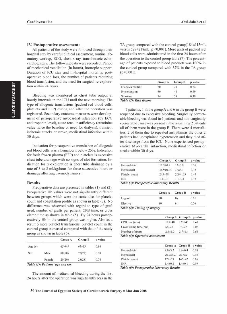
Cardiovascular Abul-dahab et al
30
Car
diov
ascu
lar
The Journal of Egyptian Society of Cardiothoracic Surgery ● Mar-Jun 2008
IV. Postoperative assessment:All patients of the study were followed through their
hospital stay by careful clinical assessment, routine lab--oratory workup, ECG, chest x-ray, transthoracic echo--cardiography. The following data were recorded: Period of mechanical ventilation (in hours), inotropic support, Duration of ICU stay and In-hospital mortality, post--operative blood loss, the number of patients requiring blood transfusion, and the need for surgical re-explora--tion within 24 hours.
Bleeding was monitored as chest tube output at hourly intervals in the ICU until the next morning. The type of allogenic transfusions (packed red blood cells, platelets and FFP) during and after the operation was registered. Secondary outcome measures were develop--ment of perioperative myocardial infarction (by ECG and troponin level), acute renal insufficiency (creatinine value twice the baseline or need for dialysis), transient ischemic attacks or stroke, mediastinal infection within 30 days.
Indication for postoperative transfusion of allogenic
red blood cells was a hematocrit below 25%. Indication for fresh frozen plasma (FFP) and platelets is excessive chest tube drainage with no signs of clot formation. In--dication for re-exploration is chest tube drainage by a rate of 3 to 5 ml/kg/hour for three successive hours or drainage affecting haemodynamics.
ResultsPreoperative data are presented in tables (1) and (2).
Preoperative Hb values were not significantly different between groups which were the same also for platelet count and coagulation profile as shown in table (3). No difference was observed with regard to type of graft used, number of grafts per patient, CPB time, or cross clamp time as shown in table (5). By 24 hours postop--eratively Hb in the control group was higher. Also as a result o more platelet transfusions, platelet count in the control group increased compared with that of the study group as shown in table (6).
Group A Group B p value
Age (y) 65.6±9 65±13 0.86
Sex Male 80(80) 72(72) 0.78
Female 20(20) 28(28) 0.74Table (1): Patients’ age and sex
The amount of mediastinal bleeding during the first 24 hours after the operation was significantly less in the
TA group compared with the control group(184±115mL versus 528±218mL, p <0.001). More units of packed red blood cells were administered in the first 24 hours after the operation to the control group table (7). The percent--age of patients exposed to blood products was 100% in the control group compared with 32% in the TA group (p<0.001).
Group A Group B p valueDiabetes mellitus 20 28 0.74Hypertension 60 44 0.39Smoking 74 58 0.39
Table (2): Risk factors
7 patients, 1 in the group A and 6 in the group B were reopened due to excessive bleeding. Surgically correct--able bleeding was found in 5 patients and non-surgically correctable cause was present in the remaining 2 patients all of them were in the group B. There were 4 mortali--ties, 2 of them due to repeated arrhythmias the other 2 patients had unexplained hypotension and they died af--ter discharge from the ICU. None experienced postop--erative Myocardial infarction, mediastinal infection or stroke within 30 days.
Group A Group B p valueHemoglobin 12.3±0.9 12±0.9 0.39Hematocrit 36.9±0.04 36±3.1 0.73Platelet count 245±58 209±103 0.47INR 1.1±0.1 1.1±0.1 0.73
Table (3): Preoperative laboratory Results
Group A Group B p value
Urgent 20 16 0.61
Elective 80 84 0.76Table (4): Timing of surgery
Group A Group B p valueCPB time(min) 125±40 133±43 0.41Cross clamp time(min) 66±23 78±27 0.08Number of grafts 2.6±1.3 2.7±1.4 0.64
Table (5): Operative assessment
Group A Group B p valueHemoglobin 8.9±3.2 9.6±0.4 0.08Hematocrit 26.9±3.2 28.7±2 0.05Platelet count 128±27 145±42 0.16INR 1.4±0.1 1.4±0.1 0.99
Table (6): Postoperative laboratory Results

Abul-dahab et al Cardiovascular
31
Car
diov
ascu
lar
The Journal of Egyptian Society of Cardiothoracic Surgery ● Volume 16, Number (1-2)
Group A Group B p value
Bleeding (ml) 184±115 528±218 <0.001
Number of patients receiving transfusion 32 100 <0.001
Packed cells 0-4 1-6 <0.001Table (7): Mediastinal bleeding and patients receiving transttfusion in the first 24 hours
Discussion
Prophylactic use of TA has been shown to reduce perioperative blood loss following primary elective heart operations including primary myocardial revasculariza--tion in several randomized, placebo-controlled studies and meta-analyses (8). Meta-analysis of multiple stud--ies has shown aprotinin and anti-fibrinolytics to reduce mediastinal chest tube drainage by 30% versus placebo. Although delivery protocols were uniform for aprotinin, they still vary widely for TA (9).
Tranexamic acid has been shown as effective as
aprotinin in reducing coagulopathy-caused bleeding af--ter CPB and cheaper than aprotinin (10). As TA is presently is the available drug of choice to reduce co--agulopathy-caused bleeding, we designed our study to gain knowledge about the benefit of using low-dose TA in terms of reducing blood loss and allogenic transfu--sion.
In the preoperative data of our patients, in both groups, there was no statistically significant difference as regard to the age and sex. The age was slightly high as all our patients were ischemic cardiac patients subjected for CABG procedure with predominance of males due to the nature of the disease, more affecting males.
As regard preoperative risk factors for ischemic heart
disease, there were no statistical differences between both groups and there were high percentage of smoking and systemic hypertension. In preoperative lab--oratory data there were no abnormalities in the platelet counts of the patients or their coagulation profiles preop--eratively. There was slight anemia preoperatively in our patients which are common figures in this community, keeping in consideration doing the research in a public hospital.
In a low-risk patient population, TA was shown to decrease mediastinal bleeding after cardiac operation as early as 1990 (8). A similar result was found in studies by Karski and associates. The first significant study of a uniform patient population undergoing coronary op--
eration was reported by Rousou and colleagues. They retrospectively studied 415 patients undergoing CABG excluding emergency and redo operations. The first 209 patients were operated on without TA and the subse--quent 206 with a 2g bolus of TA followed by 8g during the procedure. Chest tube drainage in the control group was 1114mL versus 803mL in the study group (8).
A double-blind randomized placebo-controlled study was reported from Brook Army Medical Center on patients undergoing primary coronary artery opera--tion. The dose of TA was 15 mg/kg before CPB and 1 mg/kg continued for 5 hours. The bleeding was reduced from 1202mL in the control group versus 1020mL in the TA group. Since then, multiple studies have shown the efficacy of TA in prospective studies comparing patients receiving TA to groups receiving aprotinin or ε-amino-caproic acid (EACA) (2).
In all previously mentioned studies the authors used high doses of TA. The aim of this study is to answer the question of whether the addition of low-dose TA as giv--en in our study will be beneficial. In our study although control patients only bled 528mL in 24 hours, the use o TA significantly reduced this even further to 184mL. The low amount of bleeding in the TA group is compat--ible with recently published reports.
The increased platelet transfusions in the control group most probably represent more oozing in the control group whereas the lower percentage of platelet transfusion in the study group is probably not related to any effect of TA on platelet number and function. This increased platelet transfusion in the control group ex--plains the high platelet count in the control group at 24 hours after the operation.
Extracorporeal circulation causes fibrinolysis by plasmin activation (11). Tranexamic acid is a competi--tive inhibitor of plasminogen and, at higher doses, acts as a noncompetitive inhibitor of plasmin (2). The tim--ing of TA administration to reduce postoperative blood loss is unclear. It is both logical and established that TA should be given before CPB as an initial dose (2). Whether to continue the TA after the operation is contro--versial. Tranexamic acid has been used effectively when given up to 12 hours after an operation and when only given during the operation. They postulated that because the half-life of TA is 80 minutes, a second bolus would prevent fibrinolysis after the operation. A recent study failed to prove the efficacy of postoperative administra--tion of TA (12).

Cardiovascular Abul-dahab et al
32
Car
diov
ascu
lar
The Journal of Egyptian Society of Cardiothoracic Surgery ● Mar-Jun 2008
Andreasen and Nielsen concluded in their study that an anti-fibrinolytic effect following prophylactic use of tranexamic acid in elective, primary CABG among patients with a low risk of postoperative bleeding did not result in any significant decrease in postoperative bleeding compared to placebo group (13). This was not in concordance with many other studies findings, which could be attributed to the act that they give additional heparin dose to the patients in the study group than the control group which led to significant increase in APTT ( activated partial thromboplastin time) in the treatment group next morning. Another factor may contribute to their result was their exclusion of the reopened cases for bleeding from the study which were more in the control group.
Ruel and his colleagues studied giving prophylactic TA in cases of coronary endarterectomy. They said in--tra-operative use of TA in patients undergoing coronary endarterectomy is safe and effective in reducing postop--erative blood loss (7). We concluded that administering a low dose of 2g of tranexamic acid with induction of anesthesia is associated with decreased fibrinolysis dur--ing CPB. This effect has positive clinical results in terms of bleeding and blood transfusion in patients undergoing primary CABG.
References
1. Edmunds LH. Cardiopulmonary bypass for open heart sur--gery. In: Baue AE, Geha AS, Hammond GL, Laks H, Naun--heim KS, eds. Glenn’s thoracic and cardiovascular surgery, Vol 2, 2nd ed. Stanford, CT: Appleton &Lange, 1996:1642-4.
2. Brown RS, Thwaits BK, Mongan PD. Tranexamic acid is effective in decreasing postoperative bleeding and trans--fusion in primary coronary artery bypass operations: a double-blind, randomized, placebo-controlled trial. Anesth Analg 1997; 85:963-70.
3. Engoren MC, Habib RH, Zacharias A, SchwannTA, Riordan CJ, Durham SJ. Effect of blood transfusion on long-term survival after cardiac operation. Ann Thorac Surg 2002; 74:1180-6.
4. Bill IW, Richard FM, Stephen EF, Katherine AD, Ellen MH, George TC, Bernard SG. Aprotinin and tranexamic acid for high transfusion risk cardiac surgery. Ann Thorac Surg 2000; 69:808-16.
5. Hardy J-F, Desroches J. Natural and synthetic antifibrinolyt--ics in cardiac surgery. Can J Anaesth 1992; 39: 353-65.
6. Koijima T, Gando S, Mormoto Y, Mashio H, Goda Y, Kawa--higashi H, Kemmotsu O. Systematic elucidation o effects of tranexamic acid on fibrinolysis and bleeding during and after cardiopulmonary bypass surgery. Thrombosis research 2001; 104: 301-7.
7. Ruel MA, Wang F, Bourke ME, Dupuis J-Y, Robblee JA, Keon WJ, Rubens FD. Is tranexamic acid safe in patients undergoing coronary endarterectomy? Ann Thorac Surg 2001; 71: 1508-11.
8. Pleym H, Stenseth R, Wahba A, Bjella, Karevold A, Dale O. Single-dose tranexamic acid reduces postoperative bleed--ing after coronary surgery in patients treated with aspirin until surgery. Anesth Analg 2003; 96: 923-8.
9. Zabeeda D, Medalion B, Sverdlov M, Ezra S, Schachner A, Ezri T, Cohen A. Tranexamic acid reduces bleeding and the need for blood transfusion in primary myocardial revascu--larization. Ann Thorac Surg 2002; 74: 733-8.
10. Casati V, Guzzon D, Oppizzi M. Haemostatic effects of aprotinin, tranexamic acid and ε amino aproic acid in pri--mary cardiac surgery. Ann Thorc Surgery 1999; 68: 2252-6.
11. Dignan RJ, Law DW, Seah PW. Ultra- low dose aprotinin decreases transfusion requirements, and is cost effective in coronary operations. Ann Thorac Surg 2001; 71: 158-64.
12. Casati V, Gerli C, Frano A. Activation of coagulation and fibrinolysis during coronary surgery: on-pump versus off-pump techniques. Anesthesiology 2001; 95: 1103-9.
13. Andreasen JJ, Nielsen C. Prophylactic tranexamic acid in elective, primary coronary artery bypass surgery using cardiopulmonary bypass. European J of cardio-thorac surg 2004; 26: 311-17.
Elsayed Elmistekawy et al Cardiovascular
33
Car
diov
ascu
lar
The Journal of Egyptian Society of Cardiothoracic Surgery ● Volume 16, Number (1-2)
CAN LOCAL APPLICATION OF TRANEXAMIC ACID REDUCE POST- CABG BLOOD LOSS?
Introduction: Diffuse microvascular bleeding remains a common problem after cardiac procedures. Systemic use of antifibrinolytic (tranexamic acid) reduces the postopera--tive blood loss. The purpose of this study was to examine the effectiveness of local applica--tion of tranexamic acid to reduce blood loss after coronary artery bypass grafting (CABG).Methods: Thirty eight patients scheduled for primary isolated coronary artery bypass grafting were included in this double blind, prospective, randomized, placebo controlled study. Group (I) included 19 patients who received 1 gram of tranexamic acid diluted in 100 ml normal saline. Group (II) included 19 patients who re--ceived 100ml of normal saline only as placebo. The solution was purred in the pericardial and mediastinal cavities. Postoperative blood loss and blood product transfusion were compared between the two groups.Results: Both groups were comparable with respect to baseline demo--graphic data and surgical characteristics. During the first 24 hours post-operatively, cumulative blood loss was significantly less in TA group (691.1 + 377.3 ml) compared to Placebo group (1094.1 + 672.1 ml) (P =0.03). There was no significant difference in the post-op Packed RBCs transfu--sion between both groups (1.53 + 1.47 units vs. 1.95 + 2.12 units, respec--tively) (P = 0.5). Significant less platelets transfusion required in TA group (0.47 + 0.84 units) than in placebo group (1.57 + 1.87 units) (P= 0.02). Apart from re-exploration for surgical excessive bleeding in one patient in the TA group, no difference was found in morbidity or mortality between the two groups.Conclusion: Topical application of tranexamic acid solution in patients undergoing primary coronary artery bypass grafting led to a significant reduction in postoperative blood loss without adding extra risk to the pa--tient. Consequently tranexamic acid could be advocated for routine use topically in patients undergoing coronary artery bypass grafting.
Coagulopathy remains a common problem after coronary artery bypass Grafting (CABG) using cardiopulmonary bypass (CPB). It results from many factors like thrombocytopenia, acquired platelet dysfunction, clotting factors loss, free heparin, and in--creased fibrinolysis (1-3). Lemmer and Colleagues4 found that
extracorporeal circulation results in significant fibrinolysis, as reflected by increased concentrations of plasmin and fibrin degradation products (FDP), both of which have deleterious effects on platelet function. Re-exploration for bleeding following cardiac surgery with CPB was reported to be in the range of 2-7%. Of these, 50-80% was found to be medical rather than surgi--
Accepted for publication Jul 1 , 2007
Address reprint request to Dr: Hosam
Fawzy Department of Cardiovascu--
lar Surgery , St michael’s Hospital,
Tomonto
E-mail:[email protected]
codex : 04/cord/69/0710
Elsayed Elmistekawy MD, Abdelsalam Elhenawy MD, Hosam Fawzy MD,
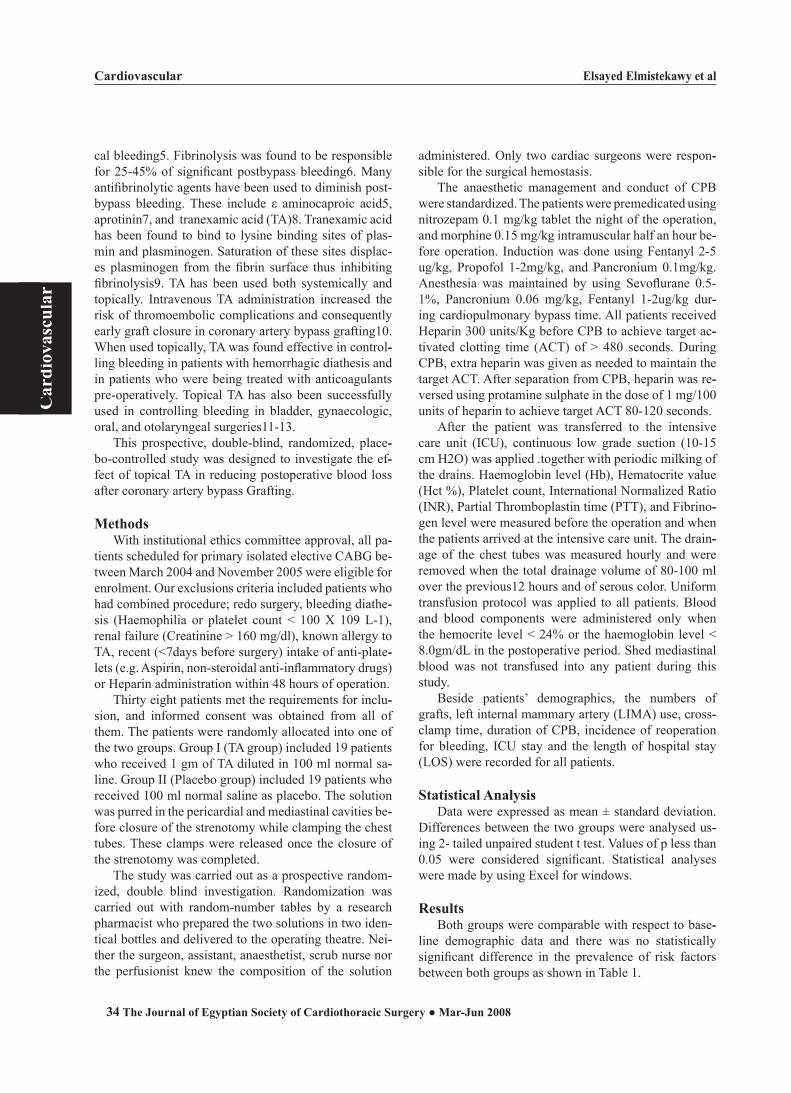
Cardiovascular Elsayed Elmistekawy et al
34
Car
diov
ascu
lar
The Journal of Egyptian Society of Cardiothoracic Surgery ● Mar-Jun 2008
cal bleeding5. Fibrinolysis was found to be responsible for 25-45% of significant postbypass bleeding6. Many antifibrinolytic agents have been used to diminish post-bypass bleeding. These include ε aminocaproic acid5, aprotinin7, and tranexamic acid (TA)8. Tranexamic acid has been found to bind to lysine binding sites of plas--min and plasminogen. Saturation of these sites displac--es plasminogen from the fibrin surface thus inhibiting fibrinolysis9. TA has been used both systemically and topically. Intravenous TA administration increased the risk of thromoembolic complications and consequently early graft closure in coronary artery bypass grafting10. When used topically, TA was found effective in control--ling bleeding in patients with hemorrhagic diathesis and in patients who were being treated with anticoagulants pre-operatively. Topical TA has also been successfully used in controlling bleeding in bladder, gynaecologic, oral, and otolaryngeal surgeries11-13.
This prospective, double-blind, randomized, place--bo-controlled study was designed to investigate the ef--fect of topical TA in reducing postoperative blood loss after coronary artery bypass Grafting.
Methods
With institutional ethics committee approval, all pa--tients scheduled for primary isolated elective CABG be--tween March 2004 and November 2005 were eligible for enrolment. Our exclusions criteria included patients who had combined procedure; redo surgery, bleeding diathe--sis (Haemophilia or platelet count < 100 X 109 L-1), renal failure (Creatinine > 160 mg/dl), known allergy to TA, recent (<7days before surgery) intake of anti-plate--lets (e.g. Aspirin, non-steroidal anti-inflammatory drugs) or Heparin administration within 48 hours of operation.
Thirty eight patients met the requirements for inclu--sion, and informed consent was obtained from all of them. The patients were randomly allocated into one of the two groups. Group I (TA group) included 19 patients who received 1 gm of TA diluted in 100 ml normal sa--line. Group II (Placebo group) included 19 patients who received 100 ml normal saline as placebo. The solution was purred in the pericardial and mediastinal cavities be--fore closure of the strenotomy while clamping the chest tubes. These clamps were released once the closure of the strenotomy was completed.
The study was carried out as a prospective random--ized, double blind investigation. Randomization was carried out with random-number tables by a research pharmacist who prepared the two solutions in two iden--tical bottles and delivered to the operating theatre. Nei--ther the surgeon, assistant, anaesthetist, scrub nurse nor the perfusionist knew the composition of the solution
administered. Only two cardiac surgeons were respon--sible for the surgical hemostasis.
The anaesthetic management and conduct of CPB were standardized. The patients were premedicated using nitrozepam 0.1 mg/kg tablet the night of the operation, and morphine 0.15 mg/kg intramuscular half an hour be--fore operation. Induction was done using Fentanyl 2-5 ug/kg, Propofol 1-2mg/kg, and Pancronium 0.1mg/kg. Anesthesia was maintained by using Sevoflurane 0.5-1%, Pancronium 0.06 mg/kg, Fentanyl 1-2ug/kg dur--ing cardiopulmonary bypass time. All patients received Heparin 300 units/Kg before CPB to achieve target ac--tivated clotting time (ACT) of > 480 seconds. During CPB, extra heparin was given as needed to maintain the target ACT. After separation from CPB, heparin was re--versed using protamine sulphate in the dose of 1 mg/100 units of heparin to achieve target ACT 80-120 seconds.
After the patient was transferred to the intensive care unit (ICU), continuous low grade suction (10-15 cm H2O) was applied .together with periodic milking of the drains. Haemoglobin level (Hb), Hematocrite value (Hct %), Platelet count, International Normalized Ratio (INR), Partial Thromboplastin time (PTT), and Fibrino--gen level were measured before the operation and when the patients arrived at the intensive care unit. The drain--age of the chest tubes was measured hourly and were removed when the total drainage volume of 80-100 ml over the previous12 hours and of serous color. Uniform transfusion protocol was applied to all patients. Blood and blood components were administered only when the hemocrite level < 24% or the haemoglobin level < 8.0gm/dL in the postoperative period. Shed mediastinal blood was not transfused into any patient during this study.
Beside patients’ demographics, the numbers of grafts, left internal mammary artery (LIMA) use, cross--clamp time, duration of CPB, incidence of reoperation for bleeding, ICU stay and the length of hospital stay (LOS) were recorded for all patients.
Statistical AnalysisData were expressed as mean ± standard deviation.
Differences between the two groups were analysed us--ing 2- tailed unpaired student t test. Values of p less than 0.05 were considered significant. Statistical analyses were made by using Excel for windows.
ResultsBoth groups were comparable with respect to base--
line demographic data and there was no statistically significant difference in the prevalence of risk factors between both groups as shown in Table 1.
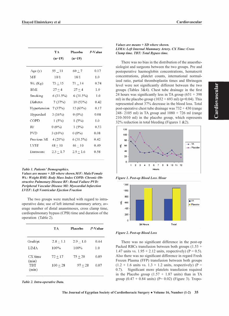
Elsayed Elmistekawy et al Cardiovascular
35
Car
diov
ascu
lar
The Journal of Egyptian Society of Cardiothoracic Surgery ● Volume 16, Number (1-2)
Table 1. Patients’ Demographics.Values are means + SD where shown.M/F: Male/Female Wt.: Weight BMI: Body Mass Index COPD: Chronic Obttstructive Pulmonary Disease RF: Renal Failure PVD: Peripheral Vascular Disease MI: Myocardial Infarction LVEF: Left Ventricular Ejection Fraction
The two groups were matched with regard to intra--operative data; use of left internal mammary artery, av--erage number of distal ananstmoses, cross clamp time, cardiopulmonary bypass (CPB) time and duration of the operation (Table 2).
Table 2. Intratoperative Data.
Values are means + SD where shown.LIMA: Left Internal Mammary Artery. CX Time: Cross Clamp time. TBT: Total Bypass time.
There was no bias in the distribution of the anaesthe--siologist and surgeons between the two groups. Pre and postoperative haemoglobin concentrations, hematocrit concentrations, platelet counts, international normal--ized ratio, partial thromboplastin times and fibrinogen level were not significantly different between the two groups (Tables 3&4). Chest tube drainage in the first 24 hours was significantly less in TA group (651 + 398 ml) in the placebo group (1032 + 693 ml) (p=0.04). This represented about 37% decrease in the blood loss. Total post-operative chest tube drainage was 732 + 430 (range 248- 2105 ml) in TA group and 1080 + 726 ml (range 210-3010 ml) in the placebo group, which represents 32% reduction in total bleeding (Figures 1 &2).
Figure 1. Posttop Blood Loss /Hour
Figure 2. Posttop Blood Loss
There was no significant difference in the post-op Packed RBCs transfusion between both groups (1.53 + 1.47 units vs. 1.95 + 2.12 units, respectively) (P = 0.5). Also there was no significant difference in regard Fresh Frozen Plasma (FFP) transfusion between both groups (1.2 + 1.6 units vs. 1.3 + 1.2 units, respectively) (P = 0.7). Significant more platelets transfusion required in the Placebo group (1.57 + 1.87 units) than in TA group (0.47 + 0.84 units) (P= 0.02) (Figure 3). Tropo--
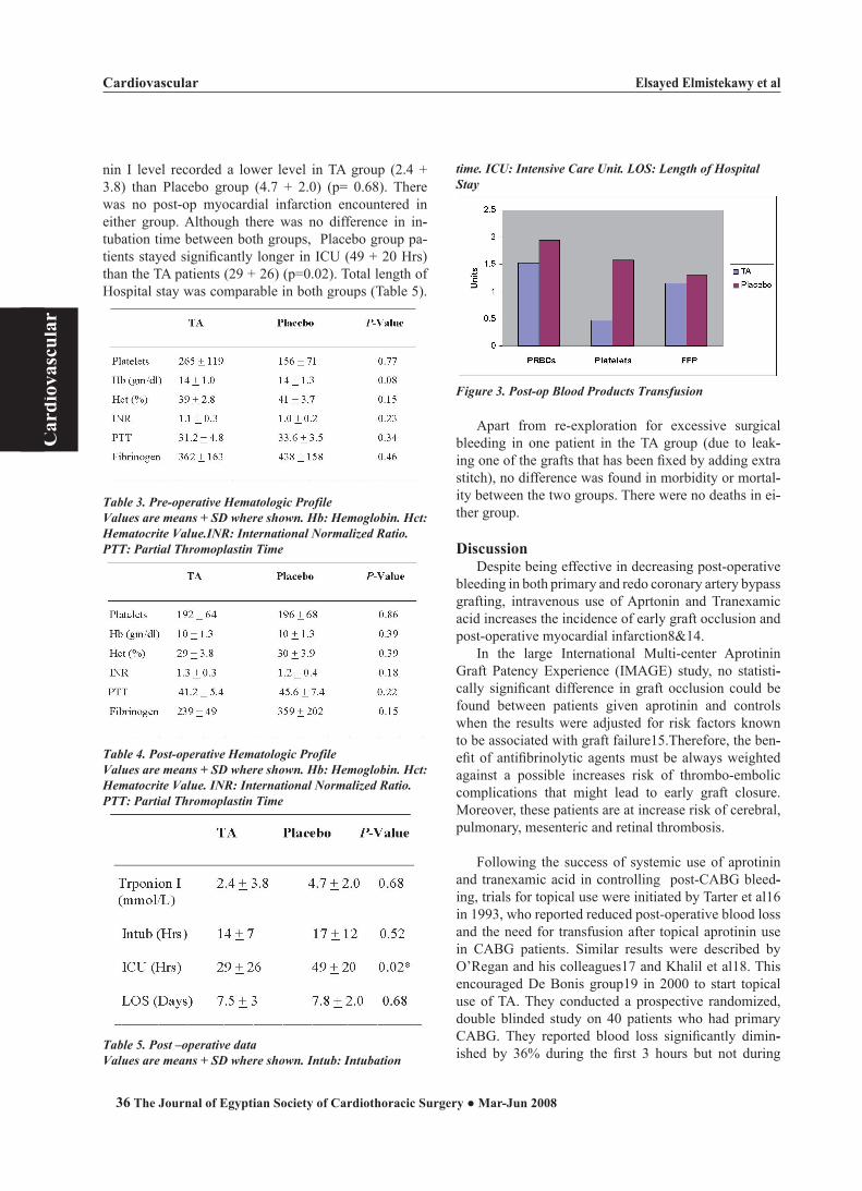
Cardiovascular Elsayed Elmistekawy et al
36
Car
diov
ascu
lar
The Journal of Egyptian Society of Cardiothoracic Surgery ● Mar-Jun 2008
nin I level recorded a lower level in TA group (2.4 + 3.8) than Placebo group (4.7 + 2.0) (p= 0.68). There was no post-op myocardial infarction encountered in either group. Although there was no difference in in--tubation time between both groups, Placebo group pa--tients stayed significantly longer in ICU (49 + 20 Hrs) than the TA patients (29 + 26) (p=0.02). Total length of Hospital stay was comparable in both groups (Table 5).
Table 3. Pretoperative Hematologic ProfileValues are means + SD where shown. Hb: Hemoglobin. Hct: Hematocrite Value.INR: International Normalized Ratio. PTT: Partial Thromoplastin Time
Table 4. Posttoperative Hematologic ProfileValues are means + SD where shown. Hb: Hemoglobin. Hct: Hematocrite Value. INR: International Normalized Ratio. PTT: Partial Thromoplastin Time
Table 5. Post –operative dataValues are means + SD where shown. Intub: Intubation
time. ICU: Intensive Care Unit. LOS: Length of Hospital Stay
Figure 3. Posttop Blood Products Transfusion
Apart from re-exploration for excessive surgical bleeding in one patient in the TA group (due to leak--ing one of the grafts that has been fixed by adding extra stitch), no difference was found in morbidity or mortal--ity between the two groups. There were no deaths in ei--ther group.
DiscussionDespite being effective in decreasing post-operative
bleeding in both primary and redo coronary artery bypass grafting, intravenous use of Aprtonin and Tranexamic acid increases the incidence of early graft occlusion and post-operative myocardial infarction8&14.
In the large International Multi-center Aprotinin Graft Patency Experience (IMAGE) study, no statisti--cally significant difference in graft occlusion could be found between patients given aprotinin and controls when the results were adjusted for risk factors known to be associated with graft failure15.Therefore, the ben--efit of antifibrinolytic agents must be always weighted against a possible increases risk of thrombo-embolic complications that might lead to early graft closure. Moreover, these patients are at increase risk of cerebral, pulmonary, mesenteric and retinal thrombosis.
Following the success of systemic use of aprotinin and tranexamic acid in controlling post-CABG bleed--ing, trials for topical use were initiated by Tarter et al16 in 1993, who reported reduced post-operative blood loss and the need for transfusion after topical aprotinin use in CABG patients. Similar results were described by O’Regan and his colleagues17 and Khalil et al18. This encouraged De Bonis group19 in 2000 to start topical use of TA. They conducted a prospective randomized, double blinded study on 40 patients who had primary CABG. They reported blood loss significantly dimin--ished by 36% during the first 3 hours but not during

Elsayed Elmistekawy et al Cardiovascular
37
Car
diov
ascu
lar
The Journal of Egyptian Society of Cardiothoracic Surgery ● Volume 16, Number (1-2)
the following 21 hours after operation compared to the placebo group. Our study demonstrates that pouring of one gram of TA into the pericardial cavity after CABG, significantly reduced post-operative blood loss in the first 24 hours after surgery (37%) with the maximum reduction at the 3rd hour (66%). Abdul Azem and his colleagues20 in 2006 described similar results in 100 patients underwent various open heart procedures. In contradictory to our findings, Yasim and his group21 did not find a statistically significant reduction in post-operative bleeding after topical application of aprotinin and tranexamic acid.
In our study the overall post-operative bleeding was modest in both groups and this could be explained by the tight inclusion criteria, meticulous surgical hemo--stasis, normothermic CPB and the use of LIMA alone as a pedicle graft in all patients. Therefore, despite the significant reduction of post-operative bleeding in TA group, the effect of its topical use might be masked by those factors. A greater effect of topical TA in reducing blood loss could be possibly evident in more prolonged and complex procedures with a higher risk of bleeding.
The use of blood products was not significantly dif--ferent between both De Bonis’ groups19, while we had a significant reduction only in platelet transfusion and Abdul Azm20 had a significant reduction only in packed cells use in TA group. This difference in reports might be explained by the difference in number of patients, type of operations and transfusion protocols used in each study. Limitation of the study
We did not measure the level of TA in the serum or in the pericardial cavity. The number of the patients was relatively small and the inclusion criteria with strictly tight. Larger multi-center prospective control trials us--ing larger dose of TA are needed to address its topical effect in procedures with higher risk of bleeding.
Conclusion: Topical use of Tranexamic acid into the pericardial
cavity in patients undergoing primary coronary artery bypass grafting significantly reduces postoperative bleeding and platelet transfusion.
References
(1) Kucuk O, Kwaan HC, Frederickson J, Wade L, Green D. Increased fibrinolytic activeity in patients undergoing car--diopulmonary bypass operation. Am J Hematol 1986; 23: 223-9.
(2) Harker LA, Malpass TW, Branson HE, Hessel EAII, Slich--ter SJ. Mechnism of abnormal bleeding in patients under--
going cardiopulmonary bypass: acquired transient platelet dysfunction associated with selective alpha-granule release. Blood 1980; 56:824-34.
(3) Depotis GJ, Santoro SA, Spitznagel E, Kater KM, Cox JL, Barnes P, et al. Prosepective evaluation and clinical utility of on-site monitoring of coagulation in patients undergoing cardiac operation. J Thorac Cardiocavsc Surg 1994; 107: 271-9.
(4) Lemmer JH Jr, Stanford W, Bonney SL, Breen JF, Chomka EV, Elderge WJ, et al. Aprotonin for coronary bypass op--erations: efficacy, safety, and influence on early saphenous graft patency- a muticenter, randomized, double blind, placebo-controlled study. J Thorac Cardiovasc Surg 1994; 107: 543-53.
(5) Daily PO, Lamphere JA, Dembitsky WP, Admson RM, Dans NF. Effect of prophylactic epsilon-aminocaproic acid on blood loss and transfusion requitments in patients under--going first-time coronary artery bypass grafting: a random--ized, prospective, double-blind study. J Thorac Cardiovasc Surg 1994; 108:99-105.
(6) Kevy SV, Glickman RM, Bernhard WF, Diamond LK, Grass RE. The pathogenesis and control of the hemorrhagic defect in open heart surgery. Surg Gynecol Obstet 1966; 123: 313-8.
(7) Cosgrove DM III, Heric B, Lytle BW, Taylor PC,Novoa R, Golding LA, et al. Aprotonin therapy for reoperative myo--cardial revascularization: a placebo-controlled study. Ann Thorac Surg 1992; 54: 1031-8.
(8) Horrow Jc, Van Riper DF, Strong MD, Brodsky I, Parmet JL. Hemostatic effects of tranexamic acid and desmopres--sin during cardiac surgery. Circulation1991; 84: 2063- 70.
(9) Longstaff C. Studies on the mechanisms of action of apro--tonin and tranexamic acid as plasmin inhibitors and antifi--brinolytic agents. Blood Coagul Fibrinolysis 1994; 5: 537-42.
(10) Ovrum E, Holen EA,Abdelnoor M, Oystese R, Ringdal ML. Tranexamic acid (Cyklokapron) in not necessary to reduce blood loss after coronary artey perations. J Thorac Cardivasc Surg 1993; 105: 78-83.
(11) Versraette M, Clinical application of inhibitors of fibri--onolysis. Drugs 1985; 29: 236-61.
(12) Valsecchi A. Further notes on the topical use of tranexam--ic acid in the treatment of gynaecological hemorrahge. Mi--nerva Ginecol 1980; 32: 825-30.
(13) Sindet-Pedersen S, Ramtro G, Bernvil S, Blomback M. Hemostatic effect of tranexamic acid mouthwash in antico--agulantant-treated patients undergoing oral surgery. N Engl J Med 1989; 320: 840-3.
(14) Store-Lesserson L, Reich DL, Vela-Cantos F, Ammar T, Ergin A. Tranexamic acid reduces transfusions and medias--tinal drainage in repeat cardiac surgery. Anesth Analg 1996; 83: 18-26.
(15) Alderman EL, Levy JH, Rich JB, et al. Analyses of coro--nary graft patency after aprotinin use: results from the In--ternational Multicenter Aprotinin Graft Patency Experience study (IMAGE) trial. J Thorac Cardiovasc Surg 1998; 116: 716-730.
(16) Tatar H, Sertac C, Ufuk D, Ertugrul O, Hikmet S, Omer
Cardiovascular Elsayed Elmistekawy et al
38
Car
diov
ascu
lar
The Journal of Egyptian Society of Cardiothoracic Surgery ● Mar-Jun 2008
O, et al. Topical use of aprotonin in open heart operations. Ann Thorac Surg 1993; 55: 659-61.
(17) O’ Regan DJ, Giannopoulos N, Mediratta N, Kendall SWH, Forni A, Pillai R, et al.Topical aprotonin in cardiac operations.Ann THorac Surg 1994; 58: 778-81.
(18) Khalil PN, Ismail M, Kalmar P, von Knobelsdorff G, Marx G. Activation of fibrinolysis in the pericardial cav--ity after cardiopulmonary bypass. Thromb Haemost 2004; 92:568-574.
(19) De Bonis M, Cavaliere F, Alessandrini F, Lapenna E, San--tarelli F, Moscato U, Schiavello R, Poosti GF. Topical use
of Tranexamic acid in coronary artey bypass operations: A double-blind, prospective, randomized, placebo-controlled study. J Thorac Cardiopvasc Surg 2000; 119:575-80.
(20) Abdul-Azm A and Abdullah KM. Effect of topical tranexamic acid in open heart surgery. Eurpean J Anaeth--esiol 2006; 23: 380-384.
(21) Yasim A, Asik R, Atahan E. Effects of topical applications of aprotinin and tranexamic acid on blood loss after open heart surgery [Abstract]. Anadolu Kardiyol Derg 2005; 5:36.

Ghada Ali et al Cardiovascular
39
Car
diov
ascu
lar
The Journal of Egyptian Society of Cardiothoracic Surgery ● Volume 16, Number (1-2)
A RANDOMIZED TRIAL OF APROTININ ON BLEEDING, BLOOD PRODUCTS REQUIREMENT AND MYOCARDIAL
INFRACTION IN PATIENTS TREATED WITH CLOPIDOGREL BEFORE CORONARY ARTERY BYPASS GRA
Background: An increased proportion of patients undergoing urgent coro--nary artery bypass graft surgery (CABG) is being treated with clopido--grel, an irreversible platelet inhibitor. Clopidogrel in combination with aspirin is known to augment bleeding, transfusion requirements, and re-operation rates after CABG. Aprotinin, a protease inhibitor, is approved for use in cardiac surgery to reduce bleeding. Aim of the work: To investigate whether or not intraoperative use of apro--tinin decreases bleeding and number of transfusions after CABG in pa--tients treated with clopidogrel less than 5 days before surgery. Methods: Patients admitted with acute coronary syndrome requiring ur--gent CABG with cardiopulmonary bypass surgery were considered for inclusion. All patients were first time cardiac surgery candidates. All pa--tients were initially loaded with 300 mg of clopidogrel followed by a daily intake of 75 mg. All patients were received aspirin 150 mg. The patients were divided into 2 equal groups, to compare of two strategies for the management of antiplatelets therapy during urgent surgery. The treat--ment group (T) continued clopidogrel and aspirin therapy until surgery, mean duration of treatment before surgery was 17±4 days. The control group (C) had both antiplatelets drugs discontinued at least 5 days before surgery. All patients continued to receive intravenous heparin which was discontinued 30 minutes before surgery. The treatment group received aprotinin (Trasylol; Bayer AG, Leverkusen, Germany) 2 million KIU be--fore start of surgery, 500 000 KIU/h during surgery, with an additional 2 million KIU in the CPB prime. None of the patients continued with apro--tinin after completion of surgery Results: The two groups were comparable with respect to baseline char--acteristics and operative data. There was a highly significant reduction in the blood loss in the treatment group compared with control group at time of removal of chest tube (811.7 ± 234.7 vs 2096 ± 238.6) respectively (table IV). The duration of chest tube drainage was greater in the control group with (p value 0.0001). The need for red blood cell transfusion was reduced from 3.333 ± 0.8841 units in the control group to 0.5667 ± 0.6261 units in the treatment group (p = 0.0001). Similarly, there was a highly significant reduction in the need for coagulation products in the treatment group: Fresh Frozen Plasma and platelets transfusion was (0.5667 ± 0.7279 vs 2.200 ± 1.3235) (1.30 ± 2.18 vs 8.00 ± 5.75) in the treatment group and control group respectively. One patient (3.3%) in each group underwent surgical re-exploration for bleeding. CI, RVSWI, and LVSWI were signifi--cantly higher in the treatment group as compared to the control group at the end of the surgery. CTnI, duration of post-operative mechanical ven--tilation, LOS-ICU, and postoperative myocardial infraction were compa--rable in the 2 groups. Conclusion: Aprotinin reduces bleeding, transfusion requirements of
Accepted for publication September
1,2007 Address reprint request to
Dr: Ghada Ali Department of anaesthe--
sien Menoufia Univeristy& Department
of Cardiothoracic Surgery , Ain Shames
Univeristy
mail:[email protected]
codex : 04/cord/70/0710
Ghada Ali M.D.,Ahmed abdel-razek M.D. ,Heba el-serwey M.D.,Ashraf El-Sebaieft M.D.,

Cardiovascular Ghada Ali et al
40
Car
diov
ascu
lar
The Journal of Egyptian Society of Cardiothoracic Surgery ● Mar-Jun 2008
packed red blood cells, platelets, and total blood units in patients on clopidogrel undergoing urgent CABG with no statistically significant increase in the prevalence of myocardial infraction in patients treated with aprotinin.
Coronary artery bypass graft (CABG) surgery has become a commonly performed operation, with more than 800,000 patirnts undergoing this procedure world--wide each year [1]. Clopidogrel is currently used to re--duce ischemic events in patients with history of myo--cardial infarction [2]. More recently, there has been a trend toward extending the use of clopidogrel to patients presenting with acute coronary syndrome and non-ST-elevation myocardial infraction [3]. Recent studies have indeed shown the clopidogrel treatment in combination with aspirin before CABG is associated with increased postoperative bleeding, transfusion, and re-exploration rates [4].
The main causes of bleeding include incomplete surgical hemostasis, defective coagulation, and platelet dysfunction after cardiac surgery (may be due to the ef--fect of cardiopulmonary bypass (CPB) or preoperative antiplatelet medications) [5].
Re-exploration due to bleeding may not only length--en the hospital stay, but has also been associated with an increase in mortality. Since clopidogrel is often given before angiography and percutaneous coronary inter--ventions, the patient may later be reffered to surgery with the additional handicap of an irreversible platelet inhibition that lasts about 5 days. Thus, the surgical team is facing the question whether the patient should have surgery delayed for 5 days at the risk of acute ischemic events, or should be operated upon earlier at the risk of increased bleeding and morbidity [6].
Several studies have demonstrated that aprotinin, an inhibitor of certain serine proteinases administered dur--ing coronary surgery, significantly reduce postoperative blood loss and the need for allogenic blood transfusion [7-10]. The major concern over the routine use of apro--tinin is, however, an increased risk of graft occlusion and perioperative myocardial infraction through pro--thrombotic mechanisms [7,8 &11].
The aim of this work is to compare of two strate--gies for the management of antiplatelets therapy during urgent coronary artery bypass graft surgery and the po--tential adverse effect of aprotinin which could be post--operative graft occlusion. This has been studied indi--rectly by electrocardiography (ECG) and by measuring enzyme levels in patients undergoing primary coronary
artery bypass grafting.
Patients and methodsAfter approval of the study design by the Hospital
Ethics Committee, this prospective randomized study was conducted on 60 adult patients of both sexes, scheduled for first elective coronary artery bypass graft (CABG) surgery with cardiopulmonary bypass (CPB).
Patients admitted with acute coronary syndrome requiring urgent CABG with cardiopulmonary bypass surgery were considered for inclusion. Acute coronary syndrome was defined as patients with unstable angina or non-ST-segment elevation myocardial infraction. All patients were first time cardiac surgery candidates. The exclusion criteria were patients in whom CABG was performed less than 5 days of the decision to operate, impaired renal function, severe left ventricular dys--function (ejection fraction ≤ 40%), previous history of stroke, peripheral vascular diseases, warfarin therapy, and other coagulopathies. Also patients need more than three grafts. All patients were initially loaded with 300 mg of clopidogrel followed by a daily intake of 75 mg. All patients were received aspirin 150 mg. The patients were divided into 2 equal groups, to compare of two strategies for the management of antiplatelets therapy during urgent surgery. The treatment group (T) contin--ued clopidogrel and aspirin therapy until surgery, mean duration of treatment before surgery was 17±4 days. The control group ( C) had both antiplatelets drugs discontin--ued at least 5 days before surgery. All patients continued to receive intravenous heparin which was discontinued 30 minutes before surgery.
On the night prior to surgery, the patients were visited where a thorough medical examination was performed and all the preoperative investigations were checked. All the cardiac medications were continued till the time of surgery.
On the day of surgery, the patient was premedicated with intramuscular morphine sulphate 0.1 mg/kg (max. 10 mg) 60-90 minutes before surgery and then was brought to the preoperative holding area where a 16-G cannula was inserted into a peripheral vein and Ringer’s infusion was started. ECG, Blood Pressure, and Pulse Oximeter monitors were connected to the patient and base-line values were recorded. After intravenous se--dation with 2-5 mg midazolam, a radial artery catheter and an internal jugular vein triple lumen catheter were inserted under local anesthesia with lidocaine 2%. Af--ter induction of general anesthesia, a pulmonary artery

Ghada Ali et al Cardiovascular
41
Car
diov
ascu
lar
The Journal of Egyptian Society of Cardiothoracic Surgery ● Volume 16, Number (1-2)
catheter was inserted through the right internal jugular vein.
General anesthesia was induced with Fentanyl 5-10 µg/kg and propofol in a dose of 10 mg bolus every 10 s until loss of consciousness. Orotracheal intubation was facilitated with Pancuronium bromide 0.1 mg/kg IV. Anesthesia was maintained using boluses of Fentanyl 1-3 µg/kg, Pancuronium 1-2 mg, and Isofluorane 0.5-2% in an oxygen/air mixture. Controlled ventilation was adjusted to maintain end-tidal carbon dioxide at 35-40 mmHg.
Activated clotting time (ACT) was performed with a Kaolin-activated hemotec device to monitor anticoagu--lation during CBP. Heparin (3-4 mg/kg) IV was injected 3-5 minutes before cannulation and every 30 minutes during CBP to ensure an ACT ≥ 400 seconds. Anesthesia during CBP was maintained by Isofluorane (a vaporizer built-in the CBP machine) and increments of Fentanyl and Pancuronium.
The heart-lung machine used in this study was SIII STOCKERT-Germany. CBP was instituted through can--nulation of the right atrium and ascending aorta. A mem--brane oxygenator was used (Jostra Quadrox hollow fiber membrane oxygenator, Germany). Non-pulsatile flow was used (2.4 L.m-2 .min-1). Systemic temperature was kept between 34oC and 36oC. Myocardial protection was achieved by using intermittent anterograde hyper--kalemic warm blood cardioplegia.
At completion of CPB, heparin was reversed by protamine sulfate given in 1:1.3 ratio. After this dose, a further dose of 100 mg protamine was administered if the ACT remained above 140 seconds. The treatment group received aprotinin (Trasylol; Bayer AG, Leverku--sen, Germany) 2 million KIU before start of surgery, 500 000 KIU/h during surgery, with an additional 2 million KIU in the CPB prime. None of the patients continued with aprotinin after completion of surgery.
Patients were transferred to the intensive care unit (ICU), on arrival in the ICU, all patients underwent a routine coagulation screening. Elevation of activated clotting time (ACT) of longer than 30 seconds when compared with baseline was treated with an additional 25 mg dose of protamine. Values of prothrombin time, activated partial thromboplastin time, and International Normalized Ratio (INR) of more than 1.5 times con--trol, suggesting factor deficiency, were corrected by in--fusion of fresh frozen plasma. A platelet count of less
than 80,000/µL was an indication for platelet transfu--sion. A hematocrit of less than 24% was corrected by transfusion of red blood cells. The total blood loss was measured starting immediately after closure of the chest in the operating theatre until the chest drains were re--moved, providing the drainage was less than 20 mL/h for 3 consecutive hours. Indications for reopening were a blood loss greater than 500 mL over the first hour, more than 300 mL for 2 consecutive hours, more than 200 mL for 3 consecutive hours, or more than 1 L over the first 8 hours.
Patients were extubated as soon as they met the fol--lowing criteria: hemodynamic stability, no excessive bleeding (<80 mL/h), normothermia, and consciousness with pain control. Aspirin 300 mg daily was started on the first postoperative day. Clinical diagnostic criteria for preoperative and postoperative myocardial infraction were new Q waves of greater than 0.04 ms or a reduc--tion in R waves greater than 25%, or both, in at least two leads.
The diagnosis of possible myocardial infraction was made when the cardiac enzymes profiles were: Cardiac troponin (CTnI) > 2.5 µg/L at 24 hours, as determined by Mair and colleagues [12].
The following data were collected:I- Data collected in the operating room (OR) included:
• CBP time and aortic cross-clamp time.• Pulmonary artery catheter measurements of cardiac
index (CI), pulmonary capillary wedge pressure (PCWP), right ventricular stroke index (RVSWI), and left ventricular stroke work index (LVSWI); were performed following induction anesthesia, at the end of surgery, and 6 hours post-extubation in the ICU.
• Rate of spontaneous defibrillation following CPB.• Need for inotropes or intra-aortic balloon pump
(IABP) following CPB-weaning.II- ICU data included:
• ECG (12 lead) at 2 and 24 hours after admission to ICU. ECG diagnosis criteria for perioperative myocardial infraction (MI) were new "Q" waves of greater than 0.04 ms and/or a reduction in R waves of greater than 25% at least 2 leads.
• Duration of mechanical ventilation.• Length of stay in the ICU (LOS-ICU).• The post-operative blood loss which was defined as
total chest tube drainage. • The amounts of allogenic whole blood, RBCs, fresh
frozen plasma, platelets transfused were noted.
Cardiovascular Ghada Ali et al
42
Car
diov
ascu
lar
The Journal of Egyptian Society of Cardiothoracic Surgery ● Mar-Jun 2008
• Patients with focal neurologic deficits (motor weak--ness, dysphasia, aphasia, cognitive deficits, seizures, or coma) were evaluated by staff intensivists. A com--puted tomography scan was performed in these pa--tients as soon as the clinical condition allowed.
• Renal complications included acute renal failure as defined by (creatine mg/dl).
• Finally, infective complications were defined by pos--itive culture and requirement for antibiotic therapy.
ResultsThe preoperative characteristics of the patients in the
two groups were comparable including: age, sex, body surface area, incidence of previous myocardial infrac--tion, ratio of stable/unstable angina, use of intravenous nitrates, ejection fraction, incidence of left main coro--nary artery stenosis, and the number of diseased vessels (table I).
Control group (n = 30)
Treatment group(n = 30) p value
Age (years)*Sex (M/F)†Body surface area (m2)*Previous MI †Stable/unstable angina †Intravenous nitrates †Ejection fraction *Left main stenosis †Three vessel disease †Two vessel disease †One vessel disease †
57.6 ± 8.122/81.77 ± 0.173 (10%)27/38/3049.87 ± 4.273 (10%)21 (70%)7 (23.3%)2 (6.7%)
59.4 ± 9.920/101.76 ± 0.145 (16.7%)25/59/3051.63 ± 3.484 (13.3%)23 (76.7%)6 (20%)1 (3.3%)
> 0.05> 0.05> 0.05> 0.05> 0.05> 0.05> 0.05> 0.05> 0.05> 0.05> 0.05
Table I: Preoperative patients’ characteristics* Data expressed as mean ± standard deviation† Data presented as number and percentage of patients
Operative data (table II) showed a comparable CPB time and aortic cross clamp time in the 2 groups. The rate of spontaneous defibrillation was significantly higher in the treatment group than in the control group. The num--ber of patients who required positive inotropic support while coming-off CPB was significantly higher in the control group (10/30) than in the treatment group (3/30). Only one patient in the control group and no patients in the treatment group needed IABP.
Treatment group(n = 30)
Control group(n = 30)
P value
CPB time (min)*Aortic cross-clamp time (min)*Spontaneous defibrillation †Inotropic support †Use of IABP †
72.5 ± 19.651.8 ± 13.828 (93.3%)3 (10%)0
77.1 ± 24.257.5 ± 19.517 (56.7%)10 (33.3%)1 (3.3%)
> 0.05> 0.05> 0.05> 0.05> 0.05
Table II: Operative data* Data expressed as mean ± standard deviation† Data presented as number and percentage of patients
The hemodynamic data measured by the pulmonary artery catheter showed no significant differences in the PCWP between the two groups at all the measurement times. The CI, RVSWI, and LVSWI were comparable in the 2 groups after induction of anesthesia, and in the ICU. The CI, RVSWI, and LVSWI were significantly higher in the treatment group as compared to the control group at the end of surgery (table III).
After Induction End of Surgery After Extubation
PCWP§ Control§ Treatment
8.9 ± 2.99.1 ± 2.7
10.6 ± 3.29.8 ± 2.5
10.4 ± 3.19.5 ± 2.4
CI§ Control§ Treatment
2.3 ± 0.42.4 ± 0.6
2.6 ± 0.83.1 ± 0.9*
2.8 ± 0.93.1 ± 1.0
RVSWI§ Control§ Treatment
11.4 ± 4.112.6 ± 3.9
13.7 ± 3.517.4 ± 6.1*
14.1 ± 4.316.7 ± 5.6
LVSWI§ Control§ Treatment
51.6 ± 2352.2 ± 23
57.8 ± 2363.1 ± 26*
58.9 ± 2460.1 ± 23
Table III: Hemodynamic Data* p < 0.05 (statistically significant difference between the two groups)PCWP: Pulmonary Capillary Wedge Pressure (mmHg)CI: Cardiac Index (L/min/m2)RVSWI: Right Ventricular Stroke Work Index (gtm/m2/beat)LVSWI: Left Ventricular Stroke Work Index (gtm/m2/beat)
There was a highly significant reduction in the blood loss in the treatment group compared with control group at time of removal of chest tube (811.7 ± 234.7 vs 2096 ± 238.6) respectively (table IV) & (figure 1). The du--ration of chest tube drainage was greater in the control group with (p value 0.0001). The need for red blood cell transfusion was reduced from 3.333 ± 0.8841 units in the control group to 0.5667 ± 0.6261 units in the treat--ment group (p = 0.0001). Similarly, there was a highly significant reduction in the need for coagulation prod--ucts in the treatment group: Fresh Frozen Plasma and platelets transfusion was (0.5667 ± 0.7279 vs 2.200 ± 1.3235) (1.30 ± 2.18 vs 8.00 ± 5.75) in the treatment group and control group respectively (figure 1). One pa--tient (3.3%) in each group underwent surgical re-explo--ration for bleeding.
The duration of postoperative mechanical ventila--tion and the length of stay in the ICU were comparable in the two groups (table V). Postoperative myocardial

Ghada Ali et al Cardiovascular
43
Car
diov
ascu
lar
The Journal of Egyptian Society of Cardiothoracic Surgery ● Volume 16, Number (1-2)
infraction developed in 2 patients in the control group and 1 patient in the treatment group. One patient in the control group died postoperatively from cerebral stroke. No mortality was recorded in the treatment group.
Treatment group Control group p value
Drain Loss* 811.7±234.8 2069±238.6 < 0.001Blood transfusion(units)*
0.566±0.626 3.33±0.884 < 0.001
Platelets (units)* 1.3±2.18 8.00±5.75 < 0.001
FFP (units)* 0.566±0.727 2.200±1.323 < 0.001
Re-exploration† 1 (3.3%) 1 (3.3%) > 0.05
Chest tube duration(hours)*
20.13±2.28 29.97±2.846 < 0.001
Table IV: Bleeding and transfusion* Data expressed as mean ± standard deviation† Data presented as number and percentage of patients
Serum creatinine one day in both groups was pre--sented. Overall, there was no significant difference be--tween the 2 groups. Although there were no significant differences in neurological complications between both groups, there were 2 patients in the control group com--plicated with stroke (table V).
Treatment group Control group p valueDuration of MV (hours)* 4.6 ± 3.1 5.2 ± 2.7 > 0.05LOS-ICU (hours)* 26 ± 7 31 ±9 > 0.05Postoperative MI† 1 (3.3%) 2 (6.7%) > 0.05Cardiac troponin* 1.33±0.23 1.21±0.19 > 0.05Ejection fraction* 51.63±3.48 49.87±4.272 > 0.05Cerebral stroke † 1 (3.3%) 2 (6.7%)Creatinine* 0.786±0.19 0.716±0.172 > 0.05Mortality† 0 1 (3.3%) > 0.05
Table V: Postoperative Data* Data expressed as mean ± standard deviation † Data presented as number and percentage of patientsMV: mechanical ventilationLOStICU: length of stay in ICUMI: myocardial infraction
05
1 01 52 02 53 0
Drain lo ss (L)
B lood tran sfu sion (u
n its )
Pla tele ts (un its)
FFP (un its)
Chest tub e du rat io
n (hou rs )
F igure 1:B leeding and transfusion
T r e atm e n t g r o u pC o n tr o l g r o u p
DiscussionAspirin is the main antiplatelet medication used in
patients with coronary artery disease, but there is grow--ing evidence that the use of the more potent antipletlet clopidogrel, on its own or in combination with aspirin, has superior outcomes in both chronic and acute settings [13 & 14].
Clopidogrel and ticlopidine are thienopyridines. They are prodrugs that are noncompetitive antagonists of the platelet adenosine diphosphate receptor, P2Y12. clopidogrel is an acetate derivative of ticlopidine and has several advantages, including more rapid onset of action, more potent antiplatelet effect, and lower inci--dence of severe neutropenia and thrombotic thrombo--cytopenic purpura [15]. The antiplatelet effect of clopi--dogrel is time and dose dependent. Maximal inhibition of platelet aggregation of 50% to 60% can be achieved with a dose of 75 mg daily (without loading dose) within 4 to 7 days, or more rapidly with a loading dose of 300 to 600 mg within 4 to 24 hours [16]. This level of platelet inhibition caused a twofold increase in the bleeding time of healthy human volunteers. Platelet function recovered completely 7 days after stopping clopidogrel in healthy volunteers [17].
Several large randomized studies have demonstrated the superior antiplatelet action of clopidogrel in combi--nation with aspirin in patients with acute coronary syn--drome without ST-segment elevation [18].
Also several studies have demonstrated that clopi--dogrel treatment within 4 days of CABG significantly increases blood loss, requires more re-operations for bleeding, and has greater transfusion requirements for red blood cells (6 to11 times), plasma (2 to 4 times), and platelets (2 to 45 times) [3,4,19&20].
Furthermore, the usual combination of aspirin and clopidogrel has synergistic antiplatelet effects, because each agent affects platelets aggregation by different mechanisms [21]. Clopidogrel acts by inhibiting adenos--ine diphosphate-dependent platelet activation and aspi--rin acts by inhibiting thromboxane-dependant platelet activation. It has been postulated that the combination of clopidogrel and aspirin produces a synergestic effect on platelet inhibition [3] that increases bleeding postopera--tively. The addition of clopidogrel to aspirin preopera--tively has been reported to significantly increase the risk of postoperative bleeding and blood transfusion [4].
Yende and Wunderink [3] found that the re-opera--
Cardiovascular Ghada Ali et al
44
Car
diov
ascu
lar
The Journal of Egyptian Society of Cardiothoracic Surgery ● Mar-Jun 2008
tion rate in patients undergoing CABG increased from 2.3% to 10.4% when the patients were treated with as--pirin only and with the combination aspirin/clopidogrel, respectively. In comparison, their re-operation rate was 0% for patients who received neither aspirin nor clopi--dogrel.
Ascione and colleagues [22] investigated the in--dependent effect of preoperative exposure to, aspirin, heparin, and clopidogrel on early clinical outcomes of in-hospital referral patients undergoing first time coro--nary artery bypass graft surgery. On average, patients prescribed clopidogrel lost 37% more blood than those not prescribed the drug (95% CI: +3% to +82%, p = 0.033) after adjustment for demographic, comorbid, car--diac and operative differences between patients. Clopi--dogrel patients were also more likely to need a re-op--eration for bleeding/tamponade (p < 0.001). Conversely, heparin and aspirin were not associated with increased blood loss, or re-operation for bleeding/tamponade (p ≥ 0.58 for all comparisons). Platelets and fresh frozen plasma transfusion rates increased > 4-fold (p ≤ 0.015 for both outcomes) when clopidogrel was used but no association with aspirin or heparin use was found (p ≥ 0.26 for all comparisons). The pattern for red blood cells usage was somewhat different; the data suggest that the combination of clopidogrel and aspirin was associated with an increased risk of a red cell transfusion, but when either drug was given alone (or with heparin) the risk was not significantly different to when the drug was not prescribed. In contrast, patients prescribed clopidogrel or heparin stayed in hospital significantly longer than other patients (p = 0.019 clopidogrel and p < 0.001 hepa--rin), while aspirin was associated with a shorter stay (p = 0.041).
In another study, clopidogrel therapy within 7 days of elective CABG resulted in increased blood loss, use of blood products, and a 10-fold increase in re-explora--tion rates. These patients had received no intraoperative aprotonin [4].
This finding was also supported by the paper by Englberger and associates [23] who reported increased bleeding and platelet and fresh frozen plasma transfu--sion in patients receiving clopidogrel within 3 days of surgery. Interestingly, all patients received low-dose aprotinin treatment. However, Karabulut and Toraman [24] showed no increase in bleeding and transfusion re--quirements after preoperative use of clopidogrel.
Aprotinin, a serine protease inhibitor with anrifibri--
nolytic activity, has successfully been used in cardiac surgery to reduce overall bleeding and transfusion re--quirements in patients exposed to aspirin [25]. Aprotinin is appealing as it not only reduces overall bleeding in cardiac surgery but also appears to preserve platelet func--tion during cardiopulmonary bypass (CPB) [5]. More--over, in animals, aprotinin has been shown to shorten prolonged bleeding induced by clopidogrel [26].
Lindvall and colleagues [27] indicate that a sig--nificant reduction of clopidogrel-induced bleeding and transfusion requirements can be achieved with full-dose aprotinin treatment. They retrospectively reviewed the medical records of all consecutive patients, with preop--erative clopidogrel exposure less than 5 days before sur--gery, who underwent urgent CABG at their institution during 1 year (n = 33). Eighteen patients received a full-dose aprotinin regime intraoperatively whereas 15 pa--tients not receiving aprotinin served as a control group. Mean postoperative bleeding was 710 ml (95% confi--dence interval [CI] 560 to 860) in the aprotinin group versus 1210 ml (95% CI 860 to 1550) in the control group (p = 0.004). The aprotinin group received fewer transfusions of packed red blood cells (0.9 U, 95% CI: 0.1 to 1.7, versus 2.7 U, 95% CI: 1-4 to 4.1; p = 0.01), platelets (0.1 U, 95% CI: 0 to 0.3, versus 0.6 U, 0.2 to 0.9; p = 0.02), and fewer blood product units (1.1 U, 95% CI: 0.1 to 2.0, versus 3.7 U, 95% CI: 2.1 to 5.4; p = 0.002).
The results of the current study showed that postop--erative blood loss and at the time of chest drain removal was greater in the control group compared with the treat--ment group who also received less blood components transfusion.
From June 2002 to July 2003, patients underwent urgent coronary artery bypass graft surgery for acute coronary syndrome were studied by Akowuah and his colleagues [28]. They concluded that the strategy of con--tinuation of aspirin and clopidogrel therapy before sur--gery, coupled with intraoperative use of aprotinin, may adopted. In view of the deaths due to embolism, further investigation in a larger trial is warranted. Neverthe--less, this strategy leads to a reduction in postoperative bleeding and blood transfusion, prevents delay to surgi--cal treatment, and may prevent major adverse cardiac events before surgery.
The results of the current study showed a significant--ly higher rate of spontaneous defibrillation and a signifi--cantly lower need for inotropic support in the treatment

Ghada Ali et al Cardiovascular
45
Car
diov
ascu
lar
The Journal of Egyptian Society of Cardiothoracic Surgery ● Volume 16, Number (1-2)
group than in the control group. The CI, RVSWI, and the LVSWI were significantly higher in the treatment group as compared to the control group at the end of surgery. The duration of postoperative mechanical ven--tilation, the length of stay in the ICU, the incidence of postoperative myocardial infraction, and mortality were comparable in the two groups.
When aprotinin administered to patients undergo--ing CPB surgery, aprotonin is consistently associated with decreased fibrinolysis and reduced bleeding in the perioperative period. However, reduction in fibrinolysis could potentially increase the risk of thrombus forma--tion and perioperative myocardial infraction. Double blinded, randomized study was undertaken in a suffi--ciently large number of subjects, using blinded readings at a core laboratory, and applying uniform entry and in--terpretation criteria to evaluate the safety of aprotinin in terms of its effect on graft patency, prevalence of myo--cardial infraction, and blood loss in patients undergoing primary coronary surgery with cardiopulmonary bypass. Patients from 13 international sites were randomized to receive intraoperative aprotinin (n = 436) or placebo (n = 434). Graft angiography was obtained a mean of 10.8 days after the operation. Electrocardiograms, cardiac en--zymes, and blood loss and replacement were evaluated. In 796 assessable patients, aprotinin reduced thoracic drainage volume by 43% (p < 0.0001) and requirement for red blood cell administration by 49% (p < 0.0001). Among 703 patients with assessable saphenous vein grafts, occlusions occurred in 15.4% of aprotinin-treated patients and 10.9% of patients receiving placebo (p = 0.03). After they had adjusted for risk factors associated with vein graft occlusion, the aprotonin versus placebo risk ratio decreased from 1.7 to 1.05 (90% CI, 0.6 to 1.8). These factors included female gender, lack of prior aspirin therapy, small and poor distal vessel quality, and possibly use of aprotinin-treated blood as excised vein perfusate. At United States sites, patients had character--istics more favorable for graft patency, and occlusions occurred in 9.4% of the aprotinin group and 9.5% of the placebo group (p = 0.72). At Danish and Israeli sites, where patients had more adverse characteristics, oc--clusions occurred in 23% of aprotinin- and 12.4% of placebo-treated patients (p = 0.01). Aprotinin did not af--fect the occurrence of myocardial infraction (aprotinin: 2.9%; placebo: 3.8%) or mortality (aprotinin: 1.4%; pla--cebo: 1.6%). So in this study, the probability of early vein graft occlusion was increased by aprotinin, but this outcome was promoted by multiple risk factors for graft occlusion [29].
The previous results were in agreement with our results which showed no difference in the incidence of perioperative MI or need for IABP support between the 2 groups. Also the results of the current study showed a higher level of CTnI in the control group than the treat--ment group (24 hours), but the difference was not sig--nificant.
Troponin I is a myofibrillar protein regulating the interaction of actin and myosin without calcium. Heart muscle isoform (cardiac troponin I, CTnI) has been shown to be a sensitive and specific marker of myocar--dial injury during open heart operations. The plasma concentration of CTnI is also unaffected by renal failure or skeletal muscle disease [30]. The specificity of CTnI is particularly beneficial for patients undergoing cardiac operation because the value of measurements of serum creatine kinase and lactate dehydrogenase is limited by enzyme release from non-cardiac tissues [31].
Mair and colleagues [12] in a study concerning pa--tients undergoing CABG concluded that a wide range of myocardial damage even in the absence of postoperative myocardial infraction is common and not always indi--cated by creatine kinase isoenzyme MB mass or activity. They reported that CTnI measurements can detect these small difference in myocardial tissue damage.
Lindvall and colleagues [27] observed lower tro--ponin-T values postoperatively in the aprotinin group, which is in accordance with the study by Taggart and coworkers [32]. These findings may be explained by the antithrombotic and anti-inflammatory mechanisms of action of aprotinin [33].
In our study postoperative complications including, stroke, incidence of mediastinitis, renal failure, and hos--pital death were not different.
The published meta-analysis of 35 randomized con--trolled trials revealed no increased risk of mortality, myocardial infraction, or renal failure in patients under--going CABG who received aprotinin [34]. This is con--sistent with the trial by Schweizer and colleagues [35], who randomized 57 patients undergoing cardiac surgery with CPB to high-dose aprotinin and placebo and de--tected no difference in several sophisticated parameter of renal function between the groups. Also the trial of Taggart and coworkers [32] did not show any adverse effects of aprotinin on renal functions as judged by se--rial serum creatinine levels. It should be noted, however, that 4 patients in the aprotinin group and 2 patients in
Cardiovascular Ghada Ali et al
46
Car
diov
ascu
lar
The Journal of Egyptian Society of Cardiothoracic Surgery ● Mar-Jun 2008
control group had postoperative creatinine levels ex--ceeding 2 mg/L on day 5. In the control group, 1 patient had a hypovolemic cardiac arrest requiring emergency thoracotomy to control bleeding, and other patient had no obvious explanation for the precipitous increase in creatinine. Of the 4 patients in the aprotinin group, 3 had elevated preoperative creatinine levels, and 4 had other complications that may have contributed to postopera--tive renal impairment. It is therefore uncertain whether it was the elevated preoperative creatinine level or a perioperative complication that contributed most to the postoperative elevation, but it would seem sensible to use aprotinin cautiously in those with preoperative renal impairment.
Conclusion Aprotinin reduces bleeding, transfusion requirements
of packed red blood cells, platelets, and total blood units in patients on clopidogrel undergoing urgent CABG with no statistically significant increase in the prevalence of myocardial infraction in patients treated with aprotinin.
References
1- Nalysnyk L, Fahrbach F, Reynolds MW, Zhao SZ, Ross S. Adverse events in coronary artery bypass graft (CABG) tri--als: a systemic review and analysis. Heart 2003;89:767-72.
2- Mitka M, Results of CURE trial for acute coronary syn--drome. JAMA 2001;285:1828-9.
3- Yends S, Wunderink RG. Effect of clopidogrel on bleed--ing after coronary artery bypass surgery. Crit Care Med 2001;29:2271-5.
4- Hongo RH, Ley J, Dick SE, Yee RR. The effect of clopido--grel in combination with aspirin when given before coronary artery bypass grafting. J Am Coll Cardiol 2002;40:231-7.
5- Weerasinghe A, Taylor KM, The platelet in cardiopulmo--nary bypass. Ann Thorac Surg 1998;66:2145-52.
6- Decey LJ, Munoz JJ, Baribeau YR, et al. Re-exploration for hemorrhage following coronary artery bypass grafting: incidence and risk factors. Northern New England Cardio--vascular Disease Study Group. Arch Surg 1998;133:442-7.
7- Cosgrove DM, Herie B, Lytle BW, Taylor PC, Novoa R, Golding LA, et al. Aprotinin therapy for reoperative myo--cardial revascularization: a placebo-controlled study. Ann Thorac Surg 1992;6:1031-6.
8- Lemmer J, Stanford W, Bonney S, Breen J, Chomka E, El--dredge WJ, et al. Aprotinin for coronary bypass operatopns: efficacy, safety, and influence on early saphenous vein graft patency. J Thorac Cardiovasc Surg 1994;107:543-53.
9- Lemmer JH, Jr, Dilling EW, Morton JR, Rich JR, Robicsek F, Bricker DL, et al. Aprotinin for primary coronary artery bypass grafting: a multicenter trial of three dose regimens. Ann Thorac Surg 1996;62:1659-68.
10- Levy JH, Pifarré R, Schaff HV, Horrow JC, Albus R, Spiess B, et al. A multicenter, double-blind, placebo-controlled tri--
al of aprotinin for reducing blood loss and the requirement for donor-blood transfusion in patients undergoing repeat coronary artery bypass grafting. Circulation 1995;92:2236-44.
11- Van der Meer J, Hillege HL, Ascoop CA, Dunselman PH, Mulder BJ, Van Ommen GV, et al. Aprotinin in aortocoro--nary bypass surgery: increased risk of vein-graft occlusion and myocardial infraction? Supportive evidence from a ret--rospective study. Thromb Haemost 1996;75:1-3.
12- Mair J, Larue C, Mair P, Balogh D, Calzolari C, Puschen--dorf B. Use of cardiac troponin I to diagnose perioperative myocardial infraction in coronary artery bypass grafting. Clin Chem 1994;40:2066-70.
13- Knight CJ. Antiplatelet treatment in stable coronary artery disease. Heart 2003;89:1273-8.
14- Harding SA, Boon NA, Flapon AD. Antiplatelet treatment in unstable angina: aspirin, clopidogrel, glycoprotein IIb/IIIa antagonist, or all three? Heart 2002;88:11-4.
15- Kam PCA, Nethery CM. The thienopyridine derivatives (platelet adenosine diphosphate receptor antagonists), pharmacology and clinical developments. Anaesthesia 2003;58:28-35.
16- Patrono C, Bachmann F, Baigent C, et al. Expert consensus document on the use of antiplatelet agents. The Task Force on the Use of Antiplatelet Agents in Patients with Athero--sclerotic Cardiovascular Disease of the European Society of Cardiology. Eur Heart J 2004;25:166-81.
17- Gurbel PA, Bliden KP, Hiatt BL, et al. Clopidogrel for coronary stenting: response variability, drug resistence, and the effect of pretreatment platelet reactivity. Circulation 2003;107:2908-13.
18- Yusuf S, Zhao F, Metha SR, for the Clopidogrel in Unsta--ble Angina to Prevent Recurrent Events Trial Investigators. Effect of clopidogrel in addition to aspirin in patients with acute coronary syndromes without ST-segment elevation. N Engl J Med 2001;345:494-502.
19- Genoni M, Tavakoli R, Hofer C, Bertel O, Turina M. Clop--idogrel before urgent coronary artery bypass graft. J Thorac Cardiovasc Surg 2003;126:288-9.
20- Chen L, Bracey AW, Radovancevic R, et al. Clopidogrel and bleeding in patients undergoing elective coronary artery bypass grafting. J Thorac Cardiovasc Surg 2004;128:425-31.
21- Payne DA, Hayes PD. Combined therapy with clopidogrel and aspirin significantly increases the bleeding time through a synergistic antiplatelet action. J Vasc Surg 2002;35:1204-9.
22- Ascione R, Ghosh A, Rogers CA, Cohen A, Monk C, An--gelini GD. In-Hospital Patients Exposed to Clopidogrel Before Coronary Artery Bypass Graft Surgery: A word of Caution. Ann Thorac Surg 2005;79:1210-6.
23- Englberger L, Faeh B, Berdat PA, Franz Eberli, Meier B, Carrel T. Impact of clopidogrel in coronary artery bypass grafting. Eur J Cardiothorac Surg 2004;26:96-101.
24- Karabulut H, Toraman F. Clopidogrel does not increase bleeding and allogenic blood transfusion in coronary artery surgery. Eur J Cardiothorac Surg 2004;25:419-23.
25- Bidstrup BP, Hunt BJ, Sheikh S, Parratt RN, Bidstrup JM,
Ghada Ali et al Cardiovascular
47
Car
diov
ascu
lar
The Journal of Egyptian Society of Cardiothoracic Surgery ● Volume 16, Number (1-2)
Sapsford RN. Amelioration of the bleeding tendency of preoperative aspirin after aortocoronary bypass grafting. Ann Thorac Surg 2000;69:541-7.
26- Herbert JM, Bernat A, Maffrand JP. Aprotinin reduces clopidogrel-induced prolongation of the bleeding time in the rat. Thromb Res 1993;71:433-41.
27- Lindvall G, Sartipy U, Linden JVD. Aprotinin Reduces Bleeding and Blood Product Use in Patients Treated With Clopidogrel Before Coronary Artery Bypass Grafting. Ann Thrac Surg 2005;80:922-7.
28- Akowuah E, Shrivastava V, Jamnadas B, Hopkinson D, Sarkar P, Storey R, Braidley P, Cooper G. Comparison of Two Strategies for the Management of Antiplatelet Therapy During Urgent Surgery. Ann Thorac Surg 2005; 80:149-152.
29- Alderman EL, Levy JH, Rich JB, Nili M, Vidne B, Schaff H, Uretzky G, Pettersson G, Thiis JJ, Hantler CB, Chait--man B, Nadel A. Analyses of coronary graft patency after aprotinin use: results from the International Multicenter Aprotinin Graft Patency Experience (IMAGE) trial. J Tho--rac Cardiovasc Surg 1998;116:716-730.
30- Etievent JPH, Chocron S, Toubin G. Use of cardiac tropo--nin I as a marker of perioperative myocardial ischemia. Ann Thorac Surg 1995;59:1192-4.
31- Adams JE, Bodor GS, Davila-Roman VG. A marker with high specificity for cardiac injury. Circulation 1993;88:101-6.
32- Taggart DP, Djapardy V, Naik M, Davies A. A randomized trial of aprotinin (trasylol) on blood loss, blood product re--quirement, and myocardial injury in total arterial grafting. J Thorac Cardiovasc Surg 2003;126:1087-94.
33- Landis RC, Asimakopoulos G, Poullis M, Haskard DO, Taylor KM. The antithrombotic and anti-inflammatory mechanisms of action of aprotinin. Ann Thorac Surg 2001;72:2169-75.
34- Sedrakyan A, Treasure T, Elefteriades JA, Effect of apro--tinin on clinical outcomes in coronary artery bypass graft surgery: a systemic review and meta-analysis of randomized clinical trials. J Thorac Cardiovasc Surg 2004;128:442-8.
35- Schweizer A, Hohn L, Morel DR, Kalangos A, Licker M. Aprotinin does not impair renal hemodynamics and func--tion after cardiac surgery. Br J Anaesth 2000;84:16-22.
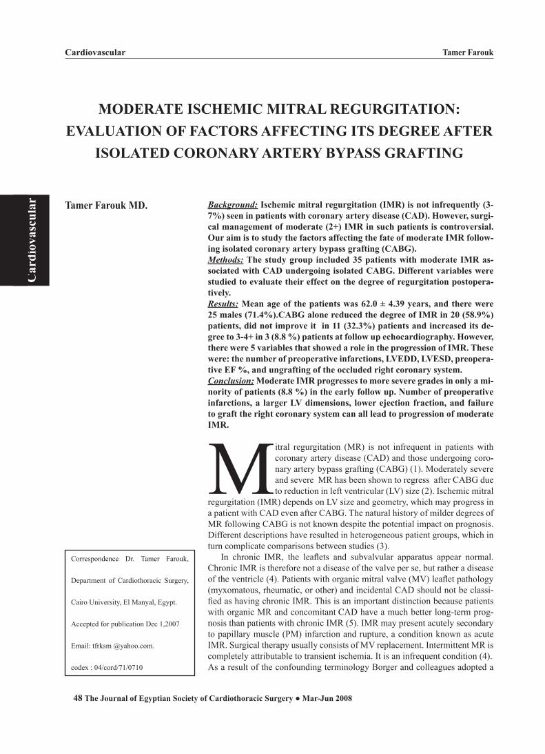
Cardiovascular Tamer Farouk
48
Car
diov
ascu
lar
The Journal of Egyptian Society of Cardiothoracic Surgery ● Mar-Jun 2008
MODERATE ISCHEMIC MITRAL REGURGITATION: EVALUATION OF FACTORS AFFECTING ITS DEGREE AFTER
ISOLATED CORONARY ARTERY BYPASS GRAFTING
Background: Ischemic mitral regurgitation (IMR) is not infrequently (3-7%) seen in patients with coronary artery disease (CAD). However, surgi--cal management of moderate (2+) IMR in such patients is controversial. Our aim is to study the factors affecting the fate of moderate IMR follow--ing isolated coronary artery bypass grafting (CABG).Methods: The study group included 35 patients with moderate IMR as--sociated with CAD undergoing isolated CABG. Different variables were studied to evaluate their effect on the degree of regurgitation postopera--tively.Results: Mean age of the patients was 62.0 ± 4.39 years, and there were 25 males (71.4%).CABG alone reduced the degree of IMR in 20 (58.9%) patients, did not improve it in 11 (32.3%) patients and increased its de--gree to 3-4+ in 3 (8.8 %) patients at follow up echocardiography. However, there were 5 variables that showed a role in the progression of IMR. These were: the number of preoperative infarctions, LVEDD, LVESD, preopera--tive EF %, and ungrafting of the occluded right coronary system.Conclusion: Moderate IMR progresses to more severe grades in only a mi--nority of patients (8.8 %) in the early follow up. Number of preoperative infarctions, a larger LV dimensions, lower ejection fraction, and failure to graft the right coronary system can all lead to progression of moderate IMR.
Mitral regurgitation (MR) is not infrequent in patients with coronary artery disease (CAD) and those undergoing coro--nary artery bypass grafting (CABG) (1). Moderately severe and severe MR has been shown to regress after CABG due to reduction in left ventricular (LV) size (2). Ischemic mitral
regurgitation (IMR) depends on LV size and geometry, which may progress in a patient with CAD even after CABG. The natural history of milder degrees of MR following CABG is not known despite the potential impact on prognosis. Different descriptions have resulted in heterogeneous patient groups, which in turn complicate comparisons between studies (3).
In chronic IMR, the leaflets and subvalvular apparatus appear normal. Chronic IMR is therefore not a disease of the valve per se, but rather a disease of the ventricle (4). Patients with organic mitral valve (MV) leaflet pathology (myxomatous, rheumatic, or other) and incidental CAD should not be classi--fied as having chronic IMR. This is an important distinction because patients with organic MR and concomitant CAD have a much better long-term prog--nosis than patients with chronic IMR (5). IMR may present acutely secondary to papillary muscle (PM) infarction and rupture, a condition known as acute IMR. Surgical therapy usually consists of MV replacement. Intermittent MR is completely attributable to transient ischemia. It is an infrequent condition (4). As a result of the confounding terminology Borger and colleagues adopted a
Correspondence Dr. Tamer Farouk,
Department of Cardiothoracic Surgery,
Cairo University, El Manyal, Egypt.
Accepted for publication Dec 1,2007
Email: tfrksm @yahoo.com.
codex : 04/cord/71/0710
Tamer Farouk MD.
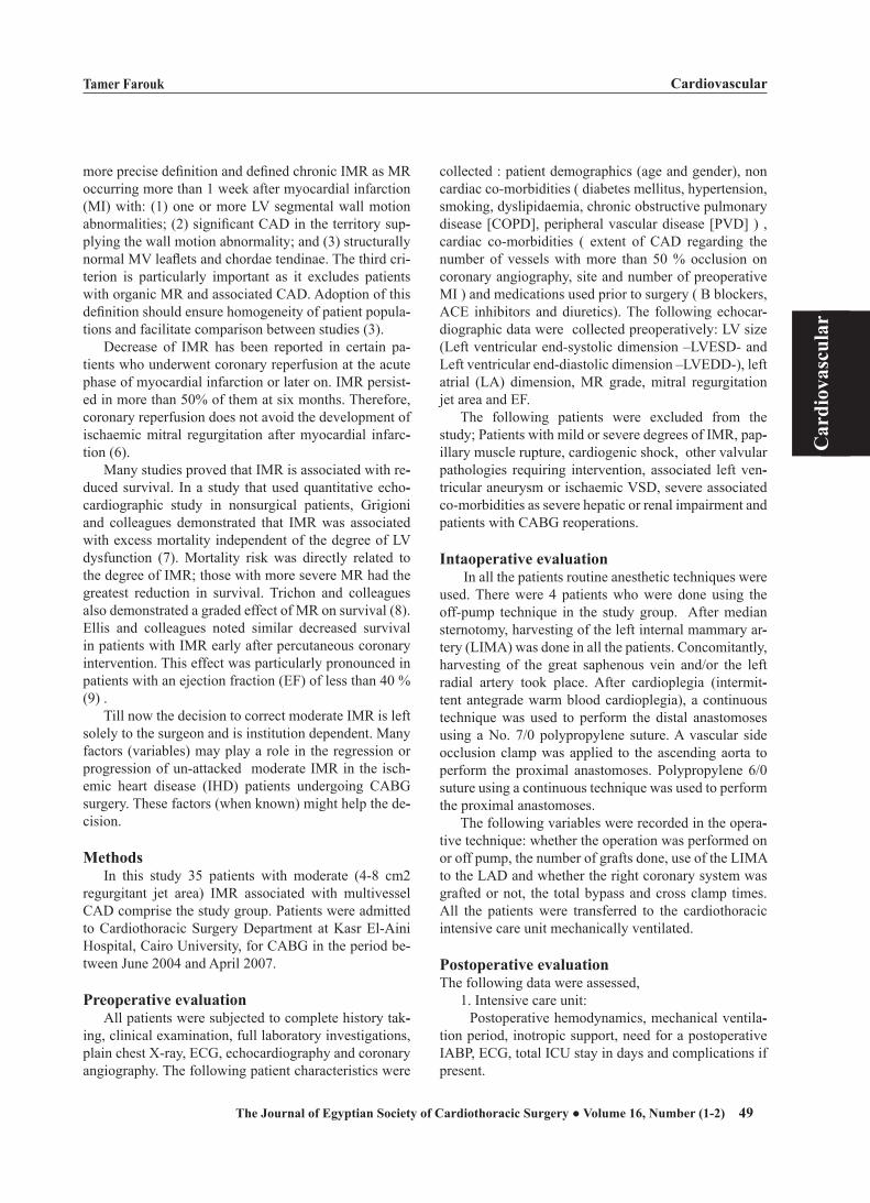
Tamer Farouk Cardiovascular
49
Car
diov
ascu
lar
The Journal of Egyptian Society of Cardiothoracic Surgery ● Volume 16, Number (1-2)
more precise definition and defined chronic IMR as MR occurring more than 1 week after myocardial infarction (MI) with: (1) one or more LV segmental wall motion abnormalities; (2) significant CAD in the territory sup--plying the wall motion abnormality; and (3) structurally normal MV leaflets and chordae tendinae. The third cri--terion is particularly important as it excludes patients with organic MR and associated CAD. Adoption of this definition should ensure homogeneity of patient popula--tions and facilitate comparison between studies (3).
Decrease of IMR has been reported in certain pa--tients who underwent coronary reperfusion at the acute phase of myocardial infarction or later on. IMR persist--ed in more than 50% of them at six months. Therefore, coronary reperfusion does not avoid the development of ischaemic mitral regurgitation after myocardial infarc--tion (6).
Many studies proved that IMR is associated with re--duced survival. In a study that used quantitative echo--cardiographic study in nonsurgical patients, Grigioni and colleagues demonstrated that IMR was associated with excess mortality independent of the degree of LV dysfunction (7). Mortality risk was directly related to the degree of IMR; those with more severe MR had the greatest reduction in survival. Trichon and colleagues also demonstrated a graded effect of MR on survival (8). Ellis and colleagues noted similar decreased survival in patients with IMR early after percutaneous coronary intervention. This effect was particularly pronounced in patients with an ejection fraction (EF) of less than 40 % (9) .
Till now the decision to correct moderate IMR is left solely to the surgeon and is institution dependent. Many factors (variables) may play a role in the regression or progression of un-attacked moderate IMR in the isch--emic heart disease (IHD) patients undergoing CABG surgery. These factors (when known) might help the de--cision.
MethodsIn this study 35 patients with moderate (4-8 cm2
regurgitant jet area) IMR associated with multivessel CAD comprise the study group. Patients were admitted to Cardiothoracic Surgery Department at Kasr El-Aini Hospital, Cairo University, for CABG in the period be--tween June 2004 and April 2007.
Preoperative evaluationAll patients were subjected to complete history tak--
ing, clinical examination, full laboratory investigations, plain chest X-ray, ECG, echocardiography and coronary angiography. The following patient characteristics were
collected : patient demographics (age and gender), non cardiac co-morbidities ( diabetes mellitus, hypertension, smoking, dyslipidaemia, chronic obstructive pulmonary disease [COPD], peripheral vascular disease [PVD] ) , cardiac co-morbidities ( extent of CAD regarding the number of vessels with more than 50 % occlusion on coronary angiography, site and number of preoperative MI ) and medications used prior to surgery ( B blockers, ACE inhibitors and diuretics). The following echocar--diographic data were collected preoperatively: LV size (Left ventricular end-systolic dimension –LVESD- and Left ventricular end-diastolic dimension –LVEDD-), left atrial (LA) dimension, MR grade, mitral regurgitation jet area and EF.
The following patients were excluded from the study; Patients with mild or severe degrees of IMR, pap--illary muscle rupture, cardiogenic shock, other valvular pathologies requiring intervention, associated left ven--tricular aneurysm or ischaemic VSD, severe associated co-morbidities as severe hepatic or renal impairment and patients with CABG reoperations.
Intaoperative evaluation In all the patients routine anesthetic techniques were
used. There were 4 patients who were done using the off-pump technique in the study group. After median sternotomy, harvesting of the left internal mammary ar--tery (LIMA) was done in all the patients. Concomitantly, harvesting of the great saphenous vein and/or the left radial artery took place. After cardioplegia (intermit--tent antegrade warm blood cardioplegia), a continuous technique was used to perform the distal anastomoses using a No. 7/0 polypropylene suture. A vascular side occlusion clamp was applied to the ascending aorta to perform the proximal anastomoses. Polypropylene 6/0 suture using a continuous technique was used to perform the proximal anastomoses.
The following variables were recorded in the opera--tive technique: whether the operation was performed on or off pump, the number of grafts done, use of the LIMA to the LAD and whether the right coronary system was grafted or not, the total bypass and cross clamp times. All the patients were transferred to the cardiothoracic intensive care unit mechanically ventilated.
Postoperative evaluationThe following data were assessed,
1. Intensive care unit: Postoperative hemodynamics, mechanical ventila--
tion period, inotropic support, need for a postoperative IABP, ECG, total ICU stay in days and complications if present.
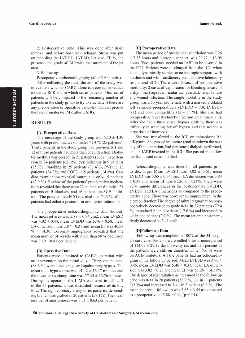
Cardiovascular Tamer Farouk
50
Car
diov
ascu
lar
The Journal of Egyptian Society of Cardiothoracic Surgery ● Mar-Jun 2008
2. Postoperative echo: This was done after drain removal and before hospital discharge. Stress was put on recording the LVEDD, LVESD, LA size, EF %, the presence and grade of IMR with measurement of the jet area.
3. Follow-up:Postoperative echocardiography (after 3-6 months).After collecting the data, the aim of the study was
to evaluate whether CABG alone can correct or reduce moderate IMR and in which set of patients. This set of patients will be compared to the remaining number of patients in the study group to try to elucidate if there are any preoperative or operative variables that can predict the fate of moderate IMR after CABG.
RESULTS
[A] Preoperative DataThe mean age of the study group was 62.0 ± 4.39
years with predominance of males 71.4 % (25 patients). Thirty patients in the study group had previous MI and 12 of those patients had more than one infarction. Diabe--tes mellitus was present in 21 patients (60%), hyperten--sion in 24 patients (68.6%), dyslipidaemia in 9 patients (25.7%), smoking in 25 patients (71.4%), PVD in 12 patients (34.3%) and COPD in 5 patients (14.3%). Car--diac examination revealed murmur in only 15 patients (42.9 %). Review of the patients’ preoperative medica--tions revealed that there were 22 patients on diuretics, 21 patients on B-blockers, and 16 patients on ACE inhibi--tors. The preoperative ECG revealed that 74.3 % of the patients had either a posterior or an inferior infarction.
The preoperative echocardiographic data showed: The mean jet area was 5.88 ± 0.94 cm2, mean LVESD was 4.02 ± 0.44, mean LVEDD was 5.76 ± 0.58, mean LA dimension was 3.87 ± 0.37 and mean EF was 46.57 % ± 14.30. Coronary angiography revealed that the mean number of vessels with more than 50 % occlusion was 3.89 ± 0.87 per patient.
[B] Operative Data Patients were submitted to CABG operation with
no intervention on the mitral valve. Thirty one patients (88.6 %) were done using cardiopulmonary bypass. The mean total bypass time was 91.42 ± 16.47 minutes and the mean cross clamp time was 57.45 ± 13.76 minutes. During the operation the LIMA was used in all but 2 of the 35 patients. It was discarded because of its low flow. The right coronary artery or its posterior descend--ing branch was grafted in 20 patients (57.1%). The mean number of anastomoses was 3.31 ± 0.63 per patient.
[C] Postoperative DataThe mean period of mechanical ventilation was 7.26
± 7.33 hours and inotropic support was 20.72 ± 13.05 hours. Two patients needed an IABP to be inserted in the ICU. Patients were discharged from the ICU when haemodynamically stable, on no inotropic support, with no drains and with satisfactory postoperative laboratory results and ECG. There were 5 cases of postoperative morbidity: 2 cases of exploration for bleeding, a case of arrhythmia (supraventricular tachycardia), renal failure and wound infection. The single mortality in the study group was a 53 year old female with a markedly dilated left ventricle preoperatively (LVEDD = 7.0, LVESD= 6.1) and poor contractility (EF= 33 %). She also had preoperative renal dysfunction (serum creatinine= 3.3). After she had a three vessel bypass grafting, there was difficulty in weaning her off bypass and that needed a large dose of inotropes.
She was transferred to the ICU on epinephrine 0.1 u/Kg/min. She passed into acute renal shutdown the next day of the operation, had peritoneal dialysis performed, and an IABP inserted in the ICU. She passed into a low cardiac output state and died.
Echocardiography was done for all patients prior to discharge. Mean LVESD was 4.02 ± 0.61, mean LVEDD was 5.65 ± 0.54, mean LA dimension was 3.84 ± 0.37 and mean EF was 51.26 ± 17.21%. There were very minute differences in the postoperative LVEDD, LVESD, and LA dimensions as compared to the preop--erative echo. There was however an improvement in the ejection fraction.The degree of mitral regurgitation post--operatively decreased to grade 0-1+ in 27 patients (79.4 %), remained 2+ in 6 patients (17.6 %) and increased to 4+ in one patient (2.9 %). The mean jet area postopera--tively decreased to 2.41 cm2.
[D]Follow up Data Follow up was complete in 100% of the 34 hospi--
tal survivors. Patients were called after a mean period of 114.09 ± 29.37 days. Twenty six and half percent of the patients were still on diuretics while 17.6 % were on ACE-inhibitors. All the patients had an echocardio--gram in the follow up period. Mean LVESD was 3.90 ± 0.46, mean LVEDD was 5.46 ± 0.37, mean LA dimen--sion was 3.82 ± 0.27 and mean EF was 51.26 ± 14.37%. The degree of regurgitation as measured in the follow up echo was 0-1+ in 20 patients (58.9 %), 2+ in 11 patients (32.3%) and increased to 3-4+ in 3 patient (8.8 %). The mean jet area at follow up was 3.65 ± 3.55 as compared to a preoperative of 5.88 ± 0.94 (p=0.01).
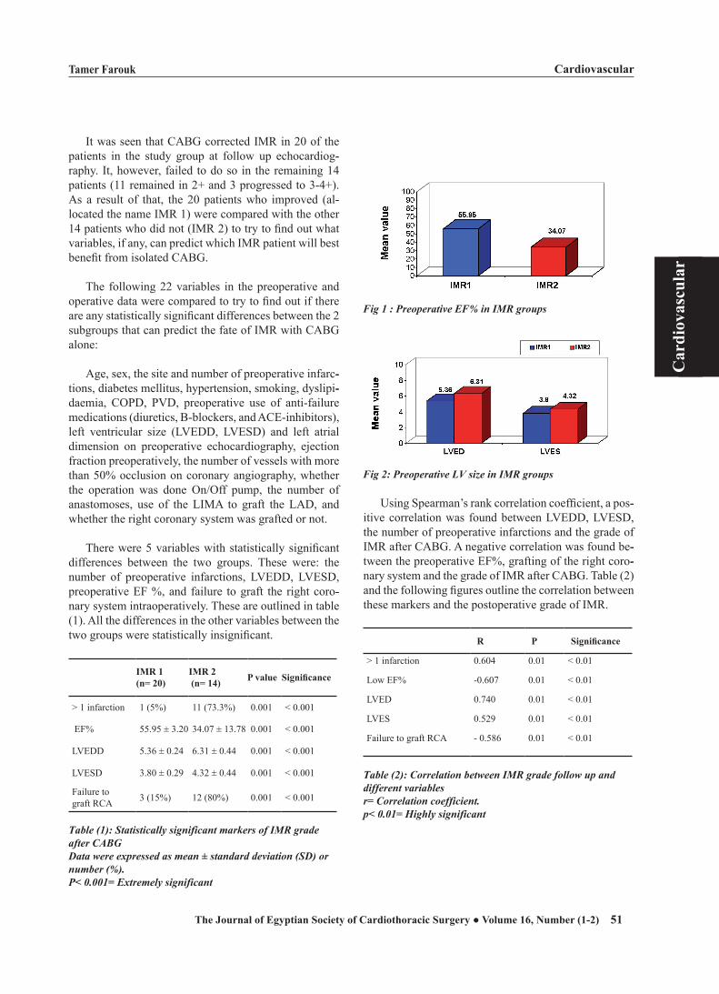
Tamer Farouk Cardiovascular
51
Car
diov
ascu
lar
The Journal of Egyptian Society of Cardiothoracic Surgery ● Volume 16, Number (1-2)
It was seen that CABG corrected IMR in 20 of the patients in the study group at follow up echocardiog--raphy. It, however, failed to do so in the remaining 14 patients (11 remained in 2+ and 3 progressed to 3-4+). As a result of that, the 20 patients who improved (al--located the name IMR 1) were compared with the other 14 patients who did not (IMR 2) to try to find out what variables, if any, can predict which IMR patient will best benefit from isolated CABG.
The following 22 variables in the preoperative and operative data were compared to try to find out if there are any statistically significant differences between the 2 subgroups that can predict the fate of IMR with CABG alone:
Age, sex, the site and number of preoperative infarc--tions, diabetes mellitus, hypertension, smoking, dyslipi--daemia, COPD, PVD, preoperative use of anti-failure medications (diuretics, B-blockers, and ACE-inhibitors), left ventricular size (LVEDD, LVESD) and left atrial dimension on preoperative echocardiography, ejection fraction preoperatively, the number of vessels with more than 50% occlusion on coronary angiography, whether the operation was done On/Off pump, the number of anastomoses, use of the LIMA to graft the LAD, and whether the right coronary system was grafted or not.
There were 5 variables with statistically significant differences between the two groups. These were: the number of preoperative infarctions, LVEDD, LVESD, preoperative EF %, and failure to graft the right coro--nary system intraoperatively. These are outlined in table (1). All the differences in the other variables between the two groups were statistically insignificant.
IMR 1 (n= 20)
IMR 2 (n= 14) P value Significance
> 1 infarction 1 (5%) 11 (73.3%) 0.001 < 0.001
EF% 55.95 ± 3.20 34.07 ± 13.78 0.001 < 0.001
LVEDD 5.36 ± 0.24 6.31 ± 0.44 0.001 < 0.001
LVESD 3.80 ± 0.29 4.32 ± 0.44 0.001 < 0.001
Failure to graft RCA 3 (15%) 12 (80%) 0.001 < 0.001
Table (1): Statistically significant markers of IMR grade after CABGData were expressed as mean ± standard deviation (SD) or number (%).P< 0.001= Extremely significant
Fig 1 : Preoperative EF% in IMR groups
Fig 2: Preoperative LV size in IMR groups
Using Spearman’s rank correlation coefficient, a pos--itive correlation was found between LVEDD, LVESD, the number of preoperative infarctions and the grade of IMR after CABG. A negative correlation was found be--tween the preoperative EF%, grafting of the right coro--nary system and the grade of IMR after CABG. Table (2) and the following figures outline the correlation between these markers and the postoperative grade of IMR.
R P Significance
> 1 infarction 0.604 0.01 < 0.01
Low EF% -0.607 0.01 < 0.01
LVED 0.740 0.01 < 0.01
LVES 0.529 0.01 < 0.01
Failure to graft RCA - 0.586 0.01 < 0.01
Table (2): Correlation between IMR grade follow up and different variablesr= Correlation coefficient. p< 0.01= Highly significant

Cardiovascular Tamer Farouk
52
Car
diov
ascu
lar
The Journal of Egyptian Society of Cardiothoracic Surgery ● Mar-Jun 2008
Fig 3: Correlation between IMR grade follow up and preoptterative number of infarctions (r= 0.604; p< 0.01)
Fig 4: Correlation between IMR grade follow up and preoptterative EF% (r= t0.607; p< 0.01).
Fig 5: Correlation between IMR grade follow up and preoptterative LVEDD (r= 0.740; p< 0.01)
Fig 6: Correlation between MR grade follow up and preoptterative LVES (r= 0.529; p< 0.01).
Fig 7: Correlation between IMR grade follow up and failure to graft RCA (r=t 0.586; p< 0.01)
DISCUSSIONThere is general agreement that patients with severe
(3+ or 4+) IMR should undergo mitral valve surgery at the time of CABG. However, the management of mod--erate (2+) IMR in such patients is controversial. The results of clinical studies differ as regards the fate of moderate IMR after CABG alone. Therefore, the course of unrepaired moderate IMR after CABG surgery alone was tracked, trying to identify the factors which could predict the fate of moderate IMR after CABG alone. The preoperative profile of patients was similar to patients investigated in other studies (10, 11&12).
In the study by Lam et al., all the patients in their study group had a previous myocardial infarction in or--der to be accepted as having IMR as opposed to 85.7% in this study. Fifty three percent of their patients had mod--
Tamer Farouk Cardiovascular
53
Car
diov
ascu
lar
The Journal of Egyptian Society of Cardiothoracic Surgery ● Volume 16, Number (1-2)
erate to severe LV dysfunction (EF < 40%) as compared to 31.4% in this series, and finally 6% of their patients had preoperative atrial fibrillation as opposed to none in this series. The series by Campwala and colleagues resembles this series closely. It was found that 74.3% of the patients had a previous posteroinferior infarc--tion (p=0.004). This validates data by other authors that states that, although anterior infarctions are commoner to occur in IHD patients, the occurrence of IMR is more common after a posteroinferior myocardial infarction (13). In their series on 102 patients with IMR, Calafiore and colleagues found that a posterior or inferior myocar--dial infarction occurred in 61.3% of their patients (14). In-hospital mortality rate in this study (2.9%) compares well with other series; 4.2% of the 168 patient cohort by Kim et al (12) and 2.9% in the study by Aklog et al. (15). There was an improvement in the ejection fraction, sustaining a 4.69 % improvement (p=0.02).
Furthermore, CABG alone was able to decrease pre--operative moderate IMR to mild or absent in 58.9 % of patients (20 out of 34). The grade remained moderate in 32.3 %, and increased in only 8.8% of cases. There is discrepancy in the literature as to agreement with these results. In their study on 49 patients with mild to moder--ate IMR, Tolis and colleagues agree with these results. In their patients, the ejection fraction improved from 22.0% to 31.5% (p < 0.05) after CABG. The mean grade of MR improved with CABG alone from 1.73 to 0.54 (p < 0.05) as measured at a mean interval of 36.9 months from CABG (16). As regards improvement in the grade of mitral regurgitation, Lam and colleagues found that moderate IMR improved to absent or mild in 73% of their patients on immediate postoperative echocardio--grams and in 40% on follow up echocardiograms (11). Disagreement, however, arises in the proportion of pa--tients who progressed to moderate-to-severe and severe mitral regurgitation. In the studies by Campwala et al., Lam et al. and Aklog et al., the proportion of patients in whom the grade of IMR progressed after CABG was 25%, 22%, and 40%, respectively (10,11& 15).
In an attempt to shed the light on factors that might influence the regression of moderate IMR after CABG, 22 preoperative and operative variables were examined that might influence this to occur and compared these between the group of patients in which the grade of IMR decreased (n= 20) to the group in which it remained the same or increased (n= 14). There were 5 variables with statistically significant differences between the two groups. These were: the number of preoperative infarc--tions, LVEDD, LVESD, preoperative EF%, and failure
to graft the right coronary system intraoperatively. Using Spearman’s rank correlation coefficient, any correlation that might existed between these variables and the grade of IMR postoperatively was studied. A positive correla--tion was found between LVEDD, LVESD, the number of preoperative infarctions and the grade of IMR after CABG. A negative correlation was found between the preoperative EF% and failure to graft the right coronary system and the grade of IMR after CABG.
The first four variables (more than one previous infarction, a larger LV dimensions as evidenced by a higher LVEDD and LVESD, and lower preoperative ejection fraction) point to the fact that patients in whom IMR does not regress are those with a sicker ventricle preoperatively. As known, the main mechanism behind the development of IMR is the apical displacement of the papillary muscles with the associated tethering of the leaflets as a result of the underlying LV remodeling. All the above factors lead to worsening of LV remodeling. Failure to graft the right coronary artery or its posterior descending branch might influence the grade of IMR postoperatively by the fact that failure to vascularize hi--bernating myocardium in this territory, may impair the improvement of left ventricular contractility postopera--tively thus preventing regression of mitral regurgitation. Furthermore, the development of new regional wall mo--tion abnormalities in the inferior-posterior LV wall ter--ritory due to the development of new ischemia without infarction, may additionally progress IMR post-CABG due to change in regional LV geometry. These results agree with other authors, Campwala and colleagues found failure to graft the PDA territory as an indepen--dent predictor of postoperative IMR progression, also the presence of pre-CABG LV dysfunction and larger LV size correlated with progression of IMR. Other fac--tors as poor targets and small vessels are associated with progression (10). Wong and colleagues also identified inferior LV dysfunction as a predictor of worsening IMR grade postoperatively (17). Further studies are requested with bigger number of patients and longer follow up pe--riod.
CONCLUSION• Moderate ischaemic mitral regurgitation progresses
to more severe grades in only a minority of patients (8.8 %) in the early follow up period following CABG.
• A higher number of preoperative infarctions, a larger LV size, lower ejection fraction, and failure to graft the right coronary artery territory can all lead to the persistence or progression of moderate IMR following isolated CABG.
Cardiovascular Tamer Farouk
54
Car
diov
ascu
lar
The Journal of Egyptian Society of Cardiothoracic Surgery ● Mar-Jun 2008
refrences
1. Connolly MW, Gelbfish JS, Jacobowitz IJ, et al.: Surgical results for mitral regurgitation from coronary artery dis--ease. J Thorac Cardiovasc Surg 1986; 91:379.
2. Campwala SZ, Bansal RC, Wang N, Razzouk A, Pai RG: Factors affecting regression of mitral regurgitation follow--ing isolated coronary artery bypass surgery. Eur J cardio--Thorac Surg 2005;28:104 -108
3. Borger MA, Alam A, Murphy PM, Doenst T, David TE: Chronic Ischaemic Mitral Regurgitation: Repair, Replace or Rethink? Ann Thorac Surg 2006; 81:1153– 61.
4. Gillinov AM, Wierup PN, Blackstone EH, et al.: Is repair preferable to replacement for ischaemic mitral regurgita--tion? J Thorac Cardiovasc Surg 2001; 122:1125.
5. Gillinov AM, Blackstone EH, Rajeswaran J, Mawad M, McCarthy PM, Sabik JF, Shiota T, Lytle BW and Cosgrove DM: Ischaemic Versus Degenerative Mitral Regurgitation: Does Etiology Affect Survival? Ann Thorac Surg 2005; 80:811-819.
6. Tcheng JE, Jackman JD, Nelson CL, et al.: Outcome of pa--tients sustaining acute ischaemic mitral regurgitation during myocardial infarction. Ann Intern Med 1992; 117:18-24.
7. Grigioni F, Enriquez-Sarano M, Zehr KJ, et al.: Ischaemic mitral regurgitation. Long-term outcome and prognostic implications with quantitative Doppler assessment. Circu--lation 2001; 103:1759–64.
8. Trichon BH, Felker GM, Shaw LK, Cabell CH, O’Connor CM: Relation of frequency and severity of mitral regurgita--tion to survival among patients with left ventricular systolic dysfunction and heart failure. Am J Cardiol 2003; 91:538-43.
9. Ellis SG, Whitlow PL, Raymond RE, Schneider JP: Impact of mitral regurgitation on long-term survival after percuta--
neous coronary intervention. Am J Cardiol 2002;89:315–8.10. Campwala SZ, Bansal RC, Wang N, Razzouk A, Pai RG:
mitral regurgitation progression following isolated coronary artery bypass surgery: frequency, risk factors, and potential prevention strategies. Eur J cardioThorac Cardiovasc Surg 2006;29:348-353.
11. Lam BK, Gillinov AM, Blackstone EH, et al.: Importance of moderate ischaemic mitral regurgitation. Ann Thorac Surg 2005; 79:462-470.
12. Kim Y-H, Czer L S.C., Soukiasian HJ, et al.: Ischaemic mitral regurgitation: Revascularization alone versus re--vascularization and mitral valve repair. Ann Thorac Surg 2005;79:1895-1901.
13. Kumanohoso T, Otsuji Y, Yoshifuku S, et al.: Mechanism of higher incidence of ischaemic mitral regurgitation in patients with inferior myocardial infarction: quantitative analysis of left ventricular and mitral valve geometry in 103 patients with prior myocardial infarction. J Thorac Cardio--vasc Surg 2003; 125:135– 43.
14. Calafiore AM, Di Mauro M, Gallina S, et al.: Mitral valve surgery for chronic ischaemic mitral regurgitation. Ann Thorac Surg 2004; 77:1989–97.
15. Aklog L, Filsoufi F, Flores KQ, et al.: Does coronary artery bypass grafting alone correct moderate ischaemic mitral re--gurgitation? Circulation 2001; 104(suppl I):I-68–75.
16. Tolis GA Jr, Korkolis DP, Kopf GS, Elefteriades JA: Re--vascularization alone (without mitral valve repair) suffices in patients with advanced ischaemic cardiomyopathy and mildto- moderate mitral regurgitation. Ann Thorac Surg 2002; 74: 1476–80; discussion 1480–1.
17. Wong DR, Agnihotri AK, Hung JW, et al.: Long-term survival after surgical revascularization for moderate isch--aemic mitral regurgitation. Ann Thorac Surg 2005;80:570-577.
Khaled Samir et al Cardiovascular
55
Car
diov
ascu
lar
The Journal of Egyptian Society of Cardiothoracic Surgery ● Volume 16, Number (1-2)
COMBINED ATRIAL FIBRILLATION (AF) ABLATION WITH MITRAL VALVE SURGERY: THE PREDICTORS OF SUCCESS
Objectives:To evaluate the efficacy and outcomes of radio frequency AF ablation in patients undergoing Mitral valve (MV) surgery.Methods: Between July 2005 and April 2007, 61 patients, (mean age 65.4 + 10 years, 65% males) underwent exclusive endo-left atrial AF ablation without LA appendage exclusion u sing radio frequency unipolar device (Cardioblate®, Medtronic, USA) in conjunction with 34 MV repairs and 27 MV replacements. AF was paroxysmal in 13 (21%) and chronic in 48 (79%) with a mean duration of 3.6 + 3.5 years. Etiology was mainly degenerative in 35 (57%,) or rheumatic in 17(28%). All patients received amiodarone® postoperatively. Results: Mortality was 0%. There were no thrombo-embolic acci--dents or device related complications. All patients had intraoperative conversion.41patients (67%) showed postoperative relapse, definitive con--version was achieved in 24 (59%) of them within 3 months. The overall success rate was 74%; it was higher in the repair group (85%) versus MV replacement group (67%, p<0.05) with a mean follow up 14 + 8.8 months. The predictors of success included younger age, smaller left atrium and shorter AF duration. Rheumatic etiology and associated CABG were pre--dictors of failure(P<0.05). The rate of success was similar in chronic and paroxysmal AF.Conclusions: Combined AF ablation with Mitral valve surgery is safe and effective, particularly, when associated with MV repair patients. However, the success rate as defined by definitive conversion to sinus rhythm may vary significantly according to patient characteristics.
Atrial Fibrillation (AF) is a well known risk factor for thrombo--embolism (stroke in 5% per year) [1]. It decreases the cardiac output and in many patients is badly tolerated, being responsi--ble for severe complaints, inspite of medical treatment. Despite extensive, decades-long research, AF remains a major cause of
substantial morbidity and mortality [2,3]. Patients with AF can also show in--tolerance for anti-arrhythmic medication and medically refractory arrhythmia.The incidence of AF increases with aging (6% of above 80y), 2.2 million pa--tients with AF in USA [1]. Several methods for the surgical treatment of AF have been described, the most common approach, in Mitral patients, being the `maze’ operation (Cox-Maze III, 1988) [4]. The aim of all these procedures is to build up an electric barrier that would prevent the transfer of the abnor--mal electrical activity. The original maze procedure that was described for the treatment of patients with atrial fibrillation (besides the surgical difficulties) was followed by an unacceptable incidence of two problems:
(1) The frequent inability to generate an appropriate sinus tachycardia in response to maximal exercise and
(2) Occasional left atrial dysfunction [4].
Accepted for publication Oct 1,2007
Address reprint request to Dr: Khaled
Samir Department of Cardiothoracic
Surgery , Ain Shames Univeristy
mail: [email protected]
codex : 04/cord/72/0710
Khaled Samir MD,Ayman Ammar MD ,Tamer El Ghobary MD,Ashraf A. ELSebaie MD
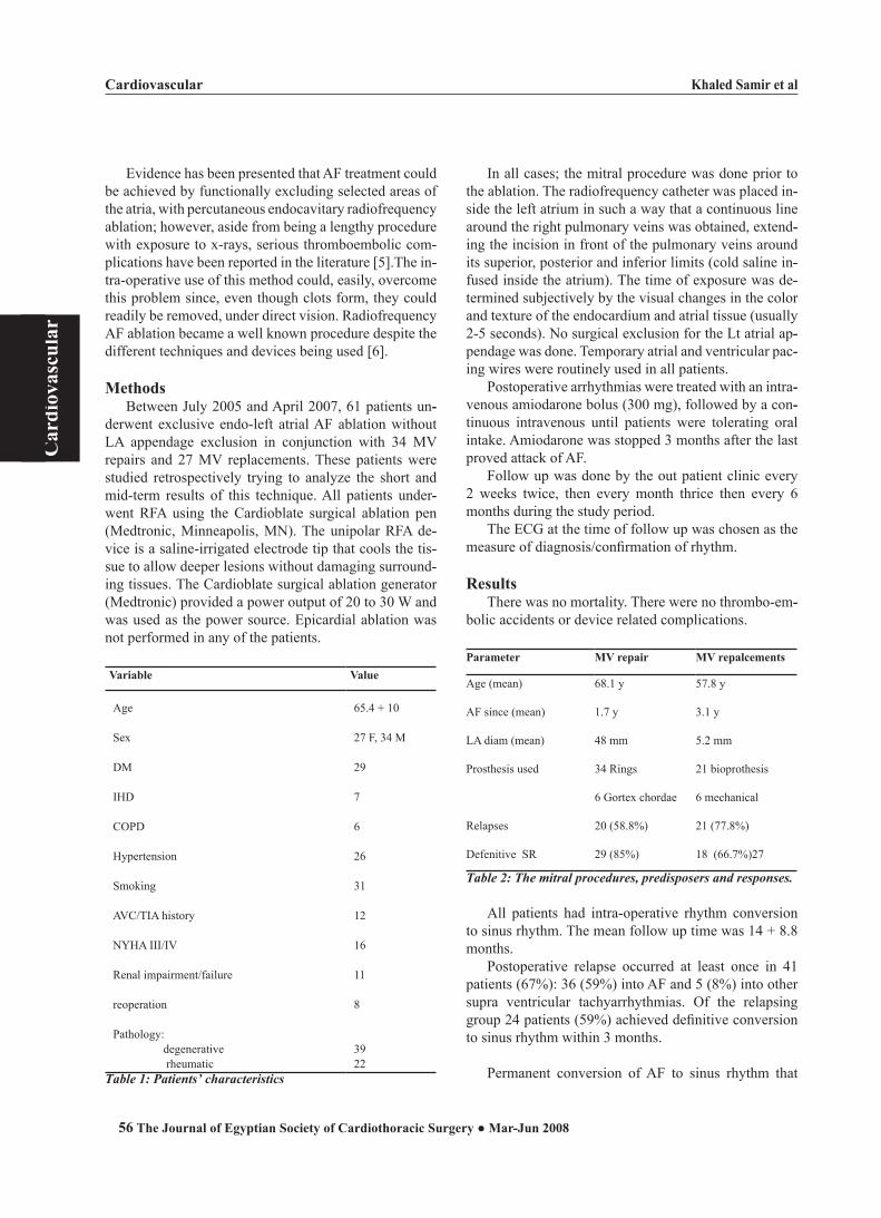
Cardiovascular Khaled Samir et al
56
Car
diov
ascu
lar
The Journal of Egyptian Society of Cardiothoracic Surgery ● Mar-Jun 2008
Evidence has been presented that AF treatment could be achieved by functionally excluding selected areas of the atria, with percutaneous endocavitary radiofrequency ablation; however, aside from being a lengthy procedure with exposure to x-rays, serious thromboembolic com--plications have been reported in the literature [5].The in--tra-operative use of this method could, easily, overcome this problem since, even though clots form, they could readily be removed, under direct vision. Radiofrequency AF ablation became a well known procedure despite the different techniques and devices being used [6].
MethodsBetween July 2005 and April 2007, 61 patients un--
derwent exclusive endo-left atrial AF ablation without LA appendage exclusion in conjunction with 34 MV repairs and 27 MV replacements. These patients were studied retrospectively trying to analyze the short and mid-term results of this technique. All patients under--went RFA using the Cardioblate surgical ablation pen (Medtronic, Minneapolis, MN). The unipolar RFA de--vice is a saline-irrigated electrode tip that cools the tis--sue to allow deeper lesions without damaging surround--ing tissues. The Cardioblate surgical ablation generator (Medtronic) provided a power output of 20 to 30 W and was used as the power source. Epicardial ablation was not performed in any of the patients.
Variable Value
Age
Sex
DM
IHD
COPD
Hypertension
Smoking
AVC/TIA history
NYHA III/IV
Renal impairment/failure
reoperation
Pathology: degenerative rheumatic
65.4 + 10
27 F, 34 M
29
7
6
26
31
12
16
11
8
3922
Table 1: Patients’ characteristics
In all cases; the mitral procedure was done prior to the ablation. The radiofrequency catheter was placed in--side the left atrium in such a way that a continuous line around the right pulmonary veins was obtained, extend--ing the incision in front of the pulmonary veins around its superior, posterior and inferior limits (cold saline in--fused inside the atrium). The time of exposure was de--termined subjectively by the visual changes in the color and texture of the endocardium and atrial tissue (usually 2-5 seconds). No surgical exclusion for the Lt atrial ap--pendage was done. Temporary atrial and ventricular pac--ing wires were routinely used in all patients.
Postoperative arrhythmias were treated with an intra--venous amiodarone bolus (300 mg), followed by a con--tinuous intravenous until patients were tolerating oral intake. Amiodarone was stopped 3 months after the last proved attack of AF.
Follow up was done by the out patient clinic every 2 weeks twice, then every month thrice then every 6 months during the study period.
The ECG at the time of follow up was chosen as the measure of diagnosis/confirmation of rhythm.
ResultsThere was no mortality. There were no thrombo-em--
bolic accidents or device related complications.
Parameter MV repair MV repalcements
Age (mean)
AF since (mean)
LA diam (mean)
Prosthesis used
Relapses
Defenitive SR
68.1 y
1.7 y
48 mm
34 Rings
6 Gortex chordae
20 (58.8%)
29 (85%)
57.8 y
3.1 y
5.2 mm
21 bioprothesis
6 mechanical
21 (77.8%)
18 (66.7%)27
Table 2: The mitral procedures, predisposers and responses.
All patients had intra-operative rhythm conversion to sinus rhythm. The mean follow up time was 14 + 8.8 months.
Postoperative relapse occurred at least once in 41 patients (67%): 36 (59%) into AF and 5 (8%) into other supra ventricular tachyarrhythmias. Of the relapsing group 24 patients (59%) achieved definitive conversion to sinus rhythm within 3 months.
Permanent conversion of AF to sinus rhythm that

Khaled Samir et al Cardiovascular
57
Car
diov
ascu
lar
The Journal of Egyptian Society of Cardiothoracic Surgery ● Volume 16, Number (1-2)
obtained intraoperatively without any relapse (primary definitive conversion) occurred in 20 patients (33%). From the relapsing group 24 patients (39%) regained and maintained sinus rhythm (secondary definitive con--version). AF definitively persisted in 17 patients (28%) after a period of temporary conversion.
Type of conversion patients
Primary (intraoperatively)
Primary definitive (intraoperative and maintained)
Secondary definitive (regained after relapses)
Failed definitive conversion
61 (100%)
20 (33%)
24 (39%)
17 (28%)
Table 3: types of conversion
Among the studied values there were statistically significant (P < 0.05) predictors for successful definitive AF ablation (table 4) and others were significantly asso--ciated with failure of achievement of definitive conver--sion (table 5).Predictors P value
Age less than 60 y
LA diameter less than 45 mm
Degenerative pathology
AF duration less than 1 year
0.032
0.02
0.041
0.029Table 4: Predictors of success
Predictors P valueAge more than 70 y
Duration more than 4 y
MV replacement
Associated IHD
0.048
0.031
0.07
0.044
Table 5: Predictors of failure
DiscussionOver the past several years, the surgical treatment of
AF has evolved rapidly, and a number of devices with various energy sources have been used to create linear lesions in the atria (endocardial, epicardial, or com--bined). These therapies have been used for both lone AF and AF associated with structural heart disease. [7-12]
The goal of these techniques, however, is to create lines of intra-atrial conduction block that will (1) pre--clude the development of macroreentrant circuits in the atria, (2) isolate the trigger or triggers for AF in or near the pulmonary vein orifices, or (3) accomplish both
goals 1 and 2, while at the same time allowing the atria to resume either a sinus or atrially paced rhythm. Recent clinical evidence has clearly demonstrated that the key mandatory step in the interventional treatment of AF is isolation of the pulmonary vein orifices. [13]
The evaluation of the outcome of the AF ablation in the same patient may vary considerably with the dif--ference in the technique (amount of microwave energy, duration, angle of the probe….etc). It can also vary with the length of follow up and the diagnostic methods used to determine the cardiac rhythm.
The patient age was statistically found to be an im--portant predictor of success if less than 60 years and failure if more than 70. The original pathology and not the type of procedures were proved to be a predictor of success in case of a degenerative pathology or failure in case of rheumatic pathology especially if associated with ischemic myocardium. The duration of AF is a difficult parameter to be objectively evaluated in many patients since a good percentage start to change their rhythm without immediate symptoms and may remain so for a considerable period. In our study we measured the duration of AF proved by objective patient findings on the first referral. Duration of less than 1 year was a predictor of success but more than 4 years of AF was a statistically significant predictor of failure.
The main aim of our study is to identify objectively statistically significant predictors that can help the sur--geon to predict the probable result of the AF ablation in concomitance with mitral valve open heart procedures. We tried to evaluate some of the factors thought to be of influence on the outcome of AF ablation. Our results shows that the nature of the associated procedures, pa--tient past history of smoking, hypertension, diabetes, COPD, renal failure, reoperation and degenerative le--sions have no statistical significance on the outcome of the ablation.
We noticed that despite the immediate 100% ablation ratio; a considerable percentage of AF relapse occurred in our patients (67%). This information is of particular importance since many patients get depressed with the first AF relapse and it would be better if they would be informed of the different mode possibilities of the out come preoperatively. Permanent ablation from the start (primary definitive conversion) occurred only in 33% of our patients. The total rate of permanent definitive conversion (72% in our study) may vary considerably between the different studies due to many factors; the most important among all is the definition adopted for permanent conversion. We didn’t consider the absence of symptoms or signs of AF relapse or follow up visit, online or on phone questionnaires as a proof of outcome.
Cardiovascular Khaled Samir et al
58
Car
diov
ascu
lar
The Journal of Egyptian Society of Cardiothoracic Surgery ● Mar-Jun 2008
We only considered a patient was permanently converted into sinus rhythm if he had had sinus rhythm in the ECG traces performed in the last follow up visit.
Our study shows that we are in a need to have a multicentric study on a large volume adopting objective methods in the microwave energy application, the esti--mation of the effect on the atrial tissue in addition to the objective estimation of the out come to be able to evalu--ate the vast difference in the results of surgical ablation published by different research groups.
The limitations of this study are: being retrospective, using a visual estimation of the ablation procedure (sub--jective) and the relatively small volume of patients.
ConclusionsCombined AF ablation with mitral valve surgery is
safe and effective, particularly, when associated with MV repair patients.
The predictors of success include patient’s age less than 60 years, AF duration less than one year, a degen--erative pathology and a left atrial diameter of less than 45mm. The predictors of failure were patient’s age older than 70 years, AF duration more than 4 years and isch--emic and/or rheumatic pathology.
However, the success rate as defined by definitive conversion to sinus rhythm may vary significantly not only according to the patient characteristics but also the methods of follow up and the definition of conversion.
References:
1. Sra J, Dhala A, Blanck Z, Deshpande S, Cooley R, Akhtar M. Atrial fibrillation: epidemiology, mechanisms, and man--agement Curr Probl Cardiol 2000;25:405-424.
2. Kannel WB, Abbott RD, Savage DD, McNamara PM. Epi--demiologic features of chronic atrial fibrillation: the Fram--ingham study N Engl J Med 1982;306:1018-1022.
3. Benjamin EJ, Wolf PA, D’Agostino RB, Silbershatz H, Kannel WB, Levy D. Impact of atrial fibrillation on the risk of death: the Framingham Heart Study Circulation 1998;98:946-952.
4. Cox JL, Schuessler RB, D’Agostino HJ, et al. The surgi--cal treatment of atrial fibrillationIII. Development of a definitive surgical procedure. J Thorac Cardiovasc Surg 1991;101:569-583.
5. Blaauw Y, Van Gelder IC, Crijns HJGM. Treatment of atrial fibrillation Heart 2002;88:432-437.
6. Deneke T, Khargi K, Grewe PH, et al. Left atrial versus
bi-atrial maze operation using intraoperatively cooled-
tip radiofrequency ablation in patients undergoing open-
heart surgery: safety and efficacy J Am Coll Cardiol
2002;39:1644-1650.
7. Cox JL. The surgical treatment of atrial fibrillation: IV. Sur--
gical technique. J Thorac Cardiovasc Surg. 1991;101:584-
589.
8. Prasad SM, Maniar HS, Camillo CJ, Schuessler RB,
Boineau JP, Sundt III TM, et al. The Cox maze III proce--
dure for atrial fibrillation. long-term efficacy in patients
undergoing lone versus concomitant procedures. J Thorac
Cardiovasc Surg 2003;126:1822-1828.
9. Haissaguerre M, Jais P, Shah DC, Takahashi A, Hocini M,
Quiniou G, et al. Spontaneous initiation of atrial fibrillation
by ectopic beats originating in the pulmonary veins. N Engl
J Med 1998;339:659-666.
10. Sueda T, Nagata H, Orihashi K, Morita S, Okada K,
Sueshiro M, et al. Efficacy of a simple left atrial procedure
for chronic atrial fibrillation in mitral valve operations. Ann
Thorac Surg 1997;63:1070-1075.
11. Kalil RA, Lima GG, Leiria TL, Abrahao R, Pires LM,
Prates PR, et al. Simple surgical isolation of pulmonary
veins for treating secondary atrial fibrillation in mitral valve
disease. Ann Thorac Surg 2002;73:1169-1173.
12. Pasic M, Bergs P, Muller P, Hofmann M, Grauhan O, Kup--
pe H, et al. Intraoperative radiofrequency maze ablation for
atrial fibrillation. the Berlin modification. Ann Thorac Surg
2001;72:1484-1491
13. Kalil RA, Lima GG, Leiria TL, Abrahao R, Pires LM,
Prates PR, et al. Simple surgical isolation of pulmonary
veins for treating secondary atrial fibrillation in mitral valve
disease. Ann Thorac Surg 2002;73:1169-1173.
14. Pasic M, Bergs P, Muller P, Hofmann M, Grauhan O, Kup--
pe H, et al. Intraoperative radiofrequency maze ablation for
atrial fibrillation. the Berlin modification. Ann Thorac Surg
2001;72:1484-1491.
Hosam Fawzy et al Cardiovascular
59
Car
diov
ascu
lar
The Journal of Egyptian Society of Cardiothoracic Surgery ● Volume 16, Number (1-2)
EFFECT OF VALVE PROSTHESIS – PATIENT MISMATCH ON SHORT TERM OUTCOME AFTER AORTIC VALVE
REPLACMENT
Background: Small- size prostheses for aortic valve replacement can be too small in relation to the body size, thus causing valve prosthesis-patient mismatch (PPM) and abnormally high transvalvular pressure gradient. This study was planned to examine the relation between the PPM and the short term outcome after the operation. Methods: Baseline risk factors and short term outcome were analyzed in 23 patients, 19 (82.6%) males and 4(17.4%) females who underwent Aor--tic valve replacement. The indexed valve effective orifice area (IEOA) was estimated for each type and size of prosthesis implanted. PPM was defined as not clinically significant if patient IEOA is > 0.85 cm2/m2 while clini--cally significant if patient IEOA is ≤ 0.85 cm2/m2 . Results: Study patients were divided into 2 groups, group “A” (9 patients with IEOA ≤0.85 cm2/m2) and group “B” (14 patients with IEOA > 0.85 cm2/m2). There was no statistical difference between both groups regard--ing pre-operative patients’ demographics. Operative characteristics were similar among patients in both groups with no statistical significance dif--ference. ICU stay was 2.8 + 1.5 days in group A vs. 3.2 + 1.9 days in group B (p =0.08). While total hospital stay was 12.5 + 9.8 days in Group A vs. 13.6 + 9.8 days in group B (p =0.59).The early mortality was encountered in two patients in group A and one patient in group B (p =0.59).Conclusion: Prosthesis-Patient Mismatch has no significant effect on short term outcome in patients undergoing Aortic Valve Replacement.
Prosthetic valve replacement represents a successful surgical thera--py for patients with symptomatic aortic valve disease. Despite an increasing proportion of high-risk patients
presenting for valve surgery, the morbidity and mortality of iso--lated aortic valve replacement (AVR) remain low1. Small aortic
annular size has been reported to be associated with increased operative mor--tality 1-3. However, considerable controversy
remains regarding the effects of small aortic prostheses on short & long-term survival 4, 5.
In particular, the relation between Prosthesis – Patient Mismatch (PPM) and short -term mortality remains to be determined and it could theoretically be important given that the left ventricle is more vulnerable at that time and that it could be more sensitive to the increased hemodynamic burden imposed by PPM.
The parameter often used in order to define the presence of PPM has been the geometric orifice area (GOA) that is, a measurement deriving from the internal diameter of the prosthesis and measured in vitro by the valve manu--facturer. However, the GOA has been shown to overestimate the ‘functional area’ of valve prosthesis. Following the introduction of echo-Doppler studies, a more reliable parameter has been validated in clinical practice, termed the
accepted for publication Oct 1,2007
Corresponding Author: Hosam Fawzy,
MD, Division of Thoracic and Cardio--
vascular Surgery, St. Michael’s Hospi--
tal,
Toronto, Ontario, M5B 1W8
E-mail:[email protected]
Codex : 04/cord/73/0710
Amr Badr MD,Hosam Fawzy MD,

Cardiovascular Hosam Fawzy et al
60
Car
diov
ascu
lar
The Journal of Egyptian Society of Cardiothoracic Surgery ● Mar-Jun 2008
effective orifice area (EOA) 6, 7.Rahimtoola8 was the first one to define PPM to be
present when the effective orifice area (EOA) of the im--planted prosthesis is less than that of the normal human valve. Based on this definition, most patients undergoing aortic valve replacement (AVR) thus have at least mild PPM.
Transvalvular gradients observed postoperatively are related not only to the EOA but also to the transvalvular flow. In turn, transvalvular flow is related to cardiac out--put that at rest is primarily related to the patient’s body size. As a consequence, the most reliable parameter to estimate the hemodynamic properties of the valve is the indexed effective orifice area (IEOA) 9, 10.
The IEOA is obtained from the relationship between effective orifice area and body surface area (EOA/BSA), with the EOA being calculated using the continuity equa--tion: EOA= SV/VTI, where SV is the stroke volume and VTI is the velocity time integral of the aortic jet Dop--pler signal. It is accepted worldwide that moderate aortic valve stenosis is present when the IEOA is <0.9 cm2/m2 and this concept should apply also to valve prostheses. In fact, several studies have demonstrated that when a prosthetic IEOA is <0.9 cm2/m2, a significant transval--vular gradient is present at rest 10, 11.
The aim of this study was to evaluate the impact of valve prosthesis-patient mismatch on the short term out--come after Aortic valve replacement.
MethodsBaseline risk factors and short term outcome were
analyzed in 23 patients, 19 (82.6%) males and 4(17.4%) females who underwent AVR with prosthetic valve in the cardiac surgery unit at North West Armed Forces Hospi--tal during the period from May 2002 to May 2004.
Clinical, echocardiography, coronary angiography, operative and post-operative outcome data were retro--spectively collected in a computerized database.
ProsthesisThe selection of prosthesis was based on the patient’s
age, history of previous thromboembolism or bleeding disorder and the preference of the patient and surgeon.
Indexed EOA (IEOA) for each prosthesis was de--rived from echocardiographic measured value of EOA divided by patient’s body surface area (BSA). PPM was defined as not clinically significant if patient IEOA is > 0.85 cm2/m2 while clinically significant if patient IEOA is ≤ 0.85 cm2/m2.
Short term mortality was defined as death from any cause within 30 days after the operation.Statistical analysis:
Data were expressed as mean ± standard deviation and compared using Chi square test to compare the re--sults obtained in patients with and without mismatch. Values of p less than 0.05 were considered significant
ResultsStudy patients were divided into 2 groups, group “A”
(9 patients with EOA/BSA ≤0.85 cm2/m2) and group “B” (14 patients with EOA/BSA > 0.85 cm2/m2).
Mean age in group A was 53 ± 26 years and was 48 ± 20 years in group B (p =0.91). There was no statistical difference between both groups regarding BSA (1.72 ± 0.16 m2 and 1.65 ± 0.15 m2 in group A & B respec--tively) (p =0.18). Rheumatic valve disease was found in 10 patients (5 in each group) while degenerative valves were found in 4 patients in group A and 9 patients in group B (p =0.34). Four of the patients were diabetic (2 in each group) (p =1.0). Two patients in group “A” and 3 patients in group “B” had LVEF ≤ 40 % (p =0.68). Euro score was 5 ± 3 for group A and 4.8 ± 2.7 for group B (p =0.80). Pre-operative patients’ demographics are pre--sented in table 1.
The following prostheses had been used: CE Standard in 6 patients, SMJ Standard in 13 patients, ON- X valve in 3 patients and SJM Toronto in one patient. Range of valve sizes implanted with in vitro internal diameter in mm, effective orifice area in cm2 and the ratio between the EOA/ BSA in cm2/m2 are shown in Table 2.
Operative characteristics were similar among pa--tients in both groups with no statistical significance dif--
Hosam Fawzy et al Cardiovascular
61
Car
diov
ascu
lar
The Journal of Egyptian Society of Cardiothoracic Surgery ● Volume 16, Number (1-2)
ference. Six Patients (2 in group A and 4 in group B) had concomitant coronary artery bypass grafting (CABG). Cardiopulmonary Bypass time was similar in both groups (176 + 120 min vs. 144 + 48 min in group A & B respectively P = 0.69). There was no difference in the Cross clamp duration between both groups (108 + 34 min vs. 103 + 26 min in group A & B respectively P = 0.74). Mechanical valves were implanted in 4 patients (44.4 % ) in group A and 12 patients (85.8 %) in group B (p =0.28) , while 5 patients (55.6 % ) had Bio-prosthesis in group A and 2 patients (14.2 % ) in group B (p =0.28). Patients’ operative characteristics are shown in table 3.
There was no statistical difference between the two groups in length of ICU stay, total hospital stay and early mortality. ICU stay was 2.8 + 1.5 days in group A vs. 3.2 + 1.9 days in group B (p =0.08). While total hospital stay was 12.5 + 9.8 days in Group A vs. 13.6 + 9.8 days in group B (p =0.59).The early mortality was encoun--tered in two patients in group A one of them died in the ICU from post-op electromechanical dissociation. The second patient had uncontrolled post operative surgical bleeding from the Aortomitral angle. Group B had only one death from post operative chest infection (p =0.59). Post-operative Data is demonstrated in table 4.
DiscussionPatient-prosthesis mismatch is a term often used but
vaguely defined, with many variations of the definition used by investigators. It was defined on the basis of small valve size, excessive gradients across the aortic valve, exercise-induced gradients, smaller in vivo pros--thetic valve effective orifice area than that of a native human valve, or various combinations of the above. Our definition of PPM has followed the generally accepted method of assessing (IEOA) to allow comparisons with other studies.
Conclusions drawn concerning the impact of PPM on short –term outcome differ greatly among reports. Our results show that there is no significant impact of PPM on the short term out come and mortality following AVR. Other studies have also purported to analyze the influence of PPM on mortality after AVR and they could not identify any major influence 7, 12.
Pibarot and coworkers5 in his series of consecutive AVR patients, the 30-day mortality was 4.6% in the patients without PPM, but was doubled with moderate PPM and increased 11-fold with severe PPM. Although patients with moderate and severe PPM had significant--ly more risk factors including impaired left ventricular function, older age, female gender, coronary artery dis--ease, and emergent/salvage surgery, the authors never--theless concluded that PPM was a strong and indepen--dent predictor of short-term mortality. Likewise, Rao et al., 6 also noted an increased operative mortality (8 vs.5%) in their series of AVR patients with PPM who similarly had a higher incidence of other risk factors. Fi--nally, Blackstone et al., 11 using multivariable hazard domain analysis with balancing score and risk factor adjustment in AVR patients from nine centers, reported that PPM increased the operative mortality by 1–2%.
A possible explanation for these apparently conflict--ing results might be an interaction of PPM with pre-ex--isting impairment of left ventricular function. In a mul--tivariate analysis of 52 patients undergoing AVR with a left ventricular ejection fraction below 35%, the only predictor of surgical mortality was smaller prosthesis size 13.
In another observational study in patients undergo--ing AVR with stentless bioprosthetic valves, baseline left ventricular mass index and PPM were the strongest predictors of the extent of postoperative LV mass regres--sion, implying a more powerful effect of PPM in the im--paired ventricle 14.
Limitation of the studyA randomized controlled study with larger number
of patients would provide better understanding of PPM and explore different risk factors that might play a role in its occurrence. Intermediate and long-term outcomes need to be addressed.
ConclusionThe effects of PPM are still controversial, and de--
spite the fact that our study showed no significant ef--fect of the PPM on the short term out come after Aortic valve replacement, larger sample size should be studied to demonstrate sufficient statistical effect.References

Cardiovascular Hosam Fawzy et al
62
Car
diov
ascu
lar
The Journal of Egyptian Society of Cardiothoracic Surgery ● Mar-Jun 2008
1. Rao V, Christakis GT, Weisel RD, et al. Changing pattern of valve surgery. Circulation. 1996; 94 (suppl II): II-113–II-120.
2. He GW, Grunkemeier GL, Gately JL, et al. Up to thirty-year survival after aortic valve replacement in the small aortic root. Ann Thorac Surg. 1995; 59: 1056–62.
3. Gonzalez-Juanetey JR, Garcia-Acuna JM, Vega Fernandez M, et al. Influence of the size of aortic valve prostheses on hemodynamics and change in left ventricular mass: impli--cations for the surgical management of aortic stenosis. J Thorac Cardiovasc Surg. 1996; 112:273–280.
4. Pibarot P, Honos GN, Durand LG, et al. The effect of pros--thesis-patient mismatch on aortic bioprosthetic valve he--modynamic performance and patient clinical status. Can J Cardiol. 1996; 12:379 –387.
5. McDonald ML, Daly RC, Schaff HV, et al. Hemodynamic performance of small aortic valve bioprostheses: is there a difference? Ann Thorac Surg. 1997;63:362–366.
6. Pibarot P, Dumesnil JG, Cartier PC, Metras J,Lemieux MD. Patient-prosthesis mismatch cannot be predicted at the time of operation. Ann Thorac Surg 2001; 7: S265- S268.
7. Dumesnil JG, Pibarot P. The approach does not disqualify prosthesis-patient mismatch. Eur J Cardiothorac Surg 2002; 21:157-158.
8. Rahimtoola SH. The problem of Valve Prosthesis-Patient mismatch. Circulation 1978 Jul; 58(1): 20-4.
9. Dumesnil JG, Honos GN, Lemieux M, Beauchemin J. Vali--dation and applications of indexed aortic prosthetic valve areas calculated by Doppler echocardiography. J Am Coll Cardiol 1990; 16: 637- 643.
10. Pibarot P, Dumesnil JG, Jobin J, Lemieux M, Honos G, Durand LG. Usefulness of the indexed effective orifice area at rest in predicting an increase in gradient during maxi--mum exercise in patients with a bioprosthesis in the aortic valve position. Am J Cardiol 1999; 83:542- 546.
11. Blackstone EH, Cosgrove DM, Jamieson WR, et al. Pros--thesis size and long-term survival after aortic valve replace--ment. J Thorac Cardiovasc Surg 2003; 126: 783–96.
12. Hanayama N, Christakis GT, Mallidi HR, et al. Patient prosthesis mismatch is rare after aortic valve replacement: valve size may be irrelevant. Ann Thorac Surg 2002;73: 1822–9.
13. Connolly HM, Oh JK, Schaff HV, Roger VL, Osborn SL, Hodge DO, Tajik AJ. Severe aortic stenosis with low trans--valvular gradient and severe left ventricular dysfunction: result of aortic valve replacement in 52 patients. Circulation 2000; 101: 1940–1946.
14. Del Rizzo DF, Abdoh A, Cartier P, Doty D, Westaby S. Factors affecting left ventricular mass regression after aor--tic valve replacement with stentless valves. Semin Thorac Cardiovasc Surg 1999; 11(4 Suppl 1): 114–120.

Hosam Fawzy et al Cardiovascular
63
Car
diov
ascu
lar
The Journal of Egyptian Society of Cardiothoracic Surgery ● Volume 16, Number (1-2)

Cardiovascular Hosam Fawzy et al
64
Car
diov
ascu
lar
The Journal of Egyptian Society of Cardiothoracic Surgery ● Mar-Jun 2008

Hosam Fawzy et al Cardiovascular
65
Car
diov
ascu
lar
The Journal of Egyptian Society of Cardiothoracic Surgery ● Volume 16, Number (1-2)
Cardiovascular Adel Ragheb et al
66
Car
diov
ascu
lar
The Journal of Egyptian Society of Cardiothoracic Surgery ● Mar-Jun 2008
RELATION OF LEFT VENTRICULAR MASS TO VOLUME AND ITS INFLUENCE ON THE OUTCOME OF AORTIC VALVE
REPLACEMENT
Abstract: Our study included 67 patients With Pure Aortic regurgitation . All of them had undergone open heart surgery for aortic valve replace--ment in National Heart Institute (NHI) and Qasr Alaini hospitals in the period from November 2004 to September 2006.Those patients have been evaluated clinically, radiologically and echo--cardiographically preoperatively, early post-operative and 6 months late post-operatively. Our results showed that those patients Who had been operated upon with the following criteria .CTR < 0.6 , LVEDD < 7.5 cm, EF >50% and diastolic radius to wall thickness (R/WTh) < 3 have good outcome with better quality of life as regard to LV function , NYHA class and ventricular arrhythmia . Those patients with CTR > 0.6 ,LVEDD > 7.5, EF < 50% , and R/WTh > 3 , showed bad outcome as LV function and NYHA did not improve and they had ventricular arrhythmi
Chronic aortic regurgitation produces LV volume overload that leads to a series of compensatory changes including LV en--largement and compensatory hypertrophy. Wall thickness must increase to compensate for increased ventricular dimensions to minimize or normalize wall stress according to Laplace law. As
long as left ventricular wall stress is maintained in the normal average, the LV reserves contractility and EF remains within normal range and patients during this chronic compensated stage remain asymptomatic for long periods. By time LV dilatation increases with change of LV from an elliptical to spherical shape with increase of wall stress, decline in the contractility and EF and the patients start to be symptomatized. LV Function is considered to be one of the most important main factors which affect the early and long term results of AVR for aortic Incompetence. Our study is to detect the parameters upon which we can predict our surgical outcome in patients who have been under--gone aortic valve replacement for chronic aortic regurge by correlating pre- and post-operative values of these parameters. .
Patients and methodsAll of our (67) patients were operated upon in NHI and Qasr Alaini hospi--
tals from November 2004 to September 2006. all of our patients had isolated chronic aortic incompetence (52) of them were males and (15) females with age range from (22) years to (47) years and the mean age of 33.6 + 3.2 Years .
All patients were examined clinically, radiologically and echocardiograph--cally pre-operatively, early post-operatively and then 6 months post-opera--tively.
Clinically our patients were assessed according to NYHA functional clas--sification. ECG data. study included comment on left ventricular hypertrophy (LVH) and LV strain and presence of arrhythmia. Plain chest X-ray PA view was done to all patients to assess cardiothoracic ratio (CTR).
Accepted for publication Dec 1 , 2007
Address reprint request to Dr: Adel ra--
gheb National heart institute, Egypt
Email: jegyptscts @gmail.com.
codex : 04/cord/74/0710
Adel Ragheb MD.,Tarek Nosseir MD.,Ahmed Amin Ms.,Mohammed Adel MD.,Nashat Abdelhamid MD.,Maher Mousa MD.
Adel Ragheb et al Cardiovascular
67
Car
diov
ascu
lar
The Journal of Egyptian Society of Cardiothoracic Surgery ● Volume 16, Number (1-2)
Echocardiographic study including M.mode, two-di--mensional and color Doppler study were done to assess LVEDV., LVESV ., LVEDD and LVESD. EF was cal--culated from the following formula
FS Percentage was calculated from the following for--mula
Both EF and FS are used as an indication for LV
systolic performance. Compensatory hypertrophy to in--creased LV volume is assessed for relation of volume to mass ratio. LV volume can be assessed by LVEDD , LV mass can be assessed by posterior wall thickness as expressed by the following formula :-
R (index of LV mass) =2 x posterior wall thickness.
Results
All our 67 patients had undergone isolated aortic valve replacement. The ratio of female to male is 1 : 3.47. The mean age of all 67 patients is 33.6+3.2. The mean age for the males was 35.3 years and for female was 29.7 years.
0
1 0
2 0
3 0
4 0
5 0
6 0
7 0
T o ta l M -F
T o ta l No . o f p a tie n tsNo . o f m a le
Male Female FIG.1 Male to Female ratio
Our (67) patients were divided into two groups ac--cording to the LVEDD and LVESD .
The first group (A) included 45 patients there mean age 25.9 + 4.2.9 of them were in NYHA class I but their LV dimensions were increased to limit which started to affect their LV functions. 30 patients were in NYHA class II ., 6 patients were in NYHA class III . in all patients group (A) CTR was less than O.6 and ECG showed LV hypertrophy by voltage criteria only .The LVEDD < 7.5 cm , LVESD < 5.5 cm EF >50% FS<25% and R/TH ratio was <3 .
The second group (B) included 22 patients with mean
age of 35.3+3.9. 10 of them were in NYHA class II ,. 7 were in NYHA class III and 5 were in NYHA class IV . CTR was > 0.6 , ECG showed strain pattern of LVH , LVEDD > 7.5 cm. ,LVESD >5.5 cm. , EF<50% , FS < 25% and R/Th ratio was >3 .
operative dataCold blood antigrade cardioplegia was used in all 67
pts. with systemic and local cooling. Mean cross clamp time was 34.1+ 3.2 min with no big difference between group A and B .the mean bypass time was longer in group B 67.6+5.2 min. while it was 49.8+3.6 min in group A. minimal inotropic support was needed in 11 pts only from group A., while inotropic support was needed in all 22 patients of group B with use of more than one inotropes in 7 patients .
postoperative data
ICU stay and total hospital stay were less in group A .one patients only from group A was re-opened for bleeding and the cause was bleeding from the site of the right upper pulmonary vein vent . No patients re-opened for bleeding in group B. 41 patients from group A were in NYHA class 1 while only 4 patients were in NYHA class 11 one of them was in NYHA class II pre-opera--tively and the other 3 were in NYHA class III pre-opera--tively . While in group B there is 12 patients in NYHA class 1 post-operatively, 8 of them were in NYHA class 11 and 3 were in NYHA class 111 and there is 6 patients in NYHA class11 post-operatively , 2 of them were in NYHA class 11 pre-operatively , another 2 of them were in NYHA class 111 pre-operatively and the last 2 were in NYHA class1V ,in NYHA class 111 there is 4 patients post-operatively ,2 of them were in NYHA class 111 pre-operatively and 2 were in NYHA class 1V pre-op--eratively. Only one patient was in NYHA class 1V post-operatively and this patient was also in NYHA class 1V pre-operatively . Total mortality was 3 pts. (4.48 %) all of them from group B and we lost 5 patients (7.46%)
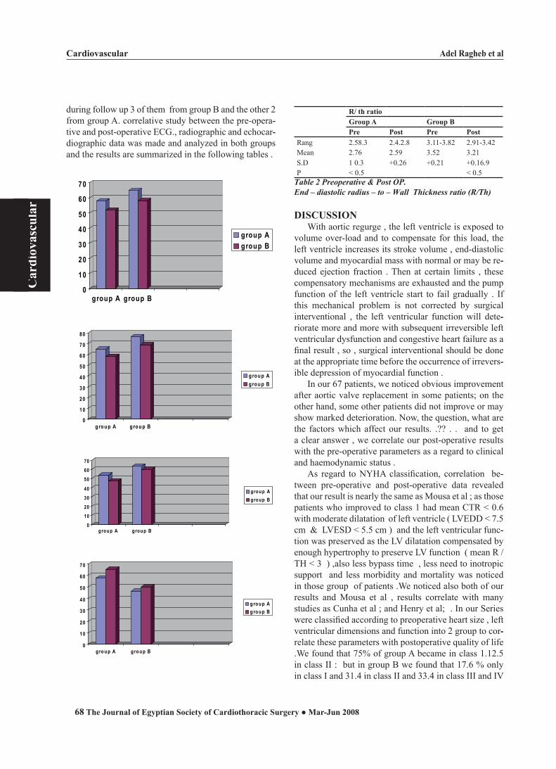
Cardiovascular Adel Ragheb et al
68
Car
diov
ascu
lar
The Journal of Egyptian Society of Cardiothoracic Surgery ● Mar-Jun 2008
during follow up 3 of them from group B and the other 2 from group A. correlative study between the pre-opera--tive and post-operative ECG., radiographic and echocar--diographic data was made and analyzed in both groups and the results are summarized in the following tables .
0
10
20
30
40
50
60
70
group A group B
group Agroup B
0
10
20
30
40
50
60
70
80
g ro u p A g ro u p B
g ro u p Ag ro u p B
010203040
506070
group A group B
group Agroup B
0
10
20
30
40
50
60
70
g ro u p A g ro u p B
g ro u p Ag ro u p B
R/ th ratio Group A Group BPre Post Pre Post
Rang 2.58.3 2.4.2.8 3.11-3.82 2.91-3.42Mean 2.76 2.59 3.52 3.21S.D 1 0.3 +0.26 +0.21 +0.16.9P < 0.5 < 0.5
Table 2 Preoperative & Post OP.End – diastolic radius – to – Wall Thickness ratio (R/Th)
DISCUSSIONWith aortic regurge , the left ventricle is exposed to
volume over-load and to compensate for this load, the left ventricle increases its stroke volume , end-diastolic volume and myocardial mass with normal or may be re--duced ejection fraction . Then at certain limits , these compensatory mechanisms are exhausted and the pump function of the left ventricle start to fail gradually . If this mechanical problem is not corrected by surgical interventional , the left ventricular function will dete--riorate more and more with subsequent irreversible left ventricular dysfunction and congestive heart failure as a final result , so , surgical interventional should be done at the appropriate time before the occurrence of irrevers--ible depression of myocardial function .
In our 67 patients, we noticed obvious improvement after aortic valve replacement in some patients; on the other hand, some other patients did not improve or may show marked deterioration. Now, the question, what are the factors which affect our results. .?? . . and to get a clear answer , we correlate our post-operative results with the pre-operative parameters as a regard to clinical and haemodynamic status .
As regard to NYHA classification, correlation be--tween pre-operative and post-operative data revealed that our result is nearly the same as Mousa et al ; as those patients who improved to class 1 had mean CTR < 0.6 with moderate dilatation of left ventricle ( LVEDD < 7.5 cm & LVESD < 5.5 cm ) and the left ventricular func--tion was preserved as the LV dilatation compensated by enough hypertrophy to preserve LV function ( mean R / TH < 3 ) ,also less bypass time , less need to inotropic support and less morbidity and mortality was noticed in those group of patients .We noticed also both of our results and Mousa et al , results correlate with many studies as Cunha et al ; and Henry et al; . In our Series were classified according to preoperative heart size , left ventricular dimensions and function into 2 group to cor--relate these parameters with postoperative quality of life .We found that 75% of group A became in class 1.12.5 in class II : but in group B we found that 17.6 % only in class I and 31.4 in class II and 33.4 in class III and IV

Adel Ragheb et al Cardiovascular
69
Car
diov
ascu
lar
The Journal of Egyptian Society of Cardiothoracic Surgery ● Volume 16, Number (1-2)
. It observed that patients with subnormal preoperative ejection fraction and fibre shortening have significantly worse late postoperative prognosis than that of patients with normal left ventricular function .
It is assumed that long standing volume load leads to irreversible left ventricular dysfunction before or coin--cident with the onset of preoperative symptoms . In the earlier phase of chronic volume sufficient left ventricular hypertrophy develops to normalize the left ventricular wall stress . Ejection fraction becomes depressed when hypertrophy is inadequate to compensate for progres--sive volume load 2.8,10,12,23 .
Mousa et al 7 , found that patients with EF of 60% or more and % ∆D of 30 had better prognosis than those with lower values Bonow et al 8 reported that the preop. EF less than 50% affected the 3 years postoperative sur--vival . H Al-Faleh et al 23 reported that impaired left ventricular function correlates with the extent of ultra--structural degenerative changes rather than with the ex--tent of fibrosis .
Therefore correct timing of operative intervention is one of the most difficult problems because , as men--tioned before, volume overload is well tolerated yet sig--nificant degenerative changes may occur in association with physiologic adaptation to chronic volume overload . As result , when corrective valve surgery is performed there may be irreversible left ventricular dysfunction which may preclude an optimal surgical result 9,13,17 . In conclusion good result were observed with prop--erative CTR <60%, EDD <68mm , ESD<57mm , EF > 55%, fibre shortening more than 25% and R/th ratio of 3 or less particularly if the patient was in functional class 1 or II .
The observed finding have led us to propose opera--tion generally at the onset of first symptoms and even before the onset of symptom in cases of voluminous chronic aortic incompetence accompanied by patent signs of left ventricular impairment.
Finally operation is indicated in1 . All symptomatic patients with moderate & sever re--
gurge .2 . Asymptomatic patients with :
a . LV dysfunction (EF < 55% &∆D < 25%) b . progressive enlargement of LV (CTR > 60 & EDD
> 68) c . Ineffective compensatory hypertrophy (R/Th <3) d . ESD of 57 mm or more , if less , for follow-up .
References
1 . Bonow RO , Epstein SE (1987) : Is preoperative left ven--tricular function predictive of survival and functional results after aortic valve replacement . JAM coll cardiol 10:713 .
2 . Pugliese P, Negri A , Muleretto C , et al (1990) : aortic insuf--ficiency : a multivariate analysis of incremental risk factors for operative mortality and functional results . J Cardiovasc Surg .(Torino) 31:213
3 . Grossman W 1984: aortic and mitral regurgitation : how to evaluate the condition and when to consider surgical inter--vention . JAMA 250:2447 .
4 . Percy RF , Miller AB, Conetta DA 1993 : usefulness of left ventricular at rest and after exercise for outcome pre--diction in asymptomatic aortic regurgitation . AM heart J 125:151.
5 . Bonow RO , Rosing DR , Maron BG , et al 1984 : Reversal of left ventricular dysfunction after aortic valve replace--ment for chronic aortic regurgitation : influence of duration of preoperative left ventricular dysfunction . Circulation 70:570.
6 . Bonow RO , Dodd JT , Maron BJ , et al 1988 : long - term serial changes left ventricular function and reversal of ven--tricular dilation after valve replacement for chronic aortic regurgitation . Circulation 78 : 1108 .
7 . A car, J.: Luxereau, Ph Ducimetiere P . et al (1981) : Prog--nosis of surgically treated chronic aortic valve disease . Predictive indicator of early Postoperative risk long-term survival Bass on 439 Cases J. Thorac. Cardiovasc . Surg 82:114 .
8 . Cunha , C.L.P, Giuliani E.R, Fuster , V. et al., (1980) : preoperative M-mode echocardiography as predictor of surgical result in chronic aortic insufficiency . J. Thorac Cardiovasc. Surg 79:256
9 . Henry , W.L : Bonow RO : Borer J.S. et al (1980): Obser--vation on the optimum time for operative intervention for aortic regurgitation .
Evaluation of the results of aortic valve replacement in symptomatic patients . Circulation 61:417 .
10 . Maher Mousa , MD , Samir Elmahmoudy MD – Lofty M.Essa MD (1992) : Optimum timing of surgical Interven--tional in chronic aortic icompetence ,THE Egyption Heart journal of the Egyption society of cardiology., volume NO. xxxx .
Cardiovascular Heba B. El-Serwi et al
70
Car
diov
ascu
lar
The Journal of Egyptian Society of Cardiothoracic Surgery ● Mar-Jun 2008
RETROGRADE VERSUS ANTEGRADE BLOOD CARDIOPLEGIA FOR CABG PATIENTS: DOES IT AFFECT
MYOCARDIAL PRESERVATION?
Background: The method of delivery of cardioplegic solution in coronary artery grafting is still controversial regarding optimal myocardial protec--tion.Aim: The current work aimed to evaluate the adequacy of myocardial protection with either ways of delivery of cardioplegic solution (antegrade versus retrograde) Methods: 40 patients scheduled for elective coronary artery bypass graft--ing were randomly assigned to 1 of 2 groups according to route of car--dioplegic administration either antegrade or retrograde. ECG, cardiac troponin (cTnI) and CK-MB were done preoperatively and at 2, 12, 24, 48 and 72 hrs postoperatively. Echocardiograhpy was done preoperatively and before discharge.Results: There were no differences in both groups regarding operative pa--rameters. The antegrade group showed significantly higher CK-MB levels at 2 hrs postoperatively as well as the sensitive and highly specific marker; cTnI concentrations till 24 hrs.Conclusions: Retrograde application of cardioplegia provides better myo--cardial protection as indicated by lower levels of cTnI concentrations.
Despite the popularisation of off-pump coronary artery bypass surgery (CABG), a large percentage of myocardial revascu--larization procedures are still performed on cardiopulmonary bypass (CPB) (1). The cardioprotective strategies available for intraoperative management during the CPB phase are numer--
ous. In the field of cardioplegia, the multiplicity of solutions available, their method of delivery, patient selection and the methods used to determine and quantify myocardial preservation versus injury have made it difficult to accu--rately decide which form of cardioplegia is best (2).
However, there is some clinical evidence that in certain subsets of patients, blood cardioplegia provides better myocardial protection (3).
The method of delivery of cardioplegic solution in coronary artery during grafting is still controversial regarding optimal myocardial protection (4). Ap--plying antegrade cardioplegia, homogenous perfusion of all segments of the heart may be limited in cases of badly collaterized coronary artery stenosis. Retrograde administration through the coronary sinus seems to offer a solution for this problem. However non homogenous cardiac protection will result if there is rare shunt or short coronary sinus (5-7).
Recently cardiac troponin I (cTnI) has been shown to be a sensitive and highly specific marker of myocardial injury during open-heart operations (8). It has already been used to compare different methods of myocardial protec--tion (9-11).
Accepted for publication Sep. 1 , 2007
Address reprint request to Dr: Heba
El-Serwi Department of anaesthesra
Menufia University & Department of
Cardiothoracic Surgery Ain Shams Uni--
versity,
Email: jegyptscts @gmail.com.
codex : 04/others/75/0710
Heba B. El-Serwi,Ahmed A-Razek Hasan,Ghada Ali,Ashraf Abdalla El-Sebaie
Heba B. El-Serwi et al Cardiovascular
71
Car
diov
ascu
lar
The Journal of Egyptian Society of Cardiothoracic Surgery ● Volume 16, Number (1-2)
The aim of this study was to compare two routes of intermittent warm blood cardioplegia delivery in pa--tients undergoing elective CABG using clinical, echo--cardiographic and biochemical results for evaluating the adequacy of myocardial protection.
METHODSAfter approval of institutional ethics review board
and informed patient consent, 40 ASA II-III patients ad--mitted for elective CABG surgeries were assigned ran--domly to 1 of 2 groups of cardioplegia administration, antegrade (group-A, n=20) and retrograde (group-R, n=20).
Exclusion criteria included Left ventricular ejection fraction (EF) <40%, previous cardiac surgery, myocar--dial infarction within 4 weeks preoperatively, associated valvular disease, chronic obstructive pulmonary disease, severe hepatic impairment (defined as serum protein <3 g% and serum bilirubin >3 mg%) or renal dysfunction (defined as predicted creatinine clearance (CLcr) <50 ml/min) and history of neurological disorder.
Anesthetic TechniquePremedication was achieved by 0.1 mg/kg morphine
intramuscularly one hour preoperatively. Induction with 0.03 mg/kg midazolam, 10-15 µg/kg fentanyl, sleep dose of propofol and atracurium 0.5-0.7 mg/kg. Anesthesia was maintained by 40 µg/kg/hr atracurium and fentanyl boluses administration at 1-3 µg/kg every 20 minutes to--gether with isoflurane up to 1.5 MAC as needed.
Standard monitoring was used in all patients includ--ing 5-lead ECG, pulse oximetry, capnography, esopha--geal temperature, continuous arterial blood pressure and central venous pressure monitoring.
Operative TechniqueAll operations were performed by one surgeon. In all
patients, median sternotomy was done, followed by dis--section of left internal mammary artery while simultane--ously harvesting the radial artery and saphenous vein. Cannulation for the CPB was carried out in the usual fashion with a 2 stage venous cannulation technique. Af--ter full heparinization (300 IU/kg) with activated clot--ting time (ACT) of 480 seconds or greater, CPB was initiated.
The standard cardiopulmonary bypass circuit was used with a membrane oxygenator and a non-pulsatile roller pump flow rate of 2.4 L/m2/min.
A cardioplegia delivery cannula with separate vent line was inserted into the ascending aorta of all patients. In patients of the retrograde group (group R) an addi--
tional coronary sinus catheter with autoinflating silicone cuff was positioned by a closed trans-arterial technique.
After aortic cross clamping, 1000 ml of blood car--dioplegia was delivered to all patients according to the group of selection either antegrade (group A) or retro--grade (group R). After the first dose, additional doses of 300-500ml were reinfused after 30 min of cross clamp time. No combinations of antegrade and retrograde car--dioplegia were given. Aortic cross clamp was removed after the distal anastomoses were done. Proximal anasto--moses were done with aortic partial clamping.
ElectrocardiogramA 12-lead ECG was recorded before the operation,
2 hrs after admission to intensive care unit (ICU), then daily after the operation for the next 3days. ECG moni--toring was continuous for 48 hrs post operatively in the ICU. New Q-waves >0.04 ms and/or a reduction in R-waves >25% in at least 2 leads were diagnostic for perioperative infarction, while transient ischemic events were diagnosed with ST-segment elevation >1mm. The ECG interpretation and analysis was done by a cardiolo--gist blinded to experimental protocol.
MeasurementsBlood samples from the central vein for measure--
ment of cardiac cTnI and CK-MB concentrations were taken preoperatively and 2, 12, 24, 48 and 72 hrs post--operatively. They were stored at -20°C until analysis. CK-MB activity was assayed by immunologic ultravio--let technique with a sensitivity of 5U/L and assay coef--ficients of variance <2% (normal value ≤ 24U/L). As for cTnI, electrochemiluminescence sandwich immunoas--say technique was utilized with a sensitivity of >0.1ηg/ml and coefficients of variance <5% (normal value, < 0.6ηg/ml).
EchocardiographyAll studies were performed by the same operator pre--
operatively and just before hospital discharge. EF was recorded as well as wall motion score index (WMSI).
Statistical analysisNumerical data are presented as mean ± SD, other--
wise total number or % of total as apropriate. Demo--graphic data were analyzed using Student’s t-test for numerical variables. Categorical data were analyzed us--ing the x2 analysis or Fisher’s exact test as appropriate. cTnI and CK-MB were analyzed using repeated measure ANOVA, where group (A) and group (R) are the inde--pendent variables, if significance is reached a tukey post.
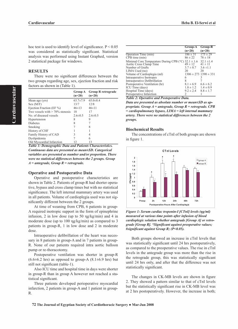
Cardiovascular Heba B. El-Serwi et al
72
Car
diov
ascu
lar
The Journal of Egyptian Society of Cardiothoracic Surgery ● Mar-Jun 2008
hoc test is used to identify level of significance. P < 0.05 was considered as statistically significant. Statistical analysis was performed using Instant Graphed, version 2 statistical package for windows.
RESULTSThere were no significant differences between the
two groups regarding age, sex, ejection fraction and risk factors as shown in (Table 1).
Group A (n=20)
Group R retrograde(n=20)
Mean age (yrs) 63.7±7.9 65.0±8.4Sex (M/F) 13/7 12/8Ejection Fraction (EF %) 46±13 46±11Two vessels with > 70% stenosis 18 17No. of diseased vessels 2.6±0.5 2.6±0.5Hypertension 8 9Diabetes 8 7Smoking 7 8History of CHF 1 1Family History of CAD 3 4Dyslipidemia 7 6Old Myocardial Infarction 12 13Table 1: Demographic Data and Patients Characteristics.Continuous data are presented as mean±SD. Categorical variables are presented as number and/or proportion. There were no statistical differences between the 2 groups. Group A = antegrade, Group R = retrograde.
Operative and Postoperative DataOperative and postoperative characteristics are
shown in Table 2. Patients of group-R had shorter opera--tive, bypass and cross clamp times but with no statistical significance. The left internal mammary artery was used in all patients. Volume of cardioplegia used was not sig--nificantly different between the 2 groups.
At time of weaning from CPB, 6 patients in group-A required inotropic support in the form of epinephrine infusion, 2 in low dose (up to 50 ηg/kg/min) and 4 in moderate dose (up to 100 ηg/kg/min) as compared to 3 patients in group-R, 1 in low dose and 2 in moderate dose.
Intraoperative defibrillation of the heart was neces--sary in 8 patients in group-A and in 7 patients in group-R. None of our patients required intra aortic balloon pump or re-thoracotomy.
Postoperative ventilation was shorter in group-R (6.6±6.2 hrs) as opposed to group-A (8.1±6.9 hrs) but still not significant (table-1).
Also ICU time and hospital time in days were shorter in group-R than in group A however not reached a sta--tistical significant.
Three patients developed perioperative myocardial infarction, 2 patients in group-A and 1 patient in group-R.
Group-A(n=20)
Group-R(n=20)
Operation Time (min) 186 ± 31 175 ± 29CPB time (min) 86 ± 22 78 ± 18Minimal Core Temperature During CPB (°C) 32.1 ± 1.6 32.1 ±1.4Aortic Cross Clamp Time 49 ± 12 41 ± 11Number of Grafts 3.7 ± 0.7 3.6 ±1.1LIMA Used (no) 20 20Volume of Cardioplegia (ml) 1306 ± 273 1390 ± 331Intraoperative Inotropes 6 3Intraoperative Defibrillation 8 7Postoperative Ventilation (hr) 8.1 ± 6.9 6.6 ± 6.2ICU Time (days) 1.8 ± 1.2 1.4 ± 0.9Hospital Time (days) 9.2 ± 2.4 8.8 ± 1.7Perioperative Infarction 2 1Table 2: Operative and Postoperative Data.Data are presented as absolute number or mean±SD as apttpropriate. Group A = antegrade, Group R = retrograde. CPB = cardiopulmonary bypass, LIMA = left internal mammary artery. There were no statistical differences between the 2 groups.
Biochemical ResultsThe concentrations of cTnI of both groups are shown
in figure 1.
C T nI Lev els
0
0 .2
0 .4
0 .6
0 .8
1
1 .2
1 .4
1 .6
1 .8
2
P reo p 2h 12h 24h 48h 72h
P o sto p era tive H o u rs After C ard io p leg ia
CTnI
(ηg/
ml)
group Agroup R
*
*
*
*
*
*
# #
#
Figure 1: Serum cardiac troponin-I [CTnI] levels (ηg/ml) measured at various time points after infusion of blood cardioplegic solution whether antegrade [Group A] or retrottgrade [Group R]. *Significant against preoperative values; #significant against Group R; (P<0.05).
Both groups showed an increase in cTnI levels that was statistically significant until 24 hrs postoperatively, as compared to the preoperative values. The rise in cTnI levels in the antegrade group was more than the rise in the retrograde group, this was statistically significant until 24 hrs only, and after that the difference was not statistically significant.
The changes in CK-MB levels are shown in figure 2. They showed a pattern similar to that of cTnI levels but the statistically significant rise in CK-MB level was at 2 hrs postoperatively. However, the increase in both;
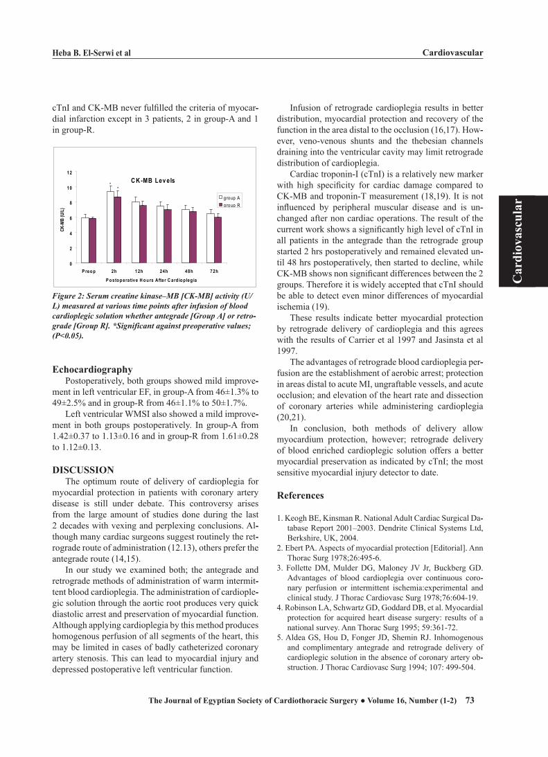
Heba B. El-Serwi et al Cardiovascular
73
Car
diov
ascu
lar
The Journal of Egyptian Society of Cardiothoracic Surgery ● Volume 16, Number (1-2)
cTnI and CK-MB never fulfilled the criteria of myocar--dial infarction except in 3 patients, 2 in group-A and 1 in group-R.
C K -MB Lev els
0
2
4
6
8
10
12
P reo p 2h 12h 24h 48h 72h
P o sto p era tive H o u rs After C ard io p leg ia
CK-M
B (U
/L)
group Agroup R
**
Figure 2: Serum creatine kinase–MB [CKtMB] activity (U/L) measured at various time points after infusion of blood cardioplegic solution whether antegrade [Group A] or retrottgrade [Group R]. *Significant against preoperative values; (P<0.05).
EchocardiographyPostoperatively, both groups showed mild improve--
ment in left ventricular EF, in group-A from 46±1.3% to 49±2.5% and in group-R from 46±1.1% to 50±1.7%.
Left ventricular WMSI also showed a mild improve--ment in both groups postoperatively. In group-A from 1.42±0.37 to 1.13±0.16 and in group-R from 1.61±0.28 to 1.12±0.13.
DISCUSSIONThe optimum route of delivery of cardioplegia for
myocardial protection in patients with coronary artery disease is still under debate. This controversy arises from the large amount of studies done during the last 2 decades with vexing and perplexing conclusions. Al--though many cardiac surgeons suggest routinely the ret--rograde route of administration (12.13), others prefer the antegrade route (14,15).
In our study we examined both; the antegrade and retrograde methods of administration of warm intermit--tent blood cardioplegia. The administration of cardiople--gic solution through the aortic root produces very quick diastolic arrest and preservation of myocardial function. Although applying cardioplegia by this method produces homogenous perfusion of all segments of the heart, this may be limited in cases of badly catheterized coronary artery stenosis. This can lead to myocardial injury and depressed postoperative left ventricular function.
Infusion of retrograde cardioplegia results in better distribution, myocardial protection and recovery of the function in the area distal to the occlusion (16,17). How--ever, veno-venous shunts and the thebesian channels draining into the ventricular cavity may limit retrograde distribution of cardioplegia.
Cardiac troponin-I (cTnI) is a relatively new marker with high specificity for cardiac damage compared to CK-MB and troponin-T measurement (18,19). It is not influenced by peripheral muscular disease and is un--changed after non cardiac operations. The result of the current work shows a significantly high level of cTnI in all patients in the antegrade than the retrograde group started 2 hrs postoperatively and remained elevated un--til 48 hrs postoperatively, then started to decline, while CK-MB shows non significant differences between the 2 groups. Therefore it is widely accepted that cTnI should be able to detect even minor differences of myocardial ischemia (19).
These results indicate better myocardial protection by retrograde delivery of cardioplegia and this agrees with the results of Carrier et al 1997 and Jasinsta et al 1997.
The advantages of retrograde blood cardioplegia per--fusion are the establishment of aerobic arrest; protection in areas distal to acute MI, ungraftable vessels, and acute occlusion; and elevation of the heart rate and dissection of coronary arteries while administering cardioplegia (20,21).
In conclusion, both methods of delivery allow myocardium protection, however; retrograde delivery of blood enriched cardioplegic solution offers a better myocardial preservation as indicated by cTnI; the most sensitive myocardial injury detector to date.
References
1. Keogh BE, Kinsman R. National Adult Cardiac Surgical Da--tabase Report 2001–2003. Dendrite Clinical Systems Ltd, Berkshire, UK, 2004.
2. Ebert PA. Aspects of myocardial protection [Editorial]. Ann Thorac Surg 1978;26:495-6.
3. Follette DM, Mulder DG, Maloney JV Jr, Buckberg GD. Advantages of blood cardioplegia over continuous coro--nary perfusion or intermittent ischemia:experimental and clinical study. J Thorac Cardiovasc Surg 1978;76:604-19.
4. Robinson LA, Schwartz GD, Goddard DB, et al. Myocardial protection for acquired heart disease surgery: results of a national survey. Ann Thorac Surg 1995; 59:361-72.
5. Aldea GS, Hou D, Fonger JD, Shemin RJ. Inhomogenous and complimentary antegrade and retrograde delivery of cardioplegic solution in the absence of coronary artery ob--struction. J Thorac Cardiovasc Surg 1994; 107: 499-504.
Cardiovascular Heba B. El-Serwi et al
74
Car
diov
ascu
lar
The Journal of Egyptian Society of Cardiothoracic Surgery ● Mar-Jun 2008
6. Allen BS, Winkelmann JW, Hanafy H, et al. Retrograde car--dioplegia does not adequately perfuse the right ventricle. J Thorac Cardiovasc Surg 1995; 109: 1116-26.
7. Kaukoranta PK, Lepojärvi MVK, Kiviluoma KT, Ylitalo KV, Peuhkurinen KJ. Myocardial protection during ante--grade versus retrograde cardioplegia. Ann Thorac Surg 1998; 66: 755-61.
8. Etievent JPH, Chocron S, Toubin G, et al. Use of cardiac troponin I as a marker of perioperative myocardial isch--emia. Ann Thorac Surg 1995; 59: 1192-4.
9. Chocron S, Alwan K, Toubin G, et al. Crystalloid cardiople--gia route of delivery and cardiac troponin I release. Ann Thorac Surg 1996; 62: 481-5.
10. Pichon H, Chocron S, Alwan K, et al. Crystalloid versus cold blood cardioplegia and cardiac troponin I release. Cir--culation 1997; 96: 316-20.
11. Chocron S, Alwan K, Yan Y, et al. Warm reperfusion and myocardial protection. Ann Thorac Surg 1998; 66: 2003-7.
12. Jasinski M, Kadziola Z, Bachowski R, Domaradzski W, Wenzel-Jasinska I, Piekarski M et al. Comparison of retro--grade versus antegrade cold blood cardioplegia: random--ized trial in elective coronary artery bypass patients. Eur J Cardiothorac Surg. 1997; 12: 620-6.
13. Carrier M, Pelletier LC, Searle NR. Does retrograde ad--ministration of blood cardioplegia improve myocardial protection during first operation for coronary artery bypass grafting? Ann Thorac Surg. 1997; 64: 1256-61.
14. Ihnken K, Morita K, Buckberg GD, Aharon A, Laks H, Pa--nos AL, et al. The safety of simultaneous arterial and coro--
nary sinus perfusion: experimental background and initial results. J Card Surg. 1994;9: 15-25.
15. Chocron S, Alwan K, Toubin G, Clement F, Kaili D, Taber--let C, et al. Crystalloid cardioplegia route of delivery and cardiac troponin I release. Ann Thorac Surg. 1996; 62: 481-5.
16. Gates RN, Laks H, Drinkwater DC Jr, Peral J, Zaragoza A, Kaczer E, et al. Gross and microvascular distribution of retrograde cardioplegia in explanted human hearts. Ann Thorac Surg. 1993; 56: 410-7.
17. Partington M, Acar C, Buckberg G, Julia P, Kofsky E, Bu--gyi H. Studies of retrograde cardioplegia. Capillary blood flow distribution to myocardium supplied by open and oc--cluded arteries. J Thorac Cardiovasc Surg. 1989; 97: 605-12.
18. Adams JE, Bodor G, Davila-Roman VG, Delmez JA, Ap--ple FS, Ladenson JH, et al. Cardiac troponin I: a marker with high specificity for cardiac injury. Circulation. 1993; 88: 101-6.
19. Cummins B,Auckland ML.,Cummins P. Cardiac-specific troponin-1 radioimmunoassay in the diagnosis of acute myocardial infarction .Am Heart J. 1987,113:1333-44.
20. Page R, Sharpe D, Bellamy C. Normothermic arrest with continuous hyperkalmic blood. Eur J Cardiothorac Surg. 1992; 6: 461-5.
21. Menasche P, Subai J, Veyssie L. Efficacy of coronary sinus cardioplegia in patients with complete coronary artery oc--clusions. Ann Thorac Surg. 1991; 51: 418-23.

Saeed Elassy et al Cardiovascular
75
Car
diov
ascu
lar
The Journal of Egyptian Society of Cardiothoracic Surgery ● Volume 16, Number (1-2)
IMPACT OF HIGH THORACIC EPIDURAL ANALGESIA ON
INCIDENCE OF PERIOPERATIVE STRESS RESPONSE IN OFF
PUMP CABG
Background: Most randomized control studies in this area have focused on hemodynamic effects and the stress response to surgery. Studies assess--ing the important issue of potential cardioprotective effects of TEA have yielded conflicting results. The aim of this prospective randomized study was to investigate effects of TEA as an adjunct to GA in patients undergo--ing Off-Pump CABG as regards hemodynamics, postoperative release of biochemical markers of myocardial damage and myocardial morbidity.Methods: 64 patients were randomly allocated into two groups (G І n=33) patients underwent Off-Pump CABG under GA without TEA and (GII n=31) patients underwent Off-Pump CABG under GA with adjunct TEA. Results: mean heart rate was increased significantly in G І during chest closure and after arrival to ICU and mean arterial pressure was lower in G II. The incidence of perioperative AF, ventricular extrasystole and peak plasma levels of ANP, BNP and catecholamines were significantly lower with TEA. The serum concentration of cTnT and CK-MB is significantly higher without TEA. Conclusion: TEA is advantageous as adjunctive analgesia to patients in Off-Pump CABG. It effectively suppressed the sympathetic activity, re--ducing incidence of post operative arrhythmia. Combination of TEA with GA during CABG exerted a beneficial effect on the perioperative stress response as indicated by reduction of Catecholamines, ANP and BNP. The use of epidural techniques as adjuncts to GA gives better results as regard level of biochemical markers cTnT and CK-MB which detect minor myo--cardial injury.
Post operative morbidity occurs because of factors related to surgi--cal procedure, concomitant medical disease, or the severity of the neuroendocrine response to the surgery (1) especially Sympatho--adrenal reflexes (2). Coronary artery bypass grafting (CABG) is associated with exaggerated hormonal and inflammatory responses
(3,4). Activation of myocardial sympathetic nerves is associated with the gen--esis of silent myocardial ischemia in patients with coronary artery disease (5). Perioperative arrhythmias such as atrial fibrillation (AF) are the most common complications in patients undergoing CABG (6). General anesthesia (GA) is the most commonly used anesthetic technique and is considered the (gold stan--dard) for CABG performed either On-pump or Off-pump (7). Within the past few years, however, thoracic epidural analgesia (TEA) as an adjunct to GA has become more prevalent and has been shown to be potentially beneficial in patients with coronary artery disease (8).
Potential advantages of TEA include thoracic sympathicolysis with subse--quent improvement of coronary perfusion, decrease heart rate, decrease endog--enous stress response, and a reduced risk of perioperative myocardial ischemia
Accepted for publication Dec. 1 , 2007
Address reprint request to Dr: Saeed
Elassy Department of Cardiothoracic
Surgery , andesthesia Ain Shams Uni--
versity, Egypt
Email: smrassy @gmail.com.
codex : 04/others/76/0710
Hala Elsheikh MD,Saeed M.R. Elassy MD,
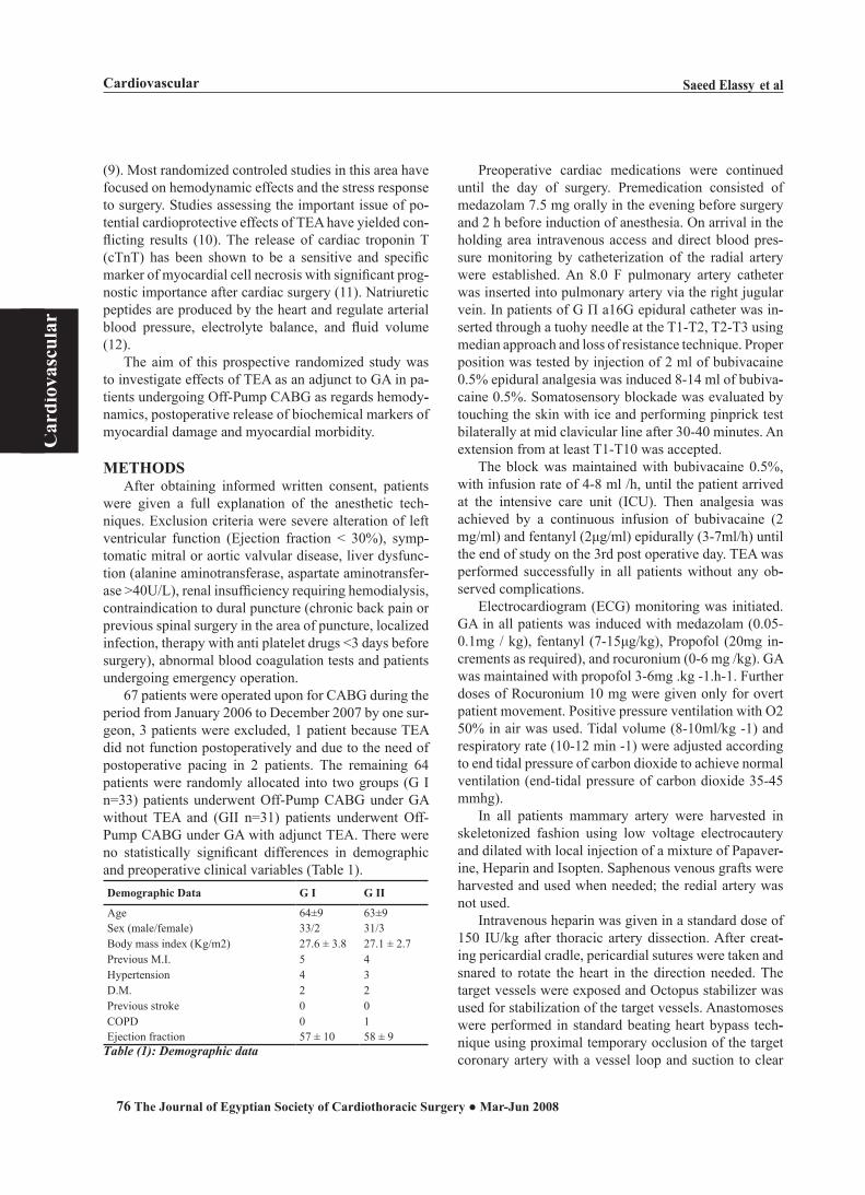
Cardiovascular Saeed Elassy et al
76
Car
diov
ascu
lar
The Journal of Egyptian Society of Cardiothoracic Surgery ● Mar-Jun 2008
(9). Most randomized controled studies in this area have focused on hemodynamic effects and the stress response to surgery. Studies assessing the important issue of po--tential cardioprotective effects of TEA have yielded con--flicting results (10). The release of cardiac troponin T (cTnT) has been shown to be a sensitive and specific marker of myocardial cell necrosis with significant prog--nostic importance after cardiac surgery (11). Natriuretic peptides are produced by the heart and regulate arterial blood pressure, electrolyte balance, and fluid volume (12).
The aim of this prospective randomized study was to investigate effects of TEA as an adjunct to GA in pa--tients undergoing Off-Pump CABG as regards hemody--namics, postoperative release of biochemical markers of myocardial damage and myocardial morbidity.
METHODSAfter obtaining informed written consent, patients
were given a full explanation of the anesthetic tech--niques. Exclusion criteria were severe alteration of left ventricular function (Ejection fraction < 30%), symp--tomatic mitral or aortic valvular disease, liver dysfunc--tion (alanine aminotransferase, aspartate aminotransfer--ase >40U/L), renal insufficiency requiring hemodialysis, contraindication to dural puncture (chronic back pain or previous spinal surgery in the area of puncture, localized infection, therapy with anti platelet drugs <3 days before surgery), abnormal blood coagulation tests and patients undergoing emergency operation.
67 patients were operated upon for CABG during the period from January 2006 to December 2007 by one sur--geon, 3 patients were excluded, 1 patient because TEA did not function postoperatively and due to the need of postoperative pacing in 2 patients. The remaining 64 patients were randomly allocated into two groups (G І n=33) patients underwent Off-Pump CABG under GA without TEA and (GII n=31) patients underwent Off-Pump CABG under GA with adjunct TEA. There were no statistically significant differences in demographic and preoperative clinical variables (Table 1).Demographic Data G I G II
Age 64±9 63±9Sex (male/female) 33/2 31/3Body mass index (Kg/m2) 27.6 ± 3.8 27.1 ± 2.7Previous M.I. 5 4Hypertension 4 3D.M. 2 2Previous stroke 0 0COPD 0 1Ejection fraction 57 ± 10 58 ± 9
Table (1): Demographic data
Preoperative cardiac medications were continued until the day of surgery. Premedication consisted of medazolam 7.5 mg orally in the evening before surgery and 2 h before induction of anesthesia. On arrival in the holding area intravenous access and direct blood pres--sure monitoring by catheterization of the radial artery were established. An 8.0 F pulmonary artery catheter was inserted into pulmonary artery via the right jugular vein. In patients of G Π a16G epidural catheter was in--serted through a tuohy needle at the T1-T2, T2-T3 using median approach and loss of resistance technique. Proper position was tested by injection of 2 ml of bubivacaine 0.5% epidural analgesia was induced 8-14 ml of bubiva--caine 0.5%. Somatosensory blockade was evaluated by touching the skin with ice and performing pinprick test bilaterally at mid clavicular line after 30-40 minutes. An extension from at least T1-T10 was accepted.
The block was maintained with bubivacaine 0.5%, with infusion rate of 4-8 ml /h, until the patient arrived at the intensive care unit (ICU). Then analgesia was achieved by a continuous infusion of bubivacaine (2 mg/ml) and fentanyl (2μg/ml) epidurally (3-7ml/h) until the end of study on the 3rd post operative day. TEA was performed successfully in all patients without any ob--served complications.
Electrocardiogram (ECG) monitoring was initiated. GA in all patients was induced with medazolam (0.05-0.1mg / kg), fentanyl (7-15μg/kg), Propofol (20mg in--crements as required), and rocuronium (0-6 mg /kg). GA was maintained with propofol 3-6mg .kg -1.h-1. Further doses of Rocuronium 10 mg were given only for overt patient movement. Positive pressure ventilation with O2 50% in air was used. Tidal volume (8-10ml/kg -1) and respiratory rate (10-12 min -1) were adjusted according to end tidal pressure of carbon dioxide to achieve normal ventilation (end-tidal pressure of carbon dioxide 35-45 mmhg).
In all patients mammary artery were harvested in skeletonized fashion using low voltage electrocautery and dilated with local injection of a mixture of Papaver--ine, Heparin and Isopten. Saphenous venous grafts were harvested and used when needed; the redial artery was not used.
Intravenous heparin was given in a standard dose of 150 IU/kg after thoracic artery dissection. After creat--ing pericardial cradle, pericardial sutures were taken and snared to rotate the heart in the direction needed. The target vessels were exposed and Octopus stabilizer was used for stabilization of the target vessels. Anastomoses were performed in standard beating heart bypass tech--nique using proximal temporary occlusion of the target coronary artery with a vessel loop and suction to clear

Saeed Elassy et al Cardiovascular
77
Car
diov
ascu
lar
The Journal of Egyptian Society of Cardiothoracic Surgery ● Volume 16, Number (1-2)
the anastomotic site. Heparin was reversed with protamine 3 mg /kg. Fur--
ther protamine (0.5-1mg/kg) was given to return the ac--tivated clotting time to baseline or as initial management of bleeding.
Heart rate (HR), Mean arterial pressure (MAP), Right atrial pressure (RAP), Mean pulmonary artery pressure (MPAP), Pulmonary capillary wedge pressure (PCWP), systemic vascular resistance (SVR) were recorded at the following interval: Prior to induction, 5 min after intuba--tion, 1-2 min post sternotomy, during chest closure, and after arrival in ICU. Patients were carefully monitored with intraoperative continuous ECG monitoring system with automatic arrhythmias and ST segment analysis. Thereafter, twice daily 12-lead ECG was performed until hospital discharge. Arrhythmias were classified as supraventricular, ventricular, or conduction defects. Supraventricular arrhythmias were further divided into atrial flutter or fibrillation. Ventricular arrhythmias were subdivided into tachycardia, fibrillation, or multiple ventricular ectopics (>6 min). Conduction defects were classified as 1º, 2ºor 3º (complete) heart block.
Postoperative myocardial infarction was diagnosed with ECG analysis (new Q waves, ST segment increase >3 mm) and confirmed by samples of blood for cTnT were collected in correlation with ECG Changes. Trans--mural infarction was defined as new Q waves, ST seg--ment increase > 3 mm and cardiac troponin I cTnT > 15μg/l.
Postoperatively, respiratory weaning was started by intermittent mandatory ventilation, follwed by continu--ous positive airway pressure breathing. Patients were extubated as soon as they fulfilled extubation criteria.
Venous blood samples were obtained preoperatively (T0), on arrival in ICU (T1), and in the morning of the first (T1) and second (T2) postoperative day, they were stored at -30 º C until analysis. The following variables were determined using commercially available laborato--ry kits: ANP for which normal values are 10-70 pg /ml in healthy men < 65 yr (13), BNP for which values >80pg /ml are indicative of neurohormonal activation (12, 14) and Catecholamines (epinephrine/or epinephrine) which was performed by using a reverse phase high perfor--mance liquid chromatography assay on anisocrotic liq--uid chromatography interfaced with an electrochemical detector.
Cardiac-specific contractile protein troponin T serum level was measured on an autoanalyzer using an enzyme immuno-method. The lower limit of detection of this test is 0.02 ng/ml. (concentration measured 30 times in the zero standard ± 3 sd). The measuring range was up to 15 ng /ml. The interassay coefficient of variation was
from 2.0% (mean concentration of troponin T 14.9 ng /ml) to 9.5% (mean concentration of troponin T 0.07ng/ml). The reference range derived from healthy subjects was 0.0-0.1 ng /ml values of > or equal 0.1ng/l were therefore considered to be positive. The monoclonal an--tibodies used in this test are highly specific for cardiac troponin T and therefore, for myocardial damage. No significant antibody cross-reaction with skeletal muscle troponin T has been observed (< or equal 0.004%). Cre--atinine kinase MB (CK-MB) measured by micriparticle enzyme immuonoassay technique, Abbot AX SYM sys--tem, Abbot laboratories, Chicago, IL. Samples collected and Data recording at preceding the induction of GA, on arrival to ICU and 24 h after ICU admission.
Statistical analysisContinuous normally distributed data were compared
using paired and unpaired studentۥs t-test or analysis of variance for repeated measures. When multiple compar--isons were made, the Bonferroni correction was applied. Data were expressed as mean ± SD.P values of <0.05 were considered statistically significant. The baseline characteristics and for both groups were compared by x 2 contingency or the fisher exact test for categorical data and the Man-Whitney U test for continuous vari--ables. Descriptive statistical analysis was done with the state view statistical software package 5.0.1 (SAS institute,Inc,Cary,NC).
RESULTSThere was no hospital mortality in either group. There
were no statistically significant difference between the two groups as regards the mean number of grafts per patient and the total operating room time excluding the time needed for TEA. The surgical results are shown in table (2).
G I G II
Number of grafts (mean ± std dev.) 2.7 ± 0.5 2.9 ±0.3
Operative time 158 ± 20 162 ±18Table (2 ):Surgical results
The mean upper sensory blockade level extended to C5-6(+/- 1.5 segments), and the lower blockade extend--ed to t10-11 (+/-3.5 segments) in both groups.
There is no significant difference in HR prior to in--duction of GA, 5 min after induction, and 1-2 min post sternotomy, however, mean heart rate was increased sig--nificantly in G І during chest closure and after arrival to ICU. (Figure 1). Mean arterial pressure was lower in G II (P=0.0006) but the other hemodynamic variables were
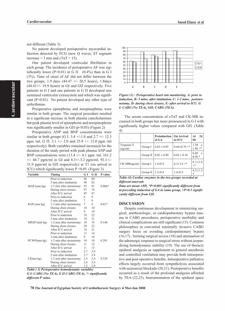
Cardiovascular Saeed Elassy et al
78
Car
diov
ascu
lar
The Journal of Egyptian Society of Cardiothoracic Surgery ● Mar-Jun 2008
not different (Table 3).No patient developed perioperative myocardial in--
farction detected by ECG (new Q waves, ST segment increase > 3 mm and cTnT > 15).
One patient developed ventricular fibrillation in each group. The incidence of perioperative AF was sig--nificantly lower (P<0.01) in G Π (0.4%) than in G І (3%). Time of onset of AF did not differ between the two groups, 1.9 days (44.47 +/- 20.5 hours), 1.8days (44.41+/- 19.9 hours) in GІ and GΠ respectively. Five patients in G I and one patients in G Π developed non sustained ventricular extrasystole and which was signifi--cant (P<0.01). No patient developed any other type of arrhythmias.
Preoperative epinephrine and norepinephrine were similar in both groups. The surgical procedure resulted in a significant increase in both plasma catecholamines but peak plasma level of epinephrine and norepinephrine was significantly smaller in GΠ (p<0.05) (Figure 2)
Preoperative ANP and BNP concentrations were similar in both groups (G І: 3.4 +/-1.8 and 2.7 +/- 12.3 pgm /ml, G Π: 3.1 +/- 2.0 and 25.9 +/- 13.0 pgm /ml respectively). Both variables remained increased for the duration of the study period with peak plasma ANP and BNP concentrations were (13.4 +/- 4.1 pgm /ml, 161.2 +/- 44.7 pgm/ml in GІ and 8.3+/-3.2 pgm/ml, 92.1+/-31.9 pgm/ml in GΠ respectively) at T1 (on arrival to ICU) which significantly lower P <0.05. (Figure 3)Variable Timing G I G II P value
MAP (mm hg)
Prior to induction 96 90
0.006*5 min after intubation 90 821-2 min after sternotomy 92 81During chest closure 97 76After ICU arrival 85 87
RAP (mm hg)
Prior to induction 6 9
0.0175 min after intubation 7 91-2 min after sternotomy 7 8During chest closure 10 10After ICU arrival 8 10
MPAP (mm hg)
Prior to induction 19 22
0.1465 min after intubation 19 211-2 min after sternotomy 18 19During chest closure 20 22After ICU arrival 18 21
PCWP(mm hg)
Prior to induction 13 14
0.2915 min after intubation 9 101-2 min after sternotomy 10 10During chest closure 11 12After ICU arrival 11 13
CI(mm hg)
Prior to induction 2.7 2.9
0.3295 min after intubation 2.7 2.81-2 min after sternotomy 2.5 2.4During chest closure 2.6 2.6After ICU arrival 3.2 2.9
Table ( 3) Perioperative hemodynamic variablesG I: CABG (No TEA), G II CABG (TEA), *: significantly different P value.
0102030405060708090
100
A B C D E
G IG II
Figure (1) : Perioperative heart rate monitoring. A: prior to induction, B: 5 mins. after intubation, C: 1t2 mins. poststerttnotomy, D: during chest closure, E: after arrival to ICU. G I: CABG (No TEA), GII: CABG (TEA).
The serum concentration of cTnT and CK-MB in--creased in both groups but more pronounced in G І with significantly higher values compared with GΠ. (Table 4)
Preinductionof GA
On Arrival to ICU
At 24 Hr
Troponin T (ng/ml) Group І 0.02 ± 0.05 0.60±0.70 +* 1.30 ±
1.98 +*
Group II 0.02 ± 0.04 0.03 ± 0.10 0.40 ± 0.50 *
CK-MB(ng/ml) Group І 1.4±0.5. 6.3±3.4 +* 8.1±3.4 +*
Group II 1.2±0.4 1.4±0.5 4.2±1.5 *
Table (4) Cardiac enzymes in the two groups recorded at different intervals Data are mean ±SD, *P<0.005 significantly different from at preceding induction of GA in same group, +P<0.5 signifittcantly different from GΠ.
DISCUSSIONDespite continuous development in minimizing sur--
gical, anethesiologic, or cardiopulmonary bypass trau--ma in CABG procedures, perioperative morbidity and clinical complications are still significant (15). Common philosophies in convential minimally invasive CABG surgery focus on avoiding cardiopulmonary bypass (16,17) , limiting surgical access (18) and attenuation of the adrenergic response to surgical stress without jeopar--dizing hemodynamic stability (19). The use of thoracic epidural analgesia as supplement to general anesthesia and controlled ventilation may provide both intraopera--tive and post operative benefits. Intraoperative palliative effects largely occurred from sympatholysis associated with neuroaxial blockade (20,21). Postoperative benefits occurred as a result of the profound analgesia afforded by TEA (22,23). Instrumentation of the epidural space
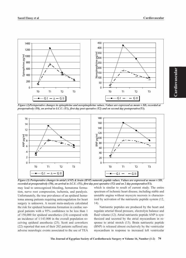
Saeed Elassy et al Cardiovascular
79
Car
diov
ascu
lar
The Journal of Egyptian Society of Cardiothoracic Surgery ● Volume 16, Number (1-2)
may lead to unrecognized bleeding, hematoma forma--tion, nerve root compression, ischemia, and paralysis. Unfortunately, the true prevalence of an epidural hema--toma among patients requiring anticoagulation for heart surgery is unknown. A recent meta-analysis calculated the risk for epidural hematoma formation in cardiac sur--gical patients with a 95% confidence to be less than 1 of 150,000 for epidural anesthetics (24) compared with an incidence of 1/143.000 in the overall population re--ceiving epidural anesthesia (25). Scott and coworkers (22) reported that non of their 202 patients suffered any adverse neurologic events associated to the use of TEA
which is similar to result of current study. The entire spectrum of ischemic heart disease, including stable and unstable angina without myocyte necrosis is character--ized by activation of the natriuretic peptide system (12, 14).
Natriuretic peptides are produced by the heart and regulate arterial blood pressure, electrolyte balance and fluid volume (12). Atrial natriuretic peptide ANP is syn--thesized and secreted by the atrial myocardium in re--sponse to atrial stretch (13). Brain natriuretic peptide (BNP) is released almost exclusively by the ventricular myocardium in response to increased left ventricular
0
200
400
600
800
1000
1200
1400
T0 T1 T2 T3
Epin
ephr
ine
pg/m
l
G I G II
0
50
100
150
200
250
300
350
400
450
T0 T1 T2 T3
Nor
epin
ephr
ine
pg/m
lG I G II
Figure (2)Perioperative changes in epinephrine and norepinephrine values. Values are expressed as mean ± SD, recorded at preoperatively (T0), on arrival to I.C.U. (T1), first day post operative (T2) and on second day postoperative(T3).
0
2
4
6
8
10
12
14
16
T0 T1 T2 T3
AN
P pg
/ml
G I G II
0
20
40
60
80
100
120
140
160
180
T0 T1 T2 T3
BNP
pg/m
l
G I G II
Figure (3) Perioperative changes in atrial (ANP) & brain (BNP) naturetic peptide values. Values are expressed as mean ± SD, recorded at preoperatively (T0), on arrival to I.C.U. (T1), first day post operative (T2) and on 2 day postoperative(T3).
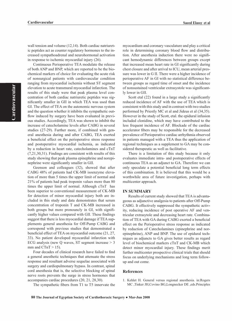
Cardiovascular Saeed Elassy et al
80
Car
diov
ascu
lar
The Journal of Egyptian Society of Cardiothoracic Surgery ● Mar-Jun 2008
wall tension and volume (12,14). Both cardiac natriuret--ic peptides act as counter regulatory hormones to the in--creased sympathoadrenal and neurohormonal activation in response to ischemic myocardial injury (26).
Continuous Perioperative TEA modulate the release of both ANP and BNP, which are reported to be the bio--chemical markers of choice for evaluating the acute risk of nonsurgical patients with cardiovascular condition ranging from myocardial ischemia without ST segment elevation to acute transmural myocardial infarction. The results of this study were that peak plasma level con--centration of both cardiac natriuretic peptides was sig--nificantly smaller in GII in which TEA was used than GІ. The effect of TEA on the autonomic nervous system and the question whether it inhibits the sympathetic out--flow induced by surgery have been evaluated in previ--ous studies. Accordingly, TEA was shown to inhibit the increase of catecholamine levels after CABG in several studies (27-29). Further more, if combined with gen--eral anesthesia during and after CABG, TEA exerted a beneficial effect on the perioperative stress response and postoperative myocardial ischemia, as indicated by a reduction in heart rate, catecholamines and cTnT (7,21,30,31). Findings are consistent with results of this study showing that peak plasma epinephrine and norepi--nephrine were significantly smaller in GII.
Geenson and colleagues (32), showed that after CABG 48% of patients had CK-MB isoenzyme eleva--tion of more than 5 times the upper limit of normal and 21% of patients had peak troponin values more than 80 times the upper limit of normal. Although cTnT has been superior to conventional measurement of CK-MB for detection of minor myocardial injury both are in--cluded in this study and data demonstrates that serum concentration of troponin T and CK-MB increased in both groups but more pronouncly in GІ, with signifi--cantly higher values compared with GII. These findings suggest that there is less myocardial damage if TEA sup--plements general anesthesia for Off-Pump CABG and correspond with previous studies that demonstrated a beneficial effect of TEA on myocardial outcome (21, 27, 33). No patient developed myocardial infarction with ECG analysis (new Q waves, ST segment increase > 3 mm and CTnT > 15).
Four decades of clinical research have failed to find a general anesthetic techniques that attenuate the stress response and resultant adverse sequelae associated with surgery and cardiopulmonary bypass. In contrast, spinal cord anesthesia that is, the selective blocking of spinal nerve roots prevents the surge in stress hormones that accompanies cardiac procedures (20, 21, 28,30).
The sympathetic fibers from T1 to T5 innervate the
myocardium and coronary vasculature and play a critical role in determining coronary blood flow and distribu--tion. After anesthesia induction there were no signifi--cant hemodynamic differences between groups except that increased mean heart rate in GІ significantly during chest closure and after arrival to ICU, mean arterial pres--sure was lower in G II. There were a higher incidence of perioperative AF in GІ with no statistical difference be--tween groups as regard time of onset and the incidence of nonsustained ventricular extrasystole was significant--ly lower in GII.
Scott etal (22) found in a large study a significantly reduced incidence of AF with the use of TEA which is consistent with this study and in contrast with two studies performed by Priestly MC et al and Jideus et al (34,35). However in the study of Scott, etal. the epidural infusion included clonidine, which may have contributed to the less frequent incidence of AF. Blockade of the cardiac accelerator fibers may be responsible for the decreased prevalence of Perioperative cardiac arrhythmia observed in patients managed with a TEA thus the application of regional techniques as a supplement to GA may be con--sidered therapeutic as well as facilitative.
There is a limitation of this study because it only evaluates immediate intra- and postoperative effects of continuous TEA as an adjunct to GA. Therefore we can only speculate a potential beneficial long term effects of this combination. It is believed that this would be a worthwhile area of future investigation, perhaps with multicenter approach.
IN SUMMARYResults of current study showed that TEA is advanta--
geous as adjunctive analgesia to patients after Off-Pump CABG. It effectively suppressed the sympathetic activ--ity, reducing incidence of post operative AF and ven--tricular extrasytole and decreasing heart rate. Combina--tion of TEA with GA during CABG exerted a beneficial effect on the Perioperative stress response as indicated by reduction of Catecholamines (epinephrine and nor--epinephrine), ANP and BNP. The use of epidural tech--niques as adjuncts to GA gives better results as regard level of biochemical markers cTnT and CK-MB which detect minor myocardial injury. These findings merit further multicenter prospective clinical trials that should focus on underlying mechanisms and long term follow-up and out come.
References
1. Kehlet H. General versus regional anesthesia. in:Rogers MC ,Tinker JH,Covino BG,Longnecker DE ,eds.Principles

Saeed Elassy et al Cardiovascular
81
Car
diov
ascu
lar
The Journal of Egyptian Society of Cardiothoracic Surgery ● Volume 16, Number (1-2)
and practice of anesthesiology.Vol 2. London:Mosby Year –Book Inc,1992.
2. Scott NB. The effects of pain and its treatment in: MoClure JH,Wildsmisth JAW,eds. Conduction blockade for postop--erative analgesia.London:Edward Arnold,1991.
3. Berendes E, Möllhoff T, Van Aken H, et al.: Increased plasma concentration of serum amyloid A: an indicator of acute-phase response after cardiopulmonary bypass. Crit Care Med .1997; 25:1527-33.
4. Loick HM, Möllhoff T, Berendes E, et al. Influence of enoxamine on systemic and splanchnic oxygen utilization and endotoxin release follwing cardiopulmonary bypass. Intensive Care Med. 1997;23:267-75.
5. Lee DD, Kimura S and DeQuattro V. Noradrenergic activ--ity and silent ischemia in hypertensive patients with stable angina, effect of metoprolol. Lancet.1989;1:403-6.
6. Mueller XM, Tevaearai HT, Ruchat P, et al. Did the intro--duction of a minimally invasive technique change the in--cidence of atrial fibrillation after single internal thoracic artery-left anterior descending artery grafting?. J Thorac Cardiovasc Surg.2001; 121:683-8.
7. Wattwil M, Sundberg A, Arvill A et al. Circulatory changes during high thoracic epidural anesthesia – influence of sym--pathetic block and systemic effect of the local anesthetic. Acta Anaesthesiol Scand.1985; 29:849-55.
8. Kessler P, Aybek T, Neidhart G, et al. Comparison of three anesthetic techniques for off pump coronary artery bypass grafting: general anesthesia, combined general and high thoracic epidural anesthesia alone. J Cardiothorac Vasc Anesth. 2005;19:32-9.
9. Farhad B, Ponagiatis T, Omer D, et al. Impact of high thoracic epidural anesthesia on incidence of perioperative atrial fibrillation in off-pump coronary bypass grafting, A prospective randomized study. J Thorac Cardiovasc Surg .2007; 134:460-4.
10. Michael JB, Roman K, Robert W, et al. Epidural Anes--thesia for Coronary Artery Bypass Surgery Compared with General Anesthesia Alone Does Not reduce Biochemi--cal Markers of Myocardial damage. Anesth Analg. 2005; 100:921-8.
11. Fellahi JL, Gue X, Richomme X, et al. Short – and long-term prognostic value of postoperative cardiac troponin I concentration in patients undergoing coronary artery by--pass grafting. Anesthesiology .2003;97:240-4.
12. Suttner SW, Boldt J. Natriuretic peptide system:physiology and clinical utility. Curr Opin Crit Care .2004;10:336-41.
13. De Zeeuw D, Jonssen WMT, DeJong PE. Atrial natriuretic factor, its (Patho)physiological significance in humans. Kidney Int .1992;41:1115-33.
14. De Lemos JA,Morrow DA, Benetley JH,et al. The prognos--tic value of B-type natriuretic peptide in patients with acute coronary syndrome N Eng J Med .2001;345:1014-21.
15. Roques F, Nashef SA, Michel P, et al. Risk factors and outcome in European cardiac surgery:analysis of the Eu--roSCORE multinational database of 19,030 patients. Eur J Cardiothorac Surg.1999;15:816-23.
16. Kirklin JK, Blackstone EH, Kirklin JW. Cardiopulmo--nary bypass:studies on its damaging effects.Blood Pu--
rif.1987;5:168-78.17. Diegeler A, Matin M, Falk V ,et al. Coronary bypass
grafting without cardiopulmonary bypass – technical cinciderations,clinical results,and follow-up .Thorac Car--diovasc Surg.1999;47:14-8.
18. Jegoden O, Mikaeloff P. Off-pump coronary artery by--pass surgery.the beginning of the end?. Eur J Cardiothorac Surg.2001; 19:237-8.
19. Stogoff S ,Keats AS ,David Y,et al. Incidence of Periopera--tive myocardial ischemia detected by electrocardiographic systems. Anesthesiology.1990;73:1074-81.
20. Rodgers A ,Walker N , Schug S,etal.Reduction of post--operative mortality and morbidity with epidural or spinal anesthesia:results from overview of randomized trials.BMJ.2000;321:1493.
21. Loick HM,Schmidt C,Van Aken H,etal.High thoracic epi--dural anesthesia ,but not clonidine,attenuates the Periop--erative stress response via sympatholysis and reduces the release of troponin T in patients undergoing coronary artery bypass grafting.Anesth Analg.1999;88:701-9.
22. Scott N, Turfrey D, Ray D, et al. A prospective random--ized study of potential benefits of thoracic epidural anes--thesia and analgesia in patients undergoing CABG.Anesth Analg.2001; 93:528-33.
23. Warner D, Warner M,Ritman E. Human chest wall function during epidural anesthesia. Anesthesiology.1996; 85:761-73.
24. HoAM, Chung DC, Joynt GM. Neuroaxial blockade and hematoma in cardiac surgery: estimating the risk of a rare adverse event that has not (yet) occurred. Chest .2000;117:551-5
25. Renck H. Neurological complications of central nerve blocks. Acta Anaesthesiol.Scand .1995; 39:859-68.
26. Baughman KL. B-type natriuretic peptide: a window to the heart.N Engl J Med.2002; 347:158-9.
27. Liem TH, Hasenbos MAWM, Booij LHDJ, etal. Coronary artery bypass grafting using two different anesthetic tech--niques. Part 2. Post operative outcome J Cardiothorac Vasc.Anesth.1992;6:156-61.
28. Moore CM, Cross MH , Desborough JP,etal .Hormonal ef--fects of thoracic extradural analgesia for cardiac surgery.Br J Anaesth.1995;75:387-93.
29. Fawcett WJ,Edwards RE,Quinn Ac, etal. Thoracic epidu--ral analgesia started after cardiopulmonary bypass .Adren--ergic, cardiovascular and respiratory sequelae. Anesthe--sia.1997;52:294-9.
30. Kirno K, Friberg P, Grzegorczyk A, etal. Thoracic epi--dural anesthesia during coronary artery bypass surgery: effects on cardiac sympathetic activity, myocardial blood flow, metabolism and central hemodynamics. Anesth Analg.1994;79:1675-81.
31. Stenseth R, Bjella L , Berg EM,etal. Thoracic epidu--ral analgesia in aortocoronary bypass surgery : effects on the endocrine metabolic response. Acta Anaesthesiol Scand.1994;38:834-9.
32. Greenson N, Macoviak J, Krishnoswamy P etal. Useful--ness of cardiac troponin I in patients undergoing open heart surgery .Am Heart J.2001;141:447-55.

Cardiovascular Saeed Elassy et al
82
Car
diov
ascu
lar
The Journal of Egyptian Society of Cardiothoracic Surgery ● Mar-Jun 2008
33. Kock M, Blomberg S, Emanuelsson H, etal. Thoracic epidural anesthesia improves global and regional left ventricular function during stress-induced myocardial ischemia in patients with coronary artery disease. Anesth Analg.1990;71:625-30.
34. Priestly MC, Cope L, Holliwell R, etal. Thoracic epi--dural anesthesia for cardiac surgery : the effects on tra--
cheal intubation time and length of hospital stay. Anesth.Analg.2002;94:275-82.
35. Jidéus L, Joachimsson PO, Stridsberg M, etal. Thoracic epidural anesthesia does not influence the occurrence of post-operative sustained atrial fibrillation. Ann Thorac Surg.2001;72:65-71.

Thoracic
83The Journal of Egyptian Society of Cardiothoracic Surgery ● Volume 16, Number (1-2)
ACQUIRED NON- OESOPHAGEAL EXTRATHORACIC BRONCHIAL FISTULAS
Background: Extra- thoracic bronchial fistulas (ETBF) are rare condi--tions that arise due to an abnormal communication between the entero-pancreatico-biliary system and the tracheo-bronchial tree though there have been very rare reports of other unusual distal locations of ETBF such as the spleen and the kidney-ureter complex. We herein review our experience in managing this entity.Methods: This was a retrospective study of patients with non-oesophageal extra-thoracic bronchial fistulas managed at the Alexandria University Hospital, Egypt between 1990 and 2007 (18years). Results: There were 23 patient with a significant male preponderance (male: female ratio of 6.7: 1) and a mean age of 41.5+/-5.3 years. Bronchobiliary fistula occurred in twenty patients (87%) due mainly to ruptured amoebic liver abscess. Gastrobronchial fistulas occurred in 2 pa--tients (8.7%) and were due to a neglected subphrenic abscess in one case while the other occurred as a complication of a gastric decompressive sur--gery for portal hypertension. One patient (4.3%) developed colobronchial fistula as a sequela of a pneumonectomy and involved the splenic flexure. 13 patients had lower lobectomy while nine patients were had pneumo--nectomy. One patient was treated by fistulectomy, primary repair of the colonic end and closure of the bronchial stump using an intercostal muscle pedicled flap. There was one mortality and ten morbidities.Conclusion: Acquired extrathoracic bronchial fistulas still remains an un--fortunate complication with attendant high morbidity. Adequate preop--erative preparation and appropriate anaesthetic and surgical techniques are emphasized to improve patient outcomes.
Fistulas are abnormal communications between two endothelial- lined surfaces (1). Extra- thoracic bronchial fistulas (ETBF) are rare conditions that arise due to an abnormal communication be--tween the entero-pancreatico-biliary system and the tracheo-bron--chial tree (2, 3). There have been reports of other unusual ETBF (4-
10). Infections such as hydatid disease (11, 12) and amoebiasis (13) represent the principal causes in the tropics and subtropics but other aetiologic factors such as iatrogenic; traumatic; malignancy; inflammatory bowel disease and pancreatico-biliary pathologies must be considered.
The diagnosis may difficult necessitating the utilization of a variety of di--agnostic tools such as plain and contrast radiographs, computed tomography, endoscopy and others depending on the suspected site of the fistula.
Management can be challenging and may be complicated by secondary
Accepted for publication Oct 1 , 2007
Address reprint request to : Dr Ahmed
Saleh Department of Cardiothoracic
surgery, Alexandria University
Email : [email protected]
Codex : 05 / other / 12 / 0710
Uvie Onakpoya MD,Ahmed Saleh MD, Bassem Ramadan MD,Abd el Meguid Ramadan MD,Munir Zeerban MD, mohamed Elhofie MD,Sahar Morad MD, Amr A. Saleh MD.
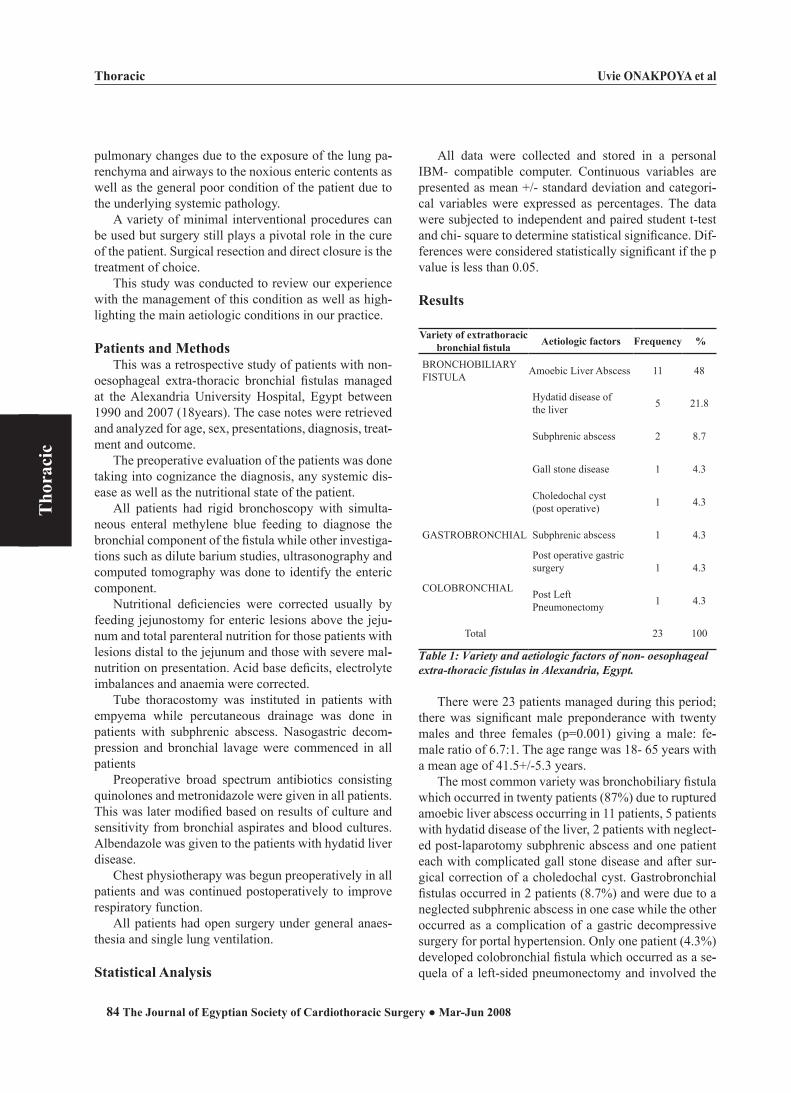
Thoracic
84
Tho
raci
cUvie ONAKPOYA et al
The Journal of Egyptian Society of Cardiothoracic Surgery ● Mar-Jun 2008
pulmonary changes due to the exposure of the lung pa--renchyma and airways to the noxious enteric contents as well as the general poor condition of the patient due to the underlying systemic pathology.
A variety of minimal interventional procedures can be used but surgery still plays a pivotal role in the cure of the patient. Surgical resection and direct closure is the treatment of choice.
This study was conducted to review our experience with the management of this condition as well as high--lighting the main aetiologic conditions in our practice.
Patients and MethodsThis was a retrospective study of patients with non-
oesophageal extra-thoracic bronchial fistulas managed at the Alexandria University Hospital, Egypt between 1990 and 2007 (18years). The case notes were retrieved and analyzed for age, sex, presentations, diagnosis, treat--ment and outcome.
The preoperative evaluation of the patients was done taking into cognizance the diagnosis, any systemic dis--ease as well as the nutritional state of the patient.
All patients had rigid bronchoscopy with simulta--neous enteral methylene blue feeding to diagnose the bronchial component of the fistula while other investiga--tions such as dilute barium studies, ultrasonography and computed tomography was done to identify the enteric component.
Nutritional deficiencies were corrected usually by feeding jejunostomy for enteric lesions above the jeju--num and total parenteral nutrition for those patients with lesions distal to the jejunum and those with severe mal--nutrition on presentation. Acid base deficits, electrolyte imbalances and anaemia were corrected.
Tube thoracostomy was instituted in patients with empyema while percutaneous drainage was done in patients with subphrenic abscess. Nasogastric decom--pression and bronchial lavage were commenced in all patients
Preoperative broad spectrum antibiotics consisting quinolones and metronidazole were given in all patients. This was later modified based on results of culture and sensitivity from bronchial aspirates and blood cultures. Albendazole was given to the patients with hydatid liver disease.
Chest physiotherapy was begun preoperatively in all patients and was continued postoperatively to improve respiratory function.
All patients had open surgery under general anaes--thesia and single lung ventilation.
Statistical Analysis
All data were collected and stored in a personal IBM- compatible computer. Continuous variables are presented as mean +/- standard deviation and categori--cal variables were expressed as percentages. The data were subjected to independent and paired student t-test and chi- square to determine statistical significance. Dif--ferences were considered statistically significant if the p value is less than 0.05.
Results
Variety of extrathoracicbronchial fistula Aetiologic factors Frequency %
BRONCHOBILIARYFISTULA Amoebic Liver Abscess 11 48
Hydatid disease ofthe liver 5 21.8
Subphrenic abscess 2 8.7
Gall stone disease 1 4.3
Choledochal cyst(post operative) 1 4.3
GASTROBRONCHIAL Subphrenic abscess 1 4.3
Post operative gastricsurgery 1 4.3
COLOBRONCHIAL Post Left Pneumonectomy 1 4.3
Total 23 100
Table 1: Variety and aetiologic factors of nont oesophageal extratthoracic fistulas in Alexandria, Egypt.
There were 23 patients managed during this period; there was significant male preponderance with twenty males and three females (p=0.001) giving a male: fe--male ratio of 6.7:1. The age range was 18- 65 years with a mean age of 41.5+/-5.3 years.
The most common variety was bronchobiliary fistula which occurred in twenty patients (87%) due to ruptured amoebic liver abscess occurring in 11 patients, 5 patients with hydatid disease of the liver, 2 patients with neglect--ed post-laparotomy subphrenic abscess and one patient each with complicated gall stone disease and after sur--gical correction of a choledochal cyst. Gastrobronchial fistulas occurred in 2 patients (8.7%) and were due to a neglected subphrenic abscess in one case while the other occurred as a complication of a gastric decompressive surgery for portal hypertension. Only one patient (4.3%) developed colobronchial fistula which occurred as a se--quela of a left-sided pneumonectomy and involved the
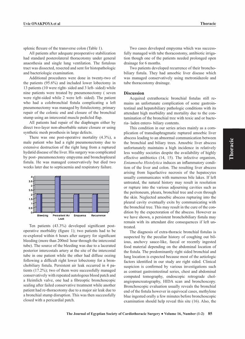
Thoracic
85
Tho
raci
c
Uvie ONAKPOYA et al
The Journal of Egyptian Society of Cardiothoracic Surgery ● Volume 16, Number (1-2)
splenic flexure of the transverse colon (Table 1).All patients after adequate preoperative stabilization
had standard posterolateral thoracotomy under general anaesthesia and single lung ventilation. The fistulous tract was dissected, resected and sent for histopathologic and bacteriologic examination.
Additional procedures were done in twenty-two of the patients (95.6%) and included lower lobectomy in 13 patients (10 were right- sided and 3 left- sided) while nine patients were treated by pneumonectomy ( seven were right-sided while 2 were left- sided). The patient who had a colobronchial fistula complicating a left pneumonectomy was managed by fistulectomy, primary repair of the colonic end and closure of the bronchial stump using an intercostal muscle pedicled flap.
All patients had repair of the diaphragm either by direct two-layer non-absorbable suture closure or using synthetic mesh prosthesis in large defects.
There was one post-operative mortality (4.3%), a male patient who had a right pneumonectomy due to extensive destruction of the right lung from a ruptured hydatid disease of the liver. His surgery was complicated by post- pneumonectomy empyema and bronchopleural fistula. He was managed conservatively but died two weeks later due to septicaemia and respiratory failure.
Ten patients (43.3%) developed significant post-operative morbidity (figure 1); two patients had to be re-explored within 6 hours after surgery for significant bleeding (more than 200ml/ hour through the intercostal tube). The source of the bleeding was due to a lacerated posterior intercostals artery at the site of the intercostal tube in one patient while the other had diffuse oozing following a difficult right lower lobectomy for a bron--chobiliary fistula. Persistent air leak occurred in 4 pa--tients (17.2%); two of them were successfully managed conservatively with repeated autologous blood patch and a Heimlich valve, one had a fibreoptic bronchoscopic sealing after failed conservative treatment while another patient had re-thoracotomy due to a major air leak due to a bronchial stump disruption. This was then successfully closed with a pericardial patch.
Two cases developed empyema which was success--fully managed with tube thoracostomy, antibiotic irriga--tion though one of the patients needed prolonged open drainage for 6 months.
Two patients developed recurrence of their broncho--biliary fistula. They had amoebic liver disease which was managed conservatively using metronidazole and tube thoracostomy drainage.
DiscussionAcquired extrathoracic bronchial fistulas still re--
mains an unfortunate complication of some gastroin--testinal and hepatobiliary pathologic conditions with its attendant high morbidity and mortality due to the con--tamination of the bronchial tree with toxic and or bacte--ria- laden entero- biliary contents.
This condition in our series arises mainly as a com--plication of transdiaphragmatic ruptured amoebic liver abscess leading to an abnormal communication between the bronchial and biliary trees. Amoebic liver abscess unfortunately maintains a high incidence in relatively less wealthy societies despite the availability of highly effective antibiotics (14, 15). The infective organism, Entamoeba Histolytica induces an inflammatory condi--tion of the liver and colon. The resulting liver abscess arising from liquefactive necrosis of the hepatocytes usually communicates with numerous bile lakes. If left untreated, the natural history may result in resolution or rupture into the various adjourning cavities such as the peritoneum, pleura, bronchial tree and even through the skin. Neglected amoebic abscess rupturing into the pleural cavity eventually exits by communicating with the bronchial tree. This may result in the cure of the con--dition by the expectoration of the abscess. However as we have shown, a persistent bronchobiliary fistula may remain with its attendant dire consequences if left un--treated.
The diagnosis of extra-thoracic bronchial fistulas is suspected by the peculiar history of coughing out bil--ious, anchovy sauce-like, faecal or recently ingested food material depending on the abdominal location of the fistula. The predominantly right sided bronchial and lung location is expected because most of the aetiologic factors identified in our study are right sided. Clinical suspicion is confirmed by various investigations such as contrast gastrointestinal series, chest and abdominal computed tomography, endoscopic retrograde chol--angiopancreatography, HIDA scan and bronchoscopy. Bronchoscopic evaluation usually reveals the bronchial end of the fistula however in equivocal cases, methylene blue ingested orally a few minutes before bronchoscopic examination should help reveal this site (16). Also, the
Thoracic
86
Tho
raci
cUvie ONAKPOYA et al
The Journal of Egyptian Society of Cardiothoracic Surgery ● Mar-Jun 2008
pH of the bronchial aspirate can hint at the possible dis--tal end of the fistula; a strongly acidic aspirate suggest--ing a gastrobronchial fistula (17) and a strongly alkaline aspirate suggesting a duodenal or pancreatobiliary site.
The patients usually present in poor general condi--tion with fluid and electrolyte derangement, anaemia, malnutrition, poor lung function, pneumonia and septi--caemia.
Attention should be paid to stabilization of any pre--operative deficits otherwise a prohibitively high mortal--ity and morbidity rate may ensue. This stabilization con--sists of correction of the fluid, electrolyte and nutritional deficits as well as preoperative broad spectrum antibi--otics. Chest physiotherapy consisting of deep breathing exercises as well as postural drainage is essential to the overall survival of the patient. We elected to commence chest physiotherapy on our patients preoperatively and continued it into the post-operative period as soon as the patient recovered from the effects of general anaesthesia until discharge from the hospital.
Associated empyema thoracis was managed by tube thoracostomy. This also helped improve the overall re--spiratory reserve of the patients making them better sur--gical candidates.
It is helpful to relieve any distal obstructing lesion on the gastro-intestinal system which may be perpetuating the fistula such as the use of endoscopic stenting and sphincterotomy of the biliary tract in cases of broncho--biliary fistulae associated with bile duct obstruction.
The use of bronchial stents is also important in pre--venting continued soilage of the airways especially in moribund patients with very poor respiratory reserve whose surgery may be delayed for adequate preopera--tive care. Biliary stenting has recently been advocated and applied in some patients with bronchobiliary fistulas (18, 19).
Surgery is the best treatment for these patients since conservative measures usually eventually fail or leave the patients with significant morbidity as well as an un--acceptably high mortality. Operative measures usually entail fistulectomy, separation and repair of the bron--chial and enteric ends of the fistula. Any diaphragmatic defect present is usually repaired at the time of the pri--mary surgery. While repair of the bronchial end by vas--cularized pedicled tissues is the ideal, this is usually not possible in most cases due to the extensive destruction of adjourning lung parenchymal tissues necessitating lo--bectomy in most cases or even pneumonectomy. In our series, thirteen of our patients (56.6%) had a lobectomy, nine (39.1%) had a pneumonectomy while only one pa--tient (4.3%) was managed by fistulectomy with separate vascularized repair of the bronchial stump as well as
closure of the enteric component.Surgery is greatly facilitated by the use of double
lumen endotracheal tubes. This allows for single lung ventilation and enhanced access to the involved non-ventilated lung.
We routinely utilize an epidural catheter for intra and especially post-operative pain management. This helps improve respiratory function by eliminating or greatly decreasing pain sensation after thoracotomy.
The high morbidity (43.3%) emphasizes the fact that these patients usually present in a grave condition. Ag--gressive perioperative management is essential to limit morbidity and mortality. This condition exerts a great burden on the health facilities due to the resources, drugs, personnel and equipment required to manage a single patient. Such a demanding care requires the understand--ing and patience of the health care providers particularly thoracic surgeons, nurses and physiotherapists to obtain good results in the long term.
References
1. Pichhardt PJ, Bhalla S, Balfe DM. Acquired gastrointesti--nal fistulas: classification, etiologies and imaging evalua--tion. Radiology 2002; 224: 9- 22.
2. Clarke HJ, Wright ES, Edstrom HW, Harris R. Enterobron--chial fistula. Can J Surg. 1984; 27: 185-6.
3. Powner DJ, Bierman MI. Thoracic and extrathoracic bron--chial fistulas. Chest. 1991; 100: 480- 6.
4. Caberwal D, Katz J, Reid R, Newman HR. A case of nephrobronchial and colobronchial fistula presenting as lung abscess. J Urol. 1977; 117:371- 3.
5. Gil- Salom M, Oron- Apluente J, Ruiz- DelCastillo J, Ch--uan- Nuez P, Carretero- Gonzalez P. Nephro- broncho- cu--taneous fistula. Br J Urol. 1989; 64: 652- 3.
6. Neff C. Splenobronchial fistula. Gastrointest Radiol. 1987; 12: 197- 9.
7. Baxter AD, Fishwick NG, Jeyapalan K. Case report: Ileo--bronchial fistula. Clin Radiol 1998; 53: 231- 3.
8. Heitmiller RF, Yeo CJ. Duodenobronchial fistula. Surgery 1991; 110: 546- 8.
9. Tzeng JJ, Lai KH, Lo GH, Hsu JH, Mok KT. Gastropleural fistula caused by incarcerated diaphragmatic herniation of the stomach. Gastrointest Endosc. 2001; 53: 382- 4.
10. Radin DR, Ray MJ, Halls JM. Strangulated diaphragmatic hernia with pneumothorax due to colopleural fistula. Am J Roentgenol 1986; 146: 321- 2.
11. Uzun K, Ozbay B, Etlik o, Kotan C, Gencer M, Sakarya ME. Bronchobiliary fistula due to hydatid disease of the liver: a case report. Acta Chir Belg. 2002; 102: 207- 9.
12. Kabiri EH, El Maslout A, Benosman A. Thoracic rupture of hepatic hydatidosis (123 cases). Ann Thorac Surg. 2001; 72: 1883- 6.
13. Roy DC, Ravindran P, Padmanabhan R. Bronchobiliary fistula secondary to amebic liver abscess. Chest 1972; 62:

Thoracic
87
Tho
raci
c
Uvie ONAKPOYA et al
The Journal of Egyptian Society of Cardiothoracic Surgery ● Volume 16, Number (1-2)
523- 4.14. Ahmed L, el Rooby A, Kassem MI, Salama ZA, Strichland
GT. Ultrasonography in the diagnosis and management of 52 patients with amoebic liver abscess in Cairo. Rev Infect Dis 1990; 12: 330- 7.
15. Valenzuela O, Moran P, Gomez A, Cordova K, Corrales N, Cardoza J, Gomez N, Cano M, Ximenez C. Epidemiology of amoebic liver abscess in Mexico: the case of Sanora. Ann Trop Parasitol 2007; 101: 533- 8.
16. Mc Manigle JE, Fletcher GL, Tenholder MF. Bronchos--copy in the management of bronchopleural fistula. Chest 1990; 97: 1235- 8.
17. Joseph JT, Krumpe PE. Diagnosis of gastrobronchial fistu--la by measurement of bronchial secretion pH. Chest 1989; 96: 935- 6.
18. Gandini R, Konda D, Tisone G, Pipitone V, Anselmo A, Simonetti G. Bronchobiliary fistula treated by self- expand--ing ePTFE- covered nitinol stent- graft. Cardiovasc Inter--vent Radiol 2005; 28: 828- 31.
19. Katsinelos P, Paroutoglou G, Chatzimavroudis G, Beltsis A, Mimidis K, Katsinelos T, Pilpilidis I, Papaziogas B. Suc--cessful treatment of intractable bronchobiliary fistula using long term biliary stenting. Surg Laparosc Endosc Percutan Tech 2007; 17: 206- 9.
Mohamed Abul-dahab et al
88 The Journal of Egyptian Society of Cardiothoracic Surgery ● Mar-Jun 2008
Thoracic
Tho
raci
c
BILATERAL BULLECTOMY THROUGH MEDIAN STERNOTOMY
Background: Emphysematous bullae represent a form of emphysematous lung destruction. Surgical resection has traditionally been indicated when there is hyperexpansion of the chest, compromised pulmonary function, and evidence of underlying, relatively normal compressed lung. Different approaches to LVRS have been proposed; these include median sternotomy, thoracosternotomy and video-assisted thoracoscopic surgery (VATS) technique. Methods: Nine patients underwent resection of emphysematous bullae at Kasr Elaini hospital between February 2006 and July 2007. All had limiting dyspnea and radiological evidence of hyperinflated bullae compressing adjacent lung parenchyma. All the patients had bilateral lesions. We did median sternotomy, with one-lung ventilation under a standard general anesthetic technique to avoid rupture of bulla, in all patients to do resections bilaterally in the same sitting. Results: Postoperative complications included persistent air-leak, failure of early extubation, readmission to ICU and chest infection. We had two mortalities one due to ARDS and the other due to chest infection.Conclusion: we recommend the use of sternotomy approach in highly selected patients with good target areas, as it is only subject the patient for one procedure instead of two and we can avoid the possibility of spontaneous pnemothorax on the opposite side, while doing bulletomy in lateral position through thoracotomy. In our experience there were no difficulties in dissection of lung adhesions or attack any target areas through this approach.
Emphysema, a form of chronic obstructive pulmonary disease, is a condition characterized by abnormal and permanent enlargement of the air spaces distal to the terminal bronchioles and accompa--nied by destruction of their walls without obvious fibrosis(1).
Lung volume reduction surgery (LVRS) is a palliative operation to be con--sidered in selected patients suffering severe dyspnea from the end stage of diffuse nonbullous emphysema, who are not responding to maximal medical management. Bullectomy has been an established technique for patients with
Accepted for publication Oct. 1 , 2007
Address reprint request to Dr: M. Abul
Eldahab Department of Cardiothoracic
Surgery , Cairo University
Email: tfrksm @yahoo.com.
codex : 05/others/13/0710
Mohamed Abul-dahab M.D, Ahmed El-Agaty M.D
Mohamed Abul-dahab et al
89The Journal of Egyptian Society of Cardiothoracic Surgery ● Volume 16, Number (1-2)
Thoracic
Tho
raci
c
bullous emphysema, since the work of Brantigan and Mueller 40 years ago (1).
A bulla is defined as an air-filled space 1 cm or great--er in diameter within the lung parenchyma that forms as a result of destruction of the alveolar walls. Rarely one or more bullae enlarge to such a degree that they occupy more than one third of the hemithorax. The term giant bulla is then applied. These easily distensible reservoirs are preferentially filled during inspiration, causing the collapse of adjacent, more normal, lung parenchyma. Because of the alveolar destruction, bullae lack any meaningful alveolar-capillary interface and the thoracic volume they occupy is wasted. The resulting hyperinfla--tion of the chest interferes with normal respiratory me--chanics, increasing the work of breathing with associ--ated exercise limitation and dyspnea (2).
The concept of LVRS was developed by Cooper and colleagues inspired by Brantigan’s earlier work. He mod--ified the approach of Brantigan and Mueller by using a median sternotomy, thus allowing access to both lungs and used a buttressed staple excision technique. The hyperinflated and relatively functionless parts of both lungs were removed and it was proposed that this would result in improvement in the function of the remaining lung, palliate the dypsnea, improve exercise ability, and provide a positive change in quality of life (3).
The fundamental concept of LVRS is to improve elastic recoil, reduce airflow limitation and improve the mechanics of respiration by excision of some of the most destroyed portions of the lung for patients with se--vere emphysema (4).
Patients suitable for LVRS are those with heterog--enous disease having target areas for resection, which is most commonly associated with centrilobular emphy--sema. Giant bullae, with relatively normal but collapsed adjacent lung, may represent the most heterogenous end of a spectrum of disease. Traditionally, bullectomy has been performed to alleviate the collapse of under-ly--ing normal lung tissue. On the other hand, the primary benefit of LVRS has been attributed to the reduction of thoracic hyperinflation and improvement in respiratory mechanics. It is probable that both mechanisms are at work after bullectomy (5).
A variety of different approaches to LVRS have been proposed; these include median sternotomy, thora--costernotomy and video-assisted thoracoscopic surgery (VATS) technique. There are both unilateral and bilateral approaches (1).
The areas or surgical removal are identified before surgery by computed tomography and radionuclide ven--tilation-perfusion scanning. Methods for sealing the site of resected lung include the use of staples or a laser;
however, prolonged air leak is a common postoperative complication (6).
Lung volume reduction surgery is a procedure that requires appropriate selection of patients who have been suitably informed of the risks of this procedure. The number of patients who qualify for LVRS are a small percentage of those originally assessed (7).
The majority of patients 5 years post LVRS experi--ence enhanced exercise capacity, a reflection of reduced hyperinflation and gas trapping (8).
The optimal surgical approach for LVRS for ad--vanced bilateral emphysema is unknown. Case series and early randomized trials have demonstrated the su--periority of stapled over laser resection and of bilateral over unilateral operations. Case series support the use of either median sternotomy (MS) or video-assisted thora--coscopic (VATS) approaches (9).
Compared with medical management, LVRS pro--duced a significant improvement in function for patients with upper lobe emphysema and improved survival for the subset of patients with upper lobe emphysema and a low baseline exercise capacity (10).
Surgical therapy has been used for bullous emphyse--ma since the mid-20th century. The best surgical candi--dates are often those with giant pulmonary bullae rather than those with diffuse disease (11).
In this study we tried to show our experience in Kasr Elaini hospital in bilateral LVRS and bullectomy through median sternotomy, the exposure, accessibility, results and outcome.
Patients and Methods:From February 2006 to July 2007, 9 patients sched--
uled for elective bilateral bullectomy through median sternotomy in Kasr Elaini hospital were enrolled in the study.
Inclusion criteria: Patient selection criteria and the clinical evaluation
consist of: (1) marked hyperinflation evident by inspira--tory and expiratory chest radiographs; (2) radiographic evidence of heterogenous emphysema with clear target zones for resection; (3) severe functional impairment (forced expiratory volume in 1 s (FEV1) < 1.2 or pre--dicted FEV1 20-35%, total lung capacity (TLC) >120%); (4) poor quality of life; (5) dyspnea despite optimized medical therapy; (6) abstinence from smoking, accept--able nutritional status; and (7) rehabilitation potential.
I Exclusion criteria: Contraindication to surgery included age older than
75 years, current cigarette smoking, symptomatic car--
Mohamed Abul-dahab et al
90 The Journal of Egyptian Society of Cardiothoracic Surgery ● Mar-Jun 2008
Thoracic
Tho
raci
c
diac disease, and pulmonary hypertension (pulmonary artery pressure >35 mmHg).
II. Preoperative Assessment: All patients of the study were assessed in the pre--
operative period by medical history taking and detailed clinical examination with a special emphasis on any re--spiratory problems, full laboratory investigations with special emphasis on complete blood picture, coagulation profile, arterial blood gases and pulmonary functions.
Radiological investigations in the form of plain chest x-ray and high resolution computed tomography.
Chest physiotherapy, started preoperatively, to help in clearance of respiratory tree from secretions.
Preoperative echocardiography was done routinely to assess the preoperative cardiac conditions.
III. Operative Management:Anesthetic technique:
All patients were not premedicated except for in--haled bronchodilators administered prior to induction of anesthesia. Anesthesia was planned to be a combination of thoracic epidural and general anesthesia.
Routine monitors were used including 5 leads ECG, continuous invasive arterial blood pressure monitor and frequent blood gases analysis via a radial 20G cannula, pulse oximeter, capnography, right internal jugular cath--eter for central venous pressure monitoring and urinary catheter.
Thoracic epidural catheter was placed under local anesthesia in the awake patient, in the sitting position between T5 and T8 via a paramedian approach using loss of resistance to saline. After confirmation of proper epidural placement using test dose 4 mL. of bupivacaine 0.5% and epinephrine 1/200,000, the catheter was firmly fixed in place and 6 mL of bupivacaine 0.5 % as well as 100 microgram fentanyl were injected as a bolus fol--lowed by a continuous infusion of 4-6 mL / h of bupi--vacaine 0.5%, throughout the operation to be reduced to 0.125% bupivacaine in the first 48 hours postopera--tively.
General anesthesia was induced intravenously using propofol 1.5 mg/ kg, fentanyl 200 microgram, and suc--cinylcholine 80 mg to facilitate intubation. A left double lumen tube was placed and anesthesia was maintained by sevoflurane 2-3% in oxygen/air mixture with a FiO2 0.5 with supplemental infusion of propofol 3-6 mg/kg/hour.
Spontaneous ventilation was resumed and continued till the time of sternotomy; when a bolus of fentanyl 100 microgram and pancuronium 6- 8 mg was given, and af--ter complete relaxation the sternum was divided while
the lungs were deflated then mechanical ventilation was started.
After strenotomy; anesthesia was maintained by 2% sevofluraine and bolus doses of pancuronium 1 mg / h. One lung ventilation was used and FiO2 was increased to 1.0.Tidal volume was set between 6-8 mL, and re--spiratory rate between 12-14/ min with a PEEP of zero. Ventilator settings were adjusted to maintain peak air--way pressure ≤ 25 cm H2O, and arterial tension of CO2 around 40 mmHg or within 20% of the awake baseline readings. When oxygen saturation dropped below 90 percent brief periods of two-lung ventilation were nec--essary.
At the end of surgery spontaneous breathing was re--sumed and muscle relaxant was reversed. The patients were extubated in the theater and transferred to the in--tensive care with an epidural infusion and supplemental oxygen.
Surgical technique:Patients underwent median sternotomy. Bilateral
procedures were performed as a rule. The side with bet--ter-preserved perfusion was operated on first. Dissection of all adhesions between the lung and chest wall, should be done firstly, to let the lung totally free.
The target zones for resections were assessed preop--eratively by high resolution computed tomography.
In many cases when there is giant bulla, a division of the anterior bulla wall done firstly, then control of the feeding bronchus was done by interrupted proline 3/0 sutures. In smaller bullae the bases are controlled either by silk suture or by continuous proline 3/0 sutures, and then bullae were excised.
Before closure of the chest, mediastinal and pleural tubes were positioned in the mediastinum, one retroster--nal two tubes in each pleural space; each of the plural tubes was placed laterally at the level of anterior axillary line.
IV. Postoperative assessment:Postoperative pain relief was achieved with a tho--
racic epidural placed prior to induction of anesthesia at the level of the fourth thoracic vertebral body.
Early and vigorous chest physiotherapy and ambula--tion were performed.
Outcomes included mortality, complications, hospi--tal stay duration, ICU stay, need for mechanical ventila--tion, and status at initial hospital discharge.
Results:A total of 9 patients underwent bullectomy through
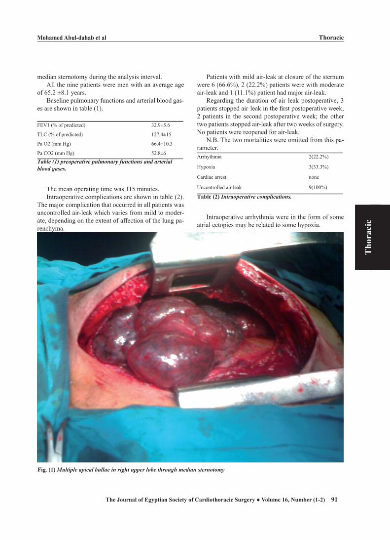
Mohamed Abul-dahab et al
91The Journal of Egyptian Society of Cardiothoracic Surgery ● Volume 16, Number (1-2)
Thoracic
Tho
raci
c
median sternotomy during the analysis interval.All the nine patients were men with an average age
of 65.2 ±8.1 years.Baseline pulmonary functions and arterial blood gas--
es are shown in table (1).
FEV1 (% of predicted) 32.9±5.6
TLC (% of predicted) 127.4±15
Pa O2 (mm Hg) 66.4±10.3
Pa CO2 (mm Hg) 52.8±6
Table (1) preoperative pulmonary functions and arterial blood gases.
The mean operating time was 115 minutes.Intraoperative complications are shown in table (2).
The major complication that occurred in all patients was uncontrolled air-leak which varies from mild to moder--ate, depending on the extent of affection of the lung pa--renchyma.
Patients with mild air-leak at closure of the sternum were 6 (66.6%), 2 (22.2%) patients were with moderate air-leak and 1 (11.1%) patient had major air-leak.
Regarding the duration of air leak postoperative, 3 patients stopped air-leak in the first postoperative week, 2 patients in the second postoperative week; the other two patients stopped air-leak after two weeks of surgery. No patients were reopened for air-leak.
N.B. The two mortalities were omitted from this pa--rameter. Arrhythmia 2(22.2%)
Hypoxia 3(33.3%)
Cardiac arrest none
Uncontrolled air leak 9(100%)
Table (2) Intraoperative complications.
Intraoperative arrhythmia were in the form of some atrial ectopics may be related to some hypoxia.
Fig. (1) Multiple apical bullae in right upper lobe through median sternotomy
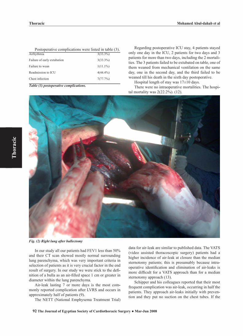
Mohamed Abul-dahab et al
92 The Journal of Egyptian Society of Cardiothoracic Surgery ● Mar-Jun 2008
Thoracic
Tho
raci
c
Postoperative complications were listed in table (3).Arrhythmia 3(33.3%)
Failure of early extubation 3(33.3%)
Failure to wean 1(11.1%)
Readmission to ICU 4(44.4%)
Chest infection 7(77.7%)
Table (3) postoperative complications.
Regarding postoperative ICU stay, 4 patients stayed only one day in the ICU, 2 patients for two days and 3 patients for more than two days, including the 2 mortali--ties. The 3 patients failed to be extubated on table, one of them weaned from mechanical ventilation on the same day, one in the second day, and the third failed to be weaned till his death in the sixth day postoperative.
Hospital length of stay was 17±10 days.There were no intraoperative mortalities. The hospi--
tal mortality was 2(22.2%). (12).
Fig. (2) Right lung after bullectomy
In our study all our patients had FEV1 less than 50% and their CT scan showed mostly normal surrounding lung parenchyma, which was very important criteria in selection of patients as it is very crucial factor in the end result of surgery. In our study we were stick to the defi--nition of a bulla as an air-filled space 1 cm or greater in diameter within the lung parenchyma.
Air-leak lasting 7 or more days is the most com--monly reported complication after LVRS and occurs in approximately half of patients (9).
The NETT (National Emphysema Treatment Trial)
data for air-leak are similar to published data. The VATS (video assisted thoracoscopic surgery) patients had a higher incidence of air-leak at closure than the median sternotomy patients; this is presumably because intra--operative identification and elimination of air-leaks is more difficult for a VATS approach than for a median sternotomy approach (13).
Schipper and his colleagues reported that their most frequent complication was air-leak, occurring in half the patients. They approach air-leaks initially with preven--tion and they put no suction on the chest tubes. If the

Mohamed Abul-dahab et al
93The Journal of Egyptian Society of Cardiothoracic Surgery ● Volume 16, Number (1-2)
Thoracic
Tho
raci
c
air-leak persists, a Heimlich valve is placed to decrease further the resistance to expelling a pnemothorax and the barotrauma on the remaining lung. They were very reluctant to return to the operating room for an air-leak, believing that reoperations create more leaks than are repaired (5).
In our study patients with persistent air-leak more than a week were 4 patients (57% of living patients), which is similar to the published data. It was the sec--ond most common complication after chest infection. In our study we had more incidence of air-leak at time of closure due to the difference in the technique of resec--tion of bullae, in different studies they used buttressed staple lines (with bovine pericardium), in our case we used continuous proline 3/0 sutures. This was only af--fecting the rate of air-leak at time of closure but did not affect the rate of persistant air-leak for more than 7 days. We managed persistent air-leak by chest physiotherapy, no suction was applied to the chest tubes, and control of chest infection. We also did not reopened any of our patients for persistent air-leak as we also believe in more damage produced by reopening than repair.
Because LVRS is a palliative, elective procedure, one of the greatest concerns was the mortality rate for the procedure. Reported operative mortality rates were generally 3.5% to 10%, but some were as high as 19.1%. Moreover, on the basis of data for Medicare pa--tients who underwent LVRS between October 1995 and January 1996, a mortality rate of 14.4% was found at 3 months. However, in the prospective multicenter study, the NETT observed 90-day mortality for LVRS of 5.9% for median sternotomy and 4.6% for VATS (13).
Schipper and his colleagues reported that their hospital mortality of 2.3% is low and consistent with the mortalities ranging from 0% to 9% reported by other au--thors(5).
Fujimoto and his colleagues reported that there should be a balance between the extent of resection that offers the maximal functional improvement and the extent that causes the minimal perioperative morbidity and mortality. On the one hand, the larger the volume of non-functional lung parenchyma is resected, the greater the functional improvement can be achieved. On the other hand, the pulmonary vascular bed, which can be represented by diffusing capacity (DLCO), may limit the possible extent of resection, (which is not routinely done) and this may lead to rapid development of adult respiratory distress syndrome (ARDS), which will lead to death (14).
Regarding our mortality we had two mortalities, one due to ARDS died on the sixth day postoperative, this was may be due to the small amount of lung tissue left
after resection (increased amount of resection with left unhealthy tissue). The other mortality was due to severe chest infection after discharge from ICU with increased bronchial tree secretions, which necessitate readmission to ICU, but patient died out of respiratory failure on fifth day postoperative. Sputum culture which appeared after his death showed infection with MRSA bacteria.
Our mortality rate is to an extent higher than most of the literature but this could be attributed to the small number of patients in our study, which make a single mortality represents more than 10%. Selection of cases may be another cause as we have no specific facilities as measuring diffusing capacity of the lung, and we depend only on pulmonary functions and CT appearance of the lung parenchyma, with the gross pathology seen by the surgeon intraoperatively.
Since the inception of the LVRS program at Barnes Jewish hospital, more than 800 patients have been eval--uated on site for potential emphysema surgery. Forty-three of these patients were found to have bullae fitting previously published criteria for bullectomy (5).
As regards the number of patients in this study, it is related to many factors as, selection criteria of the pa--tients to be indicated which were very obvious that it is a limiting factor by itself. Many of the emphysematous patients have diffuse pathology with no target areas, or affection of the lung parenchyma left small residual ap--parently healthy lung that could not permit suitable oxy--genation of the body.
Second factor is general health of the patients as they may be unfit for a major surgery like this due to their general medical condition.
Lastly, many patients refused to do surgery espe--cially when they knew the nature of this surgery being palliative one and the symptoms will recur again after few years.
Stirling and his colleagues reported 2001 that LVRS using the stapling-excision technique and the median sternotomy approach, in highly selected cases, has proved to be a safe and reasonably efficacious proce--dure. These results have been shown to be reproducible by other workers (1).
The data from the NETT showed there is no differ--ence between median sternotomy and VATS patients with respect to the functional results at 12 and 24 months after randomization. They concluded that the choice of approach is a matter of the surgeon’s prefer--ence and experience (13).
In conclusion we recommend the use of this approach in highly selected patients with good target areas and sufficient amount of residual lung parenchyma, as it is only subject the patient for one procedure instead of two
Mohamed Abul-dahab et al
94 The Journal of Egyptian Society of Cardiothoracic Surgery ● Mar-Jun 2008
Thoracic
Tho
raci
c
and we can avoid the possibility of spontaneous pnemo--thorax on the opposite side, while doing bulletomy in lateral position through thoracotomy. In our experience there were no difficulties in dissection of lung adhesions or attack any target areas through this approach, except for some difficulties in lower lobe lesions (which is not common).
We recommend doing study on long duration or mul--ticenter studies to help in getting more number of pa--tients for better results analysis.
References:
1. Stirling GR, Babidge WJ, Peacock MJ, Smith JA, Matar KS, Snell GI, Colville DJ, Maddern GJ. Lung volume reduction surgery in emphysema: a systemic review. Ann Thorc Surg 2001; 72: 641-8.
2. Morgan MDL, Edward CW, Morris J, Matthews HR. Origin and behavior of emphysematous bullae. Thorax 1989; 44: 533-8.
3. Cooper JD, Trulock EP, Triantafillou AN. Bilateral pneumec--tomy (volume reduction) for chronic obstructive pulmonary disease. J Thorc Cardiovasc Surg 1995; 109: 106-19.
4. Cooper JD, Patterson GA, Sundaresan RS, Trulock EP, Yu--sen RD, Pohl MS, Lefrak SS. Results of 150 consecutive bilateral lung volume reduction procedures in patients with severe emphysema. J Thorac Cardiovasc Surg 1996; 112: 1319-1330.
5. Shipper PH, Meyers BF, Battafarano Rj, Guthrie TG, Pat--terson GA, Cooper JD. Outcomes after resection of giant emphysematous bullae. Ann Thorc Surg 2004; 78: 976-82.
6. Brenner M, McKenna RJ, Gelb AF, Fischel RJ, Wilson AF. Rate of FEV1 change following lung volume reduction sur--gery. Chest 1998; 113: 652-9.
7. McKenna RJ Jr, Brenner M, Fischel RJ. Patient selection criteria for lung volume reduction surgery. J Thorc Cardio--vasc Surg 1997; 114: 957-67.
8. Hopkins PM, Seale H, Walsb J, Tam R, Kermeen F, Bell S,McNeil K. Long term results post conventional lung vol--ume reduction surgery exceeds outcome of lung transplan--tation for emphysema. J of heart and lung transplantation 2001;25:61.
9. Miller JI, Lee RB, Mansour KA. Lung volume reduction surgery: lessons learned. Ann Thorac Surg. 1996; 61: 1464-9.
10. National Emphysema Treatment Trial research group. A randomized trial comparing lung volume reduction surgery with medical therapy for severe emphysema. N Engl J Med 2003; 348: 2059-73.
11. Van Natta TL and Iannettoni MD. Reduction pneumoplasty for a giant right upper lobe bulla causing massive bilateral lung compression. J Thorc Cardiovasc Surg 2007; 133: 1674-5.
12. Snider GL. Reduction pneumoplasty for giant bullous em--physema. Chest. 1996; 109:540-8.
13. National Emphysema Treatment Trial research group. Safety and efficacy of median sternotomy versus video-as--sisted thoracic surgery for lung volume reduction surgery. J Thorc Cardiovasc Surg 2004; 127: 1350-60.
14. Fujimoto T, Teschler H, Hillejan L, Zaboura G, Stamatis G. Long-term results of lung volume reduction surgery. Eur J Cardio Thorac Surg 2002; 21: 483-8.