Embed Size (px)
Citation preview
©JIPBS, All rights reserved
Journal of Innovations in Pharmaceuticals and Biological Sciences
www.jipbs.com
ISSN: 2349-2759
Abstract
Novel drug delivery systems are now a days is creating a new interest in development of drug deliveries. Vesicular drug delivery system is also a part of these novel drug delivery systems. TDDS is the permeability of the skin, it is permeable to small molecules, lipophilic drug and highly impermeable to the macromolecules and hydrophilic drugs. Recent approaches have resulted in design of two vesicular carriers, ethosomes and ultra flexible lipid based elastic vesicles, transferosomes. Transferosomes have recently been introduced, which are capable of transdermal delivery of low as well as high molecular weight drugs. This offers several potential advantages over conventional routes like avoidance of first pass metabolism, predictable and extended duration of activity, minimizing undesirable side effects, utility of short half life drugs, improving physiological and pharmacological response and have been applied to increases the efficiency of the material transfer across the intact skin, by the use of penetration enhancers, iontophoresis, sonophoresis and use of colloidal carriers such as lipid vesicles (liposomes & proliposomes) and non-ionic surfactant vesicles (niosomes & proniosomes). It is suitable for controlled and targeted drug delivery and it can accommodate drug molecules with wide range of solubility. Due to its high deformability it gives better penetration of intact vesicles. They are biocompatible and biodegradable as they are made from natural phospholipids and have high entrapment efficiency. The preparation variables are depending upon the procedure involved for manufacturing of formulation and the preparation procedure was accordingly optimized and validated. Characterization of transferosomes can be done to know the vesicle size, morphology, drug content, entrapment efficiency, penetration ability, occlusion effect, surface charge, in vitro drug release, in vitro skin penetration etc., It increases stability of labile drugs and provides control release. Transferosomes thus differs from such more conventional vesicles primarily by its softer, more deformable, better adjustable artificial membrane. Key words: Transferosomes, Novel Drug Delivery System. *Corresponding Author: Y. Dastagiri Reddy, Creative Educational Society’s College of Pharmacy, Chinnatekur, Kurnool, A.P, India.
JIPBS
Review Article
Transferosomes A Novel Vesicular Carrier for Transdermal Drug Delivery System Y. Dastagiri Reddy*1, A. B. Sravani1, V. Ravisankar1, P. Ravi Prakash1, Y. Siva Rami Reddy1, N. Vijaya Bhaskar1
1Creative Educational Society’s College of Pharmacy, Chinnatekur, Kurnool, A.P, India.
Y. Dastagiri Reddy et al., JIPBS, Vol 2 (2), 193-208, 2015
194
1. Introduction
Transdermal drug delivery systems (TDDS) offer a number of potential advantages over conventional methods such as injectable and oral delivery [1]. However, the major limitation of TDDS is the permeability of the skin, it is permeable to small molecules, lipophilic drugs and highly impermeable to macromolecules and hydrophilic drugs. The main barrier and rate-limiting step for diffusion of drugs across the skin is provided by the outermost layer of the skin, the stratum corneum (SC) [2]. Recent approaches in modulating vesicle compositions have been investigated to develop systems that are capable of carrying drugs and macromolecules to deeper tissues. These approaches have resulted in the design of two novel vesicular carriers, ethosomes and ultra flexible lipid-based elastic vesicles, transferosomes [3]. Transferosomes are ultra deformable vesicles possessing an aqueous core surrounded by the complex lipid bilayer. Interdependency of local composition and shape of the bilayer makes the vesicle both self-regulating and self-optimizing [4]. Transferosomes have recently been introduced, which are capable of transdermal delivery of low as well as high molecular weight drugs [5]. Transferosomes are specially optimized, ultra flexible lipid supra molecular aggregates, which are able to penetrate the mammalian skin intact and then act as a drug carrier for non-invasive targeted drug delivery and sustained release of therapeutic agents [6]. Each transferosomes consists of at least one inner aqueous compartment, which is surrounded by a lipid bilayer with specially tailored properties, due to the incorporation of "edge activators" into the
vesicular membrane. Surfactants such as sodium cholate, sodium deoxycholate, Span 80, and Tween 80, have been used as edge activators [7]. Due to their deformability, transferosomes are good candidates for the non-invasive delivery of small, medium, and large sized drugs. Delivery via the transdermal route is an interesting option in this respect because a transdermal route is convenient and safe. This offers several potential advantages over conventional routes like avoidance of first pass metabolism, predictable and extended duration of activity, minimizing undesirable side effects, utility of short half-life drugs, improving physiological and pharmacological response, avoiding the fluctuation in drug levels, inter and intra-patient variations, and most importantly, it provides patients convenience. To date many chemical and physical approaches have been applied to increase the efficacy of the material transfer across the intact skin, by use of the penetration enhancers, enhancers, iontophoresis, sonophoresis and the use of colloidal carriers such as lipid vesicles (liposomes and proliposomes) and nonionic surfactant vesicles (niosomes and proniosomes). 1.1 Definition The name means “carrying body” and is derived from the Latin word 'transferre', meaning 'to carry across', and the Greek word 'soma', meaning 'a body'. A Transferosomes carrier is an artificial vesicle designed to exhibit the characteristics of a cell vesicle or a cell engaged in exocytosis, and thus suitable for controlled and, potentially, targeted drug delivery.
Y. Dastagiri Reddy et al., JIPBS, Vol 2 (2), 193-208, 2015
195
1.2 Discovery The term Transferosomes and the underlying concept were introduced in 1991 by Gregor Cevc. Numerous groups have since been working with similar carriers, frequently using different names (e.g., elastic vesicle, flexible vesicle, Ethosomes, etc.) to describe them. 1.3 Salient Features Transferosomes possess an infrastructure consisting of hydrophobic and hydrophilic moieties together and as a result can accommodate drug molecules with wide range of solubility. Transferosomes can deform and pass through narrow constriction (from 5 to 10 times less than their own diameter) without measurable loss. This high deformability gives better penetration of intact vesicles. They can act as a carrier for low as well as high molecular weight drugs e.g. analgesic, anesthetic, corticosteroids, sex hormone, anticancer, insulin, gap junction protein, and albumin. They are biocompatible and biodegradable as they are made from natural phospholipids similar to liposomes. They have high entrapment efficiency, in case of lipophilic drug near to 90%. They protect the encapsulated drug from metabolic degradation. They act as depot, releasing their contents slowly and gradually. They can be used for both systemic as well as topical delivery of drug. Easy to scale up, as procedure is simple, do not involve lengthy procedure and unnecessary use or pharmaceutically unacceptable additives [8]. 1.4 Advantages of Transferosomes Transferosomes can deform and pass through narrow constriction (from 5 to 10 times less than their own diameter) without measurable loss. 1. They have high entrapment efficiency,
in case of lipophilic drug near to 90%.
2. This high deformability gives better penetration of intact vesicles.
3. They can act as a carrier for low as well as high molecular weight drugs e.g. analgesic, anesthetic, corticosteroids, sex hormone, anticancer, insulin, gap junction protein, and albumin.
4. Transferosomes possess an infrastructure consisting of hydrophobic and hydrophilic moieties together and as a result can accommodate drug molecules with wide range of solubility.
5. They act as depot, releasing their contents slowly and gradually.
6. They can be used for both systemic as well as topical delivery of drug.
7. They are biocompatible and biodegradable as they are made from natural phospholipids similar to liposomes.
8. They protect the encapsulated drug from metabolic degradation.
9. Easy to scale up, as procedure is simple, do not involve lengthy procedure and unnecessary use or pharmaceutically unacceptable additives [11,12,13].
1.5 Limitations of Transferosomes 1. Transferosomes are chemically
unstable because of their predisposition to oxidative degradation.
2. Purity of natural phospholipids is another criteria militating against adoption of transferosomes as drug delivery vehicles.
3. Transferosomes formulations are expensive [10, 12, 13].
2. Transferosomes VS other Carrier Systems At first glance, transferosomes appear to be remotely related to lipid bilayer vesicle, liposomes. However in functional terms,
Y. Dastagiri Reddy et al., JIPBS, Vol 2 (2), 193-208, 2015
196
transferosomes differ vastly from commonly used liposomes in that they are much more flexible and adaptable. The extremely high flexibility of their membrane permits transferosomes to squeeze themselves even through pores much smaller than their own diameter. This is due to high flexibility of the transferosomes membrane and is achieved by judiciously combining at least two lipophilic/amphiphilic components (phospholipids plus bio surfactant) with sufficiently different packing characteristics into a single bilayer. The high resulting aggregate deformability permits transferosomes to penetrate the skin spontaneously. This tendency is supported by the high transferosomes surface hydrophilicity that enforces the search for surrounding of high water activity. It is almost certain that the high penetration potential of the transferosomes is not primarily a consequence of stratum corneum fluidization by the surfactant because micellar suspension contains much more surfactant than transferosomes (PC/Sodium cholate 65/35 w/w %, respectively). Thus, if the penetration enhancement via the solubilization of the skin lipids was the reason for the superior penetration capability of transferosomes, one would expect an even better penetration performance of the micelles. In contrast to this postulate, the higher surfactant concentration in the mixed micelles does not improve the efficacy of material transport into the skin. On the contrary, mixed micelles stay confined to the topmost part of the stratum corneum even they are applied non occlusively. The reason for this is that mixed micelles are much less sensitive to the trans-epidermal water activity gradient than transferosomes. Transferosomes differ in at least two basic features from the mixed micelles, first a transferosomes is
normally by one to two orders of magnitude (in size) greater than standard lipid micelles. Secondly and more importantly, each vesicular transferosomes contains a water filled core whereas a micelle is just a simple fatty droplet. Transferosomes thus carry water as well as fat-soluble agent in comparison to micelles that can only incorporate lipoidal substances. To differentiate the penetration ability of all these carrier systems proposed the distribution profiles of fluorescently labelled mixed lipid micelles, liposomes and transferosomes as measured by the Confocal Scanning Laser Microscopy (CSLM) in the intact murine skin. In all these vesicles the highly deformable transferosomes transverse the stratum corneum and enter into the viable epidermis in significant quantity. Chapman & Walsh also showed that the former two types of aggregates are confined to the outer half of the horny layer, where the cellular packing and intercellular seals are already compromised by the desquamation process. Pure lipid vesicles or micelles seem to have access to the low-resistance pathway only and thus very seldom reach the lower stratum corneum or even get into the viable part of the skin in significant quantities [15,16,17]. Table 1
shows the comparison of different vesicles. 3. Mechanism of Penetration 3.1 Intracellular and Transcellular routes of penetration The carrier aggregate is composed of at least one amphiphatic (such as phosphatidyl choline), which in aqueous solvents self-assembles into lipid bilayer that closes into a simple lipid vesicle. By addition of at least one bilayer softening component (such as a biocompatible surfactant or an amphiphilic drug) lipid bilayer flexibility and permeability are

Y. Dastagiri Reddy et al., JIPBS, Vol 2 (2), 193-208, 2015
197
greatly increased. The resulting, flexibility and permeability optimized, Transferosomes vesicle can therefore adapt its shape to ambient easily and rapidly, by adjusting local concentration of each bilayer component to the local stress experienced by the bilayer as shown in fig 3. In its basic organization broadly similar to a liposome), the Transfersome thus differs from such more conventional vesicle primarily by its "softer", more deformable, and better adjustable artificial membrane. Another beneficial consequence of strong bilayer deformability is the increased Transferosomes affinity to bind and retain water. An ultra deformable and highly hydrophilic vesicle always seeks to avoid dehydration; this may involve a transport process related to but not identical with forward osmosis. For example, a Transferosomes vesicle applied on an open biological surface, such as non-occluded skin, tends to penetrate its barrier and migrate into the water-rich deeper strata to secure its adequate hydration. Barrier penetration involves reversible bilayer deformation, but must not compromise unacceptably either the vesicle integrity or the barrier properties for the underlying hydration affinity and gradient to remain in place. Since it is too large to diffuse through the skin, the Transferosomes needs to find and enforce its own route through the organ. The Transferosomes vesicles usage in drug delivery consequently relies on the carrier’s ability to widen and overcome the hydrophilic pores in the skin or some other (e.g. plant cuticle) barrier. The subsequent, gradual agent release from the drug carrier allows the drug molecules to diffuse and finally bind to their target. Drug transport to an intra-cellular action site may also involve the carrier’s lipid bilayer fusion with the cell membrane, unless the vesicle is taken-up actively by
the cell in the process called endocytosis. Figure 1 shows Diagrammatic Representation of the Stratum corneum and the Intercellular and Transcellular Routes of Penetration. Propensity of Penetration The magnitude of the transport driving force, of course, also plays an important role: Flow = Area x (Barrier) Permeability x (Trans-barrier) force. Therefore, the chemically driven lipid flow across the skin always decreases dramatically when lipid solution is replaced by the some amount of lipids in a suspension [19]. 3.2 Two micro routes of penetration Transferosomes were developed in order to take the advantage of phospholipids vesicles as transdermal drug carrier. These self-optimized aggregates, with the ultra flexible membrane, are able to deliver the drug reproducibly either into or through the skin, depending on the choice of administration or application, with high efficiency. These vesicular transferosomes are several orders of magnitudes more elastic than the standard liposomes and thus well suited for the skin penetration. Transferosomes overcome the skin penetration difficulty by squeezing themselves along the intracellular sealing lipid of the stratum corneum as shown in figure 2. There is provision for this, because of the high vesicle deformability, which permits the entry due to the mechanical stress of surrounding, in a self-adapting manner. Flexibility of transferosomes membrane is achieved by mixing suitable surface-active components in the proper ratios[21]. The resulting flexibility of transferosomes membrane minimizes the risk of complete vesicle rupture in the skin and allows
Y. Dastagiri Reddy et al., JIPBS, Vol 2 (2), 193-208, 2015
198
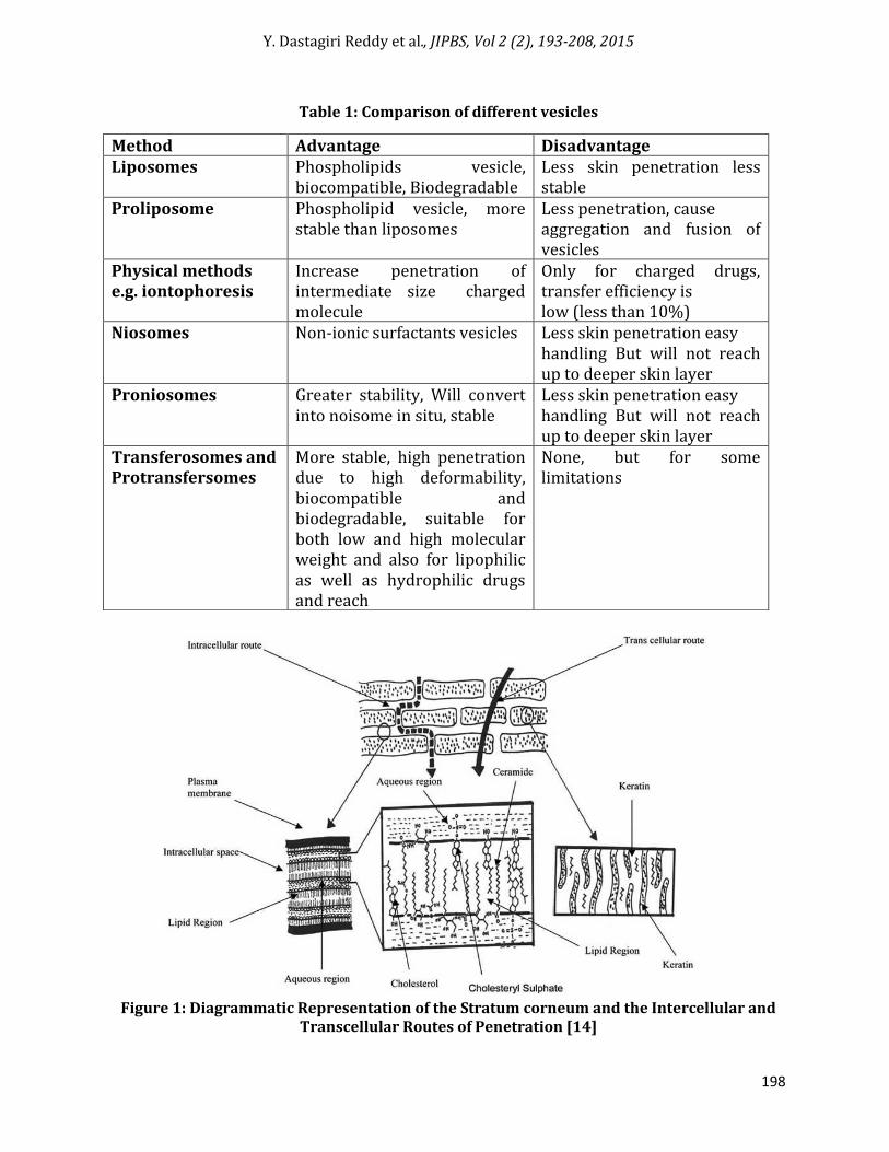
Table 1: Comparison of different vesicles
Figure 1: Diagrammatic Representation of the Stratum corneum and the Intercellular and
Transcellular Routes of Penetration [14]
Method Advantage Disadvantage Liposomes Phospholipids vesicle,
biocompatible, Biodegradable Less skin penetration less stable
Proliposome Phospholipid vesicle, more stable than liposomes
Less penetration, cause aggregation and fusion of vesicles
Physical methods e.g. iontophoresis
Increase penetration of intermediate size charged molecule
Only for charged drugs, transfer efficiency is low (less than 10%)
Niosomes Non-ionic surfactants vesicles Less skin penetration easy handling But will not reach up to deeper skin layer
Proniosomes Greater stability, Will convert into noisome in situ, stable
Less skin penetration easy handling But will not reach up to deeper skin layer
Transferosomes and Protransfersomes
More stable, high penetration due to high deformability, biocompatible and biodegradable, suitable for both low and high molecular weight and also for lipophilic as well as hydrophilic drugs and reach
None, but for some limitations
Y. Dastagiri Reddy et al., JIPBS, Vol 2 (2), 193-208, 2015
199
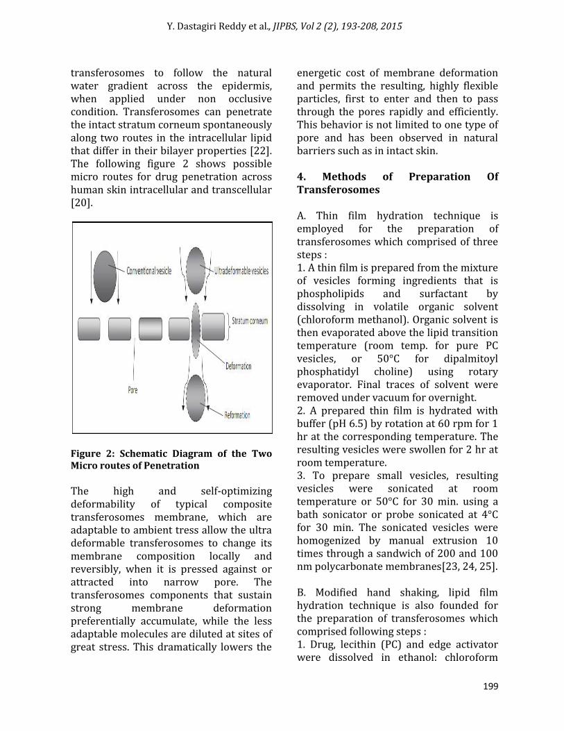
transferosomes to follow the natural water gradient across the epidermis, when applied under non occlusive condition. Transferosomes can penetrate the intact stratum corneum spontaneously along two routes in the intracellular lipid that differ in their bilayer properties [22]. The following figure 2 shows possible micro routes for drug penetration across human skin intracellular and transcellular [20].
Figure 2: Schematic Diagram of the Two Micro routes of Penetration The high and self-optimizing deformability of typical composite transferosomes membrane, which are adaptable to ambient tress allow the ultra deformable transferosomes to change its membrane composition locally and reversibly, when it is pressed against or attracted into narrow pore. The transferosomes components that sustain strong membrane deformation preferentially accumulate, while the less adaptable molecules are diluted at sites of great stress. This dramatically lowers the
energetic cost of membrane deformation and permits the resulting, highly flexible particles, first to enter and then to pass through the pores rapidly and efficiently. This behavior is not limited to one type of pore and has been observed in natural barriers such as in intact skin. 4. Methods of Preparation Of Transferosomes A. Thin film hydration technique is employed for the preparation of transferosomes which comprised of three steps : 1. A thin film is prepared from the mixture of vesicles forming ingredients that is phospholipids and surfactant by dissolving in volatile organic solvent (chloroform methanol). Organic solvent is then evaporated above the lipid transition temperature (room temp. for pure PC vesicles, or 50°C for dipalmitoyl phosphatidyl choline) using rotary evaporator. Final traces of solvent were removed under vacuum for overnight. 2. A prepared thin film is hydrated with buffer (pH 6.5) by rotation at 60 rpm for 1 hr at the corresponding temperature. The resulting vesicles were swollen for 2 hr at room temperature. 3. To prepare small vesicles, resulting vesicles were sonicated at room temperature or 50°C for 30 min. using a bath sonicator or probe sonicated at 4°C for 30 min. The sonicated vesicles were homogenized by manual extrusion 10 times through a sandwich of 200 and 100 nm polycarbonate membranes[23, 24, 25]. B. Modified hand shaking, lipid film hydration technique is also founded for the preparation of transferosomes which comprised following steps : 1. Drug, lecithin (PC) and edge activator were dissolved in ethanol: chloroform
Y. Dastagiri Reddy et al., JIPBS, Vol 2 (2), 193-208, 2015
200
(1:1) mixture. Organic solvent was removed by evaporation while hand shaking above lipid transition temperature (43°C). A thin lipid film was formed inside the flask wall with rotation. The thin film was kept overnight for complete evaporation of solvent. 2. The film was then hydrated with phosphate buffer (pH 7.4) with gentle shaking for 15 minute at corresponding temperature. The transfersome suspension further hydrated up to 1 hour at 2-8°C. [23, 26, 27]. C. In short, transferosomes were prepared by mixing an ethanolic SPC solution with the appropriate amount of an edge-active molecule, such as sodium cholate. One kind of resulting charged suspension contained 8.7 wt% SPC and 1.3 wt% cholate as well as approx. 8.5 vol% ethanol, unless stated otherwise. Other biocompatible surfactants were used with triethanolamine–HCl buffer to yield total lipid concentration of 10 wt%. The resulting suspension was sonicated, frozen, and thawed 2–3 times. And brought to the desired size as measured by means of photon correlation spectroscopy. By ultrasonication or intermediate-pressure homogenization. Vesicle suspension was ultimately sterilized by filtration through a 0.2 mm micro-porous filter Poretics, CA, USA. The final vesicle size was confirmed by the dynamic light scattering in different suspensions. The suspension was subsequently mixed with triethanolamine HCl buffer to yield total lipid concentration of 10 wt%. The resulting suspension was sonicated, frozen, and thawed 2–3 times. And brought to the desired size as measured by means of photon correlation spectroscopy. By ultrasonication or intermediate-pressure homogenization. Vesicle suspension was ultimately sterilized by filtration through
a 0.2 mm micro-porous filter Poretics, CA, USA. The final vesicle size was confirmed by the dynamic light scattering. D. Preparation of drug-Loaded Liposomes, Transferosomes, and Suspensions. Liposomes containing a controlled amount of PC and various amounts of drug were formulated. The drug concentration was varied from 2.5 to 70.0 wt. % of the PC. The sonication method was used to prepare different formulations; they were composed of bilayer-forming PC and either Chol, NaO, NaChol, or DCP in a molar ratio of 10: 2. The PC, Chol, NaO, NaChol, DCP, and drug were each briefly dissolved in chloroform: methanol (2: 1 v/v).In preparing drug-loaded liposomes and transferosomes, the materials were deposited in a test tube, and the solvent was evaporated with nitrogen gas. The lipid film was placed in a desiccator connected to a vacuum pump for a minimum of 6 hrs to remove the remaining organic solvent. The dried lipid film was hydrated with tris buffer. Following hydration, the dispersion was sonicated in a bath for 30 min and then probe-sonicated for 2 cycles of 30 min. E. Soya-phosphatidylcholine was taken in a round bottom flask. Span 80 or Tween 80 was put in the same round bottom flask. Ethanol was then added to the same flask. The drug was also loaded in the same RBM. These were then dissolved by shaking. Thin film was then formed by keeping it in the rotatory vaccum evaporator at 60C. This thin film was then hydrated by phosphate buffer saline to get the transferosomes [8]. F. The transferosomes were formulated by the conventional rotary evaporation sonication method [7, 8]. Transferosomes containing phospholipids (soya lecithin), surfactant (span 80), and the drug
Y. Dastagiri Reddy et al., JIPBS, Vol 2 (2), 193-208, 2015
201
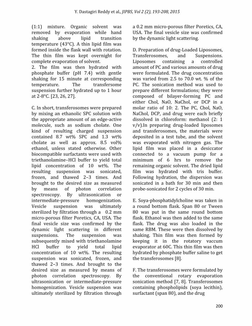
Class Example Uses Phospholipids Soya phosphatidyl choline, Dipalmitoyl
phosphatidyl choline, Distearoyl phoshatidyl choline
Vesicles forming component
Surfactant Sodium Cholate, Sodium deoxycholate, tween-80, Span-80
For providing flexibility
Alcohol Ethanol, methanol As a solvent Buffering agent Saline phosphate buffer (pH 6.4) As a hydrating medium Dye Rhodamine-123, Rhodamine-DHPE,
Fluorescein-DHPE Nilered For CSLM study
Table 2: Different additives used in formulation of transferosomes
(sertraline) were formulated. The drug concentration varied from 0.5 to 1.8 wt. % and the surfactant concentration varied from 5 to 25 wt % of the phospholipids. The phospholipids and surfactant were taken in a clean, dry, round-bottom flask and this lipid mixture was dissolved in a small quantity of ethanol. The organic solvent was removed by rotary evaporation under reduced pressure at 40°C. Final traces of solvents were removed under vacuum overnight. The deposited lipid film was hydrated with 7 % v/v ethanol i.e. solution of the drug by rotation at 60 rpm for 1 hr. The resulting vesicles were kept for 2 hrs at ambient temperature to swell and form into large multi lamellar vesicles. These were sonicated in a bath for 10 min to achieve smaller vesicles. The varied amounts of the drug were added (0.5 to 1.8 %) to determine the drug-loading capacity of transferosomes. All of the drug-loaded vesicular formulations were examined for maximum entrapment efficiency and for the appearance of drug crystals over a period of 14 days using an optical microscope. Table 2 shows the Different additives used in formulation of Transferosomes. 5. Optimization of Formulation Containing Transferosomes
There are various process variables which could affect the preparation and properties of the transferosomes. The preparation procedure was accordingly optimized and validated. The process variables are depending upon the procedure involved for manufacturing of formulation. The preparation of transferosomes involves various process variables such as, 1. Lecithin: surfactant ratio 2. Effect of various solvents 3. Effect of various surfactants 4. Hydration medium Optimization was done by selecting entrapment efficiency of drug. During the preparation of a particular system, the other variables were kept constant [28, 29]. 6. Characterization of Transferosomes The characterization of transferosomes is generally similar to liposomes, niosomes and micelles [30]. Following characterization parameters have to be checked for transferosomes. 1. Vesicle size distribution and zeta potential Vesicle size, size distribution and zeta potential were determined by Dynamic Light Scattering system by Malvern Zetasizer [31, 32].
Y. Dastagiri Reddy et al., JIPBS, Vol 2 (2), 193-208, 2015
202
2. Vesicle morphology Vesicle diameter can be determined using photon correlation spectroscopy or dynamic light scattering (DLS) method. Samples were prepared in distilled water, filtered through a 0.2 mm membrane filter and diluted with filtered saline and then size measurement done by using photon correlation spectroscopy or dynamic light scattering (DLS) measurements. Transferosomes vesicles can be visualized by TEM, phase contrast microscopy, etc. The stability of vesicle can be determined by assessing the size and structure of vesicles over time. Mean size is measured by DLS and structural changes are observed by TEM [31, 32]. 3. Number of vesicles per cubic mm This is an important parameter for optimizing the composition and other process variables. Non sonicated transferosomes formulations are diluted five times with 0.9% sodium chloride solution. Haemocytometer and optical microscope can then be used for further study [33]. The Transferosomes in80 small squares are counted and calculated using the following formula: Total number of Transferosomes per cubic mm= (Total number of Transferosomes counted × dilution factor × 4000) / Total number of square counted. 4. Entrapment efficiency The entrapment efficiency is expressed as the percentage entrapment of the drug added. Entrapment efficiency was determined by first separation of the un-entrapped drug by use of mini-column centrifugation method. After centrifugation, the vesicles were disrupted using 0.1% Triton X-100 or 50% n-propanol [32]. The entrapment efficiency is expressed as: Entrapment efficiency = (Amount entrapped / Total amount added) ×100
5. Drug content The drug content can be determined using one of the instrumental analytical methods such as modified high performance liquid chromatography method (HPLC) method using a UV detector, column oven, auto sample, pump, and computerized analysis program depending upon the analytical method of the Pharmacopoeial drug [36]. 6. Turbidity measurement Turbidity of drug in aqueous solution can be measured using nephelometer [32]. 7. Degree of deformability or permeability measurement In the case of transferosomes, the permeability study is one of the important and unique parameter for characterization. The deformability study is done against the pure water as standard. Transferosomes preparation is passed through a large number of pores of known size (through a sandwich of different microporous filters, with pore diameter between 50 nm and 400 nm, depending on the starting transferosomes suspension). Particle size and size distributions are noted after each pass by dynamic light scattering (DLS) measurements [32, 37]. 8. Penetration ability Penetration ability of Transferosomes can be evaluated using fluorescence microscopy [33, 37]. 9. Occlusion effect Occlusion of skin is considered to be helpful for permeation of drug in case of traditional topical preparations. But the same proves to be detrimental for elastic vesicles. Hydrotaxis (movement in the direction) of water is the major driving force for permeation of vesicles through the skin, from its relatively dry surface to
Y. Dastagiri Reddy et al., JIPBS, Vol 2 (2), 193-208, 2015
203
water rich deeper regions. Occlusion affects hydration forces as it prevents evaporation of water from skin [32]. 10. Surface charge and charge density Surface charge and charge density of Transferosomes can be determined using zetasizer [32, 33]. 11. In-vitro drug release In vitro drug release study is performed for determining the permeation rate. Time needed to attain steady state permeation and the permeation flux at steady state and the information from invitro studies are used to optimize the formulation before more expensive in vivo studies are performed. For determining drug release, transferosomes suspension is incubated at 320C and samples are taken at different times and the free drug is separated by mini column centrifugation. The amount of drug released is then calculated indirectly from the amount of drug entrapped at zero times as the initial amount (100% entrapped and 0% released) [32, 33]. 12. In-vitro Skin permeation Studies Modified Franz diffusion cell with a receiver compartment volume of 50ml and effective diffusion area of 2.50 cm2 was used for this study. In vitro drug study was performed by using goat skin in phosphate buffer solution (pH 7.4). Fresh Abdominal skin of goat were collected from slaughter house and used in the permeation experiments. Abdominal skin hairs were removed and the skin was hydrated in normal saline solution. The adipose tissue layer of the skin was removed by rubbing with a cotton swab. Skin was kept in isopropyl alcohol solution and stored at 0-40˚C. To perform skin permeation study, treated skin was mounted horizontally on the receptor compartment with the stratum corneum
side facing upwards towards the donor compartment of Franz diffusion cell. The effective permeation area of donor compartment exposed to receptor compartment was 2.50cm2 and capacity of receptor compartment was 50ml. The receptor compartment was filled with 50ml of phosphate buffer (pH 7.4) saline maintained at 37 ± 0.5˚C and stirred by a magnetic bar at 100RPM. Formulation (equivalent to 10mg drug) was placed on the skin and the top of the diffusion cell was covered. At appropriate time intervals 1 ml aliquots of the receptor medium were withdrawn and immediately replaced by an equal volume of fresh phosphate buffers (pH 7.4) to maintain sink conditions. Correction factors for each aliquot were considered in calculation of release profile. The samples were analyzed by any instrumental analytical technique [34, 35]. 13. Physical stability The initial percentage of the drug entrapped in the formulation was determined and were stored in sealed glass ampoules. The ampoules were placed at 4 ± 20C (refrigeration), 25 ± 20C (room temp), and 37 ± 20C (body temp) for at least 3 months. Samples from each ampoule were analyzed after 30 days to determine drug leakage. Percent drug lose was calculated by keeping the initial entrapment of drug as 100% [36, 37]. 7. Applications of Transferosomes [4,9] Transferosomes as drug delivery systems have the potential for providing controlled release of the administered drug and increasing the stability of labile drugs. Delivery of Insulin Very large molecules incapable of diffusing into skin as such can be transported across the skin with the help of Transferosomes. For example, insulin,
Y. Dastagiri Reddy et al., JIPBS, Vol 2 (2), 193-208, 2015
204
interferon can be delivered through mammalian skin. Delivery of insulin by Transferosomes is the successful means of noninvasive therapeutic use of such large molecular weight drugs on the skin. Insulin is generally administered by subcutaneous route that is inconvenient. Encapsulation of insulin into transferosomes (transferulin) overcomes the problems of inconvenience, larger size (making it unsuitable for transdermal delivery using conventional method) along with showing 50% response as compared to subcutaneous injection. Carrier for Interferons & Interleukin: Transferosomes have also been used as a carrier for interferons like leukocytic derived interferon-α (INF-α)) is a naturally occurring protein having antiviral, antiproliferative and some immune modulatory effects. Transferosomes as drug delivery systems have the potential for providing controlled release of the administered drug and increasing the stability of labile drugs. The formulation of interleukin-2 and interferone-α containing transferosomes for potential transdermal application. They reported delivery of IL-2 and INF- α trapped by Transferosomes in sufficient concentration for immunotherapy. Carrier for Other Proteins & Peptides Transferosomes have been widely used as a carrier for the transport of other proteins and peptides. Proteins and peptides are large biogenic molecules which are very difficult to transport into the body, when given orally they are completely degraded in the GI tract and transdermal delivery suffers because of their large size. These are the reasons why these peptides and proteins still have to be introduced into the body through injections. Various approaches have been developed to improve these situations.
The bioavailability obtained from transferosomes is somewhat similar to that resulting from subcutaneous injection of the same protein suspension. Human serum albumin or gap junction protein was found to be effective in producing the immune response when delivered by transdermal route encapsulated in Transferosomes. Transport of certain drug molecules that have physicochemical which otherwise prevent them from diffusing across stratum corneum can be transported. Peripheral Drug Targeting The ability of transferosomes to target peripheral subcutaneous tissues is due to minimum carrier associated drug clearance through blood vessels in the subcutaneous tissue. These blood vessels are non-fenestrated and also possess tight junctions between endothelial cells thus not allowing vesicles to enter directly into the blood stream. This automatically increases drug concentration locally along with the probability of drug to enter peripheral tissues. Transdermal Immunization Since ultra deformable vesicles have the capability of delivering the large molecules, they can be used to deliver vaccines topically. Transferosomes containing proteins like integral membrane protein, human serum albumin, gap junction protein are used for this purpose. Advantages of this approach are injecting the protein can be avoided and higher IgA levels are attained. Transcutaneous hepatitis-B vaccine has given good results. A 12 times higher AUC was obtained for zidovudine as compared to normal control administration. Selectivity in deposition in RES (which is the usual site for residence of HIV) was also increased.
Y. Dastagiri Reddy et al., JIPBS, Vol 2 (2), 193-208, 2015
205
Delivery of NSAIDS NSAIDS are associated with number of GI side effects. These can be overcome by transdermal delivery using ultra deformable vesicles. Studies have been carried out on Diclofenac and Ketotifen. Ketoprofen in a Transferosomes formulation gained marketing approval by the Swiss regulatory agency the product is expected to be marketed under the trademark Diractin. Further therapeutic products based on the transferosomes technology, according to IDEA AG, are in clinical development. Delivery of steroidal hormones and peptides Transferosomes have also used for the delivery of corticosteroids. Transferosomes improves the site specificity and overall drug safety of corticosteroid delivery into skin by optimizing the epicutaneously administered drug dose. Transferosomes based corticosteroids are biologically active at dose several times lower than the currently used formulation for the treatment of skin diseases. Flexible vesicles of ethinyl estradiol showed significant anti-ovulatory effects as compared to plain drug given orally and traditional liposomes given topically. Extensive work has been done on other drugs like hormones and peptides viz Estradiol, low molecular-weight Heparin, Retinol, Melatonin, etc. Delivery of Anaesthetics Transferosomes based formulations of local anesthetics- lidocaine and tetracaine showed permeation equivalent to subcutaneous injections. Maximum resulting pain insensitivity is nearly as strong (80%) as that of a comparable subcutaneous bolus injection, but the effect of transfersomal anesthetics last longer.
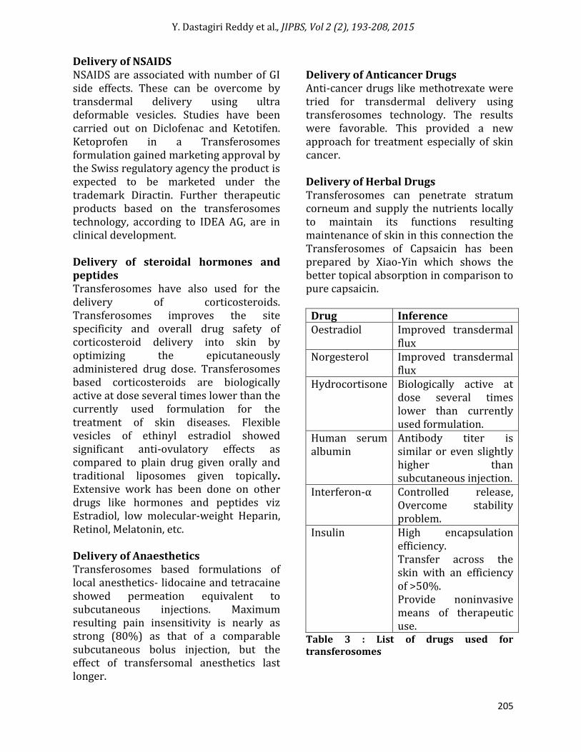
Delivery of Anticancer Drugs Anti-cancer drugs like methotrexate were tried for transdermal delivery using transferosomes technology. The results were favorable. This provided a new approach for treatment especially of skin cancer. Delivery of Herbal Drugs Transferosomes can penetrate stratum corneum and supply the nutrients locally to maintain its functions resulting maintenance of skin in this connection the Transferosomes of Capsaicin has been prepared by Xiao-Yin which shows the better topical absorption in comparison to pure capsaicin.
Drug Inference Oestradiol Improved transdermal
flux Norgesterol Improved transdermal
flux Hydrocortisone Biologically active at
dose several times lower than currently used formulation.
Human serum albumin
Antibody titer is similar or even slightly higher than subcutaneous injection.
Interferon-α Controlled release, Overcome stability problem.
Insulin High encapsulation efficiency. Transfer across the skin with an efficiency of >50%. Provide noninvasive means of therapeutic use.
Table 3 : List of drugs used for transferosomes
Y. Dastagiri Reddy et al., JIPBS, Vol 2 (2), 193-208, 2015
206
8. Conclusion Transdermal route of drug delivery does not allow transport of high mol.wt therapeutic agents and drugs because of the barrier properties of the stratum corneum layer of the skin. These Transferosomes are specially designed vesicles capable of responding to external stress by squeezing themselves through skin pores that are many times narrower than they are leading to increased transdermal flux of therapeutic agents. Transferosomes have beneficial advantages over other vesicular systems such as their high penetration power across skin, higher stability, systemic drug release possible and higher deformability than other vesicular systems such as niosomes, ethosomes etc., These will ensures reproducible and efficient transcutaneous carrier and drug transport. Transferosomes possess an infrastructure consisting of hydrophobic and hydrophilic moieties together and as a result can accommodate drug molecules with wide range of solubility. References 1. Irfan M, Verma S, Ram A. Preparation and
characterization of ibuprofen loaded transferosomes as a novel carrier for transdermal drug delivery system. Asian j Pharm. clin resear. 2012. (162-165).
2. Trommer H, Neubert RHH: Overcoming the stratum corneum. The modulation of skin penetration. A review, Skin Pharmacology and Physiology. 2006. (106-121).
3. El Zaafarany GM, Awad GAS, Holayel SM, Mortada ND. Role of edge activators and surface charge in developing ultra deformable vesicles with enhanced skin delivery. Int J Pharm. 2010. (164-172).
4. Cevc G, Grbauer D, Schatzlein A, Blume G. Ultraflexible vesicles, transferosomes, have an extremely low pore penetration resistance ant transport therapeutic amounts of insulin across the intact
mammalian skin. Biochem Biophys Act. 1998. (201-215)
5. El-Maghraby GM, Williams AC. Vesicular systems for delivering conventional small organic molecules and larger macromolecules to and through human skin. Expert Opin Drug Deliv. 2009. (149-163).
6. Walve JR, Bakliwal SR, Rane BR, Pawar SP. Transferosomes: A surrogated carrier for transdermal drug delivery system. Int J App Bio Pharm Tech. 2011.(201-214)
7. Cevc G, Blume G. Lipid vesicles penetrate into intact skin owing to transdermal osmotic gradient and hydration force. Biochem Biophys Act 1992.(226-332).
8. Jain NK. Advances in Controlled and Novel Drug Delivery. CBS Publishers and Distributers First edition2001. New Delhi.(426-451).
9. Paul, A, Cevec G. Non-invasive Administration of Protein Antigens. Epicutaneous with Bovin Serum Albumine, Vaccine Research, 1998.(145-164).
10. Modi CD, Bharadia PD. “Transferosomes: New Dominants for Transdermal Drug Delivery”. Am. J.PharmTech Res., 2012.(71-91)
11. Prajapati ST, Patel CG, Patel CN. “Transferosomes: A Vesicular Carrier System for Transdermal Drug Delivery”. Asian Journal of Biochemical and Pharmaceutical Research, 2011.(507-524).
12. Kombath RV, Minimal SK, Sockalingam A, Subadhra S, Parre S, Reddy TR, David B. “Critical issues related to transferosomes – novel Vesicular system”. Act Sci.(67-82)
13. Walve JR, Bakliwal SR, Rane BR, Pawar SP. “Transferosomes: A surrogated carrier for transdermal drug delivery system”. International Journal of Applied Biology and Pharmaceutical Technology, 2011.Pol. Technol. Aliment, 2012.(204-213).
14. Heather AE, Benson. Transdermal drug delivery: penetration enhancement technique current drug delivery, 2005.(23-33).
15. Cevc G, Blume G., Schatzlein A. Transferosomes-mediated transepidermal
Y. Dastagiri Reddy et al., JIPBS, Vol 2 (2), 193-208, 2015
207
delivery improves the region specificity and biological activity of corticosteroids in vivo. Journal of Controlled Release 1997.(211-226).
16. Rand RP and Parsegian VA: Biochem. Biophys Acta.1989.(351-377).
17. Gompper G and Kroll DM: Driven transport of fluid vesicles through narrow pores Physical Reviews 1995.(4198-4211).
18. Chapman SJ and Walsh A: Arch. Dermatol Res 1998.(304-310).
19. N. k Jain. Advances in Controlled and Novel Drug Delivery. CBS Publishers and Distributers First edition2001. New Delhi.(426-451).
20. Panchagnula R. “Transdermal delivery of drugs”. Indian Journal Pharmacology, 1997.(140-156).
21. Cevc G. “Isothermal lipid phase”, Transitions Chemistry and Physics of Lipids, 1991.(293-299).
22. Schatzlein A and Cevc G. Skin penetration by phospholipids vesicles, Transferosomes as visualized by means of the Confocal Scanning Laser Microscopy, in characterization, metabolism, and novel biological application 1995.(191-209).
23. Modi CD, Bharadia PD. “Transferosomes: New Dominants for Transdermal Drug Delivery”. Am. J.PharmTech Res., 2012.(71-91).
24. Pandey S, Goyani M, Devmurari V, Fakir J. Transferosomes: A Novel Approach for Transdermal Drug Delivery”. Der Pharmacia Letter, 2009.(143-150).
25. Jain NK. Advances in Controlled and Novel Drug Delivery. CBS Publishers and Distributers First edition. New Delhi, 2001.(426-451)
26. Jain CP, Vyas SP, Dixit VK. “Niosomal system for delivery of rifampicin to lymphatics”. Int J Pharma, 2006.(57-58).
27. Elsayed MMS, Abdallah OY, Nagar VF. Deformable liposomes and ethosomes: Mechanism of enhanced skin delivery. Int J Pharma 2006.(60-66).
28. Patel R, Singh SK, Singh S, Sheth NR, Gendle R. “Development and Characterization of Curcumin Loaded
Transfersome for Transdermal Delivery”. J. Pharm Sci. Res., 2009.(71-80)
29. Sheo DM, Shweta A, Vijay KT, Ram CD, Aklavya S, Ghanshyam M. “Enhanced Transdermal delivery of indinavir sulfate via transferosomes”. Pharmacie Globale (IJCP), 2010.(1-7)
30. Panchagnula R. “Transdermal delivery of drugs”. Indian Journal Pharmacology, 1997.(140-156)
31. Modi CD, Bharadia PD. “Transferosomes: New Dominants for Transdermal Drug Delivery”. Am. J.PharmTech Res., 2012.(71-91).
32. Prajapati ST, Patel CG, Patel CN.“Transferosomes: A Vesicular Carrier System for Transdermal Drug Delivery”. Asian Journal of Biochemical and Pharmaceutical Research, 2011.(507-524).
33. Walve JR, Bakliwal SR, Rane BR, Pawar SP. “Transferosomes: A surrogated carrier for transdermal drug delivery system”. International Journal of Applied Biology and Pharmaceutical Technology, 2011.(201-214).
34. Patel R, Singh SK, Singh S, Sheth NR, Gendle R. “Development and Characterization of Curcumin Loaded Transfersome for Transdermal Delivery”. J. Pharm Sci. Res., 2009.(71-80)
35. Sheo DM, Shweta A, Vijay KT, Ram CD, Aklavya S, Ghanshyam M. “Enhanced Transdermal delivery of indinavir sulfate via transferosomes”. Pharmacie Globale (IJCP), 2010.(1-7)
36. Sheo DM, Shweta A, Ram CD, Ghanshyam M, Girish K, Sunil KP. “Transferosomes- a Novel Vesicular Carrier for Enhanced Transdermal Delivery of Stavudine: Development, Characterization and Performance Evaluation”. J. Scientific Speculations and Res., 2010.(30-36).
37. Pandey S, Goyani M, Devmurari V, Fakir J. “Transferosomes: A Novel Approach for Transdermal Drug Delivery”. Der Pharmacia Letter, 2009.(1-7)
38. Jalon GE. Ygartua P, Santoyo S. “Topical application of acyclovir-loaded microparticles: quantification of the drug
Y. Dastagiri Reddy et al., JIPBS, Vol 2 (2), 193-208, 2015
208
in porcine skin layers”. J. Control Release, 2001.(191-197).
39. Maghraby EI, Williams GM, Barry BW, “Skin delivery of oestradiol from lipid vesicles: importance of liposome structure”. Int. J. Pharma, 2000. (59-69).
40. Trotta M, Peira E, Carlotti ME, Gallarate M. “Deformable liposomes for dermal administration of methotrexate”. Int. J. Pharma, 2004. (270,119).