Embed Size (px)
Citation preview
Journal of Colloid and Interface Science 490 (2017) 452–461
Contents lists available at ScienceDirect
Journal of Colloid and Interface Science
journal homepage: www.elsevier .com/locate / jc is
Regular Article
Biopolymer matrix for nano-encapsulation of urease – A model proteinand its application in urea detection
http://dx.doi.org/10.1016/j.jcis.2016.11.0300021-9797/� 2016 Elsevier Inc. All rights reserved.
⇑ Corresponding author at: Amity Institute of Biotechnology, Amity UniversityUttar Pradesh, Noida 201301, UP, India.
E-mail addresses: [email protected] (S. Kumar), [email protected](I.R. Epstein), [email protected] (R. Sahney).
Abhishek Saxena a, Arpita Bhattacharya b, Satish Kumar c, Irving R. Epstein d, Rachana Sahney a,⇑aAmity Institute of Biotechnology, AUUP, Noida 201301, IndiabAmity Institute of Nanotechnology, AUUP, Noida 201301, Indiac St. Stephens College, Delhi University, New Delhi 110007, IndiadDepartment of Chemistry, Brandeis University, 415 South Street, MS 015, Waltham, MA 02453, United States
g r a p h i c a l a b s t r a c t
a r t i c l e i n f o
Article history:Received 7 September 2016Revised 7 November 2016Accepted 8 November 2016Available online 9 November 2016
Keywords:NanoparticlesW/O nanoemulsionsUreaseAlginate nanogels
a b s t r a c t
Alginate microparticles and nanoparticles crosslinked with Ca+2 ions are frequently employed in biomed-ical applications. Here we use microemulsion polymerization to prepare alginate nanoparticles (nano-gels) using different crosslinking ions (Ca+2, Sr+2, Ba+2) to encapsulate a model protein, urease enzyme(jackbeans). With alginate concentrations of 0.2 wt% in the aqueous phase, emulsion droplets showedgood stability and narrow, monomodal distributions with radii �65 ± 10 nm. The size of the nanogel var-ies with the crosslinking cation and its affinity for the mannuronate and guluronate units in the linearalginate chain. The nanogels were further characterized using dynamic light scattering, scanning electronmicroscopy, energy dispersive X-ray spectrometry and zeta potential. This work demonstrates the poten-tial application of Ba-alginate as an alternative matrix for nano-encapsulation of proteins and its use forbiomedical applications.
� 2016 Elsevier Inc. All rights reserved.
1. Introduction
Alginate is a linear biopolymer obtained from brown algae. Itcontains b-d-mannuronate (M) and a-l-guluronate (G) residueslinearly linked by 1,4-glycosidic linkages in varying proportions,sequences, and molecular weights [1]. The notable biomedical
A. Saxena et al. / Journal of Colloid and Interface Science 490 (2017) 452–461 453
applications of natural polysaccharides in wound dressings [2], cellencapsulation [3–5], drug delivery [6], catalyst support [7], andbiosensors [8,9] result from the ease of forming heat-stable gelsby ionic cross-linking with multivalent cations, user-friendlypreparation methods, the possibility to encapsulate various sensi-tive molecules (synthetic or biomolecules), and the robustness ofproduction under ambient physiological conditions [10]. Thepreparation, properties and application of calcium alginate micro-gels are well documented [11], but stability studies on calciumalginate microgel particles indicate a number of drawbacks, suchas instability in physiological liquids [12] and poor thermal stabil-ity, as well as fractures and cavities (in homogeneous porous net-works) and poor mechanical resistance due to decreasing gelstrength from the surface to the core [13,14]. These microgels can-not be used for immobilization of biomolecules with molecularweights less than 300 kDa due to high leakage of biomolecules[15]. Thus, lowmechanical strength of the gel structure and seriousswelling properties greatly limit applications. Many literaturereports describe the biological application of Ba-alginate microbe-ads, which are effective in a variety of applications [3–5]. Thus, inaddition to calcium, other divalent cations are candidates as cross-links for the development of more suitable alginate gels for differ-ent applications.
The use of alginate in nanoparticles (nanogels) is less commonthan that of synthetic polymers like poly(lactic acid) (PLA), poly(glycolic acid) (PGA) and poly(lactide-co-glycolide) (PLGA). Prepa-ration of Ca-alginate nanogels for the development of nano-sizeddrug delivery systems has been reported [6,11], largely based ontwo methods [16]: (a) Complexation, where nanoparticles can beprepared by self-assembly and complexing alginate with highlycharged polycations, such as chitosan or poly l-lysine [17]. Theinclusion of polycations, however, limits versatility. Yu et al. [18]have reported nanosized aggregates with four different morpholo-gies, nanosphere, vesicle, nanoparticle and nanorod, preparedunder very mild conditions through self-assembly of alginate poly-mer with Ca+2 cations, a process that requires almost a week toobtain calcium alginate nanogels. However, solid nanoparticlescould not be obtained through this method. (b) Alginate-in-oil(W/O) emulsion systems have been used to entrap water-insoluble drug entities in calcium alginate nanoparticles, wheredevelopment of nanoemulsions requires a large input of mechani-cal energy. Shearing, ultrasonication and solvent exchange proce-dures have been employed to attain a sufficient increase in thesurface-to-volume ratio [19]. However, such harsh treatmentmay seriously damage sensitive biomacromolecules like enzymesor antibodies in comparison to milder methods. The latter, how-ever, produce particle sizes above the desired range as well as highpolydispersity [20]. Recently, Machado et al. [21] proposed a phaseinversion temperature (PIT) emulsification technique, but this isunsuitable for encapsulation of temperature-sensitive biomole-cules like proteins.
We use urease enzyme as a model protein to study the widelyused sol-gel emulsification polymerization method of proteinencapsulation, as it gives a highly selective response to the biolog-ically important molecule urea, which is of great interest (i) in clin-ical diagnosis, since it is the primary reliable index of renalfunction [22]; (ii) in environmental analysis, since it is used as anitrogenous fertilizer that causes environmental stress [23]; and(iii) in the detection of adulteration in dairy milk or in assessingthe nutritional program of lactating dairy cows [24]. Ureaseenzyme-based analytical systems are used for the monitoring ofurea in these fields when the critical issue is maintaining the sta-bility, activity, and function of the enzyme as close as possible toits native state [25]. The application of immobilized enzymes ispreferred due to their ease of handling, prolonged availability,robustness, increased resistance to environmental changes and
reusability [26]. The characteristics of immobilized enzymes arecontrolled by the properties of both the enzyme and the supportmatrix. The present study is designed to prepare alginate nanogelsusing various divalent ions- (Ca+2, Ba+2 and Sr+2) -as crosslinks andto compare the effects of these crosslinks on the morphology, sur-face charge, protein encapsulation properties and stability of theresulting nanogels. The success of urease immobilization wasdetermined by the study of various immobilization parameters,i.e., enzyme loading and specific enzyme activity over a period offour weeks. We further studied the application of encapsulatedurease for urea measurement in clinical blood serum samplesand compared the results with the those obtained from the recom-mended clinical method of blood urea nitrogen (BUN) analysisusing an auto analyser.
2. Experimental
2.1. Materials
Sodium alginate (mol. wt. 120,000–190,000), urease and hexanewere purchased from Sigma-Aldrich. Polyoxyethylene sorbitanmono-oleate (Tween 80), calcium chloride dihydrate (CaCl2�2H2O,p.a.), sodium chloride, Nessler’s reagent and urea were suppliedby Fisher Scientific. Other chemicals like strontium chloride, bar-ium chloride, Tris-acetate and sodium acetate were obtained fromQualigens India. All chemicals were used as received. Double-distilled water was used throughout the experiments.
2.2. Preparation of alginate nanoparticles (alginate nanogels) usingCa+2/Sr+2/Ba+2 crosslinkers and encapsulation of urease
The sodium alginate solution (0.2%) was prepared in Tris-acetate-saline buffer (100 mM, pH 7.2) at 4 �C. This solution wasadded dropwise to a vial containing the organic phase (hex-ane = 20.44 g) and Tween 80 under constant stirring at 500 rpm.Various phases appeared (transparent to turbid) during the processof adding alginate solution at different surfactant concentrations.For each distinct phase, the mixture was further stirred for30 min, and 10 mL of filtered solutions containing divalent cations(CaCl2, BaCl2 or SrCl2, 100 mM) were added dropwise, which led tocrosslinking of the polymer chains. After careful washing withdeionized water to remove surfactant and hexane, the vial wascentrifuged at 3000 g for 30 min, and small white pellets of algi-nate nanogel were obtained. The pellets were re-suspended inTris-acetate-saline buffer for successive studies. For the encapsula-tion of urease into the alginate nanogels, the procedure was sameas described above except that urease was pre-mixed with the 0.2%alginate solution.
2.3. Characterization
2.3.1. Size determination using dynamic light scattering (DLS)A 1 mL aliquot of the sample (emulsion and/or particle suspen-
sion) was placed in a quartz cuvette and analyzed using DLS toestimate the mean hydrodynamic diameter and the polydispersityindex (PDI). The experiments were performed on a Malvern Zeta-sizer - Nano ZS (Malvern, UK) instrument with backscatter detec-tion (173�), controlled by Dispersion Technology Software (DTS5.03, Malvern, UK). Mean hydrodynamic radii and intensity-averaged size distributions were obtained from the raw data usingthe general-purpose inverse Laplace transformmethod provided inthe instrument software. The PDIs were estimated from cumulantanalysis, which is also provided with the instrument software.
454 A. Saxena et al. / Journal of Colloid and Interface Science 490 (2017) 452–461
2.3.2. SEM-EDX measurementThe surface morphologies (shape and formation of aggregates)
and sizes of nanoparticle formulations of alginate nanoparticleswere studied using scanning electron microscopy (SEM). Specimenpreparation was performed as follows: the lyophilized nanogelswere first suspended in ethanol. The suspension thus obtainedwas mounted on stubs and sputter-coated with gold. Micrographswere taken on a SEM instrument (Model S-4800 microscope, Hita-chi, Tokyo, Japan). The nanogels mounted on stubs were furtherused to analyze the presence of metal cations by using an electronprobe X-ray microanalyzer in energy dispersive X-ray spectrome-try (EDX) mode.
2.3.3. FT-IR spectral studySamples of sodium alginate, urease and encapsulated urease in
various alginate nanogels cross-linked with Ca+2, Sr+2 and Ba+2
were lyophilized before performing FT-IR analysis. The FT-IR spec-tra were recorded on a Perkin-Elmer spectrometer. The spectrawere collected from 4000 to 400 cm�1 in the transmission mode.
2.3.4. Surface charge characteristics using zeta potentialmeasurements
The DLS instrument Malvern Zetasizer Nano ZS (Malvern, UK)was used to measure the zeta potential in the electrophoretic lightscattering (ELS) mode. The alginate nanogels containing differentcrosslink ions (Ca+2, Sr+2 and Ba+2) with 0.5 w/v particle concentra-tion were suspended in water (Milli-Q) and slowly placed in a stan-dard cell to avoid air bubbles. When the cell was inserted into theZetasizer, electrodes positioned on either side of the cell holdersupplied the voltage necessary to perform electrophoresis. The zetapotentials were calculated automatically at 25 �C by the instru-ment, determining the electrophoretic mobility using the Henryequation [27]. Each sample was run in triplicate, and the averageof the three readings was reported.
2.4. Enzyme assay
Lyophilized urease enzyme (2 mg/mL) was premixed withsodium alginate sol (0.2%) for the encapsulation of urease proteinin the nanogels using different crosslinking ions. The nanogels soobtained were washed, lyophilized and resuspended in buffer.The specific enzyme activity of immobilized enzyme and residualenzyme in the wash solution were separately determined as fol-lows: The appropriate amount of soluble enzyme in stock solutionor wash solution (0.1 mL) or immobilized urease nanogel (0.001 g)was incubated in 0.1 M urea with intermittent shaking. Theamount of NH3 liberated after incubation for a fixed time intervalwas determined using Nessler’s reagent [28]. The absorbance wasmeasured spectrophotometrically at 405 nm (Shimadzu UV–visspectrophotometer, Japan). One unit of urease activity liberates1 lmol of NH3 from 0.1 M urea per min under standard assay con-ditions (100 mM Tris-acetate-saline buffer, pH 7.2 and 25 �C).
The protein content in the alginate nanogel was determined bythe method of decrosslinking of alginate nanogels in brine solutiongiven by Pignolet et al. [29]. In this method the breaking of cation-alginate crosslinks releases urease protein, which was estimatedby Bradford’s method [30]. The residual protein content of thewash solution was also determined by the Bradford method of pro-tein estimation [30].
The enzyme loading efficiency and loading capacity of the nano-gels were calculated as follows:
A: Enzyme loading efficiency
¼ Wt: of urease in alginate sol�Wt: of residual ureaseWt: of urease in alginate sol
� 100
B: Loading Capacity
¼ Wt: of urease in alginate sol�Wt: of residual ureasePolymer weight
The percent immobilization (percent enzyme activity retentionin the alginate nanogel) was calculated as follows:
C: Percent immobilization ð%Þ
¼ Specific activity of immobilized ureaseSpecific activity of soluble urease
� 100
The specific activity of immobilized urease was determined asdetailed above. The specific activity of soluble urease was calcu-lated by subtracting the specific activity of urease during washing(unbound urease) from the specific activity of total solubleenzyme.
2.5. Steady-state kinetics
The effect of substrate concentration on urease activity wasinvestigated at 25 �C by varying the urea concentration from1 mM to 30 mM at optimum pH 7.2 for soluble and immobilizedenzyme. The activity assay was performed as described earlier.Km and Vmax were determined from a Lineweaver–Burk plot andturnover number (Kcat) was calculated as shown in Table 3.
2.6. Storage and stability studies of urease in nanogels
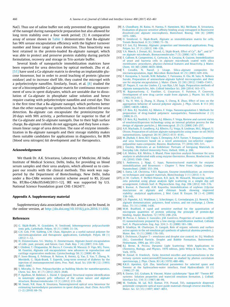
The soluble and immobilized urease were stored in 100 mMTris-acetate-saline buffer at pH 7.2 and 4 �C. The activity wasdetermined and recorded for four weeks at regular intervals forstored urease (immobilized and soluble) using assay proceduresdescribed in Section 2.4 on enzyme assay under similar conditions.The percent residual activity was plotted against the number ofdays, as shown in Fig. 7. The stability of immobilized ureaseenzyme in different nanogels with 90% residual enzyme activitywas calculated from such a plot.
3. Results and discussion
3.1. Alginate nanogels by emulsification method
The use of self-assembled surfactant as a template is one of themost promising approaches for the synthesis of nanomaterials. Thecentral idea of using surfactant templates is to turn the dynamicmolecular aggregates into a chemically and mechanically stablesupramolecular material through templating reactions. Therefore,we chose the commonly employed water-in-oil (W/O) emulsionof a nonionic surfactant for the preparation of alginate nanogelsin a water-hexane biphasic system. The preparation of emulsionswith droplet sizes in the 200 nm range may be performed by highenergy input techniques like high-shear stirring, high-pressurehomogenizers, or ultrasound generators in the presence of surfac-tant. In addition, the smaller the droplet size, the more energy and/or surfactant is required, making these preparation routes unfa-vourable for industrial applications [31]. Therefore, we chosemicellar structures, which form spontaneously in organic solventsthrough thermodynamic self-assembly, to use as templates foralginate nanogel preparation by the sol-gel method.
Tween 80 is a nonionic surfactant selected for the present workbecause of its expected advantage over anionic and cationic surfac-tants, such as AOT (sodium bis[2-ethylhexyl] sulfosuccinate) orCTAB (cetyltrimethylammonium bromide). The behavior of anionicor cationic surfactants is strongly affected by the presence of diva-lent ions (Ca+2, Sr+2, Ba+2), whereas nonionic surfactants show lesspronounced interactions with these ions. Also, the presence of a

Fig. 2. Intensity based size distribution (PDI = 0.371) of micelles formed at 7 mMTween 80 in hexane measured by DLS.
Fig. 3A. Phase diagram for a hexane/alginate solution/Tween 80 (micro)emulsion.Squares (j), circles ( ) and triangles ( ) correspond to the microemulsion regions
A. Saxena et al. / Journal of Colloid and Interface Science 490 (2017) 452–461 455
hydrophilic nonionic surfactant with a high hydrophilic-lipophilicbalance (HLB) stabilizes the aqueous phase, which promotes W/Omicroemulsion formation. The microemulsion droplets act asnanoreactors, where the high HLB value of the nonionic surfactantpromotes smaller droplet size formation, resulting in particles ofsmaller size due to a decrease in the sol-solvent interfacial tension[32,33].
It is also desirable to form emulsions/microemulsions with aminimum amount of surfactant. The critical micelle concentration(CMC) of surfactants for aqueous-surfactant systems has beenwell-studied, but few data are available for organic-surfactant sys-tems. Hence, we applied DLS to study the emulsion in the organicphase, which allows measurement of the micelles’ hydrodynamicsize and provides information on the surfactant’s CMC [34,35].The results for Tween-80 in hexane are shown in Fig. 1. Each datapoint is averaged from three independent measurements, and thestandard deviation is calculated.
The hydrodynamic diameter of Tween 80 aggregates in hexaneinitially increases with surfactant concentration and PDI value(0.3–0.8), then remains roughly constant at around 65 nm over awide range of concentration. The DLS result at [Tween 80]= 7 mM is shown in Fig. 2. It confirms that Tween 80 forms stablemicellar-type aggregates at concentrations of 7 mM or higher; thesize is consistent with published results [36,37].
In W/O-type emulsions, at the CMC and above, the sphericalmicelles enclose the sol (alginate aqueous phase) in so-calledwater pools [38,39]. For the present sol-gel process based on emul-sion polymerization, these can be called ‘‘sol pools.” The size of thepool depends on the relative quantity of the aqueous phase as wellas the number of surfactant molecules forming the micelles. Gela-tion of these sol pools in general produces spherical gel particles.Nevertheless, reasonably satisfactory alginate aqueous phase/hex-ane emulsions over a wide range of alginate sol volume fractionscould be prepared with Tween 80 dissolved in the hexane phase.Since sodium alginate sol is insoluble in oil (hexane), the alginatepolymer is confined within the aqueous nanophase. The phase sta-bility of the resulting microemulsion is significantly affected by theamount of added alginate solution. The trial compositions wereplotted on a ternary phase diagram as shown in Fig. 3A. At constantsurfactant:hexane ratio for low alginate sol concentration, a trans-parent microemulsion region appeared. The area bounded by thesquare points in the phase diagram (Fig. 3A) corresponds to the
Fig. 1. Apparent hydrodynamic diameter of aggregates formed by Tween 80 inhexane measured by DLS. Error bars are standard deviations.
where the micelle size varies from 70 nm to 110 nm, 110 nm to 150 nm, and160 nm to 300 nm, respectively, all with PDI less than 0.5.
Fig. 3B. W/O microemulsion representing points A (clear solution-microemulsion),B (slightly turbid solution) and C (milky appearance-emulsion) in Fig. 3A.
concentration range of component mixtures that produced visuallyclear and transparent microemulsions.
The droplet size was 70 ± 3.43 (std) nm at point A in the ternaryphase diagram; the corresponding microemulsion is shown inFig. 3B. As the alginate aqueous phase concentration increases,the system becomes slightly turbid/translucent, e.g., the circles inFig. 3A, where point B represents a microemulsion with droplet
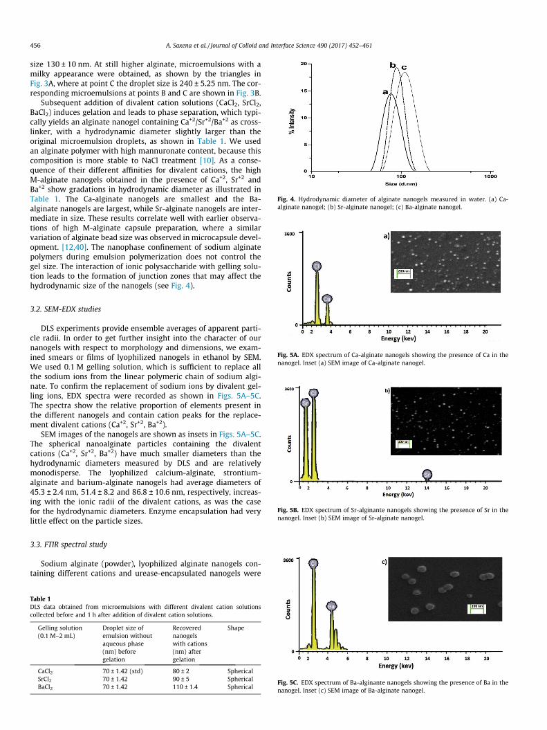
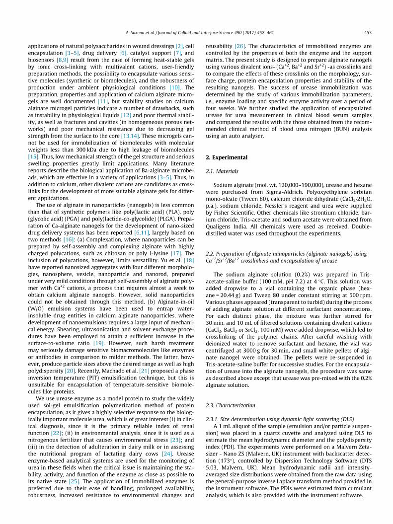
Fig. 4. Hydrodynamic diameter of alginate nanogels measured in water. (a) Ca-alginate nanogel; (b) Sr-alginate nanogel; (c) Ba-alginate nanogel.
456 A. Saxena et al. / Journal of Colloid and Interface Science 490 (2017) 452–461
size 130 ± 10 nm. At still higher alginate, microemulsions with amilky appearance were obtained, as shown by the triangles inFig. 3A, where at point C the droplet size is 240 ± 5.25 nm. The cor-responding microemulsions at points B and C are shown in Fig. 3B.
Subsequent addition of divalent cation solutions (CaCl2, SrCl2,BaCl2) induces gelation and leads to phase separation, which typi-cally yields an alginate nanogel containing Ca+2/Sr+2/Ba+2 as cross-linker, with a hydrodynamic diameter slightly larger than theoriginal microemulsion droplets, as shown in Table 1. We usedan alginate polymer with high mannuronate content, because thiscomposition is more stable to NaCl treatment [10]. As a conse-quence of their different affinities for divalent cations, the highM-alginate nanogels obtained in the presence of Ca+2, Sr+2 andBa+2 show gradations in hydrodynamic diameter as illustrated inTable 1. The Ca-alginate nanogels are smallest and the Ba-alginate nanogels are largest, while Sr-alginate nanogels are inter-mediate in size. These results correlate well with earlier observa-tions of high M-alginate capsule preparation, where a similarvariation of alginate bead size was observed in microcapsule devel-opment. [12,40]. The nanophase confinement of sodium alginatepolymers during emulsion polymerization does not control thegel size. The interaction of ionic polysaccharide with gelling solu-tion leads to the formation of junction zones that may affect thehydrodynamic size of the nanogels (see Fig. 4).
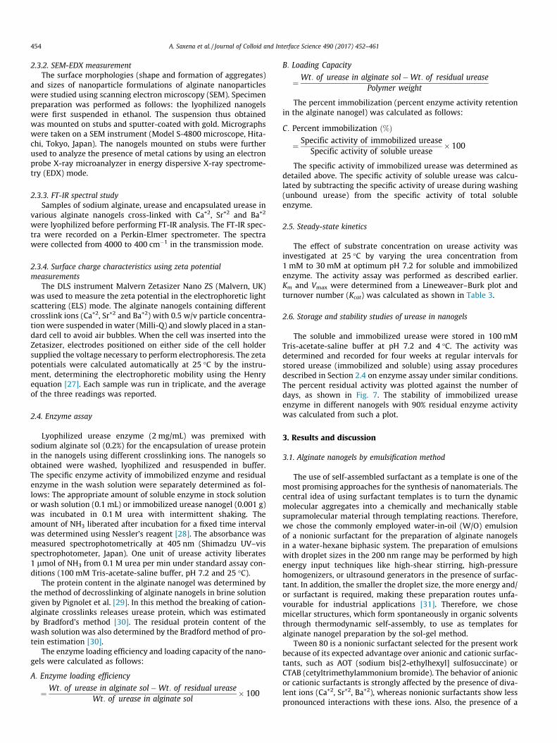
Fig. 5A. EDX spectrum of Ca-alginate nanogels showing the presence of Ca in thenanogel. Inset (a) SEM image of Ca-alginate nanogel.
Fig. 5B. EDX spectrum of Sr-alginante nanogels showing the presence of Sr in thenanogel. Inset (b) SEM image of Sr-alginate nanogel.
3.2. SEM-EDX studies
DLS experiments provide ensemble averages of apparent parti-cle radii. In order to get further insight into the character of ournanogels with respect to morphology and dimensions, we exam-ined smears or films of lyophilized nanogels in ethanol by SEM.We used 0.1 M gelling solution, which is sufficient to replace allthe sodium ions from the linear polymeric chain of sodium algi-nate. To confirm the replacement of sodium ions by divalent gel-ling ions, EDX spectra were recorded as shown in Figs. 5A–5C.The spectra show the relative proportion of elements present inthe different nanogels and contain cation peaks for the replace-ment divalent cations (Ca+2, Sr+2, Ba+2).
SEM images of the nanogels are shown as insets in Figs. 5A–5C.The spherical nanoalginate particles containing the divalentcations (Ca+2, Sr+2, Ba+2) have much smaller diameters than thehydrodynamic diameters measured by DLS and are relativelymonodisperse. The lyophilized calcium-alginate, strontium-alginate and barium-alginate nanogels had average diameters of45.3 ± 2.4 nm, 51.4 ± 8.2 and 86.8 ± 10.6 nm, respectively, increas-ing with the ionic radii of the divalent cations, as was the casefor the hydrodynamic diameters. Enzyme encapsulation had verylittle effect on the particle sizes.
3.3. FTIR spectral study
Sodium alginate (powder), lyophilized alginate nanogels con-taining different cations and urease-encapsulated nanogels were
Table 1DLS data obtained from microemulsions with different divalent cation solutionscollected before and 1 h after addition of divalent cation solutions.
Gelling solution(0.1 M–2 mL)
Droplet size ofemulsion withoutaqueous phase
Recoverednanogelswith cations
Shape
(nm) beforegelation
(nm) aftergelation
CaCl2 70 ± 1.42 (std) 80 ± 2 SphericalSrCl2 70 ± 1.42 90 ± 5 SphericalBaCl2 70 ± 1.42 110 ± 1.4 Spherical
Fig. 5C. EDX spectrum of Ba-alginante nanogels showing the presence of Ba in thenanogel. Inset (c) SEM image of Ba-alginate nanogel.
A. Saxena et al. / Journal of Colloid and Interface Science 490 (2017) 452–461 457
analyzed using an FT-IR spectrophotometer to study cation-alginate interaction before and after gelation. The spectra of thethree crosslinked alginates are quite similar. The correspondingFTIR spectra are given in the supplementary data file as Fig. 1A–1D. There are four particularly relevant spectral bands in thesenanogels prepared and tested under the same conditions. The m(OAH) (1) bands are broadened and shifted to lower wave numberscompared to linear polymeric Na-alginates, indicating that theOAH bond is weakened due to hydrogen bonding in the gel struc-ture [41]. The ratio of intensities of m(C@O) (2) and m(CAOH) (3)suggests the presence of a protonated carboxylic group in thenanogels. Band (4) indicates the presence of an O-glycosidic bondbetween b-d-mannuronic and a-l-guluronic acid residues in thelinear alginate chain. The bands in these regions are broadenedand smoothed with a shift to lower wave number relative tosodium alginate.
The FTIR spectra of nanogels containing urease are shown inFigs. 6A–6D. Three absorption bands are of particular importance.The amide A Band at 3300 cm–1 characterizes the NAH stretchvibration; the amide I band at 1650 cm–1 corresponds to the C@Ostretch vibration; the amide II band at 1550 cm–1 arises from theNAH bending vibration. CAN stretching vibrations at 1250 cm–1
are also prominent. We note that these infrared bands are broadand often overlap with neighboring bands to produce a complexabsorption profile as shown in Figs. 6A–6D [42].
Fig. 6A. FT-IR spectrum of free u
Fig. 6B. FT-IR spectrum of urease-enca
The spectra of all the crosslinked alginates are quite similar, butin the spectra of the urease-bound metal alginates, a significantnew peak (5) appears at 1250 cm�1, due to CAN stretching vibra-tions, which suggests proper encapsulation of urease in the algi-nate matrix.
3.4. Surface charge and physical stability (zeta potential)
The interaction of a protein with polymers and other biomate-rials used in medical devices has characteristic electrical proper-ties, such as the local electrostatic charge distribution and theelectrical double layer potential, which play a significant role indefining the biological interactions, aggregation behavior and sta-bility [43]. The zeta potential (ZP) is an indicator of the surfacecharge properties of a colloid or a particle in solution and dependson the surface potential and the thickness of the electric doublelayer. The zeta potentials of nanoparticles with charged functionalgroups at the surface are measured to determine their colloidal sta-bility by coulombic repulsion [44]. The alginate nanogels wereformed by electrostatic interaction between the negatively chargedcarboxylic groups of alginate and the positively charged divalentcations (Ca+2/Sr+2/Ba+2) that form a crosslinked network containinga large fraction of water. The negative zeta potential values shownin Table 2 indicate an open and porous gel network with free car-boxylic groups at the surfaces of the alginate nanogels, which
rease (lyophilized powder).
psulated calcium alginate nanogel.
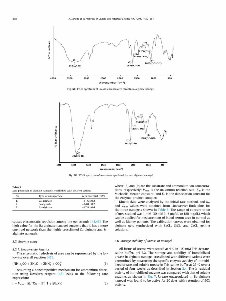
Fig. 6C. FT-IR spectrum of urease-encapsulated strontium alginate nanogel.
Fig. 6D. FT-IR spectrum of urease-encapsulated barium alginate nanogel.
Table 2Zeta potentials of alginate nanogels crosslinked with divalent cations.
No. Type of nanoparticle Zeta potential (mV)
1. Ca-alginate �11.6 ± 0.22. Sr-alginate �14.8 ± 0.23. Ba-alginate �17.8 ± 0.4
458 A. Saxena et al. / Journal of Colloid and Interface Science 490 (2017) 452–461
causes electrostatic repulsion among the gel strands [45,46]. Thehigh value for the Ba-alginate nanogel suggests that it has a moreopen gel network than the highly crosslinked Ca-alginate and Sr-alginate nanogels.
3.5. Enzyme assay
3.5.1. Steady-state kineticsThe enzymatic hydrolysis of urea can be represented by the fol-
lowing overall reaction [47]:
ðNH2Þ2COþ 2H2O ! 2NHþ4 þ CO2�
3 ð1ÞAssuming a noncompetitive mechanism for ammonium detec-
tion using Nessler’s reagent [48] leads to the following rateexpression:
r ¼ Vmax � ½S�=ðKM þ ½S�Þð1þ ½P�=KPÞ ð2Þ
where [S] and [P] are the substrate and ammonium ion concentra-tions, respectively; Vmax is the maximum reaction rate; Km is theMichaelis-Menten constant; and KP is the dissociation constant forthe enzyme-product complex.
Kinetic data were analyzed by the initial rate method, and Km
and Vmax values were obtained from Lineweaver-Burk plots forthe three nanogels shown in Table 3. The range of concentrationof urea studied was 1 mM–30 mM (�6 mg/dL to 180 mg/dL), whichcan be applied for measurement of blood serum urea in normal aswell as kidney patients. The calibration curves were obtained foralginate gels synthesized with BaCl2, SrCl2 and CaCl2 gellingsolutions.
3.6. Storage stability of urease in nanogel
All forms of urease were stored at 4 ºC in 100 mM Tris-acetate-saline buffer, pH 7.2. The storage and stability of immobilizedurease in alginate nanogel crosslinked with different cations weredetermined by measuring the specific enzyme activity of immobi-lized urease and soluble urease in Tris-saline buffer at 25 �C over aperiod of four weeks as described in Section 2.4. The % residualactivity of immobilized enzyme was compared with that of solubleenzyme, as shown in Fig. 7. Urease encapsulated in Ba-alginatenanogel was found to be active for 20 days with retention of 90%activity.
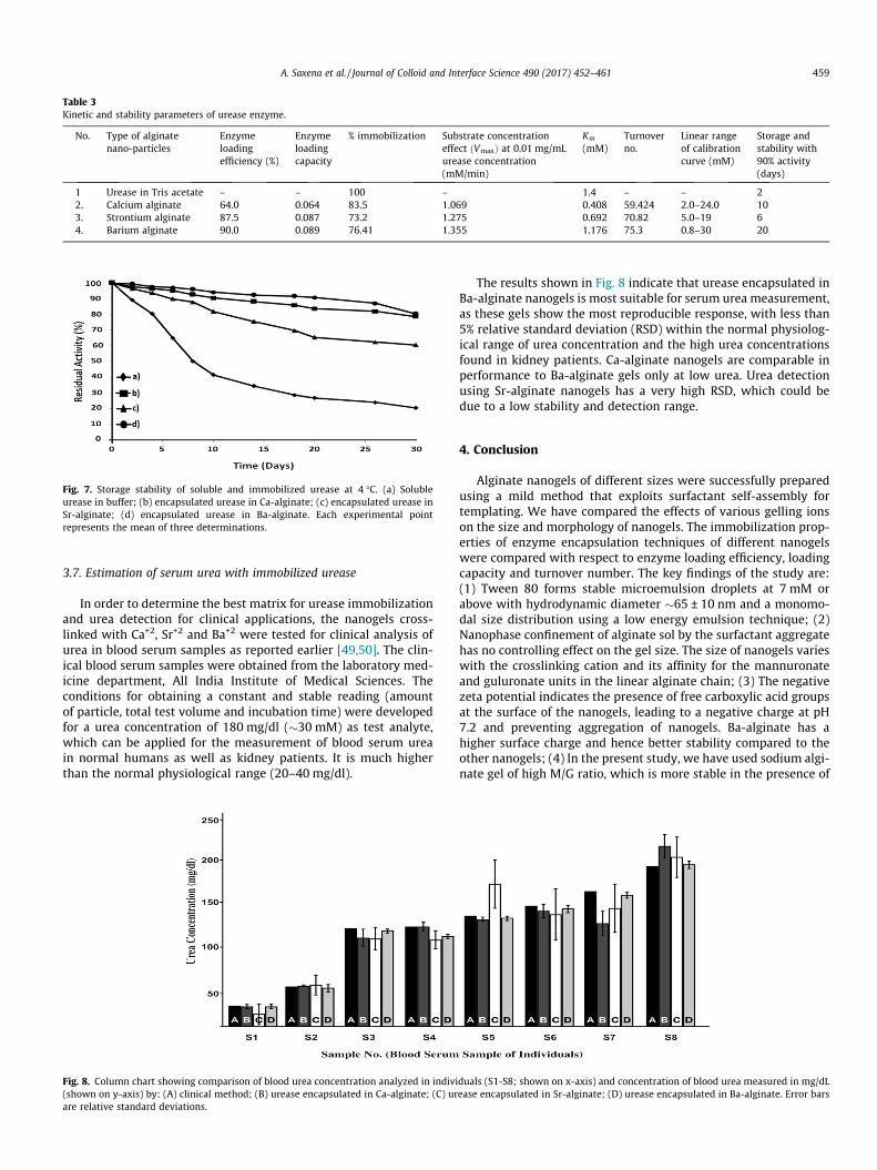
Table 3Kinetic and stability parameters of urease enzyme.
No. Type of alginatenano-particles
Enzymeloadingefficiency (%)
Enzymeloadingcapacity
% immobilization Substrate concentrationeffect ðVmaxÞ at 0.01 mg/mLurease concentration(mM/min)
Km
(mM)Turnoverno.
Linear rangeof calibrationcurve (mM)
Storage andstability with90% activity(days)
1 Urease in Tris acetate – – 100 – 1.4 – – 22. Calcium alginate 64.0 0.064 83.5 1.069 0.408 59.424 2.0–24.0 103. Strontium alginate 87.5 0.087 73.2 1.275 0.692 70.82 5.0–19 64. Barium alginate 90.0 0.089 76.41 1.355 1.176 75.3 0.8–30 20
Fig. 7. Storage stability of soluble and immobilized urease at 4 �C. (a) Solubleurease in buffer; (b) encapsulated urease in Ca-alginate; (c) encapsulated urease inSr-alginate; (d) encapsulated urease in Ba-alginate. Each experimental pointrepresents the mean of three determinations.
A. Saxena et al. / Journal of Colloid and Interface Science 490 (2017) 452–461 459
3.7. Estimation of serum urea with immobilized urease
In order to determine the best matrix for urease immobilizationand urea detection for clinical applications, the nanogels cross-linked with Ca+2, Sr+2 and Ba+2 were tested for clinical analysis ofurea in blood serum samples as reported earlier [49,50]. The clin-ical blood serum samples were obtained from the laboratory med-icine department, All India Institute of Medical Sciences. Theconditions for obtaining a constant and stable reading (amountof particle, total test volume and incubation time) were developedfor a urea concentration of 180 mg/dl (�30 mM) as test analyte,which can be applied for the measurement of blood serum ureain normal humans as well as kidney patients. It is much higherthan the normal physiological range (20–40 mg/dl).
Fig. 8. Column chart showing comparison of blood urea concentration analyzed in indivi(shown on y-axis) by: (A) clinical method; (B) urease encapsulated in Ca-alginate; (C) urare relative standard deviations.
The results shown in Fig. 8 indicate that urease encapsulated inBa-alginate nanogels is most suitable for serum urea measurement,as these gels show the most reproducible response, with less than5% relative standard deviation (RSD) within the normal physiolog-ical range of urea concentration and the high urea concentrationsfound in kidney patients. Ca-alginate nanogels are comparable inperformance to Ba-alginate gels only at low urea. Urea detectionusing Sr-alginate nanogels has a very high RSD, which could bedue to a low stability and detection range.
4. Conclusion
Alginate nanogels of different sizes were successfully preparedusing a mild method that exploits surfactant self-assembly fortemplating. We have compared the effects of various gelling ionson the size and morphology of nanogels. The immobilization prop-erties of enzyme encapsulation techniques of different nanogelswere compared with respect to enzyme loading efficiency, loadingcapacity and turnover number. The key findings of the study are:(1) Tween 80 forms stable microemulsion droplets at 7 mM orabove with hydrodynamic diameter �65 ± 10 nm and a monomo-dal size distribution using a low energy emulsion technique; (2)Nanophase confinement of alginate sol by the surfactant aggregatehas no controlling effect on the gel size. The size of nanogels varieswith the crosslinking cation and its affinity for the mannuronateand guluronate units in the linear alginate chain; (3) The negativezeta potential indicates the presence of free carboxylic acid groupsat the surface of the nanogels, leading to a negative charge at pH7.2 and preventing aggregation of nanogels. Ba-alginate has ahigher surface charge and hence better stability compared to theother nanogels; (4) In the present study, we have used sodium algi-nate gel of high M/G ratio, which is more stable in the presence of
duals (S1-S8; shown on x-axis) and concentration of blood urea measured in mg/dLease encapsulated in Sr-alginate; (D) urease encapsulated in Ba-alginate. Error bars
460 A. Saxena et al. / Journal of Colloid and Interface Science 490 (2017) 452–461
NaCl. Thus use of saline buffer not only prevented the aggregationof the nanogel during nanoparticle preparation but also allowed forlong term stability over a four week period; (5) A comparativeassay of urease shown in Table 3 demonstrates that Ba-alginatehas 90% urease encapsulation efficiency with the highest turnovernumber and linear range of urea detection. Thus bioactivity wasbest retained in the protein-loaded Ba-alginate nanogel, whichwas able to protect and preserve protein stability during particleformulation, recovery and storage in Tris-acetate buffer.
Several kinds of nanoparticle immobilization matrices havebeen reported for urea detection by optical methods. Zhu et al.[51] have used a Ca-alginate matrix to develop an implantable glu-cose biosensor, but in order to avoid leaching of protein (glucoseoxidase) and to increase shelf life, they coated the microgel witha polyelectrolyte nanofilm. Similarly, Swati, et al. [8] studied theuse of a biocompatible Ca-alginate matrix for continuous measure-ment of urea in spent dialysates, which are unstable due to disso-lution of Ca-alginate in phosphate saline solution and hencerequire coating with polyelectrolyte nanofilm and cresol dye. Thisis the first time that a Ba-alginate nanogel, which performs betterthan the other nanogels we synthesized, has been utilized for ureadetection. Ba-alginate can encapsulate the protein/enzyme for20 days with 90% activity, a performance far superior to that ofthe Ca-alginate and Sr-alginate nanogels. Due to their high surfacecharge, Ba-alginate colloids do not aggregate, and they have a max-imum linear range of urea detection. The ease of enzyme immobi-lization in Ba-alginate nanogels and their storage stability makesthem suitable candidates for applications in diagnostics, for BUN(blood urea nitrogen) kit development and for therapeutics.
Acknowledgement
We thank Dr. A.K. Srivastava, Laboratory of Medicine, All IndiaInstitute of Medical Science, Delhi, India, for providing us bloodserum samples and their urea analysis, which allowed us to com-pare our results with the clinical methods. This work was sup-ported by the Department of Biotechnology, New Delhi, Indiaunder a Bio-CARe women scientist scheme award to RS, [GrantNo. BT/Bio-CARe/05/640/2011-12]. IRE was supported by U.S.National Science Foundation grant CHE-1362477.
Appendix A. Supplementary material
Supplementary data associated with this article can be found, inthe online version, at http://dx.doi.org/10.1016/j.jcis.2016.11.030.
References
[1] G. Skjåk-Bræk, H. Grasdalen, H. Smidsrød, Inhomogeneous polysaccharideionic gels, Carbohydr. Polym. 10 (1) (1989) 31–54.
[2] C.H. Goh, P.W. SiaHeng, L.W. Chan, Alginates as a useful natural polymer formicroencapsulation and therapeutic applications, Carbohydr. Polym. 88 (1)(2012) 1–12.
[3] H. Zimmermann, S.G. Shirley, U. Zimmermann, Alginate-based encapsulationof cells: past, present, and future, Curr. Diab. Rep. 7 (4) (2007) 314–320.
[4] I. Ghidoni, T. Chlapanidas, M. Bucco, F. Crovato, M. Marazzi, D. Vigo, M.L. Torre,M. Faustini, Alginate cell encapsulation: new advances in reproduction andcartilage regenerative medicine, Cytotechnology 58 (1) (2008) 49–56.
[5] P. Soon-Shiong, E. Feldman, R. Nelson, R. Heintz, Q. Yao, Z. Yao, T. Zheng, N.Merideth, G. Skjak-Braek, T. Espevik, Long-term reversal of diabetes by theinjection of immunoprotected islets, Proc. Natl. Acad. Sci. USA 90 (12) (1993)5843–5847.
[6] S. Mizrahy, D. Peer, Polysaccharides as building blocks for nanotherapeutics,Chem. Soc. Rev. 41 (7) (2012) 2623–2640.
[7] P. Agulhon, M. Robitzer, L. David, F. Quignard, Structural regime identificationin ionotropic alginate gels: influence of the cation nature and alginatestructure, Biomacromolecules 13 (1) (2012) 215–220.
[8] M. Swati, N.K. Hase, R. Sivastava, Nanoengineered optical urea biosensor forestimating haemodialysis parameters in spent dialysate, Anal. Chim. Acta 676(1–2) (2010) 68–74.
[9] A. Chaudhary, M. Raina, H. Harma, P. Hanninen, M.J. McShane, R. Srivastava,Evaluation of glucose sensitive affinity binding assay entrapped in fluorescentdissolved-core alginate microspheres, Biotechnol. Bioeng. 104 (6) (2009)1075–1085.
[10] O. Smidsrod, G. Skjak-Braek, Alginate as immobilization matrix for cells,Trends Biotechnol. 8 (3) (1990) 71–78.
[11] K.Y. Lee, D.J. Mooney, Alginate: properties and biomedical applications, Prog.Polym. Sci. 37 (1) (2012) 106–126.
[12] Y.A. Mørch, I. Donati, B.L. Strand, G. Skjåk-Braek, Effect of Ca2+, Ba2+, and Sr2+
on alginate microbeads, Biomacromolecules 7 (5) (2006) 1471–1480.[13] E. Callone, R. Campostrini, G. Carturan, A. Cavazza, R. Guzzon, Immobilization
of yeast and bacteria cells in alginate microbeads coated with silicamembranes: procedures, physico-chemical features and bioactivity, J. Mater.Chem. 18 (40) (2008) 4839–4848.
[14] T. Coradin, N. Nassif, J. Livage, Silica–alginate composites formicroencapsulation, Appl. Microbiol. Biotechnol. 61 (5) (2003) 429–434.
[15] F. Kurayama, S. Suzuki, N.M. Bahadur, T. Furusawa, H. Ota, M. Sato, N. NoboruSuzuki, Preparation of aminosilane-alginate hybrid microcapsules and theiruse for enzyme encapsulation, J. Mater. Chem. 22 (30) (2012) 15405–15411.
[16] J.P. Paques, E. van der Linden, C.J. van Rijn, L.M. Sagis, Preparation methods ofalginate nanoparticles, Adv. Colloid Interface Sci. 209 (2014) 163–171.
[17] M. Rajaonarivony, C. Vauthier, G. Couarraze, F. Puisieux, P. Couvreur,Development of new drug carrier made from alginate, J. Pharm. Sci. 82 (9)(1993) 912–917.
[18] C. Yu, H. Wei, Q. Zhang, X. Zhang, S. Cheng, R. Zhuo, Effect of ions on theaggregation behavior of natural polymer alginate, J. Phys. Chem. B 113 (45)(2009) 14839–14843.
[19] C.P. Reis, R.J. Neufeld, A.J. Ribeiro, F. Veiga, Nanoencapsulation I. Methods forpreparation of drug-loaded polymeric nanoparticles, Nanomedicine 2 (1)(2006) 8–21.
[20] C.P. Reis, R.J. Neufeld, S. Vilela, A.J. Ribeiro, F. Veiga, Review and current statusof emulsion/dispersion technology using an internal gelation process for thedesign of alginate particles, J. Microencapsul. 23 (3) (2006) 245–257.
[21] A.H. Machado, D. Lundberg, A.J. Ribeiro, F.J. Veiga, B. Lindman, M.G. Miguel, U.Olsson, Preparation of calcium alginate nanoparticles using water-in-oil (W/O)nanoemulsions, Langmuir 28 (9) (2012) 4131–4141.
[22] M. Zhybak, V. Beni, M.Y. Vagin, E. Dempsey, A.P.F. Turner, Y. Korpan, Creatinineand urea biosensors based on a novel ammonium ion-selective copper-polyaniline nano-composite, Biosens. Bioelectron. 77 (2016) 505–511.
[23] J. Emsley, Molecules at an Exhibition: Portraits of Intriguing Materials inEveryday Life, Oxford University Press, Oxford, 1999, p. 200.
[24] G.K. Mishra, R.K. Mishra, S. Bhand, Flow injection analysis biosensor for ureaanalysis in adulterated milk using enzyme thermistor, Biosens. Bioelectron. 26(4) (2010) 1560–1564.
[25] S. Andreescu, J. Njagi, C. Ispas, Nanostructured materials for enzymeimmobilization and biosensors – Chapter 7, New Front. Organ. Compos.Nanotechnol. (2008) 355–394.
[26] S. Datta, L.R. Christena, Y.R.S. Rajaram, Enzyme immobilization: an overviewon techniques and support materials, Biotechnology 3 (1) (2013) 1–9.
[27] J.C.W. Corbett, F. McNeil-Watson, R.O. Jack, M. Howarth, Measuring surfacezeta potential using phase analysis light scattering in a simple dip cellarrangement, Colloids Surf. A: Physicochem. Eng. Asp. 396 (2012) 169–176.
[28] S. Kumar, A. Dwevedi, A.M. Kayastha, Immobilization of soybean (Glycinemax)urease on alginate and chitosan beads showing improvedstability: analytical applications, J. Mol. Catal. B: Enzym. 58 (1–4) (2009)138–145.
[29] L.H. Pignolet, A.S. Waldman, L. Schechinger, G. Govindarajoo, J.S. Nowick, Thealginate demonstration: polymers, food science, and ion exchange, J. Chem.Educ. 75 (11) (1998) 1430.
[30] M.M. Bradford, A rapid and sensitive method for the quantitation ofmicrogram quantities of protein utilizing the principle of protein-dyebinding, Analyt. Biochem. 72 (1976) 248–254.
[31] M. Porras, C. Solans, C. González, J.M. Gutiérrez, Properties of water-in-oil(W/O) nanoemulsions prepared by a low energy emulsification method, ColloidsSurf. A: Physicochem. Eng. Asp. 324 (1–3) (2008) 181–188.
[32] B. Siladitya, M. Chatterjee, D. Ganguli, Role of organic solvents and surfaceactive agents in the sol emulsion gel synthesis of spherical alumina powders, J.Mater. Res. 15 (1) (2000) 176–185.
[33] E. Dickinson, Chapter 7 – emulsions and droplet size control, in: D.J. Wedlock(Ed.), Controlled Particle, Droplet and Bubble Formation, Butterworth-Heinemann, 1994, pp. 191–216.
[34] B.J. Berne, R. Pecora, Dynamic Light Scattering: With Applications toChemistry, Biology, and Physics, 1976 edition., John Wiley & Sons Inc., NewYork, 1976.
[35] M. Zulauf, H. Friedrich, Eicke, Inverted micelles and microemulsions in theternary system water/aerosolOT/isooctane as studied by photon correlationspectroscopy, J. Phys. Chem. 83 (4) (1979) 480–486.
[36] R.K.O. Apenten, Q.H. Zhu, Interfacial parameters for selected spans andTweens at the hydrocarbon-water interface, Food Hydrocolloids 10 (1)(1996) 27–30.
[37] R. Davies, D.E. Graham, B. Vincent, Water cyclohexane ‘‘Span 80” ‘‘Tween 80”systems: Solution properties and water/oil emulsion formation, J. Colloid.Interface Sci. 116 (1) (1987) 88–99.
[38] M. Yoshida, M. Lal, N.D. Kumar, P.N. Prasad, TiO2 nanoparticle dispersedpolyimide composite optical wave guide materials through reverse micelles, J.Mater. Sci. 32 (15) (1997) 4047–4051.
A. Saxena et al. / Journal of Colloid and Interface Science 490 (2017) 452–461 461
[39] A. Aryal, L. Cot, T. Dabadie, C. Guizard, J. Ramsay, SANS investigations of oxidegel formation in inverse micelle and lamellar surfactant systems, J. Sol-Gel Sci.Technol. 2 (1) (1994) 205–209.
[40] B.L. Strand, O. Gåserød, B. Kulseng, T. Espevik, G. Skjåk-Baek, Alginate-polylysine-alginate microcapsules: effect of size reduction on capsuleproperties, J. Microencapsulation 19 (5) (2002) 615–630.
[41] E. Torres, Y.N. Mata, M.L. Blázquez, J.A. Muñoz, F. González, A. Ballester, Goldand silver uptake and nanoprecipitation on calcium alginate beads, Langmuir21 (17) (2005) 7951–7958.
[42] C.R. Cantor, P.R. Schimmel, Techniques for the study of biological structure andfunction, Biophysical Chemistry: Part II, WH Freeman and Co., Oxford, 1980, p.503.
[43] S.A.M. Tofail (Ed.), Biological Interactions With Surface Charge in Biomaterials,RSC Publishing, 2011. ISBN 978-1-84973-185-0.
[44] R.J. Hunter, Zeta Potential in Colloid Science: Principles and Publications,Academic Press, London; New York, 1981.
[45] O. Gaseroed, O. Smidsroed, G. Skjak-Braek, Microcapsules of alginate-chitosan—I: A quantitative study of the interaction between alginate andchitosan, Biomaterials 19 (20) (1998) 1815–1825.
[46] H.G. Xie, J.N. Zheng, X.X. Li, X.D. Liu, J. Zhu, F. Wang, W.Y. Xie, X.J. Ma, Effect ofsurface morphology and charge on the amount and conformation of fibrinogenadsorbed onto alginate/chitosan microcapsules, Langmuir 26 (8) (2010) 5587–5594.
[47] M. Fidaleo, R. Lavecchia, Kinetic study of enzymatic urea hydrolysis in the pHrange 4–9, Chem. Biochem. Eng. Q. 17 (4) (2003) 311–318.
[48] J.P. Hoare, K.J. Laidler, The molecular kinetics of the urea-urease system. II. Theinhibition by products, J. Am. Chem. Soc. 72 (6) (1950) 2487–2489.
[49] R. Sahney, S. Anand, B.K. Puri, A.K. Srivastava, Immobilization of urease onglass pH electrodes: A comparative study between three immobilizationtechniques and its application in urea detection in blood serum, Anal. Chim.Acta 578 (2006) 156–161.
[50] R. Sahney, S. Anand, B.K. Puri, Enzyme coated glass pH-electrode: Itsfabrication and application in the determination of urea in blood samples,Anal. Chim. Acta 542 (2005) 157–161.
[51] H. Zhu, R. Srivastava, J.Q. Brown, M.J. McShane, Combined physical andchemical immobilization of glucose oxidase in alginate microspheresimproves stability of encapsulation and activity, Bioconjugate Chem. 16 (6)(2005) 1451–1458.


![Class : I - amity.edu I_Maths.pdf · AMITY INSTITUTE FOR COMPETITIVE EXAMINATIONS: Ph. : 24336143/44, 25573111/2/3/4, 95120-2431839/42 [3] Rough Work 5. Which one of the following](https://img.dokumen.tips/doc/110x75/5d512f6188c99335538b87f5/class-i-amityedu-imathspdf-amity-institute-for-competitive-examinations.jpg)