Embed Size (px)
Citation preview

Journal of Asian Scientific Research, 2013, 3(2):157-173
157
NATURAL PRODUCTS IN ANTILEISHMANIAL DRUG DISCOVERY: A
REVIEW
Oseni Lateef Adebayo
Department of Applied Chemistry and Biochemistry, University for Development Studies, Navrongo, Ghana
Dawda Suleman
Department of Applied Chemistry and Biochemistry, University for Development Studies, Navrongo, Ghana
Abagale Aba Samson
Department of Applied Chemistry and Biochemistry, University for Development Studies, Navrongo, Ghana
ABSTRACT
Leishmaniasis is a disease caused by the protozoan parasites, which belong to the genus
Leishmania. Some known species of Leishmania include L. tropica, L. donovani, L. mexicana, L.
aethiopica, L. Infantum, L. donovani, L. mexicana, L. braziliensis, L. chagasi and L. amazonensis.
Leishmaniasis is transmitted through the bite of phlebotomine sandflies. The existence of
Leishmaniasis has been recorded in several countries in the Mediterranean, Central and South
America, Asia, Africa, the Middle East, China, India and the Caribbean. Chemotherapy and vector
control are the known available means of combating the Leishmaniasis as the development of
effective vaccines are still under way. Crude solvent extracts and isolated compounds from certain
plants have however, shown significant activity against leishmanial parasite. Some of the plants
reported to have antileishmanial activity include Tridax procumbens, Urechites andrieuxii Muell.-
Arg. (Apocynaceae), Desmodiumgangeticum, Pseudelephantopusspicatus,
Himatanthussucuuba,among others. Of the antileishmanial plants, the greatest number belonged to
the Apocynaceae. Methanol extracts of the plants were mostly found to possess antileishmanial
activity. The level of activity exhibited by the crude solvent extracts or the isolated constituent(s)
depended largely on the type of solvent used for the extraction and also on the plant part used. The
array of plants that have demonstrated antileishmanial activity suggests that the hope to discover
novel antileishmanial drugs is high.
Keywords: Antileishmanial activity; leishmaniasis; natural products; Leishmania spices,
cutaneous leishmaniasis; visceral leishmaniasis.
Journal of Asian Scientific Research
journal homepage: http://aessweb.com/journal-detail.php?id=5003
Journal of Asian Scientific Research, 2013, 3(2):157-173
158
INTRODUCTION
The Disease
Leishmaniasis is vector-borne disease caused by a protozoan endo-parasite species belonging to the
genus Leishmania that live in the blood and tissues of the host. The disease basically affects
animals but finds its way into the human population when man, flies and the animal reservoirs
coexist in the same environment. A broad range of clinical manifestations involving the skin,
mucous membranes and visceral organs with devastating consequences have been diagnosed
among infected humans. Cutaneous leishmaniasis and visceral leishmaniasis have been reported to
be the two major forms of leishmaniasis in humans. The latter happens to be the dreaded or severe
form of the disease that in certain cases can record 100% mortality of infected persons when not
treated. Cutaneous leishmaniasis unlike visceral leishmaniasis has been found to be less severe.
The less severity of cutaneous leishmaniasis has been attributed to self-healing ulcers (Bryceson,
1996). In other texts, another form of leishmaniasis called Muco-cutaneous leishmaniasis, which
causes an extensive disfiguring lesion of the nose, mouth and throat mucous membranes have also
been reported (Boakye et al., 2005). Other authors however, argue that muco-cutaneous
leishmaniasis is a subset of cutaneous leishmaniasis (The Center for Food Security & Public
Health., 2009).
Leishmaniasis has been reported to be one of the world’s most neglected diseases with about
twelve (12) million reported cases with an estimated infection rate of two (2) million new cases
yearly (about 75% cases of cutaneous leishmaniasis and 25% cases of visceral leishmaniasis)
(Luciana et al., 2012). The World Health Organization (WHO) also reports that, about 350 million
people are considered to be at risk of contracting leishmaniasis especially among people living in
the developing countries (World Health Organistion, 2010).
Causes of Leishmaniasis
Leishmaniasis is a disease caused by the protozoan called Leishmania species. Leishmania
speciesare obligate unicellular parasites that exist in two distinct forms. They exist as non-flagellar
amastigote in humans and other hosts while in culture and gut of sandflies (vector) the flagellar or
the promastigote form is visible (Chang et al., 1985). Some known species of Leishmania include
L. tropica, L. donovani, L. mexicana, L. braziliensis, L. aethiopica, L. Infantum, L. donovani, L.
mexicana, L. braziliensis, L. chagasi(Arfan and Simeen, 2008).The Leishmania parasites are said to
be motile and slender reaching about 10 to 15 µm in length with a single anterior flagellum. The
electron microscopic studies of the Leishmania parasites reveal that the amastigotes have a double
membrane supported by a layer of sub-pellicular fibrils of which one of the membranes is lost
when the transformation of the amastigotes takes place. The fibrils are however retained and a short
flagellum may be seen arising from the kinetosome. These parasites run a spiral course from the
region of the flagellar base towards the posterior apical end (Arfan and Simeen, 2008).

Journal of Asian Scientific Research, 2013, 3(2):157-173
159
Symptoms of Leishmaniasis
Clinical studies conducted on leishmaniasis have revealed diverse clinical manifestations of the
disease. The diverse clinical manifestations of the disease have been attributed to the reaction
between the virulence of the parasite species and the host’s immune response (Boakye et al.,
2005).Leishmaniasis is characterized by a wide range of clinical manifestations that include
ulcerative skin lesions developing at the site of the sand fly bite (localized cutaneous
leishmaniasis), multiple non-ulcerative nodules (diffuse cutaneous leishmaniasis), destructive
mucosal inflammation (mucosal leishmaniasis), and disseminated visceral infection (visceral
leishmaniasis) (Richard et al., 2007).
Existing Means of Control/Treatment
Before the introduction of oral and topical treatment alternatives for cutaneous leishmaniasis, the
pentavalent antimonials have been the recommended drugs used for the treatment of both
cutaneousleishmaniasis and visceral leishmaniasis (Richard et al., 2007).Thepentavalent
antimonials werediscovered over 60 years ago (Simon and Vanessa, 2006). Three new drug
formulations, liposomal amphotericin B (AmBisome), miltefosine and paromomycin for the
treatment of visceral leishmaniasis, have all been reportedto suffer from limitations of cost, specific
toxicities or parenteral administration (Simon and Vanessa, 2006).Although, new oral and topical
treatment alternatives for the treatment of cutaneous leishmaniasis have been introduced within the
past few years, a vaccine is yet to be developed (Richard et al., 2007).The idea to develop a single
drug formulation effective for the treatment of all forms of leishmaniasis have been said to be
unlikely to be achieved mainly due to the intrinsic variation in drug sensitivity of the 17
Leishmania species known to infect humans and the different pharmacokinetic requirements
imposed on the drugs to be used by the visceral and the cutaneous sites of infection(Simon and
Vanessa, 2006). However, further reports have shown that the advances made so far have been
significant as the concept of choice for treatment is now real (Simon and Vanessa, 2006).It has also
been reported in other tests that, chemotherapy and vector control are the available means of
combating leishmaniasis as the development of effective vaccines are still under way Chawla and
Madhubala (2010). Some authors believe that the prevention and control of cutaneous
leishmaniasis have proven to be difficult because of the complexity of cutaneous leishmaniasis
epizoology and the few options available for effective vector control (Richard et al.,
2007).According to a recent report The Center for Food Security & Public Health. (2009) has it
that, the parasites Leishmania species do not remain viable outside a host or in vitro culture but can
however, be inactivated by 1% sodium hypochlorite, 2% glutaraldehyde, or formaldehyde and they
are also susceptible to heat at a temperature of about 50–60°C (The Center for Food Security &
Public Health., 2009).
Mode of Transmission
The transmission of Leishmaniaparasites is largely influence by characteristics of the vector
involved in the transmission. These characteristics include the tendency of the vector to take blood

Journal of Asian Scientific Research, 2013, 3(2):157-173
160
from humans and/or animals as well as the capability of the ingested parasites to develop to the
infective stages within the specific vector (Bryceson, 1996). Leishmaniasis is a vector-transmitted
disease. The mode of transmission of Leishmania parasites is through the bite of Phlebotomine
sandflies. These sand flies (vector)have been reported to be widely distributed in the tropics and
other warm mainland areas and extend northwards to latitudes in the region of 50o N (Sewice,
1980). The vector (sand fly) belongs to the subfamily of Phlebotominae with about 600 species
distributed in five genera of which species in three genera (Phlebotomus, Lutzomyia and
Sergentomyia) are responsible for the transmission of the Leishmania parasites in vertebrates.
However, only Phlebotomus and Lutzomyiagenera transmit disease to man (Morsy, 1996).
Adult Phlebotomine sandflies are readily identified by their minute size (2-5 mm in length), their
hairy appearance, relatively large eyes and their relatively long and stilt like legs. Sandflies have
been reported to live in rodent burrows, crevices, holes in river banks, trees and houses in the Old
World whereas in the New World, sandfliesdwell in the tree canopies and forest litter (Kendrick,
1986). Sewice (1980) also described sandflies as small brownish hairy flies that are identified by
the presence of erect narrow wings covered with hair (Sewice, 1980). Sandflies have a
characteristic hopping type of flights and are said to exhibit nocturnal activity (Arfan and Simeen,
2008).
Geographical Distribution
The distribution of leishmaniasis is highly dependent on the distribution of appropriate vector
species (Boakye et al., 2005). With the exception of the Antarctica, Leishmania species(which
cause leishmaniasis) have been reported on almost every continent with their presence being
primarily endemic in tropical and sub-tropical regions (The Center for Food Security & Public
Health., 2009). The Centre for Food Security & Public Health. (2006), reported cases of the
occurrence of leishmaniasis in many countries in the Mediterranean, Central and South America,
Asia, Africa, the Middle East, China, India and the Caribbean. The report went further to say that
cases in the United States are rare and typically occurred in individuals returning from countries
that have the disease and/or the vector (sand flies) (The Centre for Food Security & Public Health.,
2006).However, sand flies capable of spreading the disease have been reported to be found in
Southern Texas and leishmaniasis has been reported in 21 states and Canada foci (The Centre for
Food Security & Public Health., 2006). An updated report in 2009 provided that, reported cases of
leishmaniasis in Europe appear to be spreading northward from its traditional foci (The Center for
Food Security & Public Health., 2009).
In Africa, leishmaniasis is endemic to countries mostly found in the North Africa, Central Africa,
East Africa, and West Africa as well as countries found in the Horn of Africa (Sheik-Mohamed and
Velema, 1999). Leishmaniasis has been reported in countries like Niger (Desjeux et al., 1981),
Mali(Lefrou, 1948), Nigeria (Dyce, 1924), Senegal(Riou and Advier, 1993), and Cameroon
(Rageu, 1951).Other countries that also have reported cases include Burkina Faso, Mauritania,
Journal of Asian Scientific Research, 2013, 3(2):157-173
161
Gambia and Guinea.Based on available information, cutaneous leishmaniasis is proposed to be
endemic in a belt running from Mauritania, Gambia and Senegal in the west to Nigeria and
Cameroon in the east. Though the cutaneous leishmaniasis belt mentioned cuts across the northern
part of Ghana no report of the disease was heard in Ghana until in 1999 when some chronic ulcers
were diagnosed as cutaneous leishmaniasis in the Volta Region (Boakye et al., 2005).
Unfortunately, in spite of the long history of the disease in West Africa, it happens to be one of the
less recognized or over-looked parasitic infections in this region (Desjeux et al., 1981). Richard et
al. (2007)also suggested the that, cutaneous leishmaniasis has become one of the so-called
neglected diseases, with little interest by financial donors, public-health authorities, and
professionals to implement activities to research, prevent, or control the disease perhaps due to the
fact that it is rarely fatal (Richard et al., 2007).The most devastating form of cutaneous
leishmaniasis (muco-cutaneous leishmaniasis) that involves the mucous membranes of the nose,
mouth and throat as well as the deadly visceral form are reported to be rare in West Africa.
However, there have been two reported cases of muco-cutaneous leishmaniasis in Senegal (Strobel
et al., 1978)and some reported cases of the deadly visceral leishmaniasis have been recorded in
Togo(de Campos et al., 1979), Burkina Faso(Andre et al., 1978),and the Gambia (Conteh and
Desjeux, 1983; Greenwood et al., 1984).
Conteh and Desjeux (1983) suggested that fevers and splenomegaly in countries neighboring the
Gambia could be due to visceral leishmaniasis (Conteh and Desjeux, 1983). Sirol et al. (1972) are
of the opinion that visceral leishmaniasis could be common in West Africa (Sirol et al., 1972).
Some casesof visceral leishmaniasis have been reported in South Asia, the Mediterranean, the
Middle East, Latin America and parts of Asia (The Center for Food Security & Public Health.,
2009).
Plants with Antileishmanial Activity
Methanol extract of Tridax procumbens L. (the whole plant)have been reported to exhibit
significant leishmanicidal activity (Peraza-Sanchez et al., 2007). In vitro studies onactivity of
Tridax procumbens extracts against promastigotes of Leishmaniamexicana revealed significant
activity (Zhelmy et al., 2009). The methanol extract of Tridax procumbensshowed inhibition of
promastigotes growth of Leishmania mexicana with a 50% inhibitory concentration (IC50) of
3µg/ml whiles oxylipin (3S)-16,17-didehydrofalcarinol, an isolated compound from Tridax
procumbens exhibited the highest inhibition at IC50 of 0.478 µg/ml. Toxicity test conducted on the
extract and oxylipin(3S)-16,17-didehydrofalcarinol however, revealed insignificant or no effect on
mammalian cells. Using the standard drugs amphotericin B and pentamidine as references,
pentamidine and oxylipin(3S)-16, 17-didehydrofalcarinol exhibited similar antileishmanial activity
(Zhelmy et al., 2009).The use of Urechites andrieuxii Muell.-Arg. (Apocynaceae) in traditional
medicine for the treatment of cutaneous leishmaniasis in the Yucatan Peninsula has been reported
(Pulido and Serralta, 1993; Argueta, 1994). It has been said that while Mayan traditional healers
recommend washing thelesion with a root infusion of Urechites andrieuxii, and then applying the

Journal of Asian Scientific Research, 2013, 3(2):157-173
162
powdered dry root over the area, other traditional doctors recommend the direct application on the
lesion with dried, ground leaves of Urechites andrieuxii (Jiu, 1966). Crude methanol extracts of
from the leaves and roots of Urechites andrieuxii Muell.-Arg. growing in four different natural
populations have also been reported to exhibit antileishmanial activity on three species of
Leishmanial parasites namely, L. braziliensis, L. amazonensis andL. donovani with extracts from
the roots showing strongest activity (Manuel et al., 2003).The first population was collected from
an area characterized by high humidityand flooded soils, second and third populations from an area
with warm humid climate and sporadically soils respectively whiles the fourth population was
obtained from an area characterized by a dry climate with well drained soils and low humidity. In
this study, crude methanol extracts from the roots of Urechites andrieuxii Muell.-Arg. obtained
from the four different natural populations in the Yucatan Peninsula all showed antileishmanial
activity against the named Leishmanial species but at different levels at a concentration of 200
µg/mL with Urechites andrieuxii Muell.-Arg collected from the first population showing a
strongest antileishmanial activity against the three Leishmania species. Urechites andrieuxii
Muell.-Arg collected from the second showed a weak antileishmanial activity whiles those
(Urechites andrieuxii Muell.-Arg) obtained from the third and fourth populations exhibited weakest
antileishmanial activity. This was revealed in an in vitro activityof methanol extracts of the four
populations of Urechitesandrieuxii against strains of promastigotes forms of L.braziliensis, L.
amazonensis andL. Donovani (Manuel et al., 2003). The difference in the activity among the
different populations suggests that biological activities of plants are influenced by environmental
factors to some extent. This falls in line with earlier report that, theproduction of plant bioactive
secondarymetabolites is influenced by environmental, ontogenetic and genetic factors (Vanhalen et
al., 1991). The most active methanol extract of the roots (from the first population) has been
reported to be cytotoxic to ovary carcinoma cells, human epidermoid carcinoma cells, cervix
carcinoma cell andcolon carcinoma cells whiles methanolic extracts from the leaves exhibited
cytotoxicity in cervix carcinoma cells only (Manuel et al., 2003).
Manuel et al. (2003) however suggested that, toxic activity observed for the most active extract
against Leishmania promastigotes, might in fact be due to the presence of metabolites with
cytotoxic, but not necessarily antileishmanial activity (Manuel et al., 2003). Manuel et al. (2003)
further suggested the presence of biologically active natural products with selective toxic activity
against Leishmania parasites in the methanol extract of the leaves Urechitesandrieuxii (Manuel et
al., 2003). The root extract of Urechites andrieuxii has been reported in other literature to have
depressant, antiatherogenic and anti-inflammatory activities (Jiu, 1966). Extracts from the
plantUrechites andrieuxii has further been reported in other texts to have toxic activity against
Leishmania mexicana (Viscencio et al., 1995). Another plant that has been reported to have
antileishmanial activity is Desmodiumgangeticum (Iwu et al., 1992). It has been reported in the
work carried out by Nasib et al. (2005) to determine theefficacy of Desmodiumgangeticumextract
and its fractions (hexane, n-butanol and aqueous) againstexperimental visceral leishmaniasis,
showed that then-butanol fraction had moderate antileishmanial activity when tested against

Journal of Asian Scientific Research, 2013, 3(2):157-173
163
established infection of Leishmania donovani in hamsters whiles the crude ethanolic extract as well
as hexane and aqueous fractions of the same plant showed insignificant inhibition of parasite
multiplication. Also, the n-butanol fraction of Desmodiumgangeticum has been reported to have the
highest efficacy of Leishmaniadonovani challenge followed by the crude methanolic extract in a
chemoprophylactic efficacy study when administered at a dosage of 250 mg/kg to hamsters 7 days
prior to the Leishmania donovani infection and also on the 7th
day after infection as booster dose.
The n-butanol fraction have further been reported to show significant (P < 0.001) non-specific
resistance to peritoneal macrophages against Leishmania infection (promastigote form) whiles the
methanol extract, hexane and aqueous fractions showed no activity. Other results have also
revealed the n-butanol fraction of Desmodiumgangeticumto be the most efficient over the other
fractionsagainst established infection of Leishmania donovani in a chemotherapeutic efficacy study
when administered orally with five doses of 100 mg/kg (Nasib et al., 2005).
Pseudelephantopusspicatus is another plant thathas been reported to possess antileishmanial
activity (Odonne et al., 2011). Isolated compounds 8,13-diacetyl-piptocarphol, 8-acetyl-13-O-
ethylpiptocarpholand ursolicobatained from Pseudelephantopusspicatusin an in vitro study
exhibited activity against Leishmaniaamazonensisaxenic amastigotes with the compounds8,13-
diacetyl-piptocarphol, and 8-acetyl-13-O-ethylpiptocarphol exhibiting significant activity than
Amphotericin B which was used as the positive control (Odonne et al., 2011). Cytotoxicity studies
conducted on the most active compounds, 8,13-diacetyl-piptocarphol and 8-acetyl-13-O-
ethylpiptocarphol showed that these compounds are not cytotoxic when tested on HeLa, L929 and
B16F10 cell lines at a concentration of 50µM (Buskuhl et al., 2010).
The bark of Himatanthussucuuba has been reported toshow significant direct activity against the
intracellular form of Leishmania amazonensis axenic amastigotes (Villegas et al., 1997). Two
known spirolactoneiridoids; plumericin and its isomer isoplumericin obtained from bio-guided
isolation of the stem bark’s ethanol extract of Himatanthussucuuba have been reported to show
strong activity against Leishmania amazonensis axenic amastigotes at IC50of 5µg/ml in
amastigotes. These two compounds have further been reported to exhibit less toxicity on mice
peritoneal macrophages and on tumoral cells than in their activity on Leishmaniaamastigotes in
cytotoxicity evaluation. However, in the case of the tumoral assaysisoplumericin is more toxic than
plumericin. In vitro study of plumericin and its isomer isoplumericin on infected macrophages,
isoplumericin exhibited toxicity against infected macrophages which did not allow an evaluation of
its activity against intracellular amastigotes whiles plumericin caused a reduction of the
macrophage infection similar to Amphotericin B, at IC50 of 0.9µM for plumericin and 1 µM for
Amphotericin B (Castillo et al., 2007). Crude methanol extract of the plants Azadirachtaindica,
Maytenussenegalensis,Eucalyptus globulus, Pseudocedrelakotscifyeand Balanitesaegyptiacahave
also been reported to possess antileishmanial activity against Leishmania major promastigotes
(Ahmed et al., 1998).Azadirchtaindica, Maytenussenegalensisand Eucalyptus globulus among the
plants showed the highest antileishmanial activity against Leishmania major promastigotesat a

Journal of Asian Scientific Research, 2013, 3(2):157-173
164
concentration <0.5 mg/mL whilePseudocedrelakotscifyeand Balanitesaegyptiaca exhibited
moderate activity (Ahmed et al., 1998). Report further has it that, liquid–liquid partitioning of the
methanol extracts showed that fractions ofM. senegalensisin dichloromethane and ethyl acetate had
the highest antileishmanial activity at 5 mg/mL.Study conducted on the effect of crude extracts on
the proliferation of lymphocytes with the addition of phytohaemagglutininstimulator showed
inhibition of lymphocyte proliferation byA. indica and P. kotscifye extracts at high concentrations
(<50 mg/mL).M.senegalensison the other hand, showed no significant toxic effect. Preliminary
phytochemical evaluation of the dichloromethane fraction of M.senegalensis revealed the presence
of terpenoids and traces of phenolic principles but no alkaloid, tannins or flavonoids (Ahmed et al.,
1998). Perhaps the antileishmanial activity of this could be as a result of the presence of these
phytochemicals (Ahmed et al., 1998).
Peschieraaustralis(Mu¨ll. Arg.) Miers, is another known plant species with antileishmanial activity
(Delorenzi et al., 2001). The chloroform fraction and an isolated indole alkaloid identified as
Coronaridine obtained from crude ethanolic stem extract of Peschieraaustralis have been reported
to show significant activityagainst both thepromastigote and amastigote formsLeishmania
amazonensis. Hexane and aqueous fractions however, showed insignificant activity towards both
forms of the parasite. Transmission electron microscopic assessment of promastigotes and
amastigotes treated with chloroform fraction or coronaridine revealed marked alterations in their
mitochondria (Delorenzi et al., 2001). In vitro study on the antitrypanosomal and antileishmanial
activity of plants used in Benin in traditional medicine and bio-guided fractionation of the most
active extract revealed that the dichloromethane extracts of aerial parts of
Acanthospermumhispidum, DC. (Asteraceae) has preferable antileishmanial activity against
Leishmanial mexicanato the methanolic and aqueous extracts. Further results have also shown that,
the dichloromethane extracts of the leaves and twigs of Keetialeucantha(K. Krause) Bridson (syn.
Plectronialeucantha Krause) have also exhibited antileishmanial activity (Joanne et al., 2011).
Methanolic extract from Lantana ukambensis has also showed significant activity against
promastigotesformof Leishmania donovani with an inhibitory concentration (IC50) of 6.9 μg/mL in
an in vitro antileishmanial evaluation (Sawadogo et al., 2012). Other study has showed that
Lantana ukambensis contained a high concentration of polyphenols, triterpenes, and saponins
(Sawadogo et al., 2011). Perhaps the antileishmanial effect of this plant could be attributed to one
of these groups of compounds (Sawadogo et al., 2012). The methanoland chloroform extracts
fromroots of ‘Indian Valerian’ Valerianawallichiihave shown activity against
Leishmaniadonovanipromastigotes and both promastigotes and amastigotes of Leishmaniamajor
(Ghosh et al., 2011).
Isolated Compounds from Plants with Antileishmanial Activity
Oxylipin (3S)-16, 17-didehydrofalcarinol isolated from Tridax procumbens has been reported by
Zhelmy et al. (2009) to have significant activity against Leishmanial Mexicana. The structure of
Oxylipin (3S)-16, 17-didehydrofalcarinol was identified by infrared spectroscopy (Nicolet,Protegé
Journal of Asian Scientific Research, 2013, 3(2):157-173
165
460), nuclear magnetic resonance experiments [1H and 13C NMR, BrukerAvance 400 (400 and
100MHz, respectively)], and mass spectrometry (Agilent Technologies gas chromatographer
6890N coupled to a mass detector 5975B) (Zhelmy et al., 2009).
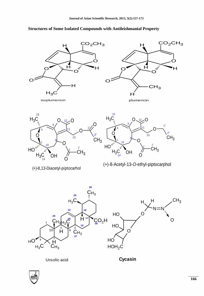
X-ray crystallography has also confirmed the presence of plumericin and isoplumericin from bio-
guided fractionation of the stem bark of Himatanthussucuuba. Both compounds in in vitro showed
marked activity against Leishmania amazonensis axenic amastigotes (Castillo et al., 2007).In other
works, the antileishmanial activity exhibited by Pseudelephantopusspicatus has been attributed to
the presence of 8,13-diacetyl-piptocarphol and the 8-acetyl-13-O-ethyl-piptocarphol and ursolic
acid (Odonne et al., 2011).Two isolated compoundsidentified as oleanolic acid and ursolic acid
obtained from bio-guided fractionation of the twigs extract of Keetialeucantha (K. Krause) Bridson
(syn. Plectronialeucantha Krause) have been reported to have antileishmanial activity (Joanne et
al., 2011). According to other results, evaluation of the antileishmanial activity of ursolic acid
showed activity against promastigotes of Leishmania amazonensisat IC50of 43.8µg/mL and 5
µg/ml, depending on the test (Torres-Santos et al., 2004; Gnoatto et al., 2008) and also against
promastigotes of Leishmania donovani at IC50of 3.5 µg/mL (Moulisha et al., 2010). Oleanolic acid
has also exhibited antileishmanial activity against promastigotes of Leishmania amazonensisat IC50
= 10 µg/mL (Torres-Santos et al., 2004).
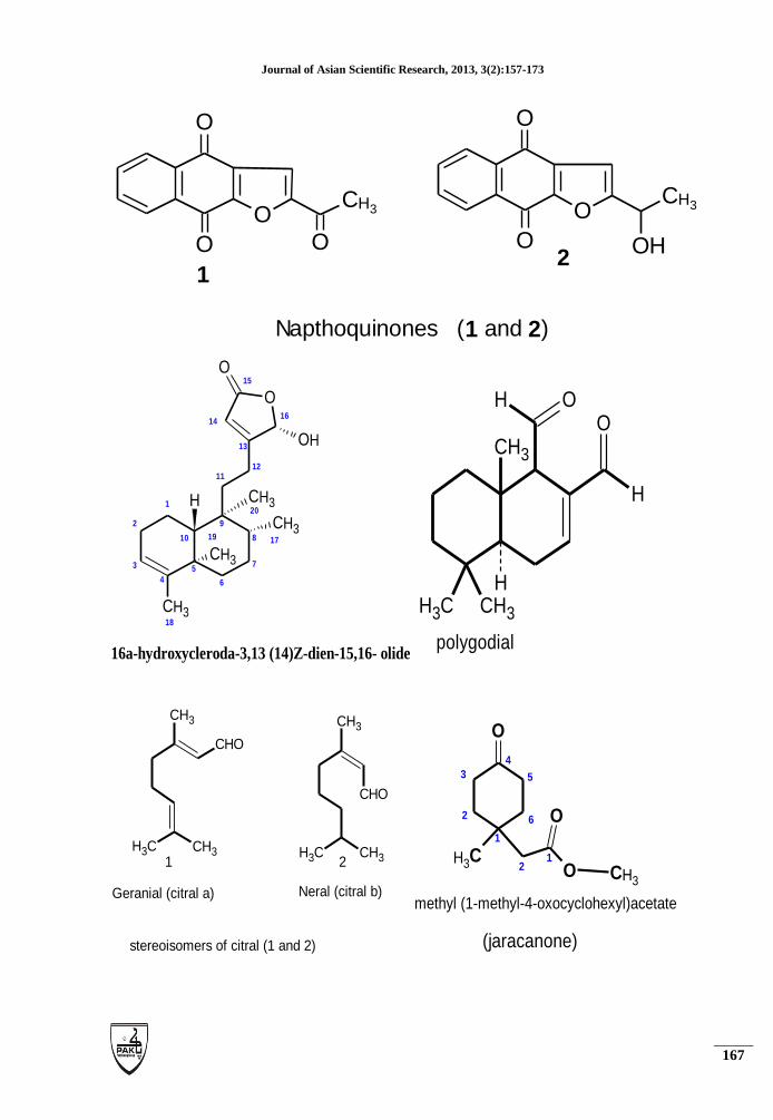
Naphtoquinones and naphtofuranes have been identified as responsible for the antileishmanial
activities from Chondodendrontomentosumbark and Cedrelaodorata (González et al., 2011). Citral
has been reported to the he main active component responsible for the antileishmanial activity of
the essential oil of Cymbopogoncitratus on Leishmania amazonensis (Santin et al., 2009). Again
16a-Hydroxycleroda-3,13 (14)Z-dien-15,16-olide an isolated compound from Polyalthialongifolia
has been found to be orally active against leishmaniadovani parasites and non-cytotoxic (Pragya et
al., 2010). Another bioactive compound obtained through the chromatographic fractionation of the
CH2Cl2 phase from methanol extract from the leaves of Pentacaliadesiderabilis (Vell.) Cuatrec.
(Asteraceae) and identified as jacaranone [methyl (1-hydroxy-4-oxo-2,5cyclohexandienyl) acetate]
have also showed significant activity against promastigotes of Leishmania (L.) chagasi,
Leishmania (V.) braziliensis, and Leishmania amazonensis (Thiago et al., 2012). Also, polygodial
(a derivative of a sesquiterpene) isolated from stem barks of DrimysbrasiliensisMiers
(Winteraceae) has been reported to show leishmanicidal effect against the promastigotes of
Leishmania braziliensis, Leishmania amazonensis, and Leishmania chagasi (Daniela et al., 2011).
Other results have shown that isolated constituents from Tridax precumbens L (an antileishmanial
plant) are saturated and unsaturated fatty acids (Gadre and Gabhe, 1988), flavonoids (Yadava and
Saurabh, 1998; Ali et al., 2001; Akbar et al., 2002), lipids (Verma and Gupta, 1988), and
polysaccharides (Raju and Davidson, 1994). Perhaps the antileishmanial activity exhibited by this
plant is as a result of the presence of some and/or combination of these compounds.
Journal of Asian Scientific Research, 2013, 3(2):157-173
166
Structures of Some Isolated Compounds with Antileishmanial Property
O
OH
CO2CH3H
HO
CH3
H
O
isoplumericin
O
OH
CO2CH3H
HO
H
CH3
O
plumericin
O
O
O
O
CH3 OH
O
CH3
O
CH3
OH
O
CH31
2'
4
10
8
6
15
12
11
13
1'
1''
2''
14
(+)-8,13-Diacetyl-piptocarhol
O
O
O
O
CH3 OH
O
CH3
O
CH3
OH
CH31
2'
4
10
8
6
15
12
11
13
1'
1''
2''
14
(+)-8-Acetyl-13-O-ethyl-piptocarphol
OH
CH3 CH3
H
CH3
H
CH3
CH3
H CO2H
CH3
CH3
1
3
10
25
26
12
27
1528
17
30
18
29
8
Ursolic acid
HOH2C
O
OH
OH
OH
O
H
N
H
N
CH3
O
Cycasin
Journal of Asian Scientific Research, 2013, 3(2):157-173
167
O
O
O O
CH3
Napthoquinones (1 and 2)
O
O
O OH
CH3
12
CH3
H
O
O
OH
CH3
CH3
CH3
1
2
3
4
5
10 19
9
20
8 17
7
6
1112
13
16
15
14
18
16a-hydroxycleroda-3,13 (14)Z-dien-15,16- olide
CH3
OH
H
O
CH3 CH3
H
polygodial
CH3
CH3CH3
CHO
Geranial (citral a)
CH3
CH3CH3
CHO
Neral (citral b)
stereoisomers of citral (1 and 2)
1 2
O
CH3 O CH3
O1
21
2
3
4
5
6
methyl (1-methyl-4-oxocyclohexyl)acetate
(jaracanone)

Journal of Asian Scientific Research, 2013, 3(2):157-173
168
Table-1.List of plants with antileishmanial property.
Plants with
antileishmanial
activity.
Plant Family Part(s) used Solvent used References
Tridax procumbens Asteraceae Whole plant Methanol Sánchez et al., 2007;
Zelmyet al., 2009).
Urechites
andrieuxii
Apocynaceae Leaves and
roots
Methanol Pulido and Serralta,
1993; Argüetaet al.,
1994; Manuel et al.,
2003
Desmodiumgangeti
cum
Fabaceae Whole plant n-butanol Iwuet al., 1992;
Nasibet al., 2005
Pseudelephantopus
spicatus
Asteraceae Leaves,
aerial parts
Ethanol Odonneet al., 2011;
Himatanthussucuub
a
Apocynaceae
Stem bark, Ethanol Villegas et al., 1997;
Castillo et al., 2007
Azadirchtaindica Meliaceae Stem bark Methanol Ahmed et al., 1998;
Maytenussenegalen
sis
Celasteraceae Stem bark Methanol Ahmed et al., 1998;
Pseudocedrelakotsc
ifye
Meliaceae Stem bark Methanol Ahmed et al., 1998;
Balanitesaegyptiac
a
Balanitaceae Seeds, stem
bark
Methanol Ahmed et al., 1998;
Eucalyptus
globulus
Myrtaceae Seeds Methanol Ahmed et al., 1998;
Acanthospermumhi
spidum
Asteraceae Aerial parts Dichlorometha
ne
Joanne et al., 2011
Cymbopogoncitratu
s
Poaceae Leaves Fresh leaves
were steam
distilled using
Clevenger’s
apparatus
Santinet al., 2001
Peschieraaustralis Apocynaceae Stem Ethanol Delorenziet al., 2000
Lantana
ukambensis
Verbenaceae Stem, leaves Methanol Sawadogo et al., 2012
Chondodendrontom
entosum
Menispermacea
e
Bark and
leaves
Ethanol González-Coloma et
al., 2011
Cedrelaodorata L Meliaceae Bark Chloroform,
hexane
González-Coloma et
al., 2011
Pentacaliadesidera
bilis
Asteraceae
Leaves Methanol Thiago et al., 2012
Drimysbrasiliensis
Miers
Winteraceae Stem barks Hexane Daniela et al., 2011).
Polyalthialongifoli
a
Annonaceae, Leaves Ethanol Pragya et al., 2010
Valerianawallichii Valerianaceae Roots Chloroform,
methanol
Ghosh et al., 2011
Journal of Asian Scientific Research, 2013, 3(2):157-173
169
CONCLUSION
The antileishmanial activity of the plants reported in this piece of work suggests that these plants
can be used to treat leishmaniasis. It should however be emphasized that, reported results in this
work have shown that, the level of activity of a particular plant depends largely on the solvent used
for extraction and to some extent the part of the plant used. Isolation and characterization of
extracts from some plants reported in this work to have antileishmanial activity have also shown
the presence of one or more natural product(s). Perhaps the antileishmanial activity of these plants
could be due presence of the natural product(s).
Isolated compounds from Pseudelephantopusspicatus (Odonne et al., 2011) and Keetialeucantha
(K. Krause) Bridson (syn. Plectronialeucantha Krause) (Joanne et al., 2011) have both shown the
presence of ursolic acid. Perhaps, that is the reason why both exhibit antileishmanial
activity.Furthermore, the report that the antileishmanial activity of Lantana ukambensis could be
due to the presence of polyphenols, triterpenes, and saponins (Sawadogo et al., 2012), is in line
with other results that diterpenoidsand triterpenoids (Tan et al., 2002), saponins (Ridoux et al.,
2001), and polyphenols (Kolodziej, 2001) are phytochemical compounds with antileishmanial
effect.
REFERENCES
Ahmed, E., M. Adil, M. Gwiria, G. Thor, K. Arsalam and A. Sami, 1998. The potential
antileishmanial activity of some sudanese medicinal plants. Phytotherapy
Research, 12: 576–579.
Akbar, E., A. Malik, N. Afza and S. Hai, 2002. Flavone glycosides and bergenin
derivatives from tridax procumbens. Heterocycles, 57: 733–739.
Ali, M., E. Ravinder and R. Ramachandram, 2001. A new flavonoid from the aerial parts
of tridax procumbens. Fitoterapia, 72: 313–315.
Andre, L., J. Sirol, C. Le Vourch, J. Lebegorre and D. Cochevelou, 1978. Sudanese kala-
azar in west africa. Med Trop (Mars), 38(4): 435-442.
Arfan, U. and B. Simeen, 2008. Cutaneous eishmaniasis: An overview of parasitology and
host-parasite-vector inter relationship. Journal of Pakistan Association of
Dermatologists, 18(42-48).
Argueta, V., 1994. Atlas de las plantas de la medicina tradicional mexicana. Instituto
Nacional Indigenista, Mexico City, 2: 784.
Boakye, D., M. Wilson and M. Kweku, 2005. A review of leishmaniasis in west africa.
Ghana Medical Journal 39(3): 94-97.
Bryceson, A., 1996. Leishmaniasis.
Buskuhl, H., F. de Oliveira, L. Blind, R. de Freitas, A. Barison, F. Capompas, Y. Corilo,
M. Eberlin, G. Caramori and M. Biavatti, 2010. Sesquiterpene lactones from

Journal of Asian Scientific Research, 2013, 3(2):157-173
170
vernonia scorpioides and their in vitro cytotoxicity. Phytochemistry 71: 1539–
1544.
Castillo, D., J. Arevalo, F. Herrera, C. Ruiz, R. Rojas, E. Rengifo, A. Vaisberg, J. Lock O.
Lemesre, H. Gornitzka and M. Sauvain, 2007. Spirolactone iridoids might be
responsible for the antileishmanial activity of a peruvian traditional remedy made
with himatanthus sucuuba (apocynaceae). Journal of Ethnopharmacology, 112:
410–414.
Chang, K., D. Fong and R. Bray, 1985. Biology of leishmania and leishmaniasis. In:
Chang kp, bray rs, eds. Leishmaniasis: Human parasitic diseases. Philadelphia:
Elsevier, 1: 1-30.
Chawla, B. and R. Madhubala, 2010. Drug targets in leishmania. J Parasit Dis, 34(1): 1-
13.
Conteh, S. and P. Desjeux, 1983. Leishmaniasis in the gambia: A case of cutaneous
leishmaniasis and a case of visceral leishmaniasis. Trans Roy Soc Trop med Hyg
77: 298-302.
Daniela, S., G. Andre, Q. Juliana, N. Noemi, A.P. Oriana, P. Murilo and H.C. Joao, 2011.
Anti-leishmanial and anti-trypanosomal potentialof polygodial isolated from stem
barks of drimys brasiliensismiers (winteraceae). Parasitology Research 109: 231–
236.
de Campos, E., A. Amedome and K. Kpodzro, 1979. Kala-azar in togo, west africa.
Presentation of a clinical case. Rev Inst Med Trop Sao Paulo, 21: 29-32.
Delorenzi, J.C., C.R. M. Attias, M. Gattass, C. Andrade, A.C. Rezende, A.T. Pinto, D.C.
Henriques, E.M.B. Bou-Habib and Saraiva, 2001. Antileishmanial activity of
indol alkaloid from peschiera australis antimicrob. Agents Chemother., Bethesda,
45: 1349-1354.
Desjeux, P., L. Waroquy and J. Dedet, 1981. La leishmaniose cutaneé humaine en afrique
de l’ouest. Bull Soc Path Exot, 4: 414-425.
Dyce, S., 1924. Oriental sore in nigeria. Trans Roy Soc Trop Med Hyg 18: 336.
Gadre, A. and S. Gabhe, 1988. Saturated and unsaturated fatty acids from tridax
procumbens. Indian Journal of Pharmaceutical Sciences 50: 168.
Ghosh, S., D. Sukalyani, H. Sudipta, H. Andreas, T. Katja, S. Martina, K. Petra, S. Uta,
M. Heidrun, H. Ulrike and H. Banasri, 2011. Valeriana wallichii root extracts and
fractions with activityagainst leishmania spp. Parasitology Research 108: 861–
871.
Gnoatto, S., V. Dalla, L. , C. Lencina, K. Dassonville, A, , S. Da Nascimento, D.
Mossalayi, J. Guillon, G. Gosmann and P. Sonnet, 2008. Synthesis and
preliminary evaluation of new ursolic and oleanolic acids derivatives as
antileishmanial agents. Journal of Enzyme Inhibition and Medicinal Chemistry,
23: 604–610.

Journal of Asian Scientific Research, 2013, 3(2):157-173
171
González, C., A. , R. Matías, S. Claudia, L. Rodney, R. Lastenia, J. Vicente, S. Jesus and
A.M. Rafael, 2011. Antileishmanial, antitrypanosomal, and cytotoxic screening of
ethnopharmacologically selected peruvian plants. Parasitology Research, 110:
1381–1392.
Greenwood, B., A. Adjukiewicz, S. Conteh, P. Hagan, D. Mabey and L. Panton, 1984.
Leishmaniasis in the gambia. Is its incidence increasing? . Trans Roy Soc Trop
Med Hyg, 78: 407-409.
Iwu, M., J. Jackson, J. Tally and D. Klayman, 1992. Evaluation of plant extracts for
antileishmanial activity using a mechanism-based radio respirometric
microtechnique (ram). Planta Medica 58: 436–441.
Jiu, J., 1966. A survey of some medicinal plants of mexico for selected biological
activities. Lloydia, 29: 250–259.
Joanne, B., H. Veronique, C. Gabrielle, F. Marie, H. and Q. Joelle, 2011. In vitro
antitrypanosomal and antileishmanial activity of plants used in benin in traditional
medicine and bio-guided fractionation of the most active extract. Journal of
Ethnopharmacology, 137: 998– 1002.
Kendrick, R., 1986. Preliminary field observations on the flight speed of a phelbotomine
sandfly. Trans R Soc Trop Med Hyg, 80: 138-142.
Kolodziej, H., 2001. Proanthocyanidins and related compounds: Antileishamanial activity
and modulatory effects on nitric oxide and tumor necrosis factor-a-release in the
murine macrophage like cell line rae 264.7. Biol Pharm Bull 24: 1016–1021.
Lefrou, G., 1948. La leishmaniose cutanée au soudan français. Fréquence de la forme
sèche a papulo-tuberculeuse. Bull Soc Path Exot 41: 622-627.
Luciana, I., M. Felipe, d.M. Eliane, Teixeira., , S.d.S. Bruna, Lima., , V. Verônica and R.
Ana, 2012. Validation of quantitative real-time pcr for the in vitro assessment of
antileishmanial drug activity. Experimental Parasitology 131: 175–179.
Manuel, J., B. Elfride, D. Eric, M. Victoria, D. Rafael and M. Luis, 2003. Variation of
leishmanicidal activity in four populations of urechites andrieuxii. Journal of
Ethnopharmacology 86: 243–247.
Morsy, T., 1996. Cutaneous leishmaniasis in egypt (review and comment). J Egypt Soc
Parasitol 26 105-130.
Moulisha, B., G.A. Kumar and H.P. Kanti, 2010. Anti-leishmanial and anti-cancer
activities of a pentacyclic triterpenoid isolated from the leaves of terminalia arjuna
combretaceae. Tropical Journal of Pharmaceutical Research, 9 135–140.
Nasib, S., K. Pushpesh, K. Aruna, R. Kamal, M. Rakesh and D. Anuradha, 2005. Efficacy
of desmodium gangeticum extract and its fractions against experimental visceral
leishmaniasis. Journal of Ethnopharmacology 98: 83–88.
Odonne, G., G. Herbette, V. Eparviera, G. Bourdye, R. Rojas, M. Sauvaine and D. Stiena,
2011. Antileishmanial sesquiterpene lactones from pseudelephantopus spicatus, a
Journal of Asian Scientific Research, 2013, 3(2):157-173
172
traditional remedy from the chayahuita amerindians (peru). Part III Journal of
Ethnopharmacology 137: 875– 879.
Peraza-Sanchez, S., F. Cen-Pacheco, A. Noh-Chimal, F. May-Pat, P. Sima-Polanco, E.
Dumonteil, M. Garcia-Miss and M. Mut-Martín, 2007. Leishmanicidal evaluation
of extracts from native plants of the yucatan peninsula. Fitoterapia, 78: 315–318.
Pragya, M., V. Koneni, P. Suriya, K. Awanish, G. Reema, S. Shailendra, S. Souvik, H.
Majumder, l.K. Ani and D. Anuradha, 2010. 16a-hydroxycleroda-3,13 (14)z-dien-
15,16-olidefrom polyalthia longifolia: A safe and orally activeantileishmanial
agent. British Journal of Pharmacology 159: 1143–1150.
Pulido, M. and L. Serralta, 1993. Lista anotada de las plantas medicinales de uso actual en
el estado de quintana roo, méxico. Centro de Investigaciones de Quintana Roo,
Chetumal, Quintana Roo, México: 6.
Rageu, J., 1951. Phlébotomes du cameroon. Bull Soc Path Exot, 44: 793-800.
Raju, T.S. and E.A. Davidson, 1994. Structural features of water-soluble novel
polysaccharide components from the leaves of tridax procumbens linn.
Carbohydrate Res, 258: 243-254.
Richard, R., C. Jean, D. , L. Hechmi, P. Claude, A. Bruce and B. Simon, 2007. Cutaneous
leishmaniasis. Lancet Infectious Diseases, 7: 581–596.
Ridoux, O., C. Di Giorigi and D. F., 2001. In vitro antileishmanial activity of three
saponins isolated from ivy, α-hederin, β-hederin and hederacolchiside a1, in
association with pentamidine and amphotericin. Phytotherapy Research 15: 298-
301.
Riou, M. and M. Advier, 1993. Leishmaniose cutanée cantractée au senegal. Bull Soc Path
Exot 26: 254-256.
Santin, M., A. dos Santos, C. Nakamura, F. Dias, I. Ferreira and N. Ueda, T. , 2009. In
vitro activity of the essential oil of cymbopogon citratus and its major component
(citral) on leishmania amazonensis. Parasitology Research 105(6): 1489–1496.
Sawadogo, W., G. Le Douaron, A. Maciuk, C. Bories, P. Loiseau, B. Figadere, I. Guissou
and O.G. Nacoulma, 2012. In vitro antileishmanial and antitrypanosomal
activities of five medicinal plants from burkina faso. Parasitology Research 110
1779–1783.
Sawadogo, W., A. Maciuk, J. Banzouzi, P. Champy, B. Figadere and I. Guissou, 2011.
Mutagenic effect, antioxidant and anticancer activities of six medicinal plants
from burkina faso. Natural ProductResearch, 26: 575-579.
Sewice, M., 1980. Phlebotomine sandflies (order diptera: Family psychodidae). A Guide
to Medical Entomology: 78-82.
Sheik-Mohamed, A. and J. Velema, 1999. Where health care has no access: The nomadic
populations of sub-saharan africa. Trop Med Int Hlth, 4: 695-707.
Simon, L.K., S. and Y. Vanessa, 2006. Current scenario of drug development for
leishmaniasis. Indian J Med Res, 123: 399-410.

Journal of Asian Scientific Research, 2013, 3(2):157-173
173
Sirol, J., P. Delpy, M. Lefevre and J. Vedy, 1972. Kala-azar in tchad republic. Does an
endemic exist in central and west africa? . Bull Acad Natl Med 156: 395-407.
Strobel, M., B.N. Diaye, J. Marchand and J. Dedet, 1978. Cas de leishmaniose cu-tanee
avec atteinte muqueuse ay senegal. Bull Soc Path Exot Filiales, 71: 423-429.
Tan, N., M. Kaloga, O. Radtke, A. Kiderlen, S. Öksüz, A. Ulubelen and H. Kolodziej,
2002. Abietane diterpenoids and triterpenoic acids from salvia cilicicaand their
antileishmanial activity. Phytochemistry, 61: 881–884.
The Center for Food Security & Public Health., 2009. Leishmaniasis (cutaneous and
visceral). Available from
http://www.cfsph.iastate.edu/Factsheets/pdfs/leishmaniasis.pdf.
The Centre for Food Security & Public Health., 2006. Leishmaniasis (fast facts).
Available from http://www.cfsph.iastate.edu/FastFacts/pdfs/leishmaniasis_F.pdf.
Thiago, R., R. Paulete, A. Oriana, Q. Juliana, C. Walkyria, G. Andre, D. Angelica, M.
Silvia, H. Joao, S. Patricia and J. Marcelo, 2012. Anti-malarial, anti-
trypanosomal, and anti-leishmanial activities of jacaranone isolated from
pentacalia desiderabilis (vell.) cuatrec. (asteraceae). Parasitology Research 110:
95–101.
Torres-Santos, E., D. Lopes, R. Oliveira, J. Carauta, C. Falcao, A. Kaplan and B. Rossi-
Bergmann, 2004. Antileishmanial activity of isolated triterpenoids from pourouma
guianensis. Phytomedicine, 11 114–120.
Vanhalen, M., J. Lejoly, M. Hanocq and L. Molle, 1991. The medicinal plant industry.
CRC Press, Boca Raton, FL.
Verma, R. and M. Gupta, 1988. Lipid constituents of tridax procumbens. Phytochemistry
27: 459-463.
Villegas, L., I. Fernandez, H. Maldonado, R. Torres, A. Zavaleta, A. Vaisberg and G.
Hammond, 1997. Evaluation of the wound-healing activity of selected traditional
medicinal plants from peru. Journal of Ethnopharmacology, 55: 193–200.
Viscencio, d.l.R., G. , S. Tamay, P. , M. Issac, AP. and D. Lezama, CM. , 1995.
Toxicidad in vitro de extractos de urechitesandrieuxii muell.-arg. En contra de l.
Mexicana. Memorias de la III Reunión de Investigacion Qu´ımica en el Sureste de
Mexico, Merida, Yucatán: 93.
World Health Organistion, W., 2010. Control of the leishmaniasis: Report of a meeting of
the who expert committee on the control of leishmaniases, geneva.
Yadava, R. and K. Saurabh, 1998. A new flavone glycoside: 5,7,4 -́trihydroxy-6,3 -́
dimethoxyflavone-5-o-α-l-rhamnopyranoside from the leaves of
tridaxprocumbens linn. Journal of Asian Natural Products Research 1: 147–152.
Zhelmy, M., M. Rosa, G. Francisco, J. Manuel, W. Luis and R. Sergio, 2009. In vitro
activity of tridax procumbens against promastigotes of leishmania mexicana.
Journal of Ethnopharmacology 122: 463–467.