Embed Size (px)
Citation preview
International Journal of Engineering Sciences &Research Technology
(A Peer Reviewed Online Journal) Impact Factor: 5.164
IJESRT
Chief Editor Executive Editor
Dr. J.B. Helonde Mr. Somil Mayur Shah
Website:www.ijesrt.com Mail:[email protected] O
IJESRT: 7(11), November, 2018 ISSN: 2277-9655
I X
ISSN: 2277-9655
[Deshpande * et al., 7(11): November, 2018] Impact Factor: 5.164
IC™ Value: 3.00 CODEN: IJESS7
http: // www.ijesrt.com© International Journal of Engineering Sciences & Research Technology
[56]
IJESRT is licensed under a Creative Commons Attribution 4.0 International License.
IJESRT INTERNATIONAL JOURNAL OF ENGINEERING SCIENCES & RESEARCH
TECHNOLOGY EFFECT OF NANOPATICLES ON PHARMACOKINETIC AND
PHARMACODYNAMIC PROPERTIES OF ANTIMALARIAL AGENTS Pallav Kaushik Deshpande1, Ragini Gothalwal1 & Anupam Kumar Pathak2
1 Department of Biotechnology, Barkatullah University,Bhopal,(M.P):462026 2 Department of Pharmacy, Sagar Institute of Research and Technology , Bhopal. (M.P)
DOI: 10.5281/zenodo.1488705
ABSTRACT Nanoparticles play an important role in a wide variety of fields including advanced materials, pharmaceuticals,
and environmental detection and monitoring. They have been used in vivo to protect the drug entity in the
systemic circulation, restrict access of the drug to the chosen sites and to deliver the drug at a controlled and
sustained rate to the site of action. Many therapeutic agents have not been successful because of their limited
ability to reach to the target site. There are various aspects of nanoparticle formulation, characterization, effect
of their characteristics and their applications in delivery of drug molecules and can be exploited to achieve
maximum potential of therapeutic agent . In present study designing nanoparticles , as a delivery system are to
control particle size, surface properties and release of pharmacologically active agents in order to achieve the
site-specific action of the drug at the therapeutically optimal rate and dose regimen against Plasmodium in mice
model. Extracts of Azadirachta indica, Cinchona officinalis and Artemisia annua having anti plasmodium
activities and chloroquine, mefloquine, artesunate synthetic drugs were selected in the study. Results obtained
signifies the ability of nanoparticle to entrap hydrophilic and hydrophobic drugs with concomitant reduction in
their toxicity potential, their versatility and their amenability for surface modification are the major factors
responsible for their popularity in drug delivery research.
KEYWORDS: Nanoparticle , pharmaceuticals, pharmacokinetic , pharmacodynamic, antimalarial
1. INTRODUCTION Nanoparticles are subnanosized colloidal structures composed of synthetic or semi synthetic polymers.
Depending on the method of preparation, nanoparticles, nanopspheres or nanocapsules can be obtained with
different properties and release characteristics for the encapsulated therapeutic agent.Nanocapsules are vesicular
systems in which the drug is confined to acavity surrounded by a unique polymer membrane, whereas
nanopspheres are matrix systems in which the drug is physically and uniformly dispersed. In recent years,
biodegradable polymeric nanoparticles, particularly those coated with hydrophilic polymer such as poly
(ethylene glycol), have been used as potential drug delivery devices because of their ability to circulate for a
prolonged period of time.(Vyas et al., 2002).
2. MATERIAL AND METHODS Plant material Plant selected for antiplasmodium activity: Artemisia annua, Cinchona officinalis, Azadirachta indica . All the
plants were authenticated and herbarium was deposited at department of pharmacy, Barkatulah
University,Bhopal.
Preparation And Extraction Procedure For Plant Material Prepration of plant material.
Materials of the three plants selected for the present study were grinded to coarse powder and stored in airtight
containers at room temperature in the dark until used.(Trease and Evans 1978).
ISSN: 2277-9655
[Deshpande * et al., 7(11): November, 2018] Impact Factor: 5.164
IC™ Value: 3.00 CODEN: IJESS7
http: // www.ijesrt.com© International Journal of Engineering Sciences & Research Technology
[57]
IJESRT is licensed under a Creative Commons Attribution 4.0 International License.
Method For Alcoholic Extraction
Extraction for Cinchona officinalis The powdered Cinchona bark (50g) was mixed with calcium oxide (30gms), water (40 ml) and 5% sodium
hydroxide to form a paste and kept overnight.Then obtained paste was packed in Soxhlet apparatus and
extracted with methanol for 8 hrs at 60ºC and processed as adopted by Kokate, 2009, detailed method was given
in Deshpande et al 2014 .Then stored at 4ºCuntil use in sealed bottle, protect against light and moisture.
Extraction of Azadirachta Indica
50 g of air‐dried powder was soaked in ethyl alcohol (100 ml) and kept for over night.Then it was filtered
through 8 layers of muslin cloth, filtrate was collected and residue was discarded. Filtrate was packed in soxhlet
apparatus and extraction was done for 8 hrs at 80-90ºC.Solvent was distilled off to one –fourth to yield alcoholic
extract( Deshpande et al 2014).
Extraction for Artimisia annua Artemisinin, a sesquiterpene lactone is an antimalarial constituent of Artimisia annua. Extraction process which
was followed in present study is: Dried leaves of artimisia annua are coarsely powdered and extracted with the
help of petroleum ether (over night). Petroleum ether was then filtered then concentrated to dryness.
Concentrated extract found was then dissolved in chloroform. Aerial part of Artemisia annua were dried in
shade, grounded to coursed powder stored in air tight amber colored bottle. Artemisinin, a sesquiterpene lactone
is an antimalarial constituent of Artimisia annua. Extraction process which was followed in this study. Dried
leaves of Artimisia annua were coarsely powdered and extracted with the help of petroleum ether (over night).
Petroleum ether was then filtered then concentrated to dryness with the help soxhlet then concentrated extract
found was then dissolved in chloroform. Acetonitrile was added to the chloroformic solution which is
responsible for the separation of impurities and pricipetation of waxes. Filtration was done to separate out the
impurities. The filtrate was again concentrated and was subjected to cool down. Crystals of artimisinin were
deposited which were further purified by washing and recrystallization with alcohol.(Trease and Evans 1978,
Kokate, 2009 , Deshpande et al 2014).
Method for Aqueous extraction The powdered material of all the selected plants (50g) separately were soaked in water (100 ml) and kept for
overnight. Then it was filtered through 8 layers of muslin cloth, filtrate was collected and residue was discarded.
Filtrate was packed in soxhlet apparatus and extraction was done for 6- 8 hrs at 80-90ºC. Solvent was distilled
off to one –fourth to yield aqueous extract. Collected extract was then weighed and stored at 4ºC until use.
Preparation and optimization of chitosan nanoparticles
Chitosen nanoparticles were prepared with suitable modification based on the ionotropic gelation with Tpp
anions. Chitosan (2mg ml-1) was dissolved in aqueous acetic acid (pH 4.0) solution and Tpp was dissolved in
distilled water at the concentration of 1mg/ml. Drug (10%) was added to Tpp solution (by dissolving Tpp in
100ml distilled water under continuous stirring on magnetic stirring).Finally 1.5 ml of drug containing Tpp
solution was added to 4 ml of chitosan solution with the help of syringe needle under magnetic stirring at room
temperature.The dispersion so formed was sonicated for 15 minutes, then disperse in water and centrifuge the
sample at 12000 rpm for 30 minute. The supernatant was discarded and pallet was resupended in distilled water
and lyophilized and stored at -20ºC till further use, and sample was further characterized.
Study of process variables and optimization involve in chitosan nanoparticles.
The number of variable observed in preparation of nanoparticles.They have different properties like surface
morphology, particle size, drug content and release profile. For optimization of nanoparticle different parameter
which are under consideration in this research work.
Polymer concentration.
Effect of acetic acid concentration.
Speed of magnetic stirrer.
Effect of TPP concentration
Drug encapsulation efficiency.
The entrapment efficiency of drug and extracts was determined by using following equation.
ISSN: 2277-9655
[Deshpande * et al., 7(11): November, 2018] Impact Factor: 5.164
IC™ Value: 3.00 CODEN: IJESS7
http: // www.ijesrt.com© International Journal of Engineering Sciences & Research Technology
[58]
IJESRT is licensed under a Creative Commons Attribution 4.0 International License.
𝑬𝒏𝒄𝒂𝒑𝒔𝒖𝒍𝒂𝒕𝒊𝒐𝒏 𝒆𝒇𝒇𝒊𝒄𝒊𝒆𝒏𝒄𝒚 (%) = 𝑻𝒐𝒕𝒂𝒍 𝒅𝒓𝒖𝒈 − 𝑭𝒓𝒆𝒆 𝒅𝒓𝒖𝒈
𝑻𝒐𝒕𝒂𝒍 𝒅𝒓𝒖𝒈 × 𝟏𝟎𝟎
About 10ml of nanoparticulate suspension was digested with minimum amount of ethanolic solution (Water:
ethanol in 7:3 ratios). The digested homogenates were centrifuged at 12000 rpm for 30 min and supernatant was
analysed for drug entrapment. Entrapped drug and extracts in this study were measured at their respective λ
maximum.
In vitro release studies
In vitro release of hydrophilic molecule out of nanoparticles was measured spectrophotometrically by
incubation of liposomes in 1OmM Tris buffer (pH: 7.4) at 37°C in mild shaking (35 rpm) water bath under
mild-shaking conditions for determined time intervals. Dialysis bags (Dialysis membrane 110, Hi Media, India)
were immersed in water for one hour to remove the preservatives followed by rinsing in phosphate buffered
saline (PBS) solution. The drug and extract encapsulated nanoparticles were placed in PBS and loaded in the
dialysis bag. The bag was sealed at both the ends and immersed in 4 mL of PBS with 10% methanol (Katrin,
1995). The release of the drug and extract was evaluated at three different pH values (1.2, 7.4 and 9.0). A pH of
1.2 was maintained using 0.1 M HCl -KCl buffer while pH 9.0 was maintained using 0.1 M phosphate buffer.
Samples were withdrawn at time intervals of 0.5, 1, 2, 4, 6, 8, 24-h, periodically( Cabral et al , 2004)
Morphological characterization
The morphology of the nanoparticles was determined using a scanning electron microscope (JEOL 6701F, from
AIIMS New Delhi).The samples were placed over a carbon paste coated stub and sputter coated with a thin
layer of platinum prior to viewing. For negative staining, 2% (w/v) phosphotungstic acid was added to the
liposome samples and incubated at room temperature for 24 hours. This sample was freeze dried and imaged
using scanning electron microscope. The transmission electron micrographs of the liposomes were obtained
using JEM 1011, JEOL. The lyophilized liposome sample was dispersed in 0.5 mL PBS. To 50 μL of this
dispersion, an equal volume of double distilled water was added and placed on a carbon coated grid. The excess
water was absorbed using a filter paper and uranyl acetate stain was added. The grid was then washed with
water to remove excess uranyl acetate and then dried before it was loaded in the specimen chamber.
3. RESULTS AND DISCUSSION All extracts which were to be screened ( Deshpande et al 2015)for the anti plasmodium activity in vitro model
were resuspended in DMSO (1mg ml-1 stock), dilutions was prepared from the stock (under aseptic conditions)
all doses were chosen based on a pre dose finding test for extracts from the literature values for plant. LD50
obtained for each extract was calculated.Similarly synthetic drugs were screened for activity against
P.falciparum culture maintained in laboratory. Culture was synchronized with sorbitol treatment before starting
the screening protocol. LD50 of drugs are calculated with probit graph .
Optimization of chitosan nanoparticles
Polymer concentration
Chitosen solution of different concentration (20mg 10ml-1 - 40mg 20ml-1) was prepared to study the effect of
polymer concentration on particle size.
Effect of acetic acid concentration
Acetic acid concentration was varied from 0.5-2% to study the effect on particle size.
Speed of magnetic stirrer Speed of magnetic stirrer was varied from (1000-5000 rpm) to study the effect on particle size.
Effect of TPP concentration The cross linking agent concentration was varies from 1mg/ml to 3mg/ml to study the effect on particle size.
Different batches were prepared by inotropic gelation method by varying the parameters mentioned above and
their effect on different properties was observed and summarized. From the above observations better results
were obtained when polymer concentration was 20mg 10ml-1, Cross linking agent concentration was 1mg/ml,
ISSN: 2277-9655
[Deshpande * et al., 7(11): November, 2018] Impact Factor: 5.164
IC™ Value: 3.00 CODEN: IJESS7
http: // www.ijesrt.com© International Journal of Engineering Sciences & Research Technology
[59]
IJESRT is licensed under a Creative Commons Attribution 4.0 International License.
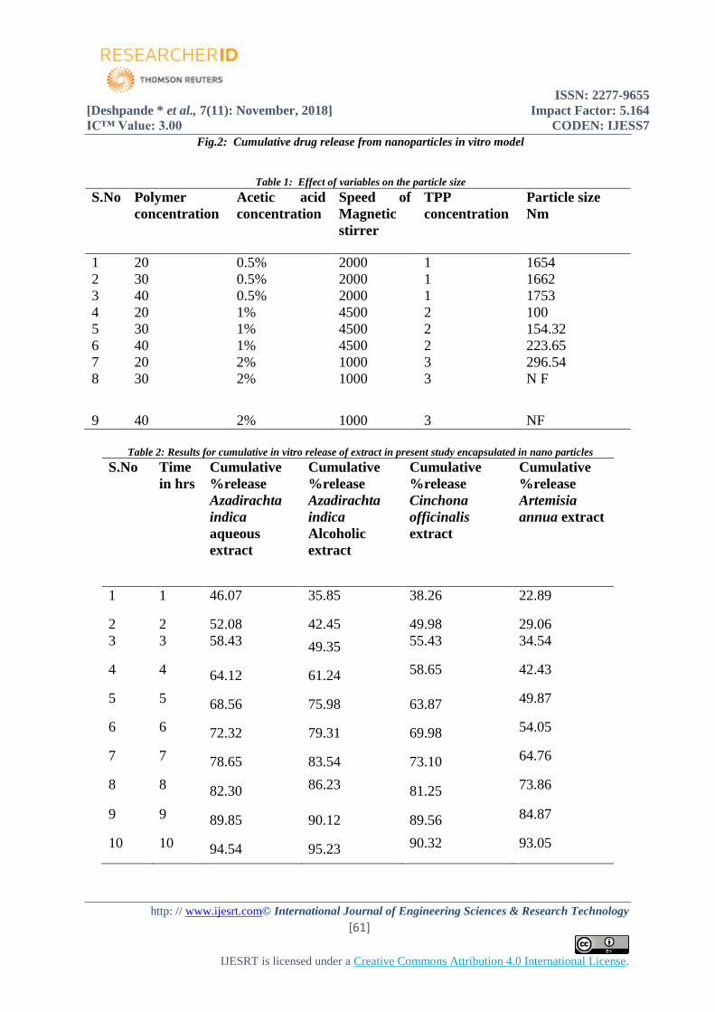
acetic acid 1% and the speed of magnetic stirrer 4500 rpm. Results of variables and their effect (Table 1) on
nanoparticle size is discussed after morphological description of nanoparticles.
Surface morphology
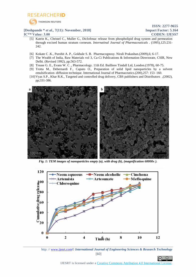
TEM was carried out to investigate the morphology of nanoparticles.TEM images reveals that drug and extract
loaded nanoparticles displayed a spherical shape with smooth surface and no aggregation were observed. No
differences in morphological properties were reported between drug/extract loaded nanoparticles and empty
nanoparticles. Nanoparticles with narrow size distribution could be achieved by appropriate stirring strength in
preparation process (Fig. 1a ,1b) TEM images of nanoparticles obtained.
Percent encapsulation efficiency
Nanoparticulate suspension was digested with minimum amount of ethanolic solution. Then digested
homogenates were centrifuged at 12000 rpm for 30 min and supernatant was (Table: 2) analysed for drug
entrapment. PBS of pH 7.4 was used as suspension media. Entrapped drug and extracts in this study were
measured at their respective λ maximum.
λ max of Azadirachta indica extract : 577nm,for Cinchona officinalis extract : 250nm, for Artemisia annua
extract: 365nm,for Chloroquine: 343 nm, for Mefloquine:283nm ,for Artesunate : 287 nm.
Cumulative percentage of in vitro release of extract encapsulated in nanoparticle
Extract encapsulated in nanoparticulate was analyzed for in vitro release pattern. The extract encapsulated
nanoparticles were placed in PBS and loaded in the dialysis bag. The bag was sealed at both the ends and
immersed in 4 ml of PBS with 10% methanol (Katrin, 1995). For each extract release test was repeated for
three times and cumulative release (Table 3 and Fig: 2) value for each extract was determined. For analysis
reading were recorded at λ max of each drug and extract. Entrapped drug and extracts in this study were
measured at their respective λ maximum for Azadirachta indica extract : 577nm,for Cinchona officinalis
extract : 250nm, for Artemisia annua extract: 365nm,for Chloroquine: 343 nm, for Mefloquine:283nm ,for
Artesunate : 287 nm. Cumulative release value for each extract is given in Table 3.
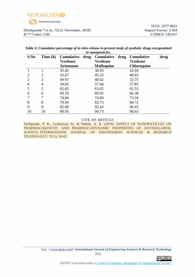
Cumulative percentage of in vitro release of synthetic drugs encapsulated in nanoparticles
Synthetic drugs encapsulated in nanoparticulate were analyzed for in vitro release pattern. The drug
encapsulated nanoparticles were placed in PBS and loaded in the dialysis bag. The bag was sealed at both the
ends and immersed in 4 ml of PBS with 10% methanol (Katrin, 1995). For each drug release test was repeated
for three times and cumulative drug release (Table 3) value for each set was determined .Cumulative release
value for each drug is given below. Graphic representation for cumulative drug release of nanoparticle
entrapped drug and extract is given in Fig. 2.
4. CONCLUSION In present study we had obtained nanoparticles of 100 to1753 nm in size, with efficiency upto 90% with plant
extracts and synthetic drugs, showing smooth surface when studied by TEM, no morphological defects were
observed in TEM study. In vitro release pattern is uniform and controlled, and follows first order of kinetics.
Similar results were reported by various authors in their study related to nanoparticle as a tool for drug delivery.
REFERENCES [1] Cabral E.C.M. , Zollner R .L., Santana M .H.A., Preparation and characterization of liosomes
entrapping allergenic proteins . Brazilian Journal of Chemical Engineering. (2004), 21, 2 ,137 – 146.
[2] Deshpande P.K., Gothalwal R. ,Pathak A.K., Evaluation for Efficiency of Drug Delivery Systems In
vitro and In vivo Plasmodium Culture using Crude Extract of Artemisia annua and artesunate . Current
Research in Pharmaceutical Sciences.(2014),4 :2; 64-69.
[3] Deshpande P.K., Gothalwal R. ,Pathak A.K., Efficiency of Liposomal Drug Delivery System Against
Plasmodium In Vitro and In Vivo Culture International Journal of Biotechnology and
Biochemistry.(2015),11:2 ; 105-114.
[4] Gayathri V. P.. Biopolymer Albumin for Diagnosis and in Drug Delivery. Drug develoement research
.(2003), 58:219–247.
ISSN: 2277-9655
[Deshpande * et al., 7(11): November, 2018] Impact Factor: 5.164
IC™ Value: 3.00 CODEN: IJESS7
http: // www.ijesrt.com© International Journal of Engineering Sciences & Research Technology
[60]
IJESRT is licensed under a Creative Commons Attribution 4.0 International License.
[5] Katrin K., Christel C., Muller G., Diclofenac release from phospholipid drug system and permeation
through excised human stratum corneum. Internatinal Journal of Pharmaceuticals . (1995),125:231-
242.
[6] Kokate C .K., Purohit A. P., Gokhale S. B. Pharmacognosy. Nirali Prakashan.(2009),6; 6-17.
[7] The Wealth of India, Raw Materials vol 3, Ca-Ci Publications & Information Directorate, CSIR, New
Delhi. (Revised 1992), pp;563-572.
[8] Trease G. E., Evans W. C ., Pharmacology. 11th Ed. Bailliere Tindall Ltd, London.(1978), 60-75.
[9] Trotta M., Debernardi F., Caputo O., Preparation of solid lipid nanoparticles by a solvent
emulsification–diffusion technique. International Journal of Pharmaceutics.(200),257: 153–160.
[10] Vyas S.P., Khar R.K., Targeted and controlled drug delivery, CBS publishers and Distributors ,(2002),
pp;331-386.
Fig. 1: TEM images of nanoparticles empty (a), with drug (b), (magnification 60000x ).
ISSN: 2277-9655
[Deshpande * et al., 7(11): November, 2018] Impact Factor: 5.164
IC™ Value: 3.00 CODEN: IJESS7
http: // www.ijesrt.com© International Journal of Engineering Sciences & Research Technology
[61]
IJESRT is licensed under a Creative Commons Attribution 4.0 International License.
Fig.2: Cumulative drug release from nanoparticles in vitro model
Table 1: Effect of variables on the particle size
S.No Polymer
concentration
Acetic acid
concentration
Speed of
Magnetic
stirrer
TPP
concentration
Particle size
Nm
1 20 0.5% 2000 1 1654
2 30 0.5% 2000 1 1662
3 40 0.5% 2000 1 1753
4 20 1% 4500 2 100
5 30 1% 4500 2 154.32
6 40 1% 4500 2 223.65
7 20 2% 1000 3 296.54
8 30 2% 1000 3 N F
9 40 2% 1000 3 NF
Table 2: Results for cumulative in vitro release of extract in present study encapsulated in nano particles
S.No Time
in hrs
Cumulative
%release
Azadirachta
indica
aqueous
extract
Cumulative
%release
Azadirachta
indica
Alcoholic
extract
Cumulative
%release
Cinchona
officinalis
extract
Cumulative
%release
Artemisia
annua extract
1 1 46.07 35.85 38.26 22.89
2 2 52.08 42.45 49.98 29.06
3 3 58.43 49.35 55.43 34.54
4 4 64.12 61.24 58.65 42.43
5 5 68.56 75.98 63.87 49.87
6 6 72.32 79.31 69.98 54.05
7 7 78.65 83.54 73.10 64.76
8 8 82.30 86.23 81.25 73.86
9 9 89.85 90.12 89.56 84.87
10 10 94.54 95.23 90.32 93.05
ISSN: 2277-9655
[Deshpande * et al., 7(11): November, 2018] Impact Factor: 5.164
IC™ Value: 3.00 CODEN: IJESS7
http: // www.ijesrt.com© International Journal of Engineering Sciences & Research Technology
[62]
IJESRT is licensed under a Creative Commons Attribution 4.0 International License.
Table 3: Cumulative percentage of in vitro release in present study of synthetic drugs encapsulated
in nanoparticles.
S.No Time (h) Cumulative drug
%release
Artesunate
Cumulative drug
%release
Mafloquine
Cumulative drug
%release
Chloroquine
1 1 35.45 38.93 42.04
2 2 43.67 45.32 49.63
3 3 49.97 49.42 53.75
4 4 54.65 57.64 57.85
5 5 62.65 63.65 61.53
6 6 69.76 69.95 66.38
7 7 74,84 74.84 73.34
8 8 79.64 83.73 84.72
9 9 82.08 92.43 90.43
10 10 89.95 94.73 96.63
CITE AN ARTICLE
Deshpande, P. K., Gothalwal, R., & Pathak, A. K. (2018). EFFECT OF NANOPATICLES ON
PHARMACOKINETIC AND PHARMACODYNAMIC PROPERTIES OF ANTIMALARIAL
AGENTS. INTERNATIONAL JOURNAL OF ENGINEERING SCIENCES & RESEARCH
TECHNOLOGY, 7(11), 56-62.


![ISSN: 2277-9655 (I2OR), Publication Impact Factor: 3.785 ... /Archive-2016/January-2016/82.pdf · [Adari, 5(1): January, 2016] ISSN: 2277-9655 (I2OR), Publication Impact Factor: 3.785](https://img.dokumen.tips/doc/110x75/5b515b5b7f8b9a6b118bf4f6/issn-2277-9655-i2or-publication-impact-factor-3785-archive-2016january-201682pdf.jpg)


![JESRT: 9(5), May, 2020 ISSN: 2277-9655 I International ... /Archive-2020/May-2020/2.pdf · ISSN: 2277-9655 [Kabeyi et al., 9(5): May, 2020] Impact Factor: 5.164 IC™ Value: 3.00](https://img.dokumen.tips/doc/110x75/5f0882f77e708231d4225fc8/jesrt-95-may-2020-issn-2277-9655-i-international-archive-2020may-20202pdf.jpg)


![JESRT: 8(1), January, 2019 ISSN: 2277-9655 I nternational …ijesrt.com/issues /Archive-2019/january-2019/38.pdf · proves potential in the management of urolithiasis [6, 7]. Antioxidants](https://img.dokumen.tips/doc/110x75/5caea69188c99323378c8bd8/jesrt-81-january-2019-issn-2277-9655-i-nternational-archive-2019january-201938pdf.jpg)

![JESRT: 7(12), December, 2018 ISSN: 2277-9655 I International … /Archive-2018/December-2018... · 2018. 12. 15. · ISSN: 2277-9655 [DEGBEGNON * et al., 7(12): December, 2018] Impact](https://img.dokumen.tips/doc/110x75/5fec535839884410451b9530/jesrt-712-december-2018-issn-2277-9655-i-international-archive-2018december-2018.jpg)