Embed Size (px)
Citation preview
Guidelines for the management of hospital-acquired pneumonia in theUK: Report of the Working Party on Hospital-Acquired Pneumonia
of the British Society for Antimicrobial Chemotherapy
R. G. Masterton1*, A. Galloway2, G. French3, M. Street4, J. Armstrong5, E. Brown6, J. Cleverley7,
P. Dilworth8, C. Fry9, A. D. Gascoigne10, Alan Knox11, Dilip Nathwani12,
Robert Spencer13 and Mark Wilcox14
1Department of Microbiology, Crosshouse Hospital, Kilmarnock, UK; 2Department of Microbiology, Royal
Victoria Infirmary, Queen Victoria Road, Newcastle-upon-Tyne, UK; 3Department of Infection, Guy’s and
St Thomas’s NHS Foundation Trust and King’s College, St Thomas’ Hospital, London, UK; 4Department of
Intensive Care, Royal Sussex County Hospital, Brighton, UK; 5Department of Public Health, North Durham
Strategic Health Authority, Earls House, Durham, UK; 6Department of Microbiology, Frenchay Hospital,
Bristol, UK; 7Department of Radiology, Royal Free Hospital, London, UK; 8Department of Thoracic Medicine,
Royal Free Hospital, London, UK; 9Department of Health, London, UK; 10Royal Victoria Infirmary, Queen
Victoria Road, Newcastle-upon-Tyne, UK; 11Respiratory Medicine Unit, City Hospital, Nottingham, UK;12Infection Unit, Ninewells Hospital and Medical School, Dundee, UK; 13Health Protection Agency,
Bristol Royal Infirmary, Marlborough St, Bristol, UK; 14University of Leeds, Leeds, UK
These evidence-based guidelines have been produced after a systematic literature review of a rangeof issues involving prevention, diagnosis and treatment of hospital-acquired pneumonia (HAP).Prevention is structured into sections addressing general issues, equipment, patient procedures andthe environment, whereas in treatment, the structure addresses the use of antimicrobials in preventionand treatment, adjunctive therapies and the application of clinical protocols. The sections dealing withdiagnosis are presented against the clinical, radiological and microbiological diagnosis of HAP.Recommendations are also made upon the role of invasive sampling and quantitative microbiology ofrespiratory secretions in directing antibiotic therapy in HAP/ventilator-associated pneumonia.
Keywords: hospital-acquired pneumonia, healthcare-associated pneumonia, evidence-based guidelines,prevention, diagnosis, antimicrobial treatment
Contents
1. Introduction2. Prevention
2.1. General issues for the prevention of HAP2.1.1. Role of staff education programmes2.1.2. Role of clinical guidelines or protocols2.1.3. Role of screening of patients or their environment
to prevent HAP2.1.4. Immunization to prevent HAP2.1.5. Importance of hand hygiene in preventing HAP2.1.6. Role of personal protective equipment
2.2. Infection control issues related to the use of equipment—best methods of sterilization or disinfection of equipmentand maintenance of instruments
2.2.1. Mechanical ventilators2.2.2. Ventilator circuits2.2.3. Heated humidifiers and HMEs2.2.4. Frequency of change of humidifiers2.2.5. Nebulizers2.2.6. Filters2.2.7. Suction equipment2.2.8. Resuscitation equipment2.2.9. Anaesthetic machines and breathing equipment2.2.10. Pulmonary function testing equipment
2.3. Patient procedures2.3.1. Closed versus open suctioning2.3.2. Use of non-invasive positive pressure ventilation2.3.3. Method of ET intubation
. . . . . . . . . . . . . . . . . . . . . . . . . . . . . . . . . . . . . . . . . . . . . . . . . . . . . . . . . . . . . . . . . . . . . . . . . . . . . . . . . . . . . . . . . . . . . . . . . . . . . . . . . . . . . . . . . . . . . . . . . . . . . . . . . . . . . . . . . . . . . . . . . . . . . . . . . . . . . . . . . . . . . . . . . . . . . . . . . . . . . . . . . . . . . . . . . . . . . . . . . . . . . . . . . . . . . . . . . . . . . . . . . . . . . . . . . . . . . . . . . . . . . . . . . . . . . . . . . . . . . . . . . . . . . . . . . . . . . . . . . . . . . . . . . . . . . . . . . . . . . . . . . . . . . . . . . . . . . . . . . . . . . . . . . . . . . . . . . . . . . . . . . . . . . . . . . . . . . . . . . . . . . . . . . . . . . . . . . . . . . . . . . . . . . . . . . . . . . . . . . . . . . . . . . . . . . . . . . . . . .
*Corresponding author. Tel: þ44-1292-614510; Fax: þ44-1292-288952; E-mail: [email protected]
Journal of Antimicrobial Chemotherapy (2008) 62, 5–34
doi:10.1093/jac/dkn162
Advance Access publication 29 April 2008
. . . . . . . . . . . . . . . . . . . . . . . . . . . . . . . . . . . . . . . . . . . . . . . . . . . . . . . . . . . . . . . . . . . . . . . . . . . . . . . . . . . . . . . . . . . . . . . . . . . . . . . . . . . . . . . . . . . . . . . . . . . . . . . . . . . . . . . . . . . . . . . . . . . . . . . . . . . . . . . . . . . . . . . . . . . . . . . . . . . . . . . . . . . . . . . . . . . . . . . . . . . . . . . . . . . . . . . . . . . . . . . . . . . . . . . . . . . . . . . . . . . . . . . . . . . . . . . . . . . . . . . . . . . . . . . . . . . . . . . . . . . . . . . . . . . . . . . . . . . . . . . . . . . . . . . . . . . . . . . . . . . . . . . . . . . . . . . . . . . . . . . . . . . . . . . . . . . . . . . . . . . . . . . . . . . . . . . . . . . . . . . . . . . . . . . . . . . . . . . . . . . . . . . . . . . . . . . . . . . . .
5
# The Author 2008. Published by Oxford University Press on behalf of the British Society for Antimicrobial Chemotherapy. All rights reserved.
For Permissions, please e-mail: [email protected]
by guest on September 4, 2015
http://jac.oxfordjournals.org/D
ownloaded from
2.3.4. Use of enteral feeding2.3.5. Different methods of enteral feeding2.3.6. Prevention of aspiration2.3.7. Use of sucralfate and stress ulcer prophylaxis2.3.8. Effect of breathing exercises2.3.9. Role of physiotherapists and respiratory therapists2.3.10. Use of incentive spirometry2.3.11. Positional strategies2.3.12. Use of kinetic beds (oscillatory therapy)2.3.13. Use of red cell transfusions
2.4. Environmental issues2.4.1. Methods to reduce transmission of Aspergillus
during building work2.4.2. Use of prophylactic antifungal agents during
building work2.4.3. Legionella control2.4.4. Cleanliness of the environment
3. Diagnosis3.1. General issues in the diagnosis of HAP
3.1.1. Definitions of HAP3.1.2. Issues in assessing the literature on diagnosis of HAP3.1.3. An assessment of lung histology and culture as a
reference standard for the diagnosis of HAP3.2. The clinical diagnosis of HAP
3.2.1. Clinical diagnostic criteria3.2.2. The clinical pulmonary infection score
3.3. The radiological diagnosis of HAP3.4. The microbiological diagnosis of HAP
3.4.1. The microorganisms of HAP3.4.2. The contribution of blood cultures in the diagnosis
of HAP3.4.3. An assessment of microbiological sampling
methods3.4.3.1. Bronchoscopy-directed PSB and BAL3.4.3.2. Blind PSB and BAL3.4.3.3. Endotracheal aspirates (EAs)3.4.3.4. The role of quantitative microbiology in
the diagnosis of HAP/VAP3.4.3.5. Quantification of intracellular organisms
in the diagnosis of HAP4. Treatment
4.1. The prevention of HAP using antimicrobials4.1.1. The role of selective decontamination of the
digestive tract (SDD)4.1.2. An assessment of the impact of SDD4.1.3. The choice of antimicrobial treatments for SDD4.1.4. The relationship of resistance development to SDD4.1.5. The cost-effectiveness of SDD4.1.6. Prevention of HAP using parenteral antibiotic
prophylaxis4.2. Treatment with antimicrobials in the management of HAP
4.2.1. Selection of antimicrobials in the management ofHAP
4.3. The role of invasive sampling and quantitative microbiologyof respiratory secretions in directing antimicrobial therapyin HAP/VAP4.3.1. Non-randomized studies4.3.2. Randomized studies4.3.3. The effect of reporting antimicrobial susceptibilities
of organisms cultured from respiratory tractsecretions
4.3.4. The effect of timely and appropriate antimicrobialtherapy in HAP/VAP
4.3.5. The duration of antimicrobial treatment in themanagement of HAP
4.3.6. Definitive treatment when the causative organism isP. aeruginosa
4.3.7. Definitive treatment when the causative organism isMRSA
4.3.8. The role of pharmacokinetic (PK) and pharmaco-dynamic (PD) antibiotic features as a guide totreatment selection in HAP
4.3.9. The choice between monotherapy and combinationtherapy in the treatment of HAP
4.3.10. Airway administration of antimicrobials in themanagement of HAP
4.3.11. Switching from intravenous to oral antimicrobialtherapy in the management of HAP
4.4. Therapeutic modalities other than antimicrobials in themanagement of HAP4.4.1. Activated protein C4.4.2. Granulocyte-colony stimulating factor (G-CSF)4.4.3. Physiotherapy4.4.4. Steroids
4.5. The use of clinical protocols for treating HAP4.5.1. Clinical outcomes4.5.2. Cost-effectiveness
5. Conclusions
1. Introduction
Hospital-acquired pneumonia (HAP) is a respiratory infectiondeveloping more than 48 h after hospital admission. HAP affects0.5% to 1.0% of inpatients and is the most commonhealthcare-associated infection (HCAI) contributing to death.1 It isestimated to increase hospital stay by 7–9 days.2 In a proportion ofpatients, HAP is associated with mechanical ventilation, in whichcase it is termed ventilator-associated pneumonia (VAP). Inpatients with VAP, there is a 24% to 50% mortality rate, whichincreases to 76% if infection is caused by multidrug-resistantpathogens.3 VAP accounts for up to 25% of all intensive care unit(ICU) infections with the risk being highest during early ICU staywhen it is estimated to be 3%/day during the first 5 days of venti-lation, followed by 2%/day up to day 10 of ventilation and there-after 1%/day.4 These features of high incidence with significantmorbidity and mortality consequences have driven considerablerecent interest in the creation of HAP guidelines. Prevention ofHAP is therefore not only desirable but also essential for providingcost-effective healthcare. The Department of Health (DH) in its‘Saving Lives’ initiative has seven high-impact interventions thatare part of the programme to reduce HCAI and one of these relatesto the care of the ventilated patient.5
Although guidelines for HAP in the UK have not been pre-viously published, a total of 10 international HAP guidelines havebeen released over the last 7 years.6 Of these, only one both useda systematic review of the literature approach and covered each ofprevention, diagnosis and treatement.7 These American ThoracicSociety guidelines, however, used a semi-qualitative approach toassessment and did not weigh fully both the strength and thequality of the evidence. Over half of the available guidelines arebased on expert opinion and cover only one or two of the three
Review
6
by guest on September 4, 2015
http://jac.oxfordjournals.org/D
ownloaded from
relevant areas of consideration. Recently, a number of guidelineshave been published relating to prevention of both VAP7,8 and thecombination of non-ventilator-associated HAP and VAP.6,9,10
These have been produced in different ways with none employinga full systematic review approach that fully meets an appraisedquality methodology.
The guidelines presented here were developed by a WorkingParty of the British Society of Antimicrobial Chemotherapy(BSAC). The overall guideline is divided into three sectionsdealing with prevention, diagnosis and treatment. In producingthe guidelines, the Working Party adopted a systematic reviewapproach using a formally evaluated quality assessment mechan-ism. The tool chosen was the guideline development processproduced by the Scottish Intercollegiate Guideline Network(SIGN).11 This methodology includes an explicit description ofthe level, definitions and volume of evidence, which is reviewedby a multidisciplinary development team. The product grades therecommendations according to the quality of supporting evidencewith the output being subject to a final expert peer assessmentprior to release (Table 1). The SIGN tool has been assessedagainst and meets the guideline quality requirements of theAppraisal of Guidelines Research and Evaluation instrument.11
Literature searches were undertaken on Medline, Embase, theCochrane Database and professional and journal Internet sites.A definitive search string was developed for each question andif necessary for each subquestion. Strings were developed forMedline searches and amended accordingly for Embasesearches. The searches were initially run in August 2002 with afinal check in July 2005. The total number of search stringsdeployed was 31 for prevention, 7 for diagnosis and 15 for treat-ment. These yielded, respectively, 971, 1753 and 3868 citationsfor review with 350, 85 and 308 articles proceeding to formalfull assessment. The scope of the guideline generally excludes
oral antiseptic treatments, severely immunocompromised patients,children ,16 years old and patients with cystic fibrosis (CF).Consultation with stakeholders took place over an 8 week periodthrough open access on the BSAC web site and through invitedcomments from relevant professional bodies and learned societies.
The guideline is divided into three main sections (prevention,diagnosis and treatment) to cover all relevant issues within thescope of the project. Prevention was divided into four sectionsto include the different modifiable aspects of patient care thatcan be used to help prevent against HAP. These include:
† General issues—staff education; use of clinical guidelinesor protocols; screening patients and their environment, immu-nization strategies; hand hygiene and the use of personalprotective equipment.
† Use of equipment—maintenance and sterilization or disinfection.† Patient procedures—suctioning; non-invasive ventilation
(NIV); method of endotracheal (ET) intubation; enteralfeeding; prevention of aspiration; stress ulcer prophylaxis;breathing exercises, physiotherapy, incentive spirometry,positional strategies, the use of kinetic beds; use of red celltransfusions.
† Environmental issues—methods to reduce transmission ofAspergillus during building work; the use of antifungal prophy-laxis; control of Legionella and cleanliness of the environment.
The work on diagnosis is divided into three sections that coverthe main diagnostic approaches for HAP: clinical assessment,radiological investigation and microbiological investigation.Similarly, treatment is divided into five sections to includethe different aspects of patient care. These cover both the useof antimicrobials in prevention and in treatment, the role ofinvasive sampling and quantitative microbiology of respiratorysecretions in directing antibiotic therapy, the use of adjunctivetherapies and the application of clinical protocols.
2. Prevention
2.1. General issues for the prevention of HAP
2.1.1. Role of staff education programmes
Only a limited number of studies addressed this issue. Theseincluded four cohort studies12 – 15 and one case–control study.16
Data from two cohort studies13,14 showed that education pro-grammes are effective in reducing the incidence of VAP by 51%and 56%, respectively. A cohort study15 also showed that intro-ducing protocols and education was effective in reducing VAPby 50%. One other cohort study12 and a case–control study16
demonstrated that as part of a broad intervention, programmededucation can be successful in controlling staff-to-staff orstaff-to-patient outbreaks of primary respiratory pathogens, e.g.pertussis and respiratory syncytial virus. A cohort study showedthat when a higher proportion of care was provided by qualifiedregistered nursing staff, there was a lower incidence of HAP.17
Studies therefore consistently provide evidence that staff edu-cation programmes both in themselves and as part of an overallinfection control programme reduce the incidence of VAP.
We recommend that hospital education programmes aspart of an overall infection control strategy should form partof the risk reduction measures for HAP. RecommendationGrade B
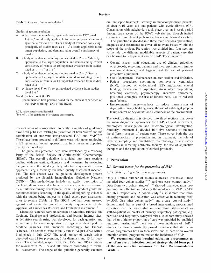
Table 1. Grades of recommendation11
Grades of recommendation
A at least one meta-analysis, systematic review, or RCT rated
1 þ þ,a and directly applicable to the target population; or A
systematic review of RCTs or a body of evidence consisting
principally of studies rated as 1 þ ,a directly applicable to the
target population, and demonstrating overall consistency of
results
B a body of evidence including studies rated as 2 þ þ,a directly
applicable to the target population, and demonstrating overall
consistency of results; or Extrapolated evidence from studies
rated as 1 þ þa or 1 þa
C a body of evidence including studies rated as 2 þ ,a directly
applicable to the target population and demonstrating overall
consistency of results; or Extrapolated evidence from studies
rated as 2 þ þa
D evidence level 3a or 4a; or extrapolated evidence from studies
level 2 þa
Good Practice Point (GPP)
Recommended best practice based on the clinical experience of
the HAP Working Party of the BSAC
RCT, randomized controlled trial.aSee ref. 11 for definitions of evidence assessments.
Review
7
by guest on September 4, 2015
http://jac.oxfordjournals.org/D
ownloaded from

Appropriate levels of experienced nursing staff should beinvolved in patient care to prevent HAP and education ofstaff on the measures that should be taken to prevent HAPshould form part of their induction and continuing pro-fessional development. Recommendation Grade GPP
2.1.2. Role of clinical guidelines or protocols
Most published guidelines relate specifically to prevention ofVAP rather than HAP. Two randomized controlled trials (RCTs)showed that care protocols in ICUs decrease the incidence ofVAP, particularly in trauma patients.18,19 Two other RCTs,specifically on the use of weaning protocols for ventilatedpatients on ICU, found that the use of protocols by nurses andrespiratory therapists resulted in reduced duration of mechanicalventilation, improved clinical outcome and reduced costs.20,21
Also the use of protocols for reducing sedation has beenreported as being effective in shortening the duration of venti-lation and ICU stay.22,23 Although there were few papers, thoseidentified provided good evidence that clinical guidelinesreduced the incidence of VAP.18 – 24 As there is no direct evi-dence that guidelines affect the incidence of HAP outside ofICUs, no recommendation can be made in respect of the valueof clinical guideline implementation in this scenario.
We recommend that care protocols and guidelines forweaning and sedation should be developed and activelyfollowed in the critical care setting to reduce the incidenceof VAP. Recommendation Grade A
In order to reduce the incidence of HAP, adherence toclinical guidelines should be monitored to ensure compliance.Recommendation Grade GPP
2.1.3. Role of screening of patients or their environment to
prevent HAP
There are no studies that examined the benefit of routine surveil-lance for HAP organisms in patients or their environment, inpreventing HAP, and so no recommendation can be made onthis topic. Future research is recommended in order to assesswhether taking routine screening samples from patients helps toreduce the incidence of HAP or assists in targeting treatmentthrough the early recognition of organisms causing HAP. Workis also needed to assess whether routine screening of theenvironment for organisms causing HAP reduces the incidenceof HAP due to multiresistant Gram-negative bacteria, e.g.Pseudomonas aeruginosa or Acinetobacter spp.
We recommend that limited and targeted surveillance oforganisms causing pneumonia in ICU patients should becarried out to identify cross-infection or outbreaks and otherinfection control problems, e.g. a single case of hospital-acquired Legionella infection.25 This type of surveillance isalso helpful in providing feedback to assist clinicians in empiri-cal antibiotic selection and on the incidence and susceptibilityof organisms causing VAP.26 Recommendation Grade GPP
2.1.4. Immunization to prevent HAP
Immunization relevant to the prevention of HAP includes, par-ticularly, influenza and pneumococcal vaccines. Most publishedpapers cover influenza (including influenza pneumonia), the
elderly and healthcare workers and mainly relate to outbreaks innursing homes. A meta-analysis27 reviewing this area included20 observational studies of HAP in the elderly and there are alsocohort studies28,29 and three RCTs.30 – 32
Existing UK guidance33 already highlights the importance ofimmunization against influenza and pneumococcal disease forhigh-risk adult and paediatric patients. Immunization of healthcareworkers involved with at-risk patients is also recommended.However, there is no direct evidence that influenza immunizationof healthcare workers or patients will directly reduce the incidenceof HAP, although one study28 found evidence to suggest thatinfluenza immunization prevents pneumonia in elderly patients.The same study reported that a failure to immunize healthcareworkers against influenza was associated with an increased mor-tality from ‘influenza like illness’ in elderly patients. There is alsono direct evidence that pneumococcal immunization of healthcareworkers or patients reduces the incidence of HAP.
We recommend that the use of influenza immunization inhealthcare workers and patients and pneumococcal immu-nization in elderly and at-risk groups should be encouraged.Recommendation Grade C
In line with the recommendations of the Joint Committeeon Vaccination and Immunization,33 influenza immuniza-tions should be actively encouraged in at-risk patients andhealthcare workers. Recommendation Grade GPP
2.1.5. Importance of hand hygiene in preventing HAP
A number of studies have assessed the effects of hand hygieneon staff-to-patient and staff-to-equipment transfer of bacteria.There is good evidence that an inverse relationship existsbetween high standards of hand hygiene and the incidence ofHCAI, but there is no good evidence of a direct relationshipwith the prevention of HAP.34 – 38 Hand hygiene is effective inreducing HCAI and the epic Project evidence-based guidelinesfor the prevention of HCAI recommend implementation of ahand hygiene policy.39
We recommend that hand hygiene guidelines are availableas part of evidence-based processes for preventing HCAIand that these should be followed. Hand hygiene practicesshould be incorporated into clinical guidelines for the pre-vention of HAP and performance audits of these should becarried out to demonstrate and maintain high levels of prac-tice. Recommendation Grade GPP
With a view to reducing the incidence of HAP, staff handhygiene should form part of routine care with hands beingdecontaminated immediately before and after every episodeof direct patient contact and after any activity or contactthat potentially results in hands becoming contaminated.Hand decontamination after glove removal should be per-formed. Recommendation Grade GPP
2.1.6. Role of personal protective equipment
There is an absence of evidence to address this issue. Thestudies available relate to HCAI and not directly to HAP. Thereare data showing that the appropriate use of Personal ProtectiveEquipment (PPE) prevents the spread of microorganisms andHCAI,39,40 which might potentially reduce the incidence ofHAP. It is essential that the choice of PPE is appropriate to therisk of infection, e.g. simple surgical masks are inadequate in
Review
8
by guest on September 4, 2015
http://jac.oxfordjournals.org/D
ownloaded from
protecting against tuberculosis and some respiratory viruses.41
Appropriate equipment needs to be readily available and thenecessary training given in its use. National health and safetyat work requirements such as PPE regulations42,43 and Controlof Substances Hazardous to Health regulations44 should befollowed.
We recommend that the role of PPE in the preventionof HAP should involve local risk assessment with referenceto national health and safety at work requirements, e.g.PPE Regulations42,43 and local infection control advice.Recommendation Grade D
We recommend high standards of hygiene includinghand hygiene and PPE, as these will protect healthcareworkers and patients against HCAI from microorganismsincluding influenza and other viral respiratory pathogens.Recommendation Grade GPP
Gloves should be put on immediately before an episode ofpatient contact or treatment and removed as soon as theactivity is completed and should be changed between caringfor different patients or between different care/treatmentactivities for the same patient. Recommendation Grade GPP
Care needs to be taken in the use of PPE to preventspreading infection between patients, e.g. gloves can con-taminate hands if not removed correctly and hence theimportance of hand decontamination after glove removal.45
Recommendation Grade GPPPersonal respiratory protection is required in certain
respiratory infections, e.g. multidrug-resistant tuberculosis,human coronavirus etc. or when patients who are severelyimmunocompromised are exposed to infection [e.g. not in ahigh efficiency particulate air (HEPA)-filtered environment].In these instances, specialized respiratory protective equip-ment should be worn. Recommendation Grade GPP
National guidelines should be followed with regard to pro-tection of staff against highly communicable infections, e.g.human coronavirus. Recommendation Grade GPP
Isolation of patients with multidrug-resistant infectionsincluding pneumonia should be performed alongside the useof PPE to prevent the spread of infection. RecommendationGrade GPP
2.2. Infection control issues related to the use of
equipment—best methods of sterilization or disinfection
of equipment and maintenance of instruments
2.2.1. Mechanical ventilators
In respect of HAP risk reduction, there is an absence of evidenceabout the best sterilization/disinfection/maintenance proceduresfor mechanical ventilators. The reuse of ‘single-use’ devices canaffect their safety, performance and effectiveness, exposingpatients and staff to unnecessary risk. It also carries legal impli-cations as anyone who reprocesses or reuses a device intendedby the manufacturer for use on a single occasion bears fullresponsibility for that item’s safety and effectiveness, includingto any organization to which the equipment is transferred.46
We recommend that, in line with the Medical DeviceAgency guidance46 on single-use medical devices, items desig-nated for ‘single-use’ must not be reused under any circum-stances. Recommendation Grade GPP
Equipment should be sterilized, disinfected and maintainedaccording to the manufacturer’s instructions. RecommendationGrade GPP
2.2.2. Ventilator circuits
A systematic review which included four RCTs and seven obser-vational studies found that changing the ventilator circuit lessfrequently than every 24 h reduced the risk of VAP.47 Anothersystematic review48 assessed three RCTs where one49 was con-sidered a higher quality trial than the other two.50,51 The reviewconcluded that the frequency of ventilator circuit changes doesnot influence the incidence of VAP; that less frequent changesof ventilator circuits are not associated with harm and that morefrequent changes are associated with increased cost. A furtherRCT found no difference in the rate of VAP in patients withventilator circuits containing heat moisture exchangers (HMEs)where 48 h circuit changes were compared with no plannedchange.52 These studies were all conducted in ventilated patientswhere circuits were changed if there were signs of visible con-tamination or damage. Further research regarding safety andinfection control criteria is required to determine the maximumlength of time between ventilator tubing changes.
Provided they are otherwise changed if they become soiledor damaged, we recommend that ventilator circuits need notbe changed before 7 days. Recommendation Grade A
New ventilator circuit tubing should be provided for eachpatient. Recommendation Grade B
In order to prevent contamination of the healthcareworker, facial protection should be used alongside PPE whenclosed breathing circuits are disconnected. This is especiallyimportant when dealing with patients with highly communic-able infections, e.g. human coronavirus. RecommendationGrade GPP
To prevent VAP, breathing circuit condensate shouldbe managed so that it does not drain towards the patientand it should be periodically drained and discarded.7,53
Recommendation Grade GPP
2.2.3. Heated humidifiers and HMEs
There are two meta-analyses47,54 and a systematic review48
which have compared the use of heat humidifiers (HHs) andHMEs. One meta-analysis54 covered all eight RCTs cited by theother papers that have examined the use of different humidifiertypes and their effect on the incidence of VAP.55 – 62 Thismeta-analysis concluded that in patients ventilated for .7 days,the use of HMEs is associated with a statistically significantreduction in the incidence of VAP when compared with HHs.Whereas concern was expressed in the earlier systematicreview48 about ET tube obstruction associated with HME use,this has not been confirmed in recent studies evaluating newerHMEs.63 Two RCTs57,62 have shown reduced costs associatedwith the use of HMEs compared with HHs.
Provided there are no contraindications to their use (e.g.patients at risk of airways obstruction), we recommend thatHMEs rather than HHs are used, as HMEs are more effec-tive in reducing the incidence of VAP. RecommendationGrade A
When HMEs are used, the type chosen should be one thathas adequate moisture output to minimize the risk of airway
Review
9
by guest on September 4, 2015
http://jac.oxfordjournals.org/D
ownloaded from
obstruction. The benefit of use of HMEs versus HHs shouldbe established for each patient and this decision shouldnot be based solely on infection control considerations.Recommendation Grade GPP
National guidelines should be followed in respect of theuse of humidifiers and HMEs for the management ofpatients with highly communicable infections, e.g. humancoronavirus. Recommendation Grade GPP
2.2.4. Frequency of change of humidifiers
A systematic review47 and three RCTs address this issue64 – 66
with two of the latter64,65 specifically evaluating the effect onVAP of a reduced frequency of change of the humidifier. Onestudy that looked at efficacy and safety by studying three differ-ent types of HMEs reported that not all HMEs performedequally with only some brands able to be used for 48 h withoutchange.64 It has also been reported that changing HMEs after 3days does not diminish the efficiency of the equipment orincrease the incidence of VAP.65 From these studies, there is noevidence that more frequent changing of HHs and HMEs thanmanufacturers recommend reduces the risk of HAP.
We recommend that where HHs and HMEs are used(except with high minute volume) these should not bechanged routinely and manufacturer’s guidance should befollowed. Recommendation Grade A
The technical performance of HMEs for more than 48 hshould be monitored, especially in patients with chronicobstructive pulmonary disease (COPD), and if there is evi-dence or suspicion of contamination, the humidifier shouldbe changed. Recommendation Grade GPP
2.2.5. Nebulizers
Nebulizers are used both in the ICU and wards and departmentsto deliver bronchodilators and other drugs. Three diagnosticstudies have reported that nebulizers can become contaminatedand act as a source of respiratory tract infection.67 – 69
We recommend that nebulizers should be single patientuse and need to be disinfected and cleaned with sterile waterbetween each use. Recommendation Grade D
Nebulizers used as part of the ventilator circuit should besingle use only and national guidelines should be followedwith regard to the use and cleaning of nebulizers.Recommendation Grade GPP
2.2.6. Filters
There are reports in the literature that provide evidence tosupport the use of filters to protect circuit systems from bacterialcontamination,68,70,71 but there is no evidence which establishesthat the use of filters specifically protects against HAP.
We recommend that appropriate filters are used toprotect mechanical ventilator circuits from bacterial con-tamination. Recommendation Grade C
National guidelines should be followed with regard tothe use of expiratory filters for patients suffering fromhighly communicable infections, e.g. human coronavirus,and who require mechanical ventilation. RecommendationGrade GPP
2.2.7. Suction equipment
Suctioning of patients on intensive care is essential to preventpooling of respiratory secretions. A systematic review47 andthree other studies72 – 74 have examined the effect of dailychanges of in-line suctioning equipment and found that whencompared with less frequent changes, this had no effect on theincidence of VAP. Whereas there is, therefore, no evidence thatchanging closed suction equipment daily reduces the risk ofVAP, the maximum duration that a closed suction catheter canbe used against safety and infection control considerations is notknown.
We recommend that daily change of suction equipment isnot required. Recommendation Grade A
Suction equipment may be changed weekly unless itbecomes contaminated or damaged, in which case it shouldbe changed immediately. Recommendation Grade GPP
2.2.8. Resuscitation equipment
Four studies on use of bag-valve mask ventilation (manual venti-lation/‘Re-breathe’) bags have reported that such resuscitationequipment can act as a source of HAP if it becomes bacteriallycontaminated.75 – 78
We recommend that in order to minimize the risk of HAPmultiuse, bag-valve mask ventilation (manual ventilation/‘Re-breathe’) bags should be decontaminated according tothe manufacturer’s guidelines between each patient use.Recommendation Grade C
All reusable resuscitation equipment should be appro-priately decontaminated according to the manufacturer’srecommendations after use and if possible single patientuse equipment (e.g. Ambu bag) should be employed.Recommendation grade GPP
2.2.9. Anaesthetic machines and breathing equipment
A diagnostic study suggested that basic hygienic management ofanaesthetic equipment was adequate to prevent cross-infection.79
Studies are required to establish the best sterilization or disinfec-tion and maintenance methods to reduce the risk of HAP fromanaesthetic machines and breathing systems.
We recommend that to reduce the risk of HAP, basichygienic measures should be adopted for anaesthetic equip-ment. Recommendation Grade D
Provided filters are in place to protect the equipment,anaesthetic equipment should be decontaminated accord-ing to the manufacturer’s instructions. RecommendationGrade GPP
Changing HMEs and anaesthetic machine valvebetween patients and weekly circuit changes should beadequate to prevent infection from anaesthetic machines.Recommendation Grade GPP
If anaesthetic equipment is used on a known infectedpatient, tubing and filters should be changed before the nextpatient use. Recommendation Grade GPP
2.2.10. Pulmonary function testing equipment
There are reports of the use of spirometers being associated withHAP caused by Acinetobacter spp.80,81 One study reported that
Review
10
by guest on September 4, 2015
http://jac.oxfordjournals.org/D
ownloaded from
the mouthpieces of spirometry tubing can become contaminatedwith bacteria during use and recommended that to prevent theacquisition of microorganisms causing HAP they should not beshared between patients.82
We recommend that spirometry mouthpieces should besingle use only. Recommendation Grade C
All respiratory equipment, where contamination by res-piratory secretions is possible, should be viewed as a poten-tial infection risk for HAP and therefore precautions shouldbe taken to reduce such risks. Recommendation grade GPP
2.3. Patient procedures
2.3.1. Closed versus open suctioning
Several studies have assessed the effect of closed versus opensuctioning on VAP, but their results are not consistent.A systematic review48 considered evidence from four RCTs83 – 86
and concluded that the type of suctioning system had no effecton the incidence of VAP. Two further RCTs confirmed thesefindings.87,88 However, one RCT reported a 3.5 times greaterrisk of VAP in patients receiving open versus closed suction-ing.84 Most studies, therefore, show that closed as opposed toopen suctioning of respiratory tract secretions does not affect therisk of VAP and there is no evidence that closed suctioningincreases the risk of VAP.
No recommendation can be made on the use of closedsuctioning to reduce the risk of HAP to patients and we rec-ommend that closed or open suctioning systems can be usedwithout affecting the risk of VAP. Recommendation Grade B
From a safety perspective, closed suctioning of respiratorytract secretions is of value in reducing the aerosolizationof respiratory tract secretions and protection of healthcareworkers. The number of disconnections of suction equipmentshould be minimized to reduce the risk of exposure to staff topotentially infected secretions. Recommendation Grade GPP
2.3.2. Use of non-invasive positive pressure ventilation
NIV involves providing respiratory support to patients withoutthe need for intubation. There is evidence that in selectedpatients NIV reduces the risk of HAP. A Cochrane systematicreview89 included five RCTs and found that in patients withCOPD, NIV reduced the risk of HAP. Although the indicationsfor this procedure are relatively narrow, the numbers of patientsto whom they apply are large.
We recommend that to reduce the risk of HAP, NIVrather than mechanical ventilation should be used in appro-priate patients. Recommendation Grade A
2.3.3. Method of ET intubation
One RCT90 specifically addressed the issue of oral versusnasotracheal intubation with regards to the development ofVAP, whereas this and four other RCTs91 – 94 also assessed thedevelopment of maxillary sinusitis. All these demonstratedan association between nasotracheal intubation and maxillarysinusitis. Another study showed that re-intubation is associatedwith an increased incidence of VAP.95
We recommend that, where possible, oral ET intubationshould be used in preference to nasotracheal intubation
and that re-intubation should be avoided if possible.Recommendation Grade C
2.3.4. Use of enteral feeding
Enteral feeding is used to prevent the development of a catabolicstate in patients requiring long-term ventilation. A cohortstudy96 of ventilated patients showed a relationship betweenenteral feeding and aspiration, but there is limited other evidenceto support this. There is also an absence of evidence that theincidence of HAP in ventilated patients is reduced by takingmeasures to reduce aspiration associated with enteral feeding. Inview of these findings, no recommendation can be made aboutthe use of enteral feeding to prevent HAP.
We recommend that in ventilated patients, the rateand volume of enteral feeding should be adjusted to avoidgastric distension and so reduce the risk of aspiration.Recommendation Grade GPP
2.3.5. Different methods of enteral feeding
There are a number of methods of providing enteral feedingand these were assessed in a systematic review.9 Four RCTs thatevaluated different methods of enteral feeding, which includedintermittent feeding,97 the use of metoclopramide and acidificationof feeding, were reviewed. No difference in the incidence of VAP ormortality was found with any of these strategies. A meta-analysis98
of seven RCTs looked specifically at post-pyloric feeding andreported that compared with gastric feeding this was associated witha significant reduction in VAP. It was suggested that further studiesare warranted. A second meta-analysis99 that included nine RCTscompared gastric versus post-pyloric feeding and found that therewas no significant difference in the incidence of VAP in each group.Further research is required to study the effect of different modesof feeding on the incidence of HAP.
We recommend that as there is no clear evidence thatintermittent feeding, small intestine feeding, the use of meto-clopramide or acidification of feeding prevent VAP, thedecision on the method of enteral feeding to be used for criti-cally ill patients should be made locally by each unit and onan individual patient basis. Recommendation Grade A
When enteral feeding is used, the method of deliveryshould be optimized for each patient. RecommendationGrade GPP
2.3.6. Prevention of aspiration
The relationship between aspiration of gastric contents and pneu-monia/pneumonitis is well known. Although there is an absenceof evidence that prevention of aspiration associated with ET intu-bation reduces the incidence of HAP, a cohort study4 reported thatwitnessed aspiration was associated with an increased risk of VAP.A meta-analysis100 found that establishing subglottic drainage waseffective in preventing early-onset VAP in patients expected toremain ventilated for more than 72 h.
We recommend that to prevent VAP, measures should betaken to reduce the risk of aspiration and this should includesubglottic drainage and positioning. Recommendation Grade B
Attention needs to be paid to the ET cuff pressure toavoid aspiration and prevent tracheal damage (>25 and< 30 cm water).101 Recommendation Grade GPP
Review
11
by guest on September 4, 2015
http://jac.oxfordjournals.org/D
ownloaded from
2.3.7. Use of sucralfate and stress ulcer prophylaxis
There is clear evidence that a reduction of gastric acid byvarious methods, including antacids and H2 antagonists used forstress ulcer prophylaxis in ICU patients on ventilation, increasesthe risk of VAP. However, the literature is not consistent withregard to the use of sucralfate in this context. A systematicreview9 that considered seven meta-analyses found evidence infour of these102 – 105 that sucralfate when compared with H2antagonists significantly reduced the incidence of VAP. Thethree other meta-analyses did not show a statistically significantreduction in VAP with the use of sucralfate, but did showa trend to this effect.106 – 108 A large RCT109 found that inmechanically ventilated patients, sucralfate therapy was associ-ated with a statistically significantly increased risk of clinicallyimportant gastrointestinal bleeding compared with H2 antagon-ists. There is, therefore, evidence that the use of sucralfate isassociated with a reduced risk of VAP when compared with theuse of other agents that raise gastric alkalinity in ventilatedpatients, but sucralfate therapy has been associated with anincreased risk of clinically important gastrointestinal bleedingwhen compared with ranitidine.
We recommend that whenever clinically appropriate,stress ulcer prophylaxis should be avoided in order to helppreserve gastric function. Recommendation Grade A
We recommend that where stress ulcer prophylaxis isindicated, sucralfate is to be preferred in order to reduce therisk of VAP, but sucralfate should only be used in patientswith low to moderate risk of gastrointestinal bleeding.Recommendation Grade A
2.3.8. Effect of breathing exercises
There is an absence of evidence that instructing patientsto cough or take deep breaths reduces the incidence of HAP.Two cohort studies110,111 and an RCT112 did not specificallylook at HAP but considered pulmonary complications ingeneral. Another study113 found that the most effective regimenof prophylaxis against pulmonary complications for low-riskpatients after abdominal surgery was deep breathing.
We recommend that coughing and early mobilizationduring the post-operative recovery period should be encour-aged in all patients in order to reduce the risk of pulmonarycomplications. Recommendation Grade GPP
2.3.9. Role of physiotherapists and respiratory therapists
We found no data on the role of physiotherapists and respiratorytherapists in reducing the incidence of HAP in general.However, one study114 showed the benefit of chest physiotherapyin preventing VAP, whereas an RCT115 demonstrated that physio-therapy with incentive spirometry reduced respiratory compli-cations in high-risk surgical patients. A systematic review116
assessed the role of respiratory therapists and reported fiveRCTs, which showed that respiratory therapists were effective inimplementing respiratory care protocols to wean patients frommechanical ventilation and in appropriately allocating respiratorycare in adult non-ICU patients but did not relate either of thesefeatures to HAP prevention. Therefore, there is evidence that res-piratory therapists, by following weaning protocols, both reducethe duration of mechanical ventilation and ICU stay and improve
outcome. Also, physiotherapy with incentive spirometry in high-risk abdominal surgery patients can reduce respiratory compli-cations, including pneumonia. Further research is required toestablish the role of physiotherapy in the prevention and man-agement of HAP.
We recommend that physiotherapists and respiratorytherapists have a role in preventing respiratory complicationsin post-operative ventilated patients. RecommendationGrade A
Physiotherapists and respiratory therapists have a holisticrole in the pre- and post-operative care of patients, especiallyin high-risk patients, where risk assessment indicates thismay be of value. Recommendation Grade GPP
2.3.10. Use of incentive spirometry
There is some evidence that the use of incentive spirometry inpost-operative, high-risk surgical patients may be beneficial,although this is poor, with only one RCT.115 There is an absenceof evidence that incentive spirometry has any impact on redu-cing the risk of HAP in low-risk surgical patients after abdomi-nal surgery. Further research is required to assess the effects ofincentive spirometry in patients requiring surgery.
We recommend that incentive spirometry has no roleto play in prevention of HAP in the low-risk (ASA grade 1or 2) surgical patient, including patients who had no pre-existing pulmonary complications, and that it should be usedin high-risk patients to prevent respiratory complications.Recommendation Grade D
2.3.11. Positional strategies
Several studies have looked at the use of semi-recumbent, proneand supine positioning in relation to the risk of HAP. AnRCT117 found that the use of semi-recumbent positioning mayprevent VAP and also reported that supine body positioning andenteral feeding were independent risk factors for the develop-ment of nosocomial pneumonia (NP). However, an earlierstudy118 with a smaller number of patients concluded that thesemi-recumbent positioning did not prevent VAP. AnotherRCT119 looked at the effect of prone positioning on patientswith acute respiratory failure and found that there was nogeneral benefit from using prone positioning and that there wereconcerns about safety. However, a cohort study120 showed thatpatients nursed in supine head positioning during the first 24 hof ventilation had an increased risk of VAP. A further study121
reported a significant increase in VAP in patients transportedout of ICU for interventions, which was most likely related topositioning.
We recommend that a positional strategy should beadopted to prevent VAP. If a patient does not require to besupine, and provided there are no contraindications, consider-ation should be given to using the semi-recumbent position(30–4588888) as a strategy to prevent VAP. RecommendationGrade B
Consideration should be given to adopting a positionalstrategy to prevent HAP in non-ventilated patients.Recommendation Grade GPP
Patients on ventilation being transported out of ICUshould if possible be maintained in the semi-recumbentposition. Recommendation Grade GPP
Review
12
by guest on September 4, 2015
http://jac.oxfordjournals.org/D
ownloaded from
In order to prevent aspiration, patients should be kept ina semi-recumbent position during enteral feeding. GPP
2.3.12. Use of kinetic beds (oscillatory therapy)
There is a limited amount of evidence on the use of kinetic bedsto prevent HAP. A meta-analysis,122 which included six studies,and a systematic review48 covering eight RCTs have reviewedthe use of kinetic (oscillating) beds. Their conclusion was thatalthough these may have an impact on reducing complicationsassociated with intensive care, it is inconclusive whether theyaffect the development of HAP. At present no recommendationfor use of kinetic therapy to prevent HAP can be made from theevidence and further research is required to assess the value ofkinetic therapy in the prevention of HAP in different patientpopulations and especially in ICUs.
2.3.13. Use of red cell transfusions
The use of transfusions has been particularly studied in post-operative patients and those receiving intensive care. Onestudy123 demonstrated that transfusion of .4 U or red cell con-centrate was associated with an increased risk of HAP in cardiacsurgery patients, whereas another cohort study showed that theuse of stored red blood cells increased the risk of HAP.124 Incontradistinction, a study125 reported that a restrictive transfusionpolicy in ICU patients is at least as effective as a liberal policywith regard to the effect on mortality and multiorgan failure.Finally, a cohort study reported that leucocyte-depleted bloodwas better than buffy-coat reduced blood in preventing pneumo-nia in patients undergoing colorectal surgery.126 There is, there-fore, some evidence that the use of red cell transfusionsincreases the risk of HAP and the evidence available is particu-larly applicable in cardiac and colorectal surgery. Furtherresearch needs to be carried out to establish the effect of red celltransfusions on the development of HAP in other patient groups.
We recommend that to prevent HAP, red cell transfusionsshould be avoided if possible and if used should be withfresh red cells. Recommendation Grade C
2.4. Environmental issues
2.4.1. Methods to reduce transmission of Aspergillus during
building work
There are a number of studies that demonstrate an associationbetween building works, environmental contamination withAspergillus and pulmonary aspergillosis.127 – 129 There is goodevidence that methods to reduce dust levels result in lowerlevels of fungal spores in the environment and reduce the inci-dence of pulmonary aspergillosis during building work.However, there are no studies that systematically look at the riskin relation to the type of building work (construction versusdemolition) and relative contributions of various strategies toreduce risk, i.e. (i) dust reduction, (ii) air handling, (iii) environ-mental and air monitoring, and (iv) antifungal prophylaxis.
There is a financial impact if measures other than simple dustreduction are used and introducing widespread environmentalcontrol is expensive, e.g. HEPA filtration. Moreover, there is anabsence of evidence that routinely monitoring spore countsduring building work is useful, although it is recommended by
some authors, especially for high-risk areas.130 – 132 Studies haveshown that although a protective environment with HEPAfiltration reduces the incidence of invasive aspergillosis (IA) inhaemopoietic stem cell transplant (HSCT) patients, this may notby itself prevent IA during building work.131,132
We recommend that during building works, considerationis given to addressing the risk of pulmonary aspergillosis.This must include:
† Identifying high-risk patients, i.e. those with acute leukae-mia, HSCT patients, patients receiving chemotherapyresulting in severe, prolonged neutropenia and otherimmunosuppressed patients including those on long-termcorticosteroids or other immunosuppressive therapy.
† Methods to reduce all patient’s exposure to Aspergillus,e.g. use of floor to ceiling barriers, sealing of windows.
† The use of HEPA filtration in high-risk units, e.g. HSCTunits and critical care.
† Dust reduction in clinical areas including cleaning (dampdusting, use of HEPA-filtered vacuum cleaners).
Recommendation Grade BThe routine monitoring of air for fungal spores during
building work outside high-risk areas is not recommended.Recommendation Grade D
During building work, environmental monitoring forfungal spores in critical areas housing at-risk patients isuseful in monitoring the effectiveness of control measures.Recommendation Grade GPP
In an outbreak situation, environmental or air monitoringmay be useful in identifying the source of infection.Recommendation Grade GPP
Ventilation systems, especially those which are not HEPAfiltered, may become contaminated during building work.During building work, all filters should be regularlyinspected and replaced as necessary. RecommendationGrade GPP
2.4.2. Use of prophylactic antifungal agents during building
work
As there have been several outbreaks of Aspergillus infectionassociated with building work, this raises issues regardingadditional patient-focused methods that can be used to preventinfection, including the use of antifungal agents. There is nogood evidence for the widespread use of antifungal treatment asprophylaxis during construction work. A small cohort study133
reported that antifungal prophylaxis in immunocompromisedpatients during construction work reduced IA. There is good evi-dence for the protective effect against IA of antifungal prophy-laxis for neutropenic patients in general134,135 and specifically tosupport the use of itraconazole in this situation.134,136,137 Thereis no clear evidence-based indication against the use of anyantifungal prophylaxis.138
We recommend that where there is a high institutionalrate of IA or building work is underway, a risk assessmentshould be undertaken. Those patients who are immunosup-pressed, especially those who are neutropenic (neutrophilcount <0.5 3 109/L for more than 2 weeks or <0.1 3 109/Lfor 1 week), who are visiting hospital regularly or stayingas an inpatient but not in a HEPA-filtered environment
Review
13
by guest on September 4, 2015
http://jac.oxfordjournals.org/D
ownloaded from
should be considered for antifungal prophylaxis.139
Recommendation Grade DThe use of antifungals to prevent IA in the immunosup-
pressed during building work should be based on a robustrisk assessment for the individual patient. RecommendationGrade GPP
The cost of using antifungal prophylaxis should thereforebe considered in all building projects (cost of drug and moni-toring). In large hospital projects, e.g. involving demolitionand reconstruction, the additional cost may need to coverseveral months. Recommendation Grade GPP
2.4.3. Legionella control
There is a good evidence base showing a relationship betweenLegionella contamination of hospital water with hospital-acquired(HA) Legionella pneumonia and also good evidence that control-ling the risk of Legionella in hospital water supplies reduces therisk of HA Legionella pneumonia. There is a large body ofevidence regarding methods to control Legionella in hospital watersupplies consisting of cohort and case–control studies.140–149 TheHealth and Safety Executive (HSE) has also issued comprehensiveguidance on the control of Legionella in hospitals,150 which hasrecently been revaluated.151
The methods available to control Legionella in water systemsinclude: (i) heat; (ii) biocides, e.g. chlorine, chlorine dioxide;(iii) ionization, e.g. copper–silver; and (iv) ultraviolet light andozone.
There are few studies comparing the different methods.Results across the methods are consistent in demonstratingthat each process has an effect in reducing Legionella load inhospital waters although the effect may be only temporary.However, it is not possible to make full comparisons betweenthe different methods. Hospitals should already be adhering toDH advice for control of Legionella in hospitals so there shouldbe no additional financial consequences. Research is requiredto confirm the relationship between environmental Legionellaand HA Legionnaire’s disease, including establishing the relativeimportance of different serogroups of Legionella pneumophilaand other Legionella spp. Research is required into the differentmethods of controlling Legionella appropriate for use in differ-ent healthcare settings.
Although no recommendation can be made about themost appropriate method, we recommend that in line withthe current guidance, appropriate Legionella control ofhospital water is required. Recommendation Grade B
UK HSE guidance on Legionnaires’ disease—control ofLegionella bacteria in water systems150,151 and all othernational guidance should be followed. RecommendationGrade GPP
For secondary prevention (i.e. preventing further casesafter a case of hospital-acquired infection), additionalmeasures may be required, e.g. use of biocides, heat flushetc. Recommendation Grade GPP
Routine culturing of hospital water for Legionella,while not recommended, is appropriate in high-risk area,e.g. for haemopoietic stem cell and solid organ transplantwards. Infection control teams should work closely withhospital engineers, management and physicians to ensureawareness of HA Legionnaires’ disease.152 RecommendationGrade GPP
2.4.4. Cleanliness of the environment
Cleanliness of the environment has been highlighted by the DH‘Saving Lives’ initiative as essential to prevent HCAI.5 Thereare a small number of cohort studies that demonstrate environ-mental risk related to poor cleaning, e.g. for infection withmethicillin-resistant Staphylococcus aureus (MRSA) andClostridium difficile, but these do not relate directly to HAP orspecifically to ICUs. There are a small number of studies thatdescribe the environment acting as a reservoir for other organ-isms causing infection in patients, though these also do notrelate directly to HAP or ICUs. These studies demonstrate thattaking action to remove the reservoir is effective in preventinginfections. The epic project review revealed little research evi-dence of an acceptable quality upon which to base guidancerelating to hospital environmental hygiene.39 However, it notedthat there is a large body of clinical evidence, derived from casereports and outbreak investigations, which show links betweenpoor environmental hygiene and the transmission of micro-organisms causing HCAI.153,154
Given that the routes of transmission for organisms causingHAP are different from those described in the available reports,where the issues relate mainly to direct contact and contami-nation spread, the applicability of the available evidence, whichdoes not directly relate to HAP, is not strong. It is not possibleto generalize from the available evidence as this is of a lowvolume and not directly related to HAP, ICUs or in all instances,cleaning. The small volume of research that is available isconsistent in demonstrating that the environment can act as areservoir for infection and that taking action to remove thereservoir is effective in preventing infection. The consistency ofthe evidence makes extrapolation to these issues reasonable.There are now published cleanliness standards for health-providing facilities and these form part of performance assess-ment reviews.155,156
We recommend that in order to reduce the risks ofHCAI (including HAP), good approved standards of hospitalcleanliness should be maintained. Recommendation Grade D
The hospital environment must be visibly clean, free fromdust and soilage and acceptable to patients, their visitors andstaff. Recommendation Grade GPP
All staff involved in hospital hygiene activities shouldundergo education and training related to the preventionof HCAI: such training to include the link betweenthese infections and the cleanliness of the environment.Recommendation Grade GPP
3. Diagnosis
3.1. General issues in the diagnosis of HAP
3.1.1. Definitions of HAP
HAP (or NP) is usually defined as pneumonia developing �48 hafter admission to hospital that was not incubating at the time ofadmission.7,157,158 VAP is usually defined as pneumonia deve-loping �48 h after implementing ET intubation and/or mechan-ical ventilation that was not present before intubation.3,7,157 – 160
HAP can be divided into early- and late-onset. Early-onsetdisease occurs within 4–5 days of admission and tends to becaused by antibiotic-susceptible community-type pathogens,
Review
14
by guest on September 4, 2015
http://jac.oxfordjournals.org/D
ownloaded from
whereas late infections tend to be caused by antibiotic-resistanthospital opportunists. However, some studies have found anincreasing frequency of early-onset HAP caused by pathogensmore commonly associated with nosocomial disease. This hascontributed to the concept of healthcare-associated pneumonia(HCAP), involving pathogens associated with recent prior hospi-talization and/or antimicrobial therapy. HCAP has been definedas pneumonia occurring in any patient who had been admittedto an acute care hospital for 2 or more days within 90 days ofthe infection; or had been a resident in a nursing home or long-term care facility; or had attended a hospital or haemodialysisclinic; or had received recent intravenous antibiotic therapy,chemotherapy, or wound care within the past 30 days of thecurrent infection.7 Rarely, HAP/VAP is due to fungal infectionand this is usually, although not exclusively, seen in severelyimmunocompromised patients.
VAP also can be divided into early- and late-onset.Early-onset VAP occurs during the first 4 days of mechanicalventilation when the pneumonia is often caused by typical com-munity organisms. Late-onset VAP develops �5 days after theinitiation of mechanical ventilation and is commonly caused bytypical opportunistic and antibiotic-resistant hospital pathogenssuch as P. aeruginosa or MRSA.3,158,159
3.1.2. Issues in assessing the literature on diagnosis of HAP
There are two fundamental problems with assessing the diagnosticliterature relating to HAP. The first is that most publications dealwith VAP, whereas most HAP occurs in non-ventilated and non-intubated patients. It may not be valid to extrapolate diagnosticcriteria for VAP to HAP; for example, the diagnosis of VAP ofteninvolves invasive microbiological sampling, which may not bepossible or appropriate in non-intubated patients. The secondproblem is that there are no universally accepted ‘gold’ or refer-ence standard diagnostic criteria for HAP or VAP. Because ofthis, it is difficult to compare different diagnostic methods. Moststudies begin by dividing patient groups into those with pneumo-nia and those without, but the criteria used for this division vary,and all have their problems. Thus, most studies have groups ofpatients that probably have pneumonia or probably do not, and ineach there is an unknown proportion that will have been wronglyclassified. For example, some patients, not thought to have HAPclinically, have been diagnosed at autopsy,161 and vice versa. Wehave not excluded such studies from this analysis, but have inter-preted the results with caution. Recently, more useful studies haveappeared that avoid these problems by analysing the outcomes ofmanagement based on different diagnostic techniques in a singlegroup of patients suspected of having HAP.
3.1.3. An assessment of lung histology and culture as a
reference standard for the diagnosis of HAP
As lung biopsies are impractical and associated with risk, theyare rarely used for the diagnosis of HAP and are not recom-mended for this purpose. Nevertheless, histology and cultures ofhomogenized lung tissue have frequently been used to validateother diagnostic tests such as the quantitative microbiology ofrespiratory specimens.
An experimental baboon model of VAP has been used toassess the severity of bronchopneumonia by lung histology and
to compare this with quantitative microbiology of lung tissueand bronchoalveolar lavage (BAL) specimens.162 Moderate/severe pneumonia as judged by histology was associated withhigh bacterial concentrations in homogenized lung biopsies, andbacterial concentrations in BALs were linearly related to tissuevalues. This led to the concept that quantitative bacteriology ofa BAL or other deep respiratory specimen could be used for theaccurate diagnosis of HAP/VAP. Many subsequent studies ofpneumonia in human patients have used lung histology (or quan-titative microbiology of respiratory secretions) as the referencestandard for assessment of other diagnostic methods.
However, several studies of lung biopsy and culture haveshown inconsistent results. In a prospective case study, postmortem lung biopsies were performed ,1 h after death on 39patients who had been mechanically ventilated for �14 days.163
Histological pneumonia was diagnosed by four independentpathologists in 7, 9, 12 and 15 cases (18% to 38%), respectively.One of the pathologists reviewed the slides blindly 6 monthslater and re-classified two patients (one diagnosed with and onewithout pneumonia). A single pathologist then reviewed theslides by the Johanson et al.162 criteria and diagnosed pneumo-nia in 14 patients (36%).163 A later prospective case study ofquantitative microbiology of open lung biopsies of ventilatedpatients found that histological lesions of pneumonia and tissueconcentrations of bacteria were unevenly distributed through thelung parenchyma.164 Quantitative tissue bacterial concentrationstended to correlate with the presence and severity of histologicallesions, but could not differentiate the histological presence orabsence of pneumonia. Similar results have been obtained byothers.165 – 167
We recommend that lung histology should not be reliedupon as a gold/reference standard for the diagnosis of HAP.Recommendation Grade D
When biopsy is used as the reference standard for otherdiagnostic methods in HAP, the histological criteria shouldbe standardized. Recommendation Grade GPP
The histological diagnosis of HAP or quantification ofbacterial lung tissue concentrations should be based onseveral specimens from different areas of the lung.Recommendation Grade GPP
Studies using histology or parenchymal cultures asthe reference standard for assessment of other diagnosticcriteria in HAP should be interpreted with caution.Recommendation Grade GPP
3.2. The clinical diagnosis of HAP
3.2.1. Clinical diagnostic criteria
The clinical diagnosis of pneumonia, HAP and VAP isdifficult and there are no universally accepted clinical criteria.A systematic review covering the clinical diagnosis of VAPconsidered publications up to 1998,161 whereas other evidencecomprises cohort studies168,169 or consensus opinion.8
Clinical diagnostic criteria described in consensus opinionstatements3,8,158,170 are the same for VAP, HAP and community-acquired pneumonia (CAP). However, there is overlap of clinicalsigns and symptoms between pneumonia and other forms ofsepsis, and the diagnosis of HAP often cannot be made on clinicalcriteria alone.
Review
15
by guest on September 4, 2015
http://jac.oxfordjournals.org/D
ownloaded from
With these reservations, the diagnostic sensitivity of clinicalsuspicion can be improved by taking account of the presence offever (core temperature .38.38C), blood leucocytosis (.10 000leucocytes/mm3) or leucopenia (,4000/mm3), purulent trachealsecretions and the presence of a new and/or persistent infiltrateon chest radiograph (CXR), which is otherwise unexplained.161
However, if all these clinical criteria were required fordiagnosis, the specificity would be poor.161 A European consen-sus group170 believed that a diagnosis of pneumonia could bebased on pulmonary infiltrates plus two of the other criteria.
In some patients, increasing severity of pneumonia may beassociated with increasing evidence of circulatory collapse(shock, tachycardia, hypotension and elevated blood urea con-centrations).171,172 However, these signs of sepsis are not specificfor pneumonia and are not required for diagnosis of HAP orVAP. In a comparative study, immediate post mortem histologyin 39 patients who had been mechanically ventilated for �14days was assessed against five clinical diagnostic criteria (fever,leucocytosis, positive sputum culture, worsening CXR changesand worsening gas exchange).173 None of the individual clinicalcriteria or any combination of them correlated with the presenceor absence of histological pneumonia. Thus, there is only amoderate amount of evidence comparing clinical diagnosticcriteria with reference criteria, including histology, and thesedata demonstrate that there can be considerable variation in theclinical presentation of HAP and that distinction from otherforms of sepsis is difficult.
3.2.2. The clinical pulmonary infection score
The clinical pulmonary infection score (CPIS) is an evolvingclinical scoring system approach to the diagnosis of HAP. It isnow based on six criteria [the original four—fever, leucocytosis,positive sputum culture and worsening CXR changes—plusoxygenation (PaO2/FiO2) and semi-quantitative cultures oftracheal aspirates with or without Gram’s stain]. These develop-ments have been claimed to further increase diagnostic sensi-tivity.174,175 A prospective case series176 studied 25 deceasedpatients who had been mechanically ventilated before death. Thepresence of both histological pneumonia and positive lungcultures immediately post mortem was used as a reference testfor VAP. In these patients, the CPIS was not superior to conven-tional clinical criteria for the diagnosis of VAP before death. Aprospective cohort study169 investigated the utility of a modifiedCPIS in the diagnosis of HAP. Conventional clinical diagnosiswas found to be inaccurate (sensitivity 50%, specificity 58%)and the CPIS was only slightly more accurate (sensitivity 60%,specificity 59%). Adding a Gram’s stain result from a BALspecimen slightly increased the accuracy of both conventional(sensitivity 85%, specificity 49%) and CPIS (sensitivity 78%,specificity 56%) diagnosis. However, although the CPIS has notbeen shown to improve diagnostic accuracy compared with con-ventional clinical assessment, it has been used successfully tomonitor and modify therapy. A prospective, multicentre cohortstudy177 of ventilated patients with suspected VAP used serialmeasurements of CPIS to monitor the clinical course of VAPand successfully identified patients with a good prognosis byday 3. A further study178 investigated patients with pulmonaryinfiltrates with suspected VAP but with a CPIS score of �6(low likelihood of pneumonia). Patients were randomized toreceive standard therapy or ciprofloxacin monotherapy with
discontinuation at day 3 if the CPIS remained �6. Comparedwith patients on standard therapy, those on ciprofloxacin mono-therapy had significantly lower antimicrobial costs, antimicrobialresistance and superinfections but no difference in length of stayor mortality. The authors concluded that the CPIS could be usedto identify patients who would benefit from a short course ofantibiotics.
We recommend that although the clinical diagnosis ofHAP is difficult, the following criteria will identify patientsin whom pneumonia should be considered in the differentialdiagnosis:
1. Purulent tracheal secretions, and new and/or persistentinfiltrate on CXR, which is otherwise unexplained
2. Increased oxygen requirement3. Core temperature >38.388888C4. Blood leucocytosis (>10 000/mm3) or leucopenia
(<4000/mm3) Recommendation Grade C
We recommend that the CPIS may be useful for selectingpatients for short-course therapy and for monitoringresponse to treatment. Recommendation Grade C
3.3. The radiological diagnosis of HAP
There is little available evidence to assess the value of imaginginvestigations in the diagnosis of HAP and VAP. There is onlyone systematic review179 that, with other reviews of diagnosticstudies, focuses on VAP and not the broader topic ofHAP.170,174,180 – 182
Publications on diagnostic imaging in VAP/HAP have beenmainly limited to the use of CXR and there is little informationon other techniques such as computed axial tomography (CT),magnetic resonance (MR) and positron emission tomography(PET). These newer techniques are not useful for initial investi-gations. CT can be useful as an additional diagnostic tool toexclude other pathology in a patient with a complex CXR, butthe role of MR and PET in the diagnosis of HAP/VAP has notbeen assessed. The diagnostic value of CXR is usually greater inHAP than VAP, because of the problems of performing mobileradiographic investigations on ventilated patients who cannot bemoved and because of other cardiothoracic co-morbidities oftenpresent in such patients. Ventilated patients may have abnormalCXR secondary to other pathology that must be distinguishedfrom infection, e.g. acute lung injury, left ventricular failure,aspiration or alveolar haemorrhage. Radiological investigationsof patients with VAP do not often influence outcome and aremore useful in patients with non-ventilated HAP where theymay provide information on differential diagnosis, complicationsor the exclusion of other pathology. Therefore, although the pre-sence of an alveolar infiltrate on the CXR raises the possibilityof HAP as well as other differential diagnoses, there are no keyimaging investigations that can establish the diagnosis of HAP,though a normal CXR excludes HAP.
We recommend that when a diagnosis of HAP is beingconsidered, a good quality CXR should be obtained andcompared with previous CXRs if available. RecommendationGrade D
CT scanning may assist in the differential diagnosis ofHAP and may guide management in patients who are notresponding to treatment and who have a complex CXR.Recommendation Grade GPP
Review
16
by guest on September 4, 2015
http://jac.oxfordjournals.org/D
ownloaded from
3.4. The microbiological diagnosis of HAP
3.4.1. The microorganisms of HAP
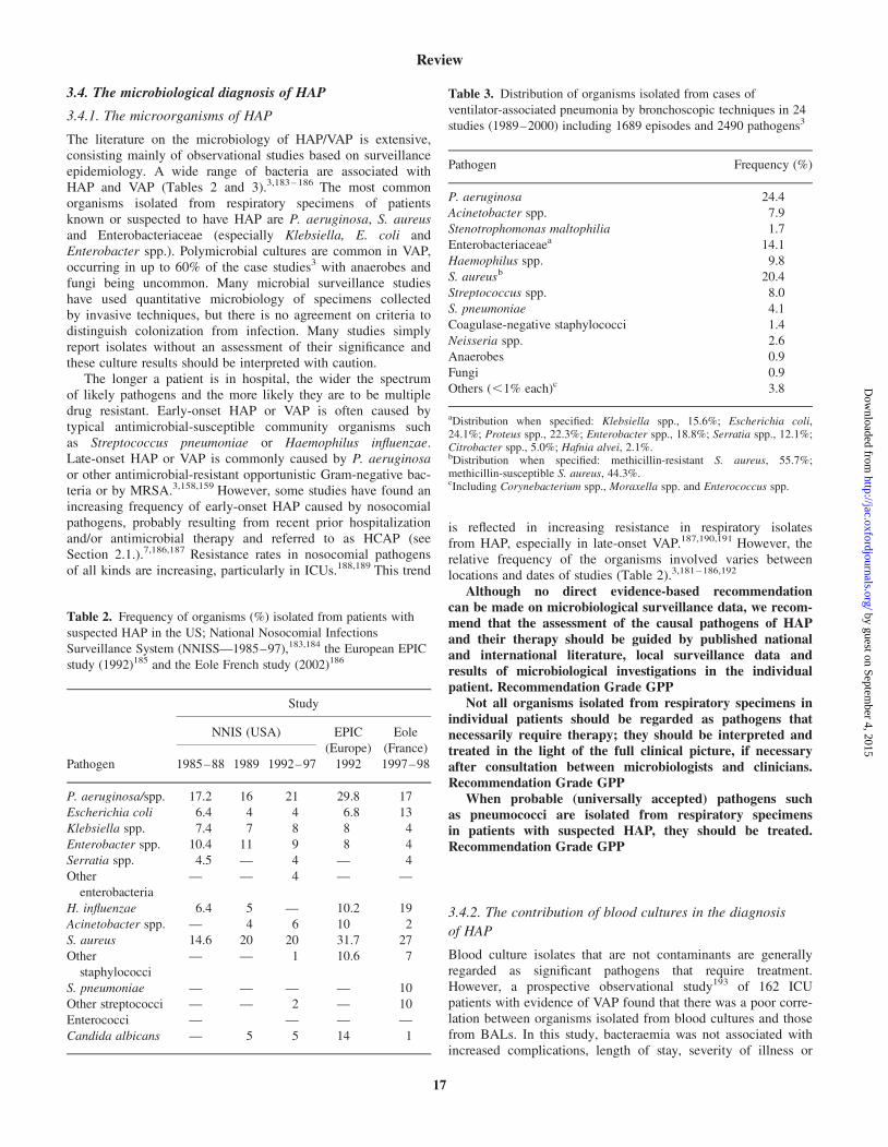
The literature on the microbiology of HAP/VAP is extensive,consisting mainly of observational studies based on surveillanceepidemiology. A wide range of bacteria are associated withHAP and VAP (Tables 2 and 3).3,183 – 186 The most commonorganisms isolated from respiratory specimens of patientsknown or suspected to have HAP are P. aeruginosa, S. aureusand Enterobacteriaceae (especially Klebsiella, E. coli andEnterobacter spp.). Polymicrobial cultures are common in VAP,occurring in up to 60% of the case studies3 with anaerobes andfungi being uncommon. Many microbial surveillance studieshave used quantitative microbiology of specimens collectedby invasive techniques, but there is no agreement on criteria todistinguish colonization from infection. Many studies simplyreport isolates without an assessment of their significance andthese culture results should be interpreted with caution.
The longer a patient is in hospital, the wider the spectrumof likely pathogens and the more likely they are to be multipledrug resistant. Early-onset HAP or VAP is often caused bytypical antimicrobial-susceptible community organisms suchas Streptococcus pneumoniae or Haemophilus influenzae.Late-onset HAP or VAP is commonly caused by P. aeruginosaor other antimicrobial-resistant opportunistic Gram-negative bac-teria or by MRSA.3,158,159 However, some studies have found anincreasing frequency of early-onset HAP caused by nosocomialpathogens, probably resulting from recent prior hospitalizationand/or antimicrobial therapy and referred to as HCAP (seeSection 2.1.).7,186,187 Resistance rates in nosocomial pathogensof all kinds are increasing, particularly in ICUs.188,189 This trend
is reflected in increasing resistance in respiratory isolatesfrom HAP, especially in late-onset VAP.187,190,191 However, therelative frequency of the organisms involved varies betweenlocations and dates of studies (Table 2).3,181 – 186,192
Although no direct evidence-based recommendationcan be made on microbiological surveillance data, we recom-mend that the assessment of the causal pathogens of HAPand their therapy should be guided by published nationaland international literature, local surveillance data andresults of microbiological investigations in the individualpatient. Recommendation Grade GPP
Not all organisms isolated from respiratory specimens inindividual patients should be regarded as pathogens thatnecessarily require therapy; they should be interpreted andtreated in the light of the full clinical picture, if necessaryafter consultation between microbiologists and clinicians.Recommendation Grade GPP
When probable (universally accepted) pathogens suchas pneumococci are isolated from respiratory specimensin patients with suspected HAP, they should be treated.Recommendation Grade GPP
3.4.2. The contribution of blood cultures in the diagnosis
of HAP
Blood culture isolates that are not contaminants are generallyregarded as significant pathogens that require treatment.However, a prospective observational study193 of 162 ICUpatients with evidence of VAP found that there was a poor corre-lation between organisms isolated from blood cultures and thosefrom BALs. In this study, bacteraemia was not associated withincreased complications, length of stay, severity of illness or
Table 2. Frequency of organisms (%) isolated from patients with
suspected HAP in the US; National Nosocomial Infections
Surveillance System (NNISS—1985–97),183,184 the European EPIC
study (1992)185 and the Eole French study (2002)186
Pathogen
Study
NNIS (USA) EPIC
(Europe)
1992
Eole
(France)
1997–981985–88 1989 1992–97
P. aeruginosa/spp. 17.2 16 21 29.8 17
Escherichia coli 6.4 4 4 6.8 13
Klebsiella spp. 7.4 7 8 8 4
Enterobacter spp. 10.4 11 9 8 4
Serratia spp. 4.5 — 4 — 4
Other
enterobacteria
— — 4 — —
H. influenzae 6.4 5 — 10.2 19
Acinetobacter spp. — 4 6 10 2
S. aureus 14.6 20 20 31.7 27
Other
staphylococci
— — 1 10.6 7
S. pneumoniae — — — — 10
Other streptococci — — 2 — 10
Enterococci — — — —
Candida albicans — 5 5 14 1
Table 3. Distribution of organisms isolated from cases of
ventilator-associated pneumonia by bronchoscopic techniques in 24
studies (1989–2000) including 1689 episodes and 2490 pathogens3
Pathogen Frequency (%)
P. aeruginosa 24.4
Acinetobacter spp. 7.9
Stenotrophomonas maltophilia 1.7
Enterobacteriaceaea 14.1
Haemophilus spp. 9.8
S. aureusb 20.4
Streptococcus spp. 8.0
S. pneumoniae 4.1
Coagulase-negative staphylococci 1.4
Neisseria spp. 2.6
Anaerobes 0.9
Fungi 0.9
Others (,1% each)c 3.8
aDistribution when specified: Klebsiella spp., 15.6%; Escherichia coli,24.1%; Proteus spp., 22.3%; Enterobacter spp., 18.8%; Serratia spp., 12.1%;Citrobacter spp., 5.0%; Hafnia alvei, 2.1%.bDistribution when specified: methicillin-resistant S. aureus, 55.7%;methicillin-susceptible S. aureus, 44.3%.cIncluding Corynebacterium spp., Moraxella spp. and Enterococcus spp.
Review
17
by guest on September 4, 2015
http://jac.oxfordjournals.org/D
ownloaded from
mortality. The authors concluded that organisms isolated fromthe blood are not necessarily those causing VAP.193
We recommend that when considering a diagnosisof HAP, the significance of blood isolates should bereviewed in the light of the patient’s clinical conditionduring consultations between clinicians and microbiologists.Recommendation grade GPP
3.4.3. An assessment of microbiological sampling methods
The microbiological investigation of HAP includes microscopyand qualitative and quantitative culture of respiratory secretions.Sputum is a poor diagnostic specimen because it may notcome from the infected area and it can be contaminated byupper respiratory flora during collection. To obtain specimensthat more accurately reflect the bacterial burden deep withinthe lung, methods have been developed to collect lowerrespiratory samples and protect them from oro-pharyngeal con-tamination. These include bronchoscopy-directed methods andnon-bronchoscopic ‘blind’ techniques. The most commonlyanalysed directed methods are bronchoscopy-directed BAL andbronchoscopy-directed protected specimen brush (PSB) sampling.Common non-directed methods are ‘blind’ BAL and ET aspira-tion. Many studies have compared qualitative and quantitativemicrobiological results on lower respiratory specimens obtainedby different sampling methods. Most of these studies are of VAP.
3.4.3.1. Bronchoscopy-directed PSB and BAL. There are severalhigh-quality reviews of the diagnostic value of quantitativecultures of respiratory secretions collected with bronchoscopy-directed PSB or BAL. A systematic review194 of studies ofdirected PSB up to 1995, a similar review195 of directed BALanalysing papers up to 1998 and a meta-analysis196 assessingPSB and BAL studies up to 1994, all concluded that there wasno significant difference between the diagnostic accuracy ofquantitative microbiology specimens collected by bronchoscopy-directed PSB or BAL.
3.4.3.2. Blind PSB and BAL. A systematic review197 of studiesup to 1998 of blind bronchial sampling, blind mini-BAL andblind sampling with PSB for the diagnosis of VAP found that thediagnostic accuracy of blind sampling was similar to that ofbronchoscopy-directed methods and side effects were also similar.Two more recent cohort studies have shown no significantdifference between the use of bronchoscopic PSB, blind PSBand bronchoscopic BAL,198 and no difference between blindand directed protected telescoping catheter and bronchoscopicBAL.199 From this evidence, it can be concluded that there isno difference between the results obtained with PSB and BAL,and there is no advantage in using bronchoscopically directedmethods compared with blind sampling.
3.4.3.3. Endotracheal aspirates (EAs). EAs are attractive speci-mens for microbiological sampling, since they are simple andsafe to obtain by unskilled staff. A systematic review200 ofreports published from 1985 to 1995 on the accuracy of EA cul-tures for the diagnosis of VAP concluded that the sensitivity andspecificity of quantitative EA cultures varied widely (sensitivity38% to 100%, and specificity 14% to 100%). Qualitative EAcultures had a high sensitivity, moderate positive predictive valueand high negative predictive value. The authors concluded that
EA cultures were unreliable for the diagnosis of VAP. The useof EA samples has been analysed in three more-recent papers.In a study199 of ventilated trauma patients with strong clinicaland radiological evidence of VAP, an assessment was made ofsampling respiratory secretions by EA, directed PSB, blind pro-tected brushing via ET tube and bronchoscopy-directed BAL.The value of quantifying the percentage of infected cells in speci-mens by Gram’s stain was also analysed. There was moderateagreement by kappa (k) analysis between quantitative cultures ofEA and BAL, and the sensitivity and specificity of quantificationof infected cells in EA samples, using quantitative culture ofBAL as the standard, were 96.7% and 48.9%, respectively.Another cohort study201 identified patients with VAP based onclinical evidence and culture results of BAL and evaluated thesignificance of quantitative cultures with a diagnostic thresholdof .105 cfu/mL for EA and .104 cfu/mL for BAL. From thisstudy the reported sensitivities, specificities, positive predictivevalues, negative predictive values and correct classification (intopneumonia or not pneumonia) were EA: 82%, 67%, 82%, 67%and 77%; and BAL: 94%, 100% 100%, 90% and 96%, respect-ively. Thus, EA performed less well than BAL, with similar sen-sitivities, lower specificities and fewer correct classifications.
Another study202 evaluated the diagnostic value of Gram’sstain identification of intracellular organisms in EA samplesfrom ventilated patients. This used two cut-off points of 5% and7% infected cells, with the usual trade-offs between sensitivityand specificity. In comparison with quantitative cultures of thesame EA samples, the presence of intracellular organisms in.5% of cells had a sensitivity of 39% to 85%, specificity 82%to 97%, positive predictive value 70% to 96% and negativepredictive value 50% to 91%. When outcome was considered,10/35 patients (28.5%) were eventually treated for VAP by theattending physician; all had EA intracellular organism counts�5% and quantitative cultures of �104 cfu/mL from at leastone specimen. However, 11/25 (44%) patients not treated forpneumonia had intracellular counts �5%. These studies supportthe conclusions that EA specimens have a high sensitivity foridentifying bacteria in respiratory specimens, but they have alow specificity and have not been demonstrated to improvepatient management.200 Thus, the evidence does not support theuse of EA specimens for the diagnosis of VAP/HAP, either byculture or microscopy, quantitatively or qualitatively.
We recommend that EA cultures are not used for thediagnosis of VAP. Recommendation Grade A
We recommend that there is no evidence that anyone invasive sampling method is better than any other.Recommendation Grade A
We recommend that the least expensive, least invasiveand most rapid sampling technique requiring minimalexpertise should be used for establishing the microbiologicaldiagnosis of HAP, e.g. non-bronchoscopy-directed (blind)BAL. Recommendation Grade GPP
3.4.3.4. The role of quantitative microbiology in the diagnosis ofHAP/VAP. Although a large amount of data assessing the diag-nostic accuracy of quantitative microbiology has been published,little of it is of a high quality. A central problem is that there isno good reference standard for the diagnosis of HAP.Microbiological results have usually been compared with one ora combination of clinical, other microbiological (e.g. tissue) andhistological diagnoses, but as described above, none of these
Review
18
by guest on September 4, 2015
http://jac.oxfordjournals.org/D
ownloaded from

reference standards is reliable. A wide variety of invasivesampling techniques and quantitative microbiological criteria(cut-off points) have been used. EAs are not reliable for micro-biological analysis, whereas, as described above, other samplingmethods give similar results to each other.
A systematic review series on the use of quantitative micro-biology for the diagnosis of HAP/VAP reviewed PSB194 andBAL,195 whereas a meta-analysis reviewed the use of PSB speci-mens.196 There are also three consensus reviews53,169,170 andfour good quality general reviews on this topic.3,7,203,204 In onesystematic review,195 the sensitivity of quantitative BAL cultureranged from 42% to 93%, with a mean of 73%; the specificityranged from 45% to 100%, with a mean of 82%. Thus, false-negative results occur in about 25% of cases and false positivesin about 20%. The other reviews have similar findings.However, an evidence-based guideline7 has concluded that nega-tive lower respiratory tract cultures in patients with no recentchange in antibiotic therapy is an indication for stopping anti-biotics (study-defined evidence level ‘moderate’). There is goodrepeatability when BAL specimens are analysed qualitativelybut not quantitatively.205 Although lavage specimens are usuallytaken from one lung, there is evidence that bilateral lavage maybe more accurate.206 Thus, quantitative cultures of respiratoryspecimens do not improve the diagnosis of HAP/VAP. Thesemethods have a wide range of accuracy and a high likelihood offalse-negative and false-positive results. They misdiagnoseHAP/VAP in 20% or more of cases.
Although they may provide an indication to the causalpathogen, we recommend that quantitative cultures of respir-atory specimens such as PSB and BAL should not be reliedon for the diagnosis of HAP/VAP. Recommendation Grade A
3.4.3.5. Quantification of intracellular organisms in the diagno-sis of HAP. Intracellular organisms in polymorphs in respiratoryspecimens can be visualized microscopically by a variety ofstaining methods. The quantification of such ‘infected’ cells hasbeen proposed as a rapid method for the diagnosis of pneumoniaand three recent cohort studies have compared this with othertechniques. A study that compared quantitative cultures ofmini-BAL specimens with the percentage of intracellular organ-isms in the same specimens found that a cut-off of .2% ofinfected cells gave a sensitivity of 80% and a specificity of82%.207 A second trial determined that the sensitivity and speci-ficity of infected cells in bronchoscopic BAL specimens with acut-off of .1% was 93.6% and 91.5%, respectively.198 In thisstudy, the therapeutic plan was considered to be correct in only65% of cases when using clinical judgment alone, but in 88%when guided by the microscopy results. Quantitative cultureof BAL specimens has been described as having diagnosticsensitivities, specificities, positive predictive values, negativepredictive values and correct classification (into pneumonia ornot pneumonia) of 94%, 100% 100%, 90% and 96%, respec-tively.201 Using quantification of intracellular organisms in thesame specimens, with a culture cut-off point of �104 cfu/mL,the values were 100%, 94%, 97%, 100% and 98%, respectively.Thus, in comparison with quantitative cultures, the detection ofintracellular organisms in BAL specimens is a rapid specific testwith high positive predictive value.
We recommend that quantification of intracellular organ-isms in BAL specimens is a rapid and specific test and can beused as a guide to initial therapy. Recommendation Grade A
4. Treatment
4.1. The prevention of HAP using antimicrobials
4.1.1. The role of selective decontamination of the digestive
tract (SDD)
The use of systemic antibiotics alone or in conjunction withtopically applied non-absorbable antibiotics as part of an SDDregimen has been investigated for the prevention of HAP,particularly VAP.
4.1.2. An assessment of the impact of SDD
Two recent systematic reviews,208,209 a recent meta-analysis210
and a Cochrane review211 which consider the benefit of SDD inthe prevention of HAP were reviewed. All of these demonstratesignificant reductions in HAP or respiratory infection. Onesystematic review also demonstrated a significant reductionin overall mortality with SDD,209 whereas another showed areduction in mortality in critically ill surgical patients only.208
The final systematic review found no significant mortalityreduction.210 However, the Cochrane review reported that a com-bination of topical and systemic prophylactic antibiotics reducesrespiratory tract infections and overall mortality in adult patientsreceiving intensive care.211 A treatment based on the use oftopical prophylaxis alone reduced respiratory infections butnot mortality.211 A more recent meta-analysis examined therelationship between methodological trial quality and the effectsof SDD and found that the better quality research papers, asdefined by their scoring method, demonstrated less benefit interms of pneumonia reduction, although a small but significantreduction in mortality was found in the high-quality studies.212
Five RCTs not included in these meta-analyses were alsoassessed and these were consistent with the above reportedbenefits from SDD.213 – 217
Quantification of the potential beneficial impact of usingSDD has been undertaken. A meta-analysis of 33 RCTs showedthat the number of patients needing treatment (NNT) to preventone case of VAP was 5 and to prevent one death was 23.209 TheCochrane review reported that on average, 5 patients needed tobe treated with SDD to prevent one respiratory tract infectionand 21 patients to prevent one death211 and the results from anRCT213 as assessed by other workers218 were consistent with anNNT to prevent one death of 12 patients.
There is thus evidence of benefit from SDD, consistentlydemonstrated across all patient groups, in the reduction of mor-bidity and mortality associated with VAP. There is also goodevidence that a reduction in respiratory tract infections willaffect mortality but, as the evidence is in critically ill patientswith VAP, it is less clear how this translates to other forms ofHAP. There is insufficient evidence to assess if particular patientsubgroups benefit significantly over others.
We recommend that where it is anticipated that mechan-ical ventilation will be for �48 h, SDD should be consideredfor ICU patients in order to prevent the development ofVAP. Recommendation Grade A
4.1.3. The choice of antimicrobial treatments for SDD
Two recent meta-analyses addressed the best methods ofSDD.208,209 Both studies found that the greatest reduction in
Review
19
by guest on September 4, 2015
http://jac.oxfordjournals.org/D
ownloaded from
respiratory infection were observed where both topical andsystemic components of treatment were administered. A morerecent RCT performed that used topical SDD alone demon-strated a significant reduction in VAP in patients receiving amodified SDD topical regimen (polymyxin, tobramycin andamphotericin). No difference in mortality in the two groups wasfound.214 A wide range of choices and drug doses have beenemployed for SDD, but there is no available evidence as towhich is the best regimen. With regard to duration of adminis-tration, most trials are consistent in using SDD throughout theICU stay, with the systemic element being given for 3–4 daysonly, and subject to modification for the treatment of sepsis.Further research into SDD is needed to determine the bestregimen and to identify the best selections of agents if unitshave particularly resistant organisms, e.g. MRSA, multidrug-resistant Acinetobacter spp. or extended-spectrum b-lactamase-producing organisms.
We recommend that SDD regimens should include topicaland parenteral agents with activity against Gram-negativebacilli and that the choice of treatment should dependon local pathogen antimicrobial susceptibility profiles.Recommendation Grade A
4.1.4. The relationship of resistance development to SDD
A meta-analysis reported that the practice of SDD led to trendstowards colonization in patients with Gram-positive organismsand pneumonia due to resistant Gram-positive organisms, butthis effect was not statistically significant.210 The Cochranereview found that there was no evidence of generalized emer-gence of resistance, with only isolated reports.211 An RCTdescribed no change comparing SDD with conventionallymanaged patients in Gram-positive resistance though there was areduction in Gram-negative resistance.213 Although another RCTfound an increase in Gram-positive organisms including MRSAin the group treated with SDD, this was felt to have been relatedpossibly to a cross-infection problem.215 Thus, there is anabsence of evidence that use of SDD results in emergence andgeneralized spread of resistance, but isolated reports of such pro-blems arising on units deploying SDD are described. In view ofthe emerging resistance patterns of the Gram-positive andGram-negative bacteria currently encountered in UK hospitals,further research is required to assess the long-term effect ofSDD on antimicrobial resistance and also to evaluate the effectof SDD on the development of C. difficile infection.
We recommend that the use of SDD should not be with-held because of concerns about the development of antibioticresistance. Recommendation Grade A
We recommend that SDD requires to be supported bygood infection control and planned prospective susceptibilitysurveillance in order that any problems can be identifiedearly and addressed. Recommendation Grade GPP
4.1.5. The cost-effectiveness of SDD
SDD carries with it costs in respect of pharmacy and otheropportunity costs as well as in the funds required to support aconcomitant antimicrobial resistance surveillance activity. Thereare RCTs from Spain,216,219 France220 and Holland221 that werestructured to assess both clinical outcome and cost-effectivenessin relation to cost per survivor. As resource costs vary from
country to country, these studies cannot be assumed to apply toeach nation either in terms of quantum or direction. However,all these four RCTs that have assessed costs per survivor havedemonstrated that SDD is a cost-effective procedure.
We recommend that although initial expenditure toimplement SDD may be higher than standard management,the use of SDD can be cost-effective in terms of individualcost per survivor. Recommendation Grade B
As costs vary between countries, we recommend that anestimate of local cost should be made for SDD protocols ineach unit. Recommendation Grade GPP
4.1.6. Prevention of HAP using parenteral antibiotic
prophylaxis
There is evidence from three RCTs that using systemic prophy-laxis alone is beneficial in reducing the risks of VAP in venti-lated patients with neurological trauma or burns.222 – 224 Noassessment of the potential for resistance emergence wasassessed in these studies. In the first, full treatment doses wereused, whereas in the second, although a trend to reduction inmortality was shown, the study was underpowered to observe asignificant difference. The final trial was the poorest in qualityenrolling the least number of patients and in it the observerswere not blinded to the treatment allocation. The magnitude oftreatment effect from systemic antibiotic prophylaxis alone isless than that from SDD, and SDD is thus to be preferred. Thereare no good studies available on the relationship of modernsurgical antibiotic prophylaxis practice to the development andoutcome of HAP.
We recommend that systemic antibiotic prophylaxis aloneshould not be used in the prevention of HAP, but only aspart of an SDD regimen. Recommendation Grade B
4.2. Treatment with antimicrobials in the management
of HAP
There is a large body of evidence about the use of antimicrobialsin the management of HAP from RCTs over the past threedecades. However, several of the earlier studies evaluated anti-biotics that are no longer regarded as appropriate empirical therapyof patients with HAP, particularly those with late-onset infections,owing to dramatic changes in the nature and susceptibility patternsof the pathogens currently recognized in most UK hospitals.Moreover, there are no well-conducted studies that have undertakenpharmacoeconomic or health economic evaluations.
4.2.1. Selection of antimicrobials in the
management of HAP
There are many limitations in the quality of the available evi-dence to assist in approaches to antimicrobial selection in HAP.Few studies have compared more than two therapeutic optionsand very few studies had sufficient power to demonstrate thesuperiority of one regimen over another. A further limitation isthat the primary microbiological evaluations were invariablyrelated to patients whose pathogens were susceptible to the trialantibiotics. Only six studies recruited 200 or more patients toboth treatment and comparator arms.225 – 230 Only five studieswere randomized double-blind studies.225,226,230 – 232 In several
Review
20
by guest on September 4, 2015
http://jac.oxfordjournals.org/D
ownloaded from
studies, so, few patients were enrolled that the likelihood of atype II error is high.178,233 – 259 Finally, none of the studiesaddressed patient demography and risk factors or assessedempirical or definitive treatment in relation to severity of illnessor duration of hospitalization. There are only a few studiesthat differentiate between Gram-positive225,226,231,260 andGram-negative pathogens alone.233,245,250,254,255,257,261,262 Moststudies looked at a mix of Gram-positive and -negative patho-gens228 – 230,232,236,237,240,241,243,248,250,252 – 254,257,263 – 283 Severalstudies enrolled patients who were either ventilated or non-ventilated. Where the therapy was active against the pathogens,there were no differences between the groups in terms ofassessed outcome whether clinical or microbiological. For allof the above reasons it is not possible to identify an optimalantibiotic regimen. However, although the pathogens and theirsusceptibility patterns associated with late-onset HAP arehighly variable, those found in early-onset infections are few innumber and remarkably consistent from centre to centre (seeSection 3.4.1).
We recommend that the choice of empirical antibiotictherapy of patients with HAP in an individual unit shouldbe based on the knowledge of the nature and susceptibilitypatterns of the pathogens that are prevalent on that unitand should also take account of such variables as durationof hospital stay (i.e. early- or late-onset infection), recentadministration of antibiotic therapy and co-morbidities.Similarly, definitive therapy should be determined by cultureand susceptibility test results. Recommendation Grade A
For patients with early-onset infections (fewer than5 days following admission to hospital) who have not pre-viously received antibiotics and in the absence of other riskfactors, the use of co-amoxiclav or cefuroxime would beappropriate. Recommendation Grade GPP
For patients with early-onset infections (fewer than 5days following admission to hospital) who have recentlyreceived antibiotics and/or who have other risk factors, athird-generation cephalosporin (cefotaxime or ceftriaxone),a fluoroquinolone or piperacillin/tazobactam would beappropriate. Recommendation Grade GPP
4.3. The role of invasive sampling and quantitative
microbiology of respiratory secretions in directing
antimicrobial therapy in HAP/VAP
As described above, invasive sampling and quantitative micro-biology of respiratory secretions has a low specificity and sensi-tivity for the diagnosis of HAP/VAP. The use of these techniquesfor directing or modifying antibiotic therapy of suspected VAPis therefore controversial.284–287 Several studies have investigatedwhether treatment based on invasive and quantitative micro-biology can improve patient outcome.
4.3.1. Non-randomized studies
A prospective, non-randomized cohort study288 of patients withand without diagnostic bronchoscopy (quantitative analysis ofbronchoscopy-directed PSB or BAL specimens) found that inthe bronchoscopy group, the durations of ventilation and ICUstay were similar but mortality was lower (18.5% versus 34.7%,P , 0.03). This study also reported that these bronchoscopy
patients received fewer antibiotics and were more likely todiscontinue antibiotics. This finding was supported by a retro-spective cohort study289 which demonstrated that managementbased on quantitative bronchoscopy rather than the CPIS,resulted in reduced antibiotic prescribing. However, in theformer work, although physicians were more confident in theirdiagnosis of VAP after diagnostic bronchoscopy, antibioticswere discontinued in only 9 of 34 patients (26.5%) who had nogrowth from PSB or BAL samples.
4.3.2. Randomized studies
Four more rigorous randomized prospective studies of ICUpatients with suspected VAP have been performed. Thesemeasured outcomes of management based on either (i) clinicalassessment and microscopy and/or quantitative or non-quantitativecultures of EAs (clinical management) or (ii) microscopy and/orquantitative cultures of PSB or BAL specimens (invasive manage-ment). A large multicentre study involving 413 patients foundinvasive management to be significantly associated with fewerdeaths at 14 days, earlier improvement of organ dysfunction andless antibiotic use. There was no difference in length of ICU stay,hospital stay or ventilation.290 In smaller studies involving 51,291
76292 and 88293 patients, respectively, no difference in length ofstay on ICU, length of ventilation or mortality was found. Inaddition, the last of these studies did not detect a difference inchanges in antibiotic therapy.293
4.3.3. The effect of reporting antimicrobial susceptibilities of
organisms cultured from respiratory tract secretions
It is widely assumed that treatment outcomes of HAP/VAP willbe affected by the susceptibilities of the infecting organisms.However, there is limited evidence to confirm this in patientswith VAP. This may be because of the difficulties of making acertain diagnosis of pneumonia and of identifying the causativepathogen. In addition, microbiological results may come toolate to influence outcome294 and/or broad-spectrum empiricaltherapy is often used before the microbiological diagnosis isconfirmed. Nevertheless, having an organism in a BAL speci-men resistant to the empirical therapy has been independentlyassociated with mortality.294,295
Infections with (usually multiply resistant) P. aeruginosa orAcinetobacter spp. have been associated with a higher mortalitythan with other pathogens,296 but in a recent retrospective cohortstudy, VAP associated with Acinetobacter baumannii was notsignificantly associated with attributable mortality or increasedlength of ICU stay, whether or not the isolates werecarbapenem-resistant.297 Another recent study where all patientswere treated with timely and appropriate antimicrobial therapyshowed no difference in outcome between patients with VAPassociated with MRSA or methicillin-susceptible S. aureus.298
4.3.4. The effect of timely and appropriate antimicrobial
therapy in HAP/VAP
There are data that demonstrate that the influence in VAP ofa delay in starting appropriate therapy, or where there is initialinappropriate antibiotic therapy, is to increase mortality rates andthe length of time patients spend on mechanical ventilation and in
Review
21
by guest on September 4, 2015
http://jac.oxfordjournals.org/D
ownloaded from
hospital.190,256,293–295,299–304 However, many of these studies havepoor design and, in some, differences have not been statisticallydifferent. Delays in instituting therapy or withholding therapywhile awaiting culture results have been associated with increasedmortality rates in two studies294,304 but not in one other.305 Thereare no studies investigating non-intubated HAP patients.
A study294 involving 132 ventilated patients with radiologicaland clinical evidence of VAP undertook quantitative culture ofbronchoscopy-directed BAL specimens within 24 h of clinical/radiological diagnosis and positive BAL specimens were definedas those that contained .104 cfu/mL bacteria. Patients with astrong clinical suspicion of VAP had a high mortality rate,regardless of whether BAL cultures confirmed the clinical diag-nosis. When the BAL-positive patients received antibiotictherapy appropriate for their cultures prior to bronchoscopy,mortality was 38%, compared with 91% (P , 0.001) if therapywas inappropriate and 60% if no treatment was given. Whenpatients were changed from inadequate to adequate therapybased on the BAL results, their mortality was comparable tothose who continued to receive inadequate therapy. Thus, evenif quantitative microbiology of bronchoscopy-directed BALspecimens correctly identifies appropriate antimicrobial therapy,this information becomes available too late to influence survival.
We recommend that there is inadequate evidence tosupport the use of invasive and quantitative microbiologyfor the diagnosis and initial therapy to improve outcome ofVAP in individual patients. Recommendation Grade C
We recommend that initial appropriate antibiotictherapy should be instituted as soon as VAP is suspected andshould be based on local microbiological surveillance data.Recommendation Grade C
We recommend that initial therapy should not be delayedwhile awaiting microbiological results. RecommendationGrade C
4.3.5. The duration of antimicrobial treatment in the
management of HAP
In a blinded, multicentre RCT, patients were treated with anti-biotics for either 8 or 15 days for bacteriologically confirmedlate-onset VAP and those who received the shorter duration treat-ment had no difference from their conventionally treatedcounterparts in terms of mortality, ICU stay or major clinicaloutcomes such as resolution of fever, white blood cell count andarterial oxygenation.306 The emergence of multiresistant patho-gens was lower in those who received the 8 day course and theiroverall use of antibiotics was also lower.
We recommend that where patients respond to therapy,the routine duration of empirical therapy should be nolonger than 8 days. Recommendation Grade C
We recommend that in order to reduce mortalityand morbidity in patients with VAP, treatment with anappropriate antibiotic should be started as soon as possible.Recommendation Grade C
4.3.6. Definitive treatment when the causative organism is
P. aeruginosa
Infection caused by P. aeruginosa is associated with a signifi-cantly higher incidence of treatment failure than those caused by
other organisms.230,274,275 Subgroup analysis of studies in whichimipenem was one of the antibiotics evaluated has shownstatistically increased incidences of treatment failure whencompared with ceftazidime (P ¼ 0.0004)243,272 or piperacillin/tazobactam (P ¼ 0.004).268 A large double-blind study in whichciprofloxacin was compared with imipenem showed that therewas no statistically significant difference between the two drugsin terms of the eradication of P. aeruginosa from patients withHAP, although, overall, according to both univariate analysis(CI difference 3.5% to 28.5%; P ¼ 0.021) and multiple logisticregression analysis (OR 2.08, 95% CI 1.04–4.16; P ¼ 0.039),ciprofloxacin was more effective in terms of clinical andbacteriological cure rates in patients with infections causedby Gram-negative bacilli other than P. aeruginosa.230 Whencompared with the combination of ceftazidime and tobramycin,the administration of meropenem has also been demonstrated tobe associated with higher incidences of treatment failure inpatients with HAP caused by P. aeruginosa, although, overall,the latter produced higher clinical and microbiological cure rates(P ¼ 0.04 and 0.006, respectively).276
We recommend that there is no proven optimal antibioticregimen for patients with HAP suspected or proven to becaused by P. aeruginosa. Treatment options include ceftazi-dime, ciprofloxacin, meropenem and piperacillin/tazobactam.Recommendation GPP
4.3.7. Definitive treatment when the causative
organism is MRSA
The efficacies of two novel antibiotics have been evaluatedas therapy of patients with infections caused specifically byGram-positive bacteria, in particular MRSA: quinapristin/dalfopristin260 and linezolid226 versus vancomycin, and linezolidversus teicoplanin.232 However, it is unlikely that any of thesestudies had sufficient power to demonstrate superiority. Therewere two retrospective analyses of the combined data from thetwo double-blind studies involving linezolid. The first concludedthat linezolid was associated with higher survival (P ¼ 0.025)and clinical cure (P ¼ 0.01) rates than vancomycin in patientswith HAP caused by MRSA.225 The second analysis in VAPpatients showed increased survival (P ¼ 0.01), bacterialeradication (P ¼ 0.001) and clinical cure (P ¼ 0.001) rates.307
However, although more than 1000 patients were evaluatedin the two trials, only 160 of these actually had documentedinfection caused by MRSA.
We recommend that on the basis of the few publishedstudies and the difficulty in interpreting results from subgroupanalysis, no firm conclusion can be reached on the use of line-zolid or a glycopeptide as optimal treatment of patients withHAP or VAP caused by MRSA. Recommendation Grade GPP
4.3.8. The role of pharmacokinetic (PK) and
pharmacodynamic (PD) antibiotic features as a guide to
treatment selection in HAP
There is only a small body of evidence available from threecohort studies on the role of PK/PD assessments in the manage-ment of pneumonia.308 – 310 However, these studies are retro-spective theoretical modelling into antimicrobial managementrather than prospective RCTs. Therefore, it is not reasonable or
Review
22
by guest on September 4, 2015
http://jac.oxfordjournals.org/D
ownloaded from
appropriate to extrapolate the findings from this small number ofstudies to treatment of HAP as a whole. Prospective RCTsshould be progressed to explore the value of PK and PD inguiding treatment choice in HAP.
We recommend that in the current state of knowledge,PK/PD modelling should not be used to guide treatmentselections in HAP. Recommendation Grade D
4.3.9. The choice between monotherapy and combination
therapy in the treatment of HAP
There are no systematic reviews directly relevant to thisquestion. Sixteen RCTs have enrolled patients with HAP andcompared patients receiving monotherapy (carbapenems, cephalo-sporins or ureidopenicillin) with those receiving combinationtherapy [cephalosporins, carbapenems or penicillins (azlocillin,carbenicillin, co-amoxiclav and ticarcillin) with anaminoglycoside].235,238,252,256-259,274 – 281,311,312 Of these, onlytwo compared the same b-lactam with and without an aminogly-coside. From the above studies, there is no evidence that clinicalor bacteriological response rates can be improved with combi-nation therapy (cephalosporins, carbapenems or penicillins andan aminoglycoside). There is also no evidence that when used incombination with an aminoglycoside, one class of b-lactam(cephalosporins, carbapenems or penicillins) is more effectivethan the others in terms of clinical or bacteriological responserates. However, few studies were sufficiently powered to enablesuperiority to be detected. A systematic review has consideredthe evidence of monotherapy versus combination therapy in thetreatment of non-neutropenic patients with serious infectionsincluding pneumonia and found these to be equivalent.313
However, increased incidences of toxicity were observed inpatients receiving combination therapy that included anaminoglycoside.
We recommend that, wherever possible, antimicrobialmonotherapy is used for the management of bacterial HAP.Recommendation Grade A
4.3.10. Airway administration of antimicrobials in the
management of HAP
PK studies314 – 316 have demonstrated that high concentrations ofaminoglycosides are achieved in bronchial secretions when thesedrugs are administered by either instillation or nebulization viaan ET tube. However, ceftazidime316 and imipenem314 achievedsignificantly higher concentrations when administered by instil-lation than by nebulization.
One RCT317 evaluated the efficacy of an antibiotic (tobra-mycin) instilled via ET tubes in patients with VAP caused byGram-negative bacteria and who were also receiving parenteraltherapy. Compared with a placebo, the bacteriological cure ratewas significantly higher in the study group. However, there wasno significant difference between the groups in terms of theclinical cure rate. In the culture-positive population, the emer-gence of resistance did not occur more frequently in the studygroup when compared with the control group. Finally, there wasno statistically significant difference between the groups interms of the incidence of adverse reactions, with the exceptionof a higher frequency of supraventricular tachycardia amongpatients in the study group. The small sample size and the large
number of patients who could not be evaluated are majordeficiencies of this study. In a second RCT, tobramycin orplacebo was nebulized via ET tubes to patients with VAP; all ofthe patients were also treated with parenteral antibiotics. Therewere no statistically significant differences between the groupsin terms of time to extubation or tolerability. This study suffersfrom a small sample size.232 A systematic review of patientswith CF has demonstrated that aerosolized antipseudomonalantibiotics are associated with a statistically significant reductionin the incidences of acute exacerbations.318 The applicability ofthese findings to patients with VAP is uncertain, but is worthy offurther study. The limitations of the existing published trials,together with the absence of other robust evidence, preclude anyfirm conclusions regarding the efficacy of topical antibiotics asadjunctive therapy in patients with VAP.
We recommend that as there is insufficient evidencethat the administration of topical antibiotics as an adjunct toparenteral therapy is beneficial to patients with VAP, theroutine implementation of this intervention is not warranted.Recommendation Grade D
If antibiotics are to be given by the ET route, instillationthrough an ET tube, as opposed to nebulization, is the pre-ferred route of administration. Recommendation Grade GPP
4.3.11. Switching from intravenous to oral antimicrobial
therapy in the management of HAP
There are three RCTs270,319,320 that assess the efficacy of switch-ing from intravenous to oral therapy in the treatment of HAP. Allthe studies were designed to examine the comparator antibioticsrather than to specifically address the benefits of switching fromintravenous to oral formulations. Each had low numbers ofpatients and were not powered to show differences between treat-ment, only comparisons. The study reports did not include defini-tive information on the criteria that were used to determine whento switch patients from intravenous to oral formulations. Hence,the available evidence is uninterruptible to determine when andhow to switch between intravenous and oral therapy in HAP sono recommendation can be made. Further targeted research isrequired into this topic.
We recommend that as there is no established evidenceupon which to base the conversion of intravenous to oraltreatment in the management of HAP, these decisions mustbe taken on a case by case basis according to the therapeuticclinical response to treatment. Recommendation Grade D
4.4. Therapeutic modalities other than antimicrobials in the
management of HAP
4.4.1. Activated protein C
There is one large, good quality RCT conducted in hospitalizedpatients with sepsis and organ failure comparing activatedprotein C with placebo. This study included 164 centres recruit-ing patients of which .90% received antibiotics in addition totrial therapy. The trial was stopped early following an interimanalysis due to mortality benefits in the activated protein Ctreated group. The final mortality results were based on 1690patients of whom 53% had pneumonia and demonstrated a19.4% reduction in the relative risk of death (P ¼ 0.005).321 Nobreakdown of HAP or CAP was provided therefore this trial
Review
23
by guest on September 4, 2015
http://jac.oxfordjournals.org/D
ownloaded from
cannot be considered as definitive proof of improvement in HAPoutcomes with activated protein C. Nevertheless, it is consideredhighly likely that a substantial proportion of the pneumoniasin this large trial were hospital-acquired. Further studies withactivated protein C in patients with diagnosed HAP are requiredto confirm these general findings.
We recommend that patients with sepsis, organ failureand HAP should be considered for treatment with activatedprotein C according to the manufacturer’s guidance for use.Recommendation Grade B
4.4.2. Granulocyte-colony stimulating factor (G-CSF)
A systematic review322 and four RCTs323 – 326 have investigatedthe role of drugs to promote increased white blood cell pro-duction in immunocompetent patients with HAP. One study323
assessed safety only and two others cannot be used to drawconclusions as the patient numbers were very small.324,326 Theremaining RCT assessed efficacy in 701 patients with acquiredor NP, though no breakdown was provided of the distributionbetween these. The patients, in addition to antibiotic therapy,were randomized to treatment with G-CSF or placebo.325 Thestudy demonstrated no improvement in mortality for pneumoniaoutcomes in terms of treatment with G-CSF or placebo.Therefore, the evidence shows that G-CSF is not beneficial inthe treatment of HAP.
We recommend that patients with sepsis and HAP shouldnot be treated with G-CSF. Recommendation Grade A
4.4.3. Physiotherapy
Chest physiotherapy is commonly used in patients with HAP.An RCT that assessed the effect of physiotherapy, in combi-nation with positive-pressure breathing, on pneumonia outcomesevaluated 54 patients with clinically diagnosed pneumonia. Nosignificant difference in outcomes (duration of fever, length ofstay and mortality) was observed. No breakdown of acquired orNP was provided and no assessment of the power of the studyto detect a difference was undertaken.327 There is, therefore,no evidence to support improved HAP outcomes with chestphysiotherapy.
Chest physiotherapy cannot be recommended for thedirect management of patients with HAP due to lack of evi-dence demonstrating improved outcomes. RecommendationGrade B
Chest physiotherapy may be of value to patients sufferingfrom or at risk of HAP and it might be appropriate toconsider it as a therapeutic option in this broader context.Recommendation Grade GPP
4.4.4. Steroids
The evidence assessing the effect of steroids on HAP is verylimited. Two cohort studies were identified.328,329 One of thesewas a pilot study and the other was based on an outbreak ofBranhamella catarrhalis. As neither of these studies is directlyrelevant to the issue of HAP, no assertive recommendation canbe made.
We recommend that in the absence of clear evidence of itsbenefit, the routine use of steroids in the management of casesof HAP cannot be promoted. Recommendation Grade D
4.5. The use of clinical protocols for treating HAP
Most of the literature investigating the use of protocols in pneu-monia is either focused on CAP or looks at the prevention anddiagnosis of HAP rather than its treatment. The literature relatingto the impact of guidelines/protocols on pneumonia has beenextensively evaluated and reviewed.330,331 These reviews con-cluded that clinical guidelines/protocols, which outline a numberof key interventions, can improve the delivery of antibiotics,procurement of microbiological tests and may improve clinicaleffectiveness. However, little information exists around whichsingle intervention works and at what cost. It would seem plaus-ible to hypothesize that on the basis of these studies, one maybe able to draw some similarities with the management of HAP/VAP as data specific to this area is limited.
4.5.1. Clinical outcomes
An RCT employed a clinical protocol utilizing a modified CPISof �6 to select patients at low risk of HAP who would bedeemed suitable for early discontinuation (at 3 days) of empiricaltherapy for HAP.178 It compared a standard duration and choiceof therapy which was at the clinician’s discretion to ciprofloxacinmonotherapy. The results confirmed that early discontinuation ofempirical therapy was not associated with increased mortality;had a shorter duration of both stay and cost and was associatedwith lower rates of antibiotic resistance and superinfection. It con-cluded that a protocol for empirical therapy that guided clinicaldecision about the duration of therapy based on the CPIS couldimprove economic and microbiological outcomes without com-promising clinical effectiveness. However, the study was notblinded, was primarily in one centre, consisted of mostly elderlymen with chronic underlying diseases and was biased by the factthat clinicians began to minimize antibiotic poly-pharmacy andthe duration of the therapy in the comparator (standard treatment)arm as the study progressed. This study, therefore, requires vali-dation in different populations and in more severe forms of HAP.
An uncontrolled before and after study examined the impact ofVAP guidelines on the primary outcome of appropriateness ofinitial empirical antibiotic therapy as judged against a subsequentpositive culture.19 The secondary study outcomes included dur-ation of therapy, mortality, secondary episodes of VAP and thelength of hospital stay. The study was imbalanced by significantdifferences in baseline prevalence of co-morbidities such asCOPD, congestive cardiac failure and serum albumin and alsoin differences in other treatments (e.g. dialysis, sucralfate).Following the implementation of VAP guidelines, this studydemonstrated improvements in the appropriateness of prescribing(48% to 94%, P . 0.001), a reduction in the duration of antibiotictherapy (14.8–8.6 days, P . 0.001) and in subsequent episodesof VAP (24% to 7.7%, P ¼ 0.03). The study did not examinecost-effectiveness or any impact on microbiological outcomes.The poor design of the study and the non-comparability of thepatient populations indicate that these data are insufficientlyrobust to definitively support the implementation of a VAP guide-line to improve any of the outcomes defined.331
4.5.2. Cost-effectiveness
An RCT in patients with VAP on a medical ICU has exploredthe relationship of a clinical protocol in respect of cost-effectiveness.332 The active antibiotic discontinuation policy,
Review
24
by guest on September 4, 2015
http://jac.oxfordjournals.org/D
ownloaded from
which did not alter patient outcome or hospital stay, resulted ina 2 day reduction of antibiotic use with associated cost savings.A non-blinded RCT178 investigated a protocol for empiricaltherapy for patients at low risk of HAP. It found that the proto-col reduced each of the duration of unnecessary antibiotictherapy, the length of hospital stay and the cost while also redu-cing the incidence of superinfection and resistance.178 Furtherwell-designed, prospective, intervention studies are required toassess the impact of specific HAP/VAP guidelines or protocolson heterogeneous populations and in severe HAP/VAP.
We recommend that clinical guidelines or protocolsfor empirical therapy of HAP/VAP should be introducedin certain clinical settings, as they are able to improve econo-mic and microbiological outcomes without compromisingefficacy. Recommendation Grade C
5. Conclusions
The preparation of this guideline is just the first step. Withoutwide dissemination of these recommendations, with theirimplementation and audit, the work of the Group will have beenin vain. There is little evidence available to support guidelineintroduction and the need for research in this area sits alongsidethe other gaps in our knowledge highlighted in this paper. Fromthis further research and feedback will come new knowledgeand it will be important that the content of this document is keptunder review to ensure that it reflects the best available contem-porary evidence.
Addendum
Since completion of the guidelines, several refer-ences33,46,128,150,151,322 have been updated on their Internet sites.The current listings are given for these publications. The BSACpublished guidelines for the management of MRSA sepsisin 2006 that included comment upon hospital-acquired lowerrespiratory tract infections.333
Acknowledgements
We would like to acknowledge Corvus Communications Ltd andparticularly Alison Crowe and Alistair Knight from within thatcompany. Corvus Communications Ltd acted as the secretariatthroughout this project and their support was from an unrest-ricted educational grant from Wyeth Pharmaceuticals Ltd, towhom we are also grateful for such funding.
Funding
Wyeth Pharmaceuticals Ltd provided an unrestricted educationalgrant, which was used for secretariat support throughout thisproject.
Transparency declarations
A. G. has received funds for speaking at a symposium organizedon behalf of Wyeth Pharmaceuticals Ltd. R. S. has received
personal remuneration for speaking at an educational meetingorganized by Wyeth. R. G. M. has received speaker honorariafrom AstraZeneca and Wyeth. D. N. presently is a member of anumber of UK and European Pharmaceutical Advisory boards(tigecycline, Wyeth; ceftobiprole, Janssen-Cilag; daptomycin,Novartis; linezolid, Pfizer; and OPT-80, Optimer). He hasreceived honoraria to speak at meetings organized by Pfizer,Wyeth and Novartis. M. W. has received honoraria for consul-tancy work, financial support to attend meetings and researchfunding from AstraZeneca, Bayer, Genzyme, Nabriva, Pfizer,Vicuron and Wyeth. A. D. G. and M. S. have received funds forspeaking at symposia organized on behalf of Wyeth andE. B. has received fees for speaking at a symposium sponsoredby Novartis and for being a member of advisory panels forChiron, Novartis and Janssen-Cilag. He has also received spon-sorship from Chiron to attend a conference. J. A., A. K., G. F.,P. D., J. C. and C. F. have none to declare.
Comment on the editorial process
This Working Party Report was put out for public consultationfrom December 2005 to February 2006 and amended in the lightof comments received, prior to submission to JAC. This nationalconsultation exercise involving major stakeholders and otherinterested parties or individuals replaced the JAC’s standard peerreview process. After submission, the final review and decision-making process was undertaken by the Editor-in-Chief in con-sultation with another member of the Editorial Board, neither ofwhom are members of the Working Party.
References
1. Gross P, Neu H, Aswapokee P et al. Deaths from nosocomial
infections: experience in a university hospital and a community hospi-
tal. Am J Med 1980; 68: 219–23.
2. Leu HS, Kaiser DL, Mori M et al. Hospital-acquired pneumonia.
Attributable mortality and morbidity. Am J Epidemiol 1989; 129:
1258–67.
3. Chastre J, Fagon JY. Ventilator-associated pneumonia. Am J
Respir Crit Care Med 2002; 165: 867–903.
4. Cook DJ, Walter SD, Cook RJ et al. Incidence of and risk
factors for ventilator-associated pneumonia in critically ill patients. Ann
Intern Med 1998; 129: 433–40.
5. Department of Health. Going further faster: Implementing
the Saving Lives delivery programme. Published May 2006. http://www.
dh.gov.uk/en/Publicationsandstatistics/Publications/PublicationsPolicy
AndGuidance/DH_4134547 (10 February 2008, date last accessed).
6. Masterton RG. The place of guidelines in hospital acquired
pneumonia. J Hosp Infect 2007; 66: 116–22.
7. American Thoracic Society. Guidelines for the management
of adults with hospital-acquired, ventilator-associated pneumonia.
Am J Respir Crit Care Med 2005; 171: 388–416.
8. Rello J, Artur J, Baaibar J et al. International conference for
the development of consensus on the diagnosis and treatment of
ventilator-associated pneumonia. Chest 2001; 120: 955–70.
9. Collard HR, Saint S, Matthay MA. Prevention of ventilator
associated pneumonia: an evidence-based systematic review. Ann
Intern Med 2003; 138: 494–501.
Review
25
by guest on September 4, 2015
http://jac.oxfordjournals.org/D
ownloaded from
10. CDC and the Healthcare Infection Control Practices Advisory
Committee. Guidelines for preventing health-care-associated
pneumonia, 2003. Respir Care 2004; 49: 926–39.
11. SIGN 50: a guideline developers’ handbook. Published
February 2001, updated May 2004. http://www.sign.ac.uk/guidelines/
fulltext/50/index.html (10 February 2008, date last accessed).
12. Christie CD, Glover AM, Willke MJ et al. Containment of pertus-
sis in the regional pediatric hospital during the Greater Cincinnati
epidemic of 1993. Infect Control Hosp Epidemiol 1995; 16: 556–63.
13. Salahuddin N, Zafar A, Sukhyani L et al. Reducing ventilator-
associated pneumonia rates through a staff education programme.
J Hosp Infect 2004; 57: 223–7.
14. Zack JE, Garrison T, Trovillion E et al. Effect of an education
program aimed at reducing the occurrence of ventilator-associated
pneumonia. Crit Care Med 2002; 30: 2407–12.
15. Baxter AD, Allan J, Beddard J et al. Adherence to simple and
effective measures reduces the incidence of ventilator-associated
pneumonia. Can J Anaesth 2005; 52: 535–41.
16. Macartney KK, Gorelick M, Manning M et al. Nosocomial
respiratory syncytial virus infections: the cost-effectiveness and
cost-benefit of infection control. Pediatrics 2000; 106: 520–6.
17. Neddleman J, Buerhaus P, Mattke P et al. Nurse-staffing levels
and the quality of care in hospitals. N Eng J Med 2002; 346: 1715–22.
18. Marelich GP, Murin S, Battistella F et al. Protocol weaning of
mechanical ventilation in medical and surgical patients by respiratory
care practitioners and nurses: effect on weaning time and incidence of
ventilator-associated pneumonia. Chest 2000; 118: 459–67.
19. Ibrahim EH, Ward S, Sherman G et al. Experience with a
clinical guideline for the treatment of ventilator-associated pneumonia.
Crit Care Med 2001; 29: 1109–15.
20. Ely EW, Baker AM, Dunagan DP et al. Effect on the duration of
mechanical ventilation of identifying patients capable of breathing
spontaneously. N Engl J Med 1996; 335: 1864–9.
21. Kollef MH, Shapiro SD, Silver P et al. A randomized, controlled
trial of protocol-directed weaning from mechanical ventilation. Crit Care
Med 1997; 25: 567–74.
22. Brook AD, Ahrens TS, Schaiff R et al. Effect of a nursing-
implemented sedation protocol on the duration of mechanical
ventilation. Crit Care Med 1999; 27: 2609–15.
23. Kress JP, Pohlman AS, O’Connor ME et al. Daily interruption
of sedative infusions in critically ill patients undergoing mechanical
ventilation. N Engl J Med 2000; 342: 1471–7.
24. MacIntyre NR, Cook DJ, Ely EW et al. Evidence-based guide-
lines for weaning and discontinuing ventilatory support: a collective
task force facilitated by American College of Chest Physicians; the
American Association for Respiratory Care; and the American College
of Critical Care Medicine. Chest 2001; 120 Suppl 6: 375–95S.
25. Haley RW, Culver DH, White JW. The efficacy of infection
surveillance and control programs in preventing nosocomial infections
in U.S. hospitals. Am J Epidemiol 1985; 121: 182–205.
26. Pearson ML. Guidelines for preventing health-care-associated
pneumonia, 2003. Recommendations of the CDC and the Healthcare
Infection Control Practices Advisory Committee. Respir Care 2004; 49:
926–39.
27. Gross PA, Hermogenes AW, Sacks HS et al. The efficacy
of influenza vaccine in elderly persons: a meta-analysis and review of
the literature. Ann Intern Med 1995; 123: 518–27.
28. Potter J, Stott DJ, Roberts MA et al. Influenza vaccination of
healthcare workers in long-term-care hospitals reduces the mortality of
elderly patients. J Infect Dis 1997; 175: 1–6.
29. Fedson DS, Kessler HA. A hospital-based influenza immuni-
zation program, 1977–1978. Am J Public Health 1983; 73: 442–5.
30. Wilde JA, McMillan JA, Serwint J et al. Effectiveness of
influenza vaccine in healthcare professionals: a randomized trial.
JAMA 1999; 281: 908–13.
31. Saxen H, Virtanen M. Randomized placebo-controlled double
blind study on the efficacy of influenza immunization on absenteeism
of healthcare workers. Pediatr Infect Dis J 1999; 18: 779–83.
32. Carman WF, Elder AG, Wallace LA et al. Effects of influenza
vaccination of health-care workers on mortality of elderly people in
long-term care: a randomised controlled trial. Lancet 2000; 355: 93–7.
33. Immunisation Against Infectious Disease 1996—‘The Green
Book’ new November 2005 chapters. http://www.dh.gov.uk/en/
Publichealth/Healthprotection/Immunisation/Greenbook/DH_4097254
(1 March 2008, date last accessed).
34. Boyce JM, Pittet D, Healthcare Infection Control Practices
Advisory Committee; HICPAC/SHEA/APIC/IDSA Hand Hygiene Task
Force. Guideline for Hand Hygiene in Health-Care Settings.
Recommendations of the Healthcare Infection Control Practices
Advisory Committee and the HIPAC/SHEA/APIC/IDSA Hand Hygiene
Task Force. Am J Infect Control 2002; 30: S1–46.
35. Pittet D, Hugonnet S, Harbarth S et al. Effectiveness of a
hospital-wide programme to improve compliance with hand hygiene.
Infection Control Programme. Lancet 2000; 356: 1307–12.
36. Adams BG, Marrie TJ. Hand carriage of Gram-negative rods
may not be transient. J Hyg 1982; 89: 33–46.
37. Adams BG, Marrie TJ. Hand carriage of aerobic Gram-negative
rods by healthcare personnel. J Hyg 1982; 89: 23–31.
38. Doebbeling BN, Pfaller MA, Houston AK et al. Removal of
nosocomial pathogens from the contaminated glove. Implications for
glove reuse and handwashing. Ann Intern Med 1988; 109: 394–8.
39. Department of Health and the Hospital Infection Society. The
epic Project: Developing National Evidence-based Guidelines for
Preventing Healthcare Associated Infections—Standard Principles for
preventing hospital-acquired infections. J Hosp Infect 2001; 47 Suppl:
S3–82.
40. Garner JS, Healthcare Infection Control Practices Advisory
Committee. Guideline for isolation precautions in hospitals. The
Hospital Infection Control Practices Advisory Committee. Infect Control
Hosp Epidemiol 1996; 17: 53–80.
41. Wake D, Bowry AC, Crook B et al. Performance of respirator
filters and surgical masks against bacterial aerosols. J Aerosol Sci
1997; 28: 1311–29.
42. Health and Safety Executive. Personal Protective Equipment at
Work Regulations 1992. Guidance on Regulations L25. London: HSE
Books, 1992, ISBN 0717604152.
43. Health and Safety Executive. Personal Protective Equipment at
Work Regulations 2002. London: HSE Books, 2002.
44. The Control of Substances Hazardous to Health Regulations
2002. Statutory Instruments 2002; London: HM Stationery Office.
No. 2677 ISBN: 0110429192.
45. Doebbeling BN, Pfaller MA, Houston AK et al. Removal of
nosocomial pathogens from the contaminated glove. Implications for
glove reuse and handwashing. Ann Intern Med 1988; 109: 394–8.
46. Medical Devices Agency. DB 2006(04) Single-use Medical
Devices: Implications and Consequences of Reuse. http://www.mhra.
gov.uk/Publications/Safetyguidance/DeviceBulletins/CON2024995
(1 March 2008, date last accessed).
47. Hess DR. Evidence based clinical practice guidelines; Care of
the ventilator circuit and its relation to ventilator-associated pneumonia.
Respir Care 2003; 48: 869–79.
48. Dodek P, Keenan S, Cook D et al. Evidence-based clinical
practice guideline for the prevention of ventilator-associated pneumo-
nia. Ann Intern Med 2004; 141: 305–13.
Review
26
by guest on September 4, 2015
http://jac.oxfordjournals.org/D
ownloaded from
49. Kollef MH, Prentice D, Shapiro SD et al. Mechanical ventilation
with or without 7-day circuit changes. A randomized controlled trial.
Ann Intern Med 1995; 123: 168–74.
50. Dreyfuss D. Prospective study of nosocomial pneumonia and of
patient and circuit colonization during mechanical ventilation with circuit
changes every 48 hours versus no change. Am Rev Respir Dis 1991;
143: 738–43.
51. Long MN, Wickstom G, Grimes A et al. Prospective, random-
ized study of ventilator-associated pneumonia in patients with one
versus three ventilator circuit changes per week. Infect Control Hosp
Epidemiol 1996; 17: 14–9.
52. Lorente L, Lecuona M, Galvan R et al. Periodically changing
ventilator circuits is not necessary to prevent ventilator-associated
pneumonia when a heat and moisture exchanger is used. Infect
Control Hosp Epidemiol 2004; 25: 1077–82.
53. Hubmayr RD. Statement of the 4th International consensus
conference in critical care on ICU-acquired pneumonia—Chicago,
Illinois, May 2002. Intensive Care Med 2002; 28: 1521–36.
54. Kola A, Eckmanns T, Gastmeier P. Efficacy of heat and moist-
ure exchangers in preventing ventilator-associated pneumonia:
meta-analysis of randomized controlled trials. Intensive Care Med
2005; 31: 5–11.
55. Martin C, Perrin G, Gevaudan MJ et al. Heat and moisture
exchangers and vaporizing humidifiers in the intensive care unit. Chest
1990; 97: 144–9.
56. Roustan JP, Kienlen J, Aubas P et al. Comparison of hydro-
phobic heat and moisture exchangers with heated humidifier during
prolonged mechanical ventilation. Intensive Care Med 1992; 18:
97–100.
57. Kollef MH, Shapiro SD, Boyd V et al. A randomized clinical trial
comparing an extended-use hygroscopic condenser humidifier with
heated-water humidification in mechanically ventilated patients. Chest
1998; 113: 759–67.
58. Kirton OC, DeHaven B, Morgan J et al. A prospective,
randomized comparison of an in-line heat moisture exchange filter
and heated wire humidifiers: rates of ventilator-associated early-onset
(community-acquired) or late-onset (hospital-acquired) pneumonia and
incidence of endotracheal tube occlusion. Chest 1997; 112: 1055–9.
59. Boots RJ, Howe S, George N. Clinical utility of hygroscopic
heat and moisture exchangers in intensive care patients. Crit Care Med
1997; 25: 1707–12.
60. Dreyfuss D. Mechanical ventilation with heated humidifiers
or heat and moisture exchangers: effects on patient colonization and
incidence of nosocomial pneumonia. Am J Respir Crit Care Med 1995;
151: 986–92.
61. Memish ZA, Oni GA, Djazmati W et al. A randomized clinical
trial to compare the effects of a heat and moisture exchanger with a
heated humidifying system on the occurrence rate of ventilator-
associated pneumonia. Am J Infect Control 2001; 29: 301–5.
62. Branson RD, Davis K Jr, Brown R et al. Comparison of
three humidification techniques during mechanical ventilation: patient
selection, cost and infection considerations. Respir Care 1996; 41:
809–16.
63. Hurni JM, Feihl F, Lazor R et al. Safety of combined heat and
moisture exchanger filters in long-term mechanical ventilation. Chest
1997; 111: 686–91.
64. Markowicz P, Ricard J-D, Dreyfuss D et al. Safety, efficacy,
and cost-effectiveness of mechanical ventilation with humidifying filters
changed every 48 hours: a prospective, randomized study. Crit Care
Med 2000; 28: 665–71.
65. Davis K Jr, Evans SL, Campbell RS et al. Prolonged use
of heat and moisture exchangers does not affect device efficiency or
frequency rate of nosocomial pneumonia. Crit Care Med 2000; 28:
1412–8.
66. Thomachot L, Leone M, Razzouk K et al. Randomized clinical
trial of extended use of a hydrophobic condenser humidifier: 1 vs 7
days. Crit Care Med 2002; 30: 232–7.
67. Mastro TD, Fields BS, Breiman RF et al. Nosocomial
Legionnaires’ disease and use of medication nebulizers. J Infect Dis
1991; 163: 667–71.
68. Craven DE, Lichtenberg DA, Goularte TA et al. Contaminated
medication nebulizers in mechanical ventilator circuits. Source of
bacterial aerosols. Am J Med 1984; 77: 834–8.
69. Mertz JJ, Scharer L, McClement JH. A hospital outbreak of
Klebsiella pneumonia from inhalation therapy with contaminated
aerosol solutions. Am Rev Respir Dis 1967; 95: 454–60.
70. Luttropp HH, Berntman L. Bacterial filters protect anaesthetic
equipment in a low-flow system. Anaesthesia 1993; 48: 520–3.
71. Berry AJ, Nolte FS. An alternative strategy for infection control
of anesthesia breathing circuits: a laboratory assessment of the Pall
HME Filter. Anesth Analg 1991; 72: 651–5.
72. Kollef MH, Prentice D, Shapiro SD et al. Mechanical ventilation
with or without daily changes of in-line suction catheters. Am J Respir
Crit Care Med 1997; 156: 466–72.
73. Darvas JA, Hawkins LG. The closed tracheal suction catheter:
24 hour or 48 hour change? Aust Crit Care 2003; 16: 86–92.
74. Stoller JK, Orens DK, Fatica C et al. Weekly versus daily
changes of in-line suction catheters: impact on rates of ventilator-
associated pneumonia and associated costs. Respir Care 2003; 48:
494–9.
75. Weber DJ, Wilson MB, Rutala WA et al. Manual ventilation
bags as a source for bacterial colonization of intubated patients. Am
Rev Respir Dis 1990; 142: 892–4.
76. Stone JW, Das BC. Investigation of an outbreak of infection
with Acinetobacter calcoaceticus in a special care baby unit. J Hosp
Infect 1986; 7: 42–8.
77. Thompson AC, Wilder BJ, Powner DJ. Bedside resuscitation
bags: a source of bacterial contamination. Infect Control 1985; 6:
231–2.
78. Fierer J, Taylor PM, Gezon HM. Pseudomonas aeruginosa
epidemic traced to delivery-room resuscitators. N Engl J Med 1967;
276: 991–6.
79. du Moulin GC, Sauberman AJ. The anesthesia machine and
circle system are not likely to be sources of bacterial contamination.
Anesthesiology 1977; 47: 353–8.
80. Cunha BA, Klimek JJ, Gracewski J et al. A common source
outbreak of Acinetobacter pulmonary infections traced to Wright
respirometers. Postgrad Med J 1980; 56: 169–72.
81. Irwin RS, Demers RR, Pratter MR. An outbreak of acinetobac-
ter infection associated with the use of a ventilator spirometer. Respir
Care 1980; 25: 232–7.
82. Rutala DR, Rutala WA, Weber DJ et al. Infection risks associ-
ated with spirometry. Infect Control Hosp Epidemiol 1991; 12: 89–92.
83. Deppe SA, Kelly JW, Thoi LL et al. Incidence of colonization,
nosocomial pneumonia, and mortality in critically ill patients using a
Trach Care closed-suction system versus an open-suction system:
prospective, randomized study. Crit Care Med 1990; 18: 1389–93.
84. Combes P, Fauvage B, Oleyer C. Nosocomial pneumonia in
mechanically ventilated patients, a prospective randomised evaluation
of the Stericath closed suctioning system. Intensive Care Med 2000;
26: 878–82.
85. Johnson KL, Kearney PA, Johnson SB. Closed versus open
endotracheal suctioning: costs and physiologic consequences. Crit
Care Med 1994; 22: 658–66.
86. Zeitoun SS, de Barros AL, Diccini S et al. Incidence of
ventilator-associated pneumonia in patients using open-suction
systems and closed-suction systems: a prospective study—preliminary
data. Rev Lat Am Enfermagem 2001; 9: 46–52.
Review
27
by guest on September 4, 2015
http://jac.oxfordjournals.org/D
ownloaded from
87. Topeli A, Harmanci A, Cetinkaya Y et al. Comparison of the
effect of closed versus open endotracheal suction systems on the
development of ventilator associated pneumonia. J Hosp Infect 2004;
58: 14–9.
88. Lorente L, Lecuona M, Martin MM et al. Ventilator-associated
pneumonia using a closed versus an open tracheal suction system.
Crit Care Med 2005; 33: 115–9.
89. Burns KEA, Adhikari NKJ, Meade MO. Noninvasive positive
pressure ventilation as a weaning strategy for intubated adults with
respiratory failure (Review). Cochrane Collab 2005; 3: 1–26.
90. Holzapfel L, Chevret S, Madinier G et al. Influence of long-term
oro- or nasotracheal intubation on nosocomial maxillary sinusitis and
pneumonia: results of a prospective, randomized, clinical trial. Crit
Care Med 1993; 21: 1132–8.
91. Rouby JJ, Laurent P, Gosnach M et al. Risk factors and clinical
relevance of nosocomial maxillary sinusitis in the critically ill. Am J
Respir Crit Care Med 1994; 150: 776–83.
92. Salord F, Gaussorgues P, Marti-Flich J et al. Nosocomial maxil-
lary sinusitis during mechanical ventilation: a prospective comparison
of orotracheal versus the nasotracheal route for intubation. Intensive
Care Med 1990; 16: 390–3.
93. Michelson A, Kamp HD, Schuster B. Sinusitis in long-term
intubated, intensive care patients: nasal versus oral intubation.
Anaesthesist 1991; 40: 100–4.
94. Bach A, Boehrer H, Schmidt H et al. Nosocomial sinusitis in
ventilated patients. Nasotracheal versus orotracheal intubation.
Anaesthesia 1992; 47: 335–9.
95. Torres A, Gatell JM, Aznar E et al. Re-intubation increases
the risk of nosocomial pneumonia in patients needing mechanical
ventilation. Am J Crit Care Med 1995: 152: 137–41.
96. Treloar DM, Stechmiller J. Pulmonary aspiration in tube-fed
patients with artificial airways. Heart Lung 1984; 13: 667–71.
97. Bonten MJM, Gaillard VA, van der Hulst R et al. Intermittent
enteral feeding: the influence of respiratory and digestive tract coloniza-
tion in mechanically ventilated intensive-care-unit patients. Am J Respir
Crit Care Med 1996; 154: 394–9.
98. Heyland DK, Drover JW, Dhaliwal R et al. Optimizing the
benefits and minimizing the risks of enteral nutrition in the critically ill:
role of small bowel feeding. J Parenter Enteral Nutr 2002; 26: S51–5.
99. Marik PE, Zaloga GP. Gastric versus post-pyloric feeding: a
systematic review. Crit Care 2003; 7: 46–51.
100. Delfuzian C, Shojania K, Collard HR et al. Subglottic secretion
drainage for preventing ventilator-associated pneumonia: a
meta-analysis. Am J Med 2005; 118: 11–8.
101. Rello J, Sonora R, Jubert P et al. Pneumonia in intubated
patients: role of respiratory airways care. Am J Respir Crit Care Med
1996; 154: 111–5.
102. Messori A, Trippoli S, Vaiani M et al. Bleeding and pneumonia
in intensive care patients given ranitidine and sucralfate for prevention
of stress ulcer: meta-analysis of randomised controlled trials. BMJ
2000; 321: 1103–6.
103. Tryba M, Cook DJ. Gastric alkalinization, pneumonia, and
systemic infections: the controversy. Scand J Gastroenterol 1995; 210:
53–9.
104. Cook DJ. Stress ulcer prophylaxis: gastrointestinal bleeding
and nosocomial pneumonia. Best evidence synthesis. Scand J
Gastroenterol Suppl 1995; 210: 48–52.
105. Tryba M. Sucralfate versus antacids or H2-antagonists for
stress ulcer prophylaxis: a meta-analysis on efficacy and pneumonia
rate. Crit Care Med 1991; 19: 942–9.
106. Cook DJ, Reeve BK, Guyatt GH et al. Stress ulcer prophylaxis
in critically ill patients. Resolving discordant meta-analyses. JAMA
1996; 275: 308–14.
107. Tryba M. Prophylaxis of stress-ulcer bleeding. A meta-analysis.
J Clin Gastroenterol 1991; 13 Suppl 2: S44–55.
108. Cook DJ, Laine LA, Guyatt GH et al. TA. Nosocomial
pneumonia and the role of gastric pH. A meta-analysis. Chest 1991;
100: 7–13.
109. Cook D, Guyatt G, Marshall J et al. A comparison of sucralfate
and ranitidine for the prevention of upper gastrointestinal bleeding in
patients requiring mechanical ventilation. N Engl J Med 1998; 338:
791–7.
110. Roukema JA, Carole EJ, Prins JG. The prevention of pulmon-
ary complications after upper abdominal surgery in patients with non-
compromised pulmonary status. Arch Surg 1988; 123: 30–4.
111. Vraciu JK, Vraciu RA. Effectiveness of breathing exercises in
preventing pulmonary complications following open heart surgery. Phys
Ther 1977; 57: 1367–71.
112. Celli BR, Rodriguez KS, Snider GL. A controlled trial of inter-
mittent positive pressure breathing, incentive spirometry, and deep
breathing exercises in preventing pulmonary complications after
abdominal surgery. Am Rev Respir Dis 1984; 130: 12–5.
113. Hall JC, Tarala RA, Tapper J et al. Prevention of respiratory
complications after abdominal surgery: a randomised clinical trial. BMJ
1996; 312: 148–52.
114. Ntoumenopoulos G, Presneill JJ, McElholum M et al. Chest
physiotherapy for the prevention of ventilator-associated pneumonia.
Intensive Care Med 2002; 28: 850–6.
115. Hall JC, Tarala R, Harris J et al. Incentive spirometry versus
routine chest physiotherapy for prevention of pulmonary complications
after abdominal surgery. Lancet 1991; 337: 953–6.
116. Stoller JK. Are respiratory therapists effective? Assessing the
evidence Respir Care 2001; 46: 56–66.
117. Drakulovic MB, Torres A, Bauer TT et al. Supine body position
as a risk factor for nosocomial pneumonia in mechanically ventilated
patients: a randomised trial. Lancet 1999; 354: 1851–8.
118. Torres A, Serra-Batlles J, Ros E et al. Pulmonary aspiration of
gastric contents in patients receiving mechanical ventilation: the effect
of body position. Ann Intern Med 1992; 116: 540–3.
119. Guerin C, Gaillard S, Lemasson S et al. Effects of systematic
prone positioning in hypoxemic acute respiratory failure. A randomized
controlled trial. JAMA 2004; 292: 2379–87.
120. Kollef MH. Ventilator-associated pneumonia. A multivariate
analysis. JAMA 1993; 270: 1965–70.
121. Kollef MH, Von Harz B, Prentice D et al. Patient transport from
intensive care increases the risk of developing ventilator-associated
pneumonia. Chest 1997; 112: 765–73.
122. Choi SC, Nelson LD. Kinetic therapy in critically ill patients.
Combined results based on meta-analysis. J Crit Care 1992; 7: 57–62.
123. Leal-Noval SR, Rincon-Ferrari MD, Garcıa-Curiel A et al.
Transfusion of blood components and postoperative infection in
patients undergoing cardiac surgery. Chest 2001; 119: 1461–8.
124. Vamvakas EC, Carven JH. Transfusion and postoperative
pneumonia in coronary artery bypass graft surgery: effect of the length
of storage of transfused red cells. Transfusion 1999; 39: 701–10.
125. Hebert PC, Wells G, Blajchman MA et al. A multicentre, ran-
domized, controlled clinical trial of transfusion requirements in critical
care. N Engl J Med 1999; 340: 409–17.
126. Jensen LS, Kissmeyer-Nielsen P, Wolff B et al. Randomised
comparison of leucocyte-depleted blood versus buffy-coat-poor blood
transfusion and complications after colorectal surgery. Lancet 1996;
348: 841–5.
127. Centers for Disease Control and Prevention. Guidelines for
preventing health-care-associated pneumonia, 2003: recommendations
of CDC and the Healthcare Infection Control Practices Advisory
Committee. MMWR 2004; 53 (No RR-3): 6–7.
Review
28
by guest on September 4, 2015
http://jac.oxfordjournals.org/D
ownloaded from
128. Sub-Committee of the Scientific Advisory Committee of the
National Disease Surveillance Centre. National Guidelines for the
Prevention of Nosocomial Invasive Aspergillosis During Construction/
Renovation Activities, 2002. http://www.ndsc.ie/hpsc/A-Z/Respiratory/
Aspergillosis/Guidance/File,896,en.pdf (1 March 2008, date last
accessed).
129. Division of Nosocomial and Occupational Infections Bureau of
Infectious Diseases Centre for Infectious Disease Prevention and
Control Population and Public Health Branch Health Canada.
Construction-Related Nosocomial Infections in Patients in Healthcare
Facilities—Decreasing the Risk of Aspergillus, Legionella and Other
Infections. Canada Communicable Disease Report 2001; 2752. http://
www.phac-aspc.gc.ca/publicat/ccdr-rmtc/01pdf/27s2e.pdf (10 February
2008, date last accessed).
130. Streifel AJ, Vesley D, Rhame FS et al. Control of airborne
fungal spores in a university hospital. Environ Int 1989; 15: 221–7.
131. Iwen PC, Davis JC, Reed EC et al. Airborne fungal spore moni-
toring in a protective environment during hospital construction, and
correlation with an outbreak of invasive aspergillosis. Infect Control
Hosp Epidemiol 1994; 15: 303–6.
132. Cornet M, Levy V, Fleury L et al. Efficacy of prevention by high-
efficiency particulate air filtration or laminar airflow against Aspergillus
airborne contamination during hospital renovation. Infect Control Hosp
Epidemiol 1999; 20: 508–13.
133. Shields ML, Joyner MV, Lee R. Invasive aspergillosis in immu-
nocompromised patients. BMJ 1990; 301: 1046–7.
134. Morgenstern GR, Prentice AG, Prentice HG et al. A random-
ized controlled trial of itraconazole versus fluconazole for the preven-
tion of fungal infections in patients with haematological malignancies.
UK Multicentre Anti-fungal Prophylaxis Study Group. Br J Haematol
1999: 105: 901–11.
135. Gøtzsche PC, Johansen HK. Routine versus selective anti-
fungal administration for control of fungal infections in patients with
cancer. Cochrane Database Syst Rev 2005; issue 3: 1–29.
136. Harousseau JL, Dekker AW, Stamatoullas-Bastard A et al.
Itraconazole oral solution for primary prophylaxis of fungal infections in
patients with hematological malignancy and profound neutropenia: a
randomized, double-blind, double-placebo, multicenter trial comparing
itraconazole and amphotericin B. Antimicrob Agents Chemother 2000;
44: 1887–93.
137. Glasmacher, Prentice A, Gorschluter M et al. Itraconazole
prevents invasive fungal infections in neutropenic patients treated for
hematologic malignancies: evidence from a meta-analysis of 3,597
patients. J Clin Oncol 2003; 21: 4615–26.
138. Cornely O, Ullman AJ, Karthaus M. Evidence based assess-
ment of primary anti-fungal prophylaxis in patients with haematological
malignancies. Blood 2003; 101: 3365–72.
139. Working Party of the British Society for Antimicrobial
Chemotherapy. Chemoprophylaxis for candidosis and aspergillosis
in neutropenia and transplantation: a review and recommendations.
J Antimicrob Chemother 1993; 32: 5–21 [published erratum appears
in J Antimicrob Chemother 1993; 32: 925].
140. Stout JE, Lin YS, Goetz AM et al. Controlling Legionella in hos-
pital water systems: experience with the superheat-and-flush method
and copper-silver ionization. Infect Control Hosp Epidemiol 1998; 19:
911–4 [erratum appears in Infect Control Hosp Epidemiol 1999; 20:
302].
141. Zacheus OM, Martikainen PJ. Effect of heat flushing on the
concentrations of Legionella pneumophila and other heterotrophic
microbes in hot water systems of apartment buildings. Can J Microbiol
1996; 42: 811–8.
142. Mermel LA, Josephson SL, Giorgio CH et al. Association of
Legionnaires’ disease with construction: contamination of potable
water? Infect Control Hosp Epidemiol 1995; 16: 76–81.
143. Liu WK, Healing DE, Yeomans JT et al. Monitoring of hospital
water supplies for Legionella. J Hosp Infect 1993; 24: 1–9.
144. Liu Z, Stout JE, Boldin M et al. Intermittent use of copper-silver
ionization for Legionella control in water distribution systems: a poten-
tial option in buildings housing individuals at low risk of infection. Clin
Infect Dis 1998; 26: 138–40.
145. Farrell ID, Barker JE, Miles EP et al. A field study of the
survival of Legionella pneumophila in a hospital hot-water system.
Epidemiol Infect 1990; 104: 381–7.
146. Edelstein PH, Whittaker RE, Kreiling RL et al. Efficacy of
ozone in eradication of Legionella pneumophila from hospital plumbing
fixtures. Appl Environ Microbiol 1982; 44: 1330–4.
147. Mietzner S, Schwille RC, Farley A et al. Efficacy of thermal
treatment and copper-silver ionization for controlling Legionella pneu-
mophila in high-volume hot water plumbing systems in hospitals. Am J
Infect Control 1997; 25: 452–7 [erratum appears in Am J Infect Control
1998; 26: 112].
148. Ezzeddine H, Van Ossel C, Delmee M et al. Legionella spp. in
a hospital hot water system: effect of control measures. J Hosp Infect
1989; 13: 121–31.
149. Hamilton E, Seal DV, Hay J. Comparison of chlorine and
chlorine dioxide disinfection for control of Legionella in a hospital
potable water supply. J Hosp Infect 1996; 32: 156–60.
150. Department of Health. Estates and Facilities Division. Water
systems: Health Technical Memorandum 04-01 Water systems: The
control of Legionella, hygiene, ‘safe’ hot water, cold water and drinking
water systems. Part A: design, installation and testing. London, 2006.
http://www.estatesknowledge.dh.gov.uk (1 March 2008, date last
accessed).
151. Department of Health. Estates and Facilities Division. Water
systems: Health Technical Memorandum 04-01 Water systems: The
control of Legionella, hygiene, ‘safe’ hot water, cold water and drinking
water systems. Part B: operational management. London, 2006. http://
www.estatesknowledge.dh.gov.uk (1 March 2008, date last accessed).
152. O’Neill E, Humphreys H. Surveillance of hospital water and
primary prevention of nosocomial legionellosis: what is the evidence? J
Hosp Infect 2005; 59: 273–9.
153. Dancer SJ. How do we assess hospital cleaning? A proposal
for microbiological standards for surface hygiene in hospitals. J Hosp
Infect 2004; 56: 10–5.
154. Gillespie TA, Johnson PR, Notman AW et al. Eradication of a
resistant Pseudomonas aeruginosa strain after a cluster of infections in
a hematology/oncology unit. Clin Microbiol Infect 2000; 6: 125–30.
155. Healthcare Associated Infection Task Force. The NHS
Scotland National Cleaning Services Specification, 2004. http://www.
scotland.gov.uk/library5/health/ncss.pdf (10 February 2008, date last
accessed).
156. Department of Health—NHS Estates. Revised Guidance on
Contracting for Cleaning, 2004. http://www.dh.gov.uk/assetRoot/04/09/
75/37/04097537.pdf (10 February 2008, date last accessed).
157. American Thoracic Society. Hospital-acquired pneumonia in
adults: diagnosis, assessment of severity, initial antimicrobial therapy,
and preventive strategies. A consensus statement. Am J Respir Crit
Care Med 1995; 153: 1711–25.
158. Craven DE, Steger K. Epidemiology of nosocomial pneumonia:
new perspectives on an old disease. Chest 1995; 108: 1–16S.
159. Grossman RF, Fein A. Evidence-based assessment of diag-
nostic tests for ventilator-associated pneumonia: executive summary.
Chest 2000; 117: 177–81S.
160. Mayhall CG. Ventilator-associated pneumonia or not?
Contemporary diagnosis. Emerg Infect Dis 2001; 7: 200–4.
161. Wunderink RG. Clinical criteria in the diagnosis of ventilator-
associated pneumonia. Chest 2000; 117 Suppl 2: 191–4S.
Review
29
by guest on September 4, 2015
http://jac.oxfordjournals.org/D
ownloaded from
162. Johanson WG, Seidenfeld JJ, Gomez P et al. Bacteriologic
diagnosis of nosocomial pneumonia following prolonged mechanical
ventilation. Am Rev Respir Dis 1988; 137: 259–64.
163. Corley DE, Kirtland SH, Winterbauer RH et al. Reproducibility
of the histologic diagnosis of pneumonia among a panel of four pathol-
ogists. Analysis of a gold standard. Chest 1997; 112: 458–65.
164. Wermert D, Marquette CH, Copin MC et al. Influence of
pulmonary bacteriology and histology on the yield of diagnostic
procedures in ventilator-acquired pneumonia. Am J Respir Crit Care
Med 1998; 158: 139–47.
165. Rouby JJ, Rossignon MD, Nicolas MH et al. A prospective
study of protected bronchoalveolar lavage in the diagnosis of nosoco-
mial pneumonia. Anesthesiology 1989; 71: 679–85.
166. Marquette CH, Copin MC, Wallet F et al. Diagnostic tests for
pneumonia in ventilated patients: prospective evaluation of diagnostic
accuracy using histology as a diagnostic gold standard. Am J Respir
Crit Care Med 1995; 151: 1878–88.
167. Marquette CH, Wallet F, Copin MC et al. Relationship between
microbiologic and histologic features in bacterial pneumonia. Am J
Respir Crit Care Med 1996; 154: 1784–87.
168. Pingleton SK, Fagon JY, Leeper KV Jr. Patient selection for
clinical investigation of ventilator-associated pneumonia. Criteria for
evaluating diagnostic techniques. Chest 1992; 102 Suppl 1: 553–6S.
169. Fartoukh M, Maitre B, Honore S et al. Diagnosing pneumonia
during mechanical ventilation: the clinical pulmonary infection score
revisited. Am J Respir Crit Care Med 2003; 168: 173–9.
170. Torres A, Carlet J. European task force on ventilator-
associated pneumonia. Eur Respir J 2001; 17: 1034–45.
171. Bone RC, Sprung CL, Sibbald WJ. Definitions for sepsis and
organ failure. Crit Care Med 1992; 20: 724–6.
172. Levy MM, Fink MP, Marshall JC et al. 2001 SCCM/ESICM/
ACCP/ATS/SIS International Sepsis Definitions Conference. Intensive
Care Med 2003; 29: 530–8.
173. Kirtland SH, Corley DE, Winterbaur RH et al. The diagnosis of
ventilator-associated pneumonia: a comparison of histologic, microbio-
logic, and clinical criteria. Chest 1997; 112: 445–57.
174. Pugin J, Auckenthaler R, Mili N et al. Diagnosis of ventilator-
associated pneumonia by bacteriologic analysis of bronchoscopic and
non-bronchoscopic ‘blind’ bronchoalveolar lavage fluid. Am Rev Respir
Dis 1991; 143: 1121–9.
175. Pugin J. Clinical signs and scores for the diagnosis of
ventilator-associated pneumonia. Minerva Anestesiol 2002; 68: 261–5.
176. Fabregas N, Ewig S, Torres A et al. Clinical diagnosis of
ventilator associated pneumonia revisited: comparative validation
using immediate post-mortem lung biopsies. Thorax 1999; 54:
867–73.
177. Luna CM, Blanzaco D, Niederman MS et al. Resolution of
ventilator-associated pneumonia: prospective evaluation of the clinical
pulmonary infection score as an early clinical predictor of outcome.
Crit Care Med 2003; 31: 676–82.
178. Singh N, Rogers P, Atwood CW et al. Short-course empiric
antibiotic therapy for patients with pulmonary infiltrates in the intensive
care unit. A proposed solution for indiscriminate antibiotic prescription.
Am J Respir Crit Care Med 2000; 162: 505–11.
179. Wunderink RG. Radiologic diagnosis of ventilator-associated
pneumonia. Chest 2000; 117 Suppl 2: 188–90S.
180. San Pedro G. Are quantitative cultures useful in the diagnosis
of hospital-acquired pneumonia? Chest 2001; 119 Suppl 2: 385–90S.
181. Garrard CS, A’Court CD. The diagnosis of pneumonia in the
critically ill. Chest 1995; 108 Suppl 2: 17–25S.
182. Meduri GU, Chastre J. The standardization of bronchoscopic
techniques for ventilator-associated pneumonia. Chest 1992; 102
Suppl 1: 557–64S.
183. Bergogne-Berezin E. Treatment and prevention of nosocomial
pneumonia. Chest 1995; 108: 26–34.
184. Richards MJ, Edwards JR, Culver DH et al. Nosocomial infec-
tions in medical intensive care units in the United States. National
Nosocomial Infections Surveillance System. Crit Care Med 1999; 27:
887–92.
185. Vincent JL, Bihari DJ, Suter PM et al. The prevalence of noso-
comial infection in intensive care units in Europe. Results of the
European Prevalence of Infection in Intensive Care (EPIC) Study.
EPIC International Advisory Committee. JAMA 1995; 274: 639–44.
186. Montravers P, Veber B, Auboyer C et al. Diagnostic and thera-
peutic management of nosocomial pneumonia in surgical patients:
results of the Eole study. Crit Care Med 2002; 30: 368–75.
187. Ibrahim EH, Ward S, Sherman G et al. A comparative analysis
of patients with early onset vs late-onset nosocomial pneumonia in the
ICU setting. Chest 2000; 117: 1434–42.
188. Kollef MH, Fraser VJ. Antibiotic resistance in the intensive care
unit. Ann Intern Med 2001; 134: 298–314.
189. Fridkin SK. Increasing prevalence of antimicrobial resistance in
intensive care units. Crit Care Med 2001; 29 Suppl 4: N64–8.
190. Trouillet JL, Chastre J, Vuagnat A et al. Ventilator-associated
pneumonia caused by potentially drug-resistant bacteria. Am J Respir
Crit Care Med 1998; 157: 531–9.
191. Bochicchio GV, Joshi M, Bochicchio K et al. A time-dependent
analysis of intensive care unit pneumonia in trauma patients. J Trauma
2004; 56: 296–303.
192. Rello J, Sa-Borges M, Correa H et al. Variations in etiology of
ventilator associated pneumonia across four treatment sites: impli-
cations for antimicrobial prescribing practices. Am J Respir Crit Care
Med 1999; 160: 608–13.
193. Luna CM, Videla A, Mattera J et al. Blood cultures have limited
value in predicting severity of illness and as a diagnostic tool in
ventilator-associated pneumonia. Chest 1999; 116: 1075–84.
194. Baughman RP. Protected-specimen brush technique in the
diagnosis of ventilator associated pneumonia. Chest 2000; 117 Suppl
2: 203–6S.
195. Torres A, El-Ebiary M. Bronchoscopic BAL in the diagnosis
of ventilator-associated pneumonia. Chest 2000; 117 Suppl 2:
198–202S.
196. de Jaeger A, Litalien C, Lacroix J et al. Protected specimen
brush or bronchoalveolar lavage to diagnose bacterial nosocomial
pneumonia in ventilated adults: a meta-analysis. Crit Care Med 1999;
27: 2548–60.
197. Campbell GD Jr. Blinded invasive diagnostic procedures in
ventilator-associated pneumonia. Chest 2000; 117 Suppl 2: 207–11S.
198. Timsit JE, Cheval C, Gachot B et al. Usefulness of a strategy
based on bronchoscopy with direct examination of bronchoalveolar
lavage fluids in the initial antibiotic therapy of suspected ventilator-
associated pneumonia. Intensive Care Med 2001; 27: 640–7.
199. Wood AY, Davit AJ, Ciraulo DL et al. A prospective assessment
of diagnostic efficacy of blind protective bronchial brushings compared
to bronchoscope-assisted lavage, bronchoscope-directed brushings,
and blind endotracheal aspirates in ventilator-associated pneumonia.
J Trauma 2003; 55: 825–34.
200. Cook D, Mandell L. Endotracheal aspiration in the diagnosis of
ventilator associated pneumonia. Chest 2000; 117 Suppl 2: 195–7S.
201. Mentec H, May-Michelangeli L, Rabbat A et al. Blind and
bronchoscopic sampling methods in suspected ventilator-associated
pneumonia. Intensive Care Med 2004; 30: 1319–26.
202. Brasel KJ, Allen B, Edmiston C et al. Correlation of intracellular
organisms with quantitative endotracheal aspirate. J Trauma 2003: 54:
141–6.
203. Baughman RP. Diagnosis of ventilator-associated pneumonia.
Curr Opin Crit Care 2003; 9: 397–4022.
Review
30
by guest on September 4, 2015
http://jac.oxfordjournals.org/D
ownloaded from
204. Ewig S, Torres A. Prevention and management of ventilator-
associated pneumonia. Curr Opin Crit Care 2002; 8: 58–69.
205. Gerbeaux P, Ledoray V, Boussuges A et al. Diagnosis of noso-
comial pneumonia in mechanically ventilated patients. Am J Crit Care
Med 1998; 157: 76–80.
206. Butler KL, Best IM, Oster RA et al. Is bilateral protected speci-
men brush sampling necessary for the accurate diagnosis of ventilator-
associated pneumonia? J Trauma 2004; 57: 316–22.
207. Sirvent J-M, Vidaur L, Gonzalez S et al. Microscopic
examination of intracellular organisms in protected bronchoalveolar
mini-lavage fluid for the diagnosis of ventilator-associated pneumonia.
Chest 2003; 123: 518–23.
208. Nathens AB, Marshall JC. Selective decontamination of the
digestive tract in surgical patients: a systematic review of the evidence.
Arch Surg 1999; 134: 170–6.
209. D’Amico R, Pifferi S, Leonetti C et al. Effectiveness of antibiotic
prophylaxis in critically ill adult patients: systematic review of random-
ised controlled trials. BMJ 1998; 316: 1275–85.
210. Kollef MH. The role of selective digestive tract decontamination
on mortality and respiratory tract infections. A meta-analysis. Chest
1994; 105: 1101–8.
211. Liberati A, D’Amico R, Pifferi et al. Antibiotic prophylaxis to
reduce respiratory tract infections and mortality in adults receiving
intensive care. Cochrane Database Syst Rev 2004; issue 1: CD000022
(DOI: 10.1002/14651858.CD000022.pub2).
212. van Nieuwenhoven CA, Buskens E, van Tiel FH et al.
Relationship between methodological trial quality and the effects of
selective digestive decontamination on pneumonia and mortality in criti-
cally ill patients. JAMA 2001; 286: 335–40.
213. de Jonge E, Schultz MJ, Spanjaard L et al. Effects of selective
decontamination of digestive tract on mortality and acquisition of resist-
ant bacteria in intensive care: a randomised controlled trial. Lancet
2003; 362: 1011–16.
214. Nardi G, Di Silvestre AD, De Monte A et al. Reduction in
Gram-positive pneumonia and antibiotic consumption following the use
of a SDD protocol including nasal and oral mupirocin. Eur J Emerg
Med 2001; 8: 203–14.
215. Lingnau W, Berger J, Javorsky F et al. Changing bacterial
ecology during a five year period of selective intestinal decontamina-
tion. J Hosp Infect 1998; 39: 195–206.
216. Sanchez GM, Cambronero Galache JA, Lopez DJ et al.
Effectiveness and cost of selective decontamination of the digestive
tract in critically ill intubated patients. A randomized, double-blind,
placebo-controlled, multicenter trial. Am J Respir Crit Care Med 1998;
158: 908–16.
217. Krueger WA, Lenhart FP, Neeser G et al. Influence of
combined intravenous and topical antibiotic prophylaxis on the
incidence of infections, organ dysfunctions and mortality in critically ill
surgical patients: a prospective, stratified, randomised, doubleblind,
placebo-controlled clinical trial. Am J Respir Crit Care Med 2002; 166:
1029–37.
218. Filos KS. Selective decontamination of the digestive tract:
implications for the critically ill patient. A critical review. Arch Hellenic
Med 2003; 20 Suppl A: 78–86.
219. Rocha LA, Martin MJ, Pita S et al. Prevention of nosocomial
infection in critically ill patients by selective decontamination of the
digestive tract. A randomized, double blind, placebo-controlled study.
Intensive Care Med 1992; 18: 398–404.
220. Korinek AM, Laisne MJ, Nicolas MH et al. Selective decontami-
nation of the digestive tract in neurosurgical intensive care unit
patients: a double-blind, randomized, placebo-controlled study. Crit
Care Med 1993; 21: 1466–73.
221. Stoutenbeek CP, van Saene HKF, Zandstra DF. Prevention of
multiple organ system failure by selective decontamination of the
digestive tract in multiple trauma patients. In: Faist E, Baue AE,
Schildberg FW eds. Immune Consequences of Trauma, Shock and
Sepsis. Lengerich: Pabst Science Publishers, 1996; 1055–66.
222. Sirvent JM, Torres A, El-Ebiary M et al. Protective effect of
intravenously administered cefuroxime against nosocomial pneumonia
in patients with structural coma. Am J Respir Crit Care Med 1997; 155:
1729–34.
223. Kimura A, Mochizuki T, Nishizawa K et al.
Trimethoprim-sulfamethoxazole for the prevention of meticillin-resistant
Staphylococcus aureus pneumonia in severely burned patients.
J Trauma 1998; 45: 383–7.
224. Acquarolo A, Urli T, Perone G et al. Antibiotic prophylaxis of
early onset pneumonia in critically ill comatose patients. A randomized
study. Intensive Care Med 2005; 31: 510–6.
225. Wunderink RG, Rello J, Cammarata SK et al. Linezolid vs
vancomycin: analysis of two double-blind studies of patients with
methicillin-resistant Staphylococcus aureus nosocomial pneumonia.
Chest 2003; 124: 1789–97.
226. Rubinstein E, Cammarata S, Oliphant T et al. Linezolid
Nosocomial Pneumonia Study Group. Linezolid (PNU-100766) versus
vancomycin in the treatment of hospitalized patients with nosocomial
pneumonia: a randomized, double-blind, multicenter study. Clin Infect
Dis 2001; 32: 402–12.
227. Norrby SR, Petermann W, Willcox PA et al. A comparative
study of levofloxacin and ceftriaxone in the treatment of hospitalized
patients with pneumonia. Scand J Infect Dis 1998; 30:
397–404.
228. Wolff M. Comparison of strategies using cefpirome and ceftazi-
dime for empiric treatment of pneumonia in intensive care patients.
Antimicrob Agents Chemother 1998; 42: 28–36.
229. Rodloff AC, Laubenthal HJ, Bastian A et al. Comparative study
of the cost-/effectiveness relationship of initial therapy with imipenem/
cilastatin in nosocomial pneumonia [German]. Anasthesiol Intensivmed
Notfallmed Schmerzther 1996; 31: 172–80.
230. Fink MP, Snydman DR, Niederman MS et al. Treatment of
severe pneumonia in hospitalized patients: results of a multicenter,
randomized, doubleblind trial comparing intravenous ciprofloxacin with
imipenem-cilastatin. Antimicrob Agents Chemother 1994; 38: 547–57.
231. Wunderink RG, Cammarata SK, Oliphant TH et al. Linezolid
Nosocomial Pneumonia Study Group. Continuation of a randomized,
double-blind, multicenter study of linezolid versus vancomycin in the
treatment of patients with nosocomial pneumonia. Clin Ther 2003; 25:
980–92.
232. Cepeda JA, Whitehouse T, Cooper B et al. Linezolid versus
teicoplanin in the treatment of Gram-positive infections in the critically ill:
a randomized, doubleblind, multicentre study. J Antimicrob Chemother
2004; 53: 345–55.
233. Le Conte G, Potel E, Clementi A et al. Administration of tobra-
mycin aerosols in patients with nosocomial pneumonia: a preliminary
study. Presse Med 2000; 29: 76–8.
234. Georges B, Archambaud M, Saivin S et al. Continuous versus
intermittent cefepime infusion in critical care patients. Preliminary
findings. Pathol Biol 1999; 47: 483–5.
235. Cone LA, Woodard DR, Stoltzman DS et al. Ceftazidime
versus treatment of pneumonia and bacteremia. Antimicrob Agents
Chemother 1985; 28: 33–6.
236. Torres A, de Celis R, Rabinad E et al. Therapeutic efficacy of
the combination of aztreonam with cefotaxime in the treatment of
severe nosocomial pneumonia. Comparative study against amikacin
combined with cefotaxime. Chemotherapy 1989; 35 Suppl 1: 15–24.
237. Rapp RP, Billeter M, Hatton J et al. Intravenous ciprofloxacin
versus ceftazidime for treatment of nosocomial pneumonia and urinary
tract infection. Clin Pharm 1991; 10: 49–55.
Review
31
by guest on September 4, 2015
http://jac.oxfordjournals.org/D
ownloaded from
238. Gao DW, Liu CL, Zhang JC. Observation of the therapeutic
effect of cefepime in the treatment of nosocomial pneumonia. Chin
Pharm J 1999; 34: 624–6.
239. Rapp RP, Young B, Foster TS et al. Ceftazidime versus
tobramycin/ticarcillin in treating hospital acquired pneumonia and
bacteremia. Pharmacotherapy 1984; 4: 211–5.
240. Nicolau DP, McNabb J, Lacy MK et al. Continuous versus inter-
mittent administration of ceftazidime in intensive care unit patients with
nosocomial pneumonia. Int J Antimicrob Agents 2001; 17: 497–504.
241. Heinrich R, Mitschka J. Multi-center, randomised comparative
study of ceftazidime vs. cefotaxime in the treatment of patients 65
years of age and older with nosocomial bacterial pulmonary and
urinary tract infections. Int J Exp Clin Chemother 1991; 4: 40–7.
242. Hartenauer U, Weilemann LS, Bodmann KF et al. Comparative
clinical trial of ceftazidime and imipenem/cilastatin in patients with
severe nosocomial pneumonias and septicaemias. J Hosp Infect 1990;
15: 61–4.
243. Trenholme GM, Schmitt BA, Spear J et al. Randomized study
of intravenous/oral ciprofloxacin versus ceftazidime in the treatment of
hospital and nursing home patients with lower respiratory tract infec-
tions. Am J Med 1989; 87 Suppl: S116–8.
244. Siami GA, Wilkins WT, Bess DT et al. Comparison of ciproflox-
acin with imipenem in the treatment of severe pneumonia in hospital-
ised geriatric patients. Drugs 1995; 49: 436–8.
245. Schentag JJ, Vari AJ, Winslade NE et al. Treatment with
aztreonam or tobramycin in critical care patients with nosocomial
Gram-negative pneumonia. Am J Med 1985; 78: 34–41.
246. Villavicencio J, Asensio de Fernandez ME, Ramirez CA et al.
Intravenous ciprofloxacin or ceftazidime in selected infections. A
prospective, randomized, controlled study. Am J Med 1989; 87 Suppl:
191–4S.
247. Fekete T, Castellano M, Ramirez J et al. A randomised com-
parative trial of aztreonam plus cefazolin versus ceftazidime for the
treatment of nosocomial pneumonia. Drug Invest 1994; 7: 117–26.
248. Bassetti D, Cruciani M, Solbiati M et al. Comparative efficacy of
ceftriaxone versus ceftazidime in the treatment of nosocomial lower
respiratory tract infections. Chemotherapy 1991; 37: 371–5.
249. Giamarellou H, Mandragos K, Bechrakis P et al. Pefloxacin
versus imipenem in the therapy of nosocomial lung infections of inten-
sive care unit patients. J Antimicrob Chemother 1990; 26: 117–27.
250. Nolen TM, Phillips HL, Hall HJ et al. Comparison of aztreonam
and tobramycin in the treatment of lower respiratory tract infections
caused by Gram-negative bacilli. Rev Infect Dis 1985; 7 Suppl:
S666–8.
251. Thomas PD, Daly S, Misan G et al. Comparison of the efficacy
and adverse effect profile of cefotaxime, 3 g day-1, and ceftriaxone,
2 g day-1, in the treatment of nosocomial lower respiratory tract infec-
tions in ICU patients. Eur Respir Rev 1994; 4: 321–8.
252. Mouton YJ, Beuscart C. Empirical monotherapy with merope-
nem in serious bacterial infections. J Antimicrob Chemother 1995; 36:
145–56.
253. Bjornson HS, Ramirez-Ronda C, Saavedra S et al. Comparison
of empiric aztreonam and aminoglycoside regimens in the treatment of
serious Gram-negative lower respiratory infections. Clin Ther 1993; 15:
65–78.
254. Beaucaire G. Evaluation of the efficacy and safety of isepami-
cin compared with amikacin in the treatment of nosocomial pneumonia
and septicaemia. J Chemother 1995; 7 Suppl 2: 165–73.
255. Rodriguez JR, Ramirez-Ronda CH, Nevarez M. Efficacy and
safety of aztreonam versus tobramycin for aerobic Gram-negative
bacilli lower respiratory tract infections. Am J Med 1985; 78: 42–3.
256. Mandell LA, Nicolle LE, Ronald AR et al. A multicentre prospec-
tive randomized trial comparing ceftazidime with cefazolin/tobramycin in
the treatment of hospitalized patients with non-pneumococcal pneumonia.
J Antimicrob Chemother 1983; 12: 9–20.
257. Kljucar S. Ceftazidime with and without tobramycin versus
azlocillin plus tobramycin in the therapy of bronchopulmonary infections
in intensive care patients. Infection 1987; 15 Suppl: S185–91.
258. Warren JW, Miller EH, Jr, Fitzpatrick B et al. A randomized,
controlled trial of cefoperazone vs. cefamandole-tobramycin in the
treatment of putative, severe infections with Gram-negative bacilli. Rev
Infect Dis 1983; 5 Suppl: S173–80.
259. Brown RB, Lemeshow S, Teres D. Moxalactam vs carbenicillin
plus tobramycin: treatment of nosocomial Gram-negative bacillary
pneumonias in nonneutropenic patients. Curr Ther Res 1984; 36:
557–64.
260. Fagon J, Patrick H, Haas DW et al. Treatment of Gram positive
nosocomial pneumonia: prospective randomized comparison of quinu-
pristin/dalfopristin versus vancomycin. Am J Respir Crit Care Med
2000; 161: 753–62.
261. Rivera-Vazquez CR, Ramirez-Ronda CH, Rodriguez JR et al.
A comparative analysis of aztreonam þ clindamycin versus
tobramycin þ clindamycin or amikacin þ mezlocillin in the treatment of
Gram-negative lower respiratory tract infections. Chemotherapy 1989;
35 Suppl 1: 89–100.
262. Giamarellou H, Perdikaris G, Galanakis N et al. Pefloxacin
versus ceftazidime in the treatment of a variety of Gram-negative-
bacterial infections. Antimicrob Agents Chemother 1989; 33: 1362–7.
263. Beaucaire G, Nicolas MH, Martin C et al. Comparative study of
combined cefepime-amikacin versus ceftazidime combined with amika-
cin in the treatment of nosocomial pneumonias in ventilated patients.
Multicenter group study. Ann Fr Anesth Reanim 1999; 18: 186–95.
264. Zanetti G, Bally F, Greub G et al. Cefepime versus imipenem-
cilastatin for treatment of nosocomial pneumonia in intensive care unit
patients: a multicenter, evaluator-blind, prospective, randomized study.
Antimicrob Agents Chemother 2003; 47: 3442–7.
265. Alvarez-Lerma F, Insausti-Ordenana J, Jorda-Marcos R et al.
Efficacy and tolerability of piperacillin/tazobactam versus ceftazidime
in association with amikacin for treating nosocomial pneumonia in
intensive care patients: a prospective randomized multicenter trial.
Intensive Care Med 2001; 27: 493–502.
266. Hopkins DW, Daniel R. Trovafloxacin vs ciprofloxacin þ/2
clindamycin/metronidazole in patients with nosocomial pneumonia.
Drugs 1999; 58: 323–5.
267. Brun-Buisson C, Sollet JP, Schweich H et al. Treatment of
ventilator-associated pneumonia with piperacillin-tazobactam/amikacin
versus ceftazidime/amikacin: a multicenter, randomized controlled trial.
VAP Study Group. Clin Infect Dis 1998; 26: 346–54.
268. Jaccard C, Troillet N, Harbarth S et al. Prospective randomized
comparison of imipenem-cilastatin and piperacillin-tazobactam in noso-
comial pneumonia or peritonitis. Antimicrob Agents Chemother 1998;
42: 2966–72.
269. Willis R, Gaines J, Nelson M et al. Cefepime vs cefotaxime in
the treatment of pneumonia. Infect Med 1998; 15: 636–43.
270. Saginur R, Garber G, Darling G et al. Prospective, randomized
comparison of intravenous and oral ciprofloxacin. Can J Infect Dis
1997; 8: 89–94.
271. Shah PM, Stille W. Cefotaxime versus ceftriaxone for the treat-
ment of nosocomial pneumonia: results of a multicenter study. Diagn
Microbiol Infect Dis 1995; 22: 171–2.
272. Norrby SR, Finch RG, Glauser M. Monotherapy in serious
hospital-acquired infections: a clinical trial of ceftazidime versus imipe-
nem/cilastatin. J Antimicrob Chemother 1993; 31: 927–37.
273. Beuscart C, Leroy O, Mouton Y et al. Prospective randomized
controlled study of the ceftazidim-pefloxacin combination versus the
ceftazidim-amikacin combination in the empirical treatment of
Review
32
by guest on September 4, 2015
http://jac.oxfordjournals.org/D
ownloaded from

pneumonia and nosocomial septicemia in intensive care units. Pathol
Biol 1989; 37: 496–9.
274. Cometta A, Baumgartner JD, Lew D et al. Prospective random-
ized comparison of imipenem monotherapy with imipenem plus netilmi-
cin for treatment of severe infections in nonneutropenic patients.
Antimicrob Agents Chemother 1994; 38: 1309–13.
275. West M, Boulanger BR, Fogarty C et al. Levofloxacin compared
with imipenem/cilastatin followed by ciprofloxacin in adult patients
with nosocomial pneumonia: a multicenter, prospective, randomized,
open-label study. Clin Ther 2003; 25: 485–506.
276. Sieger B, Berman SJ, Geckler RW et al. Empiric treatment of
hospital acquired lower respiratory tract infections with meropenem or
ceftazidime with tobramycin: a randomized study. Meropenem Lower
Respiratory Infection Group. Crit Care Med 1997; 25: 1663–70.
277. Koehler CO, Arnold H. Controlled clinical study of ceftazidime
(3 � 1 g daily) versus piperacillin þ tobramycin in patients with noso-
comial pneumonia. Int J Exp Clin Chemother 1990; 3: 211–8.
278. Alvarez LF. Efficacy of monotherapy by meropenem in
ventilator-associated pneumonia. J Chemother 2001; 13: 70–81.
279. Mandell LA, Nicolle LE, Ronald AR et al. A prospective ran-
domized trial of ceftazidime versus cefazolin/tobramycin in the treat-
ment of hospitalized patients with pneumonia. J Antimicrob Chemother
1987; 20: 95–107.
280. Rubinstein E, Lode H, Grassi C. Ceftazidime monotherapy vs.
ceftriaxone/tobramycin for serious hospital-acquired Gram-negative
infections. Antibiotic Study Group. Clin Infect Dis 1995; 20:
1217–28.
281. Mangi RJ, Greco T, Ryan J et al. Cefoperazone versus combi-
nation antibiotic therapy of hospital-acquired pneumonia. Am J Med
1988; 84: 68–74.
282. Mangi RJ, Ryan J, Berenson C et al. Cefoperazone versus
ceftazidime monotherapy of nosocomial pneumonia. Am J Med 1988;
85: 44–8.
283. Joshi M, Bernstein J, Solomkin J et al. Piperacillin/tazobactam
plus tobramycin versus ceftazidime plus tobramycin for the treatment
of patients with nosocomial lower respiratory tract infection. Piperacillin/
tazobactam Nosocomial Pneumonia Study Group. J Antimicrob
Chemother 1994; 43: 389–97.
284. Niederman MS, Torres A, Summer A. Invasive diagnostic
testing is not needed routinely to manage suspected ventilator-
associated pneumonia. Am J Respir Crit Care Med 1994; 150: 565–9.
285. Chastre J, Fagon J-Y. Invasive diagnostic testing should be
routinely used to manage ventilated patients with suspected pneumo-
nia. Am J Respir Crit Care Med 1994; 150: 570–4.
286. Niederman MS. Bronchoscopy for ventilator-associated
pneumonia: show me the money (outcome benefit)! Crit Care Med
1998; 26: 198–9.
287. Croce MA, Fabian TC, Shaw B et al. Analysis of charges
associated with diagnosis of nosocomial pneumonia: can routine
bronchoscopy be justified? J Trauma 1994; 37: 721–7.
288. Heyland DK, Cook DJ, Marshall J et al. The clinical utility of
invasive diagnostic techniques in the setting of ventilator-associated
pneumonia. Chest 1999; 115: 1076–84.
289. Luyt CE, Chastre J, Fagon JY. Value of the clinical pulmonary
infection score for the identification and management of ventilator-
associated pneumonia. Intensive Care Med 2004; 30: 844–52.
290. Fagon J-Y, Chastre J, Wolff M et al. Invasive and noninvasive
strategies for management of suspected ventilator-associated pneumo-
nia. A randomized trial. Ann Intern Med 2000; 132: 621–30.
291. Sanchez-Nieto JM, Torres A, Garcia-Cordoba F et al. Impact of
invasive and noninvasive quantitative culture sampling on outcome of
ventilator-associated pneumonia. Am J Respir Crit Care Med 1998;
157: 371–6.
292. Ruiz M, Torres A, Ewig S et al. Noninvasive versus invasive
microbiological investigation in VAP. Am J Respir Crit Care Med 2000;
162: 119–25.
293. Sole Violan J, Arroyo Fernandez J, Bordes Benitez A et al.
Impact of quantitative invasive techniques in the management and
outcome of mechanically ventilated patients with suspected pneumonia.
Crit Care Med 2000; 28: 2737–41.
294. Luna CM, Vujacich P, Niederman MS et al. Impact of BAL data
on the therapy and outcome of ventilator associated pneumonia. Chest
1997; 111: 676–85.
295. Kollef MH, Ward S. The influence of mini-BAL cultures on
patient outcomes. Implications for the antibiotic management of
ventilator-associated pneumonia. Chest 1998; 113: 412–20.
296. Kollef MH, Silver P, Murphy DM et al. The effect of late-onset
ventilator associated pneumonia in determining patient mortality. Chest
1995; 108: 1655– 62.
297. Garnacho J, Sole-Violan J, Sa-Borges M et al. Clinical impact
of pneumonia caused by Acinetobacter baumannii in intubated
patients: a matched cohort study. Crit Care Med 2003; 31: 2478–82.
298. Combes A, Luyt C-E, Fagon J-Y et al. Impact of methicillin
resistance on outcome of Staphylococcus aureus ventilator-associated
pneumonia. Am J Respir Crit Care Med 2004; 170: 786–92.
299. Alvarez-Lerma F. Modification of empiric antibiotic treatment in
patients with pneumonia acquired in the intensive care unit: ICU
Acquired Pneumonia Study Group. Intensive Care Med 1996; 22:
387–94.
300. Dupont H, Mentec H, Sollet JP et al. Impact of appropriateness
of initial antibiotic therapy on the outcome of ventilator-associated
pneumonia. Intensive Care Med 2001; 27: 355–62.
301. Kollef MH. Treatment of ventilator-associated pneumonia: get it
right from the start. Crit Care Med 2003; 31: 969–70.
302. Mehta R, Niederman MS. Adequate empirical therapy mini-
mizes the impact of diagnostic methods in patients with ventilator-
associated pneumonia. Crit Care Med 2000; 28: 3092–4.
303. Rello J, Gallego M, Mariscal D et al. The value of routine
microbial investigation in ventilator-associated pneumonia. Am J Respir
Crit Care Med 1997; 156: 196–200.
304. Iregui M, Ward S, Sherman G et al. Clinical importance of
delays in the initiation of appropriate antibiotic treatment for ventilator-
associated pneumonia. Chest 2002; 122: 262–8.
305. Bonten MJ, Bergmans DC, Stobberingh EE et al.
Implementation of bronchoscopic techniques in the diagnosis of
ventilator-associated pneumonia to reduce antibiotic use. Am J Respir
Crit Care Med 1997; 156: 1820–4.
306. Chastre J, Wolff M, Fagon JY, Chevret S et al. Comparison of
8 vs 15 days of antibiotic therapy for ventilator-associated pneumonia
in adults: a randomized trial. JAMA 2003; 290: 2588–98.
307. Kollef MH, Rello J, Cammarata SK et al. Clinical cure and sur-
vival in Gram-positive ventilator-associated pneumonia: retrospective
analysis of two doubleblind studies comparing linezolid with vancomy-
cin. Intensive Care Med 2004; 30: 388–94.
308. Forrest A, Nix DE, Ballow CH et al. Pharmacodynamics of
intravenous ciprofloxacin in seriously ill patients. Antimicrob Agents
Chemother 1993; 37: 1073–81.
309. Thomas JK, Forrest A, Bhavnani SM et al. Pharmacodynamic
evaluation of factors associated with the development of bacterial
resistance in acutely ill patients during therapy. Antimicrob Agents
Chemother 1998; 42: 521–7.
310. Kashuba AD, Nafziger AN, Drusano GL et al. Optimizing
aminoglycoside therapy for nosocomial pneumonia caused by
Gram-negative bacteria. Antimicrob Agents Chemother 1999; 43:
623–9.
311. Fernandez-Guerrero M, Gudiol F, Rodriguez-Torres A et al.
Nosocomial pneumonia: comparative multicentre trial between
Review
33
by guest on September 4, 2015
http://jac.oxfordjournals.org/D
ownloaded from
monotherapy with cefotaxime and treatment with antibiotic combinations.
Infection 1991; 19 Suppl: S320–5.
312. Speich R, Imhof E, Vogt M et al. Efficacy, safety, and tolerance
of piperacillin/tazobactam compared to co-amoxiclav plus an amino-
glycoside in the treatment of severe pneumonia. Eur J Clin Microbiol
Infect Dis 1998; 17: 313–7.
313. Paul M, Benuri-Silbiger I, Soares-Weiser K et al. b-Lactam
monotherapy versus b-lactam-aminoglycoside combination therapy for
sepsis in immunocompetent patients: systematic review and
meta-analysis of randomised trials. BMJ 2004; 328: 668.
314. Badia JR, Soy D, Adrover M et al. Disposition of instilled
versus nebulized tobramycin and imipenem in ventilated intensive care
unit (ICU) patients. J Antimicrob Chemother 2004; 54: 508–14.
315. Palmer LB, Smaldone GC, Simon SR et al. Aerosolized anti-
biotics in mechanically ventilated patients: delivery and response. Crit
Care Med 1998; 26: 31–9.
316. Bressolle F, de la Coussaye JE, Ayoub R et al. Endotracheal
and aerosol administrations of ceftazidime in patients with nosocomial
pneumonia: pharmacokinetics and absolute bioavailability. Antimicrob
Agents Chemother 1992; 36: 1404–11.
317. Brown RB, Kruse JA, Counts GW et al. Double-blind study of
endotracheal tobramycin in the treatment of Gram-negative bacterial
pneumonia. The Endotracheal Tobramycin Study Group. Antimicrob
Agents Chemother 1990; 34: 269–72.
318. Ryan G, Mukhopadhyay S, Singh M. Nebulised anti-
pseudomonal antibiotics for cystic fibrosis. Cochrane Database Syst
Rev 2003; issue 3: CD001021 (DOI: 10.1002/14651858.CD001021).
319. Bassaris HP, Williams Hopkins D, Daniel R. IV-to-oral switch
therapy with trovafloxacin compared with IV ceftazidime/oral ciproflox-
acin in the treatment of nosocomial pneumonia. Drugs 1999; 58 Suppl
2: 309–11.
320. Solbrig A, Bucher I, Sieber W et al. Comparative study on the
clinical efficacy and compatibility of fleroxacin versus ciprofloxacin.
Atemwegs-und Lungenkrkh 1996; 22 Suppl: S615–9.
321. Bernard GR, Vincent JL, Laterre PF et al. Efficacy and safety
of recombinant human activated protein C for severe sepsis. N Engl J
Med 2001; 344: 699–709.
322. Cheng AC, Stephens DP, Currie BJ. Granulocyte-colony stimu-
lating factor (GCSF) as an adjunct to antibiotics in the treatment of
pneumonia in adults (Review). Cochrane Database Syst Rev 2004;
CD004400, doi:10.1002/14651858.CD004400.pub3.
323. Wunderink RG, Leeper KV, Schein R et al. Filgrastim in
patients with pneumonia and severe sepsis or septic shock. Chest
2001; 119: 523–9.
324. Meyanci G, Oz H. Combination of granulocyte colony-stimulating
factor and antibacterial drugs for the treatment of ventilatory associated
nosocomial pneumonia. Middle East J Anesthesiol 2001; 16: 91–101.
325. Root RK, Lodato RF, Patrick W et al. Multicenter, double-blind,
placebo-controlled study of the use of filgrastim in patients hospitalized
with pneumonia and severe sepsis. Crit Care Med 2003; 31:
367–73.
326. Hartmann P, Lammertink J, Mansmann G et al. A randomized,
placebo controlled study of the use of filgrastim in non neutropenic
patients with nosocomial pneumonia. Eur J Med Res 2005; 10:
29–35.
327. Graham WG, Bradley DA. Efficacy of chest physiotherapy and
intermittent positive-pressure breathing in the resolution of pneumonia.
N Engl J Med 1978; 299: 624–7.
328. Monton C, Ewig S, Torres A et al. Role of glucocorticoids on
inflammatory response in nonimmunosuppressed patients with pneu-
monia: a pilot study. Eur Respir J 1999; 14: 218–20.
329. Patterson TF, Patterson JE, Masecar BL et al. A nosocomial
outbreak of Branhamella catarrhalis confirmed by restriction endonu-
clease analysis. J Infect Dis 1988; 157: 996–1001.
330. Barlow G. Pneumonia guidelines in practice. In: Gould I, van
der Meer JW eds. Antibiotic Policies: Theory and Practice. New York:
Kluwer Academic/Plenum Publishers, 2005; 37–61.
331. Barlow G, Lamping D, Davey P et al. Evaluating outcomes in
community acquired pneumonia: a guide for patients, physicians and
policy makers. Lancet Infect Dis 2003; 3: 476–88.
332. Micek ST, Ward S, Fraser VJ et al. A randomized controlled
trial of an antibiotic discontinuation policy for clinically suspected
ventilator-associated pneumonia. Chest 2004; 125: 1791–9.
333. Gemmell CG, Edwards DI, Fraise AP et al. Guidelines for the
prophylaxis and treatment of methicillin-resistant Staphylococcus
aureus (MRSA) infections in the UK. J Antimicrob Chemother 2006;
57: 589–608.
Review
34
by guest on September 4, 2015
http://jac.oxfordjournals.org/D
ownloaded from