Embed Size (px)
Citation preview

Isolation of Mimosine-Degrading Endophytic Bacteria from the Invasive
Plant: Leucaena leucocephala
A THESIS SUBMITTED TO THE GRADUATE DIVISION OF THE
UNIVERSITY OF HAWAI’I AT HILO IN PARTIAL FULFILLMENT OF THE
REQUIREMENTS FOR THE DEGREE OF
MASTER OF SCIENCE
IN
TROPICAL CONSERVATION BIOLOGY AND ENVIRONMENTAL
SCIENCE
DECEMBER 2016
By
Wesley J. Ulloa
Thesis Committee:
Dr. Jonathan D. Awaya, Chairperson
Dr. Michael Shintaku
Dr. Renee Bellinger

ii
ACKNOWLEDGEMENTS
I would first like to thank my advisor Dr. Jonathan Awaya for giving me a great
opportunity to be his graduate student and for guiding me through the TCBES program. With his
mentorship, I feel more confident as a graduate student and as a scientist. I would like to thank
my graduate committee members, Dr. Michael Shintaku and Dr. Renee Bellinger for dedicating
their valuable time in proofreading my manuscript and giving me advice on my research. I would
like to recognize the Hilo Genetics Core Facility (Anne Veillet) for the support on the molecular
processes in my research. I would also like to mention my colleagues Francis Kawada, Davin
Vicente and Chris Yakym for helping me improve my laboratory skills and techniques. I would
like to show my appreciation to the National Science Foundation for providing me funding
through the CREST grant. I am grateful to be well funded throughout my time at the University
of Hawaii at Hilo and allowing me to solely focus on my research. Most importantly, I would
like to thank my family and friends for their strong support and words of encouragement
throughout my graduate school experience.
iii
ABSTRACT
Leucaena leucocephala is an invasive plant in the state of Hawaii and other Pacific
regions. It was listed as one of the 100 worst invasive alien species in the world (Lowe et al.
2000). One of the reasons for its invasiveness is its ability to produce a toxic chemical called
mimosine. β-N-(3-hydroxy-4-pyridone)-α-aminopropionic acid, a non-protein forming amino
acid that acts as a strong iron chelator which inhibits growth of some rhizobacteria (Fox and
Borthakur, 2001). Mimosine is also an allelochemical, where it negatively affects the growth of
surrounding plants (Xuan et al. 2006). However, there are currently three bacterial strains (TAL
1145, Pseudomonas sp. STM 905 and Synergistes jonesii) that are capable of degrading
mimosine to its intermediate form 3-hydroxy-4-pyridone (HP) (Soedarjo et al. 1994, Awaya et
al. 2005). Mimosine degrading capabilities has shown to be an advantageous trait as
microorganism capable of doing so are able to use mimosine as a carbon/nitrogen source
(Soedarjo et al. 1994). Bacterial endophytes have been recently recognized as major contributors
to plant growth promotion, health, stress tolerance and preventing plant pathogens. Perhaps there
are bacterial endophytes that are benefiting Leucaena and aiding it in its invasiveness. The main
objectives of this study were to: 1) Isolate endophytic bacteria from the shoot tips of Leucaena
leucocephala and identify them using 16S rRNA sequencing; and,- 2) determine if isolates were
capable of degrading mimosine. After surface sterilization of shoot samples, eight total
endophytic bacterial isolates (SH01-SH08) were cultured in 869 agar. Three of the eight isolates
(SH05, SH07 and SH08) were able to degrade mimosine after monitoring their growth in AB
minimal broth with 3 mM mimosine and 1 mM FeCl3. Those three isolates are all members of
the genus Pseudomonas. Considering that the young shoots of Leucaena contain the highest

iv
concentration of mimosine, between 4-10% (Jones, 1979), this means isolates were able to
tolerate that specialized niche and translocate from the soil up to the aerial parts of the plant.
v
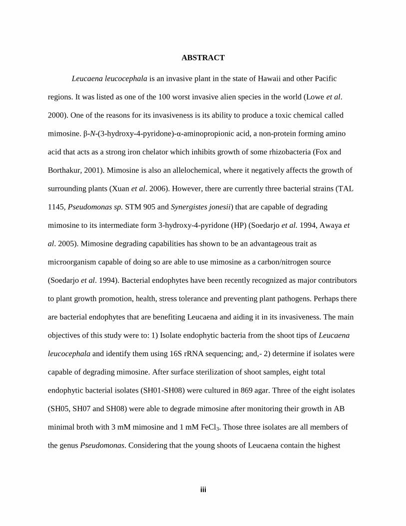
TABLE OF CONTENTS
ACKNOWLEDGEMENTS……………………………………………………………………..ii
ABSTRACT……………………………………………………………………………………..iii
TABLE OF CONTENTS……………………………………………………………...………...v
LIST OF TABLES………………………………………………………………………………vi
LIST OF FIGURES…………………………………………………………………………….vii
CHAPTER 1.......…………………..…...…………………………………….………………......1
INTRODUCTION………………………………………………………………………………...1
Leucaena leucocephala and its invasiveness……..…………………………………….....1
Mimosine and allelopathy….……………………………………………………………...2
Endophytic bacteria…………………………………………………………………….....3
Beneficial properties of bacterial endophytes......…………………………………………6
Mimosine degradation …………………………………………………………………....7
CHAPTER 2.......……………...………...........................………………………………………10
MATERIALS AND METHODS..……………....……………...…………………………..…....10
Collection of plant tissue samples......................................................................................10
Surface sterilization and culturing endophytes..................................................................10
16S rDNA extraction and sequencing................................................................................11
NCBI BLAST....................................................................................................................12
Phylogenetic analysis.........................................................................................................12
Growth of bacterial isolates in media containing mimosine and iron...............................13
vi
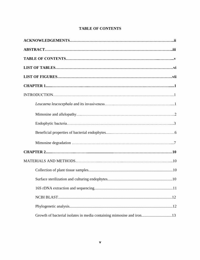
CHAPTER 3............…...…..……………………………………………..................…………..14
RESULTS…...…..……………………………………………………….....................................14
Characterization of endophytic bacteria............................................................................14
NCBI BLAST....................................................................................................................15
Phylogenetic analysis.........................................................................................................16
PCR Screen for mimosine degrading genes.......................................................................17
Growth of Bacterial isolates in media containing mimosine and iron...............................21
CHAPTER 4.........……………………………………...……….................................................22
GENERAL DISCUSSION………………………...…………...………......................................22
LITERATURE CITED………………………………………………………………………...26
vii
LIST OF TABLES
Table 1. Primer sets……………………………………………………………………………....12
Table 2. Bacterial genes………………………………………………………………………….12
Table 3. NCBI Genbank 16S rRNA sequences used in Phylogenetic analysis………………….13
Table 4. Bacterial Isolate closest NCBI GenBank BLAST neighbor……………………………16

viii
LIST OF FIGURES
Figure 1. Bacterial Isolates grown on 869 media containing 2mM mimosine…………………..15
Figure 2. Neighbor-joining tree of Bacterial isolates…………………………………………….17
Figure 3. PCR of MidD genes……………………………………………………………………18
Figure 4. PCR of PydA genes…………………………………………………………………....18
Figure 5. Growth of bacterial isolates in media containing iron-mimosine complex…………....20
Figure 6. Mimosine concentration from bacterial isolates after 48 hours of incubation…….......20
Figure 7. Growth of Bacterial isolates in 3mM mimosine and 1 mM FeCl3………………….....21
1
CHAPTER 1
INTRODUCTION
Leucaena leucocephala and its Invasiveness
Hawai’i is among the most isolated island archipelagos in the world, which unfortunately
makes it susceptible to exotic species invasions. Since the native biota is strongly endemic and
vulnerable to disturbances, the chance for recovery is low (Vitousek 1990). For instance,
Leucaena leucocephala, a member of the Mimosoideae family, is a leguminous tree/shrub that
can grow rapidly in arid climates (Shelton and Brewbaker, 1994). It is originally from Central
America and the Yucatan Peninsula of Mexico (Shelton and Brewbaker, 1998; Brewbaker et al.
1985), but has spread to Hawaii, Mariana Islands, southern Texas, southern Florida, Puerto Rico
and the Virgin Islands. (Little and Skolmen, 1989). Leucaena was listed as one of the 100 worst
invasive alien species in the world (Lowe et al. 2000). This plant was brought to tropical regions
because of its agricultural uses such as cattle fodder, reforestation, biofuels, and mitigating soil
erosion which makes it a ‘conflict species’ (Tuda et al. 2009; Elharith et al. 1980; Raghu et al.
2005, Olckers, 2011). One reason for its invasiveness is its high seed production. Each tree
produces roughly 1,700 pods (Raghu et al. 2005), each pod containing approximately 20 seeds
and there are 2-4 podding cycles per year (Raghu et al. 2005, Tuda et al. 2009). Seeds can be
easily dispersed by rodents, birds or cattle into adjacent areas. L. leucocephala is also a fast
grower, with sprouts capable of ~ 30 cm growth in a single month under optimal conditions (Kuo
et al. 2005). Once sprouts reach 2-3 m; they can escape browsing which reduces herbivory on the
species.

2
Mimosine and Allelopathy
One of the strongest defensive mechanisms for L. leucocephala is its ability to produce
mimosine. β-N-(3-hydroxy-4-pyridone)-α-aminopropionic acid, a toxic, non-protein forming
amino acid (Brewbaker and Hylin, 1965). Structurally, it is similar to dihydroxyphenylalanine
with a 3-hydroxy-4-pyridone ring instead of a 3,4-dihydroxy-phenyl ring (Xuan et al. 2006).
In animals, ingestion can result in alopecia, growth retardation, cataracts and infertility
(Crounse et al. 1962). Other symptoms include raw coronary bands, low thyroxin level and
goiter (Hegarty, 1967). The presence of -OH and -O in the pyridine ring suppresses iron-
containing enzymes, which is what makes mimosine toxic (Vickery et al. 1981). Leucaena has a
relatively high protein content in leaves, about 26.14% and in seeds for about 33.44% (Von et al.
1985) making it an ideal food source for livestock. However, mimosine is most concentrated in
the “growing tips” of Leucaena (4-10%) which prevents herbivory (Jones, 1979, Brewbaker and
Hylin, 1965). Mimosine possesses antimitotic activity which impedes the cell cycle in the G1
phase and it also disrupts DNA synthesis by changing the deoxyribonucleotide metabolism,
which prevents the formation of the DNA replication fork. (Gilbert et al. 1995; Tsvetkov et al.
1997). The free amino acid also acts as an antifungal agent (Tawata et al. 2008), and the growth
of certain root nodule bacteria is inhibited by mimosine (Fox and Borthakur, 2001). Most
importantly, mimosine acts as an iron chelator which allows it to block the biochemical activities
of the cell through iron depletion (Katoh et al. 1992; Gilbert et al. 1995; Soedarjo and Borthakur
1998).
Mimosine can have adverse effects on other plants as an allelochemical. Allelopathy is
the direct or indirect effects that a donor plant has towards other surrounding plants through
excretion of a chemical compound (Rice 1984, Mishra et al. 2013), can be positive or negative

3
towards the target plants (Ishak and Sahid 2014). Leucaena begins its allelopathy by producing
mimosine which is then leached out from the roots and plant tissues and directly absorbed by
neighboring plants (Ishak and Sahid 2014). Mimosine has been shown to significantly contribute
to weed reduction of L. glauca in paddy fields (Hong, 2004), and also been shown to inhibit
seedlings of mung bean, lettuce, wheat and rice (Smith and Fowden, 1966, Ling et al. 1969,
Xuan et al. 2006, Chou and Kuo, 1986). Because mimosine possesses strong allelopathic effects,
it has potential commercial uses as a bioherbicide compound (Xuan 2006, Ishak and Sahid
2014).
Endophytic Bacteria
Legumes are agriculturally important plants because of their nitrogen-fixing capabilities.
They can form root nodules in symbiosis with nitrogen-fixing bacteria to improve the plant's
overall growth. Because of this importance, the rhizobia-legume symbiosis has been heavily
studied (Wang et al. 2006, Mora et al. 2014). However, endophytic bacteria may contribute to
plant growth and health as much, if not more than rhizobia bacteria in legumes (Dudeja et al.
2012). Endophytes are microorganisms residing in the plant tissue which cause no visible harm
to the host plant (Hallmann et al. 1997, Wang et al. 2006). It is also defined as microorganisms
that could be isolated from surface-sterilized plant organs (Hardoim et al. 2008) All plant species
contain at least one or more endophytes (Strobel et al. 2004), but the presence of endophytes in
all tissues is highly dependent on the soil microflora (Dudeja et al. 2012). It begins with
endophytes originating from the soil as the plant is growing. Thus, bacterial communities within
the endosphere of the host plant are likely dictated by stochastic events which are tied in with
deterministic processes of colonization (Battin, et al. 2007). It can be said that the probability of
endophytic colonization depends on the initial abundance, diversity, physiological status and
4
distribution of endophytes in the soil. Other factors such as plant genotype, growth stage, type of
plant tissue and environmental soil conditions can also determine endophytic colonization. The
initial steps required in endophytic colonization are similar to that of rhizobacteria and plant
pathogens (Hallman et al, 1997). For example, well studied root-colonizing bacteria from the
genera Pseudomonas, Azospirillum and Bacillus are often found as colonizers of the internal
tissue of plants (Rosenblueth and Martinez-Romero, 2006, Hallman and Beng, 2006). However,
it is assumed that endophytes are specialized members of these groups when accepted into plant
tissue. There are several environmental and genetic factors that are presumed to have a role in
allowing a specific bacterium to become endophytic (Hardoim et al. 2008). The endosphere
region offers protection from the environment for outside bacteria that can colonize and establish
in planta (Ryan et al. 2007). The intercellular spaces provide endophytes with a significant
advantage over outside bacteria in the rhizosphere and phyllosphere because of stable pH levels,
moisture, rich nutrients and lack of competition from large number of microorganisms (Backman
and Sikora, 2008, Chebotar et al. 2015). Bacterial endophytes have been isolated from all plant
compartments, including seeds (Posada and Vega, 2005, Ryan, et al. 2007.)
How bacterial endophytes colonize regions above ground such as seeds and vegetative
plant parts is an active area of study. Endophytes are known to systematically spread throughout
the plant and colonize the stems and leaves through use of the lumen of xylem vessels (Hardoim
et al. 2008, Compant et al. 2005). But, it is unclear whether endophytes colonizing the roots or
aerial plant tissues have different effects on the plant. It is now confirmed that plant growth
promoting bacteria can migrate from one xylem compartment to another through the perforated
plates (Compant et al. 2010) Endophytes can also travel through the plant by use of the plant
transpiration stream. It is less likely for endophytes to travel through intercellular regions
5
because that would require cell wall degrading enzymes. It is important to note that only a few
endophytes are able to colonize aerial vegetative plant parts (Hallman, 2001) because they have
to pass multiple barriers as well as be physiologically capable to establish in different plant
niches (Compant et al. 2010). Endosymbiont cultivable population densities can reach up to 103-
104 CFU g
-1 of fresh weight under normal conditions (Compant et al. 2010).
Our understanding of endophytic microbial communities is being accelerated through
application of molecular techniques, e.g., 16S rRNA gene cloning, terminal restriction fragment
polymorphisms (RFLP) and sequencing (Loh et al. 2013, Franks et al. 2006). Molecular
advancements allow scientists to look into diverse microbial communities without necessarily
having to culture them. The five endophytic taxa showing the most promise for colonization and
an ability to persist were identified as Cellulomonas, Clavibacter, Curtobacterium, Pseudomonas
and Microbacterium by 16S rRNA gene sequence, carbon source uptake analyses and other
methods (Zinniel et al. 2002, Elvira-Recuenco & van Vuurde 2000). Recent studies conducted on
bacterial endophytes in Leucaena (Nimnoi, P & Pongsilp, N. 2009; Maruya, J & Saeki, K. 2010)
have successfully identified Mesorhizobium loti, Ensifer (Sinorhizobium) meliloti, Rhizobium sp.
NGR 234, R. tropici and Ensifer (Sinorhizobium) meliloti 1021, all of which were isolated from
Leucaena roots.
Our current knowledge of endophyte-host molecular interactions has developed over the
past decade (Lugtenberg et al. 2002). For example, autofluorescent protein (AFP) methods are a
key tool for studying the processes of microbe-plant interactions and biofilm formation (Ryan et
al. 2007). Artificial inoculation of plants with bacterial endophyte isolates shows us how these
bacteria colonize the endosphere and how they interact with the inoculated plants (Chebotar et al.
2014). There is also in vivo analysis of biological material for research on the molecular changes
6
of plant endospores and the spatial distribution of microorganisms over the course of the
development of the plant. A common marker system uses green fluorescent protein (GFP), which
allows the detection and counting of microorganisms in situ on plant surfaces and in planta
(Gage et al. 1996, Tombolini et al. 1997, Tombolini & Jansson, 1998.) The host plant gene
expression may be regulated by the type of bacteria endophytes.
Beneficial properties of Bacterial Endophytes
Bacterial endophytes have been shown to improve the overall health of their host plant.
Plant wellness is aided by: improved nitrogen and phosphorus nutrition to host plant, regulation
of osmotic pressure, modified root development, accelerated seedling emergence, promote plant
recruitment under extreme climate conditions and synthesis of vitamins and siderophores
(Rosenblueth & Martinez-Romero, 2006, Chebotar et al. 2015). Bacterial endophytes can reduce
or prevent the deleterious effects of pathogenic microorganisms through a variety of
mechanisms, including pathogenesis, production of various compounds and out-competing the
pathogens within the endosphere (Whipps, 2001). A variety of bacterial endophytes that show
superior antagonistic activities against plant pathogens has led to artificial inoculation
(biocontrol activity) of plants with endophytic bacteria. For example, 9 out of 137 isolates of
bacterial endophytes isolated from the tissues of stems, roots and nodules of soybean in vitro
inhibited the growth of the pathogenic fungi Macrophomina phaseolina, Fusarium udum,
Rhizoctonia bataticola and Sclerotium rolfsii (Senthikumar et al. 2009). Bacterial endophytes,
particularly the ones from the genera Pseudomonas, Burkholderia, and Bacillus are known for
producing secondary metabolites such as antibiotics, anticancer drugs, volatile organic
compounds, and fungicidal, insecticidal and immunosuppressive agents (Chebotar et al. 2015).
7
Some key properties of endophytes are their ability to promote plant growth through
signal inductions of hormones (López-López et al. 2010). Ethylene is a phytohormone that
controls plant growth and development, and has a central role in plant cellular metabolism (Ping
and Boland, 2004). Ethylene is also tied in with the plant developmental cycle, disease resistance
and microbe-plant interactions. It is a key regulator of the colonization of plant tissue by
bacteria, which are able to control plant ethylene levels by two mechanisms: first by cleaving the
ethylene precursor 1-aminocyclopropane-1-carboxylate (ACC) (Glick et al. 2007) or by
inhibiting ACC synthase or 𝛽-cystathionase, both enzymes are involved in the ethylene
biosynthesis pathway (Sugawara, M, et al. 2007) Thus, bacterial endophytes with high locally
induced ACC deaminase activities could be ideal plant-growth promoters because they can
reduce plant stress by blocking ethylene production (Cheng et al. 2007).
Indole-3-acetic acid (IAA) is the main plant growth hormone with auxin activity (Davies,
1995). IAA is responsible for several physiological processes in plants such as root proliferation,
cell division and shoot growth (Lambrecht et al. 2000, Davies, 1995). IAA production is known
to be involved in processes of plant-growth promoting bacteria and symbiotic bacteria (Hunter,
1989; Lambrecht et al. 2000). One example is Pseudomonas putida GR12-2 produces IAA
which stimulates the growth of the roots of canola seedlings (Patten and Glick, 2002).
Mimosine Degradation
Mimosine is antagonistic to a variety of plants and weeds. However, some bacterial
strains (Rhizobium sp.TAL1145, Pseudomonas sp. STM 905 and Synergistes jonesii) can
enzymatically break down mimosine into the intermediate: 3-hydroxy-4-pyridone (HP) and use it
as a carbon/nitrogen source (Soedarjo et al. 1994, Awaya et al. 2005, Pandey & Dwivedi, 2006,
Allison et al. 1992). There are specific genes (mid) that are required by the rhizobium strain TAL
8
1145 to degrade mimosine into HP (Borthakur et al. 2003). The mid genes are located within a
12.6-kb fragment of the TAL1145 chromosome. Two important steps are required to degrade
mimosine. In the first step, the alanyl side chain is hydrolyzed, forming HP. The next step
involves the expression of pydA and pydB genes respectively encoding for dioxygenase and
hydrolase enzymes that further degrade mimosine into pyruvate, formate, and ammonia. The
pydA gene is required for the cleavage reaction, opening up the aromatic ring structure of HP
(Awaya et al. 2007). The pydB gene is used to catabolize the molecule into metabolically useful
pyruvate (Awaya et al. 2007). The necessary proteins are located in the cytoplasm of TAL1145
(Soedarjo et al. 1994) Mimosine can be used as an exogenous siderophore by the rhizobial
bacterium TAL1145, giving it advantageous characteristics over other nodulating Rhizobium
(Soedarjo and Borthakur, 1998). The mechanism for degrading an allelochemical such as
mimosine is through chemotaxis where bacteria use the allelochemical as a nutrient source.
(Lugtenberg et al. 1999). It is believed that L. leucocephala originally evolved in regions with
iron-deficient soils. Mimosine released from leucaena chelates to iron, forming Fe-mimosine
complexes which are then taken up by Mid+ rhizosphere bacteria and converted to a form
available to the plant (Khanna and Lavin 1993, Gilbert et al. 1995)
Microbes outside of the rhizosphere can degrade mimosine and other pyridine ring
compounds (Allison et al. 1992, Watson et al. 1974). If there are endophytes that can degrade
mimosine, this may help explain its plant growth promoting capabilities towards Leucaena.
Overall, there is still very little research on endophytes within L. leucocephala in regions above
the root system. Perhaps mimosine degrading bacteria inhabit the regions where mimosine is
heavily concentrated such as the young shoots and stems. A better understanding of endophyte
ecology and their molecular interactions can open up a near-future application of using

9
genetically engineered endophytes as a bio-control in some main crops. A promising potential
for endophytes in agricultural uses is to increase crop yields, remove harmful chemicals, inhibit
pathogens, and produce novel substances. The full effects and functions in plants are still not
completely defined, thus this research field could contribute to economic and environmental
impacts.

10
CHAPTER TWO
MATERIALS AND METHODS
Collection of Plant Tissue Samples
Fresh, young Leucaena shoot clippings and seed pods were aseptically bagged and
collected from Pahoa-Kalapana Road (19°22’11.9”N 154°57’54.0”W) on June 23rd
and July 15th
2016. Samples were immediately placed on ice for preservation and transported to the laboratory
within 2 hours. Leucaena shoot samples were weighed and rinsed with tap water to remove
particulates. Seeds were cleaned and placed in sterile falcon tubes for long-term storage.
Surface Sterilization and Culturing Endophytes
Fresh shoot samples (~1 g) were submerged in 5% sodium hypochlorite for 10 minutes,
transferred into 70% ethanol for 2 minutes and then rinsed 3 times with sterile distilled water. A
100 ul aliquot of the third rinsate was plated on 869 minimal media petri dishes to check for
contamination. Samples were done in duplicates. There was no sign of microbial growth on the
control plates throughout the experiment.
Surface sterilized tissue samples were macerated in a sterile mortar with the addition of 5
mL of sterile water. Two 100 ul aliquots were plated on 869 media and placed in an incubator at
28ºC for 4 days. 869 minimal media was used for this experiment because it has been shown to
have the best optimal growth for bacterial endophytes (Eevers et al. 2015). The media contained
the following components per liter of deionized water: 0.035 g CaCl2.2H2O, 0.100 g Glucose D+.
0.500 g NaCl, 1.0 g Tryptone, 0.500 g Yeast Extract, 15.0 g Agar. Successful endophytes were
streak plated for single colonies and the isolates were stored in 40% glycerol at -80℃ for long
term storage. The isolates were then grown in 869 liquid broth and in AB minimal media to
detect mimosine degrading capabilities. AB minimal media contained the following components

11
per liter of deionized water: 1.5 g KH2PO4, 0.5 g NaH2PO4, 0.15 g MgSO4·7H2O, 0.075 g KCl,
0.005 g CaCl2, 0.00125 g FeSO4·7H2O and 10 mL of 40 mg/mL mimosine stock solution
(Soedarjo et al. 1994) for a final mimosine concentration of 3 mM.
16S rDNA Extraction and Sequencing
Bacterial DNA was extracted from overnight cultures of isolates using the MO BIO
UltraClean Microbial DNA Isolation Kit. DNA quality and quantity was analyzed using a
NanoDrop 1000 Spectrophotometer. The 16s, MidD and PydA primer sets (Table 1) were
obtained from Dr. Jonathan D. Awaya. Polymerase Chain Reaction (PCR) was applied to detect
mimosine degrading genes and for amplification of the 16S region for bacterial identification
(Table 2). Promega GoTaq Green Master Mix was used for our PCR reactions. We followed the
manufacturer's instructions (Part# 9PIM712) and standard protocol (Sambrook and Russell,
2001). The Eppendorf Mastercycler Pro with Vapo.protect thermocycler was set to the following
cycling condition: 15 mins at 94ºC for 1 cycle; 1 min and 30 sec at 94ºC, 1 min at 52ºC, 1 min at
72ºCfor 30 cycles; 10 min at 10ºC for 1 cycle; and held at 4ºC. All 16S PCR products were
verified for successful amplification by running gel electrophoresis using 1% agarose gel under
90 volts for 40 mins using Promega BenchTop 1kb ladder. PCR clean-up was performed using
Promega Wizard SV Gel and PCR Clean-Up system. Clean PCR products were sent to the
University of Hawaii at Hilo’s Genetics Core Lab for sequencing.
12
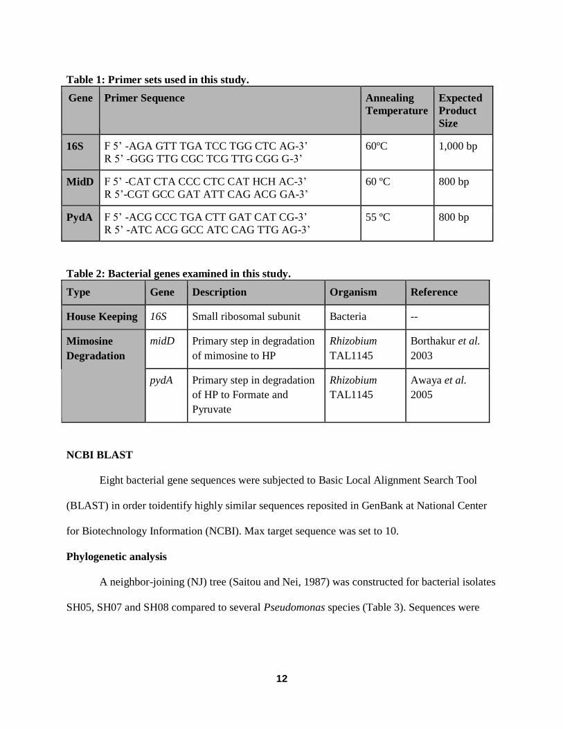
Table 1: Primer sets used in this study.
Gene Primer Sequence Annealing
Temperature
Expected
Product
Size
16S F 5’ -AGA GTT TGA TCC TGG CTC AG-3’
R 5’ -GGG TTG CGC TCG TTG CGG G-3’
60ºC 1,000 bp
MidD F 5’ -CAT CTA CCC CTC CAT HCH AC-3’
R 5’-CGT GCC GAT ATT CAG ACG GA-3’
60 ºC
800 bp
PydA F 5’ -ACG CCC TGA CTT GAT CAT CG-3’
R 5’ -ATC ACG GCC ATC CAG TTG AG-3’
55 ºC
800 bp
Table 2: Bacterial genes examined in this study.
Type Gene Description Organism Reference
House Keeping 16S Small ribosomal subunit Bacteria --
Mimosine
Degradation
midD Primary step in degradation
of mimosine to HP
Rhizobium
TAL1145
Borthakur et al.
2003
pydA Primary step in degradation
of HP to Formate and
Pyruvate
Rhizobium
TAL1145
Awaya et al.
2005
NCBI BLAST
Eight bacterial gene sequences were subjected to Basic Local Alignment Search Tool
(BLAST) in order toidentify highly similar sequences reposited in GenBank at National Center
for Biotechnology Information (NCBI). Max target sequence was set to 10.
Phylogenetic analysis
A neighbor-joining (NJ) tree (Saitou and Nei, 1987) was constructed for bacterial isolates
SH05, SH07 and SH08 compared to several Pseudomonas species (Table 3). Sequences were

13
aligned using MAFFT alignment program with default settings. Five hundred bootstrap
replications were used to evaluate support for tree topology.
Table 3. NCBI Genbank 16S rRNA sequences used in Phylogenetic analysis
NCBI GenBank
Accession #
Description Reference
NR074804 Cellvibrio japonicus (Outgroup) Deboy et al. 2008
KY024584 Pseudomonas aeruginosa Joseph et al. 2016
KX817232 Pseudomonas fluorescens Zhu and Guo 2016
KY021743 Pseudomonas mendocina Ding et al. 2016
KY021742 Pseudomonas oleovorans Ding et al. 2016
LC191549 Pseudomonas oryzihabitans Tani and Hamba 2016
KU550194 Pseudomonas psychrotolerans NA*
KX817239 Pseudomonas putida Zhu and Guo 2016
KU904408 Pseudomonas stutzeri Wang et al. 2016
* Not Applicable
Growth of bacterial isolates in media containing mimosine and iron
Bacterial isolates were grown in 15 mL of AB minimal liquid culture containing 3mM
mimosine and 1mM FeCl3 in a shaker at 28ºC. Each growth rate treatment was done in triplicate.
Optical density of bacteria was measured using a Genesys 20 spectrophotometer at 600 nm. 1
mL samples were removed every 8 hours for 48 until cultures reached stationary growth phase.
Final mimosine content for bacterial cultures were observed by measuring absorbance of 1 mL
samples at 535 nm.
14
CHAPTER THREE
RESULTS
Characterization of endophytic bacteria
A total of 8 isolates grew on the 869 media from the young shoot tissue of Leucaena.
Control plates showed no growth from the rinsate which confirms that the isolates are
endophytic. Initial growth of endophytes seemed to occur around 48 hours. All isolates seem to
grow well at 28ºC. Isolate SHO1 was distinguishable from the others because of its orange-red
color (Figure 5). It was also more difficult to form single colonies as it was a smoother, liquid
form on 869 media agar. SH02 produced a yellow opaque color. SH03 had a creamy dull white
color and smooth forming colonies. SH04 was bright yellow and creamy. SH05 was an opaque
white color. SH06 was also bright yellow like SH04. SH07 and SH08 were very similar in
morphology. Both had a translucent yellow color.
There was no successful bacterial growth from the seeds despite making adjustments to
the culturing media, surface sterilization methods and incubation time.

15
Figure 1. Bacterial Isolates (SHO1-SHO8) grown on 869 media containing 2mM mimosine.
NCBI BLAST
All 8 bacterial isolates had positive matches to 16S sequence BLAST results (Table 5).
Isolate SH01 whose closest BLAST neighbor was Rhodococcus kroppenstedtii from phylum
Actinobacteria, isolate SHO2 whose closest BLAST neighbor was Sphingomonas paucimobilis
from the phylum Proteobacteria, isolate SH03 whose closest BLAST neighbor was
Microbacterium proteolyticum from the phylum Actinobacteria, isolate SH04 whose closest
BLAST neighbor was Sphingomonas pseudosanguinis from the phylum Proteobacteria, isolate
SH05 whose closest BLAST neighbor was Pseudomonas putida from the phylum Proteobacteria,
isolate SH06 whose closest BLAST neighbor was Sphingomonas pseudosanguinis, isolate SH07
whose closest BLAST neighbor was Pseudomonas oryzihabitans and isolate SH08 whose closest
BLAST neighbor was Pseudomonas oryzihabitans.
16
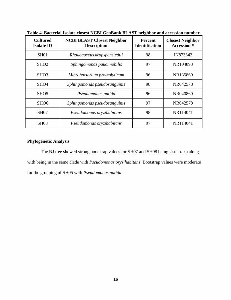
Table 4. Bacterial Isolate closest NCBI GenBank BLAST neighbor and accession number.
Cultured
Isolate ID
NCBI BLAST Closest Neighbor
Description
Percent
Identification
Closest Neighbor
Accession #
SH01 Rhodococcus kropspenstedtii 98 JN873342
SHO2 Sphingomonas paucimobilis 97 NR104893
SHO3 Microbacterium proteolyticum 96 NR135869
SHO4 Sphingomonas pseudosanguinis 98 NR042578
SHO5 Pseudomonas putida 96 NR040860
SHO6 Sphingomonas pseudosanguinis 97 NR042578
SH07 Pseudomonas oryzihabitans 98 NR114041
SH08 Pseudomonas oryzihabitans 97 NR114041
Phylogenetic Analysis
The NJ tree showed strong bootstrap values for SH07 and SH08 being sister taxa along
with being in the same clade with Pseudomonas oryzihabitans. Bootstrap values were moderate
for the grouping of SH05 with Pseudomonas putida.
17
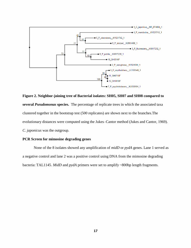
Figure 2. Neighbor-joining tree of Bacterial isolates: SH05, SH07 and SH08 compared to
several Pseudomonas species. The percentage of replicate trees in which the associated taxa
clustered together in the bootstrap test (500 replicates) are shown next to the branches.The
evolutionary distances were computed using the Jukes–Cantor method (Jukes and Cantor, 1969).
C. japonicus was the outgroup.
PCR Screen for mimosine degrading genes
None of the 8 isolates showed any amplification of midD or pydA genes. Lane 1 served as
a negative control and lane 2 was a positive control using DNA from the mimosine degrading
bacteria: TAL1145. MidD and pydA primers were set to amplify ~800bp length fragments.

18
Figure 3. PCR of MidD genes. PCR analysis of DNA from all 8 isolates with primers used to
amplify within MidD. M1 and M2 are 1.0 kb markers. Lane 1 is a MidD negative control. Lane 2
is a positive control using DNA from the rhizobium TAL1145. Lane 3-9 contains PCR products
from isolates SH01-SH08 respectively.
Figure 4. PCR of PydA genes. PCR analysis of DNA from all 8 isolates with primers used to
amplify within PydA. M1 and M2 are 1.0 kb markers. Lane 1 is a PydA negative control. Lane 2
is a positive control using DNA from the rhizobium TAL1145. Lane 3-9 contains PCR products
from isolates SH01-SH08 respectively.

19
Growth of Bacterial isolates in media containing mimosine and iron
Bacterial isolates SH05, SH07 and SH08 were able to grow in AB minimal media with
3mM mimosine and 1mM FeCl3. Bacterial cultures for the three isolates seemed to grow
exponentially around the 16 hour mark and reach the stationary phase around 32 hours. Isolate
SH05 had a greater absorbance (~0.3) than SH07 (~0.24) and SH08 (~0.21), however, there is
high variability due to minimal growth in the first replication. SH07 and SH08 seemed to have
identical absorbance throughout the trial.
Mimosine content was measured at 56 hours. There was a difference in absorbance with
isolates SH01, SH02, SH03, SH04 and SH06 having higher absorbance than SH05, SH07 and
SH08. Normal absorbance for AB minimal media with 3 mM mimosine and 1 mM FeCl3 was
roughly 1.74. Isolates SH07 and SH08 had an absorbance below 1 which translates to mimosine
degradation. Figure 11 shows a change in media color from dark red to a light yellow/orange
color. This occurs when the mimosine-iron complex structure is cleaved.

20
Figure 5. Growth of bacterial isolates in media containing iron-mimosine complex. Isolates
were grown in AB minimal broth containing 3 mM mimosine and 1 mM FeCl3. The data points
in the growth curve are the means and standard deviations of three replicates.
Figure 6. Mimosine concentration from bacterial isolates after 48 hours of incubation at
28℃. Initial concentration of mimosine was 3mM with 1mM FeCl3, which had an absorbance of
1.74 at 535 nm.

21
Figure 7. Growth of Bacterial isolates in 3mM Mimosine and 1 mM FeCl3. Change in color
of AB minimal media after 48 hour growth period.
22
CHAPTER FOUR
DISCUSSION
This study successfully demonstrates the isolation and identification of bacterial
endophytes from the vegetative regions of L. leucocephala. There was no bacterial growth from
the seeds despite making adjustments to culturing and surface sterilization methods. A total of 8
isolates were cultured from within the inner tissues of the growing tips of Leucaena sampled off
Kalapana road. The sterile control plates confirm these isolates are endophytic and not surface
dwelling bacteria. All 8 isolates have been identified and recognized as endophytes in previous
studies. For instance, SH01 was closely related (98%) to Rhodococcus kroppenstedtii. The genus
Rhodococcus is classified in the family Nocardiaceae (Stackebrandt et al. 1997). These
microorganisms are shown to have a broad metabolic diversity, especially with hydrophobic
compounds. This has piqued the interest of scientists to further study the biochemical and genetic
characterization of their metabolic processes as they make ideal candidates for use in
bioremediation (Guo-Zhen et al. 2012). Rhodococcus kroppenstedtii is Gram-positive, non-
motile, non-spore forming, coccoid-like bacteria. Other member of the Rhodococcus genus have
been isolated from the pharmaceutical plant Artemeisia annua L. (Zhao et al. 2012). Isolates
SH02, SH04 and SH06 were closely related to the genus Sphingomonas. SH02 was more
towards the species paucimobilis, whereas SH04 and SH06 were closer related to
pseudosanguinis. Members of this genus are Gram-negative, yellow-pigmented, nonspore-
forming and non-motile rods (Busse and Denner 1999). Sphingomonas paucimobilis showed it
has plant-growth promoting abilities for a traditional Chinese medicinal plant; Dendrobium
officinale. The growth of D. officinale seedlings, with Sphingomonas paucimobilis significantly
increased stem growth by 8.6% (Yang et al. 2014). It was also shown that the strain ZJSH1 was

23
able to produce various phytohormones such as salicylic acid, IAA, Zeatin and abscisic acid
(Yang et al. 2014). Isolate SH03 was closely related to Microbacterium proteolyticum which is
Gram-positive, non-motile, aerobic and short rods (Alves et al. 2015). It was recently isolated
from the roots of the salt-marsh plant Halimione portulacoides (Alves et al. 2015). Isolates
SH05, SH07 and SH08 were closely identified in the genus Pseudomonas. SH05 is closely
related to Pseudomonas putida as shown from the BLAST and NJ tree results. As mentioned
earlier, there was a Pseudomonas putida strain (STM 905) that was able to degrade mimosine
(Pandey, 2007). Isolates SH07 and SH08 might be the same species as sequence alignment. In
the article from Kodama et al. 1985, Pseudomonas oryzihabitans are yellow-pigmented, oxidase-
negative, Gram negative, rod shaped bacteria. They have been isolated from normal rice paddies
and from soybean plants grown in soil treated with glyphosate herbicide (Kuklinsky-Sobral et al.
2004). Importantly, Belimov et al. 2001 observed that P. oryzihabitans isolates from Pisum
sativum rhizoplane showed the capability to produce ACC deaminase. Furthermore, Kuklinsky-
Sobral et al. 2004 discovered that endophytic isolates from soybean plants were able to produce
auxin. These articles support that P. oryzihabitans has the potential for plant growth promotion.
Although isolates SH05, SH07 and SH08 showed the most growth in mimosine minimal
media, the gel analysis showed that none of the bacterial isolates contained the midD and pydA
genes required to degrade mimosine and HP previously identified in Rhizobium TAL 1145 (Fig.
4). This could indicate a different pathway is being used for mimosine degradation or that the
primers were not able to properly anneal to the mimosine degrading genes. To our knowledge,
this is the first attempt at isolating endophytic bacteria from Leucaena leucocephala above the
root system. This is also a new discovery of bacteria that can degrade mimosine without being
Mid+. Discovering bacterial endophytes from regions above the roots and nodules unveils

24
another area of endophytic research that can help explain how these bacteria are benefitting the
host plant. It seems that these endophytic isolates were successful in establishing themselves in a
difficult niche and successfully translocating from the soil up to the growing tips of L.
leucocephala.
Future work is needed to give us a better understanding of bacterial endophytes in
Leucaena. Unresolved questions include how isolates SH05, SH07 and SH08 are able to degrade
mimosine and which genes are induced from these isolate under high levels of mimosine?
Investigating the biochemical pathways would be an interesting research topic because scientists
can compare it with the currently known mimosine-degrading pathway from the rhizobium
isolate, TAL 1145. Another research plan could be studying how these endophytes are benefiting
the plant by detecting the presence of ACC deaminase gene. PCR can be used to assess if the
isolates cultured in this study contain the ACC deaminase gene. These endophytes also hold
potential for biocontrol activity to current plant pathogens. For example, Acacia koa is a native
legume of Hawaii that is affected by a common fungal plant pathogen: Fusarium oxysporum.
One can observe if Fusarium growth would be affected if one of the isolates was introduced to
the culture. Acacia koa seedlings could also be inoculated with Fusarium and endophytic
bacteria and observe if there is any difference in growth and symptoms.
A 16S metagenomics analysis within the endosphere of Leucaena is a good approach to
observing the microbial community that may not be culturable. Little is known of the endophytic
microbial communities within Leucaena and this could help us see which bacteria are most
prevalent. The structure of bacterial communities is based on both cultivation-dependent and
cultivation-independent techniques. Previous studies that have done this indicated that these
communities can change over time, with endophytes showing a pattern of growth that correlates

25
with plant growth and development (Hardoim et al. 2008). In addition, observing endophytic
communities is often simpler when compared to soil bacterial communities, encompassing
hundreds of different bacterial types. 16S metagenomics could provide scientists with an idea of
the bacterial community structure that resides in the tissues of Leucaena. Full genome
sequencing of the mimosine degrading isolates will also provide good insight into which unique
genes may be involved with mimosine degradation, and also which genes are involved with
communication with host plant.
Hopefully this research along with future studies will open our eyes to how important
these endophytes are to the overall success of Leucaena in its invasiveness and hopefully we can
exploit them for native and other important flora.
26
LITERATURE CITED
Allison, M.J., Mayberry, W.R., Mcweeney, C.S., and Stahl, D.A. 1992. Synergistes jonesii, gen.
nov., sp. nov.: a rumen bacterium that degrades toxic pyridinediols. Syst. Appl. Microbiol. 15:
522–529.
Awaya, J. D., Fox, P. M., & Borthakur, D. (2005). pyd Genes of Rhizobium sp. Strain TAL1145
Are Required for Degradation of 3-Hydroxy-4-Pyridone, an Aromatic Intermediate in Mimosine
Metabolism. Journal of Bacteriology. 187: 4480–4487.
Awaya, J., Walton, C. and Borthakur, D. 2007. The pydA-pydB fusion gene produces an active
dioxygenase-hydrolase that degrades 3-hydroxy-4-pyridone, an intermediate of mimosine
metabolism. Appl. Micriobiol Biotechnol. 75: 583-588.
Backman, P. and Sikora, R. 2008. Biolog. Control vol. 46, pp. 1-3.
Battin, T. et al. 2007. Microbial landscapes: new paths to biofilm research. Nat. Rev. Microbiol.
5: 76-81.
Belimov, A. et al. 2001. Characterization of plant growth promoting rhizobacteria isolated from
polluted soils and containing 1-aminocyclopropane-1-carboxylate deaminase. Can. J.
Miocrobiol. 47: 642-652.
Brewbaker, J.L., N. Hegde, E.M. Hutton, R.J. Jones, J.B. Lowry, F. Moog, and R. van den Beldt.
1985. Leucaena - Forage Production and Use. NFTA, Hawaii. 39 pp.
Brewbaker, J.L., J.W. Hylin. 1965. Variations in mimosine content among Leucaena species and
related mimosaceae. Crop Sci 5:348–349.
Brewbaker, J.L. and C.T. Sorensson. 1990. New tree crops from interspecific Leucaena hybrids.
In: Janick, J. and Simon, J.E. Ed. Advances in New Crops. Timber Press, Portland, pp.
283-289.
Busse, H., Kampfer, P., and Denner, E. 1999. Chemotaxonomic characterisation of
Sphingomonas. Journal of Industrial Microbiology & Biotechnology. 23: 242-251.
Chou, C.H., Kuo Y.L. 1986. Allelopathic research of subtropical vegetation in Taiwan. III.
Allelopathic exclusion of understory by Leucaena leucocephala (Lam.) de Wit. Journal of
Chemical Ecology. 12: 1431-1448.
Compant, S., Clement, C., and Sessitch, A. 2010. Plant growth-promoting bacteria in the rhizo-
and endosphere of plants: Their role, colonization, mechanisms involved and prospects for
utilization. Soil Biology & Biochemistry. 42: 669-678.
Crounsem R.G., Maxwell, R.D. and Blank, H. 1962. Inhibition of growth of hair by mimosine.
Nature. 194: 694-695.
27
Davies, P.J., 1995. The plant hormone concept : concentration, sensitivity, and transport. In Plan
t hormones: physiology, biochemistry and molecula r biology, Ed., Davies, P.J. Kluwer Academi
c Publishers, 13-18 .
Deboy, R.T., et al. 2008. Insights into plant cell wall degradation from the genome sequence of
the soil bacterium Cellvibrio japonicus. J. Bacteriol. 190: 5455-5463.
Ding, Y., Zhao, D. and Li, Y. 2016. Direct Submission. Shandong Agricultural University.
Dudeja et al.2012. Interaction of endophytic microbes with legumes. Journal of Basic
Microbiology. 52: 248-260.
Elvira-Recuenco, M & van Vuurde, J. (2000) Natural incidence of endophytic bacteria in pea
cultivars under field conditions. Can J Miccrobiol 46: 1036-1041.
Fox, P.M. and Bothakur, D.2001. Selection of several classes of mimosine-degradation-defective
Tn3Hogus-insertion mutants of Rhizobium sp. strain TAL1145 on the basis of mimosine-
inducible GUS activity. Canada Journal of Microbiology. 47: 488-494.
Franks, A, Ryan PR, Abbas A, Mark G & O’Gara, F. 2006. Molecular Tools for Studying Plant
Growth-Promoting Rhizobacteria (PGPR): Molecular Techniques for Soil and Rhizosphere
Microorganisms, CABI Publishing, Wallingford, Oxforshire, UK.
Gage, D., Bobo, T. & Long, S. 1996. Use of green fluorescent protein to visualize early events of
symbiosis between Rhizobium meliloti and alfalfa (Medicago sativa). J Bacteriol 178: 7159-
7166.
Gilbert, D.M., Neilson, A., Miyazawa, H., DePamphilis, M.L. and Burhans, W.C. 1995.
Mimosine arrests DNA synthesis at replication forks by inhibiting deoxyribonucleotide
metabolism. J. Biol. Chem. 270: 9597-9606.
Hallmann, J. et al.1997. Bacterial endophytes in agricultural crops. Canadian Journal of
Microbiology, 43(10), 895–914. http://doi.org/10.1139/m97-131
Hong, N.H., Xuan, T.D., Tsuzuki, E., and Khanh, T.D. 2004. Paddy weed control by higher
plants from Southeast Asia. Crop Protection. 23: 255-261.
Hallman, J. and Beng, G. 2006. Spectrum and population dynamics of bacterial root endophytes.
Microbial Root Endophytes (Shulz, B.J.E et al. eds), 15-31.
28
Hardoim, P., Overbeek, L., and Elsas, J.D. 2008 Properties of bacterial endophytes and their
proposed role in plant growth. Trends in Microbiology, 16: 463-471.
Hegarty, M.P. 1967. Leucaena as animal feed, in: F.R. Ruskin (Ed.), Leucaena, Promising
Forage and Tree Crops for the Tropics, National Academy of Science, Washington, DC. 21-41.
Hunter, W. 1989. Indole-3-acetic acid production by bacteroids from soybean root nodules.
Physiolofia Plantarum, 76: 31-36,
Ilham, Z. et al. 2015. Extraction and Quantification of Toxic Compound Mimosine from
Leucaena leucocephala Leaves. Procedia Chemistry. 16: 164-170.
Iniguez, A.L. et al. 2005. Regulation of enteric endophytic bacterial colonization by plant
defenses. Mol. Plant Microbe Interact., 18: 169-178.
Ishak, M. and Sahid, I.2014. Allelopathic effects of the aqueous extract of the leaf and seed of
Jones, R.J. 1979. The value of Leucaena leucocephala as a feed for ruminants in the tropics.
World Animal Rev. 31. 13-23.
Joseph, A., Asok, A.K. and M S, J. 2016. Direct Submission. National Institure of Plant Science
and technology, Mahatma Gandhi University, Kattayam, Kerala, India, Athirampuzha, Kattayam
686560, India.
Jube, S.L.R. 2009. Genetic transformation of Leucaena leucocephala for reduction of mimosine
content. University of Hawaii at Manoa, Doctor of Philosophy (Ph.D.) Dissertation, 119 pages.
Katoh, S., Toyama, J., Kodama, I., Kamiya, K., Akita, T. & Abe, T. 1992. Protective action of
iron-chelating agents (catechol, mimosine, diferozamine and kojic acid) against ischemia-
reperfussion injury of isolation neonatal rabbit hearts. Eur Surg Res. 24: 349-355.
Kuklinsky-Sobral, J. et al. 2004. Isolation and characterization of soybean-associated bacteria
and their potential for plant growth promotion. Environ. Microbiol. 6: 1244-1251.
Nimnoi, P., Pongsilp, N.2009. Genetic diversity and plant-growth promoting ability of the
indole-3-acetic acid (IAA) synthetic bacteria isolated from agricultural soil as well as
rhizosphere, rhizoplane and root tissue of Ficus religiosa L., Leucaena leucocephala and Piper
sarmentosum Roxb. Res. J. Agric. Biol. Sci., 5: 29-41.

29
Lambrecht, M., Okon, Y., Broek, A. and Vanderleyden, J. 2000. Indole-3-acetic acid: a
reciprocal signaling molecule in bacteria-plant interactions. Tends Microbiol. 8: 298-300.
Ling, K.H., Wen, W.H., Ling, W.I. 1969. Study on the mechanism of the toxicity of mimosine.
Journal of Formosa Medicinal Association. 68: 510-517.
Little, E. and Skolmen, R.1989. Koa haole, leucaena. Agriculture Handbook. 679.
Loh, C., et al. 2013. Diversity of endophytic bacteria in Malaysian plants as revealed by
16SrRNA encoding gene sequence based method of bacterial identification. Journal of Young
Pharmacists 5: 95-97.
Lugtenberg, B.J., Chin-A-Woeng, T.F., Bloemberg, G.V., 2002. Microbe-plant interactions:
Principles and mechanisms. Antonie van Leeuwenhoek, 81: 373-383.
Lugtenberg, BJJ, Kravchenko LV, Simmons, M. 1999. Tomato seed and root exudates sugars:
composition, utilization by Pseudomonas biocontrol strains and role in rhizosphere colonization.
Environmental Micrbiology, 1:9-13.
Miche, L & Balandreau, J (2001). Effects of rice seed surface sterilization with hypochlorite on
inoculated Burkholderia vietnamiesis. Appl Environ Microbiol 67: 3046-3052.
Mora, Y. et al.2014. Nitrogen-fixing rhizobial strains isolated from common bean seeds:
phylogeny, physiology, and genome analysis. Applied and Environmental Microbiology, 80:
5644–54.
Nimnoi, P., Pongsilp, N. 2009. Genetic diversity and plant-growth promoting ability of the
indole-3-acetic acid (IAA) synthetic bacteria isolated from agricultural soil as well as rhizospere.
Rhizoplane and root tissue of Ficus religiosa L., Leucaena leucocephala and Piper
sarmentosunm. J. Agric. Biol. Sci., 5: 29-41.
Pandey, A.K. and Dwivedi, U.N.2007. Induction, isolation and purification of mimosine
degradation enzyme from newly isolated Pseudomonas putida STM 905. Enzyme and Microbial
Technology. 40: 1059-1066.
Patten, C.L. and Glick, B.R. 2002. Role of Pseudomonas putida indole acetic acid in
development of the host plant root system. Appl. Environ. Microbiol. 68: 3795-3801.
Ping, L., and Boland, W. 2004. Signals from the underground: bacterial volatiles promote growth
in Arabidopsis, Trends Plant Sci. 9: 263-266.

30
Posada, F & Vega, F. 2005. Establishment of the fungal entomopathogen Beauveria bassiana
(Ascomycota: Hypocreales) as an endophytic in cocoa seedlings (Theobroma cacao). Mycologia
97: 1195-1200.
Reinhold-Hurek, B., Hurek, T. Living inside plants: bacterial endophytes. Current Opinion in
Plant Biology 14: 435-443.
Rosenblueth M., Martinez-Romero E.2006. Bacterial Endophytes and Their Interactions with
Hosts. The American Phytopathological Society. 19: 827-837.
Saitou, N. and Nei, M. 1987. The Neighbor-joining Method: A New Method for Reconstructing
Phylogenetic Trees. Mil. Biol. Evol. 4: 406-425.
Shelton, H. M., & Brewbaker, J. L. 1998. Leucaena leucocephala–the most widely used forage
tree legume. Forage Tree Legumes in Tropical Agriculture. St Lucia Queensland: Tropical
Grassland Society of Australia Inc. http://www. Fao.
org/ag/AGP/AGPC/doc/Publicat/Guttshel/x5556e06. htm.
Smith, I.K., Fowden, L. 1966. A study of mimosine toxicity in plant. Journal of Experimental
Botany. 17: 750-761.
Stackebrandt, E, Rainey, F.A. Ward-Rainey, N.L. 1997. Proposal for a new hierarchic
classification system, Actinobacteria classis nov. Int J Syst Bacteriol. 47: 479-491
Sugawara, M. et al. 2006. Rhizobitoxine modulates plant-microbe interactions by ethylene
inhibition. Biotechnol. Adv. 24: 382-388.
Tombolini, R., Unge, A., Davey, M., de Brujin, F. and Jansson, J. 1997. Flow cytometric and
microscopic analysis of GFP-tagged Pseudomonas fluorescens bacteria. FEMS Microbiology
Ecology. 22: 17-28.
Tsvetkov, L.M., Russev, G.C., and Anchkova, B.B. 1997. Effect of mimosine on DNA synthesis
in mammalian cells. Cencer Res. 57: 2252-2255.
Vicente, D.2012. Elucidating Unique Iron Uptake Pathways By An Environmental Bacterium,
Rhizobium sp. Strain TAL1145. ProQuest.
Von, U., ter Meulen, El Harith, E.A. 1985. Mimosine - a factor limiting the use of Leucaena
leucocephala as an animal feed.

31
Wang, Y., Wang, C. and Gao, J. 2016. Direct Submission. Agronomy College, Jilin Agricultural
University.
Raghu, S., C. Wiltshire, and K. Dhileepan.2005. Intensity of pre-dispersal seed predation in the
invasive legume Leucaena leucocephala is limited by the duration of pod retention. Austral
Ecology. 30: 310-318
Ryan RP, Germaine K, Franks A, Ryan DJ. 2008. Bacterial endophytes: recent developments and applications. FEMS Microbiol. Lett. 278:1-9
Schulz, B. et al.1993. Endophytes from Herbaceous Plants and Shrubs: Effectiveness of Surface
Sterilization Methods. Mycological Research 97: 1447–1450.
Senthikumar, M., Swarnalakshmi, K., Govindasamy, V., Lee, Y., and Annapurna, K., Current
Microbiology., 58:288-293.
Strobel G, Daisy, B, Castillo U & Harper, J. 2004. NAtural products from endophytic
microorganisms. J Nat Prod. 67: 257-268.
Wang, E. et al.2006. Diverse endophytic bacteria isolated from a leguminous tree Conzattia
multiflora grown in Mexico. Archives of Microbiology, 186: 251–259.
Tombolini, R. & Jansson, J. 1998. Monitoring of GFP-tagged bacterial cells. Methods in
Molecular Biology. Bioluminescence Methods and Protocols (LaRossa RA, ed) pp. 285-298.
Humana PRess Inc., Totowa, NJ.
Tombolini, R., Unge, A., Davey, M., de Bruijn, F. & Jansson, J. 1997. Flow cytometric and
microscopic analysis of GFP-tagged Pseudomonas fluorescens bacteria. FEMS Microbiol Ecol
22. 17-28.
Vickery, M.L., Vickery, B. 1981. Compound derived from amino acids. Secondary plant
metabolism. 220-252.
Watson, G.K., Houghton, C., and Cain, R.B. 1974. Microbial metabolism of the pyridine ring.
Biochem. J. 140: 277–292.
Whipps, J., 2001. Exp. Bot., 52: 487-511.
Xuan, T. et al.2006. Mimosine in Leucaena as a potent bio-herbicide. Agronomy for Sustainable
Development. 26: 89-97.Leucaena leucocephala on three selected weed species. AIP Conference
Proceedings 1614: 659-663.
Olckers, T. 2011. Biological Control of Leucaena leucocephala (Lam.) de Wit (Fabaceae) in
South Africa: A Tale of Opportunism, Seed Feeders and Unanswered Question. African
Entomology. 19: 356-365.
32
Tani, A. and Hamba, T. 2016. Chemotaxis fishing: direct isolation of chemotactic bacteria
toward methanol.
Tawata et al. 2008. Total utilization of tropical plants Leucaena leucocephala and Alpinia
zerumbet. J. Pestic. Sci. 33: 40-43.
Yadav, K.K., Bora, A., Datta, S., Chandel, K., Gogoi, H.K. and Veer, V. 2016. Study on bacteria
associated with midgut of Aedes albopictus (Stegomyia albopicta) (Order:Diptera) from various
regions of North-East India.
Yang, S. et al. 2014. Growth-promoting Sphingomonas paucimobilis ZJSH1 associated with
Dendrobium officinale through phytohormone production and nitrogen fixation. Microbial
biotechnology. 6: 611-620.
Zhu, Y. and Guo, Q. 2016. Direct Submission. Food enginerring, USST/DHS. Jungong road.
Zinniel, D, Lambrecht, P., Harris, B et al. 2002. Isolation and characterization of endophytic
bacteria from agronomic crops and prarie plants. Appl Environ Microbiol 68: 2198-2208.