Embed Size (px)
Citation preview
Isolation and Characterization of Postsynaptic
Densities from Various Brain Regions:
Enrichment of Different Types of Postsynaptic Densities
RICHARD K. CARLIN, DENNIS J . GRAB, ROCHELLE S. COHEN, and PHILIP SIEKEVITZDepartment of Cell Biology, The Rockefeller University, New York 10021, and Department of Anatomy,The University of Illinois at the Medical Center, Chicago, Illinois 60680
ABSTRACT Postsynaptic densities (PSDs) have been isolated from cerebral cortex, midbrain,cerebellum, and brain stem by the Triton X-100 method previously used in the isolation ofcerebral PSDs (Cohen et al ., 1977, 1. Cell Biol. 74:181) . These PSDs have been compared inprotein composition, protein phosphorylation, and morphology . Thin-section electron micros-copy revealed that cerebral cortex and midbrain PSDs were identical, being ^-57 nm thick andcomposed of apparent aggregates 20-30 nm in diameter . Isolated cerebellar PSDs appearedthinner (33 nm) than cerebral cortex PSDs and lacked the apparent 20- to 30-nm aggregates,but had a latticelike structure. In unidirectional and rotary-shadowed replicas, the cerebrumand midbrain PSDs were circular in shape with a large central perforation or hole in the centerof them . Cerebellum PSDs did not have a large perforation, but did have numerous smallerperforations in a lattice like structure. Filaments (6-9 nm) were observed connecting possible20- to 30-nm aggregates in cerebrum PSDs and were also observed radiating from one side ofthe PSD. Both cerebral cortex and midbrain PSDs exhibited identical protein patterns on SDSgel electrophoresis . In comparison, cerebellar PSDs (a) lacked the major 51,000 Mr protein, (b)contained two times less calmodulin, and (c) contained a unique protein at 73,000 Mr. Calciumplus calmodulin stimulated the phosphorylation of the 51,000 and 62,000 Mr bands in bothcerebral cortex and midbrain PSDs . In cerebellar PSDs, only the 58,000 and 62,000 Mr bandswere phosphorylated . In the PSDs from all brain regions, CAMP stimulated the phosphorylationof Protein la (73,000 Mr), Protein Ib (68,000 Mr), and a 60,000 Mr protein, although cerebrumand midbrain PSDs contained very much higher levels of phosphorylated protein than did thecerebellum .On the basis of the morphological criteria, it is possible that PSDs isolated from cerebrum
and midbrain were derived from the Gray type I, or asymmetric, synapses, whereas cerebellumPSDs were derived from the Gray type II, or symmetric, synapses, Since there is some evidencethat the type I synapses are involved in excitatory mechanisms while he type II are involved ininhibitory mechanisms, the role of the PSD and of some of its proteins in these synapticresponses is discussed .
Synapses in the central nervous system display on the internalsurface of the postsynaptic membrane a prominent structurecalled the postsynaptic density (PSD) . This is a disk-shapedstructure in which, as has previously been shown, there is alarge perforation or hole (7, 41) . The PSD has recently beenisolated from cerebral cortex with the use of Triton X-100 (1,8), and this preparation was found to consist ofsome 15 major
THE POLANAL OF CELL BIOLOGY " VOLUME 86 SEPTEMBER 1980 831-843©The Rockefeller University Press - 0021-9525/80/09/0831/13 $1 .00
and at least 20 minor proteins (1) . Electron micrographs ofthispreparation revealed cup-shaped structures -400 run long and-40 nm wide, made up of apparent particles 13-28 nm indiameter (l, 8). PSDs have also been isolated through the useof deoxycholate (33) and Sarkosyl (Geigy Chemical Corp .,Ardsley, N. Y.) (10), but, as pointed out by Matus and Taff-Jones (32), the preparation obtained using Triton X-100 seems
831
on Novem
ber 13, 2007 w
ww
.jcb.orgD
ownloaded from
to resemble more the in situ counterpart. Studies on PSDsisolated by the Triton X-100 method show the definitive pres-ence of actin (1), calmodulin (19), a cAMP phosphodiesterasethat is activated by calmodulin (20), a cAMP-dependent pro-tein kinase and two proteins that are the substrates for thiskinase (48), a calmodulin-activatable protein kinase and pro-teins which are substrates for this kinase (18, 20), and a majorunknown protein of "51,000 mol wt (5, 26) ; in addition thereare indications that tubulin (1, 35) and neurofilament protein(1) are present, but the latter is probably a contaminant (11,17, 20, 31, 37) .
In this study, the Triton X-100 method of isolation was usedto isolate PSDs from various general brain regions, to observewhether any differences existed among them, and thus gainclues to the possible function of the PSD in synaptic transmis-sion, a function unknown at present . PSDs from cerebralcortex, midbrain, and cerebellum were compared by electronmicroscope morphology, by protein composition, and by pro-tein phosphorylation . These comparisons showed that cere-brum and midbrain PSDs differ from cerebellar PSDs, andthese differences have provided some ideas as to the functionof the PSD.
MATERIALS AND METHODS
Isolation of PSDs
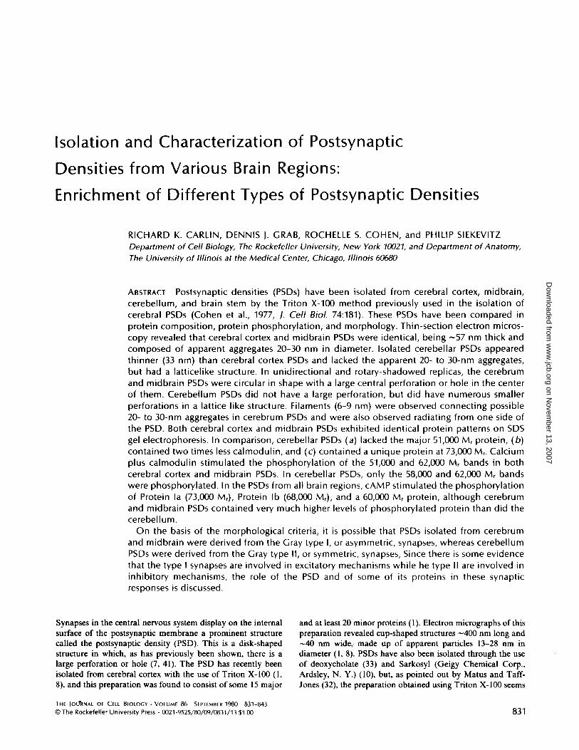
The procedure used to isolate PSDs from the different brain regions is amodification of the procedure used by Cohen et al . (8) in the case of cerebralcortex and is outlined in Fig. 1 . The brain was removed from a dog afternembutaldeath, dissected into four parts, then rinsed in solution A. The cerebrum wasconsidered as all the cortex in the forebrain, the midbrain consisted of allstructures lying below the corpus callosum and above the pons, the brain stemwas all material below and including the pons, and the cerebellum consisted ofmaterial above the cerebellar peduncles . Homogenization was performed by 12up and down strokes with a motor-operated Teflon-glass homogenizer (0.25 mmclearance), using 10 g (wet weight) brain part aliquots/40 ml of solution A (Fig .1) . The resultant homogenates were combined and diluted to 10% (wt/vol) insolution A. All the g values are average centrifugal forces . A low-speed (1,400 g)pellet was obtained and washed by resuspending the pellet with three strokes ofthe homogenizer in thesame 10% volume of solution A. The second centrifugation(710 g) was carried out for 10 min. After centrifugation the supernates werepooled and centrifuged at 13,800 g for 10 min. The resulting pellet, containingsynaptosomes and mitochondria, was resuspended with six strokes of the homog-enizer in solution B (Fig . 1) using 24 ml/10 g starting tissue . The sucrosegradients, using the Spinco SW 27 rotor, (Beckman Instruments, Inc., SpincoDiv., Palo Alto, Calif.) contained 8 ml of the resuspended material, 10 ml eachof 0.85, 1 .0, and 1.2 Msucrose solutions all containing 1.0 mM NaHCO,; thesegradients were then run for 2 h at 82,500 g. The band between 1 .0 and 1 .2 Msucrose was removed by first aspirating off the solution above the band, thenremoving the band with a 5-ml pipette gun with a plastic tip ; it was then dilutedwith solution B (60 rnl/10 g initial wt), an equal volume of 1% (vol/vol) TritonX-100 in 0.32 Msucrose-12 mM Tris-HCI (pH 8.1) was added, and the resultant,somewhat clarified suspension was stirred in the cold for 15 min. This suspensionwas spun down at 32,800 g and the pellet was resuspended in 2.5 ml of solutionB/ 10 g original wet weight brain part, and 2 ml of this material was layered ongradients in polyallomer tubes composed of 4 ml of 2.0 M sucrose, 3.0 ml of 1 .5M sucrose- I mM NaHCO:,, and 3.0 ml of 1 .0 M sucrose- I mM NaHC03. Thegradients were spun for 2 h at 201,800 g in the SW 40 rotor (Beckman Instruments,Inc., Spinco Div., 1-5-50 centrifuge) . The PSDs banded between 1 .5 and 2.0 Msucrose . The material above the band was removed by aspiration and the bandwas removed with a plastic pipette . The pipetted band was diluted to a finalvolume of 6.0 ml with solution B, and an equal volume of 1% Triton-150 mMKCI was added. This suspension was spun for 20 min at 201,800 g in the SW 40rotor. The resultant pellet was resuspended by homogenization with a motor-driven Teflon-glass homogenizer. The yields were (mg/ 10 g initial wet wt tissue) :cerebrum, 2-3: midbrain, 1 .5-2; cerebellum, I .
Gel Electrophoresis
Gel electrophoresis was performed as described before (8).
832
THE JOURNAL OF CELL BIOLOGY " VOLUME 86, 1980
Canine Brain Region-40-60gm
Homogenate in Sol. A (0.32 M Sucrose-1 mM NoHC03ImM MgCl2, 0.5 mM Cocl2 ) ; 1400 g x 10 min
S13,800g x IOmin
1 71Ogxlomin
i13
Disand
S
PDiscard
Resuspend in Sol . BJ(0.32 M sucrose- I mM NoHC03
Sucrose density gradient82,5009 x 120 min
0.85 M-I .0m-1 .2M-
Electron Microscopy
Protein Phosphorylation
-I .OM
PSD -I ,5MI
-2.0 M
Purified PSD froction
P Wash-Sol . A710g x IOmin
PDiscard
4-Synaptosome fraction1
Remove and treat for 15 min with0.5%Triton X-100 in 0.16 Msucrose, 6mM Tris -HCI pH 8.1
1 32,800 g x 20 min
Discard
I Resuspend in Sol. B.
Sucrose density gradient201,800 g x 120 min
Resuspend in 0.5%TritonX-100, 75 MM KCI201,800g x 20 min
FIGURE 1
Fractionation scheme for the preparation of PSDs fromvarious brain regions. Discussed more fully in Materials and Meth-ods.
Electron microscopy, both thin section and replicas, was performed as de-scribed before (8) . In addition, rotary shadowing was done at an angle of 20°with platinum-carbon for 6 s while the specimen was rotated six times . Thereplica was then coated at an angle of 90° with carbon for 6 s .
Calcium-dependent protein kinase activity of the PSD was performed accord-ing to the method of Schulman and Greengard (45) . The standard reactionmixture contained in a final volume of0.1 ml: 50 mM PIPES buffer, pH 7.0; 10mM MgCI 2; I mM dithiothreitol (DTT); 0.2 mM EGTA; plus 50-100 pg PSDprotein . The plus calcium samples also contained 0.5 mM CaCl_ with or without3 pg purified canine brain calmodulin (prepared by the method of Watterson etal. (53) . After a 1-min preincubation at 30°C, 5 AM ATP (5-10 pCi y-['LPIATP)was added and the mixture was further incubated at 30°C . The reaction wasterminated by the addition of 2% SDS, and SDS PAGE was performed asdescribed in Materials and Methods. Autoradiography was performed on thedried gels using Cronex 2 DC Medical Film (DuPont Instruments, Wilmington,Del.) . The cAMP protein kinase activity of the PSD was performed as describedabove, except that 10AM cAMP was used in place of the calcium and calmodulin .Isobutylmethylxanthine (I mM) was also added to inhibit the breakdown ofCAMP (48, 49).
RESULTS
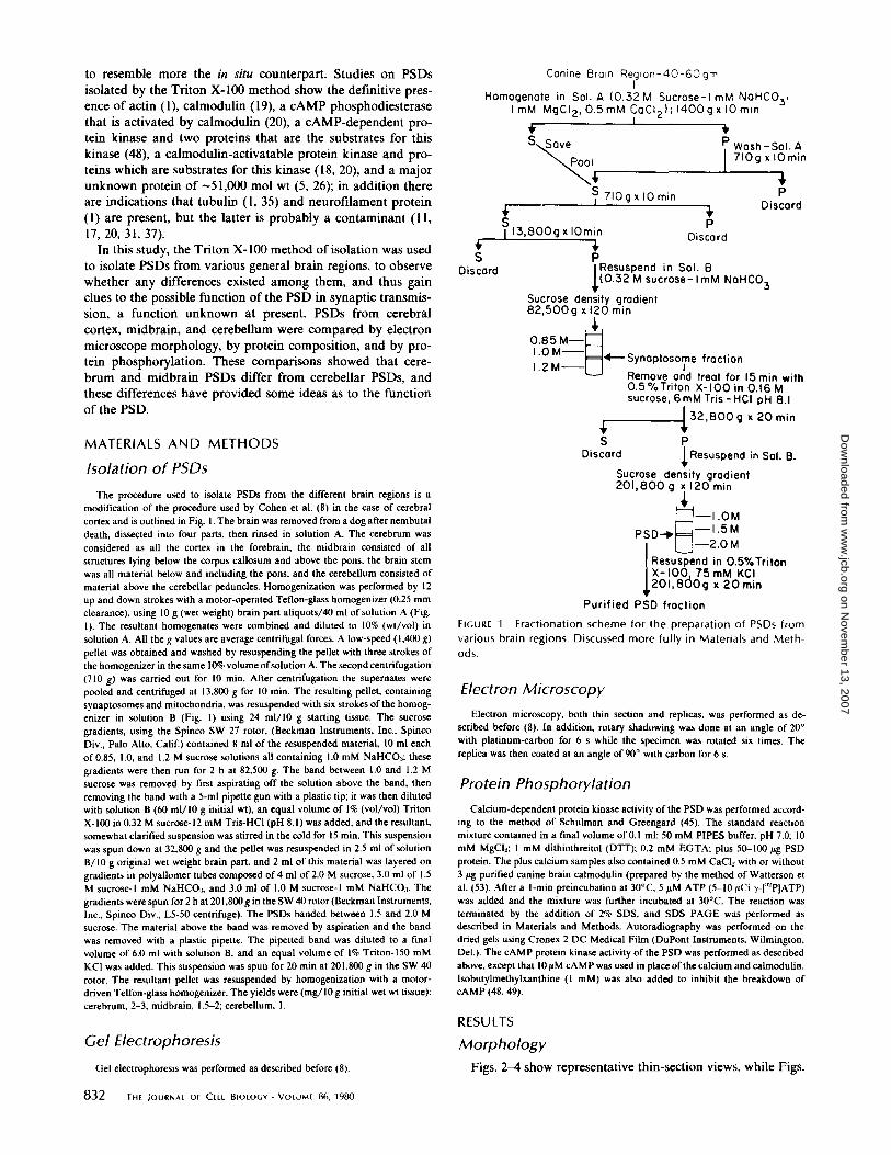
MorphologyFigs . 2-4 show representative thin-section views, while Figs .
on Novem
ber 13, 2007 w
ww
.jcb.orgD
ownloaded from
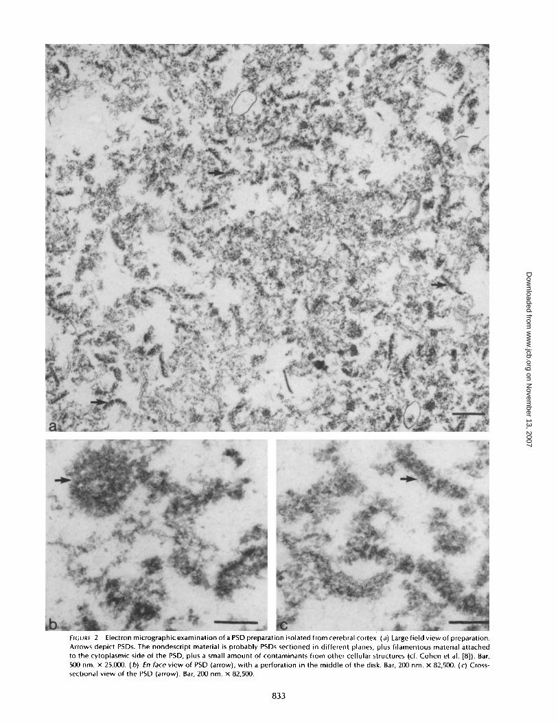
FIGURE 2
Electron micrographic examination of a PSD preparation isolated from cerebral cortex . (a) Large field view of preparation.Arrows depict PSDs. The nondescript material is probably PSDs sectioned in different planes, plus filamentous material attachedto the cytoplasmic side of the PSD, plus a small amount of contaminants from other cellular structures (cf . Cohen et al . [8]) . Bar,500 nm . x 25,000 . (b) En face view of PSD (arrow), with a perforation in the middle of the disk . Bar, 200 nm . x 82,500 . (c) Cross-sectional view of the PSD (arrow) . Bar, 200 nm . x 82,500 .
833
on Novem
ber 13, 2007 w
ww
.jcb.orgD
ownloaded from
FIGURE 3
Electron micrographic examination of a PSD preparation isolated from midbrain . (a) Large field view of preparation .Arrows indicate typical PSDs ; description of material as given in Fig. 2 . Bar, 500 nm . x 25,000 . (b) fn face view of PSD (arrow) withpossible perforation in the center . Bar, 200 nm . x 82,500 . (c) Cross-sectional view of the PSD (arrow). Bar, 200 nm . x 82,500 .
834
THE JOURNAL OF CELL BIOLOGY " VOLUME 86, 1980
on Novem
ber 13, 2007 w
ww
.jcb.orgD
ownloaded from
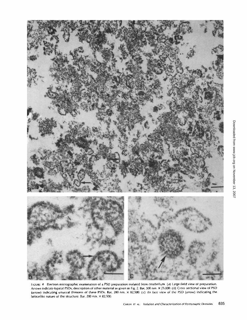
FIGURE 4
Electron micrographic examination of a PSD preparation isolated from cerebellum . (a) Large field view of preparation.Arrows indicate typical PSDs ; description of other material as given in Fig. 2. Bar, 500nm . x 25,000 . (b) Cross-sectional view of PSD(arrow) indicating unusual thinness of these PSDs . Bar, 200 nm . x 82,500. (c) En face view of the PSD (arrow) indicating thelatticelike nature of the structure. Bar, 200nm . x 82,500 .
CARLIN ET AL .
Isolation and Characterization of Postsynaptic Densities
835
on Novem
ber 13, 2007 w
ww
.jcb.orgD
ownloaded from
5 and 6 show replica views, of PSD preparations isolated fromvarious brains parts . Although the cerebrum and midbrainPSD preparations are indistinguishable from one another,those from cerebellum and brain stem present differences . Fig.2 a is a low-magnification field showing the morphology of thePSD preparation isolated from cerebral cortex. A number ofPSDs can be seen in this preparation (arrows) . A detailedmorphological description of this preparation has been givenearlier (8) . The nondescript material is probably PSDs sec-tioned in different planes, plus filamentous material attachedto the cytoplasmic side of the PSD, plus a small amount ofcontaminants from other cellular structures (8) . There is asmall amount of vesicular contaminants in the preparation .When viewed from the side (Fig . 2 c), the PSDs are flat tosemicircular, with an average thickness of 58 nm, based on 50measurements. An enface view (Fig . 2 b) shows a disk-shapedstructure with a less dense center, as has been previouslydescribed (7, 41). Both the enface and cross-sectional views ofthe PSD show the presence of 20- to 30-nm particle-likeaggregates . These have been previously described (1, 8, 35).The PSD preparation from midbrain (Fig. 3 a) shows a
preparation similar to that of cortex, wth size and shape of thePSD being the same . In a cross-sectional view (Fig . 3 c), thethickness of the PSD averaged 56 nm, based on 50 measure-ments, not significantly different from that of the PSD fromcerebral cortex . An en face view (Fig . 3 b) shows an almostidentical appearance as PSDs isolated from cerebral cortex,that is, a disk-shaped structure with 20- to 30-nm aggregatesand a less dense center.The cerebellar PSD preparation is shown in Fig. 4a. The
PSDs in this preparation are much thinner than those fromcerebral cortex or midbrain, the average cross-sectional thick-ness being 33 nm, based on 50 measurements, about half thethickness of cerebral cortex or midbrain PSDs (compare Fig.4 b with Figs . 2 c and 3 c) . Other features ofthese PSDs are thehigh degree of convexity within each PSD (Fig . 46), and thelarge amount ofsubsynaptic web material associated with them(Fig. 4b). The functional significance of both of these obser-vations is unknown at present. Enface (Fig . 4c) the PSDs aredisk shaped, but apparently lack the 20- to 30-nm aggregatespresent in cerebrum PSDs . In their place is a latticelike struc-ture (Fig . 4c) that is similar to cerebrum PSDs treated withsodium deoxycholate to produce what has been named a PSDlattice (32) . However, the sodium deoxycholate-produced lat-tice from cerebral cortex PSDs retains the original thickness ofa cerebrum PSD.The PSD preparation from brain stem represented a much
more variegated picture (not shown) than those from otherbrain areas. Though there were recognizable PSDs present,with a shape and thickness similar to that of cerebral cortexPSDs, there was much more unrecognizable material plusmuch filamentous material that was probably intermediate-filament contamination (20), as evidenced by a comparisonwith filaments isolated by the method of Liem et al . (31) .Because of the low degree of purity and low yield of thepreparation, little can be written now concerning this prepa-ration .
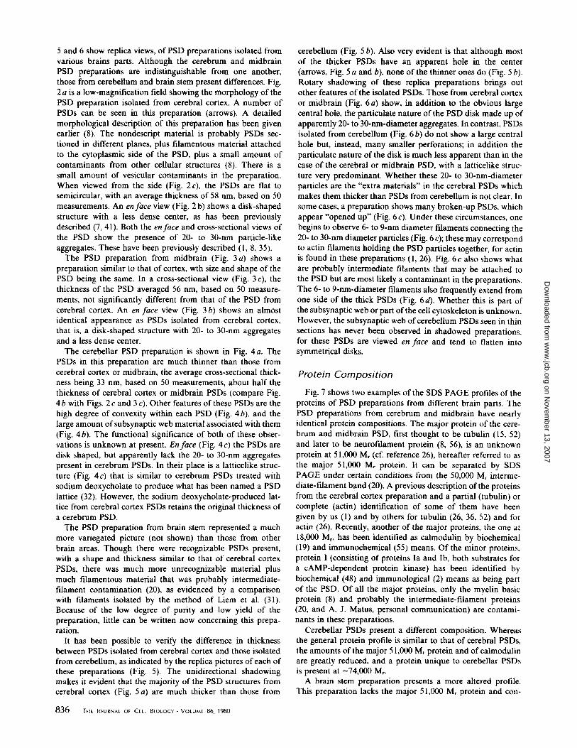
It has been possible to verify the difference in thicknessbetween PSDs isolated from cerebral cortex and those isolatedfrom cerebellum, as indicated by the replica pictures ofeach ofthese preparations (Fig. 5) . The unidirectional shadowingmakes it evident that the majority of the PSD structures fromcerebral cortex (Fig . 5 a) are much thicker than those from
836
TEIE JOURNAL OF CELL BIOLOGY " VOLUME 86, 1980
cerebellum (Fig . 5 b). Also very evident is that although mostof the thicker PSDs have an apparent hole in the center(arrows, Fig. 5 a and b), none of the thinner ones do (Fig . 5 b) .Rotary shadowing of these replica preparations brings outother features of the isolated PSDs . Those from cerebral cortexor midbrain (Fig . 6 a) show, in addition to the obvious largecentral hole, the particulate nature ofthe PSDdisk made up ofapparently 20- to 30-nm-diameter aggregates . In contrast, PSDsisolated from cerebellum (Fig. 66) do not show a large centralhole but, instead, many smaller perforations; in addition theparticulate nature of the disk is much less apparent than in thecase of the cerebral or midbrain PSD, with a latticelike struc-ture very predominant. Whether these 20- to 30-nm-diameterparticles are the "extra materials" in the cerebral PSDs whichmakes them thicker than PSDs from cerebellum is not clear . Insome cases, a preparation shows many broken-up PSDs, whichappear "opened up" (Fig . 6 c) . Under these circumstances, onebegins to observe 6- to 9-nm diameter filaments connecting the20- to 30-nm diameter particles (Fig . 6 c) ; these maycorrespondto actin filaments holding the PSD particles together, for actinis found in these preparations (1, 26). Fig. 6 c also shows whatare probably intermediate filaments that may be attached tothe PSD but are most likely a contaminant in the preparations .The 6- to 9-nm-diameter filaments also frequently extend fromone side of the thick PSDs (Fig. 6d). Whether this is part ofthe subsynaptic web or part ofthe cell cytoskeleton is unknown.However, the subsynaptic web ofcerebellum PSDs seen in thinsections has never been observed in shadowed preparations,for these PSDs are viewed en face and tend to flatten intosymmetrical disks.
Protein Composition
Fig. 7 shows two examples of the SDS PAGE profiles of theproteins of PSD preparations from different brain parts . ThePSD preparations from cerebrum and midbrain have nearlyidentical protein compositions. The major protein of the cere-brum and midbrain PSD, first thought to be tubulin (15, 52)and later to be neurofilament protein (8, 56), is an unknownprotein at 51,000 Mr (cf. reference 26), hereafter referred to asthe major 51,000 Mr protein. It can be separated by SDSPAGE under certain conditions from the 50,000 Mr interme-diate-filament band (20) . A previous description ofthe proteinsfrom the cerebral cortex preparation and a partial (tubulin) orcomplete (actin) identification of some of them have beengiven by us (1) and by others for tubulin (26, 36, 52) and foractin (26) . Recently, another of the major proteins, the one at18,000 Mr, has been identified as calmodulin by biochemical(19) and immunochemical (55) means. Of the minor proteins,protein I (consisting of proteins Ia and lb, both substrates fora cAMP-dependent protein kinase) has been identified bybiochemical (48) and immunological (2) means as being partof the PSD. Of all the major proteins, only the myelin basicprotein (8) and probably the intermediate-filament proteins(20, and A. J . Matus, personal communication) are contami-nants in these preparations .
Cerebellar PSDs present a different composition . Whereasthe general protein profile is similar to that of cerebral PSDs,the amounts of the major 51,000 Mr protein and of calmodulinare greatly reduced, and a protein unique to cerebellar PSDsis present at
74,000 Mr.A brain stem preparation presents a more altered profile .
This preparation lacks the major 51,000 Mr protein and con-
on Novem
ber 13, 2007 w
ww
.jcb.orgD
ownloaded from
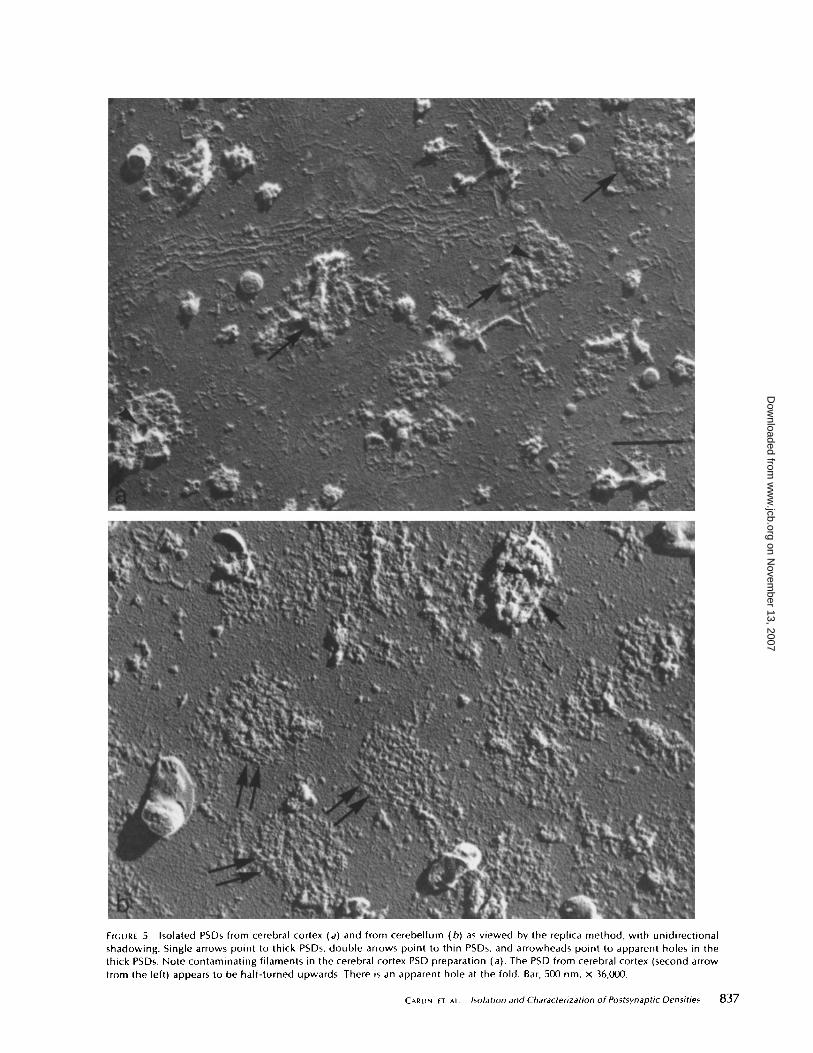
FIGURE 5
Isolated PSDs from cerebral cortex (a) and from cerebellum (b) as viewed by the replica method, with unidirectionalshadowing. Single arrows point to thick PSDs, double arrows point to thin PSDs, and arrowheads point to apparent holes in thethick PSDs . Note contaminating filaments in the cerebral cortex PSD preparation (a). The PSD from cerebral cortex (second arrowfrom the left) appears to be half-turned upwards. There is an apparent hole at the fold . Bar, 500 nm . X 36,000 .
CARLIN ET At .
Isolation and Characterization of Postsynaptic Densities
837
on Novem
ber 13, 2007 w
ww
.jcb.orgD
ownloaded from
FIGURE 6
Isolated PSDs from midbrain (a), cerebellum (b), and cerebral cortex (c and d) as viewed in high magnification by thereplica method with rotary shadowing. In a, single arrows point to possible particulate bodies, and the double arrows point to thelarge central hole. In c, the PSD is apparently broken up ; the single arrows point to 6- to 9-nm filaments and the double arrowspoint to 12- to 15-nm filaments . d Shows a PSD with extensions of 6- to 9-nm filaments (arrows) apparently arising from onesurface of the PSD . Bar, 200 nm . x 100,000.
838
THE JOURNAL OF CELL BIOLOGY " VOLUME 86, 1980
on Novem
ber 13, 2007 w
ww
.jcb.orgD
ownloaded from
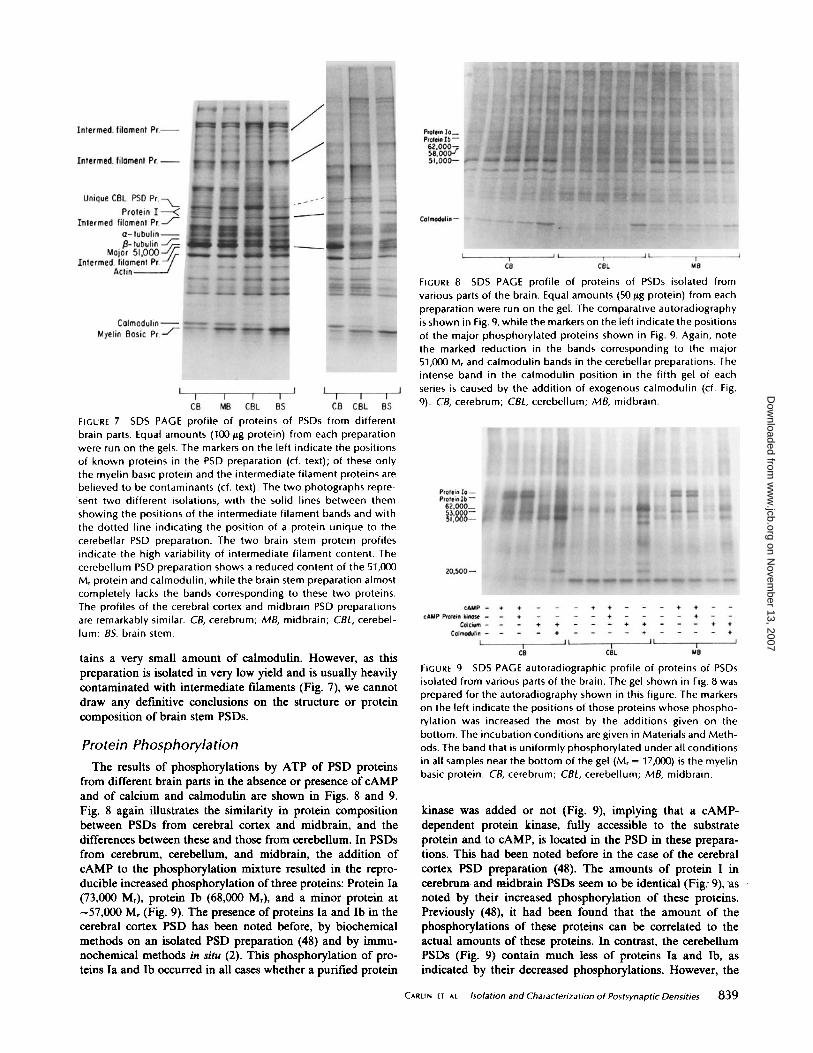
FIGURE 7 SDS PAGE profile of proteins of PSDs from differentbrain parts . Equal amounts (100 fig protein) from each preparationwere run on the gels . The markers on the left indicate the positionsof known proteins in the PSD preparation (cf. text) ; of these onlythe myelin basic protein and the intermediate filament proteins arebelieved to be contaminants (cf . text) . The two photographs repre-sent two different isolations, with the solid lines between themshowing the positions of the intermediate filament bands and withthe dotted line indicating the position of a protein unique to thecerebellar PSD preparation . The two brain stem protein profilesindicate the high variability of intermediate filament content . Thecerebellum PSD preparation shows a reduced content of the 51,000M r protein and calmodulin, while the brain stem preparation almostcompletely lacks the bands corresponding to these two proteins .The profiles of the cerebral cortex and midbrain PSD preparationsare remarkably similar . CB, cerebrum ; MB, midbrain ; CBL, cerebel-lum ; BS, brain stem .
tains a very small amount of calmodulin. However, as thispreparation is isolated in very low yield and is usually heavilycontaminated with intermediate filaments (Fig . 7), we cannotdraw any definitive conclusions on the structure or proteincomposition of brain stem PSDs .
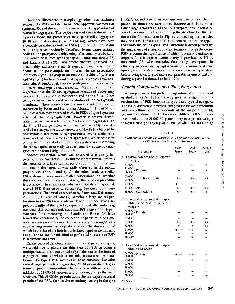
Protein PhosphorylationThe results of phosphorylations by ATP of PSD proteins
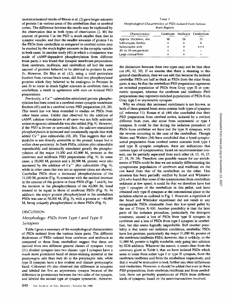
from different brain parts in the absence or presence of cAMPand of calcium and calmodulin are shown in Figs. 8 and 9 .Fig . 8 again illustrates the similarity in protein compositionbetween PSDs from cerebral cortex and midbrain, and thedifferences between these and those from cerebellum. In PSDsfrom cerebrum, cerebellum, and midbrain, the addition ofcAMP to the phosphorylation mixture resulted in the repro-ducible increased phosphorylation of three proteins: Protein Ia(73,000 M,), protein Ib (68,000 Mr ), and a minor protein at"57,000 M, (Fig . 9) . The presence ofproteins la and Ib in thecerebral cortex PSD has been noted before, by biochemicalmethods on an isolated PSD preparation (48) and by immu-nochemical methods in situ (2) . This phosphorylation of pro-teins la and Ib occurred in all cases whether a purified protein
FIGURE 8 SDS PAGE profile of proteins of PSDs isolated fromvarious parts of the brain . Equal amounts (50 fig protein) from eachpreparation were run on the gel . The comparative autoradiographyis shown in Fig. 9, while the markers on the left indicate the positionsof the major phosphorylated proteins shown in Fig . 9 . Again, notethe marked reduction in the bands corresponding to the major51,000 Mr and calmodulin bands in the cerebellar preparations . Theintense band in the calmodulin position in the fifth gel of eachseries is caused by the addition of exogenous calmodulin (cf . Fig .9) . CB, cerebrum ; C8L, cerebellum ; MB, midbrain .
FIGURE 9 SDS PAGE autoradiographic profile of proteins of PSDsisolated from various parts of the brain . The gel shown in Fig . 8 wasprepared for the autoradiography shown in this figure . The markerson the left indicate the positions of those proteins whose phospho-rylation was increased the most by the additions given on thebottom . The incubation conditions are given in Materials and Meth-ods . The band that is uniformly phosphorylated under all conditionsin all samples near the bottom of the gel (M r = 17,000) is the myelinbasic protein . CB, cerebrum ; CBL, cerebellum ; MB, midbrain .
kinase was added or not (Fig . 9), implying that a cAMP-dependent protein kinase, fully accessible to the substrateprotein and to CAMP, is located in the PSD in these prepara-tions. This had been noted before in the case of the cerebralcortex PSD preparation (48). The amounts of protein I incerebrum. and midbrain PSDs seem to be identical (Figs 9), asnoted by their increased phosphorylation of these proteins.Previously (48), it had been found that the amount of thephosphorylations of these proteins can be correlated to theactual amounts of these proteins. In contrast, the cerebellumPSDs (Fig. 9) contain much less of proteins la and lb, asindicated by their decreased phosphorylations. However, the
CARLIN ET AL .
Isolation and Characterization of Postsynaptic Densities
839
on Novem
ber 13, 2007 w
ww
.jcb.orgD
ownloaded from
immunochemical results ofBloom et al . (2) gave larger amountsof protein I in various areas of the cerebellum than in cerebralcortex . The difference between the results may be explained bythe observation that in both types of observation (2, 48) theamount of protein I in the PSD is much smaller than that insynaptic vesicles, and thus the smaller amounts of protein I inthe PSDs from cerebellum as compared to cerebral cortex maybe masked by the much higher amounts in the synaptic vesiclesin both cases. In another study (43) in which a comparison wasmade of CAMP-dependent phosphorylation from differentbrain parts, it was found that synaptic membrane preparationsfrom cerebrum, midbrain, and cerebellum all had the sameamount of proteins believed to be identical to proteins Ia andlb . However, De Blas et al . (l2), using a total particulatefraction from various brain areas, did find two phosphorylatedproteins which they believed to be analogous to Proteins laand Ib to occur in much higher amounts in cerebrum than incerebellum, a result in agreement with ours on isolated PSDpreparations.
Previously, a Ca"-calmodulin-stimulated protein phospho-rylation has been noted in a cerebral cortex synaptic membranefraction (45) and in a cerebral cortex PSD preparation (18, 20) .This result has not been extended to studies on PSDs fromother brain areas . Unlike that observed by the addition ofcAMP, calcium stimulation in all cases was not fully activatedunless exogenous calmodulin was also added (Fig. 9) . However,if the PSD is frozen or sonicated first, the calcium-stimulatedphosphorylation is increased and occasionally equals that withadded Ca" plus calmodulin (18, 20) . This suggests that cal-modulin is not directly accessible to the protein kinase but iswithin close proximity. In fresh PSDs, calcium plus calmodulinreproducibly and identically stimulated greatly the phospho-rylation of the major 51,000 and 62,000 Mr proteins in thecerebrum and midbrain PSD preparations (Fig. 9) . In somecases, a 58,000 Mr protein and a 20,500 M, protein were alsoincreased by the addition of Ca" plus calmodulin (Fig. 9),while in other cases this was not so apparent (data not shown) .Cerebellar PSDs show a decreased phosphorylation of the51,000 M, protein (Fig . 9) consistent with the marked decreasein the amount of this protein in these PSDs (Figs . 7 and 8), butthe increase in the phosphorylation of the 62,000 M, bandseemed to be equal to those of cerebrum PSDs (Fig . 9) . Inaddition, the major protein phosphorylated in the cerebellumPSDs was one at 58,000 M, (Fig. 9), with a protein at 48,000M, being uniquely phosphorylated in these PSDs (Fig . 9) .
DISCUSSIONMorphology: PSDs from Type I and Type 11SynapsesTable I gives a summary ofthe morphological characterisitcs
of PSDs isolated from the various brain parts . The differentthicknesses of PSDs isolated from cerebrum and midbrain ascompared to those from cerebellum suggest that these arederived from two different general classes of synapses . Gray(2 I) divided synapses into two classes: type I synapses have amuch more prominent band of dense-staining material at thepostsynaptic side than they do at the presynaptic side, whiletype II synapses have a less evident and thinner postsynapticband . Colonnier (9) also observed this difference in synapsesand labeled the first an asymmetric synapse because of thedifference in prominence between the two sides ofthe synapse,and labeled the second type of synapse symmetric . However,
840
THE JOURNAL OF CELL BIOLOGY " VOLUME 86, 1980
TABLE I
Morphological Characteristics of PSDs Isolated from VariousBrain Regions
the distinction between these two types may not be that clearcut (40, 42, 50) . If we assume that there is meaning to thisgeneral classification, then we can add that because the isolatedcerebellar PSDs are half as thick as PSDs from the other brainparts, it may be that the cerebellum PSD preparation representsan enriched population of PSDs from Gray type II or sym-metric synapses, whereas the cerebrum and midbrain PSDpreparations may represent enriched populations of PSDs fromGray type I or asymmetric synapses .Why we obtain this increased enrichment is not known, as
both ofthese general brain areas contain both types ofsynapses(cf. reference 51) . Rostas et al . (44) also concluded that theirPSD preparation from cerebral cortex, isolated by a methoddifferent from ours, also arose from asymmetric or type Isynapses. It could be that during the isolation procedure ofPSDs from cerebrum we have lost the type 11 synapses, withthe reverse occurring in the case of the cerebellum. ThoughMatus and Walters (34) have concluded that a lysed synapto-somal preparation from cerebral cortex contains both type Iand type II synaptic complexes, there are indications thatvarious types of synaptosomes, based on neurotransmitter con-tent, can be partially separated from each other (4, 13, 16, 25,27, 38, 39, 54) . Therefore, one possible reason for our enrich-ments of PSDs could be that we are initially differentiating thesynaptosome populations of cerebrum and midbrain on theone hand from that of the cerebellum on the other. Thissituation has been partially verified by Israel and Whittaker(24) who found that some ofthe synaptosomes from cerebellumsediment at low speed; it could be that we therefore have losttype I synapses of the cerebellum in this pellet, and haveobtained only type II synapses at the conventional place in theisolation scheme as outlined in Fig . 1 . However, a repetition ofthe Israel and Whittaker experiment did not result in anyrecognizable PSDs obtainable from this low-speed pellet bythe use of Triton X-100. Another possibility is that the laterparts of the isolation procedure, particularly the detergenttreatment, caused a loss of PSDs from type II synapses incerebrum and a loss of PSDs from type I synapses in cerebel-lum, but this seems logically improbable . Still another possi-bility is that under our isolation conditions, cerebellar PSDshave lost proteins, particularly the major 51,000 Mr protein ofthe cerebrum/midbrain PSDs ; however, this is unlikely, as the51,000 Mr protein is highly insoluble, only going into solutionby SDS addition . Whatever the reason, it seems clear from thesummary given in Table 1, that we have isolated PSDs whichseem to come from either type I or type II synapses, from thecerebrum/midbrain and from the cerebellum respectively, andthat it would be instructive to look further into their differencesand similarities . However, is should be added that even in thesePSD preparations, from cerebrum/midbrain and from cerebel-lum, there are probably populations of PSDs from differentkinds of synapses, based on the neurotransmitters involved .
Characteristics Cerebrum Midbrain Cerebellum
Approx . thickness, nm 58 56 33Subsynaptic bodies + + +Subsynaptic web + + +++20- to 30-nm particles + + -Large central hole + + -
on Novem
ber 13, 2007 w
ww
.jcb.orgD
ownloaded from
There are differences in morphology other than thicknessbetween the PSDs isolated from these apparent two types ofsynapses . One of the major differences is in the appearance ofparticular aggregates. The en face view of the cerebrum PSDtypically shows the presence of these particlelike aggregates20-30 nm in diameter (Figs . 2 and 6 a), which have beenpreviously described in isolated PSDs (1, 8) . In addition, Matuset al . (35) have previously described 20-nm dense stainingbodies at the postsynaptic thickening of isolated synapticjunc-tions which arise from type I synapses . Landis and Reese (28)and Landis et al . (29), using freeze fracture, observed thatpresumably excitatory (type I) synapses have 8- to 13-nmbodies in the postsynaptic membrane, whereas presumablyinhibitory (type II) synapses do not . And incidentally, Matusand Walters (34) have found that type II synapses have con-canavalin A binding sites on the postsynaptic junction mem-brane, whereas type I synapses do not . Matus et al. (35) havesuggested that the 20-nm aggregates mentioned above maytraverse the postsynaptic membrane and be the 8- to 13-nmparticles viewed in freeze-fracture studies of the postsynapticmembrane . These observations are reminiscent of an earliersuggestion by Peters and Kaiserman-Abramof (41) that 30-nmparticles which they observed in situ in PSDs of type I synapsesextended into the synaptic cleft. However, at present there islittle direct evidence relating the 20- to 30-nm aggregates andthe 8- to 13-nm particles . Matus and Walters (33) have de-scribed a postsynaptic lattice structure ofthe PSD, obtained bydeoxycholate treatment of synaptosomes, which could be aframework of these 20- to 30-nm aggregates . An enface viewof a typical thin cerebellum PSD shows a structure resemblingthe postsynaptic lattice (only thinner), and few apparent aggre-gates can be found (Figs. 4 and 6 b) .Another distinction which was observed consistently be-
tween cerebral/midbrain PSDs and those from cerebellum wasthe presence of a large central perforation in the former caseand not in the latter, as was easily observed in the replicapreparations (Figs. 5 and 6) . On the other hand, cerebellarPSDs showed many more smaller perforations, but whetherthis is caused by an opening-up during the isolation procedureis not known . In some cases, what is obviously an expanded,altered PSD from cerebral cortex (Fig. 6c) does show theseperforations. The initial observation by Peters and Kaiserman-Abramof (41), verified later (7), showing a large central per-foration in the PSD was made on dendritic spines, which arepredominantly of the type I synapse (20), partially confirmingour view that our cerebral/midbrain PSDs arise from type Isynapses. It is interesting that Landis and Reese (28) havefound that occasionally the collection of particles in postsyn-aptic membranes of asymmetric synapses are arranged in acircular ring around a nonparticle center, the dimensions ofwhich fit the size ofthe hole in our isolated type I or asymmetricPSDs. The reason for this kind of perforated structure of PSDis at present unknown.On the basis of the observations in this and previous papers,
we would like to picture the thin, type II PSDs as being amultiperforated disk, composed of proteins not in the form ofaggregates, some of which attach this structure to the mem-brane . The type I PSD retains this basic structure, but addsonto it large particulate aggregates, 20-30 nm in diameter; interms of protein composition, the only large difference is theaddition of 51,000 M r protein and of calmodulin to the basicstructure . This 51,000 M, protein cannot be the major structuralprotein of the PSD, for it is almost entirely lacking in the type
II PSD ; indeed, the latter contains not one protein that ispresent in abundance over others . Because actin is found inrather large amounts in all the PSD preparations, it could beone of the cementing blocks holding the structure together, asthose thin filaments seen in Fig. 6 c connecting the particlesmay be actin . The addition of the superstructure of the type IPSD onto the basic type II PSD structure is accompanied bythe appearance ofa large central perforation through the entirePSD structure the significance of which is presently unknown .Support for this superstructure theory is provided by Hindsand Hinds (23), who concluded that during development inolfactory axodendritic synaptogenesis all asymmetrical syn-apses pass through an immature symmetrical synapse stagebefore being transformed into a recognizable asymmetrical oneduring a period estimated to be 9-10 h .
Protein Composition and PhosphorylationA comparison of the protein composition of cerebrum and
cerebellum PSDs (Table II) may give an insight into themechanisms of PSD function in type I and type II synapses .The major difference in protein composition between cerebrumand cerebellum is in the amounts of the major 51,000 M,protein and calmodulin. As there is very little 51,000 M, proteinin cerebellum, the 51,000 M, protein may be a protein uniqueto asymmetric type I synapses, no matter what transmitter may
Summary of Protein Composition and Protein Phosphorylationof PSDs from Various Brain Regions
A. Relative composition of selectedproteins
74,00073,00068,000
59,00055,00051,00045,000-Actin18,000-Calmodulin
B. Increased phosphorylation uponaddition of calcium plus cal-modulin
73 '000 [ Protein I68,00057,00062,00058,00051,00048,00020,500*
Proteins (M r )
Protein I
Tubulin subunits
TABLE II
C. Increased phosphorylation uponaddition of cAMP
73,000 1 Protein 168,00057,00062,00058,00051,00020,500
* Variable .
Cere- Mid- Cerebel-brum brain lum
CARLIN ET AL .
Isolation and Characterization of Postsynaptic Densities
841
on Novem
ber 13, 2007 w
ww
.jcb.orgD
ownloaded from
be involved . This has also been inferred by Rostas et al . (44),based on the high amounts of this protein in synapticjunctioncomplexes isolated from various cerebral and midbrain regionspostulated to have asymmetric synapses. A second differenceis that calmodulin is reduced by -50% in cerebellum PSDs ascompared to,cerebrum PSDs. This confirms the work of Sobueet al . (46) in which they determined the concentration ofcalmodulin in membrane preparations from cerebrum andcerebellum . The higher concentration of calmodulin suggeststhat calmodulin may be very important in type I synapses butthe presence of still detectable amounts of calmodulin incerebellum PSDs does not rule out a role in these type synapses .The phosphorylation of some of the proteins of PSDs under
varying conditions may give some further insight into possibledifferences in function between the two classes of PSDs. TableII gives a summary of our results . In the presence of calciumplus calmodulin, cerebrum PSDs exhibit increased phospho-rylation of the major 51,000 and 62,000 M, proteins . In contrast,the cerebellum PSDs have a greatly reduced phosphorylationunder these conditions in the 51,000 M r region . As proteinphosphorylation has been proposed to be important in synapticfunction (22), the uniqueness of the major 51,000 M, protein incerebrum and midbrain PSDs may indicate that phosphoryla-tion of this protein is involved in the generation ofpostsynapticpotentials in type I synapses . On the other hand, the predom-inance ofthe increased phosphorylation upon addition ofCa"and calmodulin of the 58,000 M, protein in cerebellum mayindicate its possible involvement in the generation of postsyn-aptic potentials in type II synapses . However, the phosphoryl-ation of the 62,000 M, protein cannot entirely be assigned toType II synapses as this protein is apparently present in bothtypes of PSD . Whether this protein is involved in the action ofboth types of PSD or is present in the preparation ofcerebrumPSDs because of the presence therein of PSDs from type IIsynapses, remains to be determined.
Cyclic AMP-dependent phosphorylation of cerebrum, mid-brain, and cerebellum PSDs produces the same pattern . Themajor phosphorylated bands are in proteins la and Ib (48),though the degree of phosphorylation varies among the prep-arations . The other minor phosphorylated band (57,000 M,)possibly represents autophosphorylation ofthe regulatory sub-unit ofthe cAMP-dependent protein kinase (49) . But, cerebrumPSDs have much greater phosphorylation of proteins la andIb upon addition of CAMP than do cerebellum PSDs. Thus, itwould appear that the cAMP-dependent phosphorylations maybe involved in the function of the type I synapses and not thatof the type II synapses. However, in cerebral cortex, protein Iis much higher enriched in the synaptic vesicles than in thePSD, so that it also has some presynaptic role.
PSDs from Type I Excitatory and from Type 11Inhibitory Synapses
The general classification of synapses as type I and type IIhas led Eccles (l4) to propose that type I synapses mediateexcitation responses and type II synapses mediate inhibitoryresponses, and Colonnier (9) later suggested the same correla-tion . This hypothesis was based on work summarized by Eccles(14) and by Walberg (51), that in the hippocampus and cerebralcortex there was a correlation between type I structure andknown excitatory synapses and between type 11 structure andknown inhibitory synapses ; in the cerebellum the evidencefrom various sources seemed to be somewhat contradictory and
842
THE JOURNAL OF CELL BIOLOGY " VOLUME 86, 1980
thus not that clear-cut . Later work by Landis et al . (29) onsynapses in the olfactory bulb and by Landis and Reese (28)on synapses in the cerebellar cortex strengthened this hypoth-esis . These authors could make correlations between the knownexcitatory and inhibitory properties ofcertain synapses in theseregions and the prevalence of either type I or type 11 differen-tiations there . At the same time, results from many laboratories(summarized by Bodian [3] and by Uchizono [47]) havebrought forth another correlation, that of the appearance ofspherical synaptic vesicles in excitatory synapses and of flat-tened or oblong vesicles in inhibitory synapses. And indeed,the finding that the presence of spherical vesicles and of typeI differentiation go together and that the presence of flattenedvesicles and oftype 11 differentiation go together has promptedUchizono (47) to propose that excitatory synapses have a typeI PSD and spherical presynaptic vesicles and inhibitory syn-apses have a type II PSD and flattened presynaptic vesicles.On the basis of the above assumptions, we can summarize
our results in Table III . We first postulate that our enrichedpopulation of thick PSDs from cerebral cortex and midbrainarise from type I excitatory synapses and that the enrichedpopulation of thinner PSDs from cerebellum arise from type 11inhibitory synapses . If this is the case, then the difference inprotein composition between these two types of PSDs takes onfunctional significance . Thus, the far greater amounts of theunknown 51,000 M, protein, ofcalmodulin, and ofthe substrateproteins la and Ib for the cAMP-activatable protein kinase inthe PSDs from cerebral cortex and midbrain over that in thePSDs from the cerebellum, would suggest that these proteinsare involved in excitatory modulation ofthe transmission signalwhich occurs at these synapses . However, it must be stated thatour PSD populations from these two sources are probably stilla mixture of PSDs from different kinds ofsynapses, dependingon the neurotransmitters involved, and we thus do not knowwhether any individual PSD has both the cAMP-activatableand the calmodulin-activatable systems. A partial verificationof at least the involvement of cAMP in the modulatory excit-atory acetylcholine responses initiated by dopamine has beengiven by Libet (30) .What we know about these proteins can be summarized as
TABLE III
Summary of Postulated Characteristics of PSDs fromType i and Type 11 Synapses
Characteristics Type I Type II
Function Excitatory InhibitoryMorphology Thick disk, with Thin disk, lattice-
large perforation like structure within center ; pres- no large centralence of aggre- perforation ; littlegates or no aggregates
Enrichment of pro- Major 51,000 Mr, 74,000 M,teins Protein I, calmo-
dulin
Enrichment of cal- 51,000 M, 58,000 M,modulin-depen- 48,000 M,dent phosphoryl-ation
Enrichment of Proteins la and IbcAMP-dependentphosphorylation
on Novem
ber 13, 2007 w
ww
.jcb.orgD
ownloaded from
follows: 51,000 Mr protein (identity unknown) is phosphoryl-ated by a calmodulin-activatable protein kinase in the PSD(18, 20) and it also binds calmodulin (6, 20); another possiblefunction of calmodulin in the PSD is that it can activate acAMP-phosphodiesterase (20) . The function of proteins la andIb is unknown. While much further work needs to be done toverify this hypothesis for the involvement of the above PSDproteins in the excitatory response, we can state, even now, onthe basis of the results of this paper, that the PSDprobably hassome role in modulating the signal-conductionevents occurringat the synapse. However, it should be emphasized that thecomplete role of the PSD in some signal conduction will onlybe attained when the PSD is considered as a part of thesynapse, as a result of experiments done with systems in whichthe PSD is still attached to the postsynaptic membrane fromwhich it was isolated.
We would like to thank Dr . David Phillips, Rockefeller FoundationPopulation Council, for his advice on the replica method and hispermission to use the Sorvall Critical Point Drying System; JulieBandar for her exemplary technical assistance ; Elena Sphicas andMichael Von der Lieth for their great aid in the electron microscopypart of this work; and Asneth Kloesman for her excellent graphicswork. Dennis Grab would also like to thank Lisa Zebrawitz for herinspiration during the course of this work .We would like to acknowledge the partial support of this project by
the following granting agencies: National Institutes of Health (NIH)grant PHS-NS 12726 to P. Siekevitz; NIH Postdoctoral Fellowship 5-F32-NS06005 to R. K. Carlin; NIH Postdoctoral Fellowship 1-F32-NS05693 to D. J. Grab; NIH grant l-ROI-NS15889 to R. Cohen.
This work was presented in part at the 18th annual Meeting of theAmerican Society for Cell Biology (5) .Receivedfor publication 7April 1980, and in revised form 30 May 1980.
REFERENCES
1 . Blomberg, F ., R . S. Cohen, and P. Siekevitz. 1977. Structure of postsynaptic densitiesisolated from dog cerebral cortex 11. Characterization and arrangement of some of themajor proteins within the structure . J. Cell BioL 74:204-225 .
2 . Bloom. F . E ., T . Ueda, E . Battenberg, and P . Greengard. 1979 . Immunocytochemicallocalization, in synapses, of Protein 1, and endogenous substrate for protein kinases inmammalian brain. Proc. Natt. Acad. Scl, U. S. A . 76:5982-5986.
3. Bodian, D . 1972. Synaptic diversity and characterization by electron microscopy. InStructure and Function of Synapses. G . D . Pappas and D. P. Purpura, editors . RavenPress, New York . 45-65 .
4. Bretz, U ., M . Baggiolini, R . Hauser, and C . Hodel . 1974. Resolution of three distinctpopulations of nerve endings from rat brain homogenates by zonal isopycnic centrifuga-tion. J. Cell Biol. 61 :466-480 .
5 . Carlin, R. K ., D. 1 . Grab, P . Siekevitz, and R . S . Cohen . 1979. Characterization ofpostsynaptic densities from different brain parts. J. Cell Biol. 83 (2, Pt . 2) : 140a (Abstr.).
6. Carlin, R . K ., D . l . Grab, and P . Siekevitz. 1980. The calmodulin binding proteins of thepostsynaptic density. Fed. Proc . 39 :1658.
7. Cohen, R. S ., and P. Siekevitz . 1978 . The form of the postsynaptic density : A serial sectionstudy. J. Cell Biol. 78:36-46.
8. Cohen, R . S ., F . Blomberg, K . Berzins, and P . Siekevitz. 1977. Structure of postsynapticdensities isolated from dog cerebral cortex . 1. Overall morphology and protein composition .J. Cell Biol 74:181-203 .
9. Colonnier, M . 1968. Synaptic patterns of different cell types in the different laminae of thecat visual cortex . An electron microscopic study . Brain Res. 9:268-287 .
10. Cotman, C . W ., G . Banker, L. Churchill, and D. Taylor. 1974. Isolation of postsynapticdensities from rat brain. J. Cell Biol. 63 :441-445.
I I . Dahl, D., and A . Bignami. 1979. Astroglia and axonal proteins in isolated brain filaments .Biochim. Biophys. Acia. 578 :305-316 .
12. De Bias, A . L ., Y .J. Wang, R . Sorensen, and H. P . Mahler. 1979. Protein phosphorylationin synaptic membranes regulated by adenosine 3 :5'-monophosphate : Regional and sub-cellular distribution of the endogenous substrates. J Neurochem. 33 :647-659 .
13. De Robertis, E., A. Pellegrino de Iraldi, G . R. de L. Amaiz, and L. Salganicoff. 1962 .Cholinergic and non-cholinergic nerve endings in rat brain. 1. Isolation and subcellulardistribution of acetylcholine and acetylcholinesterase . J. Neurochem. 9:23-35 .
14. Eccles, J . C . 1964. The physiology of synapses . Springer-Verlag, New York .I5 . Feit, H ., P. Kelly, and C . W. Colman. 1977. Identification of a protein related to tubulin
in the postsynaptic density. Proc. Nat. Acad. Sci. U. S. A . 74 :1047-1051 .16 . Gfeller, E ., M . J . Kuhar, and S. H. Snyder . 1971 . Neurotransmitter-specific synaptosomes
in rat corpus striatum: Morphological variations . Proc. Nall. Acad. Sci. U. S. A . 68 :155-159.
17 . Goldman,1 . E., H. H . Schaumburg, and W . T . Norton . 1978. Isolation and characterizationof glial filaments from human brain . J. Cell Biol. 78 :426-440 .
18 . Grab, D. J ., and P. Siekevitz . 1979 . Calmodulin-dependent protein kinase activity in
postsynaptic densities . J. Cell Mal 83 (2, Pt. 2): 131 a (Abstr.) .19. Grab, D . J ., K . Begins, R. S . Cohen, and P . Siekevitz . 1979 . Presence of calmodulin in
postsynaptic densities isolated from canine cerebral cortex . J. Biol. Chem. 254:8690-8696.20. Grab, D . 1 ., R. K . Carlin, and P. Siekevitz . 1980. The presence and possible function of
calmoduhn in the post-synaptic density . Ann. N. Y. Acad. Sci. I n press .21 . Gray, E. G . 1959 . Axo-somatic and axo-dendritic synapses of the cerebral cortex: an
electron microscopic study. J. Anat. 93 :420-433 .22. Greengard, P. 1978 . Cyclic nucleotides, phosphorylated proteins, and neuronal function.
Raven Press, New York .23. Hinds, 1 . W., and P . L. Hinds. 1976 . Synapse formation in the mouse olfactory bulb. J.
Comp. Neural 169:41-62.24. Israel, M ., and V. P . Whittaker. 1965. Th e isolation of mossy fiber endings from the
granular layer of cerebellar cortex. Experientia (Basell . 21 :325-326.25. Iversen, L . L., and S. H. Snyder. 1968 . Synaptosomes: Different populations storing
catecholamines and gamma-aminobutyric acid in homogenates of brain . Nature (Land.).220:796-798 .
26. Kelly, P. T ., and C . W. Colman. 1978. Synapti c proteins : Characterization of tubulin andactin and identification of a distinct postsynaptic density protein. J. Cell Biol. 79:173-183 .
27. Kuhar, M. J., E . G . Shaskan, and S . H . Snyder . 1971 . Th e subcellular distribution ofendogenous and exogenous serotonin in brain tissue : Comparison of synaptosomes storingserotonin, norepinephrine, and gamma-amino butyric acid. J. Neurochem. 18:333-343 .
28. Landis, D. M . D ., and T. S . Reem . 1974 . Differences in membrane structure betweenexcitatory and inhibitory synapses in cerebellar cortex. J. Comp . Neural 155 :93-125 .
29. Landis, D . M . D., T. S . Reese, and E. Raviola . 1974. Differences in membrane structurebetween excitatory and inhibitory components of the reciprocal synapse in the olfactorybulb. J. Comp. Neural. 155:93-126.
30 . Libet, B . 1979. Which postsynaplic action of dopamine is mediated by cyclic AMP? LifeSri. 24 :1043-1058 .
31 . Liem, R. K . H ., S .-H. Yen, G . D . Salomon, and M . L . Shelanski. 1978. Intermediatefilaments in nervous tissue. J. Cell Blot. 79:637-645.
32. Matus, A. 1 ., and D. H . Taff-Jones. 1978 . Morphologica l and molecular composition ofisolated postsynaplicjunctional structures . Proc. R. Soc., Lond B. Biol. Sci. 203 :135-151 .
33. Matus, A. L, and B. B. Walters. 1975 . Ultrastructure of the synaptic junctional latticeisolated from mammalian brain . J. Neurocytol. 4 :369-375.
34 . Matus, A. 1 ., and B . B . Walters. 1976. Type I and 2 synaptic junctions : Differences indistribution of concanavalin A binding sites and stability of the junctional adhesion. BrainRes. 108:249-256.
35 . Matus, A. 1 ., B. B . Walters, and D . H. Jones. 1975. Junctiona l ultrastructure in isolatedsynaptic membranes . J. Neurocytol. 4 :357-367.
36 . Matus, A . 1 ., B . B . Waiters, and S. Mughal. 1975 . Immunohistochemical demonstration oftubulin associated with microtubules and synaptic junctions in mammalian brain. J.Neurocytol. 4:733-744.
37. Matus, A. 1., M. Ng, and D . H. Jones. 1979 . Immunohistochemica l localization ofneurofilament antigen in rat cerebellum . J Neurocytol 8 :513-525 .
38 . Michaelson, I . A., and V. P. Whittaker. 1963 . Th e subcellular localization of 5-hydroxy-tryptamine in guinea pig brain . Biochem . Pharmacol. 12:203-211 .
39. Osborne, R. H.. 1 . R . Duce, and P. Keen. 1976. Amino acids in "light" and "heavy"synaptosomal fractions from rat olfactory lobes and their release on electrical stimulation .J Neurochem. 27:1483-1488 .
40 . Pappas, G . D . 1966. Electron microscopy of neuronal junction transmission in CNS . InNerve as a Tissue. K . Rodahl and B . Issekutz, editors . Harper and row, New York . 49-87 .
41 . Peters, A., and 1 . R . Kaiserman-Abramof 1969. The small pyramidal neuron of the ratcerebral cortex . The synapses from dendritic spines. Z. Zellforch. Mikrosk . Anat. 100 :487-506.
42. Peters, A., S . L. Palay, and H. de F. Webster. 1976 . The Fine Structure of the NervousSystem . Harper and Row, New York.
43. Reddington, M ., and E. Mehl . 1979 . Synaptic membrane proteins as substrates for cAMP-stimulated protein phosphorylation in various regions of rat brain. Biochim. Biophys. Acta.555:230-238 .
44. Rostas, J. A . P ., P . T . Kelly, R . H . Pesin, and C. W . Cotman. 1979 . Protei n and glycoproteincomposition of synaptic junctions prepared from discrete synaptic regions and differentspecies . Brain Res . 168 :151-167.
45 . Schulman, H . . and P . Greengard . 1978. Cá"-dependent protein phosphorylation systemin membranes from various tissues, and its activation by "calcium-dependent regulator."Proc. Nall. Acad. Sci. U. S. A . 75 :5432-5436.
46. Sobue, K ., Y. Muramoto, R . Yamazaki, and S . Kakiuchi. 1979. Distribution in rat tissueof modulator-binding protein of particulate nature. FEBS Lett. (Fed. Eur. Biochem . Soc. )105:105-109.
47. Uchizono, K . 1968 . Inhibitory and excitatory synapses in vertebrate and invertebrateanimals. In Structure and Function of Inhibitory Neuronal Mechanisms. C . van Euler, S.Skoglund, and V . Soderberg, editor. Pergamon Press, New York. 33-59 .
48 . Ueda, T ., P. Greengard, K. Berzins, R . S. Cohen, F. Blomberg, D . 1 . Grab, and P.Siekevitz . 1979 . Subcellular distribution in cerebral cortex of two proteins phosphorylatedby a cAMP dependent protein kinase. J. Cell BioL 83:308-319 .
49. Uno, L, T. Ueda, and P . Greengard . 1976 . Adenosine 3':5'-monophosphate regulatedphosphorylation system of neuronal membranes. II . Solubilization, purification, and someproperties of an endogenous adenosine 3':5'-monophosphate-dependent protein kinase. J.Biol. Chem. 252:5164-5174 .
50. Van de Loos, M. 1965 . In The synapse: Morphological and clinical correlation of function.Neurosei. Res. Program. Bull. 3:23-24 .
51 . Walberg, F. 1968 . Morphological correlates of postsynaptic inhibitory processes . /nStructure and Function of Inhibitory Neuronal Mechanisms . C. van Euler, S . Skoglund,and V . Soderberg, editors. Pergamon Press, New York . 7-13 .
52 . Walters, B . B ., and A . 1 . Mattis. 1975 . Tubuli n in post-synaptic junctional lattice . Nature(Lond. ) . 257 :496498 .
53 . Watterson, D. M ., W . G . Harrelson, Jr ., P. M . Keller, F . Sharief, and T. C. Vanaman .1976 . Structural similarities between the Cá''-dependent regulatory proteins of 3':5'-cyclicnucleotide phosphodiesterase and actomyosin ATPase. J. Biol. Chem. 251 :4501-4513 .
54 . Wofsey, A . R .. M . J. Kuhar, and S. H . Snyder . 1971 . A unique synaptosomal fraction,which accumulates glutamic and aspartic acids, in brain tissue. Proc. Nail Acad. Sci. U.S. A. 68:1102-1106.
55 . Wood, 1. G., R. W. Wallace, J . N. Whitaker, andW. Y . Cheung. 1980 . Immunocytochem-ica l localization of calmodulin and a heat labile calmodulin-binding protein (CAM-BP.,)in basal ganglia from mice brain . J Cell Biol 84:66-76.
56. Yen, S . H ., P . Kelly, R . Liem, C . Colman, and M . Shelanski. 1977. Membrane-linkedproteins at CNS synapses . Brain Res. 132:172-175.
CARLIN ET AL .
Isolation and Characterization of Postsynaptic Densities
843
on Novem
ber 13, 2007 w
ww
.jcb.orgD
ownloaded from