Embed Size (px)
Citation preview
Introduction to Immunology I. The normal Immune response Abul K. Abbas, MD
1
INTRODUCTION TO IMMUNOLOGY I. The Normal Immune Response
(Lecture)
OBJECTIVES • List the principal cells and tissues of the immune system. • Summarize the main differences between innate and adaptive immunity. • Describe how lymphocytes respond to foreign antigens. • Describe how humoral and cell-mediated immune responses are induced by microbes and
other foreign substances. KEY WORDS • Antigen • Antigen-presenting cell • Antibodies • B lymphocyte • Cytotoxic (cytolytic) T lymphocyte • Helper T lymphocyte • Lymphocyte RECOMMENDED READING
For the purpose of Prologue, the first part of Chapter 5 in ROBBINS BASIC PATHOLOGY, 8th edition, Elsevier, 2007 is adequate; this is the required textbook for Pathology throughout the essential core curriculum. Pages 108-119 summarize basic concepts of normal immune responses. Chapter 1 in BASIC IMMUNOLOGY: Functions and Disorders of the Immune System (A.K. Abbas and A.H. Lichtman), 3rd edition, 2008, Elsevier. This is the recommended textbook for the Immunology portion of the Infection, Immunity & Inflammation (I-3) course at the beginning of the second year. There is a Glossary at the end of the book that should be a useful source for definitions, especially because the language of Immunology may be unfamiliar to many of you.
INTRODUCTION Immunity refers to protection against infection. The immune system is the collection of cells, tissues and molecules that functions to defend us against infectious microbes. The coordinated reaction of the immune system against infections (and other foreign substances) is known as the immune response. Abnormalities of the immune system that result in defective immune responses make individuals susceptible to infections by viruses, bacteria, fungi and parasites. This anti-microbial defense function of the immune system is essential for our ability to survive in an environment that is teeming with potentially deadly microbes. However, immune responses are also capable of causing damage. Many common diseases are caused by uncontrolled or excessive immune responses (examples include rheumatic fever, asthma and glomerulonephritis, which you will discuss in the Organs block; inflammatory bowel disease and autoimmune thyroiditis in M&N; and multiple sclerosis in BMB). In this and the next lecture, we will help you understand the basic features of normal immune responses and immune-mediated diseases. We will return to Immunology in much greater depth in I-3, when we will discuss how the immune system combats microbes and how its abnormalities cause a variety of diseases.
Introduction to Immunology I. The normal immune response Abul K. Abbas, MD
2
A. The concept of innate and adaptive immunity
Defense against microbes consists of two types of reactions. Innate immunity is mediated by cells and proteins that are always present and ready to fight against microbes (hence, “innate”), and are called into action immediately in response to infection. The four major components of innate immunity are:
1. epithelial barriers of the skin, GI tract and respiratory tract, which prevent microbe entry (and have to be breached for a microbe to establish infection, e.g. by cuts and burns);
2. phagocytic leukocytes (neutrophils and macrophages); 3. a specialized cell type called the natural killer (NK) cell; and 4. several circulating plasma proteins, the most important of which are the proteins of
the complement system (about which we will hear more later).
The innate immune response is able to prevent and control many infections. However, many pathogenic microbes have evolved to overcome innate immune defenses, and to protect ourselves against these infections we have to call in the more powerful mechanisms of adaptive immunity. Adaptive immunity is normally silent, and responds (or “adapts”) to the presence of infectious microbes by becoming active, expanding, and generating potent mechanisms for neutralizing and eliminating the microbes. The components of the adaptive immune system are lymphocytes and their products. The cells of innate and adaptive immunity are recruited to sites of infection and injury, and activated to get rid of the infectious agents and dead tissues. This process, called inflammation, is beneficial, but also the basis of many human diseases. We will discuss inflammation in more detail later in Prologue, and you will hear about inflammation and “inflammatory diseases” throughout medical school and beyond. By convention, the terms “immune system” and “immune response” refer to adaptive immunity. In this introduction to immunology, we will focus on the adaptive immune system.
B. Cells and tissues of the immune system You were introduced to the cells of the immune system in an earlier lecture on “Blood Cells”. The key points that are relevant to the immune system are the following: • All the cells of the immune system develop from precursors in the bone marrow, after
which they circulate in the blood and live in lymphoid organs (lymph nodes, spleen, tonsils) and in virtually all the tissues of the body. They can rapidly migrate to any site of infection, where they are needed.
• Among circulating white blood cells (leukocytes), the major phagocytes are neutrophils and monocytes. These cells ingest and destroy microbes, other injurious agents, and one’s own dead and damaged cells. Neutrophils respond rapidly to foreign stimuli and to injury; their reaction is part of acute inflammation. When blood monocytes enter tissues, they mature, and are called macrophages. These cells are present under epithelia, in connective tissues, and in all organs. Macrophages respond more slowly than do neutrophils but for longer times; this reaction is typical of chronic inflammation. Macrophages also help to repair damaged tissue.
• The most important cells of adaptive immunity are lymphocytes. There are two main classes of lymphocytes. B lymphocytes (so called because they mature in the bone marrow) secrete proteins called antibodies, which bind to and eliminate extracellular microbes. T lymphocytes (which mature in the thymus) function mainly to combat microbes that have learned to live inside cells (where they are inaccessible to
Introduction to Immunology I. The normal immune response Abul K. Abbas, MD
3
antibodies). There are two main types of T lymphocytes: helper T cells “help” B lymphocytes to make the most effective antibodies and “help” macrophages to kill ingested microbes, and cytotoxic (cytolytic) T lymphocytes (CTLs) kill infected host cells and thus serve to eliminate reservoirs of infection. A third, small population of lymphocytes is called “regulatory T cells” because they control immune responses and prevent inappropriate reactions. There are several other small populations of lymphocytes. These various types of lymphocytes can be distinguished by their expression of surface proteins. The surface proteins are named according to the standardized “CD” nomenclature. For instance, helper T cells are CD4+ and cytotoxic T lymphocytes are CD8+.
• In order to get immune responses started, foreign substances have to be captured and displayed to lymphocytes. The cells that perform this task of displaying antigens are called antigen-presenting cells (APCs). The best defined APCs are specialized type of cells called dendritic cells.
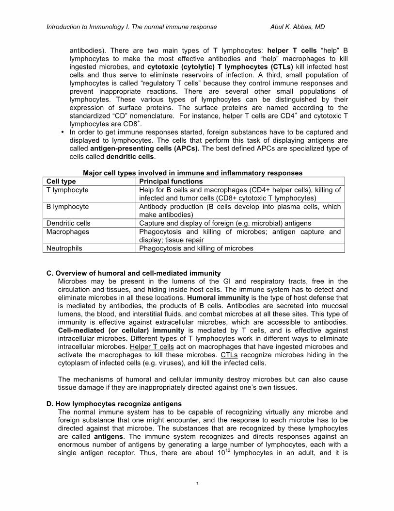
Major cell types involved in immune and inflammatory responses
Cell type Principal functions T lymphocyte Help for B cells and macrophages (CD4+ helper cells), killing of
infected and tumor cells (CD8+ cytotoxic T lymphocytes) B lymphocyte Antibody production (B cells develop into plasma cells, which
make antibodies) Dendritic cells Capture and display of foreign (e.g. microbial) antigens Macrophages Phagocytosis and killing of microbes; antigen capture and
display; tissue repair Neutrophils Phagocytosis and killing of microbes
C. Overview of humoral and cell-mediated immunity Microbes may be present in the lumens of the GI and respiratory tracts, free in the circulation and tissues, and hiding inside host cells. The immune system has to detect and eliminate microbes in all these locations. Humoral immunity is the type of host defense that is mediated by antibodies, the products of B cells. Antibodies are secreted into mucosal lumens, the blood, and interstitial fluids, and combat microbes at all these sites. This type of immunity is effective against extracellular microbes, which are accessible to antibodies. Cell-mediated (or cellular) immunity is mediated by T cells, and is effective against intracellular microbes. Different types of T lymphocytes work in different ways to eliminate intracellular microbes. Helper T cells act on macrophages that have ingested microbes and activate the macrophages to kill these microbes. CTLs recognize microbes hiding in the cytoplasm of infected cells (e.g. viruses), and kill the infected cells. The mechanisms of humoral and cellular immunity destroy microbes but can also cause tissue damage if they are inappropriately directed against one’s own tissues.
D. How lymphocytes recognize antigens The normal immune system has to be capable of recognizing virtually any microbe and foreign substance that one might encounter, and the response to each microbe has to be directed against that microbe. The substances that are recognized by these lymphocytes are called antigens. The immune system recognizes and directs responses against an enormous number of antigens by generating a large number of lymphocytes, each with a single antigen receptor. Thus, there are about 1012 lymphocytes in an adult, and it is
Introduction to Immunology I. The normal immune response Abul K. Abbas, MD
4
estimated that these are able to recognize at least 107 – 109 different antigens. Thus, only a few thousand lymphocytes express identical antigen receptors and recognize the same antigen. The molecular mechanisms responsible for the production of this enormous and diverse collection of antigen receptors are beyond our scope, and will be discussed in I-3. The antigen receptors of B cells are membrane-bound antibodies (also called immunoglobulins, or Ig). Antibodies are Y-shaped structures. The tops of the Y recognize the antigen and, in B cells, the “tail” of the Y anchors the molecule in the plasma membrane. Antibodies are able to recognize whole microbes and macromolecules as well as small chemicals. These could be in the circulation (e.g. a bacterial toxin) or attached to cells (e.g. a microbial cell wall component). The antigen receptors of T cells are structurally similar to antibodies, but T cell receptors (or TCRs) recognize only small peptides that are displayed on specialized peptide-display molecules. These peptide display molecules are called MHC (major histocompatibility complex) molecules, because they were discovered in the context of graft acceptance or rejection (tissue compatibility); human MHC molecules are called HLA (for human leukocyte antigens, because they were detected by antibodies made against leukocytes). HLA molecules pick up peptides from intracellular microbes and display these peptides for recognition by T cells; this is how T cells sense the presence of microbes inside infected cells. Although the immune system is capable of recognizing millions of foreign antigens, it normally does not react against one’s own (self) antigenic substances. As we shall see in the next lecture, this is because lymphocytes that happen to express receptors for self antigens are killed or shut off when they recognize these antigens. This phenomenon is called self-tolerance, implying that we “tolerate” our own antigens; the breakdown of this process results in autoimmune diseases.
E. Steps in immune responses
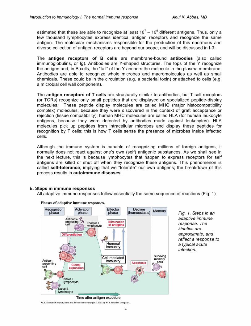
All adaptive immune responses follow essentially the same sequence of reactions (Fig. 1).
Fig. 1. Steps in an adaptive immune response. The kinetics are approximate, and reflect a response to a typical acute infection.
Introduction to Immunology I. The normal immune response Abul K. Abbas, MD
5
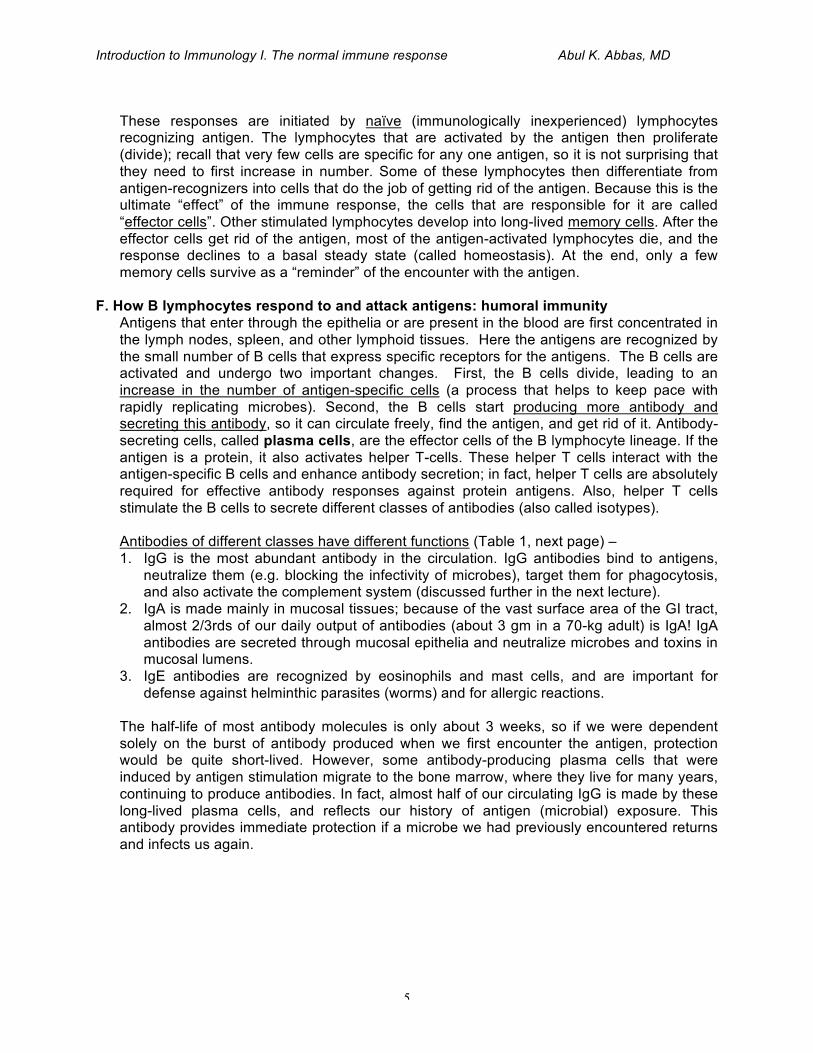
These responses are initiated by naïve (immunologically inexperienced) lymphocytes recognizing antigen. The lymphocytes that are activated by the antigen then proliferate (divide); recall that very few cells are specific for any one antigen, so it is not surprising that they need to first increase in number. Some of these lymphocytes then differentiate from antigen-recognizers into cells that do the job of getting rid of the antigen. Because this is the ultimate “effect” of the immune response, the cells that are responsible for it are called “effector cells”. Other stimulated lymphocytes develop into long-lived memory cells. After the effector cells get rid of the antigen, most of the antigen-activated lymphocytes die, and the response declines to a basal steady state (called homeostasis). At the end, only a few memory cells survive as a “reminder” of the encounter with the antigen.
F. How B lymphocytes respond to and attack antigens: humoral immunity
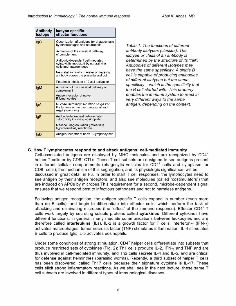
Antigens that enter through the epithelia or are present in the blood are first concentrated in the lymph nodes, spleen, and other lymphoid tissues. Here the antigens are recognized by the small number of B cells that express specific receptors for the antigens. The B cells are activated and undergo two important changes. First, the B cells divide, leading to an increase in the number of antigen-specific cells (a process that helps to keep pace with rapidly replicating microbes). Second, the B cells start producing more antibody and secreting this antibody, so it can circulate freely, find the antigen, and get rid of it. Antibody-secreting cells, called plasma cells, are the effector cells of the B lymphocyte lineage. If the antigen is a protein, it also activates helper T-cells. These helper T cells interact with the antigen-specific B cells and enhance antibody secretion; in fact, helper T cells are absolutely required for effective antibody responses against protein antigens. Also, helper T cells stimulate the B cells to secrete different classes of antibodies (also called isotypes). Antibodies of different classes have different functions (Table 1, next page) – 1. IgG is the most abundant antibody in the circulation. IgG antibodies bind to antigens,
neutralize them (e.g. blocking the infectivity of microbes), target them for phagocytosis, and also activate the complement system (discussed further in the next lecture).
2. IgA is made mainly in mucosal tissues; because of the vast surface area of the GI tract, almost 2/3rds of our daily output of antibodies (about 3 gm in a 70-kg adult) is IgA! IgA antibodies are secreted through mucosal epithelia and neutralize microbes and toxins in mucosal lumens.
3. IgE antibodies are recognized by eosinophils and mast cells, and are important for defense against helminthic parasites (worms) and for allergic reactions.
The half-life of most antibody molecules is only about 3 weeks, so if we were dependent solely on the burst of antibody produced when we first encounter the antigen, protection would be quite short-lived. However, some antibody-producing plasma cells that were induced by antigen stimulation migrate to the bone marrow, where they live for many years, continuing to produce antibodies. In fact, almost half of our circulating IgG is made by these long-lived plasma cells, and reflects our history of antigen (microbial) exposure. This antibody provides immediate protection if a microbe we had previously encountered returns and infects us again.
Introduction to Immunology I. The normal immune response Abul K. Abbas, MD
6
G. How T lymphocytes respond to and attack antigens: cell-mediated immunity
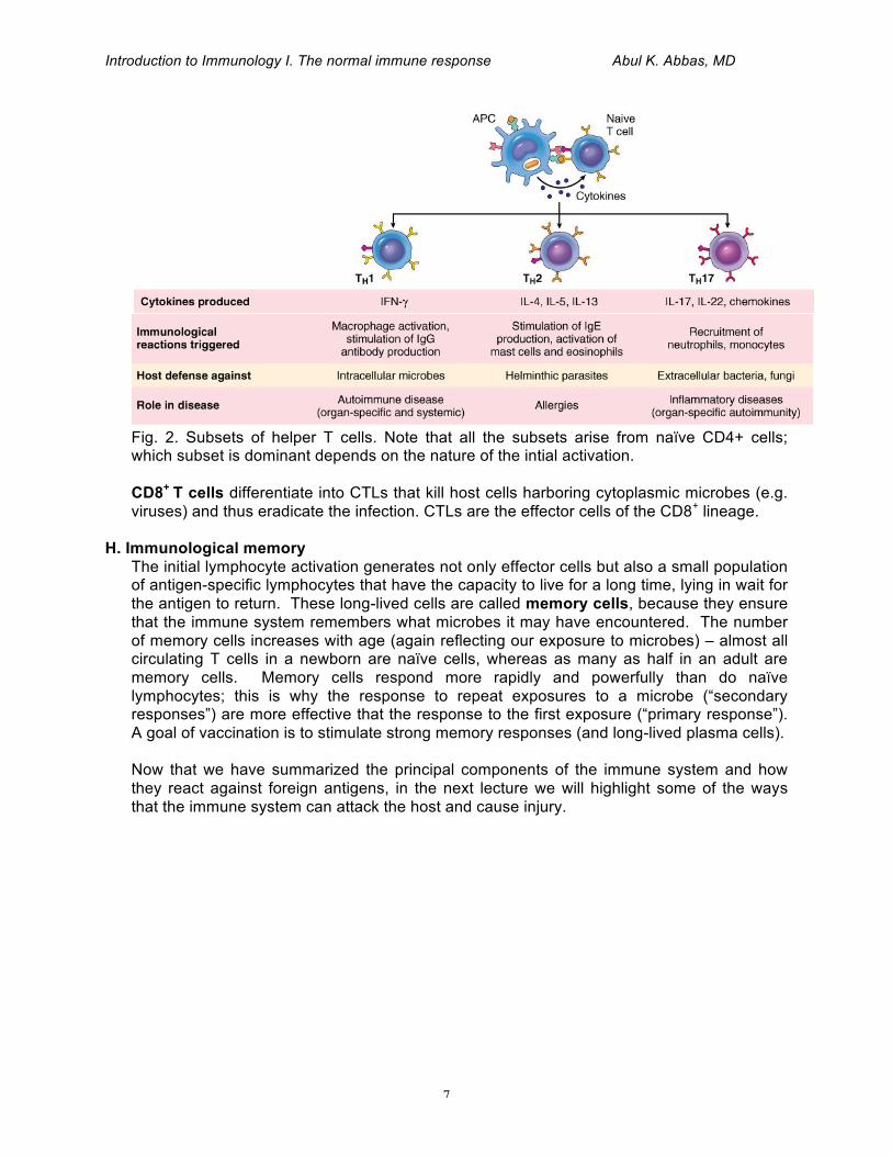
Cell-associated antigens are displayed by MHC molecules and are recognized by CD4+ helper T cells or by CD8+ CTLs. These T cell subsets are designed to see antigens present in different cellular compartments (phagocytic vesicles for CD4+ cells and cytoplasm for CD8+ cells); the mechanism of this segregation, and its physiologic significance, will be discussed in great detail in I-3. In order to start T cell responses, the lymphocytes need to see antigen by their antigen receptors, and also see molecules (called “costimulators”) that are induced on APCs by microbes.This requirement for a second, microbe-dependent signal ensures that we respond best to infectious pathogens and not to harmless antigens. Following antigen recognition, the antigen-specific T cells expand in number (even more than do B cells), and begin to differentiate into effector cells, which perform the task of attacking and eliminating microbes (the “effect” of the immune response). Effector CD4+ T cells work largely by secreting soluble proteins called cytokines. Different cytokines have different functions; in general, many mediate communications between leukocytes and are therefore called interleukins (ILs). IL-2 is a growth factor for T cells; interferon-γ (IFN-γ) activates macrophages; tumor necrosis factor (TNF) stimulates inflammation; IL-4 stimulates B cells to produce IgE; IL-5 activates eosinophils. Under some conditions of strong stimulation, CD4+ helper cells differentiate into subsets that produce restricted sets of cytokines (Fig. 2): Th1 cells produce IL-2, IFN-γ and TNF and are thus involved in cell-mediated immunity, and Th2 cells secrete IL-4 and IL-5, and are critical for defense against helminthes (parasitic worms). Recently, a third subset of helper T cells has been discovered, called Th17 cells because their signature cytokine is IL-17. These cells elicit strong inflammatory reactions. As we shall see in the next lecture, these same T cell subsets are involved in different types of immunological diseases.
Table 1. The functions of different antibody isotypes (classes). The isotype or class of an antibody is determined by the structure of its “tail”. Antibodies of different isotypes may have the same specificity. A single B cell is capable of producing antibodies of different isotypes but the same specificity – which is the specificity that the B cell started with. This property enables the immune system to react in very different ways to the same antigen, depending on the context.
Introduction to Immunology I. The normal immune response Abul K. Abbas, MD
7
Fig. 2. Subsets of helper T cells. Note that all the subsets arise from naïve CD4+ cells; which subset is dominant depends on the nature of the intial activation. CD8+ T cells differentiate into CTLs that kill host cells harboring cytoplasmic microbes (e.g. viruses) and thus eradicate the infection. CTLs are the effector cells of the CD8+ lineage.
H. Immunological memory The initial lymphocyte activation generates not only effector cells but also a small population of antigen-specific lymphocytes that have the capacity to live for a long time, lying in wait for the antigen to return. These long-lived cells are called memory cells, because they ensure that the immune system remembers what microbes it may have encountered. The number of memory cells increases with age (again reflecting our exposure to microbes) – almost all circulating T cells in a newborn are naïve cells, whereas as many as half in an adult are memory cells. Memory cells respond more rapidly and powerfully than do naïve lymphocytes; this is why the response to repeat exposures to a microbe (“secondary responses”) are more effective that the response to the first exposure (“primary response”). A goal of vaccination is to stimulate strong memory responses (and long-lived plasma cells). Now that we have summarized the principal components of the immune system and how they react against foreign antigens, in the next lecture we will highlight some of the ways that the immune system can attack the host and cause injury.
Introduction to Immunology I. The normal immune response Abul K. Abbas, MD
8
INTRODUCTION TO IMMUNOLOGY II. How The Immune System Causes Disease
(Lecture)
OBJECTIVES • Understand the meaning of hypersensitivity and autoimmunity. • Describe the mechanisms by which antibodies cause tissue injury. • Describe the mechanisms by which helper and cytotoxic T lymphocytes cause tissue injury. KEY WORDS • Autoimmunity • Hypersensitivity • Complement • Inflammation • Delayed type hypersensitivity • Immune complexes RECOMMENDED READING
Chapter 5 in ROBBINS BASIC PATHOLOGY 8th edition, Elsevier, 2007. (Pages 119-131 and 135-139 summarize the mechanisms of injury in immune-mediated “hypersensitivity” diseases and the pathogenesis of autoimmunity, which are the themes of this lecture. The remainder of the chapter is devoted to specific immunological diseases, and is not necessary at this stage.)
Chapter 11 in BASIC IMMUNOLOGY: Functions and Disorders of the Immune System (A.K. Abbas and A.H. Lichtman), 2nd edition, updated 2006, Elsevier. OR INTRODUCTION
An active immune system is vital for protecting us from deadly infections, and when it fails (e.g. in congenital immunodeficiencies or advanced HIV infection), individuals become very susceptible to infections and may not survive unless treated. On the other hand, immune responses themselves can become harmful. There are three main situations in which immune responses are pathologic. 1. When self-tolerance fails and the immune system begins to attack an individual’s own
tissues (causing autoimmune diseases). 2. When the immune response becomes excessive or uncontrolled, either against microbial
antigens or against normally harmless environmental antigens. 3. As part of an entirely normal reaction against some microbes.
You will come across diseases caused by all these reactions as you go through your education, and will hear about various such diseases in Organs, M&N and BMB. In this lecture, we will introduce you to some of the basic principles of tissue injury caused by immune responses; we will return to these ideas in more depth in the I-3 course.
A. Hypersensitivity and autoimmunity
Disorders caused by pathologic immune responses are called hypersensitivity diseases. This term is derived from the idea that an individual who has previously encountered an

Introduction to Immunology I. The normal immune response Abul K. Abbas, MD
9
antigen is “sensitive” to a second encounter with that antigen (i.e. mounts a stronger response upon the second encounter); “hypersensitivity” denotes an abnormal, or pathologic, reaction. There are four types of hypersensitivity, which differ in their pathogenesis, effector mechanisms, and clinical and pathological manifestations (Table 1). We will describe each of these in more detail later.
Autoimmunity refers to immune responses against self-antigens. When autoimmunity develops, antibodies and/or T cells begin to react against self antigens and to attack the tissues where these antigens are located. These reactions are examples of hypersensitivity; in other words, autoimmunity is one cause of hypersensitivity. Note, however, that hypersensitivity reactions can also be triggered by foreign (microbial or environmental) antigens, so the terms hypersensitivity and autoimmunity are not synonymous. A common clinical and pathologic manifestation of immunological diseases is inflammation, especially chronic inflammation. Inflammation is a vascular and cellular reaction to a wide variety of injurious and dangerous stimuli. We will discuss the causes and consequences of inflammation, and how leukocytes destroy microbes as well as host tissues, in later lectures. Many types of inflammation are caused by abnormal immune responses (i.e., hypersensitivity). A modern name for diseases that are caused by abnormal immune responses (e.g. against self antigens) and in which inflammation is an important component is immune-mediated inflammatory diseases. Because of the prominent role that abnormal immune responses play in many common inflammatory diseases, an introduction to Immunology is important as a prelude to subsequent courses in which these disorders will be described in much more detail. Before discussing the mechanisms of tissue injury in hypersensitivity diseases, we will touch upon the situations in which immune responses against self or foreign antigens might become the cause of diseases.
Table 1. Types of hypersensitivity diseases. (It is preferable to use the descriptive terms rather than the less informative numerical classification.)

Introduction to Immunology I. The normal immune response Abul K. Abbas, MD
10
B. Autoimmunity: why self-tolerance fails
The normal immune system does not react against self-antigens. During the generation of a large number of specificities, all individuals produce lymphocytes (T and B) whose antigen receptors can recognize self-antigens. If these lymphocytes encounter self-antigens during their maturation (i.e. in the bone marrow and thymus), the self-reactive lymphocytes are killed. Even if some self-reactive lymphocytes complete their maturation, their encounter with the self-antigens in peripheral tissues results in the death or permanent inactivation of the lymphocytes. Thus, the collection of lymphocytes in healthy individuals is “purged” of cells capable of reacting against self. However, in some individuals, self-tolerance fails, and T and/or B lymphocytes specific for self-antigens survive, become activated, and the cells or their products (antibodies) attack the tissues in which those antigens are present. Although we are beginning to understand the mechanisms of self-tolerance (based largely on animal experiments), we do not know why self-tolerance fails in any human autoimmune diseases. Likely underlying mechanisms include the inheritance of susceptibility genes, infections and other causes of tissue injury (leading to release of self-antigens and activation of specific lymphocytes) (Fig. 1). This is similar to the pathogenesis of most multifactorial diseases, which involve some combination of susceptibility genes and environmental triggers.
C. Disorders caused by immune responses against foreign antigens
Some immunological diseases are the result of poorly controlled responses to foreign antigens; in other diseases, these responses are quite normal, and tissue injury is an unfortunate accompaniment. Examples include the following. • In allergic disease (immediate hypersensitivity), genetically susceptible individuals make
strong Th2 responses to environmental antigens (pollen, foods, insect proteins, some drugs, and so on) that are well tolerated in most individuals. Examples include asthma (Organs block).
Fig. 1. Hypothesis for how susceptibility genes and environmental triggers work together to cause autoimmune diseases.
Introduction to Immunology I. The normal immune response Abul K. Abbas, MD
11
• Sometimes, large amounts of antibodies are produced in response to a microbe and the microbial antigen is persistent, resulting in the formation of antigen-antibody complexes (as in poststreptococcal glomerulonephritis, discussed in Organs). In other situations, antibodies produced against a microbe happen to bind to (”cross-react” with) a self-antigen, and therefore attack the individual’s own tissues (rheumatic fever, also Organs).
• In some cases, the immune system reacts strongly against bacteria that are normal inhabitants of our GI tract (called “commensals”); Crohn’s disease (M&N) is thought to be an example of one such disease.
• Some microbes are difficult to eradicate and elicit strong T-cell responses and chronic inflammation, with large numbers of activated macrophages. The macrophages produce substances intended to kill the microbes, but these same substances damage normal tissues.
• In some cases, the immune response is entirely normal, but results in damage to host cells. For instance, the hepatitis virus is not cytopathic. When it infects liver cells, host CTLs are activated and they kill infected cells in an attempt to eradicate the infection – even though the virus itself may be quite harmless! Thus, viral hepatitis is initiated by the virus, but liver damage is the result of a normal host response that is trying to get rid of the virus (M&N).
Now that we have reviewed the main conditions that lead to disease-producing immune responses, we will conclude by describing how these responses actually result in tissue injury. A general principle that is important to remember is that the effector mechanisms of tissue injury in immunological diseases are the same as the effector mechanisms used by the immune system to combat and destroy microbes.
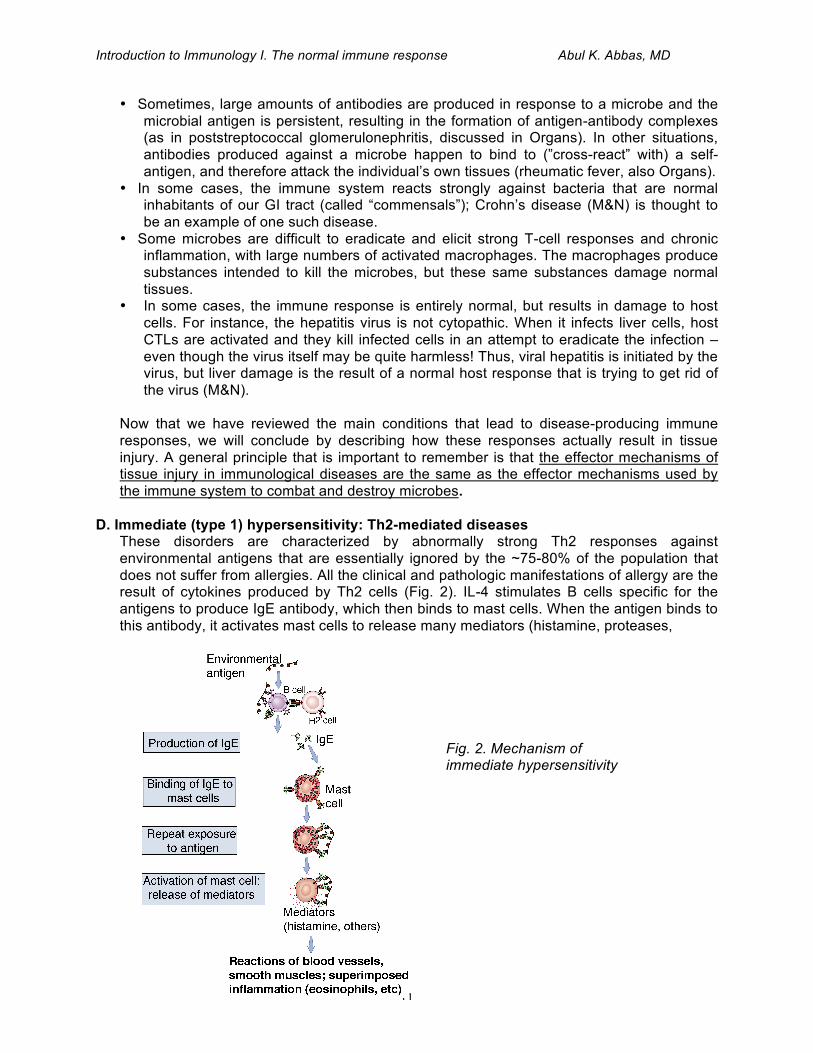
D. Immediate (type 1) hypersensitivity: Th2-mediated diseases These disorders are characterized by abnormally strong Th2 responses against environmental antigens that are essentially ignored by the ~75-80% of the population that does not suffer from allergies. All the clinical and pathologic manifestations of allergy are the result of cytokines produced by Th2 cells (Fig. 2). IL-4 stimulates B cells specific for the antigens to produce IgE antibody, which then binds to mast cells. When the antigen binds to this antibody, it activates mast cells to release many mediators (histamine, proteases,
Fig. 2. Mechanism of immediate hypersensitivity
Introduction to Immunology I. The normal immune response Abul K. Abbas, MD
12
cytokines) that cause the acute vascular and smooth muscle reactions and inflammation that are typical of allergies. IL-5 made by Th2 cells activates eosinophils, which can exacerbate tissue damage. Th2 cells also secrete IL-13, which acts on mucosal epithelial cells to stimulate secretion of mucus. Bronchial asthma is a Th2-mediated disease about which you will hear more in the “Organs” block. The propensity to develop allergies is genetic, but the actual genes that may be causative have not been definitively identified.
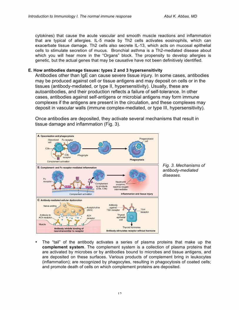
E. How antibodies damage tissues: types 2 and 3 hypersensitivity Antibodies other than IgE can cause severe tissue injury. In some cases, antibodies may be produced against cell or tissue antigens and may deposit on cells or in the tissues (antibody-mediated, or type II, hypersensitivity). Usually, these are autoantibodies, and their production reflects a failure of self-tolerance. In other cases, antibodies against self-antigens or microbial antigens may form immune complexes if the antigens are present in the circulation, and these complexes may deposit in vascular walls (immune complex-mediated, or type III, hypersensitivity). Once antibodies are deposited, they activate several mechanisms that result in tissue damage and inflammation (Fig. 3).
• The “tail” of the antibody activates a series of plasma proteins that make up the
complement system. The complement system is a collection of plasma proteins that are activated by microbes or by antibodies bound to microbes and tissue antigens, and are deposited on these surfaces. Various products of complement bring in leukocytes (inflammation); are recognized by phagocytes, resulting in phagocytosis of coated cells; and promote death of cells on which complement proteins are deposited.
Fig. 3. Mechanisms of antibody-mediated diseases.
Introduction to Immunology I. The normal immune response Abul K. Abbas, MD
13
• The tail of the antibody (called the Fc piece because this fragment has a propensity to crystallize in solution) is also recognized by Fc receptors on phagocytes (macrophages and neutrophils).
Once these pathways are activated, they cause disease in several ways (see Fig. 4).
A. If the antibody is deposited on a cell (e.g. erythrocyte or platelet), the combined action of complement and Fc receptors results in that cell being eaten and destroyed by phagocytes. (This is the basis of red blood cell and platelet depletion in autoimmune hemolytic anemia and thrombocytopenia, respectively.)
B. If the antibody is deposited on a solid surface the phagocytes may be activated and release toxic substances that induce inflammation and damage the tissue (as in some forms of glomerulonephritis).
C. Less commonly, antibodies can cause disease by interfering with normal molecules (such as hormones and hormone receptors), without any actual tissue injury; examples include myasthenia gravis (BMB) and Graves’ disease (M&N).
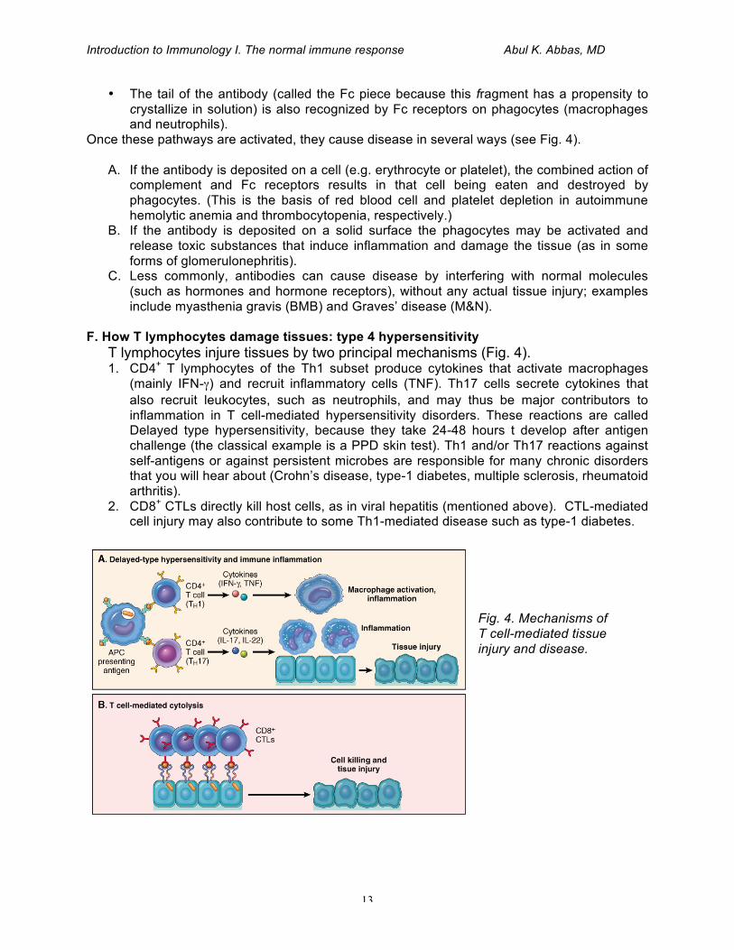
F. How T lymphocytes damage tissues: type 4 hypersensitivity
T lymphocytes injure tissues by two principal mechanisms (Fig. 4). 1. CD4+ T lymphocytes of the Th1 subset produce cytokines that activate macrophages
(mainly IFN-γ) and recruit inflammatory cells (TNF). Th17 cells secrete cytokines that also recruit leukocytes, such as neutrophils, and may thus be major contributors to inflammation in T cell-mediated hypersensitivity disorders. These reactions are called Delayed type hypersensitivity, because they take 24-48 hours t develop after antigen challenge (the classical example is a PPD skin test). Th1 and/or Th17 reactions against self-antigens or against persistent microbes are responsible for many chronic disorders that you will hear about (Crohn’s disease, type-1 diabetes, multiple sclerosis, rheumatoid arthritis).
2. CD8+ CTLs directly kill host cells, as in viral hepatitis (mentioned above). CTL-mediated cell injury may also contribute to some Th1-mediated disease such as type-1 diabetes.
Fig. 4. Mechanisms of T cell-mediated tissue injury and disease.

Introduction to Immunology I. The normal immune response Abul K. Abbas, MD
14
Recent remarkable successes in treating these diseases are based largely on improved understanding of the underlying immune abnormalities and the role of lymphocytes and cytokines in the pathogenesis of these diseases. Such successes have fueled tremendous interest in studying immune-mediated inflammatory diseases and in developing new therapeutic strategies.