Embed Size (px)
Citation preview
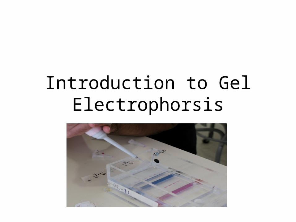
Introduction to Gel Electrophorsis
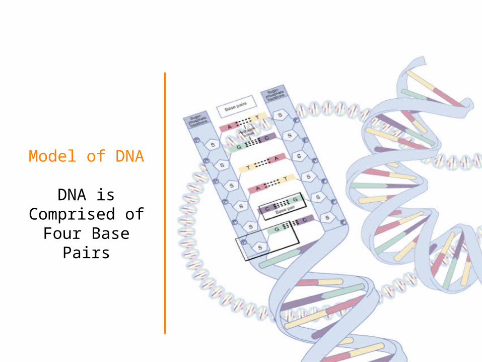
Model of DNA
DNA is Comprised of Four Base Pairs
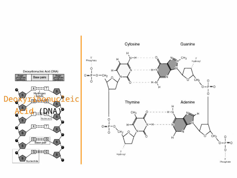
Deoxyribonucleic Acid
(DNA)
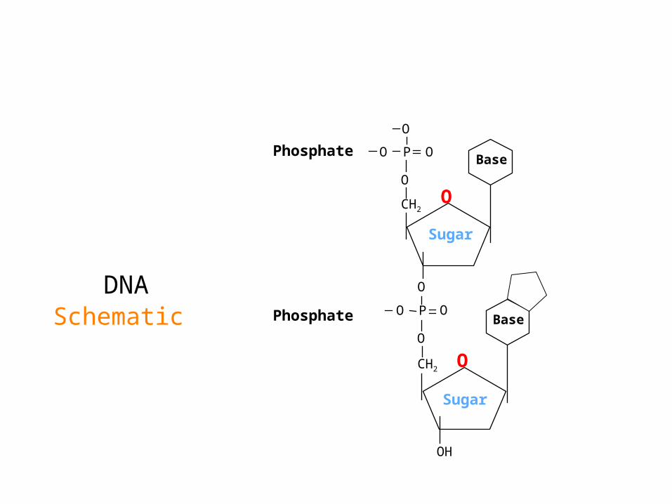
DNA Schematic
OCH2
O
P O
O
OBase
CH2
O
P
O
O
O
Base
OH
Sugar
Sugar
O
Phosphate
Phosphate
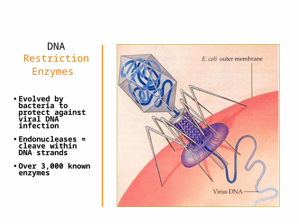
DNA Restriction Enzymes
• Evolved by bacteria to protect against viral DNA infection
• Endonucleases = cleave within DNA strands
• Over 3,000 known enzymes
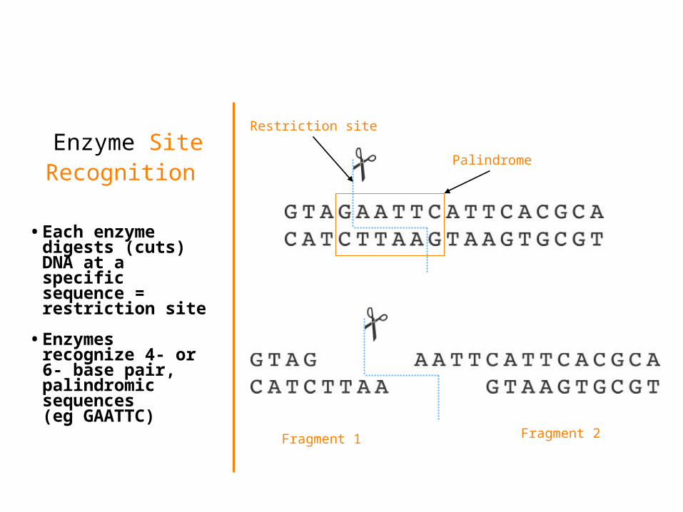
Enzyme Site Recognition
• Each enzyme digests (cuts) DNA at a specific sequence = restriction site
• Enzymes recognize 4- or 6- base pair, palindromic sequences (eg GAATTC)
Palindrome
Restriction site
Fragment 1 Fragment 2

5 vs 3 Prime Overhang
• Generates 5 prime overhang
Enzyme cuts
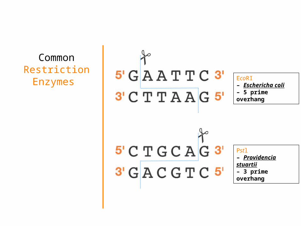
Common Restriction Enzymes EcoRI
– Eschericha coli– 5 prime overhang
Pstl– Providencia stuartii– 3 prime overhang
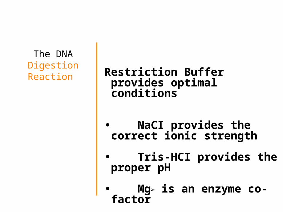
The DNA DigestionReaction Restriction Buffer provides
optimal conditions
• NaCI provides the correct ionic strength
• Tris-HCI provides the proper pH
• Mg2+ is an enzyme co-factor
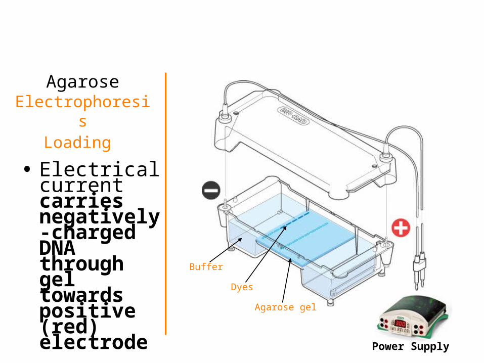
AgaroseElectrophoresis
Loading
• Electrical current carries negatively-charged DNA through gel towards positive (red) electrode
Power Supply
Buffer
Dyes
Agarose gel
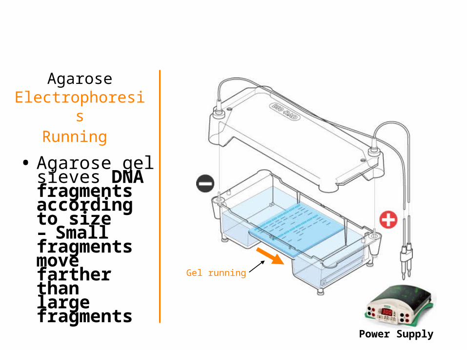
AgaroseElectrophoresis
Running
• Agarose gel sieves DNA fragments according to size– Small fragments
move farther than
large fragments
Power Supply
Gel running
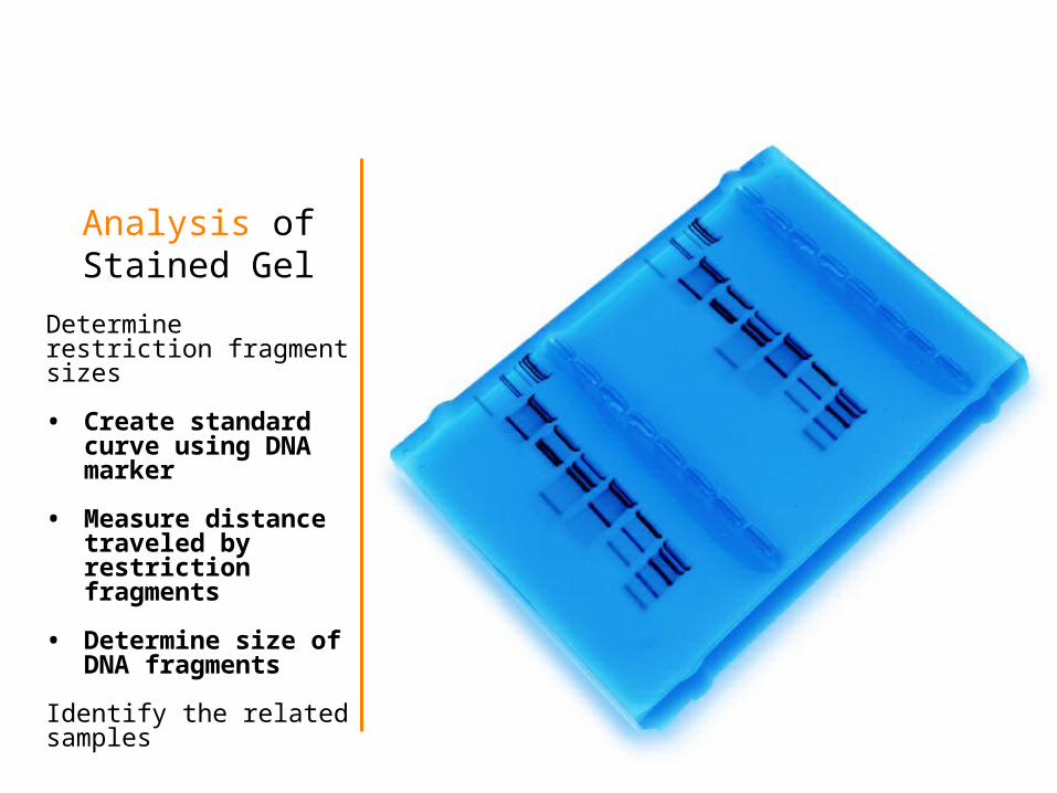
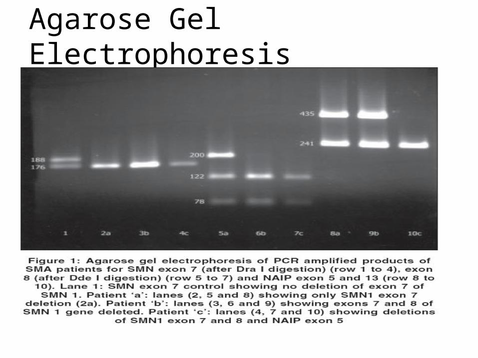
Analysis of Stained Gel
Determinerestriction fragmentsizes
• Create standard curve using DNA marker
• Measure distance traveled by restriction fragments
• Determine size of DNA fragments
Identify the relatedsamples
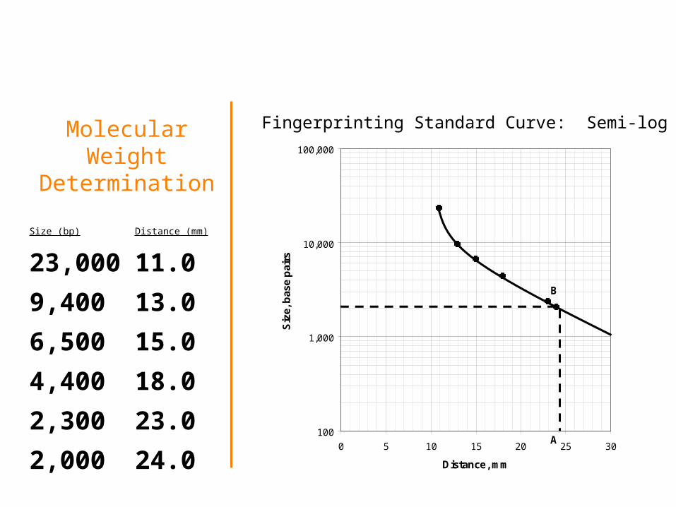
Molecular Weight Determination
Size (bp) Distance (mm)
23,000 11.0 9,400 13.0
6,500 15.0
4,400 18.0
2,300 23.0
2,000 24.0100
1,000
10,000
100,000
0 5 10 15 20 25 30
Distance, mm
Siz
e, b
ase
pai
rsB
A
Fingerprinting Standard Curve: Semi-log
Agarose Gel Electrophoresis
• The standard method for separating DNA fragments is electrophoresis through agarose gels.
Agarose Gel Electrophoresis
• The standard method for separating DNA fragments is electrophoresis through agarose gels.
• Agarose is a polysaccharide like agar or pectin derived from seaweed
Agarose Gel Electrophoresis
• The standard method for separating DNA fragments is electrophoresis through agarose gels.
• Agarose is a polysaccharide like agar or pectin derived from seaweed
• It dissolves in boiling water and then gels as it cools
Agarose Gel Electrophoresis
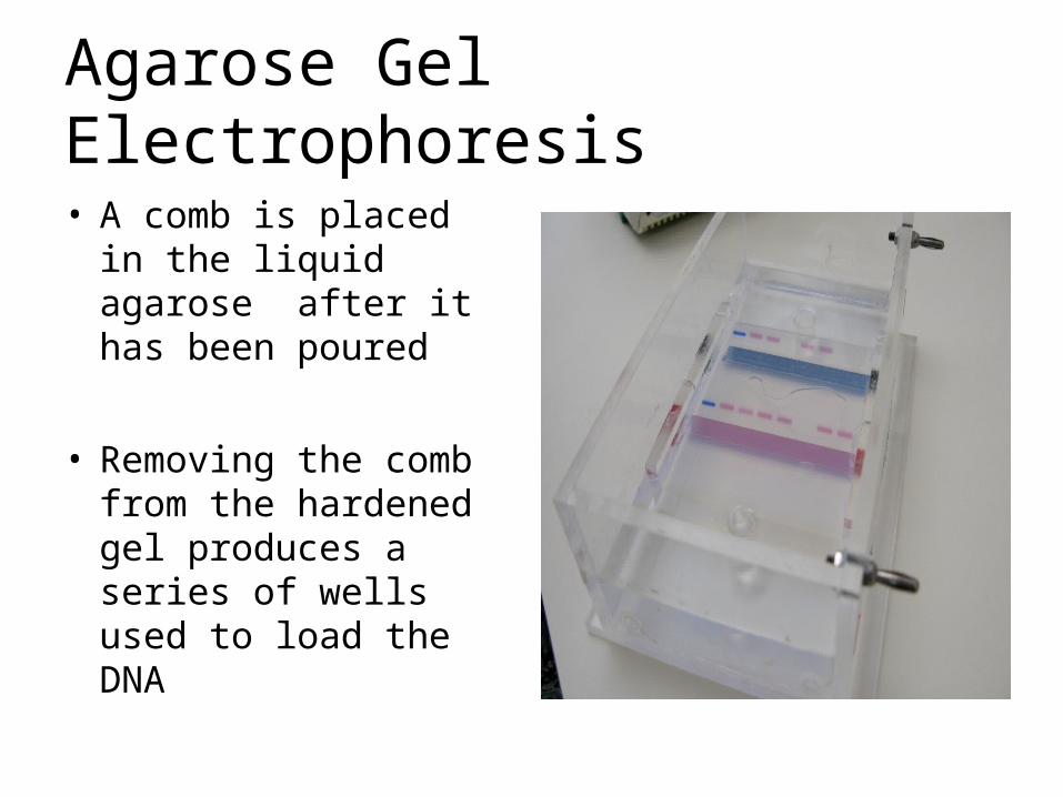
• A comb is placed in the liquid agarose after it has been poured
• Removing the comb from the hardened gel produces a series of wells used to load the DNA
Agarose Gel Electrophoresis
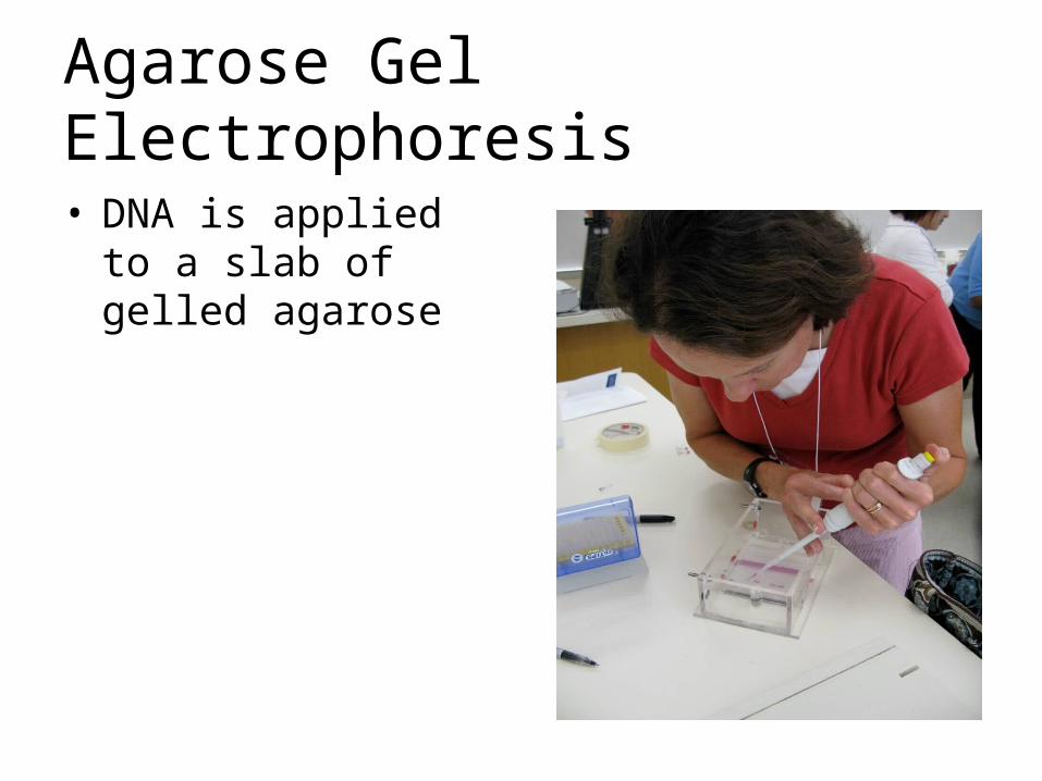
• DNA is applied to a slab of gelled agarose
Agarose Gel Electrophoresis
• DNA is applied to a slab of gelled agarose
• The sample is loaded with a loading buffer—containing dyes and glycerol or sugar
Agarose Gel Electrophoresis
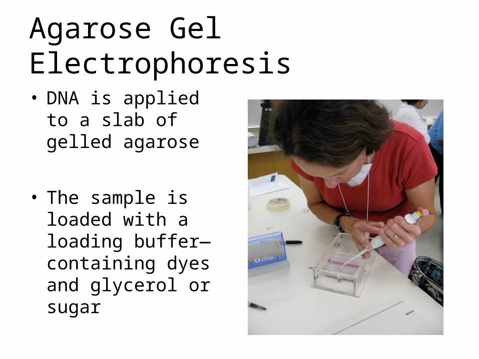
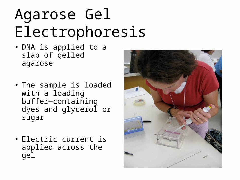
• DNA is applied to a slab of gelled agarose
• The sample is loaded with a loading buffer—containing dyes and glycerol or sugar
• Electric current is applied across the gel
Agarose Gel Electrophoresis
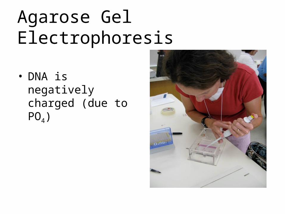
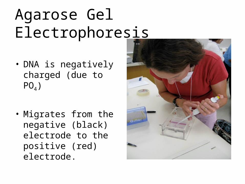
• DNA is negatively charged (due to PO4)
Agarose Gel Electrophoresis
• DNA is negatively charged (due to PO4)
• Migrates from the negative (black) electrode to the positive (red) electrode.
Agarose Gel Electrophoresis
• Rate of migration of DNA through agarose depends on the size of DNA
Agarose Gel Electrophoresis
• Rate of migration of DNA through agarose depends on the size of DNA
• Smaller DNA fragments move more quickly
Agarose Gel Electrophoresis
• Rate of migration of DNA through agarose depends on the size of DNA
• Smaller DNA fragments move more quickly
• Rate of migration is inversely proportional to the log10 of molecular weight
Agarose Gel Electrophoresis
Agarose Gel Electrophoresis
• Concentration of agarose also affects migration
Agarose Gel Electrophoresis
• Concentration of agarose also affects migration
• Higher concentration of agarose, the more it retards the movement of all DNA fragments
Agarose Gel Electrophoresis
• Concentration of agarose also affects migration
• Higher concentration of agarose, the more it retards the movement of all DNA fragments
• Small DNA fragments require higher concentrations of agarose/ Lg fragments low concentrations
Agarose Gel Electrophoresis
• Agarose gels must be prepared and run in a buffer containing ions.
Agarose Gel Electrophoresis
• Agarose gels must be prepared and run in a buffer containing ions.
• Ions are charged particles (like those found in salt) and are necessary to carry a charge
Agarose Gel Electrophoresis
• During electrophoresis water undergoes hydrolysis : H2O H+ and OH-
Agarose Gel Electrophoresis
• During electrophoresis water undergoes hydrolysis : H2O H+ and OH-
• The anode (+ /red) pole becomes alkaline because OH- will accumulate at this pole
• The cathode (-/black) pole becomes acidic because H+ will accumulate at this pole
Agarose Gel Electrophoresis
• Buffers prevent the pH from changing by reacting with the H+ or OH- products
Agarose Gel Electrophoresis
• The buffer is either TBE or TAE
– TBE is made with Tris/Boric Acid/EDTA
– TAE is made with Tris/Acetic Acid/ EDTA
Agarose Gel Electrophoresis• The voltage applied to the gel affects how
quickly the gel runs
Agarose Gel Electrophoresis• The voltage applied to the gel affects how
quickly the gel runs
• The higher the voltage, the more quickly the gel runs………But that often reduces the quality of the DNA separation
Agarose Gel Electrophoresis• The voltage applied to the gel affects how
quickly the gel runs
• The higher the voltage, the more quickly the gel runs………But that often reduces the quality of the DNA separation
• >>>>>>>>>>It also generates heat which reduces the quality of the DNA separation
Agarose Gel Electrophoresis
• To make DNA fragments visible after electrophoresis, the DNA must be stained

Agarose Gel Electrophoresis
A gel stained with Methylene blue