Embed Size (px)
Citation preview
Intravenous and interstitial photodynamic laser therapy:
New options in oncology
By Dr. M. WEBER
Summary
Objective: Introduction of a new treatment option in oncology: Photodynamic tumor
therapy.
Method:
Combined application of natural and chemically derived highly specific photosensitizers
and new catheter- based laser technology for light activation of these photosensitizers
to induce selective destruction of tumor tissue. Additional low-dose chemotherapy with
light activation of chemotherapeutic drugs.
The combination with hyperbaric oxygenation is an option to optimize therapeutic
results because oxygen radicals are responsible for tumor necrosis and apoptosis.
Combination with immunotherapy like macrophage activating factor GcMAF can be of
benefit as well.
Results: Different case reports indicate the great potential of the treatment regime.
Conclusion: Photodynamic tumor therapy and additional low-dose chemotherapy with
light activation of chemotherapeutic drugs seems to be a promising new treatment
regime in future oncology.
Keywords: PDT (photodynamic therapy), intravenous laser therapy, interstitial laser
therapy, low-dose chemotherapy, hyperbaric oxygen, macrophage activating factor
GcMAF, light activation.
1) Introduction
Photodynamic therapy is one of the most interesting and promising approaches for
treatment of various cancers. It has a long history and is already approved for some
superficial cancers on the skin or on superficial layers in the esophagus or bronchial
system.(1,2,3,4,5) Treatments are easy to perform and – in contrast to chemotherapy –
go normally along without severe side effects. The principle is the stimulation of a light
sensitive drug (photosensitizer) that is either applied on the skin as a cream or injected
into the blood circulation. Through endocytosis the photosensitizer will be transported
into the tumor cells anywhere in the body with high specificity. The process takes
several hours (depending on the photosensitizer) and tumor cells will be light sensitive
-2-
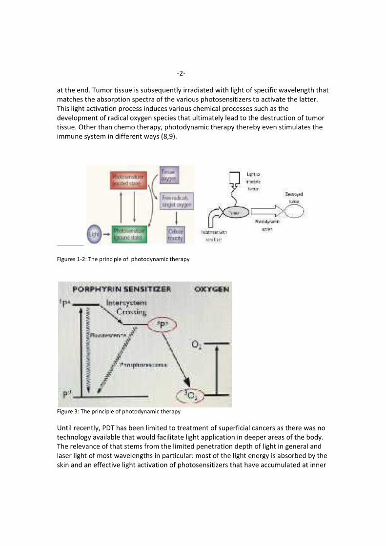
at the end. Tumor tissue is subsequently irradiated with light of specific wavelength that
matches the absorption spectra of the various photosensitizers to activate the latter.
This light activation process induces various chemical processes such as the
development of radical oxygen species that ultimately lead to the destruction of tumor
tissue. Other than chemo therapy, photodynamic therapy thereby even stimulates the
immune system in different ways (8,9).
Figures 1-2: The principle of photodynamic therapy
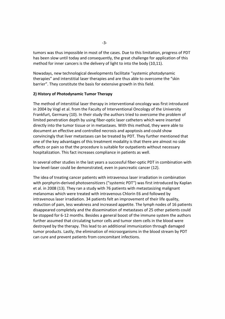
Figure 3: The principle of photodynamic therapy
Until recently, PDT has been limited to treatment of superficial cancers as there was no
technology available that would facilitate light application in deeper areas of the body.
The relevance of that stems from the limited penetration depth of light in general and
laser light of most wavelengths in particular: most of the light energy is absorbed by the
skin and an effective light activation of photosensitizers that have accumulated at inner
-3-
tumors was thus impossible in most of the cases. Due to this limitation, progress of PDT
has been slow until today and consequently, the great challenge for application of this
method for inner cancers is the delivery of light to into the body (10,11).
Nowadays, new technological developments facilitate “systemic photodynamic
therapies” and interstitial laser therapies and are thus able to overcome the “skin
barrier”. They constitute the basis for extensive growth in this field.
2) History of Photodynamic Tumor Therapy
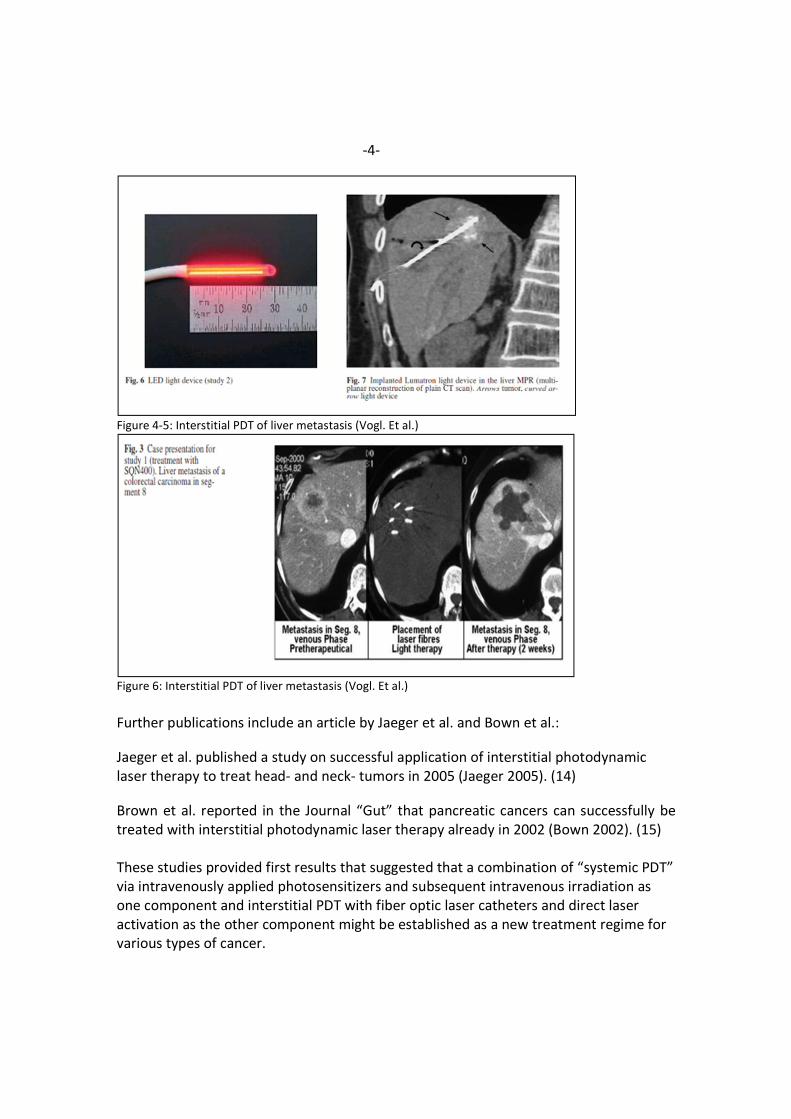
The method of interstitial laser therapy in interventional oncology was first introduced
in 2004 by Vogl et al. from the Faculty of Interventional Oncology of the University
Frankfurt, Germany (10). In their study the authors tried to overcome the problem of
limited penetration depth by using fiber-optic laser catheters which were inserted
directly into the tumor tissue or in metastases. With this method, they were able to
document an effective and controlled necrosis and apoptosis and could show
convincingly that liver metastases can be treated by PDT. They further mentioned that
one of the key advantages of this treatment modality is that there are almost no side
effects or pain so that the procedure is suitable for outpatients without necessary
hospitalization. This fact increases compliance in patients as well.
In several other studies in the last years a successful fiber-optic PDT in combination with
low-level-laser could be demonstrated, even in pancreatic cancer (12).
The idea of treating cancer patients with intravenous laser irradiation in combination
with porphyrin-derived photosensitizers (“systemic PDT“) was first introduced by Kaplan
et al. in 2008 (13). They ran a study with 76 patients with metastasizing malignant
melanomas which were treated with intravenous Chlorin E6 and followed by
intravenous laser irradiation. 34 patients felt an improvement of their life quality,
reduction of pain, less weakness and increased appetite. The lymph nodes of 16 patients
disappeared completely and the dissemination of metastases of 25 other patients could
be stopped for 6-12 months. Besides a general boost of the immune system the authors
further assumed that circulating tumor cells and tumor stem cells in the blood were
destroyed by the therapy. This lead to an additional immunization through damaged
tumor products. Lastly, the elimination of microorganisms in the blood stream by PDT
can cure and prevent patients from concomitant infections.
-4-
Figure 4-5: Interstitial PDT of liver metastasis (Vogl. Et al.)
Figure 6: Interstitial PDT of liver metastasis (Vogl. Et al.)
Further publications include an article by Jaeger et al. and Bown et al.:
Jaeger et al. published a study on successful application of interstitial photodynamic
laser therapy to treat head- and neck- tumors in 2005 (Jaeger 2005). (14)
Brown et al. reported in the Journal “Gut” that pancreatic cancers can successfully be
treated with interstitial photodynamic laser therapy already in 2002 (Bown 2002). (15)
These studies provided first results that suggested that a combination of “systemic PDT”
via intravenously applied photosensitizers and subsequent intravenous irradiation as
one component and interstitial PDT with fiber optic laser catheters and direct laser
activation as the other component might be established as a new treatment regime for
various types of cancer.
-5-
3) Photosensitizers
3.1) Overview
Photosensitizers are mostly porphyrin molecules and derivatives either from the human
haem (without the central iron atom) or plant-derived chlorophyll (without the central
magnesium atom) (6,7) Accordingly, they are called hematoporphyrins or chlorines.
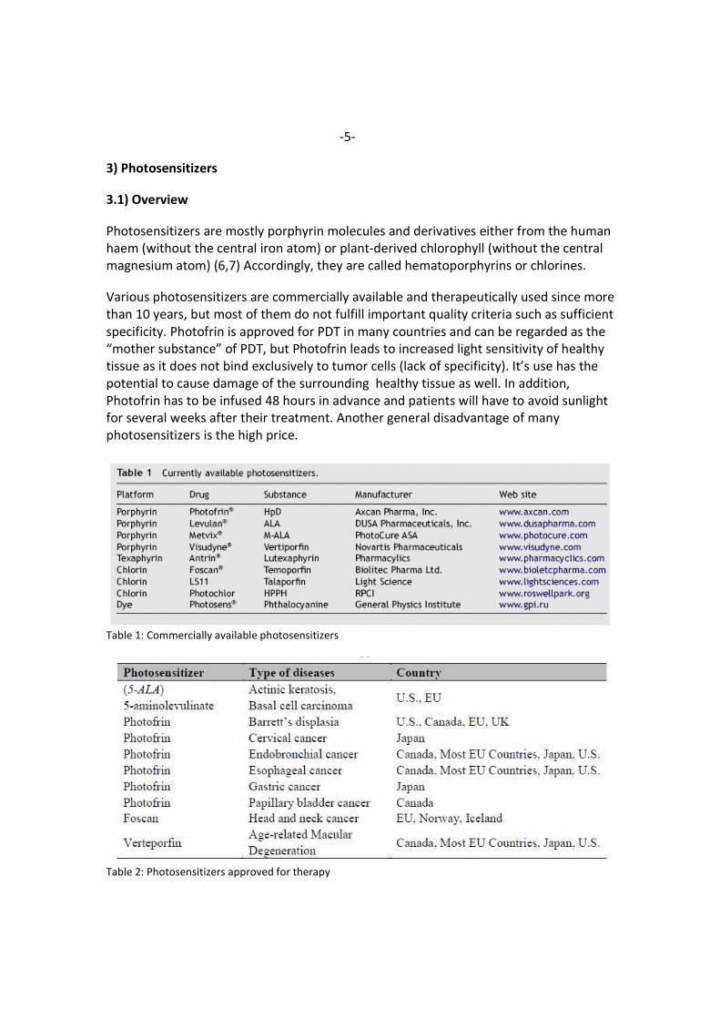
Various photosensitizers are commercially available and therapeutically used since more
than 10 years, but most of them do not fulfill important quality criteria such as sufficient
specificity. Photofrin is approved for PDT in many countries and can be regarded as the
“mother substance” of PDT, but Photofrin leads to increased light sensitivity of healthy
tissue as it does not bind exclusively to tumor cells (lack of specificity). It’s use has the
potential to cause damage of the surrounding healthy tissue as well. In addition,
Photofrin has to be infused 48 hours in advance and patients will have to avoid sunlight
for several weeks after their treatment. Another general disadvantage of many
photosensitizers is the high price.
Table 1: Commercially available photosensitizers
Table 2: Photosensitizers approved for therapy
-6-
The following photosensitizers are the most promising ones right now as they fulfill
much better the specificity quality criteria.
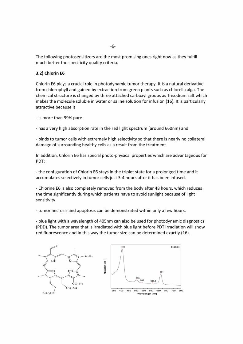
3.2) Chlorin E6
Chlorin E6 plays a crucial role in photodynamic tumor therapy. It is a natural derivative
from chlorophyll and gained by extraction from green plants such as chlorella alga. The
chemical structure is changed by three attached carboxyl groups as Trisodium salt which
makes the molecule soluble in water or saline solution for infusion (16). It is particularly
attractive because it
- is more than 99% pure
- has a very high absorption rate in the red light spectrum (around 660nm) and
- binds to tumor cells with extremely high selectivity so that there is nearly no collateral
damage of surrounding healthy cells as a result from the treatment.
In addition, Chlorin E6 has special photo-physical properties which are advantageous for
PDT:
- the configuration of Chlorin E6 stays in the triplet state for a prolonged time and it
accumulates selectively in tumor cells just 3-4 hours after it has been infused.
- Chlorine E6 is also completely removed from the body after 48 hours, which reduces
the time significantly during which patients have to avoid sunlight because of light
sensitivity.
- tumor necrosis and apoptosis can be demonstrated within only a few hours.
- blue light with a wavelength of 405nm can also be used for photodynamic diagnostics
(PDD). The tumor area that is irradiated with blue light before PDT irradiation will show
red fluorescence and in this way the tumor size can be determined exactly.(16).
-7-
Figure 7-9: Structure, absorption spectrum and application of Chlorin E6
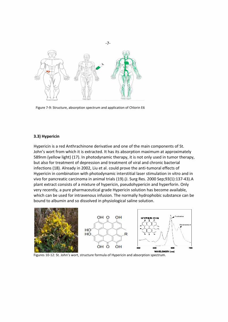
3.3) Hypericin
Hypericin is a red Anthrachinone derivative and one of the main components of St.
John’s wort from which it is extracted. It has its absorption maximum at approximately
589nm (yellow light) (17). In photodynamic therapy, it is not only used in tumor therapy,
but also for treatment of depression and treatment of viral and chronic bacterial
infections (18). Already in 2002, Liu et al. could prove the anti-tumoral effects of
Hypericin in combination with photodynamic interstitial laser stimulation in vitro and in
vivo for pancreatic carcinoma in animal trials (19).(J. Surg Res. 2000 Sep;93(1):137-43).A
plant extract consists of a mixture of hypericin, pseudohypericin and hyperforin. Only
very recently, a pure pharmaceutical grade Hypericin solution has become available,
which can be used for intravenous infusion. The normally hydrophobic substance can be
bound to albumin and so dissolved in physiological saline solution.
Figures 10-12: St. John’s wort, structure formula of Hypericin and absorption spectrum.
-8-
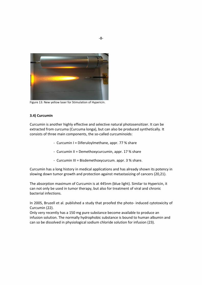
Figure 13: New yellow laser for Stimulation of Hypericin.
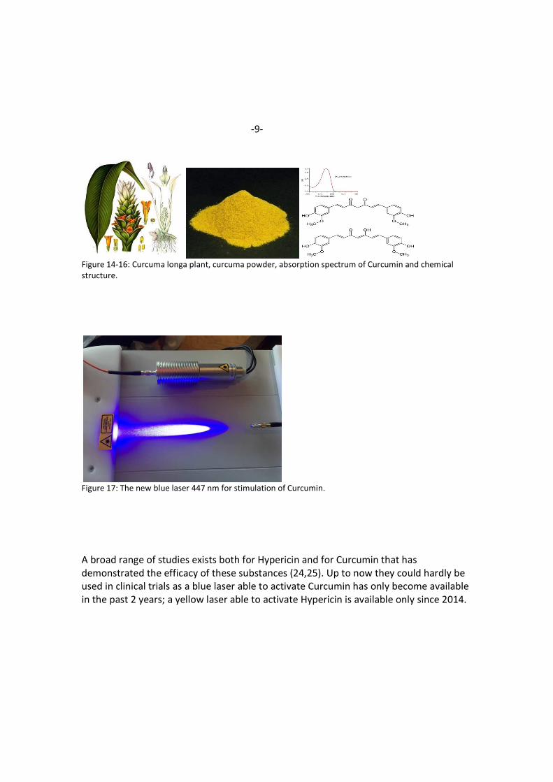
3.4) Curcumin
Curcumin is another highly effective and selective natural photosensitizer. It can be
extracted from curcuma (Curcuma longa), but can also be produced synthetically. It
consists of three main components, the so-called curcuminoids:
- Curcumin I = Diferuloylmethane, appr. 77 % share
- Curcumin II = Demethoxycurcumin, appr. 17 % share
- Curcumin III = Bisdemethoxycurcum. appr. 3 % share.
Curcumin has a long history in medical applications and has already shown its potency in
slowing down tumor growth and protection against metastasizing of cancers (20,21).
The absorption maximum of Curcumin is at 445nm (blue light). Similar to Hypericin, it
can not only be used in tumor therapy, but also for treatment of viral and chronic
bacterial infections.
In 2005, Bruzell et al. published a study that proofed the photo- induced cytotoxicity of
Curcumin (22).
Only very recently has a 150 mg pure substance become available to produce an
infusion solution. The normally hydrophobic substance is bound to human albumin and
can so be dissolved in physiological sodium chloride solution for infusion (23).
-9-
Figure 14-16: Curcuma longa plant, curcuma powder, absorption spectrum of Curcumin and chemical
structure.
Figure 17: The new blue laser 447 nm for stimulation of Curcumin.
A broad range of studies exists both for Hypericin and for Curcumin that has
demonstrated the efficacy of these substances (24,25). Up to now they could hardly be
used in clinical trials as a blue laser able to activate Curcumin has only become available
in the past 2 years; a yellow laser able to activate Hypericin is available only since 2014.
-10-
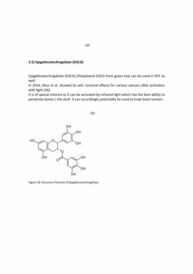
3.5) Epigallocatechingallate (EGCG)
Epigallocatechingallate (EGCG) (Polyphenol EGCG from green tea) can be used in PDT as
well.
In 2014, Mun et al. showed its anti- tumoral effects for various cancers after activation
with light (26).
It is of special interest as it can be activated by infrared light which has the best ability to
penetrate bones / the skull. It can accordingly potentially be used to treat brain tumors.
-10-
Figure 18: Structure formula of Epigallocatechingallate
-11-
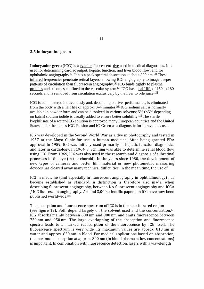
3.5 Indocyanine green
Indocyanine green (ICG) is a cyanine fluorescent dye used in medical diagnostics. It is used for determining cardiac output, hepatic function, and liver blood flow, and for ophthalmic angiography.[2] It has a peak spectral absorption at about 800 nm.[3] These infrared frequencies penetrate retinal layers, allowing ICG angiography to image deeper patterns of circulation than fluorescein angiography.[4] ICG binds tightly to plasma proteins and becomes confined to the vascular system.[2] ICG has a half-life of 150 to 180 seconds and is removed from circulation exclusively by the liver to bile juice.[2]
ICG is administered intravenously and, depending on liver performance, is eliminated from the body with a half life of approx. 3–4 minutes.[6] ICG sodium salt is normally available in powder form and can be dissolved in various solvents; 5% (<5% depending on batch) sodium iodide is usually added to ensure better solubility.[7] The sterile lyophilisate of a water-ICG solution is approved many European countries and the United States under the names ICG-Pulsion and IC-Green as a diagnostic for intravenous use.
ICG was developed in the Second World War as a dye in photography and tested in
1957 at the Mayo Clinic for use in human medicine. After being granted FDA
approval in 1959, ICG was initially used primarily in hepatic function diagnostics
and later in cardiology. In 1964, S. Schilling was able to determine renal blood flow
using ICG. From 1969, ICG was also used in the research and diagnosis of subretinal
processes in the eye (in the choroid). In the years since 1980, the development of
new types of cameras and better film material or new photometric measuring
devices has cleared away many technical difficulties. In the mean time, the use of
ICG in medicine (and especially in fluorescent angiography in ophthalmology) has
become established as standard. A distinction is therefore also made, when
describing fluorescent angiography, between NA fluorescent angiography and ICGA
/ ICG fluorescent angiography. Around 3,000 scientific papers on ICG have now been
published worldwide.[8]
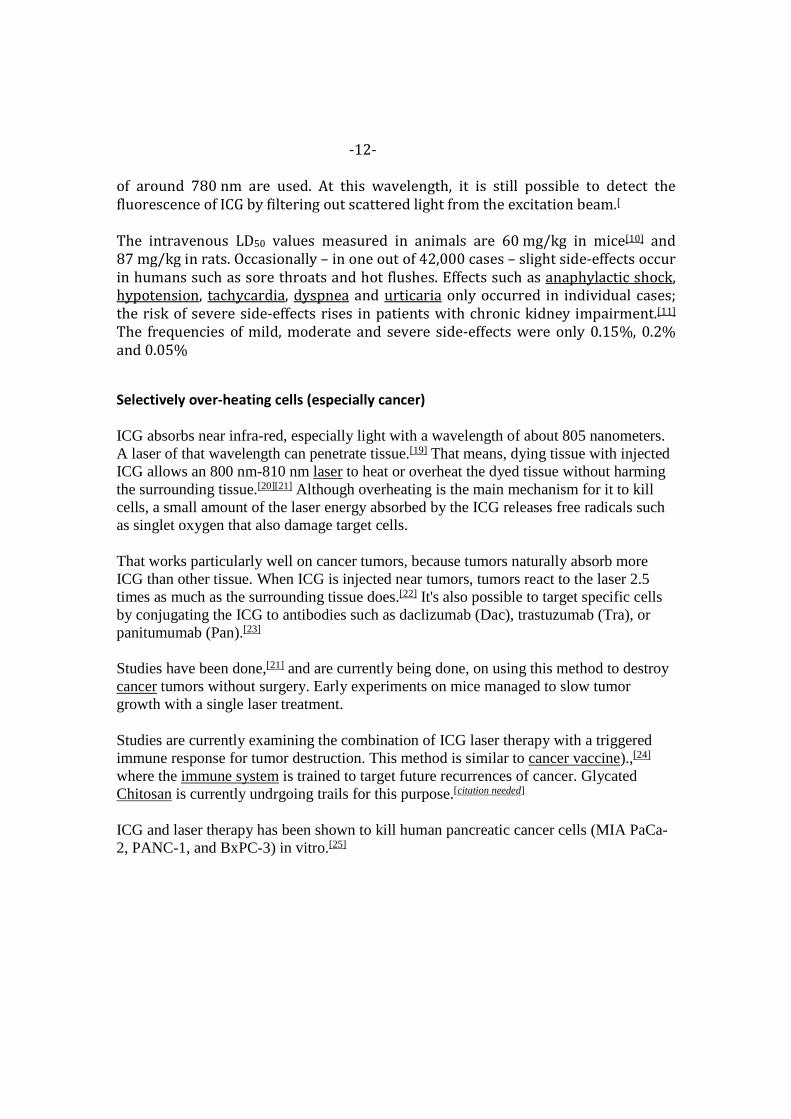
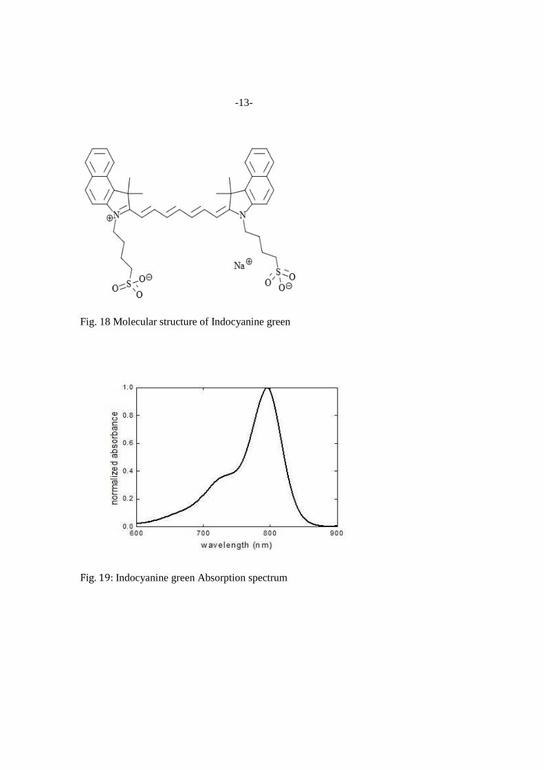
The absorption and fluorescence spectrum of ICG is in the near infrared region
(see figure 19). Both depend largely on the solvent used and the concentration.[9]
ICG absorbs mainly between 600 nm and 900 nm and emits fluorescence between
750 nm and 950 nm. The large overlapping of the absorption and fluorescence
spectra leads to a marked reabsorption of the fluorescence by ICG itself. The
fluorescence spectrum is very wide. Its maximum values are approx. 810 nm in
water and approx. 830 nm in blood. For medical applications based on absorption,
the maximum absorption at approx. 800 nm (in blood plasma at low concentrations)
is important. In combination with fluorescence detection, lasers with a wavelength
-12-
of around 780 nm are used. At this wavelength, it is still possible to detect the
fluorescence of ICG by filtering out scattered light from the excitation beam.[
The intravenous LD50 values measured in animals are 60 mg/kg in mice[10] and
87 mg/kg in rats. Occasionally – in one out of 42,000 cases – slight side-effects occur
in humans such as sore throats and hot flushes. Effects such as anaphylactic shock,
hypotension, tachycardia, dyspnea and urticaria only occurred in individual cases;
the risk of severe side-effects rises in patients with chronic kidney impairment.[11]
The frequencies of mild, moderate and severe side-effects were only 0.15%, 0.2%
and 0.05%
Selectively over-heating cells (especially cancer)
ICG absorbs near infra-red, especially light with a wavelength of about 805 nanometers. A laser of that wavelength can penetrate tissue.[19] That means, dying tissue with injected ICG allows an 800 nm-810 nm laser to heat or overheat the dyed tissue without harming the surrounding tissue.[20][21] Although overheating is the main mechanism for it to kill cells, a small amount of the laser energy absorbed by the ICG releases free radicals such as singlet oxygen that also damage target cells.
That works particularly well on cancer tumors, because tumors naturally absorb more ICG than other tissue. When ICG is injected near tumors, tumors react to the laser 2.5 times as much as the surrounding tissue does.[22] It's also possible to target specific cells by conjugating the ICG to antibodies such as daclizumab (Dac), trastuzumab (Tra), or panitumumab (Pan).[23]
Studies have been done,[21] and are currently being done, on using this method to destroy cancer tumors without surgery. Early experiments on mice managed to slow tumor growth with a single laser treatment.
Studies are currently examining the combination of ICG laser therapy with a triggered immune response for tumor destruction. This method is similar to cancer vaccine).,[24] where the immune system is trained to target future recurrences of cancer. Glycated Chitosan is currently undrgoing trails for this purpose.[citation needed]
ICG and laser therapy has been shown to kill human pancreatic cancer cells (MIA PaCa-2, PANC-1, and BxPC-3) in vitro.[25]
-13-
Fig. 18 Molecular structure of Indocyanine green
Fig. 19: Indocyanine green Absorption spectrum
-14-
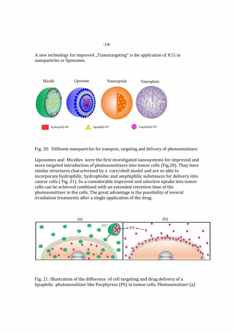
A new technology for improved „Tumortargeting“ is the application of ICG in nanoparticles or liposomes.
Fig. 20: Different nanoparticles for transport, targeting and delivery of photosensitizers
Liposomes and Micelles were the first investigated nanosystems for improved and
more targeted introduction of photosensitizers into tumor cells (Fig.20). They have
similar structures characterized by a core/shell model and are so able to
incorporate hydrophilic, hydrophobic and amphiphilic substances for delivery into
cancer cells ( Fig. 21). So a considerable improved and selective uptake into tumor
cells can be achieved combined with an extended retention time of the
photosensitizer in the cells. The great advantage is the possibility of several
irradiation treatments after a single application of the drug.
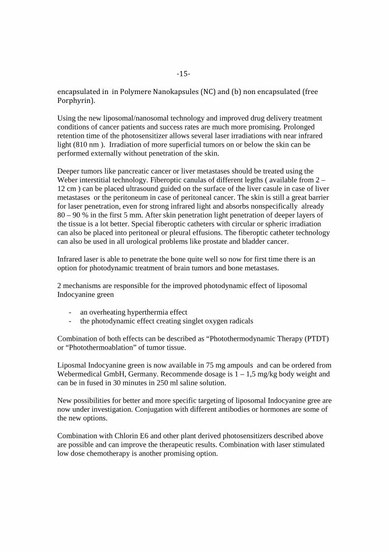
Fig. 21: Illustration of the difference of cell targeting and drug delivery of a
lipophilic photosensitizer like Porphyrins (PS) in tumor cells. Photosensitizer (a)
-15-
encapsulated in in Polymere Nanokapsules (NC) and (b) non encapsulated (free
Porphyrin).
Using the new liposomal/nanosomal technology and improved drug delivery treatment conditions of cancer patients and success rates are much more promising. Prolonged retention time of the photosensitizer allows several laser irradiations with near infrared light (810 nm ). Irradiation of more superficial tumors on or below the skin can be performed externally without penetration of the skin.
Deeper tumors like pancreatic cancer or liver metastases should be treated using the Weber interstitial technology. Fiberoptic canulas of different legths ( available from 2 – 12 cm ) can be placed ultrasound guided on the surface of the liver casule in case of liver metastases or the peritoneum in case of peritoneal cancer. The skin is still a great barrier for laser penetration, even for strong infrared light and absorbs nonspecifically already 80 – 90 % in the first 5 mm. After skin penetration light penetration of deeper layers of the tissue is a lot better. Special fiberoptic catheters with circular or spheric irradiation can also be placed into peritoneal or pleural effusions. The fiberoptic catheter technology can also be used in all urological problems like prostate and bladder cancer.
Infrared laser is able to penetrate the bone quite well so now for first time there is an option for photodynamic treatment of brain tumors and bone metastases.
2 mechanisms are responsible for the improved photodynamic effect of liposomal Indocyanine green
- an overheating hyperthermia effect - the photodynamic effect creating singlet oxygen radicals
Combination of both effects can be described as “Photothermodynamic Therapy (PTDT) or “Photothermoablation” of tumor tissue.
Liposmal Indocyanine green is now available in 75 mg ampouls and can be ordered from Webermedical GmbH, Germany. Recommende dosage is 1 – 1,5 mg/kg body weight and can be in fused in 30 minutes in 250 ml saline solution.
New possibilities for better and more specific targeting of liposomal Indocyanine gree are now under investigation. Conjugation with different antibodies or hormones are some of the new options.
Combination with Chlorin E6 and other plant derived photosensitizers described above are possible and can improve the therapeutic results. Combination with laser stimulated low dose chemotherapy is another promising option.
-16-
4) Immunological effects of photodynamic therapy
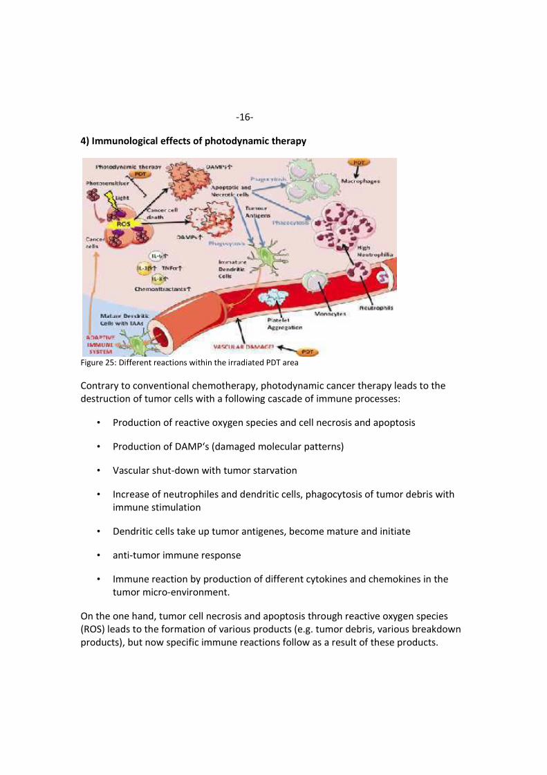
Figure 25: Different reactions within the irradiated PDT area
Contrary to conventional chemotherapy, photodynamic cancer therapy leads to the
destruction of tumor cells with a following cascade of immune processes:
• Production of reactive oxygen species and cell necrosis and apoptosis
• Production of DAMP‘s (damaged molecular patterns)
• Vascular shut-down with tumor starvation
• Increase of neutrophiles and dendritic cells, phagocytosis of tumor debris with
immune stimulation
• Dendritic cells take up tumor antigenes, become mature and initiate
• anti-tumor immune response
• Immune reaction by production of different cytokines and chemokines in the
tumor micro-environment.
On the one hand, tumor cell necrosis and apoptosis through reactive oxygen species
(ROS) leads to the formation of various products (e.g. tumor debris, various breakdown
products), but now specific immune reactions follow as a result of these products.
-17-
The tumor vessels supplying the tumors will be capped (vascular shut- down of neo-
angiogenesis) and tumor tissue is being starved. Proliferation and activation of
macrophages takes place as well, together with maturation of dendritic cells. The latter
will absorb the tumor antigens and produce a specific immune response with
production of cytokines and chemokines both in direct proximity to the tumor tissue
and circulating in the rest of the body.
5) Combination of PDT with other methods
The literature shows different options to combine PDT with other treatment regimes:
- Combination with traditional chemotherapy (27)
- Combination with light sensitive chemo drugs (using chemotherapeutic drugs as
photosensitizers) (28)
- Combination with antioxidants (29)
- Combination with antiangionesis inhibitors (30)
- Combination with Cox-2 inhibitors (31)
- Combination with antibodies (32)
- Combination with different natural compounds
- Combination with immunotherapy (33)
In the following, only combinations with low-dose chemotherapeutic drugs and
immunotherapy are presented as their potential to improve the results of PDTT is
mostly significant.
5.1 Combination with traditional chemotherapy: Chemotherapeutic drugs used as
photosensitizers in low dose
In case of big tumor masses or multiple metastases even the described new methods of
PDT are often not effective enough. The author’s own experience has shown that
tumors or metastases larger than 3-4 cm mostly cannot be destroyed by PDT alone or
maybe only with multiple follow-up treatments.
The conventional procedures in such cases are well known: If possible, tumor mass has
-18-
to be reduced by surgical procedures. In many cases, especially for diagnosed multiple
metastases, chemotherapy has to be used as well. However, the side effects of high
dose chemotherapy are well known. In this context, a new approach can contribute to
the alleviation of the destructive and immune suppressing effects of traditional
chemotherapy.
Like the above described natural derived photosensitizers also the different
chemotherapeutic drugs have a specific light absorption spectrum and their efficacy can
be enhanced by stimulation with appropriate laser light (using chemotherapeutic drugs
as photosensitizers). As a consequence, the drug´s dose can be reduced significantly and
the well known side effects are alleviated. Studies show that chemo drugs in a low dose
even have the potential to enhance the immune system in combination with PDT (34).
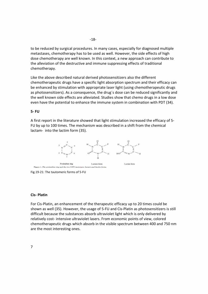
5- FU
A first report in the literature showed that light stimulation increased the efficacy of 5-
FU by up to 100 times. The mechanism was described in a shift from the chemical
lactam- into the lactim form (35).
Fig.19-21: The tautomeric forms of 5-FU
Cis- Platin
For Cis-Platin, an enhancement of the therapeutic efficacy up to 20 times could be
shown as well (35). However, the usage of 5-FU and Cis-Platin as photosensitizers is still
difficult because the substances absorb ultraviolet light which is only delivered by
relatively cost- intensive ultraviolet lasers. From economic points of view, colored
chemotherapeutic drugs which absorb in the visible spectrum between 400 and 750 nm
are the most interesting ones.
7
-19-
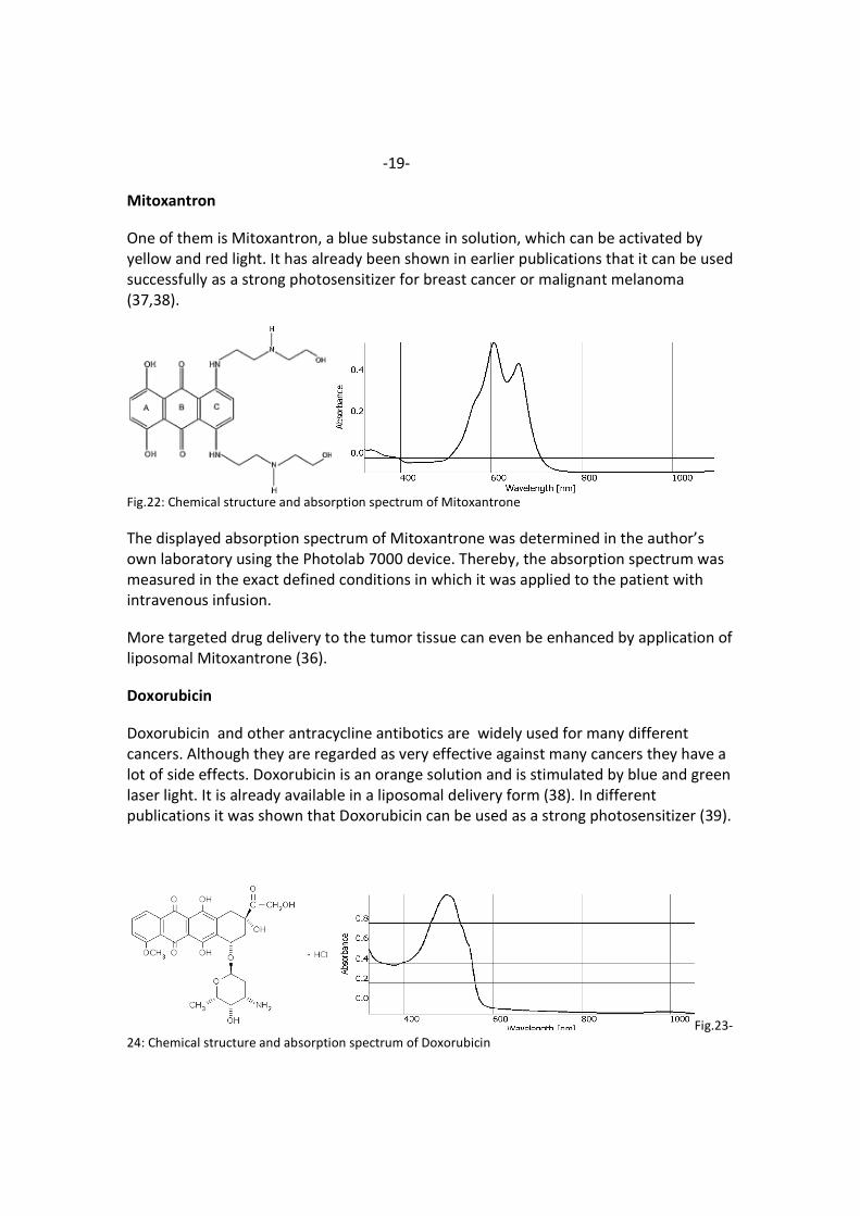
Mitoxantron
One of them is Mitoxantron, a blue substance in solution, which can be activated by
yellow and red light. It has already been shown in earlier publications that it can be used
successfully as a strong photosensitizer for breast cancer or malignant melanoma
(37,38).
Fig.22: Chemical structure and absorption spectrum of Mitoxantrone
The displayed absorption spectrum of Mitoxantrone was determined in the author’s
own laboratory using the Photolab 7000 device. Thereby, the absorption spectrum was
measured in the exact defined conditions in which it was applied to the patient with
intravenous infusion.
More targeted drug delivery to the tumor tissue can even be enhanced by application of
liposomal Mitoxantrone (36).
Doxorubicin
Doxorubicin and other antracycline antibotics are widely used for many different
cancers. Although they are regarded as very effective against many cancers they have a
lot of side effects. Doxorubicin is an orange solution and is stimulated by blue and green
laser light. It is already available in a liposomal delivery form (38). In different
publications it was shown that Doxorubicin can be used as a strong photosensitizer (39).
Fig.23-
24: Chemical structure and absorption spectrum of Doxorubicin
-20-
5.2 Hyperbaric oxygenation
The success of photodynamic tumor therapies ultimately depends on the availability of
tissue oxygen as the final tumor toxicity is triggered by oxygen radicals.
Unfortunately, tumor tissue is often poor in tissue oxygen. PDT very soon expends the
available oxygen. Additive oxygen therapies that increase the amount of available
oxygen can thus improve the success of photodynamic tumor therapies.
Therefore, oxygen devices by Regelsberger can be used for intravenous oxygen
therapies (..).
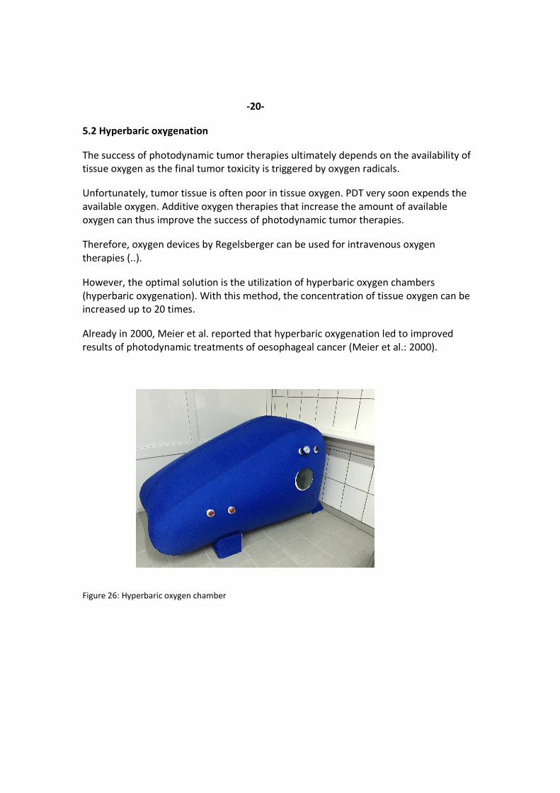
However, the optimal solution is the utilization of hyperbaric oxygen chambers
(hyperbaric oxygenation). With this method, the concentration of tissue oxygen can be
increased up to 20 times.
Already in 2000, Meier et al. reported that hyperbaric oxygenation led to improved
results of photodynamic treatments of oesophageal cancer (Meier et al.: 2000).
Figure 26: Hyperbaric oxygen chamber
-21-
5.3) GcMAF
Another recommendation to support immune reactions after PDT is immunization with
macrophage activating substances, e.g. GcMAF from Japan. It can be injected
subcutaneously for several months after the PDT treatment. It also can be injected
directly into the treated tumor area to enhance the immunological effect of the PDT.
Macrophage activation factor (MAF) are glycoproteins that increase macrophage activity
and transform them into natural killer (NK) cells. Vitamin DBP (Gc Protein) is the primary
MAF. The glycosylated Gc Protein is the best MAF. Unfortunately, tumor cells produce
an enzyme which destroys the body’s own GcMAF by a specific enzyme named
Nagalase.
Nagalase is an enzyme produced in small amount by liver cells but in large amounts by
cancer cells. Nagalase deglycosylates the Gc-protein and so has an immunosuppressive
effect. Nagalase is also produced by different viruses, bacteria and fungi. GcMAF
macrophage activation therapy is useful in the treatment of many diseases, such as
cancer, HIV AIDS, Hepatitis B virus (HBV), Hepatitis C virus (HCV), Herpes Simplex virus
(HSV), Tuberculosis, Pneumonia infection, Epstein-Barr virus (EBV), cystitis/urinary tract
infection. The therapeutic effects of GcMAF were first described by Nobuto Yamoto from
Japan.
General goals of GcMAF therapy are to:
• Improve well-being and quality of life (QOL)
• Return the patient to good health so that they are able to participate in
regular lifestyle activities
• Achieve long term survival
• Enhance the effect of other therapies
• Repair the immune system
• Increase the number of monocytes (macrophages) and activate them to
destroy cancer cells, viruses, bacteria and other pathogens in the body
• Increase the rate of maturation of dendritic cells (DCs)
Different sources of GcMAF are available today. There are many claims for GcMAF
therapy in the literature, especially cancer, autoimmune diseases and autism.
However more research is necessary to approve the therapeutic effects.
-22-
6) Treatment examples
6.1) Treatment of metastasizing small intestine cancer with Chlorin E6
In a first pilot study, the author of this article employed the combination of “systemic
PDT” via intravenously applied photosensitizers and subsequent intravenous irradiation
as one component and interstitial PDT with fiber optic laser catheters and direct laser
activation as the other component to treat a patient with small intestine cancer and 4
liver metastases from May 2010 to May 2011.
The small intestine cancer had been removed operationally in 2009, but 2 new liver
metastases developed soon.
The patient had received chemotherapy with different chemotherapeutical agents, but
the treatments did not lead to significant improvements and had strong side effects.
A first systemic PDT was given in May 2010 that led to a surprising improvement of life
quality and after a second treatment course in June 2010 both metastases were no
longer visible in a July 2010 MRI. However, in December 2010 the liver metastases
reappeared. In January 2011 the patient was treated with 3 sessions of systemic PDT.
Despite this the metastases were still growing slowly.
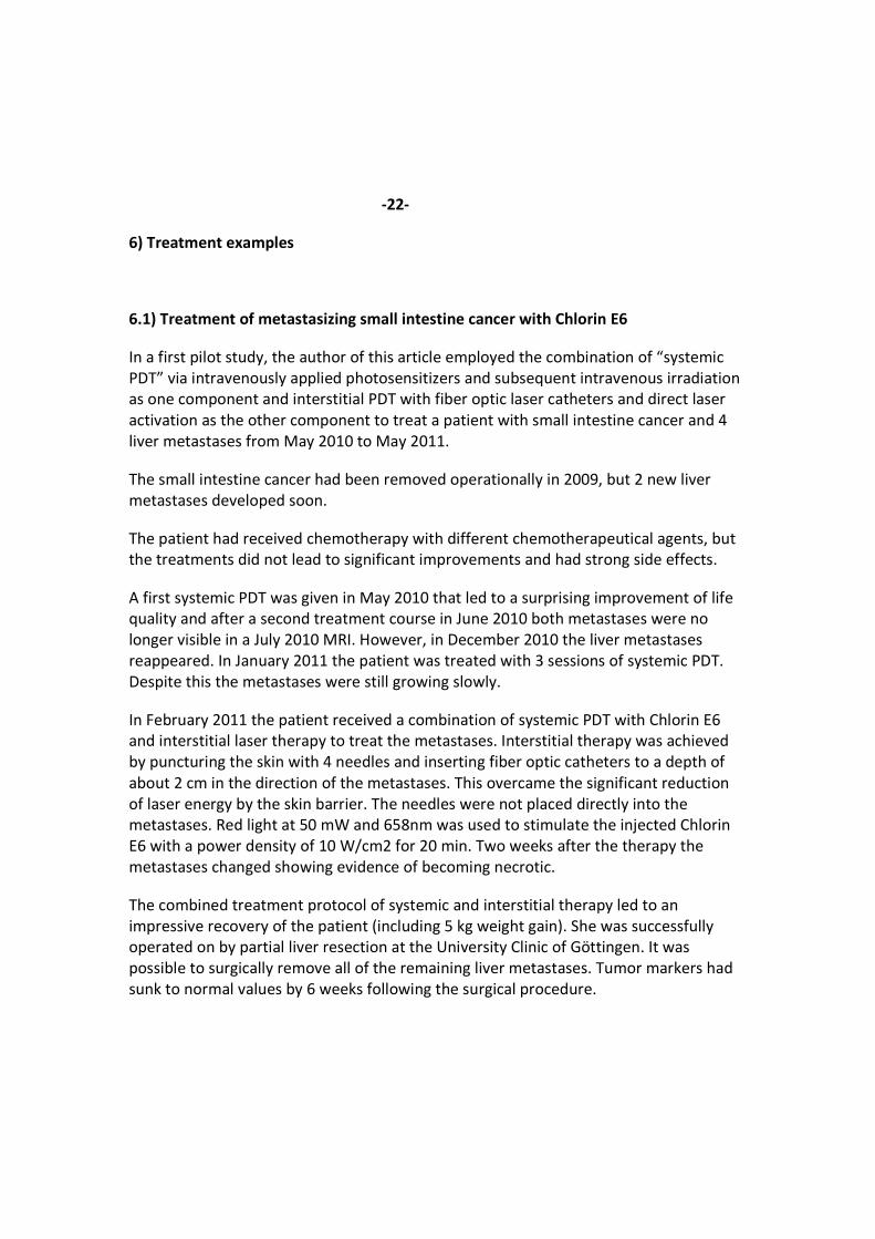
In February 2011 the patient received a combination of systemic PDT with Chlorin E6
and interstitial laser therapy to treat the metastases. Interstitial therapy was achieved
by puncturing the skin with 4 needles and inserting fiber optic catheters to a depth of
about 2 cm in the direction of the metastases. This overcame the significant reduction
of laser energy by the skin barrier. The needles were not placed directly into the
metastases. Red light at 50 mW and 658nm was used to stimulate the injected Chlorin
E6 with a power density of 10 W/cm2 for 20 min. Two weeks after the therapy the
metastases changed showing evidence of becoming necrotic.
The combined treatment protocol of systemic and interstitial therapy led to an
impressive recovery of the patient (including 5 kg weight gain). She was successfully
operated on by partial liver resection at the University Clinic of Göttingen. It was
possible to surgically remove all of the remaining liver metastases. Tumor markers had
sunk to normal values by 6 weeks following the surgical procedure.
-23-
Figure 27: First pilot study with combination of systemic and interstitial PDT
6.2) Treatment of metastasizing pancreas cancer
Another patient (76 years) had been suffering from a cancer in the head of the pancreas
and that was operated on in August 2012. Only a few months after the cancer
reoccurred in the form of malignant ascites. Chemotherapy with Gemzar (Gemcitabine)
had to be abandoned due to intolerance to the side effects by the patient. At this point
the ascites was out of control, malignant pleural effusions developed as well and a
secondary tumor-related anemia set in that required ongoing blood transfusions.
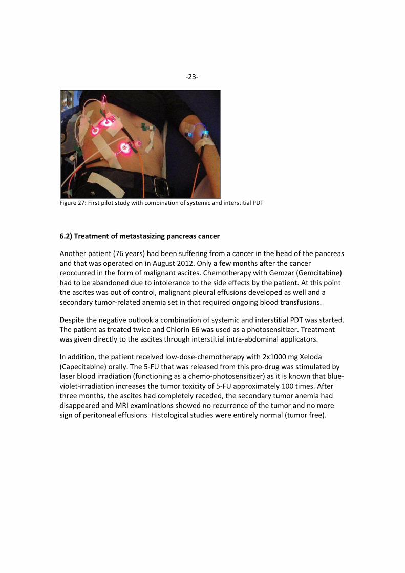
Despite the negative outlook a combination of systemic and interstitial PDT was started.
The patient as treated twice and Chlorin E6 was used as a photosensitizer. Treatment
was given directly to the ascites through interstitial intra-abdominal applicators.
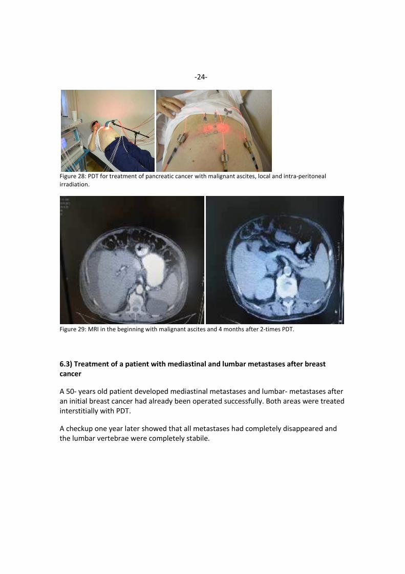
In addition, the patient received low-dose-chemotherapy with 2x1000 mg Xeloda
(Capecitabine) orally. The 5-FU that was released from this pro-drug was stimulated by
laser blood irradiation (functioning as a chemo-photosensitizer) as it is known that blue-
violet-irradiation increases the tumor toxicity of 5-FU approximately 100 times. After
three months, the ascites had completely receded, the secondary tumor anemia had
disappeared and MRI examinations showed no recurrence of the tumor and no more
sign of peritoneal effusions. Histological studies were entirely normal (tumor free).
-24-
Figure 28: PDT for treatment of pancreatic cancer with malignant ascites, local and intra-peritoneal
irradiation.
Figure 29: MRI in the beginning with malignant ascites and 4 months after 2-times PDT.
6.3) Treatment of a patient with mediastinal and lumbar metastases after breast
cancer
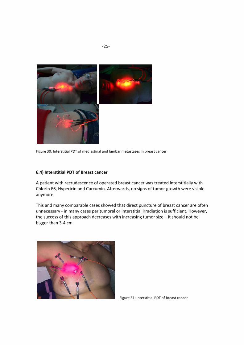
A 50- years old patient developed mediastinal metastases and lumbar- metastases after
an initial breast cancer had already been operated successfully. Both areas were treated
interstitially with PDT.
A checkup one year later showed that all metastases had completely disappeared and
the lumbar vertebrae were completely stabile.
-25-
Figure 30: Interstitial PDT of mediastinal and lumbar metastases in breast cancer
6.4) Interstitial PDT of Breast cancer
A patient with recrudescence of operated breast cancer was treated interstitially with
Chlorin E6, Hypericin and Curcumin. Afterwards, no signs of tumor growth were visible
anymore.
This and many comparable cases showed that direct puncture of breast cancer are often
unnecessary - in many cases peritumoral or interstitial irradiation is sufficient. However,
the success of this approach decreases with increasing tumor size – it should not be
bigger than 3-4 cm.
Figure 31: Interstitial PDT of breast cancer
-26-
Another patient from New York was treated as explained above. She refused
conventional treatments.At the time of treatment the tumor had a size of 5 cm, but no
metastases were evident. 6 weeks after the treatment no sign of cancer was evident.
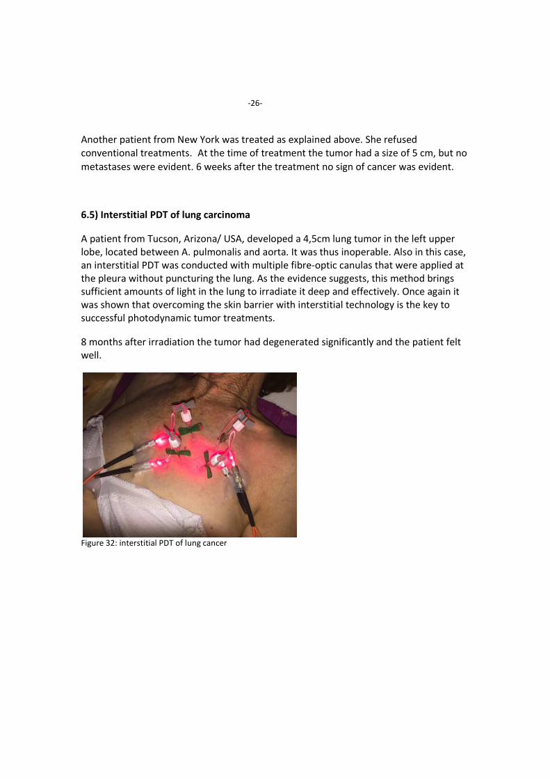
6.5) Interstitial PDT of lung carcinoma
A patient from Tucson, Arizona/ USA, developed a 4,5cm lung tumor in the left upper
lobe, located between A. pulmonalis and aorta. It was thus inoperable. Also in this case,
an interstitial PDT was conducted with multiple fibre-optic canulas that were applied at
the pleura without puncturing the lung. As the evidence suggests, this method brings
sufficient amounts of light in the lung to irradiate it deep and effectively. Once again it
was shown that overcoming the skin barrier with interstitial technology is the key to
successful photodynamic tumor treatments.
8 months after irradiation the tumor had degenerated significantly and the patient felt
well.
Figure 32: interstitial PDT of lung cancer

-27-
6.6) PDT for urological cases
It is well known that prostate cancer is one of the most frequent malignancies in men.
Conventional treatments consist of operations and/or radiation with severe side effects.
In addition, there are thousands of patients who undergo disputable biopsies because of
borderline of high PSA levels that are done for screening of prostate cancer. These
patients live in permanent fear of prostate cancer.
For treatment of prostate cancer with photodynamic therapy a special catheter system
with integrated fiber optic technology has been developed. A thin fiber optic that is
attached to a transparent permanent catheter emits radiation in the area of the
prostate that penetrates the whole organ. Apart from its usage for prostate cancer, the
treatment is also suitable for chronic prostatitis.
Whether an improvement can also be achieved in treatments of benign prostate
enlargement is not clarified yet, but previous positive experiences with cases like this
raise hopes that good results can also be achieved in this field of application. The new
catheter-facilitated fiber optic procedure can also be used for the treatment of bladder
cancer in a similar manner. In this case a ball-shaped steel fiber optic is inserted in the
catheter.
Procedure:
Patients receive the following infusions: 90mg Chlorin E6, 10mg Hypericin and 150mg
Curcumin. 3 hours later, the photodynamic intra-prostatic irradiation therapy is given.
For 20 minutes irradiation with red light (658nm), yellow light (589nm) and blue light
(447nm) is given each for a total of 60 minutes. It is applied through a ball- shaped
radiating probe that is attached to a transparent silicon catheter. It leads to the
spherical irradiation of the bladder from inside.
Figure 33: Spherically radiating probe for intravesical irradiation of bladder cancer
-28-
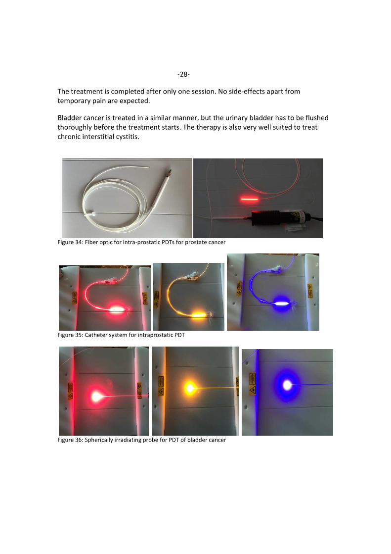
The treatment is completed after only one session. No side-effects apart from
temporary pain are expected.
Bladder cancer is treated in a similar manner, but the urinary bladder has to be flushed
thoroughly before the treatment starts. The therapy is also very well suited to treat
chronic interstitial cystitis.
Figure 34: Fiber optic for intra-prostatic PDTs for prostate cancer
Figure 35: Catheter system for intraprostatic PDT
Figure 36: Spherically irradiating probe for PDT of bladder cancer
-29-
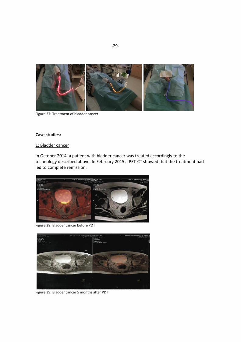
Figure 37: Treatment of bladder cancer
Case studies:
1: Bladder cancer
In October 2014, a patient with bladder cancer was treated accordingly to the
technology described above. In February 2015 a PET-CT showed that the treatment had
led to complete remission.
Figure 38: Bladder cancer before PDT
Figure 39: Bladder cancer 5 months after PDT
-30-
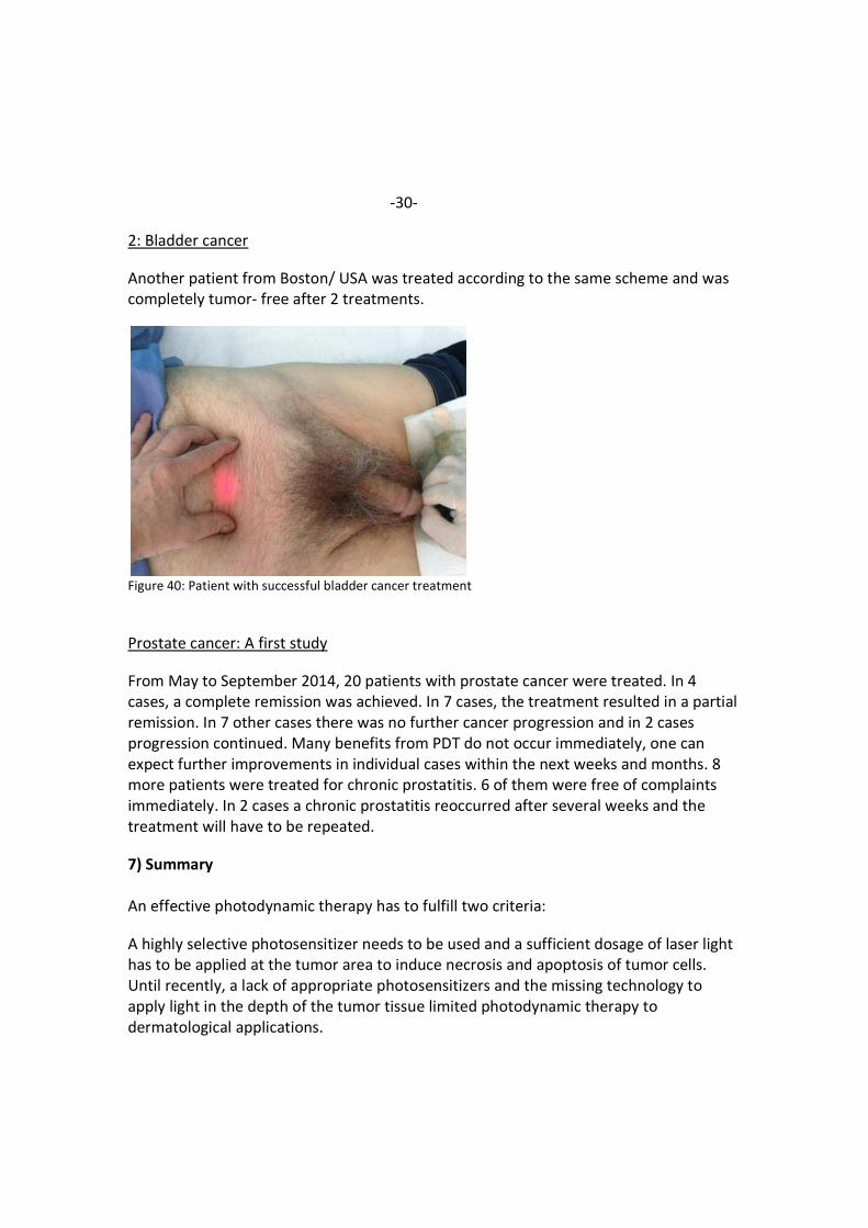
2: Bladder cancer
Another patient from Boston/ USA was treated according to the same scheme and was
completely tumor- free after 2 treatments.
Figure 40: Patient with successful bladder cancer treatment
Prostate cancer: A first study
From May to September 2014, 20 patients with prostate cancer were treated. In 4
cases, a complete remission was achieved. In 7 cases, the treatment resulted in a partial
remission. In 7 other cases there was no further cancer progression and in 2 cases
progression continued. Many benefits from PDT do not occur immediately, one can
expect further improvements in individual cases within the next weeks and months. 8
more patients were treated for chronic prostatitis. 6 of them were free of complaints
immediately. In 2 cases a chronic prostatitis reoccurred after several weeks and the
treatment will have to be repeated.
7) Summary
An effective photodynamic therapy has to fulfill two criteria:
A highly selective photosensitizer needs to be used and a sufficient dosage of laser light
has to be applied at the tumor area to induce necrosis and apoptosis of tumor cells.
Until recently, a lack of appropriate photosensitizers and the missing technology to
apply light in the depth of the tumor tissue limited photodynamic therapy to
dermatological applications.
-31-
Today, a number of highly selective photosensitizers (e.g. Chlorin E6, Hypericin,
Curcumin and Epigallocatechingallate) and a new fiber- optic catheter technology that
facilitates the application of laser light deep in the tissue (by interstitial therapy) set the
stage for the establishment of PDT as a new treatment option in oncology.
WeberMedical, a German company, manufactures the laser system that is used with the
new fiber optic catheter technology. Many immune reactions go hand in hand with the
primary effect of the introduced treatment regime, the destruction of tumor cells:
Through intravenous blood irradiation circulating tumor cells and tumor stem cells can
be destroyed and concomitant infections are treated after a photosensitizer has been
administered intravenously.
Even though the results of PDT are very promising by itself, the therapy should be
regarded as a complementary approach to traditional chemo therapy, not only as an
alternative therapy. PDT contributes to lessen the side effects of chemotherapies.
Additionally, many chemotherapeutic agents have an absorption spectrum that allows
laser treatments to stimulate them as well. In the above mentioned cases where low-
dose chemotherapy was used with Capecitabine or Cis-Platin both chemotherapeutics
were used as photosensitizers. If they are used in low dosages and in combination with
light they can even have immune- stimulating effects.
PDT is a very promising new treatment option in oncology; it is easy to apply and had
only been limited due to technological reasons in the past. As shown in this article,
these limitations have now been overcome and PDT will in all probability be established
as a mainstream treatment for cancer soon.
Address of the author:
Dr. med. Dipl. Chem. Michael Weber, Sohnreystrasse 4 37697 Lauenförde
Email: [email protected]
-32-
Literature:
1. Allison, R.R., Sibata, C. H.: Photodynamic Therapy; Chapter 24. Perez and Brady’s
Principles and Practice of Radiation Oncology, 5th Edition, 2008
2. Allison, R.R., Sibata, C. H.: Oncologic photodynamic therapy photosensitizers: A
clinical review. Photodiagnosis and Photodynamic Therapy 7, 61-75, 2010
3. Bown et al.: Photodynamic therapy for cancer of the pancreas
In: Gut 2002, April, 50(4):549-557
4. Braathen, L. R., Hunger, R. E., Kernland Lang, K.: Photodynamische Therapie (PDT),
Kap. 4.5. Tumoren der Haut, 2010, Georg Thieme Verlag KG
5. Bruzell, E. Morisbak, H. Tønnesen, 2005 Studies on curcumin and curcuminoids. XXIX.
Photoinduced cytotoxicity of curcumin in selected aqueous preparations. Photochemical
& photobiological sciences. 4 7 (July), 523 530 , 0147-4905X
6. Calin, M. A., Parasca, S. V.: Photodynamic therapy in oncology. Journal of
Optoelectronics and advanced materials Vol. 8, No. 3, June 2006, p. 1173-1179
7. Eichler, K., Engelmann, K., Mack, M. G., Straub, R., Vogl, T. J., Zangos, S.: Interstitielle
Photodynamische Lasertherapie zur Behandlung von Lebermetastasen: Erste Ergebnisse
einer in vivo Phase I-Studie. Fortschr Röntgenstr 2003; 175:682-687, Georg Thieme
Verlag Stuttgart
8. Eichler, K., Engelmann, K., Herzog, C., Mack, M. G., Thalhammer, A., Vogl, T. J.,
Zangos, S.: Interstitial photodynamic laser therapy in interventional oncology. Eur Radiol
(2004) 14:1063–1073

-33-
9. Jaeger et al.: A New Finite Element Approach for Near Real-Time Simulation of Light
Propagation in Locally Advanced Head and Neck Tumors. In: American Journal for
Neuroradiology (AJNR Am J Neuroradiol 26:1193–1200, May 2005.).
10. Kaplan et al: Systemic Photodynamic Therapy in the Combined Therapy of Patients
with Malignant Neoplasms with Metastases; Book of Abstracts, int. Laser conference
Helsinki 2008
11. Liu et al.: Hypericin and photodynamic therapy decreases human pancreatic cancer
in vitro and in vivo. In: (J Surg Res. 2000 Sep;93(1):137-43).
(22) (Photochemical & photobiological sciences. 4 7 (July), 523 530 , 0147-4905)