Embed Size (px)
Citation preview
Neurilemmoma is a benign neoplasm that originates fromthe Schwann cells that cover the peripheral nerves.1,2 This tumorpresents as a painless, slow-growing mass and it may develop atany age.2 Neurilemmomas are most frequently located in the softtissues of the head and neck, but they are also encountered onthe flexor surfaces of the upper and lower extremities.2,3 Intrao-sseous neurilemmoma is extremely rare and fewer than 200 exam-ples of this entity have been described in the literature. It accountsfor less than 0.2% of all primary bone tumors.4 We present herea rare case of intraosseous neurilemmoma of the mandible in apatient who was substantially older than the average neurilem-moma patient and the tumor occurred more anterior than itsusual location in the mandible.
CASE REPORT
A 77-year-old woman presented with a painful swelling inthe right chin, which had evolved over approximately 3 years
and recently become aggravated. A clinical examination revealedan asymmetrical facial appearance due to swelling of the rightchin. No skin color change or sensation of heat was evident. Thepatient did not complain of facial numbness, but she did com-plain of paresthesia of the right lower lip and chin area. She worecomplete dentures on both sides of the jaw. Moreover, althoughshe complained of tenderness on intraoral palpation of the rightlower vestibule, there was no pus discharge. Panoramic radiog-raphy showed a 3×2 cm-sized well-delimitated radiolucentlesion in the anterior region of the right mandibular body (Fig.1A). A block CT scan revealed an expansile, well-defined, uniloc-ular lesion with thinning of the buccal and lingual cortical platesof the right mandibular body (Fig. 1B). Surgical resection ofthe lesion was performed under general anesthesia. After an inci-sion on the buccal mucosa 1 cm away from the lesion, the muco-periosteal flap was elevated and a bone window was made togain access to the lesion. The right mental nerve was sacrificeddue to its adhesion to the lesion. There were no signs of inva-sion to the adjacent bone. The mass and overlying thinned cor-
88
The Korean Journal of Pathology 2009; 43: 88-91DOI: 10.4132/KoreanJPathol.2009.43.1.88
Neurilemmoma (Schwannoma) is a benign nerve sheath tumor that’s composed entirely of well-differentiated Schwann cells. Intraosseous neurilemmomas are rare and they represent lessthan 1% of all benign primary bone tumors. We report here on an additional case of intraosseousneurilemmoma that was located in the mandible of a 77-year-old woman. CT revealed anexpansile, well-defined lesion on the right side of the mandibular body with thinning of the cor-tex. The lesion was surgically removed and it was found to be a 2××1.7 cm-sized, bright yel-lowish, hard mass with hemorrhage and cyst formation. Histologically, the mass was a mod-erately cellular neoplasm and it showed distinct nuclear palisading, numerous Verocay bod-ies and tumor cells that were positively immunohistostained for S-100 protein. Two monthsafter the operation, the patient has remained in a good condition with no signs or symptomsof tumor recurrence.
Key Words : Mandibular neoplasms; Neurilemmoma; Bone neoplasms
Kyu Yun Jang∙∙Woo Sung MoonHo Sung Park
88
Intraosseous Neurilemmoma of the Mandible
- A Case Report -
88 88
Corresponding AuthorHo Sung Park, M.D.Department of Pathology, Chonbuk National University Medical School, San 2-20 Geumam-dong,Deokjin-gu, Jeonju 561-180, Korea Tel: 063-270-3073Fax: 063-270-3135E-mail: [email protected]
*This paper was supported by research funds ofChonbuk National University in 2005.
Department of Pathology, Institute forMedical Science, Chonbuk NationalUniversity Medical School, Jeonju,Korea
Received : August 11, 2008Accepted : September 9, 2008
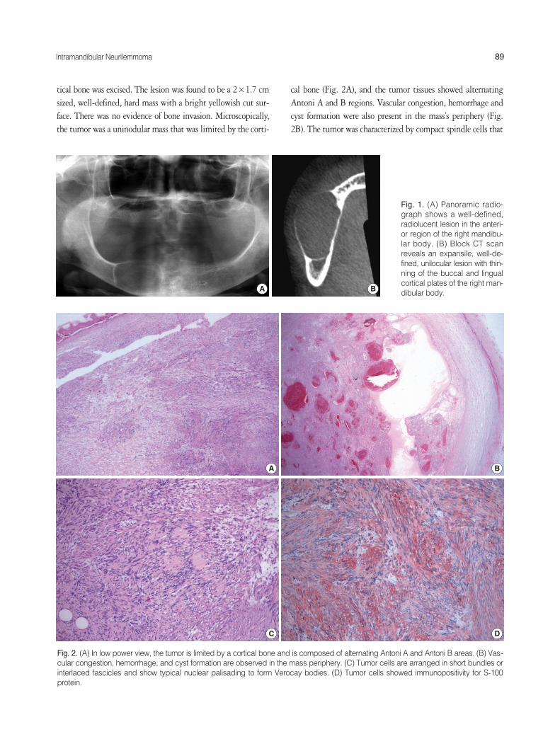
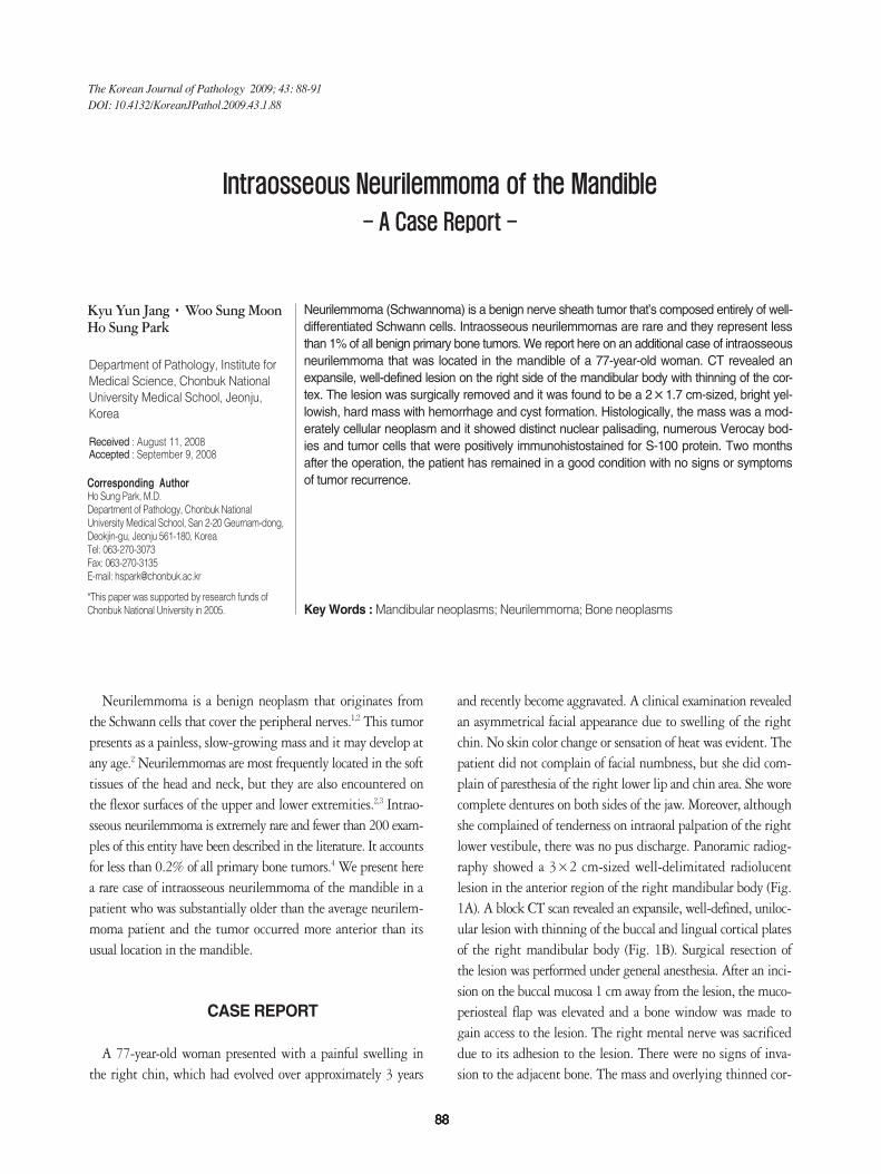
tical bone was excised. The lesion was found to be a 2×1.7 cmsized, well-defined, hard mass with a bright yellowish cut sur-face. There was no evidence of bone invasion. Microscopically,the tumor was a uninodular mass that was limited by the corti-
cal bone (Fig. 2A), and the tumor tissues showed alternatingAntoni A and B regions. Vascular congestion, hemorrhage andcyst formation were also present in the mass’s periphery (Fig.2B). The tumor was characterized by compact spindle cells that
Intramandibular Neurilemmoma 89
A B
Fig. 1. (A) Panoramic radio-graph shows a well-defined,radiolucent lesion in the anteri-or region of the right mandibu-lar body. (B) Block CT scanreveals an expansile, well-de-fined, unilocular lesion with thin-ning of the buccal and lingualcortical plates of the right man-dibular body.
Fig. 2. (A) In low power view, the tumor is limited by a cortical bone and is composed of alternating Antoni A and Antoni B areas. (B) Vas-cular congestion, hemorrhage, and cyst formation are observed in the mass periphery. (C) Tumor cells are arranged in short bundles orinterlaced fascicles and show typical nuclear palisading to form Verocay bodies. (D) Tumor cells showed immunopositivity for S-100protein.
C D
A B

were arranged in short bundles or as interlaced fascicles. In addi-tion, the tumor cells had twisted nuclei and indistinct cytoplas-mic borders, and some of these nuclei formed typical palisadesaround an acellular eosinophilic area (Verocay bodies) (Fig. 2C).Mitotic figures and necrosis were not observed. The tumor cellswere immunopositive for S-100 protein (Fig. 2D) but immu-nonegative for smooth muscle actin and CD34. The histopatho-logic diagnosis was intraosseous neurilemmoma. Two monthsafter the operation, the patient has remained in good conditionwith no signs or symptoms of tumor recurrence.
DISCUSSION
Neurilemmoma is a benign neoplasm that originates fromSchwann cells of the neural sheath. Intraosseous neurilemmo-ma is a rare tumor and it accounts for less than 0.2% of all pri-mary bone tumors.4 However, the mandible is the most com-mon site when encountering an intraosseous neurilemmoma.5-7
In the mandible, the posterior segments of the body and ramusare the most frequent sites of occurrence because of the protractedintraosseous path of the inferior alveolar nerve.5-7 The presentcase is somewhat less common because of the tumor’s anteriorlocation. Neurilemmoma may involve bone via three mecha-nisms: 1) it may arise intramedullary within bone, 2) it may arisewithin the nutrient canal and cause canal enlargement, or 3) itmay initially arise as a soft tissue or periosteal tumor and laterpenetrate bone.4,8 The last two occurrences are most frequent.In this described case, we were able to suggest the second mech-anism because the proximal part of the right mental nerve hadadhered to the mass and we exclude the third mechanism becauseno cortical bone destruction had occurred.
Clinically, neurilemmoma is a slow-growing tumor and itmay be present for years before becoming symptomatic.2,9 Chiet al.9 has reported that swelling is the most common symptomof intraosseous neurilemmoma involving the mandible, but painor paresthesia is present in about 37% and 11% of the casesrespectively. It has shown a slight female predilection, with a1.5:1 female-to-male ratio and 82% of the patients are youngerthan 50 years of age with the peak prevalence in the second andthird decades of life.9 Furthermore, it occurs more frequently inthe posterior body/ascending ramus than in the anterior body,with a ratio of 2.5:1. Radiographically, neurilemmomas may beeither uniloculated or multiloculated, and they invariably pro-duce well-defined radiolucencies in the posterior mandible, whichmay resemble many benign processes such as odontogenic ker-
atocysts, periodontal cysts or ameloblastoma.10,11 In the presentcase, the possibility of neurilemmoma was not considered dur-ing the first radiologic study because of its less common intra-osseous location in the anterior region of the mandibular body.Histopathologically, neurilemmoma provides a characteristicalternation of two types of tissue arrangement, i.e., Antoni Aand Antoni B.1,4 The Antoni A areas are relatively cellular, andwhen they are more differentiated, they may exhibit nuclearpalisading, whorling and Verocay bodies. In contrast, the AntoniB areas are less cellular and less organized, and they often con-tain prominent thickened blood vessels.1,4 In addition to classicneurilemmoma, there are several histopathologic variants thatinclude the cellular, plexiform, epithelioid, ancient, and melan-otic types.4 Microscopically, intraosseous neurilemmoma shouldbe differentiated from desmoplastic fibroma, well-differentiat-ed fibrosarcoma, fibrous dysplasia and nonossifying fibroma.4
Diffuse immunoreactivity for S-100 protein is routinely observedin neurilemmomas, but this is not seen in other lesions. Whenattempting to differentiate neurilemmoma from malignant spin-dle cell neoplasm, it is important to remember that atypia maybe visualized as a degenerative change, but atypical mitoses arenever present in benign neurilemmoma.12 Fortunately, malig-nant transformation of neurilemmoma is exceedingly rare,13 andno such transformation has been reported for intraosseous neu-rilemmoma.
Because it is a well-encapsulated lesion, the treatment of choicefor neurilemmomas is conservative surgical enucleation withperiodic follow-up.4 Recurrence is uncommon and in the pre-sent case, the patient has been followed up for two months withno clinical or radiographic signs of recurrence.
We present here a rare case of intraosseous neurilemmoma ofthe mandible. The majority of such tumors reported in the man-dible have involved a posterior location, so our described case issomewhat unusual because of its anterior location and the patienthad an older age at onset.
REFERENCES
1. Asaumi J, Konouchi H, Kishi K. Schwannoma of the upper lip:
ultrasound, CT and MRI findings. J Oral Maxillofac Surg 2000; 58:
1173-5.
2. Murphy J, Giunta JL. Atypical central neurilemmoma of the man-
dible. Oral Surg Oral Med Oral Pathol 1985; 59: 275-8.
3. Villanueva J, Gigoux C, Sole F. Central neurilemmoma of maxilla.
Oral Surg Oral Med Oral Pathol Oral Radiol Endod 1995; 79: 41-3.
90 Kyu Yun Jang∙Woo Sung Moon∙Ho Sung Park

Intramandibular Neurilemmoma 91
4. Dorfman HD, Czerniak B. Bone tumors. 1st ed. St. Louis: Mosby,
1998; 825-8.
5. Artzi Z, Taicher S, Nass D. Neurilemmoma of the mental nerve. J
Oral Maxillofac Surg 1991; 49: 196-200.
6. Minic AJ. Central schwannoma of the maxilla. Int J Oral Maxillofac
Surg 1992; 21: 297-8.
7. Kim IK, Kim JW, Cha SK, Yoo JB, Kwak HJ. A peripheral and cen-
tral neurilemmoma of the lower jaw. J Korean Assoc Oral Maxillo-
fac Surg 2005; 31: 89-93.
8. Park Y, Kim YW, Yang MH, Kim EJ, Ryu DM. Neurilemmoma of
the mandible. Skeletal Radiol 1999; 28: 536-9.
9. Chi AC, Carey J, Muller S. Intraosseous schwannoma of the man-
dible: a case report and review of the literature. Oral Surg Oral Med
Oral Pathol Oral Radiol Endod 2003; 96: 54-65.
10. Musgrove BT, Moody GH. Central neurilemma of the mandible.
Br Dent J 1990; 169: 206-7
11. Shimura K, Allen EC, Kinoshita Y, Takaesu T. Central neurilem-
moma of the mandible: report of case and review of the literature. J
Oral Surg 1973; 31: 363-7.
12. White W, Shiu MH, Rosenblum MK, Erlandson RA, Woodruff JM.
Cellular schwannoma: a clinicopathologic study of 57 patients and
58 tumors. Cancer 1990; 66: 1266-75.
13. Agha FP, Lilienfeld RM. Roentgen features of osseous neurilem-
moma. Radiology 1972; 102: 325-6.