Embed Size (px)
Citation preview
BioMed Central
International Seminars in Surgical Oncology
ss
Open AcceResearchThe expression and prognostic value of the guanine nucleotide exchange factors (GEFs) Trio, Vav1 and TIAM-1 in human breast cancerJane Lane*, Tracey A Martin, Robert E Mansel and Wen G JiangAddress: Metastasis research Group, University Department of Surgery, Cardiff University School of Medicine, Heath Park, Cardiff, CF4 4XN, UK
Email: Jane Lane* - [email protected]; Tracey A Martin - [email protected]; Robert E Mansel - [email protected]; Wen G Jiang - [email protected]
* Corresponding author
AbstractBackground: Development of metastasis in breast cancer is a multi-step process comprisingchanges in cytoskeletal structure and gene expression of tumour cells leading to changes in celladhesion and motility. The Rho GTPase proteins, which function as guanine nucleotide regulatedbinary switches, govern a variety of cellular processes including cell motility and migration, changesin cell adhesion as well as actin cytoskeletal reorganisation and gene expression/transcription. Onegroup of activators which regulate the Rho-GTPases is the guanine nucleotide exchange factors(GEFs), and this study looked at three such GEFs, Trio, Vav1 and TIAM-1. The purpose of this studywas to investigate the expression of these GEFs, in human breast cancer and assess the affect onclinical outcome.
Methods: Specimens of fresh, frozen breast tumour tissue (n = 113) and normal background tissue(n = 30) were processed for quantitative PCR analysis. The expression and levels of expression ofTrio, Vav1 and TIAM-1 were analysed using RT-PCR and real-time Q-PCR respectively. Sectionswere also immunostained with Trio and Tiam-1 antibodies.
Results: Tumour tissue exhibited high levels of all three Rho activators Trio, Vav1 and TIAM-1compared with normal background breast tissue, reaching a level of significance for the GEF Trio(p = 0.013). Trio levels also increased significantly in patients with a poor prognostic index (p =0.04).
Levels of TIAM-1 were significantly higher in tumour tissue from patients who died from breastcancer compared with those who survived (p = 0.04). No significant correlation was found betweentumour grade and histology types.
Conclusion: High expression levels of Trio, Vav1 and TIAM-1 were seen in breast tumours,especially in those with poor prognosis. This suggests that aberrant regulation of Rho familyactivities by GEFs may have an important prognostic value in breast cancer.
Published: 16 October 2008
International Seminars in Surgical Oncology 2008, 5:23 doi:10.1186/1477-7800-5-23
Received: 22 August 2008Accepted: 16 October 2008
This article is available from: http://www.issoonline.com/content/5/1/23
© 2008 Lane et al; licensee BioMed Central Ltd. This is an Open Access article distributed under the terms of the Creative Commons Attribution License (http://creativecommons.org/licenses/by/2.0), which permits unrestricted use, distribution, and reproduction in any medium, provided the original work is properly cited.
Page 1 of 7(page number not for citation purposes)
International Seminars in Surgical Oncology 2008, 5:23 http://www.issoonline.com/content/5/1/23
BackgroundDuring the development of metastasis in breast cancer,tumour cells undergo numerous changes in their cytoskel-etal structure and gene expression promoting changes incell adhesion, motility and morphology leading to metas-tasis and tissue invasion. The Rho GTPases, which func-tion as guanine nucleotide regulated binary switches,control the regulation of the actin cytoskeleton, and assuch, have been implicated in promoting a variety of cel-lular processes including cell motility and migration,changes in cell adhesion as well as actin cytoskeletal reor-ganisation [1-3] and gene expression/transcription [4].Increased expression of Rho proteins has been demon-strated in a variety of tumours with raised levels of Rho-C,Rho-G and Rho-6 detected in breast tumour tissue [5], aswell as increase in the expression of the ROCK proteins,which function as downstream effectors of the RhoGTPases [6]. Therefore this study was initiated to investi-gate the expression levels of the activators of the Rho-GTPase cycle.
In part, the Rho-GTPases are activated by guanine nucle-otide exchange factors (GEFs), a group of regulators whichfunction as modulators of the activation/inactivationcycle of the Rho family GTPases by binding to inactiveGTPases and inducing a conformational change leadingto GDP release. The GTPases then bind free cytoplasmicGTP to become reactivated.
There are a large number of guanine nucleotide-bindingproteins requiring an equally large range of GEFs toensure signalling specificity and, as such, a number of GEFfamilies exist. A recent review of the GEFs and GAPs(GTPase activating proteins), which both function as reg-ulators of the Rho GDP/GTP cycle, has suggested thatthese proteins may be potential therapeutic targets fordeveloping drug treatments for various cancers [7]. Threesuch GEFs which regulate the Rho family of GTPases areTrio, Vav1 and TIAM-1 (T-lymphoma invasion and metas-tasis gene).
Trio acts as a cytoskeletal modulator activating the Rhoand/or Rac pathways and has been shown to play a vitalrole in axon guidance, neuronal cell migration and cellmotility [8] as well as in the regulation of focal adhesiondynamics [9].
The Vav family of guanine nucleotide exchange factorshave been shown to modulate activity of Rho, Rac and/orCdc42 to effect changes in cytoskeletal organisation [10].
Vav proteins couple tyrosine kinase signals with the acti-vation of the Rho-GTPases and are likely to play an inte-gral role in the regulation of cell differentiation in manytissues. Vav1 has been shown to function as an oncogene
involved in malignant transformation. This protein alsoacts as a growth stimulatory protein in primary pancreaticadenocarcinoma. [11].
Studies have shown that over expression of TIAM-1 pro-tein confers an invasive phenotype in T-lymphoma cellssuggesting that increased TIAM-1 levels may lead totumour progression and invasion [12]. This GEF has alsobeen shown to interact with the cytoskeletal proteinankyrin which promotes Rac activation leading to breasttumour cell invasion and migration [13].
To look for evidence to support their role in the motilityand invasion of breast tumour cells we have analysed theexpression of the Rho GTPase regulators Trio, Vav1 andTIAM-1 in normal breast tissue and compared this withthe expression in breast tumour tissue and with the gradeof tumour and clinical outcome.
MethodsSurgical specimens of fresh, frozen breast tissue compris-ing breast tumours (n = 113) and normal background tis-sue (n = 30) were collected during surgery. Informationwas available on the Nottingham Prognostic Index (NPI),grade of tumour, degree of nodal involvement and clinicaloutcome for all patients with a mean follow up period of72.2 months. The expression and levels of expression ofTrio, Vav1 and TIAM-1 were analysed using RT-PCR andreal-time quantitative PCR respectively.
RNA extraction and RT-PCRRNA was isolated from breast cancer cell and tissue linesusing a standard RNA-zol procedure, as we previouslyreported [6]. For RT-PCR, cDNA was synthesised in a 25μl reaction mixture, as described in the manufacturer'sprotocol (ABgene Reverse Transcription System, ABgene,Surrey, UK). The cDNA obtained was amplified by astandard PCR mixture (as supplied in Pre-aliquotedReddy-Load Mix, Advanced Biotechnologies). Cyclingconditions for the 25 μl reaction mix were 94°C for 4 min,followed by 36 cycles of 94°C for 15 s, 55°C annealing for15 s and 72°C for 30 s, followed by a final extension of 7min at 72°C. The products were visualised on a 0.8% aga-rose gel following staining with ethidium bromide.
Quantitative-PCR analysisThe Q-PCR system used the Amplifluor™ Uniprimer™ sys-tem (Intergen Company Oxford, UK) and Thermo-Start®
(ABgene, Epsom, Surrey, UK) [5,6]. Specific primers weredesigned by the authors and manufactured by Invitrogen(Invitrogen Life Technologies, Paisley, Scotland, UK).Using the Icycler IQ system (Bio-Rad, Hemel Hempstead,UK), which incorporates a gradient thermocycler and a96-channel optical unit, the plasmid standards and breastcancer cell cDNA were simultaneously assayed in dupli-
Page 2 of 7(page number not for citation purposes)
International Seminars in Surgical Oncology 2008, 5:23 http://www.issoonline.com/content/5/1/23
cated 10 μl reactions as follows: Q-master mix (5 μl), for-ward primer (0.3 μl), probe (0.3 μl), reverse primer (0.3μl), plasmid, (internal standard) or specimen cDNA (4μl), water (0.1 μl). Q-PCR conditions were as follows:enzyme activation at 95°C for 12 min, 1 cycle, followedby 60 cycles of denaturing: 95°C for 15 s; annealing: 55°Cfor 40 s; extension: 72°C for 25 s. Using purified plasmidsas internal standards, the level of each molecule cDNA(copies/50 ng RNA) in the breast cancer samples was cal-culated. Q-PCR for β-actin was also performed on thesame samples, to correct for any residual differences in theinitial level of RNA in the specimens (in addition to spec-trophotometry). The products of Q-PCR were verified onagarose gels. Primer pairs for Q-PCR were as follows: TrioF1 (5'-accgttgttcttagatgtcg); Trio ZR (5'-actgaacctgaccgta-caggagatgctgtagtgaccat; Vav1 F1 (5'-agtctctggacaccacctt);Vav1 ZR (5'-actgaacctgaccgtacaccaaaatactttgtgcttcc); TIAM-1 F1 (5'-ctttaagaagaaacgctccca); TIAM-1 ZR (5'-actgaacct-gaccgtacacttggctcagatcagagagt).
ImmunohistochemistryFrozen sections of breast tumour and background tissuewere cut with a cryostat at a thickness of 6 μm. The sec-tions were mounted on super frost plus microscope slides,air dried and then fixed in a mixture of 50% acetone and50% methanol. The sections were then placed in "Opti-max" wash buffer for 5 – 10 minutes to rehydrate andincubated for 20 mins in a 0.6% BSA blocking solutionand probed with either Trio (sc-6060) or Tiam-1 (sc-872)primary antibody (Santa Cruz Biotechnologies Inc., CA,USA). Following extensive washings in buffer, sectionswere incubated for 30 min in the secondary biotinylatedantibody (Multilink Swine anti-goat/mouse/rabbitimmunoglobulin, Dako Inc.). Following washings, Avi-din/Biotin Complex (Vector Laboratories) was applied tothe sections followed by extensive washings. Diaminobenzidine chromogen (Vector Labs) was then added tothe sections which were incubated in the dark for 5 min.Sections were counterstained in Gill's Haematoxylin anddehydrated in ascending grades of methanol before clear-ing in xylene and mounting under a cover slip.
Statistical analysisStatistical analysis was performed with MINITAB version11.2 (Minitab Inc., State College, PA) using two sampleStudent's t-tests.
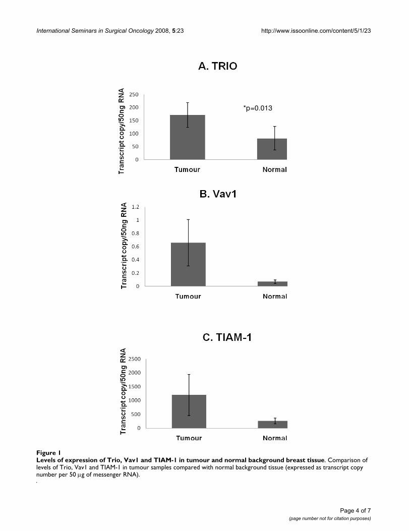
ResultsAberrant expression of TRIO, VAV1 and TIAM-1 in human breast cancerTumour tissue exhibited significantly high levels of Trio,when compared with normal background breast tissue(171.5 ± 46.7 v 82.0 ± 45; p = 0.013). Levels of Vav 1 andTIAM-1 were also higher in tumours but were not signifi-cantly different (0.66 ± 0.35 v 0.07 ± 0.027; p = 0.095 for
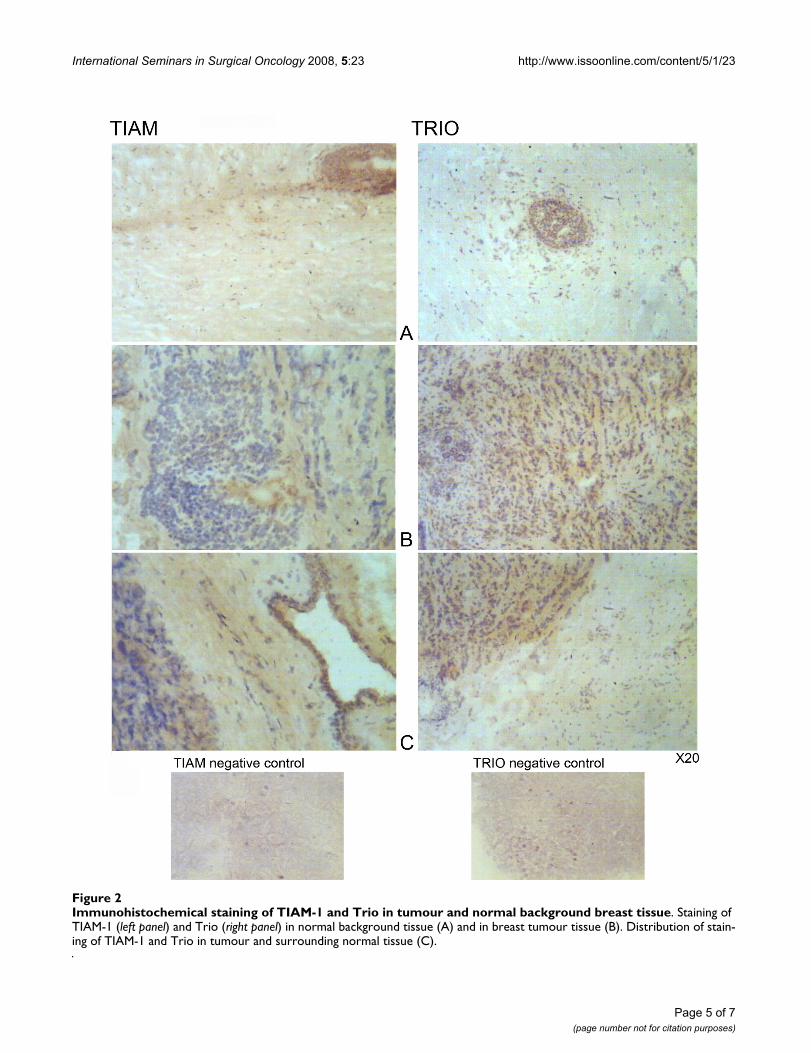
Vav1 and 1196 ± 743 v 261 ± 107; p = 0.22 for TIAM-1)(Figure 1). These results were confirmed for Trio andTIAM-1 by immunohistochemistry staining (Figure 2);with very little staining seen in normal background tissue(Figure 2A) while tumour tissue stained strongly for bothTrio and TIAM-1 (Figure 2B). This difference of stainingintensity between normal and tumour tissue is shown inFigure 2C.
The expression levels of all three GEFs, assessed by Q-PCR,were compared with a series of predictive factors (Tables1, 2, 3). Trio levels were seen to increase significantly withan increase in Nottingham Prognostic Index (109 ± 44NPI1; 234 ± 113 NPI2; 315 ± 176 NPI3 p = 0.04 compar-ing NPI1 and NPI2) Figure 3.
Expression of Vav1 showed no definitive pattern, increas-ing in NPI2 tumours compared with NPI1 tumours butshowing no change in NPI3 tumours (mean ± SEM 0.45 ±0.22 NPI1; 1.18 ± 1.06 NPI2; 0.45 ± 0.41 NPI3). TIAM-1expression showed a sequential decrease with increase inNPI status (1957 ± 1466 NPI1; 506 ± 205 NPI2; 298 ± 123NPI3) however these changes did not reach a level of sig-nificance (p > 0.3 in all cases) Table 1.
TIAM-1 expression increased with tumour-node-metasta-sis status (mean ± SEM 430 ± 117 TNM1 and 2613 ± 2245TNM2 p = 0.34), as did Vav1 levels (mean ± SEM 0.407 ±0.22 TNM 1 and 1.28 ± 1.04 TNM 2 p = 0.42) however,Trio expression showed an increase from TNM1 to TNM3(160 ± 50 TNM1 and 669 ± 554 TNM4 p = 0.39) but adecrease in Trio expression in TNM2 tumours (117 ± 62)Table 2.
Expression levels of the three GEFs studied were alsorelated to the follow-up data available on clinical out-come for the patients (Table 3). Based on the outcomes ofthe follow up data, patients were grouped into those whowere disease free; with metastatic disease; with local recur-rence of breast cancer, and died as a consequence of breastcancer. Trio levels showed little change of expressionbetween those patients who survived and those who diedfrom their breast cancer (mean ± SEM 174.1 ± 62.1 surv 1;263 ± 151 surv 2; 152.4 ± 94.5 surv 3; 188.6 ± 97.2 surv4). Vav1 showed the highest expression in patients whoremained disease free (0.89 ± 0.48) with much lowerexpression in those patients with metastatic disease (0.01± 0.003); with local recurrence (0.02 ± 0.012) and inpatients who died from breast cancer (0.03 ± 0.016). Lev-els of TIAM-1 were much lower in tumour tissue frompatients who remained disease free compared with thosewho died from breast cancer (median value disease free,60.3 v median value died from breast cancer, 224.5; p =0.04, with range Q1–Q3 disease free 0 – -251.1; died frombreast cancer 0 – -3.2).
Page 3 of 7(page number not for citation purposes)
International Seminars in Surgical Oncology 2008, 5:23 http://www.issoonline.com/content/5/1/23
Page 4 of 7(page number not for citation purposes)
Levels of expression of Trio, Vav1 and TIAM-1 in tumour and normal background breast tissueFigure 1Levels of expression of Trio, Vav1 and TIAM-1 in tumour and normal background breast tissue. Comparison of levels of Trio, Vav1 and TIAM-1 in tumour samples compared with normal background tissue (expressed as transcript copy number per 50 μg of messenger RNA).
*p=0.013
International Seminars in Surgical Oncology 2008, 5:23 http://www.issoonline.com/content/5/1/23
Page 5 of 7(page number not for citation purposes)
Immunohistochemical staining of TIAM-1 and Trio in tumour and normal background breast tissueFigure 2Immunohistochemical staining of TIAM-1 and Trio in tumour and normal background breast tissue. Staining of TIAM-1 (left panel) and Trio (right panel) in normal background tissue (A) and in breast tumour tissue (B). Distribution of stain-ing of TIAM-1 and Trio in tumour and surrounding normal tissue (C).
International Seminars in Surgical Oncology 2008, 5:23 http://www.issoonline.com/content/5/1/23
Comparison of tumour types showed lower levels of Trio(p = 0.33) and TIAM-1 (p = 0.37) but higher levels of Vav1(p = 0.47) in ductal tumours than in all other tumourtypes Table 4.
DiscussionThe guanine nucleotide exchange factors TRIO, VAV1 andTIAM-1 activate the Rho-GTPases by initiating GDP/GTPexchange. The GEF Trio is associated with cell motility byregulation of FAK and effects changes in the actin cytoskel-eton. This study has shown for the first time that Trio lev-els are significantly raised in breast tumour tissuecompared with normal tissue (p = 0.013) and higher lev-els of Trio are also found in tumours with a poor predic-tive outcome (NPI > 5.4) (p = 0.04). This pattern isconfirmed by immunohistochemical staining.
Vav1 functions as an oncogene involved in malignanttransformation [14] and the Vav family appears to play anessential role in angiogenesis [15] and androgen receptortranscriptional activity in prostate cancer [16]. It isexpressed in human neuroblastomas originating from tis-sues which do not normally express this protein [17]. Theresults of our study show that Vav 1 expression was higherin tumour tissue than in normal background tissue butthis did not reach a level of significance (p = 0.095). Vav1 over expression has been previously shown to be associ-ated with poorer survival (9,10) however our study indi-cates that decreased levels of Vav 1 are associated withpoorer survival (p = 0.07 in all cases).
Higher grade breast tumours have a higher expression ofTIAM-1 which can disturb intercellular adhesiveness byinducing cellular ruffles and may be correlated with theinvasive phenotypes of human breast tumours [18]. Theresults of our study would tend to agree with this findingas we have shown that TIAM-1 expression is higher in
Levels of expression of Trio in relation to NPI statusFigure 3Levels of expression of Trio in relation to NPI status. Trio levels increased sequentially with an increase in the Nottingham Prognostic Index.
Table 1: Expression levels of the GEFs studied related to NPI status of the tumours
Guanine Nucleotide Exchange Factors
NPI status TRIO VAV1 TIAM-1
(NPI < 3.4) 109 ± 44 0.45 ± 0.22 1957 ± 1466
(NPI 3.4–5.4) 234 ± 113 1.18 ± 1.06 506 ± 205
(NPI > 5.4) 315 ± 176 0.45 ± 0.41 298 ± 123
NPI = Nottingham Prognostic Index.Levels of Trio showed a sequential increase with increased NPI status with a significant increase from NPI1 to NPI2 (p = 0.04). Expression of Vav1 showed no definitive pattern while TIAM-1 expression showed a sequential decrease with an increase in NPI status.
Table 2: Expression levels of the three GEFs studied related to tnm status
Guanine Nucleotide Exchange Factors
TNM status TRIO VAV1 TIAM-1
1 160 ± 50.5 0.41 ± 0.22 430 ± 117
2 116.5 ± 61.6 1.28 ± 1.04 2613 ± 2245
3 669 ± 544 0.148 ± 0.12 2022 ± 1817
tnm = tumour-node-metastasis.TIAM-1 expression increased with tumour-node-metastasis (tnm) status as did Vav1 levels however, Trio expression showed an increase from tnm1 to tnm3 (160 ± 50 tnm1 and 669 ± 554 tnm4 p = 0.39) but a decrease in Trio expression in tnm2 tumours (117 ± 62).
Table 3: Expression levels of the three GEFs studied related to clinical outcome
Guanine Nucleotide Exchange Factors
Survival status TRIO VAV1 TIAM-1
1 Disease free 174.1 ± 62.1 0.89 ± 0.48 60.3
2 Metastatic disease 263 ± 151 0.01 ± 0.003 169
3 Local recurrance 152.4 ± 94.5 0.02 ± 0.012 26
4 Died from breast cancer 188.6 ± 97.2 0.03 ± 0.016 225
Trio levels show little change of expression between patients who survived and patients who died from breast cancer. Vav1expression was highest in patients who remained disease free while TIAM-1 expression was much lower in patients who remained disease free compared with those who died from breast cancer.
Page 6 of 7(page number not for citation purposes)
International Seminars in Surgical Oncology 2008, 5:23 http://www.issoonline.com/content/5/1/23
tumour tissue than in normal background breast tissue,although this did not reach a level of significance (p =0.22). The results from immunohistochemical stainingagree with this finding. However when looking at clinicaloutcome for these patients, levels of TIAM-1 were signifi-cantly higher in tumour tissue from patients who diedfrom breast cancer compared with those who survived p =0.04.
ConclusionHigh levels of expression of all three GEFs studied Trio,Vav1 and TIAM-1 were seen in breast tumours comparedwith normal background breast tissue. The statistical evi-dence in this study leads to the conclusion that the GEFTRIO may be particularly useful as a prognostic factor inbreast cancer as its level is significantly increased intumour tissue with Tiam-1 showing a significant increasein tumour tissue from patients with poor prognosis. Thissuggests that aberrant regulation of Rho family activitiesby GEFs may have an important prognostic value in breastcancer and that these GEFs may be important targets fortherapeutic intervention.
Competing interestsThe authors declare that they have no competing interests.
Authors' contributionsJL carried out the RNA extraction, PCR and Q-PCR analy-sis and drafted the manuscript. TM carried out tissue prep-aration and PCR analysis and helped with drafting themanuscript and statistical analysis. REM provided thepatient samples for this study. WGJ conceived the study,participated in its design and helped to draft the manu-script. All authors have read and approved the final man-uscript.
AcknowledgementsThe authors wish to thank Mr Anthony Douglas-Jones for his invaluable help in evaluating the histology of the tissue specimens and Mr Gareth Wat-kins for his technical expertise in producing the immunohistochemical staining. Thanks also to the Emma Jane Demery Bequest Fund and to Can-cer Research Wales for supporting this work.
References1. Hall A: Rho GTPases and the actin cytoskeleton. Science 1998,
11:2295-2322.2. Ridley AJ: Rho GTPases and cell migration. J Cell Sci 2001,
114:2713-2722.3. Takaishi K, Sasaki T, Kotani H, Nishioka H, Takai Y: Regulation of
cell-cell adhesion by rac and rho small G proteins in MDCKcells. J Cell Biol 1997, 137:1421-1431.
4. Jaffe AB, Hall A: Rho GTPases in transformation and metasta-sis. Adv Cancer Res 2002, 84:57-80.
5. Jiang WG, Watkins G, Lane J, Cunnick GH, Douglas-Jones A, MokbelK, Mansel RE: Prognostic value of Rho-GTPases and Rho Gua-nine Nucleotide Dissociation Inhibitors (GDI's) in humanbreast cancers. Clin Cancer Res 2003, 9(17):6432-6440.
6. Lane J, Martin TA, Watkins G, Mansel RE, Jiang WG: The expres-sion and prognostic value of ROCK I and ROCK II and theirrole in human breast cancer. Int J Oncol 2008, 33:585-593.
7. Bos Jl, Rehmann H, Wittinghofer A: GEFs and GAPs: Critical ele-ments in the control of small G proteins. Cell 2007,129:865-877.
8. Bateman J, van Vactor D: The Trio family of guanine-nucleotideexchange factors: regulators of axon guidance. J Cell Science2001, 114:1973-1980.
9. Medley QG, Buchbinder EG, Tachibana K, Ngo H, Serra-Pages C,Streuli M: Signalling between focal adhesion kinase and Trio.J Biol Chem 2003, 278:13265-13270.
10. Hornstein I, Alcover A, Katzav S: Vav proteins, masters of theworld of cytoskeletal organisation. Cell Signal 2004, 16:1-11.
11. Fernandez-Zapico ME, Gonzalez-Paz NC, Weiss E, Savoy DN, MolinaJR, Fonseca R, Smyrk TC, Chari ST, Urrutia R, Billadeau DD: Ectopicexpression of VAV1 reveals an unexpected role in pancreaticcancer tumorigenesis. Cancer Cell 2005, 7:39-49.
12. Habets GGM, Schlotes EHM, Zuydgeest D, Kamma RA van der, StamJC, Berns A, Collard JG: Identification of an invasion inducinggene Tiam-1, that encodes a protein with homology to GDP-GTP exchangers for Rho-like proteins. Cell 1994, 77:537-549.
13. Borguignon LY, Zhu H, Shao L, Chen YW: Ankyrin-Tiam1 inter-action promotes Rac1 signaling and metastatic breast tumorcell invasion and migration. J Cell Biol 2000, 150:177-191.
14. Bustelo XR: Regulatory and signalling properties of the Vavfamily. Mol Cell Biol 2000, 20:1461-1477.
15. Hunter SG, Zhuang G, Brantley-Sieders D, Swat W, Cowan CW,Chen J: Essential role of Vav family guanine nucleotideexchange factors in EphA receptor-mediated angiogenesis.Mol Cell Biol 2006, 26:4830-4842.
16. Lyons LS, Burnstein KL: Vav3, a Rho GTPase guanine nucleotideexchange factor, increases during progression to androgenindependence in prostate cancer cells and potentiatesandrogen receptor transcriptional activity. Mol Endocrinol2006, 20:1061-1072.
17. Hornstein I, Pikarsky E, Groysman M, Amir G, Peylan-Ramu N, Shu-lamit K: The haematopoietic specific signal transducer Vav1 isexpressed in a subset of human neuroblastomas. J Pathol 2003,199:526-533.
18. Adam L, Vadlamudi RK, McCrea P, Kumar R: Tiam1 overexpres-sion potentiates heregulin-induced lymphoid enhancer fac-tor-1/beta-catenin nuclear signaling in breast cancer cells bymodulating the intercellular stability. J Biol Chem 2001,276:28443-28450.
Table 4: Expression levels of the three GEFs studied related to type of breast cancer
Guanine Nucleotide Exchange Factors
Breast cancer type TRIO VAV1 TIAM-1
Ductal 130 ± 34 0.73 ± 0.44 487 ± 152
Other 578 ± 410 0.36 ± 0.24 12029 ± 11862
P = 0.33 P = 0.47 P = 0.37
Lower levels of Trio and TIAM-1 were found in ductal tumours than in all other tumour types
Page 7 of 7(page number not for citation purposes)