Embed Size (px)
Citation preview

International Journal of Pharmaceutics 496 (2015) 1057–1068
Integrin-mediated active tumor targeting and tumormicroenvironment response dendrimer-gelatin nanoparticles for drugdelivery and tumor treatment
Guanlian Hua,1, Huiqing Zhangb,1, Li Zhanga, Shaobo Ruana, Qin Hea, Huile Gaoa,*aKey Laboratory of Drug Targeting and Drug Delivery Systems, West China School of Pharmacy, Sichuan University, No. 17, Block 3, Southern Renmin Road,Chengdu 610041,ChinabHuashan Hospital North, Fudan University, 108 Lu Xiang Road, Shanghai 201907, China
A R T I C L E I N F O
Article history:Received 7 October 2015Received in revised form 8 November 2015Accepted 13 November 2015Available online 17 November 2015
Keywords:Tumor penetration and retentionBreast cancerMultistage drug delivery systemMatrix metalloproteinase-2Gelatin nanoparticles
A B S T R A C T
Due to the high morbidity and mortality of cancer, it has become an urgent matter to develop an effectiveand a safe treatment strategy. Nanoparticles (NP) based drug delivery systems have gained muchattention nowadays but they faced a paradoxical issue in delivering drugs into tumors: NP with large sizewere characterized with weak tumor penetration, meanwhile NP with small size resulted in poor tumorretention. To solve this problem, we proposed a multistage drug delivery system which could intelligentlyshrink its size from large size to small size in the presence of matrix metalloproteinase-2 (MMP-2) whichwere highly expressed in tumor tissues, therefore the multistage system could benefit from its large sizefor better retention effect in tumor and then shrunk to small size to contribute to better penetrationefficiency. The multistage drug delivery system, RGD–DOX–DGL–GNP, was constructed by 155.4 nmgelatin NP core (the substrate of MMP-2) and surface decorated with doxorubicin (DOX) and RGD peptideconjugated dendritic poly-L-lysine (DGL, 34.3 nm in diameter). In vitro, the size of multistage NP couldeffectively shrink in the presence of MMP-2. Thus, the RGD–DOX–DGL–GNP could penetrate deep intotumor spheroids. In vivo, this multistage drug delivery system showed higher tumor retention anddeeper penetration than both DOX–DGL and DOX–GNP. Consequently, RGD–DOX–DGL–GNP successfullycombined the advantages of dendrimers and GNP in vivo, resulting in an outstanding anti-tumor effect. Inconclusion, the multistage drug delivery system could intelligently shrink from large size to small size inthe tumor microenvironment and displayed better retention and penetration efficiency, making it animpressing system for cancer treatment.
ã 2015 Elsevier B.V. All rights reserved.
Contents lists available at ScienceDirect
International Journal of Pharmaceutics
journal homepage: www.elsev ier .com/locate / i jpharm
1. Introduction
Nanoparticles (NP) based drug delivery has gained increasingattention in diagnosis and treatment of various diseases includingcancer. Until now, several nanomedicines have been approved(Weissig et al., 2014). Although these approved nanomedicinesenhanced the antitumor effect and reduced the drug originatedside effect, the outcome is still modest. Commonly, nanomedicinescould passively accumulate in tumor because of the enhancedpermeability and retention (EPR) effect (Fang et al., 2011).However, tumor is a heterogeneous tissue with unique microenvi-ronment, such as dense tumor extracellular matrix and high
* Corresponding author.E-mail address: [email protected] (H. Gao).
1 Contributed equally to this work.
http://dx.doi.org/10.1016/j.ijpharm.2015.11.0250378-5173/ã 2015 Elsevier B.V. All rights reserved.
interstitial fluid pressure, which extremely restrict the distributionof NP through tumor (Jain, 2001; Junttila & de Sauvage, 2013;Marte, 2013). Therefore, it is important to develop novel NP thatcould adapt the tumor microenvironment and improve thehomogenous distribution of drugs.
For this purpose, several kinds of size changeable NP wereconstructed to adapt the controversial particle size requirement oftumor penetration and tumor retention because good tumorpenetration required small particle size while good tumorretention required large particle size (Li et al., 2015; Ruan et al.,2015a,b; Tong et al., 2012; Wong et al., 2011). When these sizechangeable NP distributed in tumor according to EPR effect, thelarge size made them easy to retain in tumor. Then the particle sizecould considerably reduce, leading to well tumor penetration.Recently, we constructed a matrix metalloproteinase-2 (MMP-2)sensitive NP: DOX–DGL–GNP. The model drug doxorubicin (DOX)was firstly conjugated onto small sized dendritic poly-L-lysine

1058 G. Hu et al. / International Journal of Pharmaceutics 496 (2015) 1057–1068
(DGL), then the DOX–DGL was fabricated onto gelatin NP (GNP) toform a large particle (Hu et al., 2015b). The GNP could be degradedby MMP-2, which overexpressed in tumor (Ruan et al., 2015a,b;Zhu et al., 2012), thus the size of DOX–DGL–GNP could reduce andthe tumor penetration could be improved. Although somepreliminary study showed the DOX–DGL–GNP had better pene-tration efficiency, rare in vivo study was performed (Hu et al.,2015a,b).
To further improve the tumor targeting effect, active targetingligand was used in this study (Gao et al., 2013). Neovesselendothelial cells and malignant tumor cells were highly overex-pressed integrin receptors, such as avb3 (Schottelius et al., 2009),which could specifically bind with arginine-glycine-aspartic acid(RGD) peptide (Ruoslahti, 1996; Ruan et al., 2015c), so RGD wasused as a targeting ligand to improve the tumor targeting effect ofparticles.
In this study, we have proposed a multistage drug deliverysystem: RGD–DOX–DGL–GNP (Fig. 1). The system could effectivelytarget and retain in tumor which benefited from EPR effect andavb3mediated active targeting. After the degradation by MMP-2 intumor, the size reduced and RGD–DOX–DGL was released tofurther penetrate into the core of tumor. To evaluate the tumortargeting and penetration of this system, several experiments wereperformed.
2. Materials and methods
2.1. Materials
Doxorubicin hydrochloride was obtained from Beijing HuafengUnited Technology Co., Ltd. (Beijing, China). Gelatin type A wasobtained from MP Biomedicals Co., Ltd. (California, USA).Glutaraldehyde solution (Grade II, 25%) was obtained from BeijingSolarbio Technology Co., Ltd. (Beijing, China). DGL-G3 dendrimerwas purchased from Colcom (Montpellier Cedex, France). a-Mal-eimide-PEG-succinimidyl carbonate (MW3500), a-mPEG-v-ami-no (MW5000) and amine-PEG-carboxyl (MW5000) were obtainedfrom Seebio Biotechnology Co., Ltd. (Shanghai, China). RGDyC wasobtained from Phtdpeptides Co., Ltd. (Zhengzhou, China). Aconiticanhydride (CA) was obtained from Alfa Aesar Chemical Co., Ltd.(Tianjin, China). 1-Ethyl-3-(3-dimethylaminopropyl) carbodiimide(EDC) and N-hydroxy-succinimide (NHS) were obtained fromChengdu Best Reagent Co., Ltd. (Chengdu, China). 6-Diamidino-2-
Fig. 1. Schematic illustration to show the size shrinkage of the RGD–DOX–DGL–GNP from large size to small size triggered by MMP-2, a protease highly expressed intumor extracellular matrix, thus penetrating into deep tumor tissue.
pheylindole (DAPI) was obtained from Beyotime Insitute Biotech-nology (Haimen, China). Purified rabbit mAb to CD34 waspurchased from Abcam Ltd. (Hong Kong, China). MMP-2 protein(rat) was purchased from Abcam Ltd. (Hong Kong, China). AlexaFluor1 594-conjugated AffiniPure donkey anti-rabbit was pur-chased from Jackson Immuno Research Laboratories Inc. (WestGrove, PA, USA). RPMI1640, 3-(4,5-dimethylthialzol-a-yl)-2,5-diphenyltetrazolium bromide (MTT), fetal bovine serum (FBS),penicillin, streptomycin and trypsin were purchased from com-mercial sources (Chengdu, China). Mouse mammary breast tumorcell line (4T1) and human umbilical vein endothelial cells (HUVEC)were purchased from Shanghai Institute of Cell Biology (Shanghai,China). All other reagents and solvents were of analytical or HPLCgrade and were used without further purification.
Female BALB/C mice (4-week old, 16–18 g) were purchasedfrom experiment animal center of Sichuan University (Chengdu,China). The mice were maintained under standard housingconditions. All animal experiments were performed in accordancewith the principles approved by the experiment animal adminis-trative committee of Sichuan University.
2.2. Synthesis of RGD–DOX–PEG–DGL conjugates
RGD–DOX–DGL–PEG was synthesized according previousprocedure (Hu et al., 2015a,b). Aftercis-aconityl doxorubicin(CAD) was synthesized using published procedure (Du et al.,2013), DGL was reacted with NHS-PEG3400-MAL at the ratio 1:8(molar ratio) in PBS (pH 8.0) for 2 h followed with RGDyC at theratio of 1:2 (DGL to peptide, molar ratio) in PBS (pH 7.0) for 24 h.After dialyzed in deionized water, RGD–DGL–PEG was freeze-driedand analyzed in a 400 MHz spectrometer. Then DGL–PEG or RGD–DGL–PEG was reacted with CAD in the presence of EDC and NHS inthe dark for 12 h. At last, the mixtures were purified byultrafiltration through a membrane (MWCO10,000), and theamount of DOX conjugation was confirmed by UV–vis spectropho-tometry at 480 nm.
2.3. Preparation of RGD–DOX–DGL–GNP
Firstly, GNP were prepared by a two-step desolvation method asprevious described with some modification (Wong et al., 2011).Then, EDC (0.8 mg) and NHS (0.8 mg) were added to the GNPsolution (pH 6.0, 1 mL) to activate carboxyl groups. After the pHvalue was adjusted to 8.0, 20 mg of SCM-PEG5000-NH2was added tothe mixture. 2 h later, the pH was adjusted to 6.0 and additionalsolution of EDC (0.8 mg) and sulfo-NHS (0.8 mg) was added. Afterstirring for 30 min, the pH value was adjusted to 8.0 again, DOX–DGL–PEG or RGD–DOX–DGL–PEG was added to the resultingmixture. The mixtures were purified by ultrafiltration through amembrane (MWCO 100,000) (4500 g � 30 min). After hydrolyzedby 1 N HCl, the doxorubincinone was determined using HPLC asdescribed in Section 2.9 to determine the drug loading capacity(Zhu et al., 2010).
2.4. In vitro release
DOX, DOX–DGL–PEG, RGD–DOX–DGL–PEG, DOX–DGL–GNPand RGD–DOX–DGL–GNP (equivalent to 0.1 mg DOX) were addedinto dialysis bags secured with clamps respectively, and thenplaced in plastic tubes containing 40 mL PBS (pH 7.4). Theexperiment was carried out at 37 �C and a horizontal shakingspeed of 50 rpm. At appropriate time intervals (0, 1, 2, 3, 5, 7, 9, 12and 24 h), 1 mL media was taken away and the corresponding freshbuffer was supplemented. The amount of released DOX wasdetected by fluorescence spectrophotometer. Each drug releasetest was performed thrice.

Fig. 2. (A) Synthetic scheme of RGD-DOX-DGL-PEG. (B) 1H NMR spectra of RGD–DGL–PEG. (C) Mass spectra of CAD.
G. Hu et al. / International Journal of Pharmaceutics 496 (2015) 1057–1068 1059

Fig. 3. Absorption spectra of the synthesized RGD–DOX–DGL–PEG, DOX, CAD, RGD,DGL–PEG, RGD–DGL–PEG, DOX–DGL–PEG in PBS.
1060 G. Hu et al. / International Journal of Pharmaceutics 496 (2015) 1057–1068
2.5. In vitro cytotoxicity assays
The in vitro anti-proliferation activity of various DOX-loadedGNP was evaluated by the MTT assay. 4T1 and HUVEC cells wereseeded at a density of 3 � 103 cells/well into 96-well plates, after12 h incubation at 37 �C in 5% CO2 atmosphere, these cells weretreated with a series of different concentration (from 0.391 mM to50 mM) of DOX, DOX–DGL–PEG, RGD–DOX–DGL–PEG, DOX–GNP,DOX–DGL–GNP and RGD–DOX–DGL–GNP for 48 h. To assess cellviability, MTT (5 mg/mL, 20 mL) was added into each well andincubated at 37 �C for 4 h. The medium was removed and 150 mL ofDMSO was added to each well at 75 rpm for 15 min. After that thepercentage of cell viability was detected at 570 nm by fluorescencespectrophotometer.
2.6. Tumor spheroid penetration assay
To obtain tumor spheroid, 4T1 cells were seeded into lowmelting point agarose precoated 96-well plate at the density of8 � 103 cells per well (Gao et al., 2014a,b). Several days later, theuniform spheroids were selected and treated with DOX–DGL–PEG,RGD–DOX–DGL–PEG, DOX–DGL–GNP, RGD–DOX–DGL–GNP, DOX–DGL–GNP (MMP-2) and RGD–DOX–DGL–GNP (MMP-2) at DOXconcentration of 12.5 mg/mL for 24 h. After washed with PBS forthree times, the spheroids were fixed with 4% paraformaldehyde
Fig. 4. In vitro DOX release pattern of DOX, DOX–DGL–PEG, RGD–DOX–DGL–PEG, DOX–pH7.4; B: pH5.5). Data represented mean � SD (n = 3).
and then the fluorescence distribution was observed via confocalmicroscopy.
2.7. Surface plasmon resonance (SPR) analysis
SPR assay was performed at 25 �C using a Biacore T200instrument (GE, USA) equipped with a GM5 chip. Integrin avb3
was coupled on the surface of the GM5 chip. Then the dilutedsamples flow through the surface of chip and the signal wasdetected. Subsequently, all the samples were eluted with buffer orregenerated reagent.
2.8. In vitro cellular uptake study
The cellular uptake of RGD–DOX–DGL–GNP was evaluated byqualitative and quantitative methods. For flow cytometry experi-ment, HUVEC were seeded on 6-well plates at 2 � 105 cells/welland incubated for 24 h. Then, the cells were treated with varioussamples (12.5 mg/mL DOX equal) under 37 �C for 2 h. Afterincubation, the cells were washed twice with PBS, trypsinizedand resuspended in proper volume of PBS for flow cytometeranalysis (CytomicsTM FC 500, Beckman Coulter, Miami, FL, USA).
For qualitative assay, HUVEC were seeded at a density of 1 �105
cells/well on microscope slides in 6-well plates. After 24 hincubation, the cells were treated with various samples(12.5 mg/mL DOX equal) for 2 h. After incubation, the microscopeplates were washed with cold PBS for three times, fixed with 4%paraformaldehyde for 20 min and then stained with DAPI for 5 min.Finally, slides were imaged by fluorescent microscope (Nikon,Japan).
2.9. In vivo imaging and tumor distribution
At two weeks after 4T1 tumor implantation, DOX, DOX–GNP,DOX–DGL–PEG, RGD–DOX–DGL–PEG, DOX–DGL–GNP and RGD–DOX–DGL–GNP were intravenously injected into the mice at thedose of 5 mg/kg DOX (equal) per mouse (Ruan et al., 2015a). Afteradministration for 24 h, the mice were anesthetized by 4% chloralhydrate and sacrificed, the main organs were taken to observe theaccumulation condition of the NP using fluorescent imagingsystem (IVIS, Caliper, USA).
For quantitative analysis, tissues were weighted and homoge-nate was prepared. After 1 N HCl was added to the homogenate tohydrolyze conjugates, 20 mL of daunorubicin (200 mg/mL) asinternal standard solution was precisely added to 200 mL of tissuehomogenates for vortex mixing. Then 1 mL of extracted solution(chloroform:methanol = 4:1) were added for vortex-ultrasonicextraction several times. After centrifugation (12,000 rpm,
GNP, DOX–DGL–GNP, RGD–DOX–DGL–GNP under different pH value condition (A:

Fig. 5. The cell viability of 4T1 (A) and HUVEC (B) incubated with DOX, DOX–DGL–PEG, RGD–DOX–DGL–PEG, DOX–DGL–GNP, RGD–DOX–DGL–GNP, DOX–DGL–GNP (MMP-2)and RGD–DOX–DGL–GNP (MMP-2).
G. Hu et al. / International Journal of Pharmaceutics 496 (2015) 1057–1068 1061
5 min), the organic phase was moved to another centrifuge tubeand dried under a stream of nitrogen at 37 �C. The residue wasredissolved in 100 mL of mobile phase. After centrifugation(12,000 rpm, 10 min), the supernatant was collected for HPLCanalysis. Samples were analyzed by a HPLC method using UV-detection. Angilent liquid chromatographic system (Angilent 1200,USA) was equipped witha UV detectorwith 479 nm. Apollo C18(250 � 4.6 mm,5 mm) was used at 30 �C. The mobile phase wasmethanol:0.01 M sodium acetate:acetic acid = 70:30:1 (v/v/v), theinjection volume was 40 mL and the flow rate was 1 mL/min. Thepeak area ratio of the sample peak and the internal standard peakwere recorded for quantitative analysis. In order to quantitativeevaluation of the interior and exterior of the tumors, the tumorswere divided into two parts, namely the interior and exterior, eachparts were weighted and treated as described above.
For confocal microscopy, the tumors were fixed in 4%paraformaldehyde for 48 h, followed by dehydration with 15%sucrose solution and 30% sucrose solution overnight sequentiallyuntil subsidence. Thereafter, the tumors were embedded in OCT(Leica, Germany) at �20 �C, sectioned at 16 mm via freezingmicrotome section. Finally, Rabbit mAb to CD34 antibody (1:100)was added to sections, then the slices stained with 300 nM DAPI for5 min and observed via confocal microscopy.
2.10. Degradation of AuNP–GNP in vivo
In order to validate the degradation of this multistage nano-carrier in vivo, AuNP–GNP (200 mg/mL, 0.1 mL) were injected intosubcutaneous tumor in situ. The mice were anesthetized afterinjection for 2 h, then the hearts of mice were perfused withphosphate buffer and paraformaldehyde, thereafter the tumortissues were sampled from the injection site and the region awayfrom the injection site (about 1 mm3) respectively, and fixed with25% glutaraldehyde. After a series of dehydration step, theultrathin sections were stained with osmium for TEM observation.
2.11. Anti-tumor efficacy
BALB/c mice bearing 4T1 tumor were randomly dividedinto seven groups (n = 5). After the size of tumors reached100 mm3, mice were given DOX, DOX–DGL–PEG, RGD–DOX–DGL–PEG, DOX–GNP, DOX–DGL–GNP and RGD–DOX–DGL–GNP (at an equal dose of 5 mg/kg) and PBS (the control group)every three days for four times, respectively. The tumor size andanimal body weight were measured every three days during
the study and the tumor volume was calculated by the formula:
Tumor volume ¼ width2=length
� �=2. After 27 days, all the mice
were sacrificed and the tumors were harvested for imaging,weighing and HE staining.
2.12. Statistical analysis
Data were analyzed using the IBM SPSS 19.0 software. PairedStudent’s t test was used for comparison. A probability (p) less than0.05 was considered statistically difference.
3. Results
3.1. Synthesis and characterization of DGL derivatives
DGL was first decorated with PEG and PEG–RGD (Fig. 2A). InNMR spectra of RGD–DGL–PEG (Fig. 2B), the peak of PEG’s repeatunits (��O��CH2��CH2��) was presented a sharp peak at 3.4–3.6 ppm (Huang et al., 2013) and the double peak of RGD was foundat 6.873 ppm, 6.886 ppm, 7.189 ppm and 7.211 ppm (Miura et al.,2013). Then DOX was conjugated to the residual primary aminogroups of RGD–PEG–DGL by cis-aconityl linkage. Mass spectrome-try exhibited the mass value of cis-aconitic anhydride-doxorubicin(CAD) was 700.5 (M-H) (Fig. 2C), which was in agreement withprevious study (Zhu et al., 2010). The analysis of HPLC alsoindicated the successive synthesis of CAD. Finally, the activatedcarboxyl groups of CAD were connected to the amino groups ofRGD–DGL–PEG to obtain the final product: RGD–DOX–DGL–PEG.The purified RGD–DOX–DGL–PEG, RGD, and CAD were evaluatedby UV–vis absorption spectrophotometer (Fig. 3). The characteris-tic absorption peaks of RGD and CAD were at 275 nm and 490 nm,respectively, and the above characteristic absorption peaks wereobserved in the absorption spectrum of the RGD–DOX–DGL–PEG,indicating the successful synthesis of the RGD–DOX–DGL–PEGconjugates (Liu et al., 2012). The calculated numbers of PEG, RGDand DOX per DGL were 7.1, 2.5 and 13.3, respectively.
3.2. Characterization of RGD–DOX–DGL–GNP
The diameters of RGD–DOX–DGL–PEG were 34.4 nm with a PDIof 0.273. After decorated onto GNP, the diameters of RGD–DOX–DGL–GNP were considerably increased to 193.1 nm with a PDI of0.272. The increase of particle size indicated that small-sized RGD–DOX–DGL–PEG was successfully modified on the surface of GNP.

Fig. 6. (A) Fluorescent images of 4T1 tumor spheroids upon incubation with RGD–DOX–DGL–PEG, RGD–DOX–DGL–GNP and RGD–DOX–DGL–GNP (MMP-2). (MMP-2) meansNP were incubated with MMP-2 for 24 h before added to tumor spheroid. Scale bars represents 100 mm. (B) The ratio of fluorescence intensity of core and surface at 100 mm.(C) The fluorescence intensity profile from surface to core of DOX, DOX–DGL–PEG, RGD–DOX–DGL–PEG, DOX–GNP, DOX–DGL–GNP, RGD–DOX–DGL–GNP, DOX–DGL–GNP(MMP-2), RGD–DOX–DGL–GNP (MMP-2) at 100 mm.
1062 G. Hu et al. / International Journal of Pharmaceutics 496 (2015) 1057–1068
The DOX loading capacity of RGD–DOX–DGL–PEG was4.81% � 0.4%.
3.3. In vitro release of DOX
Compared with the rapid release of free DOX, all particles wereshowed with sustained release profiles and no burst initial release(Fig. 4). After 24 h incubation, there were lower than 40% of DOXreleased. The release amounts of DOX–DGL–GNP and RGD–DOX–DGL–GNP were less than 20% at pH 7.4 while the release rates ofDOX–DGL–GNP and RGD–DOX–DGL–GNP rapidly increased withthe reduction of the pH value of the medium, The phenomenonabove mainly attributed to the acid-sensitivity of cis-aconityl bondwhich was also in accordance with literature (Du et al., 2013). Thereleased profiles suggested this multistage NP would be stableunder the normal physiological pH value conditions.
3.4. In vitro cytotoxicity
Cytotoxicity is a primary concern in the development of drugdelivery system. The viabilities of mouse mammary breast tumorcells (4T1) (Fig. 5A) and human umbilical vein endothelial cells(HUVEC) (Fig. 5B) were determined after 48 h of incubation withvarious formulations. All formulations showed dose-related anti-tumor cells activity, and at as high as 50 mM of DOX, the survival ofcells treated by all formulations was lower than 40%. Specifically,RGD functionalized particles, RGD–DOX–DGL–PEG and RGD–DOX–DGL–GNP, showed better anti-proliferation effect than theunmodified particles.
3.5. Penetration efficiency of RGD–DOX–DGL–GNP
As shown in Fig. 6A, small sized RGD–DOX–DGL–PEG possessedbetter penetration efficiency than large sized RGD–DOX–DGL–

Fig. 7. (A) SPR studies of NP-Integrin avb3 interactions. (B) Cellular uptake of HUVEC determined by flow cytometry (n = 3). **p < 0.01. (C) The images of HUVEC incubatedwith DOX, DOX–DGL–PEG, RGD–DOX–DGL–PEG, DOX–GNP, DOX–DGL–GNP and RGD–DOX–DGL–GNP for 2 h taken by fluorescence microscope. Blue represents DAPI stainednuclei and red represent DOX. Scale bar represents 100 mm. (For interpretation of the references to color in this figure legend, the reader is referred to the web version of thisarticle.)
G. Hu et al. / International Journal of Pharmaceutics 496 (2015) 1057–1068 1063
GNP, while RGD–DOX–DGL–GNP incubated with MMP-2 couldenhance the penetration efficiency, demonstrating smaller sizedparticles had better penetration ability, which was consistent withour previous reports (Hu et al., 2015a,b; Ruan et al., 2015a,b). Inorder to better qualitatively compare the penetration efficiency ofvarious NP, the core/surface coefficient was introduced, and thehigher the core/surface coefficient was, the stronger the penetra-tion efficiency of NP was. At 100 mm depth, the core/surfacecoefficient of RGD–DOX–DGL–GNP (MMP-2) was much higherthan RGD–DOX–DGL–GNP (Fig. 6B). Besides, the fluorescenceintensity profiles of NP at 100 mm depth also indicated that thepenetration ability of RGD–DOX–DGL–GNP after degradation wasapparently stronger than that before (Fig. 6C). All the resultsconfirmed that this multistage nanocarrier RGD–DOX–DGL–GNPcould benefit from RGD active targeting and MMP-2 triggereddegradation, which facilitated penetration in tumor.
3.6. SPR analysis
The whole uptake process of NP by cells included in two steps,surface binding on cell membrane and internalization (Guo et al.,2014). SPR was promised to detect the interaction between thesensor surface and samples. Fig. 7 showed representative sensor
grams of the binding of RGD–DOX–DGL–GNP or DOX–DGL–GNP toIntegrin avb3 on the CM5 chip. The binding proportion of RGD–DOX–DGL–GNP was significantly higher than that of DOX–DGL–GNP (Fig. 7), indicating RGD was successfully modified on thesurface of DOX–DGL–GNP and contributed to binding on avb3
positive cells.
3.7. In vitro cellular uptake study
Fluorescence microscopy and flow cytometry were used forqualitative and quantitative evaluation of the drug deliverycapacity of the RGD–DOX–DGL–GNP into human umbilical veinendothelial cells (HUVEC) in vitro. Both results expressed thecellular uptake of RGD–DOX–DGL–GNP was higher than DOX–DGL–GNP, which meant that RGD–DOX–DGL–GNP possessed theability of active targeting to tumor angiogenesis (Fig. 7B and C).
3.8. In vivo tumor distribution and penetration
4T1 breast cancer was used as model and ex vivo imaging wasutilized to evaluate the tumor distribution and penetration ofRGD–DOX–DGL–GNP. At 24 h after tail vein injection, the fluores-cent intensity of large-sized NP (DOX–GNP, DOX–DGL–GNP and

Fig. 8. (A) Ex vivo imaging of 4T1-tumor bearing mice at 24 h post injection of DOX, DOX–DGL–PEG, RGD–DOX–DGL–PEG, DOX–GNP, DOX–DGL–GNP and RGD–DOX–DGL–GNP at a dose of 5 mg/kg. (B) Semi-quantitative analysis of fluorescent intensity in tumors at 24 h post injection (n = 3). (C) Quantitative analysis of DOX in the main organs(n = 3). (D) Quantitative analysis of DOX in the interior and exterior of tumors (n = 3). (E) Fluorescent distribution in slices of 4T1 tumors. Blue represents nuclei, red representsDOX, green represents neovasculature and scale bars represent 50 mm. (For interpretation of the references to color in this figure legend, the reader is referred to the webversion of this article.)
1064 G. Hu et al. / International Journal of Pharmaceutics 496 (2015) 1057–1068

Fig. 9. In vivo degradation and penetration of AuNP–GNP after in situ injection to tumor. (A) The mostly integral AuNP–GNP in tumor tissue at injection site. (B) The partialdegraded AuNP–GNP in tumor tissue. (C) The AuNP–GNP with complete degradation of tumor tissue, and a part of AuNP are obviously uptaken by lysosome. (D) The AuNP oftumor tissue in the area distanced from the injection site, AuNP–GNP were completely degraded by MMP-2 and released AuNP. Scale bars represents 100 nm.
G. Hu et al. / International Journal of Pharmaceutics 496 (2015) 1057–1068 1065
RGD–DOX–DGL–GNP) in tumor was apparently stronger than thatof small-sized NP (DOX, DOX–DGL–PEG and RGD–DOX–DGL–PEG)(Fig. 8A and B), demonstrating large-sized NP possessed bettertumor distribution and retention effect. In tumor sites DOX–DGL–GNP exhibited the higher fluorescent intensity than that of DOX–GNP, suggesting the multistage delivery system possessed bettertumor targeting efficiency that large sized NP, which might due tonot only better tumor retention but also tumor penetration effect.Besides, RGD-modified NP showed stronger fluorescent intensitythan non-targeted NP, which due to the active targeting ability ofRGD and was consistent with previous studies (Gao et al., 2014a,b;Zhan et al., 2010). Simultaneously, semi-quantitative analysis ofthe fluorescent intensity in tumor tissues also demonstrated thatthe intensity of RGD–DOX–DGL–GNP was 1.18-, 1.53- and 1.93-foldhigher than that of DOX–DGL–GNP, DOX–GNP and RGD–DOX–DGL–PEG, respectively (Fig. 8B), which confirmed this multistageshrinkable RGD–DOX–DGL–GNP could more effectively accumu-late in tumor compared with other NP. To further validate thistargeted multistage nanocarrier could effectively accumulate intumor sites, reduce cardiotoxicity of DOX and penetrate to deeparea of tumor tissues, the amounts of DOX in the heart, the internaland external tumor sites were measured by HPLC. The distribu-tions of NP in heart were notably decreased compared with freeDOX groups (Fig. 8C), which may contribute to the lowcardiotoxicity of these NP. Besides, RGD–DOX–DGL–GNP showedhighest DOX concentrations in both the internal and outer of tumor(Fig. 8D), confirming the multistage nanocarrier could not onlyeffectively accumulate in the peripheral of tumor but alsopenetrate into the core of the tumor.
Then vasculature was stained to green and the distribution ofDOX (red) in the tumor slices was determined. The fluorescentintensity of fixed-sized NP (DOX, DOX–DGL–PEG, RGD–DOX–DGL–PEG and DOX–GNP) was weaker than that of multistage drugdelivery systems (DOX–DGL–GNP and RGD–DOX–DGL–GNP),which was consistent with quantitative analysis by HPLC,indicating the multistage drug delivery systems displayed bettertumor targeting and retention effect. In addition, the fluorescencesignal of DOX–DGL–GNP and RGD–DOX–DGL–GNP distributedhomogeneously in the whole tumor including areas that both richand lack of neovasculature, which further demonstrated thesuperiority of this multistage drug delivery system (Fig. 8E).
3.9. In vivo shrink of DOX–DGL–GNP
In order to validate the in vivo shrink effect and tumorpenetration of this multistage drug delivery system, gold nano-particles (AuNP) with 23.2 nm were used to replace DOX–DGL–PEGbecause of their strong contrast under TEM. Two hours afterinjection, the mostly integral AuNP–GNP could still be seen in theinjection site in situ (Fig. 9A). At the same time, the partialstructure of AuNP–GNP could be observed (Fig. 9B), suggesting theGNP based particles could be degraded in tumor due to the highlyexpressed MMP-2. Furthermore, in the area distanced from theinjection site, the released AuNP monomers and oligomers couldbe clearly observed (Fig. 9C and D), indicating the AuNP with smallsize could penetrate much deeper than the integral AuNP–GNPwith large size. This experiment demonstrated the GNP basedmultistage delivery system could be successfully degraded in

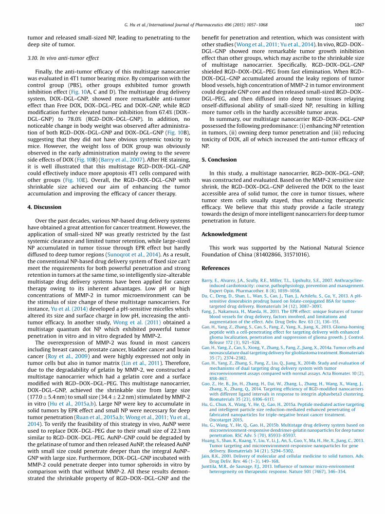
Fig. 10. In vivo anti-tumor effect. (A) 4T1 tumor growth curves of different groups after treatments (mean � SD, n = 5). *p < 0.05, **p < 0.01. (B) Body weights of mice aftervarious treatments. (C) Photos of the tumors collected from different groups of mice at the end of treatments (day 27). (D) Average weights of tumors in each treatment groupon day 27 (mean � SD, n = 5). *P < 0.05, **P < 0.01. (E) HE staining of tumor tissue after therapy. The image magnification is 200�. Scale bars represent 100 mm.
1066 G. Hu et al. / International Journal of Pharmaceutics 496 (2015) 1057–1068
G. Hu et al. / International Journal of Pharmaceutics 496 (2015) 1057–1068 1067
tumor and released small-sized NP, leading to penetrating to thedeep site of tumor.
3.10. In vivo anti-tumor effect
Finally, the anti-tumor efficacy of this multistage nanocarrierwas evaluated in 4T1 tumor bearing mice. By comparison with thecontrol group (PBS), other groups exhibited tumor growthinhibition effect (Fig. 10A, C and D). The multistage drug deliverysystem, DOX–DGL–GNP, showed more remarkable anti-tumoreffect than Free DOX, DOX–DGL–PEG and DOX–GNP, while RGDmodification further elevated tumor inhibition from 67.4% (DOX–DGL–GNP) to 78.0% (RGD–DOX–DGL–GNP). In addition, nonoticeable change in body weight was observed after administra-tion of both RGD–DOX–DGL–GNP and DOX–DGL–GNP (Fig. 10B),suggesting that they did not have obvious systemic toxicity tomice. However, the weight loss of DOX group was obviouslyobserved in the early administration mainly owing to the severeside effects of DOX (Fig. 10B) (Barry et al., 2007). After HE staining,it is well illustrated that this multistage RGD–DOX–DGL–GNPcould effectively induce more apoptosis 4T1 cells compared withother groups (Fig. 10E). Overall, the RGD–DOX–DGL–GNP withshrinkable size achieved our aim of enhancing the tumoraccumulation and improving the efficacy of cancer therapy.
4. Discussion
Over the past decades, various NP-based drug delivery systemshave obtained a great attention for cancer treatment. However, theapplication of small-sized NP was greatly restricted by the fastsystemic clearance and limited tumor retention, while large-sizedNP accumulated in tumor tissue through EPR effect but hardlydiffused to deep tumor regions (Sunoqrot et al., 2014). As a result,the conventional NP-based drug delivery system of fixed size can'tmeet the requirements for both powerful penetration and strongretention in tumors at the same time, so intelligently size-alterablemultistage drug delivery systems have been applied for cancertherapy owing to its inherent advantages. Low pH or highconcentrations of MMP-2 in tumor microenvironment can bethe stimulus of size change of these multistage nanocarriers. Forinstance, Yu et al. (2014) developed a pH-sensitive micelles whichaltered its size and surface charge in low pH, increasing the anti-tumor efficacy. In another study, Wong et al. (2011) obtained amultistage quantum dot NP which exhibited powerful tumorpenetration in vitro and in vitro degraded by MMP-2.
The overexpression of MMP-2 was found in most cancersincluding breast cancer, prostate cancer, bladder cancer and braincancer (Roy et al., 2009) and were highly expressed not only intumor cells but also in tumor matrix (Lin et al., 2011). Therefore,due to the degradability of gelatin by MMP-2, we constructed amultistage nanocarrier which had a gelatin core and a surfacemodified with RGD–DOX–DGL–PEG. This multistage nanocarrier,DOX–DGL–GNP, achieved the shrinkable size from large size(177.0 � 5.4 nm) to small size (34.4 � 2.2 nm) stimulated by MMP-2in vitro (Hu et al., 2015a,b). Large NP were key to accumulate insolid tumors by EPR effect and small NP were necessary for deeptumor penetration (Ruan et al., 2015a,b; Wong et al., 2011; Yu et al.,2014). To verify the feasibility of this strategy in vivo, AuNP wereused to replace DOX–DGL–PEG due to their small size of 22.3 nmsimilar to RGD–DOX–DGL–PEG. AuNP–GNP could be degraded bythe gelatinase of tumor and then released AuNP, the released AuNPwith small size could penetrate deeper than the integral AuNP–GNP with large size. Furthermore, DOX–DGL–GNP incubated withMMP-2 could penetrate deeper into tumor spheroids in vitro bycomparison with that without MMP-2. All these results demon-strated the shrinkable property of RGD–DOX–DGL–GNP and the
benefit for penetration and retention, which was consistent withother studies (Wong et al., 2011; Yu et al., 2014). In vivo, RGD–DOX–DGL–GNP showed more remarkable tumor growth inhibitioneffect than other groups, which may ascribe to the shrinkable sizeof multistage nanocarrier. Specifically, RGD–DOX–DGL–GNPshielded RGD–DOX–DGL–PEG from fast elimination. When RGD–DOX–DGL–GNP accumulated around the leaky regions of tumorblood vessels, high concentration of MMP-2 in tumor environmentcould degrade GNP core and then released small-sized RGD–DOX–DGL–PEG, and then diffused into deep tumor tissues relayingonself-diffusional ability of small-sized NP, resulting in killingmore tumor cells in the hardly accessible tumor areas.
In summary, our multistage nanocarrier RGD–DOX–DGL–GNPpossessed the following predominance: (i) enhancing NP retentionin tumors, (ii) owning deep tumor penetration and (iii) reducingtoxicity of DOX, all of which increased the anti-tumor efficacy ofNP.
5. Conclusion
In this study, a multistage nanocarrier, RGD–DOX–DGL–GNP,was constructed and evaluated. Based on the MMP-2 sensitive sizeshrink, the RGD–DOX–DGL–GNP delivered the DOX to the leastaccessible area of solid tumor, the core in tumor tissues, wheretumor stem cells usually stayed, thus enhancing therapeuticefficacy. We believe that this study provide a facile strategytowards the design of more intelligent nanocarriers for deep tumorpenetration in future.
Acknowledgment
This work was supported by the National Natural ScienceFoundation of China (81402866, 31571016).
References
Barry, E., Alvarez, J.A., Scully, R.E., Miller, T.L., Lipshultz, S.E., 2007. Anthracycline-induced cardiotoxicity: course, pathophysiology, prevention and management.Expert Opin. Pharmacother. 8 (8), 1039–1058.
Du, C., Deng, D., Shan, L., Wan, S., Cao, J., Tian, J., Achilefu, S., Gu, Y., 2013. A pH-sensitive doxorubicin prodrug based on folate-conjugated BSA for tumor-targeted drug delivery. Biomaterials 34 (12), 3087–3097.
Fang, J., Nakamura, H., Maeda, H., 2011. The EPR effect: unique features of tumorblood vessels for drug delivery, factors involved, and limitations andaugmentation of the effect. Adv. Drug Deliv. Rev. 63 (3), 136–151.
Gao, H., Yang, Z., Zhang, S., Cao, S., Pang, Z., Yang, X., Jiang, X., 2013. Glioma-homingpeptide with a cell-penetrating effect for targeting delivery with enhancedglioma localization, penetration and suppression of glioma growth. J. Control.Release 172 (3), 921–928.
Gao, H., Yang, Z., Cao, S., Xiong, Y., Zhang, S., Pang, Z., Jiang, X., 2014a. Tumor cells andneovasculature dual targeting delivery for glioblastoma treatment. Biomaterials35 (7), 2374–2382.
Gao, H., Yang, Z., Zhang, S., Pang, Z., Liu, Q., Jiang, X., 2014b. Study and evaluation ofmechanisms of dual targeting drug delivery system with tumormicroenvironment assays compared with normal assays. Acta Biomater. 10 (2),858–867.
Guo, Z., He, B., Jin, H., Zhang, H., Dai, W., Zhang, L., Zhang, H., Wang, X., Wang, J.,Zhang, X., Zhang, Q., 2014. Targeting efficiency of RGD-modified nanocarrierswith different ligand intervals in response to integrin alphavbeta3 clustering.Biomaterials 35 (23), 6106–6117.
Hu, G., Chun, X., Wang, Y., He, Q., Gao, H., 2015a. Peptide mediated active targetingand intelligent particle size reduction-mediated enhanced penetrating offabricated nanoparticles for triple-negative breast cancer treatment.Oncotarget 2015.
Hu, G., Wang, Y., He, Q., Gao, H., 2015b. Multistage drug delivery system based onmicroenvironment-responsive dendrimer-gelatin nanoparticles for deep tumorpenetration. RSC Adv. 5 (70), 85933–85937.
Huang, S., Shao, K., Kuang, Y., Liu, Y., Li, J., An, S., Guo, Y., Ma, H., He, X., Jiang, C., 2013.Tumor targeting and microenvironment-responsive nanoparticles for genedelivery. Biomaterials 34 (21), 5294–5302.
Jain, R.K., 2001. Delivery of molecular and cellular medicine to solid tumors. Adv.Drug Deliv. Rev. 46 (1–3), 149–168.
Junttila, M.R., de Sauvage, F.J., 2013. Influence of tumour micro-environmentheterogeneity on therapeutic response. Nature 501 (7467), 346–354.

1068 G. Hu et al. / International Journal of Pharmaceutics 496 (2015) 1057–1068
Li, L., Sun, W., Zhou, J., Yang, Q., Zhu, X., Zhou, Z., Zhang, Z., Huang, Y., 2015.Multistage nanovehicle delivery system based on stepwise size reduction andcharge reversal for programmed nuclear targeting of systemically administeredanticancer drugs. Adv. Funct. Mater. 28 (3), 474–479.
Lin, X., Xie, J., Zhu, L., Lee, S., Niu, G., Ma, Y., Kim, K., Chen, X., 2011. Hybrid ferritinnanoparticles as activatable probes for tumor imaging. Angew. Chem. Int. Ed.Engl. 50 (7), 1569–1572.
Liu, S., Guo, Y., Huang, R., Li, J., Huang, S., Kuang, Y., Han, L., Jiang, C., 2012. Gene anddoxorubicin co-delivery system for targeting therapy of glioma. Biomaterials 33(19), 4907–4916.
Marte, B., 2013. Tumour heterogeneity. Nature 501 (7467), 327.Miura, Y., Takenaka, T., Toh, K., Wu, S., Nishihara, H., Kano, M.R., Ino, Y., Nomoto, T.,
Matsumoto, Y., Koyama, H., Cabral, H., Nishiyama, N., Kataoka, K., 2013. CyclicRGD-linked polymeric micelles for targeted delivery of platinum anticancerdrugs to glioblastoma through the blood–brain tumor barrier. ACS Nano 7 (10),8583–8592.
Roy, R., Yang, J., Moses, M.A., 2009. Matrix metalloproteinases as novel biomarkersand potential therapeutic targets in human cancer. J. Clin. Oncol. 27 (31), 5287–5297.
Ruan, S., Cao, X., Cun, X., Hu, G., Zhou, Y., Zhang, Y., Lu, L., He, Q., Gao, H., 2015a.Matrix metalloproteinase-sensitive size-shrinkable nanoparticles for deeptumor penetration and pH triggered doxorubicin release. Biomaterials 60, 100–110.
Ruan, S., He, Q., Gao, H., 2015b. Matrix metalloproteinase triggered size-shrinkablegelatin-gold fabricated nanoparticles for tumor microenvironment sensitivepenetration and diagnosis of glioma. Nanoscale 7 (21), 9487–9496.
Ruan, S., Qian, J., Shen, S., Chen, J., Cun, X., Zhu, J., Jiang, X., He, Q., Gao, H., 2015c. Non-invasive imaging of breast cancer using RGDyK functionalized fluorescentcarbonaceous nanospheres. RSC Adv. 5, 25428–25436.
Ruoslahti, E., 1996. RGD and other recognition sequences for integrins. Annu. Rev.Cell Dev. Biol. 12, 697–715.
Schottelius, M., Laufer, B., Kessler, H., Wester, H.J., 2009. Ligands for mappingalphavbeta3-integrin expression in vivo. Acc. Chem. Res. 42 (7), 969–980.
Sunoqrot, S., Bugno, J., Lantvit, D., Burdette, J.E., Hong, S., 2014. Prolonged bloodcirculation and enhanced tumor accumulation of folate-targeted dendrimer-polymer hybrid nanoparticles. J. Control. Release 191, 115–122.
Tong, R., Hemmati, H.D., Langer, R., Kohane, D.S., 2012. Photoswitchablenanoparticles for triggered tissue penetration and drug delivery. J. Am. Chem.Soc. 134 (21), 8848–8855.
Weissig, V., Pettinger, T.K., Murdock, N., 2014. Nanopharmaceuticals (part 1):products on the market. Int. J. Nanomed. 9, 4357–4373.
Wong, C., Stylianopoulos, T., Cui, J., Martin, J., Chauhan, V.P., Jiang, W., Popovic, Z.,Jain, R.K., Bawendi, M.G., Fukumura, D., 2011. Multistage nanoparticle deliverysystem for deep penetration into tumor tissue. Proc. Natl. Acad. Sci. U. S. A. 108(6), 2426–2431.
Yu, Y., Zhang, X., Qiu, L., 2014. The anti-tumor efficacy of curcumin when deliveredby size/charge-changing multistage polymeric micelles based on amphiphilicpoly(beta-amino ester) derivates. Biomaterials 35 (10), 3467–3479.
Zhan, C., Gu, B., Xie, C., Li, J., Liu, Y., Lu, W., 2010. Cyclic RGD conjugated poly(ethyleneglycol)-co-poly(lactic acid) micelle enhances paclitaxel anti-glioblastomaeffect. J. Control. Release 143 (1), 136–142.
Zhu, L., Kate, P., Torchilin, V.P., 2012. Matrix metalloprotease 2-responsivemultifunctional liposomal nanocarrier for enhanced tumor targeting. ACS Nano6 (4), 3481–3488.
Zhu, S., Hong, M., Tang, G., Qian, L., Lin, J., Jiang, Y., Pei, Y., 2010. Partly PEGylatedpolyamidoamine dendrimer for tumor-selective targeting of doxorubicin: theeffects of PEGylation degree and drug conjugation style. Biomaterials 31 (6),1360–1371.