Embed Size (px)
Citation preview
Research Article
The Muscular Suspension of the Hip-An Anatomical Study - Kaj Klaue1* and Antoine Klaue2
1Clinica Luganese Moncucco, via Soldino 5, CH 6900 Lugano TI, Switzerland2Institute for Chemical and Bioengineering, Department of Chemistry and Applied Bioscience, Federal Institute of Technology (ETH) Zurich; Vladimir-Prelog-Weg 1-5/10, CH 8093 Zürich ZH, Switzerland
*Address for Correspondence: Kaj Klaue, Clinica Luganese Moncucco, via Soldino 5, CH 6900 Lugano Switzerland, Tel: +41-919-662-212; Fax: +41-919-662-236; E-mail:
Submitted: 19 July 2019; Approved: 23 July 2019; Published: 25 July 2019
Cite this article: Klaue K, Klaue A. The Muscular Suspension of the Hip-An Anatomical Study. Int J Ortho Res Ther. 2019;3(1): 001-005.
Copyright: © 2019 Klaue K, et al. This is an open access article distributed under the Creative Commons Attribution License, which permits unrestricted use, distribution, and reproduction in any medium, provided the original work is properly cited.
International Journal ofOrthopedics: Research & Therapy
International Journal of Orthopedics: Research & Therapy
SCIRES Literature - Volume 3 Issue 1 - www.scireslit.com Page -002
INTRODUCTIONFriedrich Pauwels postulated that osteoarthrosis could be
caused by excessive static articular pressure between femoral head and acetabulum [1]. Augmentation procedures [2-4] and pericoxal reorientation osteotomies [5-7] have proven in selected cases to halt progression in the degenerative process of arthrosis [8]. Th ose operative corrections aim to optimize the mechanics of the joint by altering the levers of the abductor muscles and of the gravity forces. In fact, the levers of those forces can be optimized by e.g. lengthening the femoral neck (abductor forces) [9] and medialising the centre of rotation through pelvic osteotomies (gravity forces) [10]. However, the abductor muscles are not the only muscles which link the pelvis to the proximal femur. Th ere are a series of muscles originating on the pelvic bone and inserting on the proximal femur which are not categorized as “hip abductors”. Th eir precise anatomy has not attracted much attention in the past and their exact function remains thus to be elucidated. Precisely, their anatomical orientation in relation to the cranio-caudal axis, hence their static eff ect on the hip joint have not been described yet at this day in the literature. Our aim was to study the anatomy and more precisely the direction of the muscular fi bres of the
• m. glutaeus medius
• m. piriformis
• m. obturatorius internus with gemelli
• m. obturatorius externus
• m. quadratus femoris to assess their function under weight bearing conditions.
METHODS5 fresh cadavers (10 hip joints) were dissected. Th e bodies were
positioned horizontally prone, both legs parallel and symmetric in relation to the pelvis. Attention was drawn to the parallel position of both feet. A metallic bar was positioned in the rima ani and fi xed to the underlying surface at the level of the knees to simulate the vertical axis within the sagittal plane of symmetry. Th is bar was used as reference to the angular measurements. Quantitative assessments were performed on 3 cadavers (6 hip joints, n = 6).
A skin incision was performed along the pelvic crest and the lateral edge of the sacrum. Th e m. glutaeus maximus was refl ected towards lateral distal. Th e underlying muscles were indentifi ed and
ABSTRACTMuscle forces are essential to provide functional stability of the hip joint. Although the mechanics of the gluteal muscles are well
described in the literature, little is known about the short pelvi-trochanteric muscles. Aim of the study is to elucidate the mechanical effect of those muscles onto the hip joint by evaluating the orientation of their axes. We dissected 10 cadaveric hip joints to study the exact orientation of the muscles situated behind the hip joint within the frontal plane. The angle of the upper and lower margins of the muscles to the vertical axis of the body and their mid substance perimeter were measured.
The m. obturatorius externus, the m. obturatorius internus with both m. gemelli and the m. quadratus femoris are oriented towards cranial in a way to stabilize the hip joint in a standing position by stretching.
In contrast to the m. glutaeus medius which stabilizes the hip joint by increasing the pressure on the sourcil, the muscles listed above decrease the pressure on the sourcil and “unload” the joint by a spring-suspension mechanism. To preserve this mechanism, one should save the integrity of the muscles and their tendons at the trochanter major. Surgical techniques should be adapted to this anatomy.
Keywords: Hip joint; Hip suspension; Hip joint pressure; Total joint replacement; Surgical approach
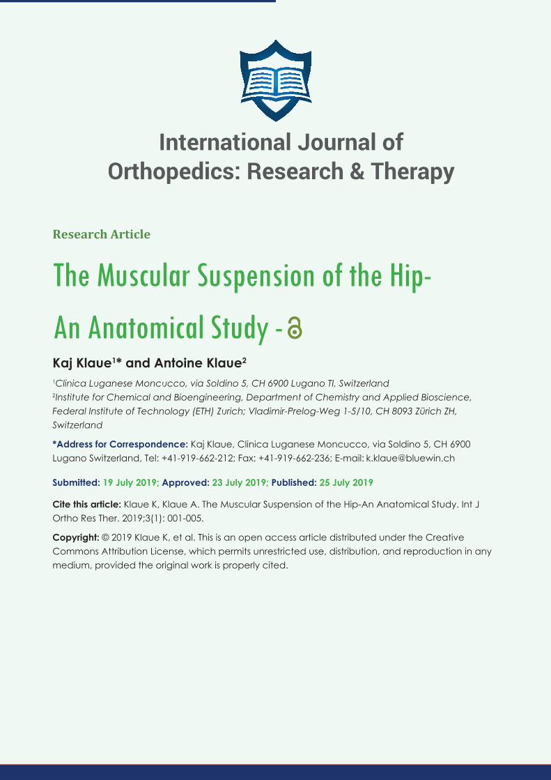
well individualized between the distal margins of the m. glutaeus medius and the m. adductor magnus at the posterior aspect of the hip (Figures 1-3).
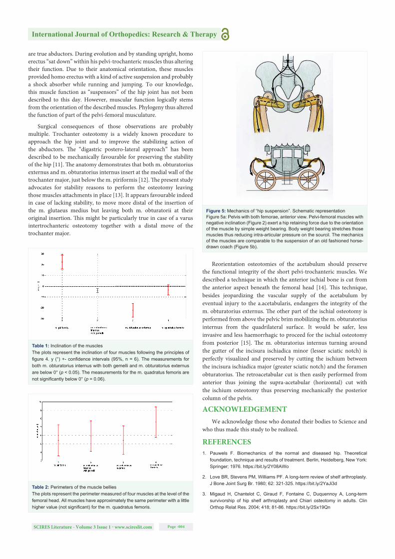
A hinged goniometer was used to measure the angle between the straight margins of the muscles listed above and the horizontal line (Figure 4).
We defi ne the inclination of a muscle by considering the mean value of both angles formed by the proximal and the distal margins and the vertical axis (angle bisector). Negative inclination of a muscle means that the muscle is directed towards lateral proximal. Positive inclination of a muscle means that the muscle is directed towards lateral distal (Figure 4).
A centimeter strap was used to evaluate the perimeter of the middle part of the muscle belly.
Following parameters were thus measured successively:
• Angle of the inferior margin of the m. glutaeus medius
• Angle of the superior margin of the m. piriformis
• Angle of the inferior margin of the m. piriformis
Figure 1: Posterior dissection of the left hip The m. maximus is reclined towards lateral distal. Following structures are well visualized: a: m.gluteus medius, b: m. piriformis, c: nervus ischiadicus, d: m. obturatorius internus with m. gemellii, e: m. quadratus femoris. The sacro-tuberal ligament has been removed.
International Journal of Orthopedics: Research & Therapy
SCIRES Literature - Volume 3 Issue 1 - www.scireslit.com Page -003
• Perimeter of the m. piriformis
• Angle of the superior margin of the m. gemellus superior (m. obturatorius internus)
• Angle of the inferior margin of the m. gemellus inferior (m. obturatorius internus)
• Perimeter of the m. obturatorius internus and both m. gemelli
• Angle of the superior margin of the m. quadratus femoris
• Angle of the inferior margin of the m. quadratus femoris
• Perimeter of the m. quadratus femoris
• Angle of the superior margin of the m. obturatorius externus
• Angle of the inferior margin of the m. obturatorius externus
• Perimeter of the m. obturatorius externus
RESULTS4 hips in two cadavers were used for further dissection and were
not included in the quantitative series. 6 hips in 3 cadavers were assessed quantitatively and the results are plotted in table 1 and 2.
Th e inclinations are plotted in table 1 considering the confi dence intervals (95%). Th e inclination angle of the distal margin of the m. glutaeus medius was not plotted because it does not correspond to the direction of the whole muscle. Its inclination however is clearly positive: yº +- confi dence intervalº = 37º +- 6º. All 3 muscles: m. obturatorius internus with m. gemelli, m. quadratus femoris and m. obturatorius externus have a negative inclination. Th e negativity of the inclination of the m. quadratus femoris is however not signifi cant (p = 0.06). Th e inclination of the m. piriformis is clearly positive (p < 0.05).
Th e perimeters of the muscle bellies are plotted in table 2 considering the confi dence intervals ((95%). Interestingly, all muscle bellies have about the same dimension at the level of the femoral head and their diff erences are not signifi cant (p > 0.05).
DISCUSSIONPauwels calculated that considering the single abductor muscles,
to maintain equilibrium of the joint, the muscular force must be about three times greater than the weight supported [1]. He asserted that the resultant force always crosses the upper part of the facies semilunaris (sourcil) of the acetabulum. Our measurements of the orientation of both m. obturatorii and gemelli together with m. quadratus femoris demonstrate a “passive spring mechanism” which stabilizes the joint. Due to the active contractility of the muscles, the spring mechanism can become an “active spring mechanism” reducing the articular pressure on the sourcil. Th e weight bearing pelvic bone is “suspended” on both proximal femurs. Mechanically, such spring mechanism is known in various mechanical constructions requiring shock absorption (Figure 5a and 5b).
Phylogeny of homo erectus demonstrates how the small hip abductors in fl exion became hip “braces”. Th e ancestor of homo erectus was probably a quadruped mammal with a dimension of a dog. At this stage, the hips bore the weight of the posterior part of the body having the hips fl exed. In this position, the m. glutaei are rather extensors-external rotators and the shorter pelvi-trochanteric muscles
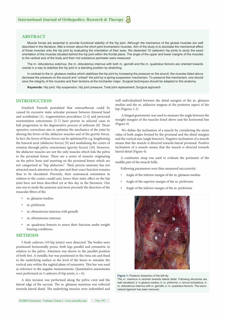
Figure 2: Posterior dissection of the left hip joint, detailed viewThe nervus ischiadicus and the m. piriformis have been removed. Them. obturatorius internus (a) is refl ected around the ischiatic bone within the incisura ischiadica minor. It originates on a wide area on the foramen obturatorius and the quadrilateral surface. The m. gemellus superior (b) originates at the spina ischiadica. The m. gemellus inferior is a quite strongmuscle which originates at the inferior part of the incisura ischiadica minor. The m. quadratus femoris. (c) originates at the anterior aspect of the tuberischiadicum.
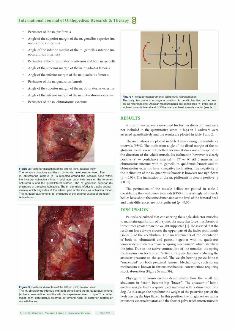
Figure 3: Posterior dissection of the left hip joint, detailed view.The m. obturatorius internus with both gemelli and the m. quadratus femoris (a) have been reclined and the articular capsule removed. b: tip of Trochanter major. c: m. obturatorius externus. d: femoral neck. e: posterior acetabular rim with limbus.
Figure 4: Angular measurements. Schematic representationThe body lies prone in orthogonal position. A metallic bar lies on the rima ani as reference line. Angular measurements are considered “+” if the line is inclined towards lateral and “-” if the line is inclined towards medial (see text).
International Journal of Orthopedics: Research & Therapy
SCIRES Literature - Volume 3 Issue 1 - www.scireslit.com Page -004
are true abductors. During evolution and by standing upright, homo erectus “sat down” within his pelvi-trochanteric muscles thus altering their function. Due to their anatomical orientation, these muscles provided homo erectus with a kind of active suspension and probably a shock absorber while running and jumping. To our knowledge, this muscle function as “suspensors” of the hip joint has not been described to this day. However, muscular function logically stems from the orientation of the described muscles. Phylogeny thus altered the function of part of the pelvi-femoral musculature.
Surgical consequences of those observations are probably multiple. Trochanter osteotomy is a widely known procedure to approach the hip joint and to improve the stabilizing action of the abductors. Th e “digastric postero-lateral approach” has been described to be mechanically favourable for preserving the stability of the hip [11]. Th e anatomy demonstrates that both m. obturatorius externus and m. obturatorius internus insert at the medial wall of the trochanter major, just below the m. piriformis [12]. Th e present study advocates for stability reasons to perform the osteotomy leaving those muscles attachments in place [13]. It appears favourable indeed in case of lacking stability, to move more distal of the insertion of the m. glutaeus medius but leaving both m. obturatorii at their original insertion. Th is might be particularly true in case of a varus intertrochanteric osteotomy together with a distal move of the trochanter major.
Table 1: Inclination of the muscles The plots represent the inclination of four muscles following the principles of fi gure 4. y (°) +- confi dence intervals (95%, n = 6). The measurements for both m. obturatorius internus with both gemelli and m. obturatorius externus are below 0° (p < 0.05). The measurements for the m. quadratus femoris are not signifi cantly below 0° (p = 0.06).
Table 2: Perimeters of the muscle belliesThe plots represent the perimeter measured of four muscles at the level of the femoral head. All muscles have approximately the same perimeter with a little higher value (not signifi cant) for the m. quadratus femoris.
Figure 5: Mechanics of “hip suspension”. Schematic representationFigure 5a: Pelvis with both femorae, anterior view. Pelvi-femoral muscles with negative inclination (Figure 2) exert a hip retaining force due to the orientation of the muscle by simple weight bearing. Body weight bearing stretches those muscles thus reducing intra-articular pressure on the sourcil. The mechanics of the muscles are comparable to the suspension of an old fashioned horse-drawn coach (Figure 5b).
Reorientation osteotomies of the acetabulum should preserve the functional integrity of the short pelvi-trochanteric muscles. We described a technique in which the anterior ischial bone is cut from the anterior aspect beneath the femoral head [14]. Th is technique, besides jeopardizing the vascular supply of the acetabulum by eventual injury to the a.acetabularis, endangers the integrity of the m. obturatorius externus. Th e other part of the ischial osteotomy is performed from above the pelvic brim mobilizing the m. obturatorius internus from the quadrilateral surface. It would be safer, less invasive and less haemorrhagic to proceed for the ischial osteotomy from posterior [15]. Th e m. obturatorius internus turning around the gutter of the incisura ischiadica minor (lesser sciatic notch) is perfectly visualized and preserved by cutting the ischium between the incisura ischiadica major (greater sciatic notch) and the foramen obturatorius. Th e retroacetabular cut is then easily performed from anterior thus joining the supra-acetabular (horizontal) cut with the ischium osteotomy thus preserving mechanically the posterior column of the pelvis.
ACKNOWLEDGEMENTWe acknowledge those who donated their bodies to Science and
who thus made this study to be realized.
REFERENCES1. Pauwels F. Biomechanics of the normal and diseased hip. Theoretical
foundation, technique and results of treatment. Berlin, Heidelberg, New York: Springer; 1976. https://bit.ly/2Y08AWo
2. Love BR, Stevens PM, Williams PF. A long-term review of shelf arthroplasty. J Bone Joint Surg Br. 1980; 62: 321-325. https://bit.ly/2YaJi3d
3. Migaud H, Chantelot C, Giraud F, Fontaine C, Duquennoy A, Long-term survivorship of hip shelf arthroplasty and Chiari osteotomy in adults. Clin Orthop Relat Res. 2004; 418; 81-86. https://bit.ly/2Sx19Qn
International Journal of Orthopedics: Research & Therapy
SCIRES Literature - Volume 3 Issue 1 - www.scireslit.com Page -005
4. Fawzy E, Mandellos G, De Steiger R, Mc.Lardy-Smith P, Benson MK, Murray D. Is there a place for shelf actabuloplasy in the management of adult acetabular dysplasia? A survivorship study. J Bone Joint Surg Br. 2005; 87: 1197-1202. https://bit.ly/30PbMB8
5. Toyama H, Endo N, Sofue M, Dohmae Y, Takahashi HE. Relief from pain after Bombelli’s valgus-extension osteotomy, and effectiveness of the combined shelf operation. J Orthop Sci. 2000; 5: 114-123. https://bit.ly/2OeUQCD
6. Debnath UK, Guha AR, Karlakki S, Varghese J, Evans GA. Combined femoral and Chiari osteotomies for reconstruction of the painful subluxation or dislocation of the hip in cerebral palsy. A long-term outcome study. J Bone Joint Surg Br. 2006; 88: 1373-1378. https://bit.ly/2Z7mS45
7. Spence G, Hocking R, Wedge JH, Roposch A. Effect of innominate and femoral varus derotation osteotomy on acetabular development in developmental dysplasia of the hip. J Bone Joint Surg Am. 2009; 91: 2622-2636. https://bit.ly/2OeWF2r
8. Mechlenburg I. Evaluation of Bernese periacetabular osteotomy: prospective studies examining projected load-bearing area, bone density, cartilage thickness and migration. Acta Orthop Suppl. 2008; 79: 4-43. https://bit.ly/2M7m1wI
9. Maquet P. Importance of the position of the greater Trochanter. Acta Orthop Belg. 1990; 56: 307-322. https://bit.ly/2ZaEmMU
10. Klaue K, Ganz R. Pelvic Osteotomies in the Adult. In: Chapman MW, Madison M. Operative Orthopaedics. 2nd ed. Philadelphia: J.B. Lippincott Company; 1993. p. 1835-1844. https://bit.ly/2Z7Ubnv
11. Mercati E, Guary A, Myquel C, Bourgeon A. A postero-external approach to the hip joint. Value of the formation of a digastric muscle. J Chir (Paris). 1972; 103: 499-504. https://bit.ly/2Z8of2n
12. MacMinn RMH, Hutchings RT. A Colour Atlas of Human Anatomy. Wolfe Medical Publications Ltd; London. 1993. https://bit.ly/2GrAyPZ
13. Lakstein D, Kosashvili Y, Backstein D, Safi r O, Gross AE. Modifi ed extended trochanteric osteotomy with preservation of posterior structures. Hip Int. 2010; 20: 102-108. https://bit.ly/30RL9f2
14. Ganz R, Klaue K, Vinh TS, Mast JW. A New periacetabular osteotomy for the treatment of hip dysplasias. Technique and preliminary results. Clin Orthop Relat Res. 1988; 232: 26-36. https://bit.ly/2Gne5Dy
15. Janssen D, Kalchschmidt K, Katthagen BD. Triple pelvic osteotomy as treatment for osteoarthritis secondary to developmental dysplasia of the hip. Int Orthop. 2009; 33: 1555-1559. https://bit.ly/2M1r2Xw