Embed Size (px)
Citation preview
Intercalary bone allografts 23 tumor cases followed for 3 years Jose Antonio Cara, Antonio Laclériga and Jose Cañadell Department of Orthopedics, Navarra University Hospital. Av Pio XII S/N , E-31080, Pamplona, Spain. ABSTRACT From 1987 to 1991, 26 patients with malignant bone tumors (osteosarcoma, Ewing's sarcoma, chondrosarcoma and malignant fibrous histiocytoma) in the diaphysis or metaphysis of long bones have been treated by chemotherapy, radiotherapy and intercalary bone allograft replacement alter resection. The mean follow-up was 3 years in 23 patients, 2 died from tumors, and 1 had an amputation because of local recurrence. Allograft incorporation in 11 metaphyseal anastomoses was excellent in all cases. In the 23 diaphyseal anastomoses there were 6 nonunions. Other complications were deep infection in 3 cases, leg length discrepancy in 2 and allograft fracture in 2. Function became satisfactory in 14 cases. INTRODUCTION Advances in tumor and treatment have made limb salvage possible. Use of a bone allograft is common for reconstruction of large bone defects after tumor surgery (Mankin et al. 1982, Alho et al. 1987, Delépine and Delépine 1988, Alho et al. 1989, Kattapuram et al. 1989, Shinohara et al. 1990, Cara et al. 1991b, 1992). We report our observations on results, allograft incorporation and complications in a consecutive series of patients. PATIENTS AND METHODS From 1987 through 1991, 13 men and 13 women with stage IIB carcomas in the diaphysis (14 cases) or metaphysis (12 cases) of long bones have been treated by chemotherapy, radiotherapy, resection and intercalary bone allograft replacement. The mean age was 16 (5-48) years. The length of the defect was 20 (10-32) cm and the mean follow-up was 3 (1-6) years (Table 1) The treatment followed the Cancer Protocol of our hospital (Cañadell 1984). This treatment is osteosarcoma: preoperative intra-arterial chemotherapy, surgery and intraoperative radiotherapy, and postoperative intravenous chemotherapy; Ewing's sarcoma: preoperative intravenous chemotherapy and radiotherapy, surgery and intraoperative radiotherapy, and postoperative intravenous chemotherapy; chondrosarcoma: only surgery; malignant fibrous histiocytoma: preoperative intraarterial chemotherapy, surgery and intraoperative radiotherapy and postoperative intravenous chemotherapy and radiotherapy. We obtained bone allografts from cadavers meeting the selection criteria of the American Association of Tissue Banks
(Friedlaender and Mankin 1981, Amillo and Cañadell 1990, Amillo et al. 1990). Antibiotics (Cefazolina) were administered intravenously intraoperatively and postoperatively for at least 1 week and then orally for 3 weeks. The surgical procedure was resection of the lesion, including biopsy scars with a wide bone and soft-tissue margin, 1 cm transversely and 5 cm longitudinally in the diaphyseal lesion, based on the radiologic studies (Figure 1). Physeal resection, according to the technique of Cañadell et al. (1991) was used in 10 patients. In these cases, the margin was marginal. This technique comprises 3 phases: 1. The external fixator is attached: 2 pins are inserted into the epiphysis, and a further 2 in the diaphysis at least 8-10 cm above the tumor. Distraction should be commenced in the operating room, and should continue at the rate of 1-2 mm/day until a distraction of 2 cm is reached. This first phase can be carried out when the patient is finishing the course of neoadjuvant chemotherapy. 2. En bloc resection of the tumor tissue: once the epiphysiolysis has been checked by radiographs, we proceed to resected the tumor. The resected piece is sent for histologic studies, and chains of PMMA with gentamicin are inserted to fill the space; the fixator is left in position. 3. Reconstruction of the bone defect: once the histology report has confirmed the absence of tumor in the edges of the resected piece, we proceed to withdraw the chains of gentamicin PMMA, and to intercalate a bone graft. Where the histology report is positive, the epiphysis is resected, leaving a broad margin, and the limb is reconstructed by means of a prosthesis or arthrodesis. In the diaphyseal union, the osteosynthesis used was screws and plates (DCS®, DHS® or DCP®) in all cases. In 4 cases, we added Ender's nail. In the metaphyseal union, we used Ender's nail and staples. The patients performed active exercises under Glose supervision until a reasonable range of joint motion was achieved. When the graft was in a lower extremity, unrestricted weight bearing was not advised until radiographs showed a solid union at the anastomosis sites. Patients were seen as outpatients at regular intervals and were studied by appropriate radiographs, laboratory determinations and bone scans. The functional results of surgery were evaluated following the classification adopted from the MTST (Enneking 1987). The radiographic evaluation of the bone allografts was analyzed following the ISOLS protocol (Cara et al. 1992). RESULTS We evaluated 23 patients because 2 died from metastases or local recurrence, and 1 required an amputation for a local recurrence. Complications There were 1 superficial and 3 deep infections. The deep infections were treated by removal of the infected allograft. The length of the limb was maintained either by an antibiotic-impregnated polymethylmethacrylate spacer (Gentamicin-PMMA beads) or by bone cement with gentamicin (Cara et al. 1991a) and external fixation. Antibiotics were then administered for 1-3 months. Gram-positive organisms were the most common cause of infection, with 2 infections due to Staphylococus epidermidis and the other mixed flora.
The infection healed in 2 of the 3 patients and a new allograft was implanted. We had 6 delayed unions, 5 in the femur and 1 in the tibia, all were in diaphyseal anastomoses (Figure 2). They were treated by reosteosynthesis plus autograft bone. 4 patients had an excellent consolidation, 1 good and 1 poor. 2 patients presented bone allograft fractures Type III (Zehr et al. 1991) 2.5 and 3 years after the operation and 1 was treated with a monolateral external fixator. Later, he presented infection of the graft (Case 19, see above). The other was treated with a condylar plate osteosynthesis plus autograft; 6 months later, the fracture had consolidated. 5 children presented limb length discrepancies because of the growth of the patient. 2 of them were treated by bone lengthening (Cañadell et al. 1991 and Cara et al. 1991c). Other complications were 2 permanent common peroneal nerve palsies. Radiographic evaluation All the patients presented metaphyseal and diaphyseal unions. The radiographic evaluation of the metaphyseal reconstruction was found to be excellent in all cases as regards allograft incorporation (Figure 3). The result in the diaphysis was worse. The evaluations of diaphyseal union are listed in Table 2. Function Function was excellent in 9 cases, good in 5, fair in 5 and poor in 4 cases. DISCUSSION 14 of our 23 patients had a good or excellent function, similar to other series (Kattapuram et al. 1989, Capanna and Donati 1991). Mankin (1982) reports a better functional result (100 percent graded excellent). Delayed union or non-union in the diaphysis was the main problem. It may be related to suboptimal bone contact at the host-donor junction site or to inadequate osteosynthesis. It is also possible that it represent a subtle form of rejection. Currently, our technique of fixing the graft to the host bone aims to make surfaces as plane as possible and anastomose them using either an AO DCP® compression plate or DCS/DHS®. In several recent cases we have used an oblique osteotomy, rigid fixation and cancellous bone graft at the site of osteotomy to reduce the development of this complications. We have seen a faster consolidation in these patients. We prefer to use plates and screws because we have more experience with these methods of osteosynthesis. Other authors recommend intramedullary nailing with interlocking screw fixation. We do not know if the postoperative chemotherapy and radiotherapy have an adverse effect on allograft healing. Many series of bone allografts demonstrate that infection is the principal complication (Tomford et al. 1981, Lord et al. 1988 and Tomford et al. 1990). Our incidence (3/23) is like that in these series. The high rate of infection may have been related to predisposing factors, such as duration of the operation, blood loss, soft tissue injury, chemotherapy and radiation.
REFERENCES
1. Alho A, Karaharju E O, Korkala O, Laasonen E. Hemijoint allografts in the treatment of low-grade malignant and aggressive bone turnours about the knee. Int Orthop 1987: 11 (1) : 35-41.
2. Alho A, Karaharju E O, Korkala O, Laasonen E M, Holmstrom T, Muller C. Allogeneic grafts for bone tumor. 21 cases of osteoarticular and segmental grafts. Acta Orthop Scand 1989: 60 (2): 143-53.
3. Amillo S, Cañadell J. Banco de huesos y de otros tejidos del sistema musculoesquelético. EUNSA. Pamplona 1989.
4. Amillo S, Cara J A, Valenti J R. Banco de tejidos del sistema musculoesquelético. Aplicaciones clínicas. Rev Med Univ Navarra 1990 34 (4): 227-34.
5. Cañadell J. Protocolos terapéuticos del cáncer de la Clínica Universitaria de Navarra. Departamento de Cirugía Ortopédica y Traumatología. EUNSA, Pamplona 1984.
6. Cañadell J, Cara J A, Ganoza C. Physeal distraction and bone lengthening in the conservative treatment of malignant bone tumors in children. In: Treatment of Malignant Bone Tumors in Children and Adolescents. (Eds. Cañadell J, Sierrasesumaga L, Calvo F, y Ganoza C) EUNSA, Pamplona 1991: 293-305.
7. Capanna R, Donati, D. Joint study on intercalary and arthrodesis allografts. In: Proc 4th Meet Europ Musculoskeletal Oncology Soc (EMSOS), Birmingham 1991:7.
8. Cara J A, Cañadell J, Laclériga A. Infection in grafting procedures. In: Complications of Limb Salvage (Ed. Brown K) Publisher ISOLS, Montreal 1991a: 41-4.
9. Cara J A, Gil Albarova J, Amillo S, Cañadell J. Bone allograft after segmental resection of bone tumors. In: New Trends in Bone Grafting (Ed. Lindholm T S) University of Tampere 1991b: 191-9.
10. Coral A, Gil Albarova J, Cañadell J. Correction of late limb length discrepancies in limb salvage surgery. In: Complictions of Limb Salvage (Ed. Brown K) Publisher ISOLS, Montreal 1991c: 533-8.
11. Cara J A, Amillo S, Cañadell J. La prótesis de resección de rodilla en la cirugía reconstructiva tumoral. Rev Ortop Traum 1992; 36: 39-42
12. Delépine G, Delépine N. Résultats préliminaires de 79 altogreffes osseuses massives dans le traitement conservateur des tumeurs malignes de l'adulte et de l'enfant. Int Orthop 1988; 12 (11: 21-9.
13. Enneking W F. Limb salvage in musculoskeletal oncology. Churchill Livingstone, New York 1987: 626.
14. Friedlaender G E, Mankin H J. Bone banking: current methods and suggested guidelines. Instr Course Lect 1981; 30: 36-55.
15. Kattapuram S V, Phillips W C, Mankin H J. Intercalary bone allografts: radiographic evaluation. Radiology 1989; 170 (1 Pt 1): 137-41.
16. Lord C F, Gebhardt M C, Tomford W W, Mankin El J. Infection in bone allografts. Incidente, nature, and treatment. J Bone Joint Surg (Am) 1988; 70 (3): 369-76.
17. Mankin H J, Doppelt S H, Sullivan T R, Tomford W W. Osteoarticular and intercalary allograft transplantation in the management of malignant tumors of bone. Cancer 1982; 50 (4): 613-30.
18. Shinohara N, Sumida S, Masuda S. Bone allografts after segmental resection of tumours. Int Orthop 1990; 14 (3): 273-6.
19. Tomford W W, Starkweather R J, Goldman M H. A study of the clinical incidence of infection in the use of banked allograft bone. J Bone Joint Surg (Am) 1981; 63 (2): 244-8.
20. Tomford W W, Thongphasuk J, Mankin H 1, Ferrar() M J. Frozen musculoskeletal allografts. A study of the clinical incidence and causes of infection associated with their use. 1 Bone Joint Surg (Am) 1990; 72 (8): 1137-43.
21. Zehr R J, Enneking W F, Heare T, Liang T S. Fractures in large structural allografts. In: Complications of Limb Salvage (Ed. Brown K) Publisher ISOLS, Montreal 1991: 3-7.
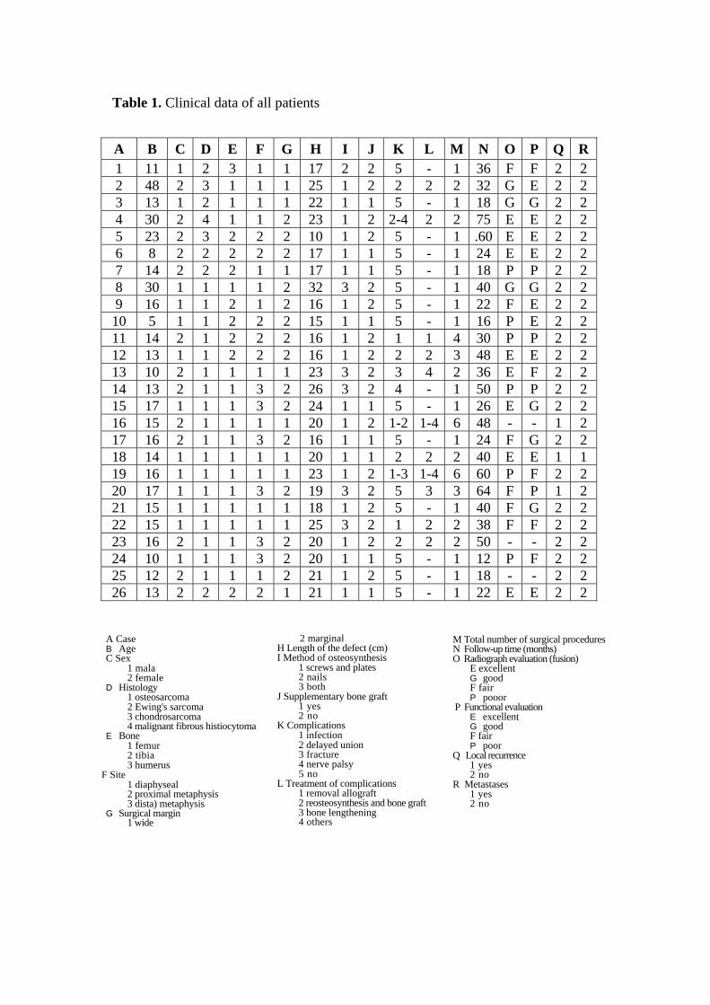
Table 1. Clinical data of all patients
A B C D E F G H I J K L M N O P Q R 1 11 1 2 3 1 1 17 2 2 5 - 1 36 F F 2 22 48 2 3 1 1 1 25 1 2 2 2 2 32 G E 2 23 13 1 2 1 1 1 22 1 1 5 - 1 18 G G 2 24 30 2 4 1 1 2 23 1 2 2-4 2 2 75 E E 2 25 23 2 3 2 2 2 10 1 2 5 - 1 .60 E E 2 26 8 2 2 2 2 2 17 1 1 5 - 1 24 E E 2 27 14 2 2 2 1 1 17 1 1 5 - 1 18 P P 2 28 30 1 1 1 1 2 32 3 2 5 - 1 40 G G 2 29 16 1 1 2 1 2 16 1 2 5 - 1 22 F E 2 210 5 1 1 2 2 2 15 1 1 5 - 1 16 P E 2 211 14 2 1 2 2 2 16 1 2 1 1 4 30 P P 2 212 13 1 1 2 2 2 16 1 2 2 2 3 48 E E 2 213 10 2 1 1 1 1 23 3 2 3 4 2 36 E F 2 214 13 2 1 1 3 2 26 3 2 4 - 1 50 P P 2 215 17 1 1 1 3 2 24 1 1 5 - 1 26 E G 2 216 15 2 1 1 1 1 20 1 2 1-2 1-4 6 48 - - 1 217 16 2 1 1 3 2 16 1 1 5 - 1 24 F G 2 218 14 1 1 1 1 1 20 1 1 2 2 2 40 E E 1 119 16 1 1 1 1 1 23 1 2 1-3 1-4 6 60 P F 2 220 17 1 1 1 3 2 19 3 2 5 3 3 64 F P 1 221 15 1 1 1 1 1 18 1 2 5 - 1 40 F G 2 222 15 1 1 1 1 1 25 3 2 1 2 2 38 F F 2 223 16 2 1 1 3 2 20 1 2 2 2 2 50 - - 2 224 10 1 1 1 3 2 20 1 1 5 - 1 12 P F 2 225 12 2 1 1 1 2 21 1 2 5 - 1 18 - - 2 226 13 2 2 2 2 1 21 1 1 5 - 1 22 E E 2 2
A Case B Age C Sex
1 mala 2 female
D Histology 1 osteosarcoma 2 Ewing's sarcoma 3 chondrosarcoma 4 malignant fibrous histiocytoma
E Bone 1 femur 2 tibia 3 humerus
F Site 1 diaphyseal 2 proximal metaphysis 3 dista) metaphysis
G Surgical margin 1 wide
2 marginal H Length of the defect (cm) I Method of osteosynthesis
1 screws and plates 2 nails 3 both
J Supplementary bone graft 1 yes 2 no
K Complications 1 infection 2 delayed union 3 fracture 4 nerve palsy 5 no
L Treatment of complications 1 removal allograft 2 reosteosynthesis and bone graft 3 bone lengthening 4 others
M Total number of surgical procedures N Follow-up time (months) O Radiograph evaluation (fusion)
E excellent G good F fair P pooor
P Functional evaluation E excellent G good F fair P poor
Q Local recurrence 1 yes 2 no
R Metastases 1 yes 2 no
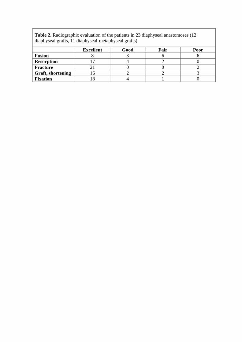
Table 2. Radiographic evaluation of the patients in 23 diaphyseal anastomoses (12 diaphyseal grafts, 11 diaphyseal-metaphyseal grafts)
Excellent Good Fair Poor Fusion 8 3 6 6 Resorption 17 4 2 0 Fracture 21 0 0 2 Graft, shortening 16 2 2 3 Fixation 18 4 1 0

Figure 1. Case 3 Ewing's sarcoma of the diaphysis of the femur in a 13-year-old boy. After resection and an intercalary allograft replacement.

Figure 2. Case 18 A fatigue fracture of the graft and plate 2 years after the transplantation was treated by reosteosynthesis and autogenous subperiosteal graft.
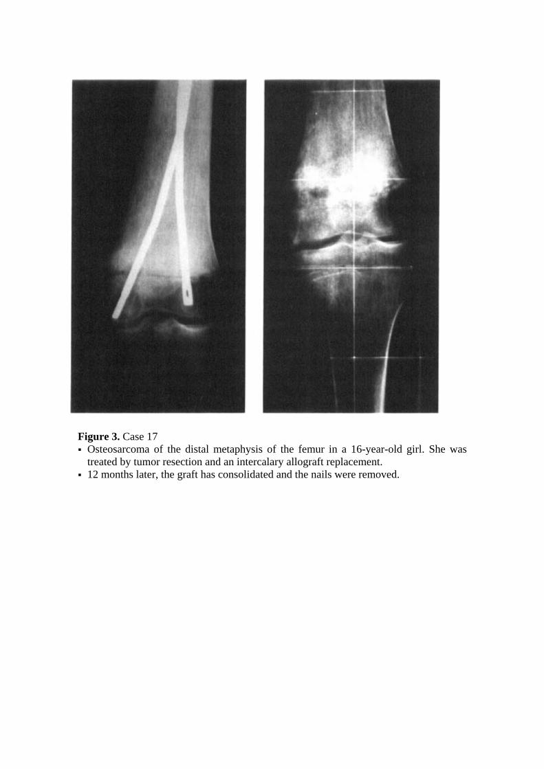
Figure 3. Case 17 Osteosarcoma of the distal metaphysis of the femur in a 16-year-old girl. She was
treated by tumor resection and an intercalary allograft replacement. 12 months later, the graft has consolidated and the nails were removed.


![Natural Coral-Based Bone Substitutescause loss of essential bone-like and resorption qualities. Furthermore, in Europe, the use of allografts is limited due to legal issues [5, 6]](https://img.dokumen.tips/doc/110x75/5e70c77407cb5744ce4fdb48/natural-coral-based-bone-substitutes-cause-loss-of-essential-bone-like-and-resorption.jpg)