Embed Size (px)
Citation preview
16
Integrated PET/CT in the Staging of NSCLC
Walter De Wever University Hospitals – Leuven
Belgium
1. Introduction
Lung cancer is a common disease with approximately 3-million new cases per year worldwide and is the leading cause of cancer-related death in many countries. Eighty percent of the lung cancers are non-small cell lung cancers (NSCLC) and 20% are small cell lung cancers (SCLC) [1]. The imaging diagnostic assessment of patients with lung cancer includes morphological imaging modalities such as chest X-ray, Computed Tomography (CT) and Magnetic Resonance (MR) as well as metabolic imaging modalities such as nuclear medicine procedures, including Positron Emission Tomography (PET) and PET/CT. Staging a patient with lung cancer implies an accurate determination of the size of the tumor, the potential infiltration of the tumor into the adjacent structures, the involvement of hilar and mediastinal lymph nodes and the detection of distant metastases. Till recently, CT was the routine imaging procedure for staging patients with NSCLC. The success of CT is related to the very detailed imaging information of the localization and extent of the tumor, the presence of enlarged lymph nodes and the presence of metastatic disease [2]. PET has more recently been introduced in tumor staging and it has been used successfully for detection of primary tumors, metastases, early tumor recurrence, and for the detection of metastatic lymph nodes [3, 4]. This imaging modality possesses a greater sensitivity for detection of malignancy though it is inhibited by relatively poor spatial resolution and anatomical localization of disease. Combining detailed anatomical information obtained by CT with metabolic information from PET seems logical, therefore. Integrated PET/CT is the most recent approach to post hoc image fusion. It combines these image modalities into one scanner that acquires accurately aligned anatomical and metabolical images in the same scanning session [1].
2. Evaluation of a solitary pulmonary nodule
Lung lesions detected in a chest radiograph, incidentally or by systematic investigation, need a definite confirmation of diagnosis. The key point is the evaluation of malignancy in peripheral lung nodules. A solitary pulmonary nodule (SPN) is defined as a focal round or oval lung lesion with a diameter smaller than 3 cm, completely surrounded by normal lung tissue, not associated with atelectasis or adenopathies. Lung lesions greater than 3 cm are classified as masses [5]. Non-invasive evaluation of SPN is usually performed by different imaging procedures including CT, MR, PET and PET/CT. CT is considered to be an excellent tool for the detection and localization of SPNs with a good sensitivity (96%, range 91–98%) but a poor specificity (50%, range 41–58%) [6]. CT provides data regarding the nodule shapes, borders and density [7]. Central or concentric calcifications, round shape or a morphologic stability
www.intechopen.com
Computed Tomography – Clinical Applications
256
over 2 years are features of benignancy. On the contrary, non-demarcated borders, calcifications with eccentric appearance or spiculated pattern, a doubling time of <10 month and cavitation or pseudocavitation are features of malignancy [8, 9]. Contrast enhanced CT can be used to characterize SPN. Enhancement of the pulmonary nodule with 15 Hounsfield Units (HU) has a sensitivity and specificity of 98% and 58% in the detection of malignancy, respectively and the absence of lung nodule enhancement is strongly predictive of a benign diagnosis (negative predictive value of 96.5%) [10]. Several studies showed that PET had similar sensitivity (92–95%) but superior specificity (72–83%) as compared to CT for the characterization of SPN [11, 12]. In one of the largest published studies (450 patients) with lung nodules evaluated with PET, Gould et al. reported a high PET sensitivity (94.2%) and specificity (83.3%) [12]. Lower sensitivity (91.7%) but similar specificity (82.3%) was observed by Fletcher et al. in a population of 344 patients [13]. Malignant lesions with a size of more than 10 mm in diameter are detected with a sensitivity of 96% [14]. Gupta et al. showed that PET using Fluor-Deoxy-Glucose (FDG) as tracer, is highly accurate in differentiating malignant from benign solitary pulmonary nodules for sizes from 6 to 30 mm when radiographic findings are indeterminate [15]. An important contribution of two more recent meta-analyses [16, 17] is the assessment of the performance of FDG-PET in small lung nodules. Nomori et al. demonstrated that the sensitivity clearly decreases for malignant lung lesions of less than 10 mm in diameter [18]. For technical reasons, the lower limit has to be set in dependence from the spatial resolution and will be around 6–10 mm according to the most common PET scanners used. Thus, FDG-PET is not indicated for the evaluation of solitary pulmonary nodules of less than 6–10 mm. The meta-analysis of Ung et al. mentioned that the accuracy for the characterization of lung lesions by FDG-PET depends on the amount of tumoral FDG uptake given as Standardized Uptake Value (SUV) [16]. Grgic et al. reported that it is possible to estimate the individual risk for malignancy considering the SUVmax of a given nodule and clinically relevant information [19]. The authors reported that the mean SUVmax of malignant SPNs was higher than benign lesions (SUVmax 9.7 ± 5.5 vs. 2.6 ± 2.5; P < 0.01). False positive FDG-PET findings are represented by lung inflammatory conditions such as pneumonia, pyogenic abscesses, aspergillosis, granulomatous diseases (tuberculosis, sarcoidosis, histoplasmosis, Wegener's granulomatosis) [20]. False negative findings on PET images can be the result of small lesion size (<1 cm) or tumor types characterized by low glucose metabolism (such as Neuro Endocrine Tumors (NET), Broncho Alveolar Cell Carcinomas (BAC) or pulmonary carcinoids [21, 22]. The widespread introduction in clinical practice of FDG PET/CT has allowed a more accurate assessment of SNPs. With integrated PET/CT, additional certainty to the presence or absence of FDG uptake in the pulmonary nodule can be achieved because morphologic CT criteria and metabolical PET criteria are available simultaneously [23]. Kim et al. reported a sensitivity of 97% and a specificity of 85% for the detection of SPNs, concluding that the combination of the anatomical and metabolic images preserves the sensitivity of the CT and the specificity of the PET scan, but improves significantly the overall accuracy [24, 25].
3. Assessment of T stage
The most significant improvement in staging results with combined PET/CT compared with PET alone relates to T staging. This superiority is attributed entirely to the CT component of the examination [26]. The major benefit of PET/CT lies in the direct link between the information on metabolic changes of structures and the highly detailed anatomic CT
www.intechopen.com
Integrated PET/CT in the Staging of NSCLC
257
information of these structures. Recent published studies showed that PET/CT is the best noninvasive imaging technique for correct prediction of T-stage: PET/CT correctly predicts the T-stage in +/_ 82% of cases, in comparison with 55, 68 and 76% for PET, CT and visual correlation of PET and CT, respectively [20]. Halpern et al.[27] demonstrated an accuracy rate of 97% with PET/CT compared with 67% with PET only. Another study by Cerfolio et al. [28] showed that PET/CT more accurately predicted T status (70% of cases) than did PET alone (47%). Lardinois et al. described accuracy rates for T staging with PET/CT and CT as 88% and 58%, respectively [29]. Pauls et al. found that the advantages of integrated PET/CT also depend on the histological T-stage of the primary tumor [30]. Changes of the therapeutic strategy due to PET/CT are especially seen in T3 and T4 tumors. It has been demonstrated that integrated PET/CT is a useful tool for the detection of tumor invasion into the chest wall [1, 29, 31]. Due to the exact anatomic correlation to the FDG uptake, the delineation of the primary tumor can be defined precisely. Integrated PET/CT provides important information on mediastinal infiltration as well. In addition, PET can be helpful in evaluating the cause of pleural effusions [29] (figure 1). One of the most important attributes of PET/CT is the ability to distinguish between tumor and distal atelectasis (figure 2). This is particularly important for the planning of radiotherapy in patients with lung cancer associated with atelectasis [23]. Table 1 summarizes the most important studies concerning T-staging with PET/CT.
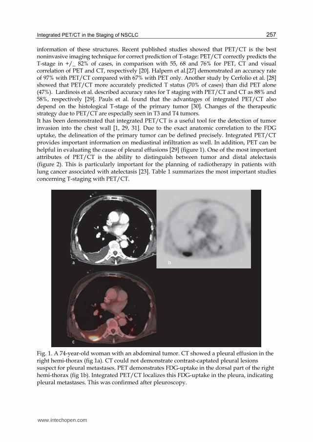
Fig. 1. A 74-year-old woman with an abdominal tumor. CT showed a pleural effusion in the right hemi-thorax (fig 1a). CT could not demonstrate contrast-captated pleural lesions suspect for pleural metastases. PET demonstrates FDG-uptake in the dorsal part of the right hemi-thorax (fig 1b). Integrated PET/CT localizes this FDG-uptake in the pleura, indicating pleural metastases. This was confirmed after pleuroscopy.
www.intechopen.com
Computed Tomography – Clinical Applications
258
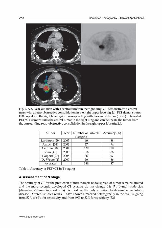
Fig. 2. A 57-year-old man with a central tumor in the right lung. CT demonstrates a central mass with a retro-obstructive consolidation in the right upper lobe (fig 2a). PET demonstrates FDG uptake in the right hilar region corresponding with the central tumor (fig 2b). Integrated PET/CT demonstrates the central tumor in the right lung and can delineate the tumor from the surrounding retro-obstructive consolidation in the right upper lobe (fig 2c).
Author Year Number of Subjects Accuracy (%)
T stagingLardinois [29] 2003 40 88Antoch [31] 2003 27 94Cerfolio [28] 2004 129 70
Shim [41] 2005 106 86Halpern [27] 2005 36 97De Wever [1] 2007 50 86
Average 388 87
Table 1. Accuracy of PET/CT in T staging
4. Assessment of N stage
The accuracy of CT for the prediction of intrathoracic nodal spread of tumor remains limited and the more recently developed CT systems do not change this [7]. Lymph node size (diameter >10 mm in short axis) is used as the only criterion to determine metastatic disease. Different studies with CT have shown a marked heterogeneity in the results, going from 52% to 69% for sensitivity and from 69% to 82% for specificity [32].
www.intechopen.com
Integrated PET/CT in the Staging of NSCLC
259
Over the past years, several studies have found that FDG-PET has a significantly higher sensitivity and specificity than CT in the detection of tumoral involvement of mediastinal lymph nodes[33-35]. Meta-analyses have confirmed sensitivities ranging between 79–85% and specificities between 89–92% [36-38]. The clinical importance of FDG-PET lies in the high negative predictive value in lymph node staging, which has been estimated as >90% in several studies [39]. Some studies have demonstrated that the accuracy of PET imaging in the mediastinum is dependent on the size of lymph nodes. PET scanning is more sensitive but less specific when CT imaging identifies enlarged nodes. A median sensitivity and specificity of PET scans of 100 and 78%, respectively, in patients with enlarged lymph nodes has been reported [40]. It has been concluded that PET scanning is very accurate in identifying malignant lymph nodes when lymph nodes are enlarged. Conversely, PET scanning is less sensitive but more specific in patients with normal-sized mediastinal nodes on CT. It has been shown that CT of the mediastinum is falsely negative in about 20% of patients with normal-sized malignant lymph nodes. Gould et al. reported a median sensitivity and specificity in these patients of 82 and 93%, respectively [38]. There is an ongoing controversy whether a negative PET scan can be used to obviate further invasive mediastinal staging in patients with enlarged lymph nodes on CT. Microscopic foci of metastases cannot be detected with any imaging modality. In our institution, in patients with negative PET scan but with enlarged mediastinal lymph nodes on CT further evaluation by EBUS or mediastinoscopy will be performed. PET/CT result in improvement of nodal staging compared with PET alone due to ability to reveal the exact location of metastatic lymph nodes [26]. Accurate anatomic correlation is of benefit for exact localization of a solitary lymph node metastasis and thus allows exact classification as N1 or N2 disease. PET/CT is also important when identifying supraclavicular N3 disease [26]. Initial studies demonstrated a pooled average sensitivity, specificity, positive predictive value, negative predictive value and accuracy of PET/CT of 73%, 80%, 78%, 91% and 87%, respectively. Shim and colleagues [41] demonstrated accuracy rate for PET/CT and CT in N disease of 84% and 69%, respectively. In a recent study by Kim et al.[42] the overall sensitivity, specificity and accuracy of PET/CT for mediastinal nodal staging were 61%, 96% and 86%, respectively Another study by Yi et al. [43] reported that in T1 stage NSCLC, contrast-enhanced helical dynamic CT better predicts mediastinal nodal metastasis than PET/CT, whereas PET/CT shows perfect specificity and higher accuracy than helical dynamic CT [26]. Table 2 summarizes the most important studies concerning N stage with PET/CT. The benefit of PET/CT compared with PET for nodal staging appears to lie in an increase in specificity and positive predictive value, and the benefit in accuracy of PET/CT is due to the appropriate assignment of focally increased FDG uptake. This emphasizes the importance of anatomic information in conjunction with PET imaging for appropriate PET image interpretation. False-negative results can occur when the cancer involvement of the mediastinal nodes is low (micro metastases). Because of limitations in spatial resolution of PET, it is often not possible to distinguish between a central tumor and hilar lymph nodes or adjacent mediastinal lymph nodes. Additionally, differentiation between malignant lymph node and residual brown fat or inflammatory lymph nodes can be challenging [44]. Therefore, mediastinal staging with mediastinoscopy or endo-bronchial ultrasonography or endo-eosophageal ultrasonography remains the standard for mediastinal staging, even if not all mediastinal lymph nodes can be accessed with each technique alone [45] (figures 3-5). On the other hand, previous or concomitant inflammatory and infectious conditions even as granulomatous diseases are mainly responsible for false-positive results of PET/CT.
www.intechopen.com
Computed Tomography – Clinical Applications
260
Author YearNumber of
SubjectsSensitivity
(%)Specificity
(%)PPV (%)
NPV (%)
Accuracy (%)
N StagingLardinois [29] 2003 40 81 Cerfolio [28] 2004 129 77-92 78
Shim [41] 2005 106 85 84 Halpern [27] 2005 36 78
Kim [59] 2006 150 47 100 100 87 88 De Wever [1] 2007 50 80
Yi [43] 2007 143 56 100 100 88 90 Lee 2007 126 86 81 56 95 82
Melek [60] 2008 170 74 73 55 87 74 Yang [61] 2008 122 85 Billé [62] 2009 159 45 94 67 88 85 Average 1231 82
PPV: Positive Predictive Value NPV: Negative Predictive Value
Table 2. Accuracy of PET/CT in N staging
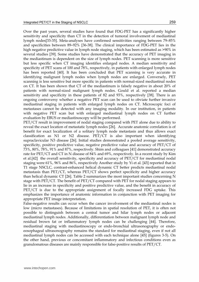
Fig. 3. A 55-year-old woman with an atypical carcinoid tumor in the right middle lobe. CT showed enlarged lymph nodes in the subcarinal (fig 3a) and right paratracheal (fig 3b) region. These lymph nodes are FDG positive on PET (fig 3 c,d). PET/CT (fig 3e,f) confirmed the correlation of these findings. On histopathology these nodes were metastatic lymph nodes.
www.intechopen.com
Integrated PET/CT in the Staging of NSCLC
261
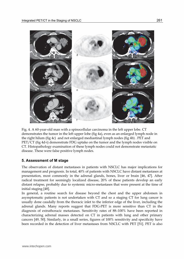
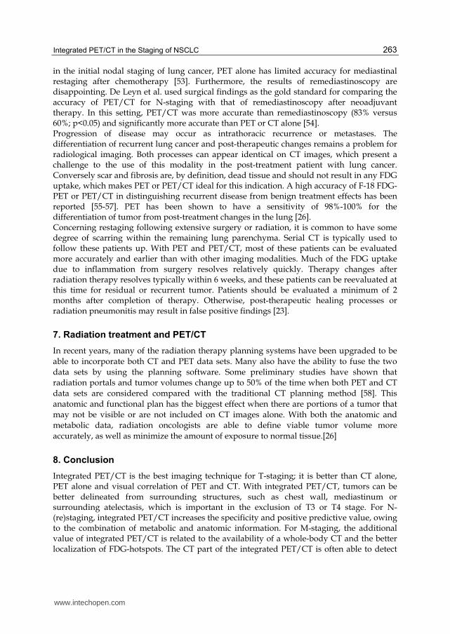
Fig. 4. A 60-year-old man with a spinocellular carcinoma in the left upper lobe. CT demonstrates the tumor in the left upper lobe (fig 4a), even as an enlarged lymph node in the right hilum (fig 4c) and not enlarged mediastinal lymph nodes (fig 4b) . PET and PET/CT (fig 4d-i) demonstrate FDG uptake on the tumor and the lymph nodes visible on CT. Histopathology examination of these lymph nodes could not demonstrate metastatic disease. These were false positive lymph nodes.
5. Assessment of M stage
The observation of distant metastases in patients with NSCLC has major implications for
management and prognosis. In total, 40% of patients with NSCLC have distant metastases at
presentation, most commonly in the adrenal glands, bones, liver or brain [46, 47]. After
radical treatment for seemingly localized disease, 20% of these patients develop an early
distant relapse, probably due to systemic micro-metastases that were present at the time of
initial staging [48].
In general, a routine search for disease beyond the chest and the upper abdomen in
asymptomatic patients is not undertaken with CT and so a staging CT for lung cancer is
usually done caudally from the thoracic inlet to the inferior edge of the liver, including the
adrenal glands. Many reports suggest that FDG-PET is more sensitive than CT in the
diagnosis of extrathoracic metastases. Sensitivity rates of 88–100% have been reported in
characterizing adrenal masses detected on CT in patients with lung and other primary
cancers [49, 50]. Similarly, in a small series, figures of 100% sensitivity and specificity have
been recorded in the detection of liver metastases from NSCLC with PET [51]. PET is also
www.intechopen.com
Computed Tomography – Clinical Applications
262
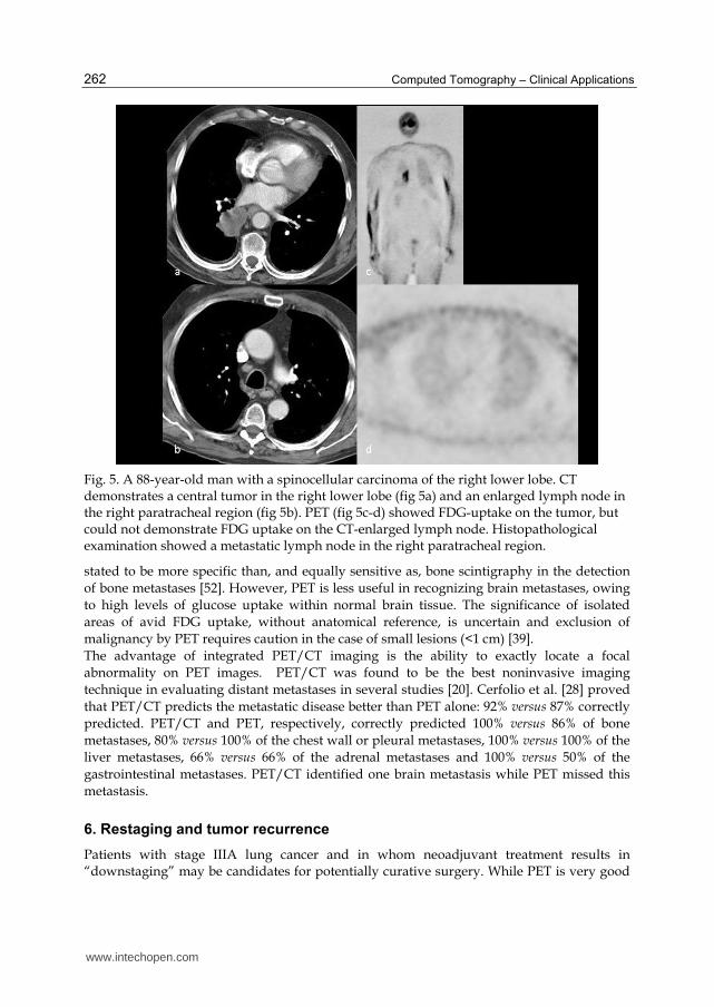
Fig. 5. A 88-year-old man with a spinocellular carcinoma of the right lower lobe. CT demonstrates a central tumor in the right lower lobe (fig 5a) and an enlarged lymph node in the right paratracheal region (fig 5b). PET (fig 5c-d) showed FDG-uptake on the tumor, but could not demonstrate FDG uptake on the CT-enlarged lymph node. Histopathological examination showed a metastatic lymph node in the right paratracheal region.
stated to be more specific than, and equally sensitive as, bone scintigraphy in the detection
of bone metastases [52]. However, PET is less useful in recognizing brain metastases, owing
to high levels of glucose uptake within normal brain tissue. The significance of isolated
areas of avid FDG uptake, without anatomical reference, is uncertain and exclusion of
malignancy by PET requires caution in the case of small lesions (<1 cm) [39].
The advantage of integrated PET/CT imaging is the ability to exactly locate a focal
abnormality on PET images. PET/CT was found to be the best noninvasive imaging
technique in evaluating distant metastases in several studies [20]. Cerfolio et al. [28] proved
that PET/CT predicts the metastatic disease better than PET alone: 92% versus 87% correctly
predicted. PET/CT and PET, respectively, correctly predicted 100% versus 86% of bone
metastases, 80% versus 100% of the chest wall or pleural metastases, 100% versus 100% of the
liver metastases, 66% versus 66% of the adrenal metastases and 100% versus 50% of the
gastrointestinal metastases. PET/CT identified one brain metastasis while PET missed this
metastasis.
6. Restaging and tumor recurrence
Patients with stage IIIA lung cancer and in whom neoadjuvant treatment results in “downstaging” may be candidates for potentially curative surgery. While PET is very good
www.intechopen.com
Integrated PET/CT in the Staging of NSCLC
263
in the initial nodal staging of lung cancer, PET alone has limited accuracy for mediastinal restaging after chemotherapy [53]. Furthermore, the results of remediastinoscopy are disappointing. De Leyn et al. used surgical findings as the gold standard for comparing the accuracy of PET/CT for N-staging with that of remediastinoscopy after neoadjuvant therapy. In this setting, PET/CT was more accurate than remediastinoscopy (83% versus 60%; p<0.05) and significantly more accurate than PET or CT alone [54]. Progression of disease may occur as intrathoracic recurrence or metastases. The differentiation of recurrent lung cancer and post-therapeutic changes remains a problem for radiological imaging. Both processes can appear identical on CT images, which present a challenge to the use of this modality in the post-treatment patient with lung cancer. Conversely scar and fibrosis are, by definition, dead tissue and should not result in any FDG uptake, which makes PET or PET/CT ideal for this indication. A high accuracy of F-18 FDG-PET or PET/CT in distinguishing recurrent disease from benign treatment effects has been reported [55-57]. PET has been shown to have a sensitivity of 98%-100% for the differentiation of tumor from post-treatment changes in the lung [26]. Concerning restaging following extensive surgery or radiation, it is common to have some degree of scarring within the remaining lung parenchyma. Serial CT is typically used to follow these patients up. With PET and PET/CT, most of these patients can be evaluated more accurately and earlier than with other imaging modalities. Much of the FDG uptake due to inflammation from surgery resolves relatively quickly. Therapy changes after radiation therapy resolves typically within 6 weeks, and these patients can be reevaluated at this time for residual or recurrent tumor. Patients should be evaluated a minimum of 2 months after completion of therapy. Otherwise, post-therapeutic healing processes or radiation pneumonitis may result in false positive findings [23].
7. Radiation treatment and PET/CT
In recent years, many of the radiation therapy planning systems have been upgraded to be
able to incorporate both CT and PET data sets. Many also have the ability to fuse the two
data sets by using the planning software. Some preliminary studies have shown that
radiation portals and tumor volumes change up to 50% of the time when both PET and CT
data sets are considered compared with the traditional CT planning method [58]. This
anatomic and functional plan has the biggest effect when there are portions of a tumor that
may not be visible or are not included on CT images alone. With both the anatomic and
metabolic data, radiation oncologists are able to define viable tumor volume more
accurately, as well as minimize the amount of exposure to normal tissue.[26]
8. Conclusion
Integrated PET/CT is the best imaging technique for T-staging; it is better than CT alone, PET alone and visual correlation of PET and CT. With integrated PET/CT, tumors can be better delineated from surrounding structures, such as chest wall, mediastinum or surrounding atelectasis, which is important in the exclusion of T3 or T4 stage. For N-(re)staging, integrated PET/CT increases the specificity and positive predictive value, owing to the combination of metabolic and anatomic information. For M-staging, the additional value of integrated PET/CT is related to the availability of a whole-body CT and the better localization of FDG-hotspots. The CT part of the integrated PET/CT is often able to detect
www.intechopen.com
Computed Tomography – Clinical Applications
264
and diagnose metastatic disease, obviating specific diagnostic CT examinations while the additional FDG hotspots detected with PET are better characterized when the CT information is used. However, there are still many indeterminate lesions that need histopathological proof, and integrated PET/CT can be helpful in guiding these interventional procedures.
9. References
[1] De Wever W, Ceyssens S, Mortelmans L, Stroobants S, Marchal G, Bogaert J, Verschakelen JA. Additional value of PET-CT in the staging of lung cancer: comparison with CT alone, PET alone and visual correlation of PET and CT. Eur Radiol 2007: 17(1): 23-32.
[2] Hany TF, Steinert HC, Goerres GW, Buck A, von Schulthess GK. PET diagnostic accuracy: improvement with in-line PET-CT system: initial results. Radiology 2002: 225(2): 575-581.
[3] Scott WJ, Gobar LS, Terry JD, Dewan NA, Sunderland JJ. Mediastinal lymph node staging of non-small-cell lung cancer: a prospective comparison of computed tomography and positron emission tomography. J Thorac Cardiovasc Surg 1996: 111(3): 642-648.
[4] Weder W, Schmid RA, Bruchhaus H, Hillinger S, von Schulthess GK, Steinert HC. Detection of extrathoracic metastases by positron emission tomography in lung cancer. Ann Thorac Surg 1998: 66(3): 886-892; discussion 892-883.
[5] Tan BB, Flaherty KR, Kazerooni EA, Iannettoni MD. The solitary pulmonary nodule. Chest 2003: 123(1 Suppl): 89S-96S.
[6] Swensen SJ, Jett JR, Hartman TE, Midthun DE, Sloan JA, Sykes AM, Aughenbaugh GL, Clemens MA. Lung cancer screening with CT: Mayo Clinic experience. Radiology 2003: 226(3): 756-761.
[7] Verschakelen JA, De Wever W, Bogaert J. Role of computed tomography in lung cancer staging. Curr Opin Pulm Med 2004: 10(4): 248-255.
[8] Gurney JW, Lyddon DM, McKay JA. Determining the likelihood of malignancy in solitary pulmonary nodules with Bayesian analysis. Part II. Application. Radiology 1993: 186(2): 415-422.
[9] Seemann MD, Seemann O, Luboldt W, Bonel H, Sittek H, Dienemann H, Staebler A. Differentiation of malignant from benign solitary pulmonary lesions using chest radiography, spiral CT and HRCT. Lung Cancer 2000: 29(2): 105-124.
[10] Swensen SJ, Viggiano RW, Midthun DE, Muller NL, Sherrick A, Yamashita K, Naidich DP, Patz EF, Hartman TE, Muhm JR, Weaver AL. Lung nodule enhancement at CT: multicenter study. Radiology 2000: 214(1): 73-80.
[11] Divisi D, Di Tommaso S, Di Leonardo G, Brianzoni E, De Vico A, Crisci R. 18-fluorine fluorodeoxyglucose positron emission tomography with computerized tomography versus computerized tomography alone for the management of solitary lung nodules with diameters inferior to 1.5 cm. Thorac Cardiovasc Surg: 58(7): 422-426.
[12] Gould MK, Maclean CC, Kuschner WG, Rydzak CE, Owens DK. Accuracy of positron emission tomography for diagnosis of pulmonary nodules and mass lesions: a meta-analysis. JAMA 2001: 285(7): 914-924.
www.intechopen.com
Integrated PET/CT in the Staging of NSCLC
265
[13] Fletcher JW, Kymes SM, Gould M, Alazraki N, Coleman RE, Lowe VJ, Marn C, Segall G, Thet LA, Lee K. A comparison of the diagnostic accuracy of 18F-FDG PET and CT in the characterization of solitary pulmonary nodules. J Nucl Med 2008: 49(2): 179-185.
[14] Hellwig D, Baum RP, Kirsch C. FDG-PET, PET/CT and conventional nuclear medicine procedures in the evaluation of lung cancer: a systematic review. Nuklearmedizin 2009: 48(2): 59-69, quiz N58-59.
[15] Gupta NC, Maloof J, Gunel E. Probability of malignancy in solitary pulmonary nodules using fluorine-18-FDG and PET. J Nucl Med 1996: 37(6): 943-948.
[16] Ung YC, Maziak DE, Vanderveen JA, Smith CA, Gulenchyn K, Lacchetti C, Evans WK. 18Fluorodeoxyglucose positron emission tomography in the diagnosis and staging of lung cancer: a systematic review. J Natl Cancer Inst 2007: 99(23): 1753-1767.
[17] Wahidi MM, Govert JA, Goudar RK, Gould MK, McCrory DC. Evidence for the treatment of patients with pulmonary nodules: when is it lung cancer?: ACCP evidence-based clinical practice guidelines (2nd edition). Chest 2007: 132(3 Suppl): 94S-107S.
[18] Nomori H, Watanabe K, Ohtsuka T, Naruke T, Suemasu K, Uno K. Evaluation of F-18 fluorodeoxyglucose (FDG) PET scanning for pulmonary nodules less than 3 cm in diameter, with special reference to the CT images. Lung Cancer 2004: 45(1): 19-27.
[19] Grgic A, Yuksel Y, Groschel A, Schafers HJ, Sybrecht GW, Kirsch CM, Hellwig D. Risk stratification of solitary pulmonary nodules by means of PET using (18)F-fluorodeoxyglucose and SUV quantification. Eur J Nucl Med Mol Imaging: 37(6): 1087-1094.
[20] De Wever W, Stroobants S, Coolen J, Verschakelen JA. Integrated PET/CT in the staging of nonsmall cell lung cancer: technical aspects and clinical integration. Eur Respir J 2009: 33(1): 201-212.
[21] Erasmus JJ, McAdams HP, Patz EF, Jr., Coleman RE, Ahuja V, Goodman PC. Evaluation of primary pulmonary carcinoid tumors using FDG PET. AJR Am J Roentgenol 1998: 170(5): 1369-1373.
[22] Higashi K, Ueda Y, Seki H, Yuasa K, Oguchi M, Noguchi T, Taniguchi M, Tonami H, Okimura T, Yamamoto I. Fluorine-18-FDG PET imaging is negative in bronchioloalveolar lung carcinoma. J Nucl Med 1998: 39(6): 1016-1020.
[23] Steinert HC. PET and PET-CT of lung cancer. Methods Mol Biol: 727: 33-51. [24] Kim SK, Allen-Auerbach M, Goldin J, Fueger BJ, Dahlbom M, Brown M, Czernin J,
Schiepers C. Accuracy of PET/CT in characterization of solitary pulmonary lesions. J Nucl Med 2007: 48(2): 214-220.
[25] Martins Rde C, Almeida SA, Siciliano AA, Landesmann MC, Silva FB, Franco CA, Fonseca LM. [Value of [18F]-FDG-PET/CT as a predictor of cancer in solitary pulmonary nodule]. J Bras Pneumol 2008: 34(7): 473-480.
[26] Mattar EH. Integrated PET/CT in imaging of non-small cell lung cancer. J Egypt Natl Canc Inst 2007: 19(4): 263-274.
[27] Halpern BS, Schiepers C, Weber WA, Crawford TL, Fueger BJ, Phelps ME, Czernin J. Presurgical staging of non-small cell lung cancer: positron emission tomography, integrated positron emission tomography/CT, and software image fusion. Chest 2005: 128(4): 2289-2297.
www.intechopen.com
Computed Tomography – Clinical Applications
266
[28] Cerfolio RJ, Ojha B, Bryant AS, Raghuveer V, Mountz JM, Bartolucci AA. The accuracy of integrated PET-CT compared with dedicated PET alone for the staging of patients with nonsmall cell lung cancer. Ann Thorac Surg 2004: 78(3): 1017-1023; discussion 1017-1023.
[29] Lardinois D, Weder W, Hany TF, Kamel EM, Korom S, Seifert B, von Schulthess GK, Steinert HC. Staging of non-small-cell lung cancer with integrated positron-emission tomography and computed tomography. N Engl J Med 2003: 348(25): 2500-2507.
[30] Pauls S, Buck AK, Hohl K, Halter G, Hetzel M, Blumstein NM, Mottaghy FM, Glatting G, Kruger S, Sunder-Plassmann L, Moller P, Hombach V, Brambs HJ, Reske SN. Improved non-invasive T-Staging in non-small cell lung cancer by integrated 18F-FDG PET/CT. Nuklearmedizin 2007: 46(1): 9-14; quiz N11-12.
[31] Antoch G, Stattaus J, Nemat AT, Marnitz S, Beyer T, Kuehl H, Bockisch A, Debatin JF, Freudenberg LS. Non-small cell lung cancer: dual-modality PET/CT in preoperative staging. Radiology 2003: 229(2): 526-533.
[32] Dillemans B, Deneffe G, Verschakelen J, Decramer M. Value of computed tomography and mediastinoscopy in preoperative evaluation of mediastinal nodes in non-small cell lung cancer. A study of 569 patients. Eur J Cardiothorac Surg 1994: 8(1): 37-42.
[33] Pieterman RM, van Putten JW, Meuzelaar JJ, Mooyaart EL, Vaalburg W, Koeter GH, Fidler V, Pruim J, Groen HJ. Preoperative staging of non-small-cell lung cancer with positron-emission tomography. N Engl J Med 2000: 343(4): 254-261.
[34] Steinert HC, Hauser M, Allemann F, Engel H, Berthold T, von Schulthess GK, Weder W. Non-small cell lung cancer: nodal staging with FDG PET versus CT with correlative lymph node mapping and sampling. Radiology 1997: 202(2): 441-446.
[35] Vansteenkiste JF, Stroobants SG, De Leyn PR, Dupont PJ, Bogaert J, Maes A, Deneffe GJ, Nackaerts KL, Verschakelen JA, Lerut TE, Mortelmans LA, Demedts MG. Lymph node staging in non-small-cell lung cancer with FDG-PET scan: a prospective study on 690 lymph node stations from 68 patients. J Clin Oncol 1998: 16(6): 2142-2149.
[36] Birim O, Kappetein AP, Stijnen T, Bogers AJ. Meta-analysis of positron emission tomographic and computed tomographic imaging in detecting mediastinal lymph node metastases in nonsmall cell lung cancer. Ann Thorac Surg 2005: 79(1): 375-382.
[37] Dwamena BA, Sonnad SS, Angobaldo JO, Wahl RL. Metastases from non-small cell lung cancer: mediastinal staging in the 1990s--meta-analytic comparison of PET and CT. Radiology 1999: 213(2): 530-536.
[38] Gould MK, Kuschner WG, Rydzak CE, Maclean CC, Demas AN, Shigemitsu H, Chan JK, Owens DK. Test performance of positron emission tomography and computed tomography for mediastinal staging in patients with non-small-cell lung cancer: a meta-analysis. Ann Intern Med 2003: 139(11): 879-892.
[39] Schrevens L, Lorent N, Dooms C, Vansteenkiste J. The role of PET scan in diagnosis, staging, and management of non-small cell lung cancer. Oncologist 2004: 9(6): 633-643.
[40] Toloza EM, Harpole L, McCrory DC. Noninvasive staging of non-small cell lung cancer: a review of the current evidence. Chest 2003: 123(1 Suppl): 137S-146S.
www.intechopen.com
Integrated PET/CT in the Staging of NSCLC
267
[41] Shim SS, Lee KS, Kim BT, Chung MJ, Lee EJ, Han J, Choi JY, Kwon OJ, Shim YM, Kim S. Non-small cell lung cancer: prospective comparison of integrated FDG PET/CT and CT alone for preoperative staging. Radiology 2005: 236(3): 1011-1019.
[42] Kim YK, Lee KS, Kim BT, Choi JY, Kim H, Kwon OJ, Shim YM, Yi CA, Kim HY, Chung MJ. Mediastinal nodal staging of nonsmall cell lung cancer using integrated 18F-FDG PET/CT in a tuberculosis-endemic country: diagnostic efficacy in 674 patients. Cancer 2007: 109(6): 1068-1077.
[43] Yi CA, Lee KS, Kim BT, Shim SS, Chung MJ, Sung YM, Jeong SY. Efficacy of helical dynamic CT versus integrated PET/CT for detection of mediastinal nodal metastasis in non-small cell lung cancer. AJR Am J Roentgenol 2007: 188(2): 318-325.
[44] Truong MT, Erasmus JJ, Macapinlac HA, Marom EM, Mawlawi O, Gladish GW, Sabloff BS, Bruzzi JF, Munden RF. Integrated positron emission tomography/computed tomography in patients with non-small cell lung cancer: normal variants and pitfalls. J Comput Assist Tomogr 2005: 29(2): 205-209.
[45] Janssen-Heijnen ML, Coebergh JW. Trends in incidence and prognosis of the histological subtypes of lung cancer in North America, Australia, New Zealand and Europe. Lung Cancer 2001: 31(2-3): 123-137.
[46] De Wever W, Bruyeer E, Demaerel P, Wilms G, Coolen J, Verschakelen J. Staging of lung cancer. Do we need a diagnostic CT of the brain after an integrated PET/CT for the detection of brain metastases? JBR-BTR: 93(2): 71-76.
[47] Quint LE, Tummala S, Brisson LJ, Francis IR, Krupnick AS, Kazerooni EA, Iannettoni MD, Whyte RI, Orringer MB. Distribution of distant metastases from newly diagnosed non-small cell lung cancer. Ann Thorac Surg 1996: 62(1): 246-250.
[48] Pantel K, Izbicki J, Passlick B, Angstwurm M, Haussinger K, Thetter O, Riethmuller G. Frequency and prognostic significance of isolated tumour cells in bone marrow of patients with non-small-cell lung cancer without overt metastases. Lancet 1996: 347(9002): 649-653.
[49] Jana S, Zhang T, Milstein DM, Isasi CR, Blaufox MD. FDG-PET and CT characterization of adrenal lesions in cancer patients. Eur J Nucl Med Mol Imaging 2006: 33(1): 29-35.
[50] Kumar R, Xiu Y, Yu JQ, Takalkar A, El-Haddad G, Potenta S, Kung J, Zhuang H, Alavi A. 18F-FDG PET in evaluation of adrenal lesions in patients with lung cancer. J Nucl Med 2004: 45(12): 2058-2062.
[51] Marom EM, McAdams HP, Erasmus JJ, Goodman PC, Culhane DK, Coleman RE, Herndon JE, Patz EF, Jr. Staging non-small cell lung cancer with whole-body PET. Radiology 1999: 212(3): 803-809.
[52] Bury T, Barreto A, Daenen F, Barthelemy N, Ghaye B, Rigo P. Fluorine-18 deoxyglucose positron emission tomography for the detection of bone metastases in patients with non-small cell lung cancer. Eur J Nucl Med 1998: 25(9): 1244-1247.
[53] Hoekstra CJ, Stroobants SG, Smit EF, Vansteenkiste J, van Tinteren H, Postmus PE, Golding RP, Biesma B, Schramel FJ, van Zandwijk N, Lammertsma AA, Hoekstra OS. Prognostic relevance of response evaluation using [18F]-2-fluoro-2-deoxy-D-glucose positron emission tomography in patients with locally advanced non-small-cell lung cancer. J Clin Oncol 2005: 23(33): 8362-8370.
[54] De Leyn P, Stroobants S, De Wever W, Lerut T, Coosemans W, Decker G, Nafteux P, Van Raemdonck D, Mortelmans L, Nackaerts K, Vansteenkiste J. Prospective
www.intechopen.com
Computed Tomography – Clinical Applications
268
comparative study of integrated positron emission tomography-computed tomography scan compared with remediastinoscopy in the assessment of residual mediastinal lymph node disease after induction chemotherapy for mediastinoscopy-proven stage IIIA-N2 Non-small-cell lung cancer: a Leuven Lung Cancer Group Study. J Clin Oncol 2006: 24(21): 3333-3339.
[55] Hicks RJ, Kalff V, MacManus MP, Ware RE, McKenzie AF, Matthews JP, Ball DL. The utility of (18)F-FDG PET for suspected recurrent non-small cell lung cancer after potentially curative therapy: impact on management and prognostic stratification. J Nucl Med 2001: 42(11): 1605-1613.
[56] Inoue T, Kim EE, Komaki R, Wong FC, Bassa P, Wong WH, Yang DJ, Endo K, Podoloff DA. Detecting recurrent or residual lung cancer with FDG-PET. J Nucl Med 1995: 36(5): 788-793.
[57] Keidar Z, Haim N, Guralnik L, Wollner M, Bar-Shalom R, Ben-Nun A, Israel O. PET/CT using 18F-FDG in suspected lung cancer recurrence: diagnostic value and impact on patient management. J Nucl Med 2004: 45(10): 1640-1646.
[58] Klopp AH, Chang JY, Tucker SL, Sulman EP, Balter PA, Liu HH, Bucci MK, Macapinlac HA, Komaki R, Cox JD. Intrathoracic patterns of failure for non-small-cell lung cancer with positron-emission tomography/computed tomography-defined target delineation. Int J Radiat Oncol Biol Phys 2007: 69(5): 1409-1416.
[59] Kim BT, Lee KS, Shim SS, Choi JY, Kwon OJ, Kim H, Shim YM, Kim J, Kim S. Stage T1 non-small cell lung cancer: preoperative mediastinal nodal staging with integrated FDG PET/CT--a prospective study. Radiology 2006: 241(2): 501-509.
[60] Melek H, Gunluoglu MZ, Demir A, Akin H, Olcmen A, Dincer SI. Role of positron emission tomography in mediastinal lymphatic staging of non-small cell lung cancer. Eur J Cardiothorac Surg 2008: 33(2): 294-299.
[61] Yang W, Fu Z, Yu J, Yuan S, Zhang B, Li D, Xing L, Zhao D, Mu D, Sun X, Fang Y, Huang Y, Li W. Value of PET/CT versus enhanced CT for locoregional lymph nodes in non-small cell lung cancer. Lung Cancer 2008: 61(1): 35-43.
[62] Bille A, Pelosi E, Skanjeti A, Arena V, Errico L, Borasio P, Mancini M, Ardissone F. Preoperative intrathoracic lymph node staging in patients with non-small-cell lung cancer: accuracy of integrated positron emission tomography and computed tomography. Eur J Cardiothorac Surg 2009: 36(3): 440-445.
www.intechopen.com
Computed Tomography - Clinical ApplicationsEdited by Dr. Luca Saba
ISBN 978-953-307-378-1Hard cover, 342 pagesPublisher InTechPublished online 05, January, 2012Published in print edition January, 2012
InTech EuropeUniversity Campus STeP Ri Slavka Krautzeka 83/A 51000 Rijeka, Croatia Phone: +385 (51) 770 447 Fax: +385 (51) 686 166www.intechopen.com
InTech ChinaUnit 405, Office Block, Hotel Equatorial Shanghai No.65, Yan An Road (West), Shanghai, 200040, China
Phone: +86-21-62489820 Fax: +86-21-62489821
Computed Tomography (CT), and in particular multi-detector-row computed tomography (MDCT), is apowerful non-invasive imaging tool with a number of advantages over the others non- invasive imagingtechniques. CT has evolved into an indispensable imaging method in clinical routine. It was the first method tonon-invasively acquire images of the inside of the human body that were not biased by superimposition ofdistinct anatomical structures. The first generation of CT scanners developed in the 1970s and numerousinnovations have improved the utility and application field of the CT, such as the introduction of helical systemsthat allowed the development of the "volumetric CT" concept. In this book we want to explore the applicationsof CT from medical imaging to other fields like physics, archeology and computer aided diagnosis. Recentlyinteresting technical, anthropomorphic, forensic and archeological as well as paleontological applications ofcomputed tomography have been developed. These applications further strengthen the method as a genericdiagnostic tool for non- destructive material testing and three-dimensional visualization beyond its medical use.
How to referenceIn order to correctly reference this scholarly work, feel free to copy and paste the following:
Walter De Wever (2012). Integrated PET/CT in the Staging of NSCLC, Computed Tomography - ClinicalApplications, Dr. Luca Saba (Ed.), ISBN: 978-953-307-378-1, InTech, Available from:http://www.intechopen.com/books/computed-tomography-clinical-applications/integrated-pet-ct-in-the-staging-of-nsclc