Embed Size (px)
Citation preview

Iqbal et al. Molecular Cancer 2013, 12:72http://www.molecular-cancer.com/content/12/1/72
RESEARCH Open Access
Insulin enhances metabolic capacities of cancercells by dual regulation of glycolytic enzymepyruvate kinase M2Mohd Askandar Iqbal, Farid Ahmad Siddiqui, Vibhor Gupta, Shilpi Chattopadhyay, Prakasam Gopinath,Bhupender Kumar, Siddharth Manvati, Noor Chaman and Rameshwar NK Bamezai*
Abstract
Background: Insulin is tightly associated with cancer progression; however, mechanistic insights into suchobservations are poorly understood. Recent studies show that metabolic transformation is critical to cancer cellproliferation. Here, we attempt to understand the role of insulin in promotion of cancer metabolism. To this end,the role of insulin in regulating glycolytic enzyme pyruvate kinase M2 (PKM2) was examined.
Results: We observed that insulin up-regulated PKM2 expression, through PI3K/mTOR mediated HIF1α induction,but significantly reduced PKM2 activity independent of this pathway. Drop in PKM2 activity was attributed tosubunit dissociation leading to formation of low activity PKM2 oligomers, as assessed by density gradientcentrifugation. However, tyrosine 105 phosphorylation of PKM2, known for inhibiting PKM2 activity, remainedunaffected on insulin treatment. Interestingly, insulin-induced ROS was found responsible for PKM2 activityreduction. The observed changes in PKM2 status led to augmented cancer metabolism. Insulin-induced PKM2up-regulation resulted in enhanced aerobic glycolysis as confirmed by PKM2 knockdown studies. Further, PKM2activity reduction led to characteristic pooling of glycolytic intermediates and increased accumulation of NADPH;suggesting diversion of glucose flux towards macromolecular synthesis, necessary for cancer cell growth.
Conclusion: The study identifies new PKM2-mediated effects of insulin on cancer metabolism, thus, advancing theunderstanding of insulin’s role in cancer.
Keywords: Insulin, Cancer, Metabolism, Lactate, Glycolysis, HepG2, PKM2
BackgroundCancer cell metabolism differs from that of normal cellsand is characterized by high glucose uptake and lactateproduction even in presence of sufficient oxygen, anobservation made by Otto Warburg in 1920’s. Thephenomenon has been referred to as Warburg effector aerobic glycolysis [1] which forms the basis of18fluorodeoxyglucose-positron emission tomography(FDG-PET) – a diagnostic technique used for clinicaldetection of cancer [2]. Although, inefficient in terms ofATP production, aerobic glycolysis, nonetheless, is vitalto maintain macromolecule synthesis needed to producedaughter cells [3]. Further, aerobic glycolysis is believedto provide cancer cells a selective advantage to grow in
* Correspondence: [email protected] Centre of Applied Human Genetics, School of Life Sciences,Jawaharlal Nehru University, New Delhi 110067, India
© 2013 Iqbal et al.; licensee BioMed Central LtCommons Attribution License (http://creativecreproduction in any medium, provided the or
regions with low oxygen concentration [4]. Consideringthe significance of aerobic glycolysis to cancer cells,metabolic behaviour is now perceived as a critical targetfor anti-cancer therapeutics [5,6]. Among the severalglycolytic genes that are up-regulated in cancer [7],pyruvate kinase is of prime significance because of itskey position in glycolytic sequence [8,9]. It catalyzes thelast step of glycolysis i.e. conversion of phosphoenolpyr-uvate (PEP) to pyruvate with concomitant ATP produc-tion [10]. Out of four isoforms of pyruvate kinase (PK)in mammals- L, R, M1 and M2 [11]; tumor cells pre-dominantly express M2 isoform [11]. Switch to this iso-form is considered essential for aerobic glycolysis andtumor growth [12].Activity of PKM2 is critical to determine a shift in me-
tabolism required for tumor growth. Decrease in PKM2activity causes pooling of glycolytic intermediates which
d. This is an Open Access article distributed under the terms of the Creativeommons.org/licenses/by/2.0), which permits unrestricted use, distribution, andiginal work is properly cited.

Figure 1 PKM2 is the major isoform in HepG2 cells. Immunoblotshowing that PKM2 expression is many-fold higher than other PKisoforms in hepatoma cells. PKL, which is typically present in normalliver cells, is expressed at very low levels in cancerous HepG2 cellline. M1, which is present in energy requiring tissues such as brainand muscle, is completely absent in HepG2 cells. L6 cell line (ratskeletal muscle) is used as a positive control for M1 and a negativecontrol for PKL isoform.
Iqbal et al. Molecular Cancer 2013, 12:72 Page 2 of 12http://www.molecular-cancer.com/content/12/1/72
are then shunted to pentose phosphate pathway (PPP) forNADPH generation for macromolecular synthesis, essen-tial for cellular growth [8,9,11,13]. Therefore, decreasedPKM2 activity is believed to favour rapidly proliferatingcancer cells; however, the phenomenology of PKM2 activ-ity regulation by external factors is not well understood.PKM2 exists in highly active (tetramer) and less active(dimer/monomer) oligomeric forms. The ratio of theseforms decides the overall PKM2 activity in cellular milieu[11,14,15]. In cancer cells, disruption of active PKM2 tet-ramers results in accumulation of less active PKM2 mono-mers/dimers, thus, promoting anabolic synthesis [9,13].Our previous work has shown how two missense muta-tions in PKM2 stabilized less active PKM2 form to pro-mote cellular growth and polyploidy [16].Evidences in recent years have suggested a crucial role of
insulin in cancer cell growth and survival [17-20]. Highlevels of endogenous insulin among type-2 diabetics andnon-diabetics have also been associated with increased can-cer risk in epidemiological studies [21]; with highest risk forliver and pancreatic cancer [22-24]. Significantly, insulin re-ceptor has been reported to provide resistance to IGF-1Rtargeted therapies [25]; emphasizing the importance oftargeting insulin signalling. The role of insulin, however, inregulation of cancer cell metabolism is still obscure.In this study, the role of insulin in promotion of can-
cer metabolism is examined. We show that insulinpromotes cancer metabolism by upregulating PKM2 ex-pression and decreasing its activity. Insulin-inducedchanges in PKM2 status directly resulted in amplifica-tion of cancer-metabolism-specific parameters like glu-cose uptake, lactate production, glycolytic pooling andmacromolecular synthesis. Our results contribute to bet-ter understanding of the role of insulin in cancer metab-olism and thus cancer progression.
ResultsPKM2 is the predominant isoform of pyruvate kinasein cancer cellsPK exists in four isoforms in mammals; therefore, isoformstatus of PK in HepG2, H1299 and PC3 cells was exam-ined. Immunoblotting analysis in HepG2 showed thatPKM2 was the predominant isoform in these cells withcomplete absence of PKM1 (Figure 1). Whereas, PKLisoform, which is predominant in normal liver cells,expressed at a much lower level in HepG2 cells (Figure 1).Similarly, PKM2 was the predominant isoform inH1299 and PC3 cells with negligible PKM1 expression(Additional file 1: Figure S1).
Insulin upregulates PKM2 expression in a PI3K/mTORdependent manner by inducing HIF1α expressionTo examine the effect of insulin on PKM2 expression,serum starved HepG2 cells were treated with insulin for
0, 2, 4, 6 and 8 hours with maximum increase in PKM2expression at 8 hours of insulin treatment (Figure 2A).Dose dependent effect of insulin on PKM2 expression

Figure 2 Insulin increased PKM2 expression via HIF1α up-regulation, in a PI3K/mTOR dependent manner. (A) Serum starved HepG2 cellswere treated with 100 nM insulin for 0–8 hours, maximum increase in PKM2 protein observed after 8 hours of 100 nM treatment. (B) Dosedependent increase in PKM2 expression. (C) Total RNA and protein was extracted and subjected to real time PCR and immunoblottingrespectively, PKM2 mRNA increased by ~2 fold in a PI3K/mTOR dependent manner (as LY294002 and rapamycin decreased PKM2 mRNA). ForPI3K/mTOR inhibition, cells were pretreated with 50 μM LY294002 or 20 nM rapamycin for 30 minutes before stimulating cells with insulin. (D)Representative immunoblot showing insulin-induced up-regulation of PKM2 and its transcriptional regulator HIF1α in a PI3K/mTOR sensitivemanner. However, PKL expression remained unchanged on insulin treatment. For PI3K/mTOR inhibition, cells were pretreated with 50 μMLY294002 or 20 nM rapamycin for 30 minutes. (E) Densitometric analysis was done using Alpha Imager EP densitometer (Alpha Innotech Corp.,USA). (F) CoCl2 treatment (100 μM for 8 hours) resulted in accumulation of HIF1α with concomitant increase in PKM2 expression, siRNA mediatedHIF1α silencing resulted in decreased PKM2 expression. These results suggest that insulin promoted PKM2 expression through HIF1α induction, ina PI3K/mTOR dependent manner. β actin was used as endogenous control in real time experiments and loading control in immunoblotting.mRNA data is expressed as mean ± SE. *P ≤ 0.05.
Iqbal et al. Molecular Cancer 2013, 12:72 Page 3 of 12http://www.molecular-cancer.com/content/12/1/72
revealed that 1 nM insulin (near physiological concen-tration) substantially induced PKM2 expression, how-ever, maximum increase in PKM2 expression wasobserved with 100 nM insulin for 8 hours (Figure 2B);suggesting to choose 100 nM insulin for further experi-ments. A two fold increase in PKM2 mRNA and proteinexpression was observed upon 100 nM insulin treatmentfor 8 hours (Figure 2C-E), whereas, PKL expressionremained unaltered under similar conditions (Figure 2D).Insulin treatment up-regulated PKM2 in H1299 (lungcancer) and PC3 (prostate cancer) cells also (Additionalfile 2: Figure S2), replicating the observations made inHepG2 cells.PI3K/mTOR signalling was inhibited to find out if
insulin-induced PKM2 up-regulation is PI3K dependent.
PI3K is the chief mediator of insulin signalling in liverand is frequently mutated in liver and other cancers[26-28]. As a result of PI3K/mTOR inhibition, PKM2 ex-pression was down-regulated (Figure 2C-E). These re-sults confirmed that insulin regulates PKM2 expressionthrough PI3K/mTOR pathway. To elucidate how PI3K/mTOR promoted PKM2 expression, the role of HIF1αprotein, a transcription factor that binds to hypoxia re-sponse element (HRE) in PKM2 gene to induce its ex-pression, was examined [29,30]. Insulin induced HIF1αexpression in a PI3K/mTOR sensitive manner, as wasevident from the decrease in HIF1α protein after treat-ment with PI3K/mTOR inhibitors (Figure 2D). Further,to corroborate this observation, HIF1α was silenced aswell as induced, using siRNA and cobalt chloride

Iqbal et al. Molecular Cancer 2013, 12:72 Page 4 of 12http://www.molecular-cancer.com/content/12/1/72
(CoCl2) treatment, respectively, to observe the effect onPKM2 expression [31,32]. Expectedly, HIF1α silencingled to a decreased PKM2 expression; whereas CoCl2-in-duced HIF1α accumulation increased PKM2 expression(Figure 2F), confirming that HIF1α is crucial for PKM2up-regulation. These results demonstrated that insulinupregulates PKM2 expression through PI3K/mTORdependent induction of HIF1α transcription factor.
Insulin inhibits PKM2 activity, in a PI3K/mTORindependent manner, by promoting subunit dissociationPKM2 activity is crucial in determining the glycolyticflux; it was pertinent therefore to examine the potentialrole of insulin in altering PKM2 activity. Interestingly,~30% drop in PKM2 activity was observed in HepG2
Figure 3 PKM2 activity decreased as a result of subunit dissociation u100 nM insulin treatment of 15 minutes reduced PKM2 activity in HepG2 cLY294002 or 20 nM rapamycin for 30 minutes) did not significantly alter PKused to monitor decrease in OD due to oxidation of NADH at 340 nm. (B)oligomeric forms of PKM2. Activity was assessed in different fractions to deconcomitant fall in peak II is suggestive of disruption of highly oligomerisecorresponds to the respective amount of particular oligomeric form. (C) Retetrameric PKM2 has optimal activity [15], it can be concluded that PKM2 a(D) Dose-dependent decrease in PKM2 activity upon insulin treatment. Dat
cells within 15 minutes after insulin treatment with nofurther decrease on increasing the duration of treatment(Figure 3A, Additional file 3: Figure S3). However, PI3K/mTOR inhibition did not reverse insulin-induced de-crease in PKM2 activity (Figure 3A). Insulin-treatedH1299 and PC3 cells also exhibited decrease in PKM2activity (Additional file 4: Figure S4). To analyse if theinsulin-induced drop in PKM2 activity is due to subunitdissociation, which governs the overall activity of PKM2[9,11,15,33,34], glycerol density gradient centrifugationwas performed using HepG2 cell lysate. Density gradientcentrifugation is known to separate different oligomericforms of an enzyme with highest quaternary form at thebottom of the gradient and the magnitude of activityrepresenting the respective content of an oligomeric
pon insulin treatment, in a PI3K/mTOR independent manner. (A)ells by ~30%. Pretreatment with PI3K/mTOR inhibitors (50 μMM2 activity. For activity measurement: NADH/LDH coupled assay wasGlycerol density gradient centrifugation was used to separatetect any shift in peaks i.e. subunit dissociation. Rise in peak I withd PKM2 (tetramer) in less oligomerised forms. Activity in a fractionlative peak areas show significant shift in peaks I and II. Asctivity decreased as a result of dissociation of active tetramers.a is expressed as mean ± SE. *P≤ 0.05.
Iqbal et al. Molecular Cancer 2013, 12:72 Page 5 of 12http://www.molecular-cancer.com/content/12/1/72
form [35]. PKM2 activity was measured in fractions col-lected from top to bottom of the glycerol gradient. Ob-servation of two peaks suggested two oligomeric formsof PKM2 and the vertical shift in peaks indicated subunitdissociation (Figure 3B). As evident from relative peakareas, a significant fall in peak II with concomitantincrease in peak I, compared to control peaks, wasobserved upon insulin treatment (Figure 3C). Dosedependent insulin treatment showed that even 1nM in-sulin treatment decreased PKM2 activity significantlywith maximum decrease on 100 nM insulin (Figure 3D).Results, thus, suggested that decrease in PKM2 activitywas due to subunit dissociation upon insulin treatment.As expected, PI3K/mTOR inhibition did not abolish theeffect of insulin on PKM2 oligomeric status (Additionalfile 5: Figure S5). In an attempt to understand how insu-lin promoted subunit dissociation, we checked the tyro-sine 105 phosphorylation of PKM2, which is known todiminish its activity by disrupting the formation of activetetramer [33], but, no change in phosphorylation was
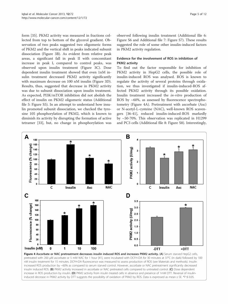
Figure 4 Ascorbate or NAC pretreatment decreases insulin induced Rpretreated with 200 μM ascorbate or 5 mM NAC for 1 hour [41], were incunM insulin treatment for 15 minutes. DCFH-DA fluorescence was measuredincreased ROS production by ~60% as compared to serum starved controlinsulin induced ROS. (B) PKM2 activity increased in ascorbate or NAC pretreincrease in ROS production by insulin. (D) PKM2 activity from insulin treateinduced decrease in PKM2 activity by DTT suggests the possibility of oxida
observed following insulin treatment (Additional file 6:Figure S6 and Additional file 7: Figure S7). These resultssuggested the role of some other insulin-induced factorsin PKM2 activity regulation.
Evidence for the involvement of ROS in inhibition ofPKM2 activityTo find out the factor responsible for inhibition ofPKM2 activity in HepG2 cells, the possible role ofinsulin-induced ROS was analysed. ROS is known toregulate the activity of several proteins through oxida-tion, we thus investigated if insulin-induced-ROS af-fected PKM2 activity through its possible oxidation.Insulin treatment increased the in-vitro production ofROS by ~60%, as assessed by fluorescence spectropho-tometry (Figure 4A). Pretreatment with ascorbate (Asc)or N-acetyl-L-cysteine (NAC), well-known ROS scaven-gers [36-41], reduced insulin-induced-ROS markedlyby ~50-70%. This observation was replicated in H1299and PC3 cells (Additional file 8: Figure S8). Interestingly,
OS and increases PKM2 activity. (A) Serum starved HepG2 cells,bated with DCFH-DA for 30 minutes at 37°C (in dark) followed by 100to assess production of ROS (see Materials and methods). Insulin
. However, ascorbate or NAC pretreatment significantly decreasedated cells compared to untreated control. (C) Dose dependentd cells in absence and presence of 1mM DTT. Reversal of insulin-tion of PKM2 by ROS. Data is expressed as mean ± SE. *P ≤ 0.05.

Iqbal et al. Molecular Cancer 2013, 12:72 Page 6 of 12http://www.molecular-cancer.com/content/12/1/72
ascorbate or NAC pretreatment, followed by incubationwith insulin, reversed the insulin-induced decrease inPKM2 activity in HepG2 (Figure 4B), H1299 and PC3(Additional file 4: Figure S4) cells, suggesting the role ofinsulin-induced-ROS in PKM2 activity inhibition. Fur-ther, dose-dependent increase in ROS production by in-sulin correlated with dose-dependent decrease in PKM2activity (compare Figures 3D and 4C). To understandhow ROS contributed to reduction in PKM2 activity, thepossibility of ROS-induced- oxidation of PKM2 wasstudied. Addition of dithiothreitol (DTT), a strong redu-cing agent, to activity reaction mixture abolishedinsulin-induced-decrease in PKM2 activity (Figure 4D).In fact, addition of DTT increased PKM2 activity to thelevels similar to that of control without insulin treatment(Figure 4D and B). These data indicated the role of
Figure 5 Insulin promoted aerobic glycolysis, partly, by upregulatingcells was collected for measurement of glucose uptake and lactate productionglucose uptake and lactate production in a PI3K/mTOR sensitive manner. (B) P(C) Glucose uptake and lactate production decreased on silencing PKM2 in PTpartly, through PKM2 up-regulation. (D) Cellular proliferation decreased on PK*P≤ 0.05.
ROS-induced-oxidation of PKM2 [34], resulting in itsactivity-inhibition.
PKM2 up-regulation is crucial in insulin-induced aerobicglycolysisPKM2 expression has been suggested to promote aerobicglycolysis [12,30], which is characterized by high glucoseuptake and lactate production even in presence of oxygen,and is believed to be the hallmark of nearly all cancer cells[4]. Both glucose uptake and production of lactate weresubstantially enhanced in cells treated with insulin ascompared to untreated control (Figure 5A). Further, inhib-ition of PI3K/mTOR pathway reduced aerobic glycolysiswhich is consistent with earlier reports (Figure 5A)[42,43]. To show that PI3K/mTOR dependent PKM2 up-regulation contributed to insulin-induced augmentation in
PKM2. Media used by insulin treated (100 nM for 8 hours) or untreatedas described in materials and methods section. (A) Insulin promotedKM2 knockdown efficiency as checked by real time and Western blotting.EN negative PC3 cells; indicating that insulin stimulated aerobic glycolysis,M2 knockdown. C-serum starved cells. Data is expressed as mean ± SE.

Iqbal et al. Molecular Cancer 2013, 12:72 Page 7 of 12http://www.molecular-cancer.com/content/12/1/72
aerobic glycolysis, PKM2 was knocked down in cells withhyperactive PI3K/mTOR signalling (to mimic insulin-induced activation of PI3K/mTOR), followed by anassessment of glucose uptake and lactate production,approximately 48 hours after transfecting control shRNAor PKM2-shRNA (to knock down PKM2 expression).Interestingly, silencing of PKM2 partially inhibited glucoseuptake and lactate production, signifying that PKM2 is re-quired for aerobic glycolysis (Figure 5B-C). Moreover,PKM2 knockdown also retarded cellular proliferation(Figure 5D), consistent with our previous observation [44].
Glycolytic intermediates and NADPH accumulated as aresult of decreased PKM2 activityCharacteristic accumulation of glycolytic intermediates,as a result of decreased PKM2 activity, is another keyfeature of cancer cells and has been shown to be import-ant for anabolic synthesis required for tumor growth[8,11]. To explore the implications of insulin-inducedsuppression of PKM2 activity on glycolytic pooling,intracellular levels of fructose-1,6-bisphosphate (FBP)and phosphoenolpyruvate (PEP), the glycolytic metabo-lites upstream of PKM2, were measured [15]. Conse-quently, an increased build-up of both metabolites was
Figure 6 Increased glycolytic pooling and NADPH accumulation duecells were treated with or without 100 nM insulin for 15 minutes or pretrea200 μM for 30 minutes followed by 100 nM insulin treatment. Intracellularmaterials and methods section. (A) Increased accumulation of PEP (~1.7 foLY294002 and rapamycin decreased glycolytic pooling modestly, howeverpooling of FBP and PEP. (B) NADPH accumulated in insulin treated cells, suPI3K/mTOR inhibitors decreased NADPH accumulation; however, pretreatmextent, bringing NADPH levels similar to that of control. These results suggmainly by decreasing PKM2 activity, and also indicates the diversion of gluconcentrations is expressed as nmol per million cells. Data is expressed as
observed compared to control (Figure 6A). Glycolyticpooling is known to facilitate pentose phosphate path-way for macromolecular synthesis [8,9,13]. Therefore,intracellular levels of NADPH, which is produced as aresult of PPP and provides reducing power for macro-molecular synthesis [45], were determined. Accumula-tion of NADPH increased significantly (P < 0.05) ininsulin treated cells (Figure 6B).To demonstrate that decreased PKM2 activity is con-
tributing to the observed accumulation of FBP, PEP andNADPH, PI3K/mTOR pathway was inhibited as it in-creases glucose uptake which might have contributed toobserved accumulation. PI3K/mTOR inhibition did notcompletely block the insulin-induced pooling of FBP,PEP and NADPH. However, pretreatment with ROSscavengers NAC or ascorbate abolished the insulin-induced glycolytic pooling and accumulation of NADPH.These results indicated that insulin promoted accumula-tion of glycolytic intermediates and NADPH through de-creased PKM2 activity.
DiscussionIn recent years, PKM2 has emerged as a key regulator ofcancer metabolism. Considering the PKM2-mediated
to insulin-induced PKM2 activity inhibition. Serum starved HepG2ted with 50 μM LY294002 or 20 nM rapamycin or 5 mM NAC orPEP, FBP and NADPH were extracted and measured as described inld) and FBP (~1.6 fold) was observed in insulin stimulated cells.ROS scavengers NAC and ascorbate substantially decreased glycolyticggesting enhanced anabolic synthesis via PPP [8,9]. Pretreatment withent with ROS scavengers reduced NADPH accumulation to greaterest that insulin promoted accumulation of glycolytic intermediatescose flux towards PPP [8,9]. Actual change in intracellularmean ± SE. *P≤ 0.05.

Iqbal et al. Molecular Cancer 2013, 12:72 Page 8 of 12http://www.molecular-cancer.com/content/12/1/72
pro-cancerous effects of insulin, our results seeminglyprovide a mechanistic understanding into insulin’s rolein cancer metabolism.The predominance of PKM2 over other PK isoforms
in HepG2, H1299 and PC3 cells (Figure 1 and Additionalfile 1: Figure S1) [30,46], indicated the importance ofthis PK isoform in cancer, and is consistent with thenotion that switch to PKM2 isoform is required for can-cer progression [12]. The insulin induced-up- regulationof PKM2, with evident increase at as low as 1 nM insu-lin (Figure 2), is an important observation, since PKM2has been reported essential for aerobic glycolysis andtumor growth [30]. PI3K-dependence of PKM2 expres-sion supports the notion that PI3K pathway is central toinsulin signalling in cancer cells and correlates with highfrequency of PI3K mutations in several cancer types,which may lead to increased expression of PKM2[26,27]. PI3K/mTOR dependent induction of HIF1α oninsulin treatment, and CoCl2-induced concomitant HIF1α and PKM2 accumulation, along with HIF1α silencingresults (with decreased PKM2 expression) clearly sug-gested that insulin promoted PKM2 expression throughHIF1α (Figure 2D and F). Our results are consistent withprevious observations of insulin up-regulation of PKM2in adipocytes [47] and decreased PKM2 expression onPI3K/mTOR inhibition [44,48]. These results also ex-plain the recent observation of high PKM2 in PTEN(negative regulator of PI3K pathway) null fatty liver cells[49]. Decreased aerobic glycolysis on PKM2 knock-downin PTEN deficient (hyper-activated PI3K/mTOR signal-ling) PC3 cells suggested that insulin promoted aerobicglycolysis, at least in part, through PKM2 (Figure 5). Im-portance of PKM2 in aerobic glycolysis could be realizedfrom the observation that PKM2 transactivates expres-sion of genes like glucose transporter-1 (GLUT1) andlactate dehydrogenase-A (LDHA), required for glucoseuptake and lactate production respectively [29]. Appar-ently, these results present an important mechanismwhich may underlie insulin’s role in carcinogenesis.Decreased PKM2 activity fuels macromolecular syn-
thesis by accumulating glycolytic intermediates that areprecursors for PPP. Suppression of PKM2 activity pre-sents yet another dimension of insulin’s role in promo-tion of cancer metabolism. Rise in peak I withsimultaneous fall in peak II suggested subunit dissoci-ation leading to increased formation of low activity olig-omeric form of PKM2 (Peak I in Figure 3B), thusjustifying the observed decrease in PKM2 activity(Figure 3A). Intriguingly, activity of PKM2 was notaffected on PI3K/mTOR inhibition (Figure 3A andAdditional file 6: Figure S5), indicating the involvement ofother factors in activity regulation. Increased PKM2 activityin ascorbate or NAC pretreated cells suggests the involve-ment of ROS in activity regulation (Figure 4 and Additional
file 4: Figure S4). Reversal of insulin-induced decrease inPKM2 activity by DTT suggests the possibility of ROS-induced oxidation of cysteine residues in PKM2 [34].Correlation between dose dependent changes in PKM2activity and ROS production further support our conclu-sion of ROS mediated PKM2 activity inhibition (Figures 3Dand 4C). Notably, ROS induced decrease in PKM2 activityhas been linked with ability of lung cancer cells to with-stand oxidative stress [34]. However, further research isneeded to support this observation in liver and prostatecancer cells.The decrease in the activity of PKM2, positioned at
the end of glycolytic sequence, in cancer cells is sug-gested to accumulate important glycolytic intermediatesrequired for cellular growth. On the contrary, a decreasein the activity of enzyme, like hexokinase, which appearearlier in glycolytic sequence, does not provide the sameadvantage to the cancer cells; thus the biosynthetic pro-cesses required for cellular growth would be inhibited.Accumulation of glycolytic intermediates PEP and FBP(Figure 6) indicated the diversion of glucose flux towardsbiosynthetic pathway PPP [8], an observation which wasconfirmed when PPP product NADPH accumulatedupon insulin treatment (Figure 6B). The decrease inglycolytic pooling and NADPH accumulation on PI3K/mTOR inhibition correlated with glucose uptake regula-tion by PI3K/mTOR pathway (Figures 5A and 6). Inter-estingly, metabolite accumulation decreased almost tothe levels of untreated control upon treatment with ROSscavengers NAC or ascorbate, suggesting a crucial roleof decreased PKM2 activity in insulin-induced metabol-ite build-up. ROS scavenging increased PKM2 activitywhich negatively affected metabolite pooling and macro-molecular synthesis (Figures 4B and 6).On insulin treatment a maximum PKM2 expression was
observed at 8 hours; whereas a decrease in activity was ob-served within 15 minutes, which was almost constant till 8hours. Since no further decrease in activity was observedby prolonging insulin treatment till 8 hours (Additionalfile 3: Figure S3), we chose 15 minutes as the earliest timepoint at which PKM2 activity decreased. The up-regulation of PKM2 expression with concomitant decreasein activity was associated with enhanced aerobic glycolysisand increased macromolecular synthesis (Figures 4 and 5).PKM2 expression is critical for aerobic glycolysis as ittransactivates expression of GLUT1 (glucose transporter)and LDHA (lactate dehydrogenase) [29]. We have shownthat knockdown of PKM2 expression decreased aerobicglycolysis (Figure 5C) while inhibition of PKM2 activitypromoted pooling of glycolytic intermediates which arethen shunted to PPP for anabolic synthesis [8,13,33,34].Apparently, dual regulation of PKM2 expression and ac-tivity by insulin ensures the promotion of both aerobicglycolysis and anabolic synthesis, required for cancer cell

Iqbal et al. Molecular Cancer 2013, 12:72 Page 9 of 12http://www.molecular-cancer.com/content/12/1/72
proliferation (Figure 7). This possibly explains the link be-tween high insulin levels and elevated cancer risk. More-over, reduced PKM2 activity might explain liver cirrhosisdue to fatty liver since PKM2 activity reduction is knownto promote lipid synthesis [13].
ConclusionOur study highlights previously unknown PKM2-mediated effects of insulin in promotion of cancermetabolism, which probably explains the observations ofincreased cancer risk under hyperinsulinemic condition[17,24,50].
Materials and methodsCell culture, drug treatment, knockdown and proliferationstudiesHepG2 and PC3 cell lines were procured from the Na-tional Centre for Cell Science, Pune, India. H1299 cellswere a kind gift from Dr. Uttam Pati, School of Biotech-nology, Jawaharlal Nehru University. All the cell lineswere maintained in DMEM (Sigma) with 10% heat-inactivated FBS (Biowest, France), 1% penicillin/strepto-mycin (Sigma) at 37°C and 5% CO2 in a humifiedatmosphere (Heraeus, UK). Cells were grown in mono-layer and passaged routinely two-three times a week. Fordrug treatment; LY294002 (Sigma) and rapamycin (Sigma)were dissolved in DMSO; single use aliquots were storedat −80°C For insulin treatment: cells were seeded in
Figure 7 Graphical summary. Effect of insulin on PKM2 expression(through PI3K/mTOR/HIF axis) and activity (ROS-dependent),resulting in amplified cancer metabolism.
triplicate at a density of 0.4 million cells/well of six wellplates, serum starved for 20–24 hours and then stimu-lated with insulin (Sigma) in presence or absence of in-hibitors. DMSO treated cells were used as mock control.For PKM2 knockdown: short hairpin RNA (shRNA)constructs in lentiviral pGIPZ vector were purchasedfrom Open Biosystems. Control and shRNA containingpGIPZ vectors were transfected using polymer basedtransfection reagent- Arrestin from Thermo-Scientific,as per manufacturer’s instructions. Knock down effi-ciency was then checked by real time PCR and Westernblotting. For HIF1α silencing: pre-designed siRNA wasused and transfected using Lipofectamine (Invitrogen) asper manufacturer’s protocol and efficiency was checked byWestern blotting. For proliferation assay: cells werecounted before and after 48 hours of transfection, usinga hemocytometer.
RNA isolation, cDNA preparation and real time PCRCellular RNA was extracted from cell lines using TRIzol(Sigma), according to manufacturer’s protocol. RNA qual-ity was analyzed by A260/A280 absorbance and by electro-phoresis on a 1.2% agarose formaldehyde gel. 3–4 μg oftotal RNA was reverse transcribed into single strandedDNA using cDNA preparation kit (Applied Biosystems,USA). Commercially available Taqman gene expressionassay (Applied Biosystems, USA) was used for quantitatingmRNA levels of PKM2. β-actin was used as endogenouscontrol. Real time PCR was carried out on ABI Prism7000 Sequence Detection System (Applied Biosystems).ΔΔCt (Cycle threshold) method of relative quantificationwas used to calculate fold change in gene expression bySDS 1.1 RQ software (Applied Biosystems).
Cell lysate preparation, protein estimation and WesternblottingWhole cell lysate was prepared by incubating cells, onice for 30 minutes, in buffer containing 50 mM Tris pH7.2, 150 mM NaCl, 0.5% sodium deoxycholate, 10% gly-cerol, 1% Triton X-100, 0.1% SDS, 1 mM DTT, 1 mMPMSF, 5 mM NaF, 1 mM NaV, phosphatase inhibitorcocktail (Sigma), 4 μg/ml aprotinin, 4 μg/ml leupeptinand 4 μg/ml pepstatin (Sigma). The lysate was centri-fuged at high speed in a cooling centrifuge (CM 12,Remi, India) for 30 minutes and supernatant was col-lected in pre-chilled fresh tubes. Protein concentrationwas estimated using BCA method as per manufacturerprotocol (Thermo Scientific). Proteins were separated on8% SDS-PAGE, transferred to nitrocellulose membrane(mdi) at 4°C (wet transfer) and probed with primaryantibodies. Membrane was incubated with appropriatesecondary antibody for one hour at room temperatureand proteins were detected using Luminata forte(Millipore). Primary antibodies used were: anti-HIF1α

Iqbal et al. Molecular Cancer 2013, 12:72 Page 10 of 12http://www.molecular-cancer.com/content/12/1/72
(Novus Biologicals, USA), anti PKM2, anti-phospho-PKM2 (Tyr105), anti-AKT, anti-phospho-AKT, anti-phosphoS6, anti-S6 protein and anti-β-actin (Cell SignallingTechnology).
PKM2 activity assay and glycerol gradient centrifugationFor activity, cells were lysed in buffer as described previ-ously [12]. Activity was measured using NADH/lactatedehydrogenase (LDH) coupled assay. Decrease in OD at340 nm due to oxidation of NADH was monitoredusing a double beam spectrophotometer (UV-1800,Shimadzu). Reaction was started by adding 2 μg cell lys-ate to mixture containing 50 mM Tris pH 7.5, 100 mMKCl, 5 mM MgCl2, 1.25 mM ADP, 0.5 mM PEP, 0.28mM NADH and 8 units of LDH. Specific activity per mgof cell lysate was calculated as:
U=mg ¼ OD340=min6:22�mg lysate=ml reaction mixture
U/mg = specific enzyme activity per mg of protein.OD340 = Change in absorbance due to oxidation ofNADH in one minute at 340 nm wavelength.
For glycerol gradient experiment, 500 μg of cell lysateprotein was loaded on the top of 11-25% glycerol gradi-ent and centrifuged at 45000 rpm for 16 hours at 4°C inSW55Ti rotor (Beckman Coulter) and rest of the pro-cedure was followed as described [16].
Metabolites, glucose and lactate measurementMetabolite extract was prepared from 20 million cells in 0.5ml of chilled 90% ethanol containing 0.2% formic acid andcentrifuged at 15000 rpm in a refrigerated centrifuge. Super-natant was dried using nitrogen flow and then reconstitutedin 0.2 ml of MilliQ water. PEP was assessed using NADH/LDH coupled assay as mentioned above with 30 ng of re-combinant PKM2. FBP was measured as described [51]. Forboth FBP and PEP, concentration was determined againststandard curve. NADPH was analyzed using kit fromBioVision- USA, as per the manufacturer’s protocol. For glu-cose and lactate: media was collected from wells; spun downat high speed to remove any cell debris, deproteinized usingTCA, pH was adjusted between 7.0-7.5 and then glucose up-take was analyzed using glucose assay kit (Sigma), accordingto manufacturer’s protocol. Lactate and was analysed usingkit (BioVision, USA) as per manufacturer’s instructions. Allthe measurements were normalized to cell numbers.
Ascorbate, NAC treatment and ROS analysisCells pretreated with or without ascorbate or NAC(Sigma) were incubated with DCFH-DA for 30 minutes indark at 37°C, followed by insulin stimulation. Cells werethen washed with PBS, trypsinized and resuspended in 0.5
ml PBS, kept on ice, in dark until ROS analysis by fluores-cence spectrophotometry. Excitation and emission wave-lengths used were 500 nm and 510 nm respectively.
Statistical analysisEach experiment was performed in triplicate. All experi-ments were repeated at least 3 times. Significance wascalculated using student’s t-test. P value less than 0.05was considered statistically significant.
Additional files
Additional file 1: Figure S1. PKM2 is the predominant isoform inH1299 and PC3 cell. PKM1 expression is negligibly low.
Additional file 2: Figure S2. Insulin up-regulated PKM2 expression inH1299 and PC3 cells (C = Control, I = 100 nM insulin).
Additional file 3: Figure S3. PKM2 activity after different time points of100 nM insulin treatment. Data is expressed as mean ± SE.
Additional file 4: Figure S4. Insulin treatement decreased PKM2activity in H1299 and PC3 cells. Pretreatment with ROS scavenger- NAC,reversed insulin-induced decrease in activity. Data is expressed asmean ± SE. *P≤ 0.05.
Additional file 5: Figure S5. Glycerol gradient of cells pre-treated withDMSO or 50 μM LY294002 or 20 nM rapamycin, followed by 100 nMinsulin treatment for 15 minutes.
Additional file 6: Figure S6. Representative Western blot showing nochange in phosphor-tyr-105-PKM2 upon insulin treatment (100 nM for 15minutes) and inhibition with 50 μM LY294002 and 20 nM rapamycin, inHepG2 cells.
Additional file 7: Figure S7. Immunoblot showing no change inphosphor-tyr-105-PKM2 upon insulin treatment (100 nM for 15 minutes)in H1299 and PC3 cells.
Additional file 8: Figure S8. Insulin treatement increase ROS in H1299and PC3 cells. Pretreatment with 5 mM NAC decreased insulin-inducedROS. Data is expressed as mean ± SE. *P ≤ 0.05.
AbbreviationsPKM2: Pyruvate kinase M2; PI3K: Phosphoinositide-3-kinase;mTOR: Mammalian target of rapamycin; PEP: Phosphoenolpyruvate;FBP: Fructose 1,6-bisphosphate; PPP: Pentose phosphate pathway;NADPH: Nicotinamide adenine dinucleotide phosphate; ROS: Reactiveoxygen species; NAC: N-acetyl-L-cysteine.
Competing interestsThe authors declare that they have no competing interests.
Authors’ contributionsMAI designed study; acquired, analysed and interpreted data; performedstatistical analysis and drafted manuscript. FAS participated in acquisition ofdata, statistical analysis and manuscript preparation. VG, SC, PG, BK, SM andNC participated in experimental data acquisition and revision of manuscript.RNKB conceived the study, critically reviewed manuscript for intellectualcontent and gave final approval for submission. All authors read andapproved the final version of manuscript.
AcknowledgementsRNKB acknowledges University Grants Commission (UGC), Government ofIndia, for providing research funds to NCAHG. MAI acknowledges UGC forproviding research fellowship.
Received: 27 May 2013 Accepted: 13 June 2013Published: 9 July 2013

Iqbal et al. Molecular Cancer 2013, 12:72 Page 11 of 12http://www.molecular-cancer.com/content/12/1/72
References1. Warburg O: On the origin of cancer cells. Science 1956, 123:309–314.2. Weber WA, Avril N, Schwaiger M: Relevance of positron emission
tomography (PET) in oncology. Strahlenther Onkol 1999, 175:356–373.3. Vander Heiden MG, Cantley LC, Thompson CB: Understanding the
Warburg effect: the metabolic requirements of cell proliferation. Science2009, 324:1029–1033.
4. Gatenby RA, Gillies RJ: Why do cancers have high aerobic glycolysis? NatRev Cancer 2004, 4:891–899.
5. Tennant DA, Duran RV, Gottlieb E: Targeting metabolic transformation forcancer therapy. Nat Rev Cancer 2010, 10:267–277.
6. Vander Heiden MG: Targeting cancer metabolism: a therapeutic windowopens. Nat Rev Drug Discov 2011, 10:671–684.
7. Majumder PK, Febbo PG, Bikoff R, Berger R, Xue Q, McMahon LM, Manola J,Brugarolas J, McDonnell TJ, Golub TR, et al: mTOR inhibition reverses Akt-dependent prostate intraepithelial neoplasia through regulation ofapoptotic and HIF-1-dependent pathways. Nat Med 2004, 10:594–601.
8. Lv L, Li D, Zhao D, Lin R, Chu Y, Zhang H, Zha Z, Liu Y, Li Z, Xu Y, et al:Acetylation targets the M2 isoform of pyruvate kinase for degradationthrough chaperone-mediated autophagy and promotes tumor growth.Mol Cell 2011, 42:719–730.
9. Deberardinis RJ, Sayed N, Ditsworth D, Thompson CB: Brick by brick:metabolism and tumor cell growth. Curr Opin Genet Dev 2008, 18:54–61.
10. Gupta V, Bamezai RN: Human pyruvate kinase M2: a multifunctionalprotein. Protein Sci 2010, 19:2031–2044.
11. Mazurek S, Boschek CB, Hugo F, Eigenbrodt E: Pyruvate kinase type M2and its role in tumor growth and spreading. Semin Cancer Biol 2005,15:300–308.
12. Christofk HR, Vander Heiden MG, Harris MH, Ramanathan A, Gerszten RE,Wei R, Fleming MD, Schreiber SL, Cantley LC: The M2 splice isoform ofpyruvate kinase is important for cancer metabolism and tumour growth.Nature 2008, 452:230–233.
13. Christofk HR, Vander Heiden MG, Wu N, Asara JM, Cantley LC: Pyruvate kinaseM2 is a phosphotyrosine-binding protein. Nature 2008, 452:181–186.
14. Mazurek S, Michel A, Eigenbrodt E: Effect of extracellular AMP on cellproliferation and metabolism of breast cancer cell lines with high andlow glycolytic rates. J Biol Chem 1997, 272:4941–4952.
15. Zwerschke W, Mazurek S, Massimi P, Banks L, Eigenbrodt E, Jansen-Durr P:Modulation of type M2 pyruvate kinase activity by the human papillomavirustype 16 E7 oncoprotein. Proc Natl Acad Sci USA 1999, 96:1291–1296.
16. Gupta V, Kalaiarasan P, Faheem M, Singh N, Iqbal MA, Bamezai RN:Dominant negative mutations affect oligomerization of human pyruvatekinase M2 isozyme and promote cellular growth and polyploidy. J BiolChem 2010, 285:16864–16873.
17. Gallagher EJ, LeRoith D: Minireview: IGF, Insulin, and Cancer. Endocrinology2011, 152:2546–2551.
18. Szabolcs M, Keniry M, Simpson L, Reid LJ, Koujak S, Schiff SC, Davidian G,Licata S, Gruvberger-Saal S, Murty VV, et al: Irs2 inactivation suppressestumor progression in Pten+/− mice. Am J Pathol 2009, 174:276–286.
19. Zhang H, Fagan DH, Zeng X, Freeman KT, Sachdev D, Yee D: Inhibition ofcancer cell proliferation and metastasis by insulin receptor down-regulaion. Oncogene 2010, 29:2517–2527.
20. Fierz Y, Novosyadlyy R, Vijayakumar A, Yakar S, LeRoith D: Insulin-sensitizingtherapy attenuates type 2 diabetes-mediated mammary tumorprogression. Diabetes 2010, 59:686–693.
21. Hemkens LG, Grouven U, Bender R, Gunster C, Gutschmidt S, Selke GW,Sawicki PT: Risk of malignancies in patients with diabetes treated withhuman insulin or insulin analogues: a cohort study. Diabetologia 2009,52:1732–1744.
22. Hemminki K, Li X, Sundquist J, Sundquist K: Risk of cancer followinghospitalization for type 2 diabetes. Oncologist 2010, 15:548–555.
23. Coughlin SS, Calle EE, Teras LR, Petrelli J, Thun MJ: Diabetes mellitus as apredictor of cancer mortality in a large cohort of US adults. Am JEpidemiol 2004, 159:1160–1167.
24. Balkau B, Kahn HS, Courbon D, Eschwege E, Ducimetiere P:Hyperinsulinemia predicts fatal liver cancer but is inversely associatedwith fatal cancer at some other sites: the paris prospective study.Diabetes Care 2001, 24:843–849.
25. Ulanet DB, Ludwig DL, Kahn CR, Hanahan D: Insulin receptor functionallyenhances multistage tumor progression and conveys intrinsic resistanceto IGF-1R targeted therapy. Proc Natl Acad Sci USA 2010, 107:10791–10798.
26. Lee JW, Soung YH, Kim SY, Lee HW, Park WS, Nam SW, Kim SH, Lee JY, YooNJ, Lee SH: PIK3CA gene is frequently mutated in breast carcinomas andhepatocellular carcinomas. Oncogene 2005, 24:1477–1480.
27. Samuels Y, Wang Z, Bardelli A, Silliman N, Ptak J, Szabo S, Yan H, Gazdar A,Powell SM, Riggins GJ, et al: High frequency of mutations of the PIK3CAgene in human cancers. Science 2004, 304:554.
28. Taniguchi CM, Tran TT, Kondo T, Luo J, Ueki K, Cantley LC, Kahn CR:Phosphoinositide 3-kinase regulatory subunit p85alpha suppressesinsulin action via positive regulation of PTEN. Proc Natl Acad Sci USA 2006,103:12093–12097.
29. Luo W, Hu H, Chang R, Zhong J, Knabel M, O'Meally R, Cole RN, Pandey A,Semenza GL: Pyruvate kinase M2 is a PHD3-stimulated coactivator forhypoxia-inducible factor 1. Cell 2011, 145:732–744.
30. Sun Q, Chen X, Ma J, Peng H, Wang F, Zha X, Wang Y, Jing Y, Yang H, ChenR, et al: Mammalian target of rapamycin up-regulation of pyruvate kinaseisoenzyme type M2 is critical for aerobic glycolysis and tumor growth.Proc Natl Acad Sci USA 2011, 108:4129–4134.
31. Treins C, Giorgetti-Peraldi S, Murdaca J, Semenza GL, Van Obberghen E:Insulin stimulates hypoxia-inducible factor 1 through aphosphatidylinositol 3-kinase/target of rapamycin-dependent signalingpathway. J Biol Chem 2002, 277:27975–27981.
32. Piret JP, Mottet D, Raes M, Michiels C: CoCl2, a chemical inducer ofhypoxia-inducible factor-1, and hypoxia reduce apoptotic cell death inhepatoma cell line HepG2. Ann N Y Acad Sci 2002, 973:443–447.
33. Hitosugi T, Kang S, Vander Heiden MG, Chung TW, Elf S, Lythgoe K, Dong S,Lonial S, Wang X, Chen GZ, et al: Tyrosine phosphorylation inhibits PKM2to promote the Warburg effect and tumor growth. Sci Signal 2009, 2:ra73.
34. Anastasiou D, Poulogiannis G, Asara JM, Boxer MB, Jiang JK, Shen M,Bellinger G, Sasaki AT, Locasale JW, Auld DS, et al: Inhibition of pyruvatekinase M2 by reactive oxygen species contributes to cellular antioxidantresponses. Science 2011, 334:1278–1283.
35. Lad PM, Hammes GG: Physical and chemical properties of rabbit musclephosphofructokinase cross-linked with dimethyl suberimidate.Biochemistry 1974, 13:4530–4537.
36. Erkekoglu P, Baydar T: Evaluation of the protective effect of ascorbic acidon nitrite- and nitrosamine-induced cytotoxicity and genotoxicity inhuman hepatoma line. Toxicol Mech Methods 2010, 20:45–52.
37. Frei B: Ascorbic acid protects lipids in human plasma and low-densitylipoprotein against oxidative damage. Am J Clin Nutr 1991, 54:1113S–1118S.
38. Dasgupta S, Hoque MO, Upadhyay S, Sidransky D: Mitochondrialcytochrome B gene mutation promotes tumor growth in bladder cancer.Cancer Res 2008, 68:700–706.
39. Macip S, Igarashi M, Fang L, Chen A, Pan ZQ, Lee SW, Aaronson SA:Inhibition of p21-mediated ROS accumulation can rescue p21-inducedsenescence. EMBO J 2002, 21:2180–2188.
40. Staal FJ, Roederer M, Herzenberg LA: Intracellular thiols regulate activationof nuclear factor kappa B and transcription of human immunodeficiencyvirus. Proc Natl Acad Sci USA 1990, 87:9943–9947.
41. Kattan Z, Minig V, Leroy P, Dauca M, Becuwe P: Role of manganesesuperoxide dismutase on growth and invasive properties of humanestrogen-independent breast cancer cells. Breast Cancer Res Treat 2008,108:203–215.
42. Pankratz SL, Tan EY, Fine Y, Mercurio AM, Shaw LM: Insulin receptorsubstrate-2 regulates aerobic glycolysis in mouse mammary tumor cellsvia glucose transporter 1. J Biol Chem 2009, 284:2031–2037.
43. Elstrom RL, Bauer DE, Buzzai M, Karnauskas R, Harris MH, Plas DR, Zhuang H,Cinalli RM, Alavi A, Rudin CM, Thompson CB: Akt stimulates aerobicglycolysis in cancer cells. Cancer Res 2004, 64:3892–3899.
44. Iqbal MA, Bamezai RNK: Resveratrol inhibits Cancer cell metabolism bydown regulating pyruvate kinase M2 via inhibition of mammalian targetof rapamycin. PLoS One 2012, 7:e36764.
45. Cairns RA, Harris IS, Mak TW: Regulation of cancer cell metabolism. Nat RevCancer 2011, 11:85–95.
46. Bluemlein K, Gruning NM, Feichtinger RG, Lehrach H, Kofler B, Ralser M: Noevidence for a shift in pyruvate kinase PKM1 to PKM2 expression duringtumorigenesis. Oncotarget 2011, 2:393–400.
47. Asai Y, Yamada K, Watanabe T, Keng VW, Noguchi T: Insulin stimulatesexpression of the pyruvate kinase M gene in 3T3-L1 adipocytes. BiosciBiotechnol Biochem 2003, 67:1272–1277.
48. Komazawa N, Matsuda M, Kondoh G, Mizunoya W, Iwaki M, Takagi T,Sumikawa Y, Inoue K, Suzuki A, Mak TW, et al: Enhanced insulin sensitivity,

Iqbal et al. Molecular Cancer 2013, 12:72 Page 12 of 12http://www.molecular-cancer.com/content/12/1/72
energy expenditure and thermogenesis in adipose-specific Ptensuppression in mice. Nat Med 2004, 10:1208–1215.
49. Panasyuk G, Espeillac C, Chauvin C, Pradelli LA, Horie Y, Suzuki A, AnnicotteJS, Fajas L, Foretz M, Verdeguer F, et al: PPARgamma contributes to PKM2and HK2 expression in fatty liver. Nat Commun 2012, 3:672.
50. Hsu IR, Kim SP, Kabir M, Bergman RN: Metabolic syndrome,hyperinsulinemia, and cancer. Am J Clin Nutr 2007, 86:s867–s871.
51. Ryu H, Walker JK, Kim S, Koo N, Barak LS, Noguchi T, Kang BY, Kim KM:Regulation of M2-type pyruvate kinase mediated by the high-affinity IgEreceptors is required for mast cell degranulation. Br J Pharmacol 2008,154:1035–1046.
doi:10.1186/1476-4598-12-72Cite this article as: Iqbal et al.: Insulin enhances metabolic capacities ofcancer cells by dual regulation of glycolytic enzyme pyruvate kinaseM2. Molecular Cancer 2013 12:72.
Submit your next manuscript to BioMed Centraland take full advantage of:
• Convenient online submission
• Thorough peer review
• No space constraints or color figure charges
• Immediate publication on acceptance
• Inclusion in PubMed, CAS, Scopus and Google Scholar
• Research which is freely available for redistribution
Submit your manuscript at www.biomedcentral.com/submit


![Insulin Enhances the In Vitro Osteogenic Capacity of ...cation in repair tendon [10, 11] are some pathologies reported in people with DM. Increased calcium deposition at Achilles tendon](https://img.dokumen.tips/doc/110x75/60ad6acb907c6b12d93a9c2d/insulin-enhances-the-in-vitro-osteogenic-capacity-of-cation-in-repair-tendon.jpg)