Embed Size (px)
Citation preview
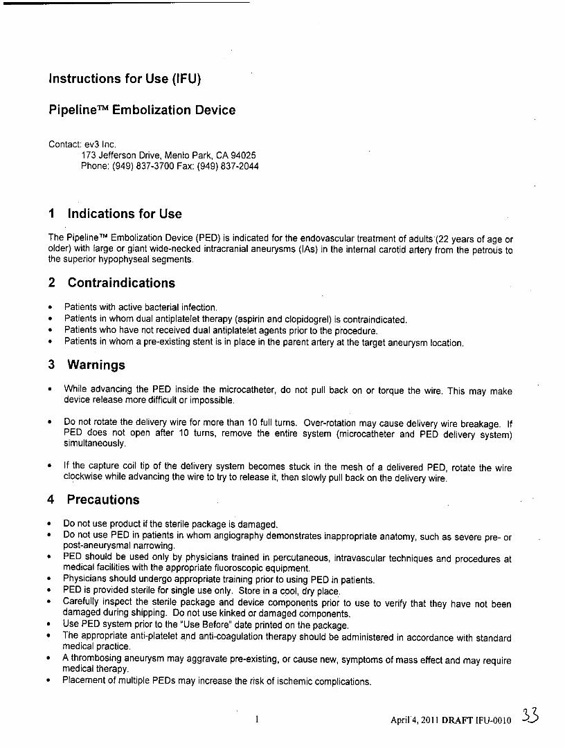
Instructions for Use (IFU)
Pipeline Embolization Device
Contact: ev3 Inc.173 Jefferson Drive, Menlo Park, CA 94025Phone: (949) 837-3700 Fax: (949) 837-2044
1 Indications for Use
The PipelineTM Embolization Device (PED) is indicated for the endovascular treatment of adults (22 years of age orolder) with large or giant wide-necked intracranial aneurysms (lAs) in the internal carotid artery from the petrous tothe superior hypophyseal segments.
2 Contraindications* Patients with active bacterial infection.* Patients in whom dual antiplatelet therapy (aspirin and clopidogrel) is contraindicated.* Patients who have not received dual antiplatelet agents prior to the procedure.* Patients in whom a pre-existing stent is in place in the parent artery at the target aneurysm location.
3 Warnings* While advancing the PED inside the microcatheter, do not pull back on or torque the wire. This may make
device release more difficult or impossible.
* Do not rotate the delivery wire for more than 10 full turns. Over-rotation may cause delivery wire breakage. IfPED does not open after 10 turns, remove the entire system (microcatheter and PED delivery system)simultaneously.
* If the capture coil tip of the delivery system becomes stuck in the mesh of a delivered PED, rotate the wireclockwise while advancing the wire to try to release it, then slowly pull back on the delivery wire.
4 Precautions* Do not use product if the sterile package is damaged.* Do not use PED in patients in whom angiography demonstrates inappropriate anatomy, such as severe pre- or
post-aneurysmal narrowing.* PED should be used only by physicians trained in percutaneous, intravascular techniques and procedures at
medical facilities with the appropriate fluoroscopic equipment.* Physicians should undergo appropriate training prior to using PED in patients.* PED is provided sterile for single use only. Store in a cool, dry place.* Carefully inspect the sterile package and device components prior to use to verify that they have not been
damaged during shipping. Do not use kinked or damaged components.* Use PED system prior to the "Use Before" date printed on the package.* The appropriate anti-platelet and anti-coagulation therapy should be administered in accordance with standard
medical practice.* A thrombosing aneurysm may aggravate pre-existing, or cause new, symptoms of mass effect and may require
medical therapy.* Placement of multiple PEDs may increase the risk of ischemic complications.
I April 4,2011 DRAFT IFU-0010 33
5 Potential Complications
Potential complications, some of which could be fatal, include, but are not limited to the following:
Adverse reaction to antiplatelet/anticoagulation agents or contrast media, intracerebral, bleeding, coma,device fracture, device migration or misplacement, dissection of the parent artery, embolism, groin injury,headache, hemorrhage, hydrocephalus, infection, intracerebral bleeding, ischemia, mass effect,neurological deficits, parent artery stenosis, perforator occlusion, post-procedure bleeding, ruptured orperforated aneurysm, seizure, stroke, thromboembolism, transient ischemic attack (TIA), vasospasm,vessel occlusion, vessel perforation and vision impairment.
6 Device Description
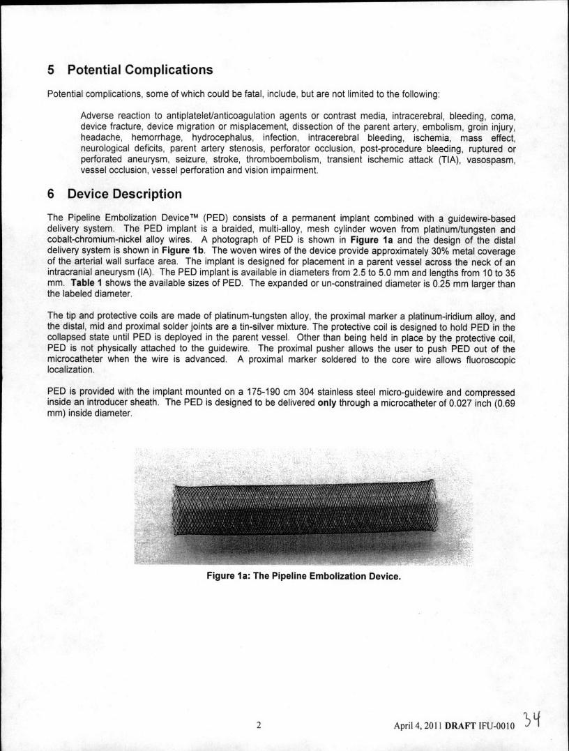
The Pipeline Embolization Devicem (PED) consists of a permanent implant combined with a guidewire-baseddelivery system. The PED implant is a braided, multi-alloy, mesh cylinder woven from platinum/tungsten andcobalt-chromium-nickel alloy wires. A photograph of PED is shown in Figure la and the design of the distaldelivery system is shown in Figure lb. The woven wires of the device provide approximately 30% metal coverageof the arterial wall surface area. The implant is designed for placement in a parent vessel across the neck of anintracranial aneurysm (IA). The PED implant is available in diameters from 2.5 to 5.0 mm and lengths from 10 to 35mm. Table I shows the available sizes of PED. The expanded or un-constrained diameter is 0.25 mm larger thanthe labeled diameter.
The tip and protective coils are made of platinum-tungsten alloy, the proximal marker a platinum-iridium alloy, andthe distal, mid and proximal solder joints are a tin-silver mixture. The protective coil is designed to hold PED in thecollapsed state until PED is deployed in the parent vessel. Other than being held in place by the protective coil,PED is not physically attached to the guidewire. The proximal pusher allows the user to push PED out of themicrocatheter when the wire is advanced. A proximal marker soldered to the core wire allows fluoroscopiclocalization.
PED is provided with the implant mounted on a 175-190 cm 304 stainless steel micro-guidewire and compressedinside an introducer sheath. The PED is designed to be delivered only through a microcatheter of 0.027 inch (0.69mm) inside diameter.
Figure Ia: The Pipeline Embolization Device.
2 April 4,2011 DRAFT IFU-0010
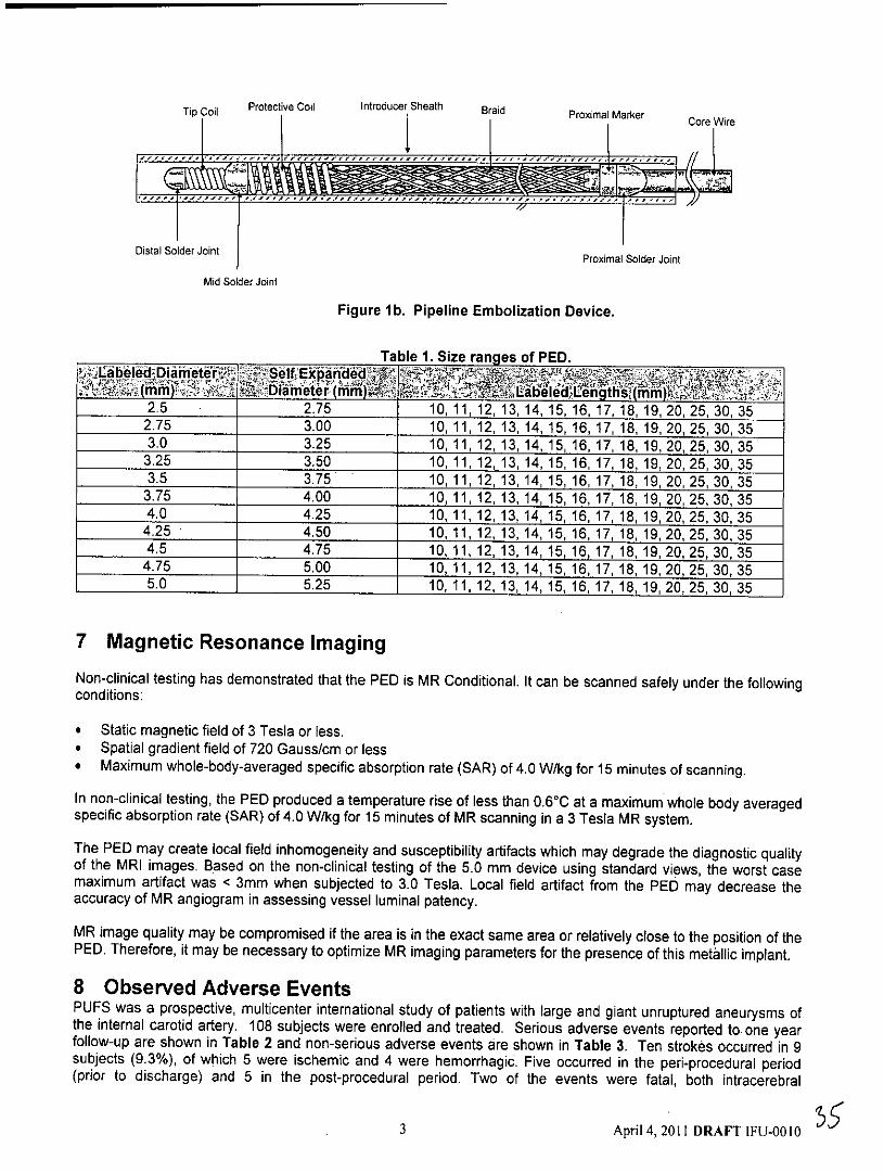
T Protective Coil Introducer Sheath Braid Proximal Marker
Distal Solder JointProximal Solder Joint
Mid Solder Joint
Figure lb. Pipeline Embolization Device.
Table 1. Size ranges of PED.
2.5 2.75 10, 11, 12, 13, 14, 15, 16, 17, 18, 19,20,25, 30, 352.75 3.00 10, 11, 12, 13, 14, 15, 16, 17. 18, 19,20,25, 30,353.0 3.25 10, 11, 12, 13,14, 15, 16, 17, 18, 19, 20, 25, 30,35
3.25 3.50 10, 11, 12, 13, 14, 15, 16, 17, 18, 19,20,25,30,353.5 3.75 10, 11, 12, 13, 14, 15, 16, 17, 18, 19, 20, 25, 30,35
3.75 4.00 10, 11, 12, 13, 14, 15, 16, 17, 18, 19,20,25,30,354.0 4.25 10, 11, 12, 13, 14, 15, 16, 17, 18, 19,20,25,30,35
4.25 4.50 10, 11, 12, 13, 14, 15, 16, 17, 18, 19,20,25,30,354.5 4.75 10, 11, 12, 13, 14, 15, 16, 17, 18, 19, 20,25, 30,35
4.75 5.00 10, 11, 12, 13, 14, 15, 16, 17, 18, 19,20,25, 30,355.0 5.25 10, 11, 12, 13, 14, 15, 16, 17, 18, 19, 20, 25, 30,35
7 Magnetic Resonance ImagingNon-clinical testing has demonstrated that the PED is MR Conditional. It can be scanned safely under the followingconditions:
* Static magnetic field of 3 Tesla or less.* Spatial gradient field of 720 Gauss/cm or less* Maximum whole-body-averaged specific absorption rate (SAR) of 4.0 W/kg for 15 minutes of scanning.
In non-clinical testing, the PED produced a temperature rise of less than 0.60C at a maximum whole body averagedspecific absorption rate (SAR) of 4.0 W/kg for 15 minutes of MR scanning in a 3 Tesla MR system.
The PED may create local field inhomogeneity and susceptibility artifacts which may degrade the diagnostic qualityof the MRI images. Based on the non-clinical testing of the 5.0 mm device using standard views, the worst casemaximum artifact was < 3mm when subjected to 3.0 Tesla. Local field artifact from the PED may decrease theaccuracy of MR angiogram in assessing vessel luminal patency.
MR image quality may be compromised if the area is in the exact same area or relatively close to the position of thePED, Therefore, it may be necessary to optimize MR imaging parameters for the presence of this metallic implant.
8 Observed Adverse EventsPUFS was a prospective, multicenter international study of patients with large and giant unruptured aneurysms ofthe internal carotid artery. 108 subjects were enrolled and treated. Serious adverse events reported to one yearfollow-up are shown in Table 2 and non-serious adverse events are shown in Table 3. Ten strokes occurred in 9subjects (9.3%), of which 5 were ischemic and 4 were hemorrhagic. Five occurred in the peri-procedural period(prior to discharge) and 5 in the post-procedural period. Two of the events were fatal, both intracerebral
3 April 4, 2011 DRAFT IFU-0010
hemorrhages. One peri-procedural ischemic stroke and 2 post-procedural ischemic strokes were associated withparent artery occlusion.
Table 2. Serious adverse events in PUFS by MedDRA@ *category and term - cumulativeincidence at 180 days and one year (N=107 subjects).
MedDRA category MedDRA' term 180 days I yearNervous system disorders 17(15.9%) 19(178%
Headache 5 (4.7%) 5 (4.7%)Haemorrhage intracranial 4(3.7%) 4 (3.7%)Amaurosis fugax 3(2.8%) 5(4.7%)Ischemic stroke 3(2.8%) 3(2.8%)Cerebral haematoma 1 (0.9%) 1 (0.9%)Thrombotic stroke 1 (0.9%) 1 (0.9%)
Neurological disorders NEC Dizziness 0 (0%) 2 (0.9%)Vascular disorders NEC Arteriovenous fistula 2(1.9%) 2(1.9%)Arteriosclerosis, stenosis, vascular insufficiency and Compartment syndrome I (0.9%) 1 (0.9%)necrosis
Carotid artery occlusion 0(0%) 1 (0.9%)Cardiac arrhythmias Atrial fibrillation 1 (0.9%) 1 (0.9%)
Sinus bradycardia 1 (0.9%) 1 (0.9%)Sudden cardiac death 1 (0.9%) 1 (0.9%)
Decreased and nonspecific blood pressure disorders and Procedural hypotension 1 (0.9%) 1 (0.9%)shockEar and labyrinth disorders Tinnitus 1 (0.9%) 1 (0.9%)Embolism and thrombosis Deep vein thrombosis postoperative 1 (0.9%) 1 (0.9%)
Retinal artery thrombosis 1 (0.9%) 1 (0.9%)Gastrointestinal hemorrhages Colitis (excl infective) 1 (0.9%) 1 (0.9%)
Rectal haemorrhage 1 (0.9%) 1 (0.9%)Infections - pathogen unspecified - Urinary tract infection 1 (0.9%) 1 (0.9%)Neoplasms benign, malignant and unspecified Breast cancer recurrence 0 (0%) 1 (0.9%)Pulmonary vascular disorders Post procedural pulmonary 1 (0.9%) 1 (0.9%)
embolismReproductive system and breast disorders Female genital tract fistula 1 (0.9%) 1 (0.9%)Respiratory tract neoplasms Lung squamous cell carcinoma stage 1 0(0%) 1 (0.9%)Vascular disorders Aneurysms and dissections site specific 1 (0.9%) 1 (0.9%)
NECVascular hemorrhagic disorders Epistaxis 1 (0.9%) 1 (0.9%)
Retroperitoneal haemorrhage 1 (0.9%) 1 (0.9%)Vision disorders Diplopia 1 (0.9%) 1 (0.9%)Visual field disorders Visual field defect 1 (0.9%) 1 (0.9%)Total 37 (34.6%) 44 (41.1%)MedDRA': Medical Dictionary for Regulatory Activities
Table 3. Non-serious adverse events occurring in PUFS by 180 days - by decreasing incidence(N=107 subjects).
MedDRA Category MedDRA' Term 180 DaysNervous system disorders - headache 19 (17.8%)
Headache 18 (16.8%)Post-traumatic headache 1 (0.9%)
Procedural and device related injuries and complications NEC* Procedural headache 16 (15%)Vascular hemorrhagic disorders 14(13.1%)
Conjunctival haemorrhage 1 (0.9%)Epistaxis 3 (2.8%)Subcutaneous haematoma 1 (0.9%)Urogenital haemorrhage 2(1.9%)Vessel puncture site hemorrhage 7 (6.5%)
Gastrointestinal signs and symptoms 13(12.1%)Nausea 5 (4.7%)Procedural nausea 7(6.5%)Procedural vomiting 1 (0.9%)
4 April 4,2011 DRAFT IFU-0010

MedDRA' Category MedDRA' Term 180 DaysVision disorders 10(9.3%)
Diplopia 6 (5.6%)Photopsia 3 (2.8%)Vision blurred 1 (0.9%)
Ocular neuromuscular disorders _ _ 9 (8.4%)Eyelid ptosis 4 (3.7%)
lilrd nerve disorder 1(0.9%)lVth nerve disorder 1 (0.9%)VIth nerve disorder 3 (2.8%)
Infections - pathogen unspecified 6 (5.6%)Acute sinusitis 1 (0.9%)Pharyngitis 2 (1.9%)Puncture site infection 1(0.9%)Urinary tract infection 2 (1.9%)
Neurological disorders NEC 5 (4.7%)Dizziness 2 (1.9%)Hyperesthesia 1 (0.9%)Hypoesthesia 1 (0.9%)Hypoesthesia facial 1 (0.9%)
Vascular disorders Ecchymosis 4 (3.7%)Visual field disorders Visual field defect 3(2.8%)Blood and lymphatic system disorders Anemia 2 (1.9%)Body temperature conditions Postoperative fever 2(1.9%)Embolism and thrombosis Deep vein thrombosis postoperative 2(1.9%)Allergic conditions Drug eruption 1 (0.9%)Ear and labyrinth disorders Tinnitus 1 (0.9%)Epidermal and dermal conditions Pruritis 1 (0.9%)Eye disorders NEC Eye pain 1 (0.9%)Gastrointestinal disorders Constipation i (0.9%)Gastrointestinal hemorrhages Lower gastrointestinal haemorrhage 1 (0.9%)General system disorders Discomfort 1 (0.9%)
Facial pain 1,(0.9%)Peripheral edema 1 (09%)
Iniuries NEC Corneal abrasion 1 (0.9%)Musculoskeletal and connective tissue disorders NEC Back pain 1 (0.9%)
Pain in extremity 1 (0.9%)Reproductive system and breast disorders Menometrorrhagia 1 (0.9%)
Menorrhagia 1 (0.9%)Skin and subcutaneous tissue disorders Skin bacterial infection 1 (0.9%)Skin appendage conditions Application site alopecia 1 (0.9%)Total 12
*NEC; not elsewhere classified
5 April 4,2011 DRAFT IFU-0010 5
9 Potential Adverse EventsPotential adverse effects, some of which can be fatal, from use of PED, the PED placement procedure and generalanesthesia include:
* Bleeding, including intracerebral, retroperitoneal or in other locations* Blindness* Complications of arterial puncture including pain, local bleeding, local infection and injury to the artery, vein
or adjacent nerves* Confusion, coma or other change in mental status* Cranial neuropathy* Device fracture, migration or misplacement* Dissection or perforation of the parent artery* Embolism of air, blood clots, cholesterol fragments or device components* Headache* Hydrocephalus* Infection* Mass effect* Neurologic deficits* Perforation or rupture of aneurysm sac or parent artery* Reactions to antiplatelet/anticoagulant agents* Reactions due to radiation exposure* Reactions to anesthesia and related procedures* Reactions to contrast agents including allergic reactions and renal failure* Seizure* Stenosis or thrombosis of the parent artery within PED or a branch vessel covered by PED* Transient ischemic attack (TIA) or ischemic stroke* Vasospasm* Visual impairment
10 Clinical Trial Results - PUFS (Eipeline for Uncoilable or FailedAneurysms) Study
10.1 PurposeThe purpose of the PUFS study was to evaluate the safety and effectiveness of PED for the endovasculartreatment of patients with unruptured large and giant intracranial aneurysms of the internal carotid artery from thepetrous to superior hypophyseal segments.
10.2 DesignPUFS was a prospective, multi-center, single-arm, open label clinical study conducted at 8 sites in the US and 2sites outside of the US. PUFS subjects were adults with a single target aneurysm on the internal carotid artery withsize 210 mm and neck a4 mm. Patients were excluded if they had recent surgery or subarachnoid hemorrhage, ifthey had a bleeding disorder and if a stent was already in place. All patients received perioperative aspirin (325 mgdaily for 2 days prior to PED and 325 mg daily for 6 months after PED) and clopidogrel (75 mg daily for 7 days [or a650 mg oral bolus the day prior to the procedure] and 75 mg daily for 3 months after PED).
The primary effectiveness endpoint of the study was complete occlusion of the target aneurysm on 180-daycerebral angiography in the absence of use of other treatments and in the absence of major (>50%) stenosis of theparent artery. The primary effectiveness endpoint was judged by a core radiologic laboratory. The primary safetyendpoint was the occurrence of major ipsilateral stroke or neurologic death by 180 days. The primary safetyendpoint was judged by a clinical events committee. Based on a literature review, PUFS was designed to beconsidered a success if the primary effectiveness endpoint rate was statistically greater than 50% and the primarysafety endpoint rate was statistically <20%. A Bayesian statistical approach with non-informative prior distributionswas used for the primary endpoint analysis.
The use of antiplatelet agents after these time periods was at the discretion of the treating physician.
6 April 4,2011 DRAFT IFU-0010
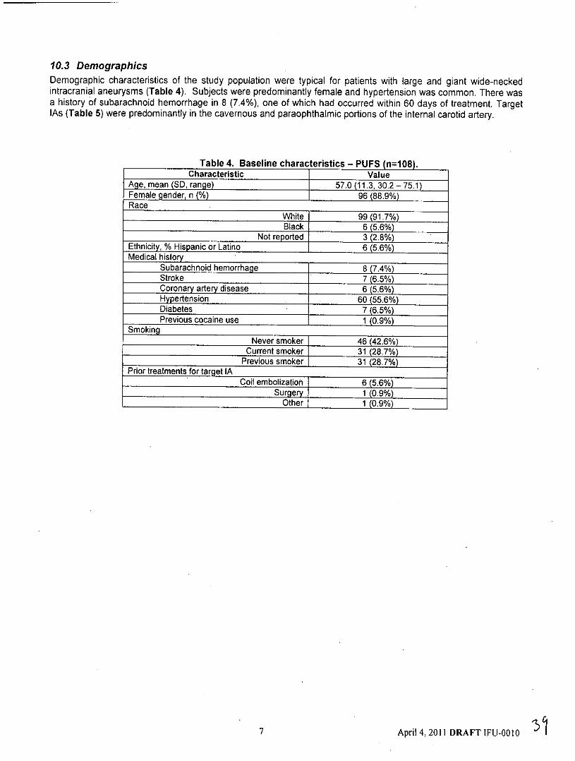
10.3 DemographicsDemographic characteristics of the study population were typical for patients with large and giant wide-neckedintracranial aneurysms (Table 4). Subjects were predominantly female and hypertension was common. There wasa history of subarachnoid hemorrhage in 8 (7.4%), one of which had occurred within 60 days of treatment. TargetIAs (Table 5) were predominantly in the cavernous and paraophthalmic portions of the internal carotid artery.
Table 4. Baseline characteristics - PUFS (n=108).Characteristic Value
Age, mean (SD, range) 57.0 (11.3, 30.2-75.1)Female gender, n (%) 96 (88.9%)Race
W~hite 99(91.7%)Black 6 (5.6%)
Not reported 3(2.8%)Ethnicity, % Hispanic or Latino 6 (5.6%)Medical history
Subarachnoid hemorrhage 8 (7.4%)Stroke 7 (6.5%)Coronary artery disease 6 (5.6%)Hypertension 60 (55.6%)Diabetes 7 (6.5%)Previous cocaine use 1 (0.9%)
Smoking
Never smoker 46 (42.6%)Current smoker 31 (28.7%)
Previous smoker [ 31 (28.7%)Prior treatments for target IA
Coil embolization 6 (5.6%)Surgery 1 (0.9%)
Other 1 (0.9%)
7 April 4,2011 DRAFT IFU-0010
Table 5. Target IA characteristics in PUFS (n=108).Characteristic N (%) or Mean (range)Side
Left 57(52.8%)Right 51 (47.2%)
LocationPetrous 4 (3.7%)
Cavernous 45 (41.7%)Carotid cave 2(1.9%)
Superior hypophyseal 10(9.3%)Lateral clinoidal 2 (1.9%)Paraophthalmic 35 (32.4%)
Supracinoid 9 (8.3%)Posterior communicating 1 (0.9%)
Maximum fundus diameter (mm), mean (SD, 18.2 (6.4, 6.2 - 36.1)range)
"Small" (<10 mm), N (%) 1 (0.9%)tLarge" (>10 mm), N (%) 85(78.7%)"Giant" (>25 mm), N (%) 22(20.4%)
Neck (mm), mean (SD, range) 8.8 (4.3, 4.1-36.1)Dome (mm), mean (SD, range) 14.6 (5.5, 4.4 - 29.5)Dome/neck ratio, mean (SD, range) 1.8 (0.6, 0.6 - 4.1)Target IA partially thrombosed, N (%) 17(15.7%)
10.4 Technical ResultsPED was placed successfully in 107 of 108 attempted (99.0%) subjects. In one subject, the parent artery distal tothe IA could not be catheterized and the PED procedure was abandoned. A mean of 3.1 PEDs was placed persubject (Table 6). PEDs of most diameters and lengths were used (Table 7). Mean procedure time was 124minutes and mean fluoroscopy time was 48.4 minutes.
Table 6. Number of PEDs placed per subject in PUFS (n = 107 subjects)# of PEDs placed N (%)
1 2(2%)2 34 (32%)3 50(47%)4 12(11%)
5 or more 9 (8%)Mean (range) 3.1 (1-15)
Lengths greater than 20 mm were not available during the study.
8 April 4,2011 DRAFT IFU-0010 i d

Table 7. Length and diameter of PEDs used in PUFSLength, mm N
10 1312 5514 6216 6718 6320 81
Diameter, mm N3.25 33.50 313.75 884.00 914.25 644.50 394.75 125.00 13Total 341
10.5 Patient Follow-UpOf the 104 subjects with 106 lAs in the lAs treated population, 97 subjects with 99 treated lAs had angiography 180days after treatment and 89 subjects with 91 treated lAs had angiography 1 year after treatment. Clinical andangiographic follow-up was obtained in 96% of available subjects at 180 days.
10.6 ResultsThe analysis of effectiveness was evaluated in three populations (Table 8). The posterior probability that the studymet its primary effectiveness endpoint was >0.9999 in all three analyses. Complete IA occlusion was seen in81.8% of subjects at 180 days and 85.7% at 1 year (Table 9).
Table 8. Analyses of proportion of PUFS subjects who met the primary effectiveness endpoint.Posterior
Population 180 day Probability .. 1 yearIntracranial aneurysms 78/106; 73.6% (64.4, 81,0)* >0.9999 75/106; 70.8% (61.1, 79.2)**treated (N=106)
Subjects treated 76/104; 73.1% (63.8, 80.7)* >0.9999 73/104; 70.2% (60.4, 78.7)**(N=104)Intracranial aneurysms 80/110; 72.7% (63.7, 80.2)* >0.9999 77/110; 70.7% (58.6, 76.7)*attempted (N=110)
S% posterior credible interval: 95% exact confidence interval
.. Probability that observed effectiveness rate was >50%
Table 9. IA occlusion status at 180 days and 1 year for subjects with angiographic data.180 days 1 year
Occlusion ranking (N=99 As) (N=91 lAs)Complete occlusion 81 (81.8%) 78(85.7%)Residual neck 8(8.1%) 5(5.5%)Residual aneurysm 6(6.1%) 5(5.5%)Other 4' (4.0%) 3" (3.3%)Total 99 (100%) 91 (100%)1 subject with carotid-cavernous fistula and 3 subjects with carotid occlusion in whom IA not visualized:2 subjects with carotid occlusion, 1 transvenous coil embolization in whom IA not visualized
9 April 4,2011 DRAFT IFU-0010 4

The analysis of the primary safety endpoint was based on the safety cohort of 107 subjects treated with PED. Thestudy's primary safety endpoint, ipsilateral major stroke or neurologic death by 180 days after treatment, occurredin 6 subjects (5.6%, 95% posterior credible interval Cl 2.6 - 11.7%). The posterior probability that the major safetyendpoint rate was less than 20%, the predetermined safety success threshold, was 0.999979.
Both the effectiveness and safety endpoint posterior probability values exceeded the pre-study probability thresholdof 0.975, indicating that both results were statistically significant.
Adverse events are listed in Section 8.
10.7 ConclusionsThe study met the pre-specified primary effectiveness and safety endpoints at 180 days which remained statisticallysignificant at one year.
11 Directions for Use1. Using standard interventional radiographic technique, place the microcatheter tip at least 20mm past the distal
edge of the aneurysm. Gently retract the microcatheter to reduce slack in the microcatheter prior to insertingPED.
2. Choose a PED with labeled diameter that approximates the target vessel diameter.
3. Choose a PED with labeled length that is at least 6 mm longer than the aneurysm neck.
4. Remove packaging hoop from the pouch and detach wire from the white rubber wire-holder.
5. Carefully remove delivery wire and introducer sheath out of the packaging coil.
6. Insert introducer sheath into the rotating hemostatic valve at the catheter hub. Visually confirm that the tip of thesheath is seated deeply in the hub of the microcatheter.
7. Secure introducer sheath to the hub by locking down the rotating hemostatic valve tightly.
8. Advance the PED into the microcatheter by pushing the delivery wire until the tip of the delivery wire aligns withthe tip of the microcatheter.
Caution: Do not torque or pull back on delivery wire during insertion.
9. Once the tip of delivery system and microcatheter are aligned, verify that the PED is in the desired location.Distal end of PED should be placed at least 2-3 mm past the distal edge of the aneurysm.
10. Unsheath the PED by slowly retract the microcatheter while maintaining the position of the PED until the tip ofthe microcatheter is proximal to the distal end of the PED
11. Push the delivery wire to continue to expose the PED. After about 10mm of PED is exposed the distal end maydetach from the delivery wire. Detachment can be facilitated by slowly rotating the delivery wire in theclockwise direction.
Warning: Never rotate the delivery wire more than 10 full turns. If PED does not open after 10 turns, remove theentire system (microcatheter and PED delivery system together).
12. After the distal end of PED has successfully expanded, deploy the remainder of PED by alternately advancingthe delivery core wire and allowing the microcatheter to retract slightly.
Caution: Under fluoroscopy, carefully monitor the tip of the core wire during PED deployment. The core wire canbe rotated and maneuvered as needed after the distal end of the PED has detached.
10 April 4, 2011 DRAFT IFU-0010

13. After the entire PED is deployed, advance the microcatheter through the PED. When the microcatheter tip isdistal to the PED, retract while gently rotating the delivery core wire clockwise to prevent entanglement withthe deployed PED and the microcatheter tip.
14. Carefully inspect the deployed PED under fluoroscopy to confirm that it is completely apposed to the vesselwall and not kinked. If the device is not fully opposed or is kinked, consider using an angioplasty balloon to fullyopen it.
* Select an appropriately sized PED such that it is fully expanded diameter is equivalent to that of the proximalparent vessel. An incorrectly sized PED may result in inadequate device placement, incomplete opening ordistal migration.
* Anchor PED at least 2-3 mm into the proximal and distal segments of the parent artery, preferably in a straightportion of the parent artery.
* Use fluoroscopy to carefully monitor the tip of the core wire during PED deployment.* PED foreshortens substantially (50-60%) during deployment. Take device foreshortening into account when
deploying PED.* If the delivery wire cannot be retracted into the microcatheter, carefully remove the delivery core wire and
microcatheter simultaneously.* Rotate the delivery wire only in a clockwise direction. Rotating in a counter-clockwise direction may make
device release more difficult or impossible.
12 Packaging and Storage
Store in a cool, dry place.
Questions and Answers
Q If excessive friction is experienced during the insertion of delivery system at anytime during the delivery ofPED, what should I do?
A Carefully remove the entire. system simultaneously (microcatheter and delivery system).
Q Can I retrieve the PED if the distal end of the PED has expanded at an undesirable location?A Yes. A partially deployed PED can be retrieved. Carefully pull back the delivery core wire until the PED is
secured at the tip of the microcatheter. Then, if there is no resistance, simultaneously remove the entire system(microcatheter and delivery system).
Q Can I retrieve a fully deployed PED?A Once fully deployed, the PED cannot be removed. A second PED can be deployed if needed.
0 Can I place a second PED inside another PED?A Yes. A second PED can be placed inside another PED. After placing the first PED, advance the microcatheter
over the delivery wire while keeping the delivery core wire across the PED. Position the microcatheter at thedesired location and retrieve the delivery wire. Select a new appropriate PED and deploy it as normal.
Caution: Placement of multiple PEDs may increase the risk of ischemic complications.
Q If there.is a difference between the proximal and distal diameter, which PED diameter do I choose?A Choose a PED that matches larger (typically proximal) vessel diameter to ensure proper anchoring.
Definitions of Symbols
Caution: Federal law (USA)O For Single Use Only restricts this device to sale by orLW (Do not Re-use) oni on the order of a (licensed
healthcare practitioner).
11 April 4,2011 DRAFT IFU-0010
SEE Sterile -Ethylene Oxide Non-pyrogenic
Red Instructions Prior Store in a cool, dry place
Ato Use
Manufacturer:ev3, Inc.Menlo Park, CA 94025 USA
Manufactured By:ev3, Inc.173 Jefferson DriveMenlo Park, CA 94025 USA
For Technical Information:ev3, Inc.173 Jefferson DriveMenlo Park, CA 94025 USA
Product Services:Tel: (949) 837-3700Fax: (949) 837-2044
A Pipeline Embolization Device Patient Information Card that includes both patient information, implant informationand MRI guidelines is included with each device. All patients should be instructed to keep this card in theirpossession at all times for procedure/device identification.
12 April 4,2011 DRAFT IFU-0010