Embed Size (px)
Citation preview
2013 www.kce.fgov.be
KCE REPORT 198CS
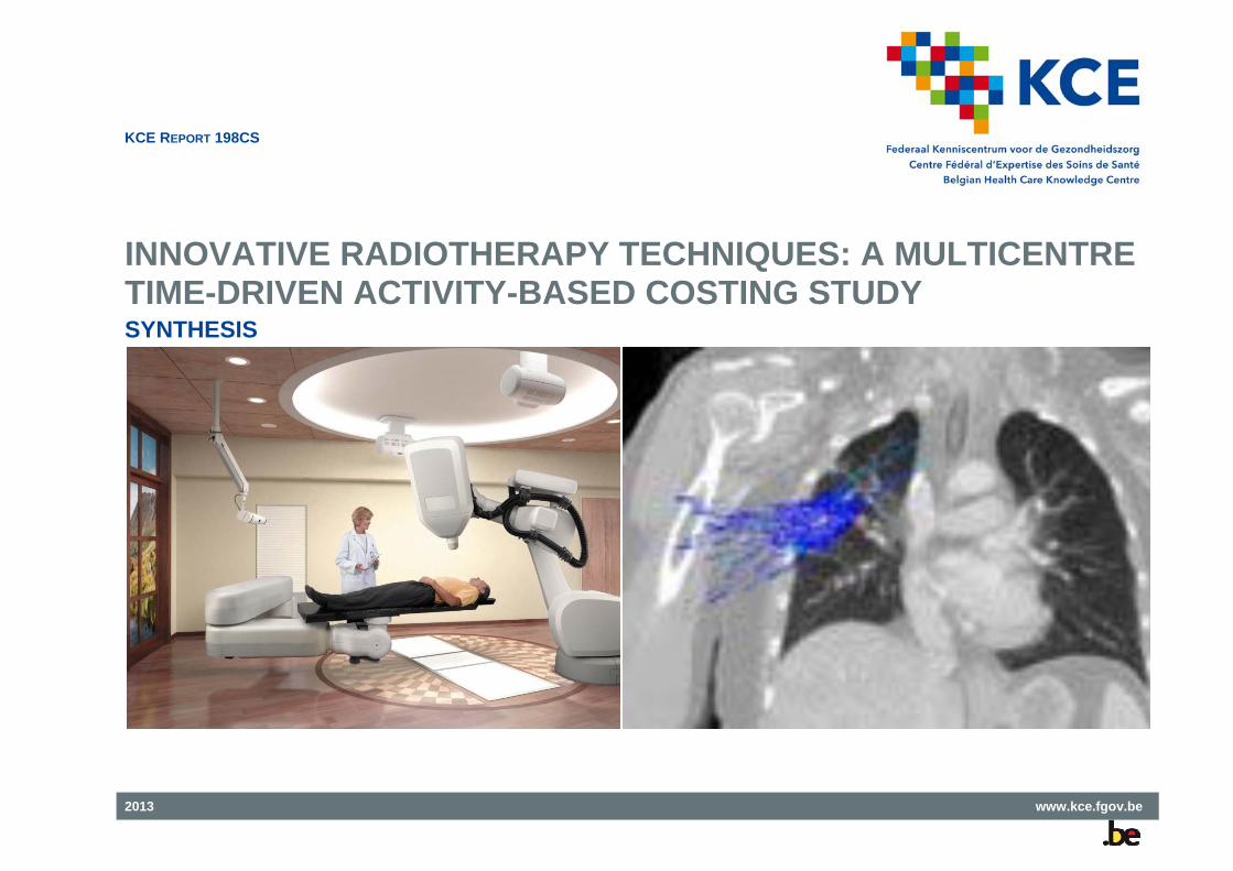
INNOVATIVE RADIOTHERAPY TECHNIQUES: A MULTICENTRE TIME-DRIVEN ACTIVITY-BASED COSTING STUDY SYNTHESIS
Belgian Health Care Knowledge Centre The Belgian Health Care Knowledge Centre (KCE) is an organization of public interest, created on the 24th of December
2002 under the supervision of the Minister of Public Health and Social Affairs. KCE is in charge of conducting studies that support the political decision making on health care and health insurance.
Executive Board Actual Members Substitute Members
President Pierre Gillet CEO - National Institute for Health and Disability Insurance (vice
president) Jo De Cock Benoît Collin
President of the Federal Public Service Health, Food Chain Safety and Environment (vice president)
Dirk Cuypers Christiaan Decoster
President of the Federal Public Service Social Security (vice president) Frank Van Massenhove Jan Bertels General Administrator of the Federal Agency for Medicines and Health
Products Xavier De Cuyper Greet Musch
Representatives of the Minister of Public Health Bernard Lange Brieuc Van Damme Bernard Vercruysse Annick Poncé Representatives of the Minister of Social Affairs Lambert Stamatakis Vinciane Quoidbach Ri De Ridder Koen Vandewoude Representatives of the Council of Ministers Jean-Noël Godin Philippe Henry de
Generet Daniël Devos Wilfried Den Tandt Intermutualistic Agency Michiel Callens Frank De Smet Patrick Verertbruggen Yolande Husden Xavier Brenez Geert Messiaen Professional Organisations - representatives of physicians Marc Moens Roland Lemye
Jean-Pierre Baeyens Rita Cuypers Professional Organisations - representatives of nurses Michel Foulon Ludo Meyers
Myriam Hubinon Olivier Thonon Hospital Federations Johan Pauwels Katrien Kesteloot Jean-Claude Praet Pierre Smiets Social Partners Rita Thys Leo Neels Paul Palsterman Celien Van Moerkerke House of Representatives Lieve Wierinck
Control Government commissioner Yves Roger
Management Chief Executive Officer
Assistant Chief Executive Officer Raf Mertens Christian Léonard
Manager Program Management Kristel De Gauquier Contact Belgian Health Care Knowledge Centre (KCE)
Doorbuilding (10th Floor) Boulevard du Jardin Botanique, 55 B-1000 Brussels Belgium T +32 [0]2 287 33 88 F +32 [0]2 287 33 85 [email protected] http://www.kce.fgov.be

2013 www.kce.fgov.be
KCE REPORT 198CS HEALTH TECHNOLOGY ASSESSMENT
INNOVATIVE RADIOTHERAPY TECHNIQUES: A MULTICENTRE TIME-DRIVEN ACTIVITY-BASED COSTING STUDY SYNTHESIS FRANK HULSTAERT, ANNE-SOPHIE MERTENS, CAROLINE OBYN, DRIES VAN HALEWYCK, BRIEUC VAN DER STRATEN, YOLANDE LIEVENS
COLOPHON Title: Innovative radiotherapy techniques: a multicentre time-driven activity-based costing study – synthesis
Authors: Frank Hulstaert (KCE), Anne-Sophie Mertens (Möbius), Caroline Obyn (KCE), Dries Van Halewyck (Möbius), Brieuc van der Straten (Möbius), Yolande Lievens (UZ Gent)
Reviewers: Francis Colardyn (Consultant), Irina Cleemput (KCE), Sophie Gerkens (KCE), Raf Mertens (KCE), Bert Winnen (RIZIV – INAMI), Hilde Engels (RIZIV – INAMI), Saskia Van den Bogaert (FOD Volksgezondheid – SPF Santé Publique)
External experts: An Fremout (FANC – AFCN), Isabelle De Pau (FANC – AFCN); Carolien Claeys (FANC – AFCN), Liesbeth Van Eycken (Cancer Registry), Nancy Van Damme (Cancer Registry), Karen Vos (Cancer Registry) The Belgian radiotherapy specialists and their collaborators who contributed in the working group meetings and participated to the cost study CHU de Liège, Liège: Philippe Coucke, Eric Lenaerts, Jean Vanderick, Nicolas Jansen, Philippe Martinive UZ Gent, Ghent: Yolande Lievens, Barbara Vanderstraeten, Marc Van Eijkeren GZA Ziekenhuizen Sint-Augustinus, Antwerp: Philippe Huget, Katrien Erven, Rita Reymen, Reinhilde Weytjens, Mark Vos UZ Brussel, Brussels: Mark De Ridder, Thierry Gevaert Institut Jules Bordet, Brussels: Paul Vanhoutte, Luigi Moretti, John Vercauteren UZ KULeuven, Leuven: Karin Haustermans, Jan Verstraete OLV Ziekenhuis, Aalst: Luc Verbeke, Samuel Bral, Heidi Roelstraete, Stijn Van Der Heyden, Pol Decroos Jessa Ziekenhuis, Hasselt: Marc Brosens, Paul Bulens, Katrien Spaas, Goele Kippers Centre Hospitalier Jolimont: Carine Mitine, Christian Nicolas, Jean-François Rosier Hôpital Sainte-Elisabeth, Namur: Vincent Remouchamps, Hervé Marchal, Christian Sprumont, François Sergent Experts from other Belgian radiotherapy centres who participated to the working groups: URA, Antwerp: Danielle Van den Weyngaert, Bie De Ost, Dirk Van Gestel AZ Saint-Jan, Brugge-Oostende: Geertrui De Meestere, Sarah Roels AZ Saint-Lucas, Ghent: Frank Bouttens, Pieternel Thysebaert AZ Saint-Maarten Duffel: Philippe Spaas, Valerie Vandeputte CHIREC, Brussels: Richard Burette CH Peltzer-La Tourelle, Verviers: Olivier De Hertogh CHU Charleroi: Anne-Rose Henry
Clinique Saint-Jean Kliniek St. Jan: Sophie Cvilic Cliniques de l’Europe-Europaziekenhuizen: Carl Salambier Heilig Hartziekenhuis, Roeselare: Ivan Staelens Grand Hôpital de Charleroi St. Joseph, Gilly: Françoise Gilsoul AZ Groeninge, Kortrijk: Antoon Lambrecht RHMS – Clinique Louis Caty, Baudour: Joelle Fraikin AZ Turnhout, Turnhout: Jean Meyskens, Michel Martens, Katrien Geboers UCL Saint-Luc, Brussels: Pierre Scalliet, Xavier Geets
Acknowledgements: Sara Appeltants (FOD Economie – SPF Economie),: Kris Engels (RIZIV – INAMI), Chris Hubin (RIZIV – INAMI), Alphonse Thijs (RIZIV – INAMI), Herwin De Kind (Vlaams Agentschap Zorg en Gezondheid), Hilde Smets (Vlaams Agentschap Zorg en Gezondheid), Vincent Ronflé (Varian), Bart van Acker (UMC St Radboud, The Netherlands), Hans Kaanders (UMC St Radboud)
External validators: Lieven Annemans (Health economics, Ghent University and Brussels University), Peter Dunscombe (Medical Physics and Oncology, University of Calgary, Canada), Lionel Perrier (Health technology assessment, Cancer Centre Léon Bérard, Lyon, France)
Other reported interests: None declared
Layout: Ine Verhulst, Sophie Vaes
Disclaimer: The external experts were consulted about a (preliminary) version of the scientific report. Their comments were discussed during meetings. They did not co-author the scientific report and did not necessarily agree with its content. Subsequently, a (final) version was submitted to the validators. The validation of the report results from a consensus or a voting process between the validators. The validators did not co-author the scientific report and did not necessarily all three agree with its content. Finally, this report has been approved by common assent by the Executive Board. Only the KCE is responsible for errors or omissions that could persist. The policy recommendations are also under the full responsibility of the KCE.
Publication date: 25 March 2013
Domain: Health Technology Assessment (HTA)
MeSH: Radiotherapy; Cost and Cost Analysis
NLM Classification: QZ 269
Language: English
Format: Adobe® PDF™ (A4)
Legal depot: D/2013/10.273/8
Copyright: KCE reports are published under a “by/nc/nd” Creative Commons Licence http://kce.fgov.be/content/about-copyrights-for-kce-reports.
How to refer to this document? Hulstaert F, Mertens A-S, Obyn C, Van Halewyck D, Van Der Straten B, Lievens Y. Innovative radiotherapy techniques: a multicentre time-driven activity-based costing study – synthesis. Health Technology Assessment (HTA) Brussels: Belgian Health Care Knowledge Centre (KCE). 2013. KCE Reports 198Cs. D/2013/10.273/8.
This document is available on the website of the Belgian Health Care Knowledge Centre.

KCE Report 198Cs Innovative radiotherapy techniques 1
FOREWORD
When the National Health and Disability Insurance Institute (RIZIV – INAMI) asked us to study innovative radiotherapy techniques, frankly, we did not greet the project with a lot of enthusiasm. It looked as if we were to face another instance of the well know scenario of technology push in the field, versus the lack of high level evidence in the literature. Added to this, is the emotional context of cancer patients who deserve to get all the best chances… In brief, this project looked as if it was programmed to lead to yet another disappointment. But, since KCE was not created to provide only the easy answers, we met with RIZIV – INAMI and the Ministry of Public Health (FOD Volksgezondheid – SPF Santé Publique), and looked if we could not explore new avenues. In collaboration with Federal Agency for Nuclear Control (FANC – AFCN) and the national cancer registry we looked for an intermediate solution between the extremes of either bluntly rejecting the innovative techniques or allowing an uncontrolled introduction. The road we took was that of limited access with evidence generation, also referred to as coverage with evidence generation. A first piece of required information which was not readily available, was the real cost for the hospital of the radiotherapy treatments. Fortunately, we could build on the experience of the University Hospitals of the KULeuven in this area. Also the KCE manual of cost studies (KCE report 178) published last year proved to be of use. All ten Belgian radiotherapy centres we contacted were prepared to open their books for the financial analysts of Möbius, who executed this study. In this report you will find the results of this first step. The next steps are in the hands of the RIZIV – INAMI. by means of research financing of the centres (“conventions”) a stepwise introduction will be realized, together with the registration of the technique used and the indications – crucial information to document an introduction. After about 4 years, the data should be made available to take a more informed decision on the reimbursement of these treatments. We hope our study is contributing to a more evidence-based model of financing, and we wish to express our gratitude to the many radiotherapy centres for their hard work and their courage to go this route.
Christian LÉONARD Deputy general director
Raf MERTENS General director
2 Innovative radiotherapy techniques KCE Report 198Cs
ABSTRACT OBJECTIVES The primary aim of this study is to calculate the cost of innovative radiotherapy techniques that are being introduced in Belgium. We focus on (1) stereotactic body radiation therapy (SBRT) in selected indications, (2) accelerated partial breast irradiation (APBI) and (3) intraoperative radiotherapy (IORT) as a boost to whole breast irradiation (WBI) schemes. The results will serve as input for a planned research financing of these radiotherapy techniques by the National Institute for Health and Disability Insurance (RIZIV–INAMI). This study is part of a larger project that aims for a staged introduction of promising innovative radiotherapy techniques in the Belgian health care system. The secondary aim is to calculate the costs of the most commonly performed routine radiotherapy treatments. These cost data could support any future revision of the radiotherapy financing in Belgium.
METHODS We performed a time-driven activity-based costing (TDABC) of radiotherapy treatments in 10 centres representative for Belgium. Time measurements were performed during a 4 week period in the second half of 2012. Results are expressed in 2011 euros.
KCE Report 198Cs Innovative radiotherapy techniques 3
RESULTS SBRT of the lung shortens the radiotherapy course as only 3 (1 to 10) very high doses of external irradiation are precisely targeted. The average SBRT cost of € 6 222 is similar to the cost of 3D or intensity modulated radiotherapy (IMRT) schemes with hypo- or standard fractionation, which range from € 3 993 to € 7 379. The cost of SBRT varied by centre from about € 3 000 euro to over € 12 000, by technique used in the centre (tracking SBRT had a slightly higher cost than other modalities) and by fractionation (higher cost for higher fractionation). Based on few patients, the cost for SBRT (para)spinal, liver, and pancreas was € 3573 (3 fractions), € 5 586 (3 to 10 fractions) and € 5 341 (10 fractions), respectively. Costs for SBRT of oligometastatic disease varied from € 3 342 (3 fractions) to € 5 076 (10 fractions). APBI can be used in specific cases of low-risk early breast cancer, shortening the irradiation course from 3-7 weeks to 1-2 weeks, or to single fraction IORT. The average cost of IORT APBI delivered as MeV electrons using a mobile linear accelerator system (Mobetron) was € 3 063 euro and € 2 744 euro if delivered as kV photons (Intrabeam). These costs are at the low end of whole breast irradiation (WBI) schemes without boost (€ 3 008 to € 4 683 euro). We found a higher cost of € 6 693 for APBI using high dose rate (HDR) interstitial multicatheter brachytherapy. The cost of an external beam radiotherapy boost is about € 500 to € 1 500. This additional cost is much lower than the cost of a brachytherapy boost with HDR or pulsed dose rate (€ 2 000 to € 2500), an IORT kV boost (€ 2 000 to € 2 500 euro) or an IORT MeV boost (€ 2 500 to € 4 000). Some costs are to be added. In case of brachytherapy these are the cost of the isotope and possibly additional general anesthesia and hospitalization. In case of IORT the cost associated with extending breast surgery is to be added. The average costs of IMRT of head and neck (€ 8 237) and prostate cancer (€ 7 278) were higher than the cost of IMRT for rectum cancer (€ 4 889) and standard fractionated WBI (€ 4 587). The cost of palliative treatments varies from € 1 032 (1 fraction) to € 2 655 (10 to 13 fractions).
CONCLUSION TDABC in parallel at 10 radiotherapy centres was feasible. We obtained average cost estimates both for the innovative radiotherapy treatments as well as for the routine treatments. The data may be of use to better align the financing of radiotherapy in Belgium with the costs.
4 Innovative radiotherapy techniques KCE Report 198Cs
SYNTHESIS TABLE OF CONTENTS FOREWORD ......................................................................................................................................... 1
ABSTRACT ........................................................................................................................................... 2 SYNTHESIS .......................................................................................................................................... 4
1. INTRODUCTION ................................................................................................................................... 6 1.1. OBJECTIVES ........................................................................................................................................ 6 1.2. FINANCING OF RADIOTHERAPY IN BELGIUM ................................................................................. 6 1.3. THE CHALLENGE OF INNOVATION IN RADIOTHERAPY ................................................................. 6 1.4. INNOVATIVE RADIOTHERAPY TECHNIQUES AND THEIR INDICATIONS ...................................... 7
1.4.1. Novel radiotherapy techniques for treating early breast cancer. ............................................ 7 1.4.2. Stereotactic body radiation therapy (SBRT). .......................................................................... 7
1.5. RESEARCH FUNDING, REGISTRATION OF ACTIVITIES AND CLINICAL OUTCOME. ................... 8 2. METHODS ............................................................................................................................................. 9 2.1. WHY AN ACTIVITY-BASED COSTING ANALYSIS? ........................................................................... 9 2.2. APPLICATION OF THE KCE COST MANUAL ..................................................................................... 9 2.3. THE COSTS REFLECT THE REALITY ................................................................................................ 9 2.4. SELECTION AND PARTICIPATION OF RADIOTHERAPY CENTRES ............................................. 10 2.5. DEFINITION OF RADIOTHERAPY TREATMENTS ........................................................................... 10 2.6. STEPWISE APPROACH ..................................................................................................................... 10 2.7. ACTIVITY CATEGORIES .................................................................................................................... 11 2.8. OUT OF SCOPE ACTIVITIES ............................................................................................................. 11 2.9. COSTS AND RESOURCE USE .......................................................................................................... 11
2.9.1. Personnel cost ....................................................................................................................... 11 2.9.2. Equipment costs .................................................................................................................... 11 2.9.3. Indirect material costs ........................................................................................................... 13 2.9.4. Pharmaceuticals and radio-isotopes ..................................................................................... 13 2.9.5. Overhead ............................................................................................................................... 13
KCE Report 198Cs Innovative radiotherapy techniques 5
2.9.6. Direct costs ............................................................................................................................ 13 2.10. COST ALLOCATION ........................................................................................................................... 13
2.10.1. Allocation of personnel and equipment to activities .............................................................. 13 2.10.2. Allocation of activities, materials and overhead to treatments .............................................. 14
3. DISCUSSION OF THE RESULTS ...................................................................................................... 15 3.1. GENERAL FINDINGS ......................................................................................................................... 15
3.1.1. Number of type of treatments ................................................................................................ 15 3.1.2. Total cost by centre ............................................................................................................... 15 3.1.3. Cost structure ........................................................................................................................ 16 3.1.4. Process validation ................................................................................................................. 16 3.1.5. Efficiency ............................................................................................................................... 16 3.1.6. Limitations ............................................................................................................................. 17
3.2. COST OF SBRT COMPARED WITH OTHER MODALITIES ............................................................. 18 3.2.1. Focus on the lung .................................................................................................................. 18 3.2.2. SBRT of spinal, liver, pancreas, bone and oligometastases................................................. 20
3.3. BREAST CANCER, FOCUS ON APBI AND IORT BOOST ................................................................ 20 3.4. COST OF OTHER COMMON TREATMENTS .................................................................................... 23 3.5. COSTS, FINANCING AND BUDGET CONSIDERATIONS ................................................................ 23 3.6. INTRODUCING INNOVATIONS IN HEALTH CARE .......................................................................... 24
REFERENCES .................................................................................................................................... 25 RECOMMENDATIONS ....................................................................................................................... 27
6 Innovative radiotherapy techniques KCE Report 198Cs
1. INTRODUCTION 1.1. Objectives The primary aim of this study is to calculate the cost of innovative radiotherapy techniques that are being introduced in Belgium. We focus on (1) stereotactic body radiation therapy (SBRT) in selected indications, (2) accelerated partial breast irradiation (APBI) and (3) intraoperative radiotherapy (IORT) as a boost to whole breast irradiation (WBI) schemes. The results will serve as input for a planned research financing of these radiotherapy techniques by the National Institute for Health and Disability Insurance (RIZIV–INAMI). The secondary aim is to calculate the costs of the most commonly performed routine radiotherapy treatments. These cost data could support any future rework of the radiotherapy financing in Belgium.
1.2. Financing of radiotherapy in Belgium Radiotherapy in Belgium is offered at 25 hospital-based centres that have been authorized for this activity. Eight of these centres serve one or more satellite centres, which are based in another hospital. The Belgian compulsory health insurance provides coverage for radiotherapy treatments in these centres when performed by one of the 155 radiotherapy specialists. Billing of activities is performed using a set of billing codes or “nomenclature codes”, managed by the RIZIV–INAMI. Adapting this financing system, including the tariffs and the content of the “nomenclature”, is a lengthy process, involving negotiations with multiple stakeholders. For 2012, the overall amount paid for radiotherapy specific billing codes is 111 million euros. In addition, the Belgian hospital financing (Budget Financiële Middelen-Budget des Moyens Financiers, BFM – BMF) includes specific funds for radiotherapy (within the A3 and B3 component) amounting to 40 million euros per year. The A3 amount for radiotherapy depends on the number of linear accelerators that are less than 10 years old (each such linear accelerator is financed at about 90 000 euros per year) and on the number and type of acts performed by the centre. The B3 part is aimed at covering costs of nursing and technical personnel, administrative and general costs, cost of consumables and maintenance
cost of the equipment and rooms of the radiotherapy centre. The B3 amount is a lump sum in function of the number and type of acts performed by the centre. The radiotherapy centres also make use of centrally provided hospital services (such as building depreciation, financial costs, central administration, management) that are financed through other BFM – BMF components, notably A1, A2, B1 and B4. The radiotherapy centres furthermore also have a number of D-beds for hospitalized patients, financed mostly through the B2 part.
1.3. The challenge of innovation in radiotherapy Linear accelerators and other equipment used to deliver radiotherapy are medical devices. They are not clinically validated for specific indications when they obtain a CE mark and enter the European market.1, 2 The low regulatory barrier creates a challenge for the health care authorities. The case of radiotherapy techniques is even more complex as the innovation often concerns delivering the right radiation dose precisely to the target volume, based on multiple medical devices (software and hardware systems) from different manufacturers that are expected to communicate flawlessly. Furthermore, oncology clinical trials with hard outcomes take many years to conduct. Such large confirmatory clinical trials for innovative radiotherapy techniques in specific indications are often still ongoing when some centres already want to offer these novel therapeutic options to their patients. In some cases, evidence based on limited case series can be convincing enough to justify this move. In many other cases, where the evidence based on case series is not yet reassuring, it seems appropriate to only use the innovative technique in the context of a well monitored clinical trial, such that side-effects of the new radiotherapy techniques are timely identified. Awaiting further clinical validation of specific innovative techniques for specific indications (technique/indication pairs), the health insurance decision makers have no other option than a staged introduction of promising new techniques based on robust cost calculations and the creation of a registry of innovative radiotherapy treatments and their clinical outcome.
KCE Report 198Cs Innovative radiotherapy techniques 7
The interpretation of clinical outcome data based on such registries remains a challenge and the level of evidence that can be generated based on such data is low at best.
1.4. Innovative radiotherapy techniques and their indications First, the promising innovative radiotherapy techniques and their indications were identified by a working group of interested Belgian radiation oncologists and representatives from KCE, RIZIV–INAMI, FOD–SPF Public Health, and the Federal Nuclear Control Agency (FANC–AFCN). The techniques considered were novel radiotherapy techniques for treating early breast cancer and stereotactic body radiation therapy (SBRT). For both novel strategies of radiotherapy, indications were specified and the current level of evidence was determined based on literature reviews and consensus statements of professional societies. No systematic review of the literature was however performed, which is a limitation of the project. 1.4.1. Novel radiotherapy techniques for treating early breast
cancer. For early stage breast cancer the focus of innovation is on accelerated partial breast irradiation (APBI) as an alternative to whole breast irradiation (WBI) following breast-conservative surgery. We also study the use of intraoperative radiation therapy (IORT) to deliver a “boost” dose in addition to a WBI scheme. The advantage of APBI is that the period during which irradiation is scheduled is shortened from the current 3 to 7 weeks for hypo- and standard fractionated WBI to 1 or 2 weeks, or even to just the stay in the operating theatre. This is convenient for patients. In addition, targeting the right area with an IORT boost is more straightforward compared with a brachytherapy boost delivered after completion of the WBI, despite any markers the surgeon may have left behind to indicate the target area. Techniques for APBI identified in the literature range from brachytherapy using radio-isotopes or electrons, to external beam irradiation techniques using MV photons or MeV electrons, or intraoperative radiation therapies (IORT) delivered with kV photons or MeV electrons.3-5 6 Different techniques may show substantial differences in dose delivery, but the
clinical relevance of it has not yet been documented.7 At least seven large RCTs are ongoing (www.clinicaltrials.gov). Consensus statements on APBI have been published by the American Society for Therapeutic Radiology and Oncology (ASTRO)8 and by the GEC-ESTRO Breast Cancer Working Group (www.estro.org). Based on these statements a Belgian working group of radiotherapists proposed three categories: a low risk group eligible for APBI without clinical trial, a medium risk group where APBI should only be performed in the context of a clinical trial and high risk group for whom APBI is contra indicated, detailed in appendix. Belgian radiotherapy centres have currently invested in the following APBI techniques: • MeV electrons delivered in a single intraoperative fraction via
Mobetron (IntraOp): a mobile external irradiation system which can be placed in a shielded operating theatre for IORT. The entire procedure lasts for about 15 to 20 minutes.5 Unnecessary radiation to the underlying normal tissue is avoided by mobilizing the mammary gland during surgery and placing a lead plate for shielding on its dorsal surface. The Mobetron can also be used for IORT for other indications.
• 50 kV photons delivered in a single intraoperative fraction via Intrabeam (Zeiss): a smaller device with an applicator that fits in the cavity after lumpectomy, for IORT.
• In one centre multicatheter brachytherapy with high dose range (HDR) Iridium-192 isotope is delivered in 8 fractions over 4 days. The same technique usually administered in 10 fractions over 5 days is used in another centre when the same breast had already been treated by WBI in the past. Other brachytherapy systems such as balloon catheter radiation (MammoSite) are currently not used in Belgium.
1.4.2. Stereotactic body radiation therapy (SBRT). SBRT is also referred to as stereotactic ablative body radiotherapy (SABR) and as stereotactic ablative radiation therapy. By using X-ray imaging devices for stereotactic positioning and sometimes implanted markers in the body, radiation oncologists are able to deliver a much higher radiation dose to a precise target than with traditional radiation therapy, while sparing healthy tissue. SBRT can be delivered using various formats with
8 Innovative radiotherapy techniques KCE Report 198Cs
systems that may or may not be dedicated for SBRT. SBRT is under advanced evaluation for the treatment of early-stage non-small cell lung cancer (NSCLC), (oligo)metastases in the liver and lung, as well as (para)spinal tumours. Evidence for additional indications is still very limited.9 In France, indications for SBRT (lung cancer and spinal tumours) were already defined in 2006.10 The 2010 SBRT evidence review document by the National Health Service (NHS), UK,11 provides important guidance on the implementation of the SBRT technique by indication. Five large RCTs are ongoing in NSCLC, one in spine metastasis and one in colorectal carcinoma liver metastases (www.clinicaltrials.gov). For patients with T1-2 N0 NSCLC in the periphery of the lung and unfit or unwilling to undergo surgery, there is now considerable non-randomised evidence supporting SBRT as superior to conventional radiotherapy with respect to local control and survival.12-14 Ongoing randomized controlled trials of SBRT versus surgery will help to determine their relative effectiveness. For the patient SBRT is also more convenient because of fewer treatment sessions (typically 1 to 5 or sometimes up to 10 sessions, instead of the standard 30-35 sessions for 3D conformal RT or IMRT or less protracted hypofractionated schedules). Four different modalities (free breathing with tracking, free breathing without tracking, gating, breath-hold) exist to deliver SBRT treatment for lung tumours. The relative effectiveness of these options is currently unknown. Systems with tracking of the tumour include CyberKnife (Accuray) and Vero (Brainlab).15 Other SBRT systems include Hi-Art (TomoTherapy), Novalis (BrainLab), Novalis TX (Varian and Brainlab), Trilogy (Varian), TrueBeam (Varian), Elekta Axesse (Elekta).
1.5. Research funding, registration of activities and clinical outcome.
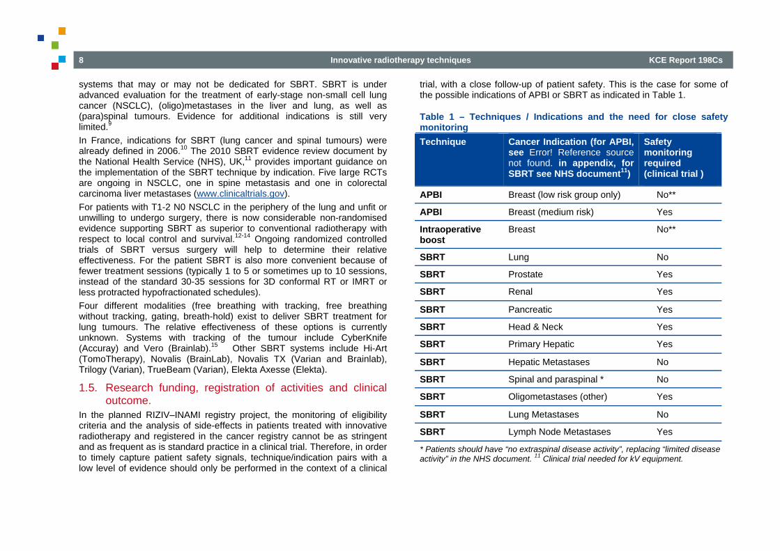
In the planned RIZIV–INAMI registry project, the monitoring of eligibility criteria and the analysis of side-effects in patients treated with innovative radiotherapy and registered in the cancer registry cannot be as stringent and as frequent as is standard practice in a clinical trial. Therefore, in order to timely capture patient safety signals, technique/indication pairs with a low level of evidence should only be performed in the context of a clinical
trial, with a close follow-up of patient safety. This is the case for some of the possible indications of APBI or SBRT as indicated in Table 1.
Table 1 – Techniques / Indications and the need for close safety monitoring Technique Cancer Indication (for APBI,
see Error! Reference source not found. in appendix, for SBRT see NHS document11)
Safety monitoring required (clinical trial )
APBI Breast (low risk group only) No**
APBI Breast (medium risk) Yes
Intraoperative boost
Breast No**
SBRT Lung No
SBRT Prostate Yes
SBRT Renal Yes
SBRT Pancreatic Yes
SBRT Head & Neck Yes
SBRT Primary Hepatic Yes
SBRT Hepatic Metastases No
SBRT Spinal and paraspinal * No
SBRT Oligometastases (other) Yes
SBRT Lung Metastases No
SBRT Lymph Node Metastases Yes
* Patients should have “no extraspinal disease activity”, replacing “limited disease activity” in the NHS document. 11 Clinical trial needed for kV equipment.
KCE Report 198Cs Innovative radiotherapy techniques 9
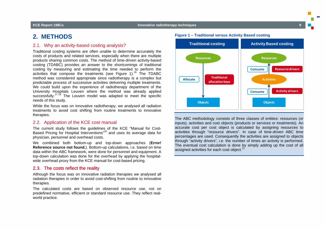
2. METHODS 2.1. Why an activity-based costing analysis? Traditional costing systems are often unable to determine accurately the costs of products and related services, especially when there are multiple products sharing common costs. The method of time-driven activity-based costing (TDABC) provides an answer to the shortcomings of traditional costing by measuring and estimating the time needed to perform the activities that compose the treatments (see Figure 1).16 The TDABC method was considered appropriate since radiotherapy is a complex but predictable process of successive activities delivering multiple treatments. We could build upon the experience of radiotherapy department of the University Hospitals Leuven where the method was already applied successfully.17-19 The Leuven model was adapted to meet the specific needs of this study. While the focus was on innovative radiotherapy, we analysed all radiation treatments to avoid cost shifting from routine treatments to innovative therapies.
2.2. Application of the KCE cost manual The current study follows the guidelines of the KCE “Manual for Cost-Based Pricing for Hospital Interventions”20 and uses its average data for physician, personnel and overhead costs. We combined both bottom-up and top-down approaches (Error! Reference source not found.). Bottom-up calculations, i.e. based on time data within the ABC framework, were done for personnel and equipment. A top-down calculation was done for the overhead by applying the hospital-wide overhead proxy from the KCE manual for cost-based pricing.
2.3. The costs reflect the reality 2.3. The costs reflect the reality Although the focus was on innovative radiation therapies we analysed all radiation therapies in order to avoid cost-shifting from routine to innovative therapies. The calculated costs are based on observed resource use, not on predefined normative, efficient or standard resource use. They reflect real-world practice.
Figure 1 – Traditional versus Activity Based costing
The ABC methodology consists of three classes of entities: resources (or inputs), activities and cost objects (products or services or treatments). An accurate cost per cost object is calculated by assigning resources to activities through “resource drivers”. In case of time-driven ABC time percentages are used. Consequently the activities are assigned to objects through “activity drivers”, i.e. the number of times an activity is performed. The eventual cost calculation is done by simply adding up the cost of all assigned activities for each cost object.21
Traditional costing Activity Based costing
Resources
Objects
Traditional allocationkeys
Resources
Objects
Activities
Consume
Consume Resource drivers
Activity drivers
Allocate
10 Innovative radiotherapy techniques KCE Report 198Cs
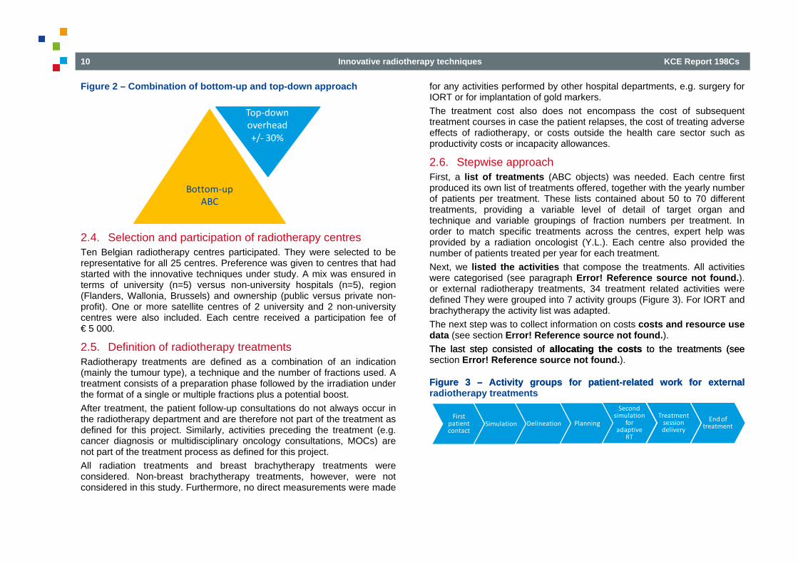
Figure 2 – Combination of bottom-up and top-down approach
2.4. Selection and participation of radiotherapy centres Ten Belgian radiotherapy centres participated. They were selected to be representative for all 25 centres. Preference was given to centres that had started with the innovative techniques under study. A mix was ensured in terms of university (n=5) versus non-university hospitals (n=5), region (Flanders, Wallonia, Brussels) and ownership (public versus private non-profit). One or more satellite centres of 2 university and 2 non-university centres were also included. Each centre received a participation fee of € 5 000.
2.5. Definition of radiotherapy treatments Radiotherapy treatments are defined as a combination of an indication (mainly the tumour type), a technique and the number of fractions used. A treatment consists of a preparation phase followed by the irradiation under the format of a single or multiple fractions plus a potential boost. After treatment, the patient follow-up consultations do not always occur in the radiotherapy department and are therefore not part of the treatment as defined for this project. Similarly, activities preceding the treatment (e.g. cancer diagnosis or multidisciplinary oncology consultations, MOCs) are not part of the treatment process as defined for this project. All radiation treatments and breast brachytherapy treatments were considered. Non-breast brachytherapy treatments, however, were not considered in this study. Furthermore, no direct measurements were made
for any activities performed by other hospital departments, e.g. surgery for IORT or for implantation of gold markers. The treatment cost also does not encompass the cost of subsequent treatment courses in case the patient relapses, the cost of treating adverse effects of radiotherapy, or costs outside the health care sector such as productivity costs or incapacity allowances.
2.6. Stepwise approach First, a list of treatments (ABC objects) was needed. Each centre first produced its own list of treatments offered, together with the yearly number of patients per treatment. These lists contained about 50 to 70 different treatments, providing a variable level of detail of target organ and technique and variable groupings of fraction numbers per treatment. In order to match specific treatments across the centres, expert help was provided by a radiation oncologist (Y.L.). Each centre also provided the number of patients treated per year for each treatment. Next, we listed the activities that compose the treatments. All activities were categorised (see paragraph Error! Reference source not found.). or external radiotherapy treatments, 34 treatment related activities were defined They were grouped into 7 activity groups (Figure 3). For IORT and brachytherapy the activity list was adapted. The next step was to collect information on costs costs and resource use data (see section Error! Reference source not found.). The last step consisted of allocating the costs to the treatments (see The last step consisted of allocating the costs to the treatments (see section Error! Reference source not found.).
Figure 3 – Activity groups for patient-related work for external Figure 3 – Activity groups for patient-related work for external radiotherapy treatments
Bottom‐upABC
Top‐downoverhead +/‐ 30%
DelineationSimulationFirst patientcontact
PlanningSimulation Delineation Planning
Second simulation
for adaptive
RT
Treatment session delivery
End of treatment
KCE Report 198Cs Innovative radiotherapy techniques 11
2.7. Activity categories Activities performed by radiotherapy personnel were grouped in several categories. • Care-related activities
o Patient-related radiotherapy activities performed for a specific patient (see Figure 3 for the activity groups).
o Supportive radiotherapy activities performed for multiple patients, e.g. meetings, equipment maintenance and quality assurance (QA).
o Non-radiotherapy care-related activities performed in order to treat non-radiotherapy patients, e.g. time spent on consultations for patients receiving chemotherapy, mutidisciplinary consultations for non-radiotherapy patients.
• Non-care related activities. These are activities that are not part of routine patient care, e.g. teaching or research. These occurs mainly at university hospitals.
2.8. Out of scope activities Among the activities, some were out of scope for one or several of the following reasons: • Activity is financed by a source other than RIZIV – INAMI and Budget
of Financial Means (BFM – BMF), e.g. psychologist and other personnel financed by the national cancer plan or separately financed research.
• Activity is not part of the radiotherapy treatment as we defined it, e.g. multidisciplinary consultation, follow-up consultations.
• Activity is part of radiotherapy treatment as defined in this project, e.g. multidisciplinary consultations, follow-up consultations.
• The activity concerns non-radiotherapy patients. • The activity is part of the overall radiotherapy treatment but is
performed outside of the radiotherapy department and by non-radiotherapy personnel, e.g. surgical activities for placing gold markers, for IORT or brachytherapy.
• Activities related to the hospitalisation of patients.
By “out of scope”, we mean that the cost of the activity was not allocated to radiotherapy treatments. Surgery and hospitalisation For radiotherapy treatments that include surgical activities or hospitalisation no total cost was calculated. For information, we list the costs per hour or half a day for these activities. These costs are thus to be added to the costs presented in the report.
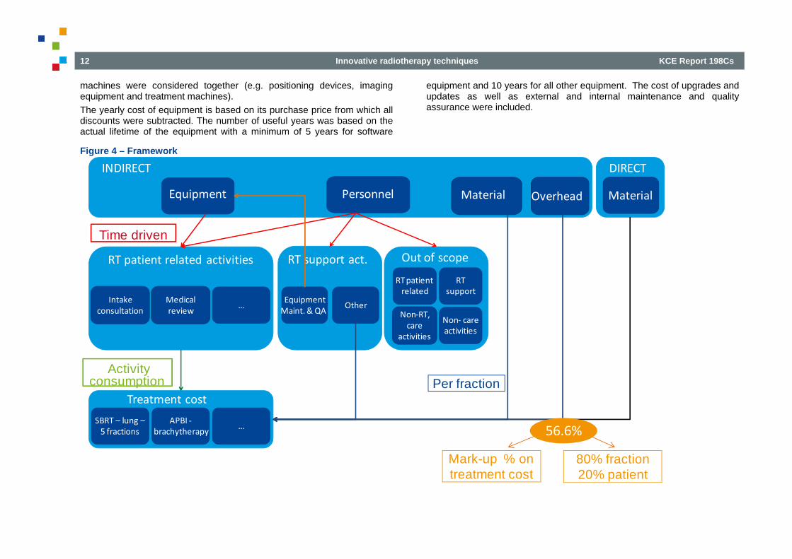
2.9. Costs and resource use Radiotherapy costs were split into indirect and direct costs. In this report, indirect costs are defined as costs that were not directly assigned to a single treatment, but allocated to the treatments through activities. Four types of indirect costs were distinguished: personnel costs (including physicians), equipment costs, indirect material costs and overhead. For direct cost there was only some direct material cost. Costs are based on 2011 prices and include Value Added Tax (VAT). Figure 4 shows an overview of the applied framework. 2.9.1. Personnel cost In accordance with the KCE manual for cost-based pricing,20 the following personnel categories were analysed separately: • senior radiation oncologists (including chief radiation oncologist), • junior radiation oncologists (i.e. radiation oncologists in training), • physicists who calculate the radiation plans and are responsible for
equipment quality assurance, • dosimetrists or planners who calculate the radiation plans, and • nurses (including head nurse). Physician cost per half day and personnel costs per hour were derived from the KCE manual.20 2.9.2. Equipment costs All types of equipment were included in the model: simulators, treatment machines, verification systems, dosimetrical equipment, planning systems, positioning devices, imaging equipment, stereotactic frames, gating modules and other equipment. In some cases, several devices and
12 Innovative radiotherapy techniques KCE Report 198Cs
machines were considered together (e.g. positioning devices, imaging equipment and treatment machines). The yearly cost of equipment is based on its purchase price from which all discounts were subtracted. The number of useful years was based on the actual lifetime of the equipment with a minimum of 5 years for software
equipment and 10 years for all other equipment. The cost of upgrades and updates as well as external and internal maintenance and quality assurance were included.
Figure 4 – Framework
Out of scope
Treatment cost
DIRECTINDIRECT
Personnel MaterialEquipment
RT patient related activities RT support act.
Intake consultation
Medical review
…
APBI ‐brachytherapy
SBRT – lung –5 fractions
…
Overhead
OtherEquipment Maint. & QA
Material
Time driven
Activity consumption Per fraction
Non‐RT, care
activities
Non‐ care activities
RT patient related
RT support
Mark-up % on treatment cost
80% fraction20% patient
56.6%
KCE Report 198Cs Innovative radiotherapy techniques 13
2.9.3. Indirect material costs Indirect material costs comprise all consumables used in the radiotherapy department (bandages, paper for the examination table …) that could not be linked to a specific treatment. 2.9.4. Pharmaceuticals and radio-isotopes No pharmaceuticals (e.g. anti-emetics) nor radio-isotopes were included in the cost analysis as they are financed separately from the radiotherapy activity and are generally not registered on the cost accounts of the radiotherapy department. The use of pharmaceuticals in radiotherapy departments is low. For brachytherapy, possible costs of general anaesthesia, surgery and hospitalisation are to be added, as well as the cost of the radio-isotope. For more information on the cost of radio-isotopes we refer to KCE report 79.22 2.9.5. Overhead A hospital-wide overhead percentage Overhead costs were estimated using the hospital-wide overhead rate from the KCE cost manual.20 They include costs of administrative personnel, blue-collar workers and engineers, top and middle management, all depreciations (except on medical equipment), general, cleaning, maintenance (except of medical equipment), heating, financial and administration costs. The overhead rate is 56.6% of all costs minus physician costs. Two scenarios for overhead allocation were analysed. Unless specified otherwise, the cost presented is the average of the two scenarios. The first overhead scenario In the first scenario (“mark-up” scenario), the overhead percentage was applied at the treatment-level. This means that for each treatment the cost minus the physician cost was multiplied by 56.6%. In this scenario more overhead is allocated to “expensive” products than to “cheap” products. This may not always reflect actual overhead costs. Stereotactic treatments, which are relatively expensive treatments in proportion to the number of fractions, are attributed a relatively large overhead cost, although one could expect a lower overhead given the smaller number of fractions.
The second overhead scenario Therefore a second scenario was also analysed. In this scenario (““80/20” scenario), the 56.6% is calculated at the departmental level. This resulted in a departmental overhead-pool, which was consequently allocated to the treatments based on a combination of the number of fractions (80%) and number of patients (20%). This method results in higher overhead costs for the treatments with high number of fractions and lower overhead for the treatments with low number of fractions. 2.9.6. Direct costs Direct costs can be traced to a specific treatment. They consist of the cost of masks or other fixation systems and markers which can directly be assigned to a specific treatment.
2.10. Cost allocation 2.10.1. Allocation of personnel and equipment to activities The allocation of personnel and equipment costs to activities was based on time registrations and estimates for radiotherapy patient-related activities and equipment maintenance and QA. Time registrations For all activities that were expected to have a significant impact on the treatment cost (Table 1), either because they are repeated several times or because they require expensive equipment, times were registered by the personnel of all radiotherapy departments during 4 weeks in the second half of 2012. Algorithm for missing time registrations Given the 4-week period, not all treatments or not all activities of that treatment occurred during this period and could be measured. Consequently, a number of time measurement extrapolations were done by the project team to estimate the lacking time data. A standard algorithm was developed and used for this extrapolation. Briefly, for the purpose of extrapolations, the time of the following activities was assumed to be organ-dependent: simulation, image import and fusion, delineation of target volume and organs at risk. For the other activities, including planning and treatment delivery, we assumed that the most important determinant of time spent was the technique. When we were
14 Innovative radiotherapy techniques KCE Report 198Cs
confronted with missing data for innovative techniques the centres were asked for a time estimate. Rescaling of the time data The initial time data were rescaled in order to adjust for: • In scope radiotherapy support activities, such as weekly meetings,
morning discussions, starting up and closing down equipment and training activities exceeding the 3 days estimated in the KCE cost manual
• Idle time, such as coffee and toilet breaks • Imperfections of the time measurements and estimates Equipment cost For each piece of equipment the number of hours used was calculated bottom-up starting from the duration of each activity, the frequency of this
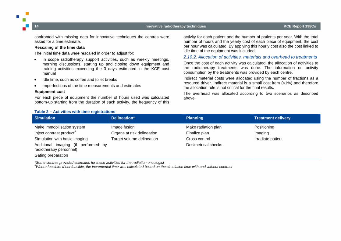
activity for each patient and the number of patients per year. With the total number of hours and the yearly cost of each piece of equipment, the cost per hour was calculated. By applying this hourly cost also the cost linked to idle time of the equipment was included. 2.10.2. Allocation of activities, materials and overhead to treatments Once the cost of each activity was calculated, the allocation of activities to the radiotherapy treatments was done. The information on activity consumption by the treatments was provided by each centre. Indirect material costs were allocated using the number of fractions as a resource driver. Indirect material is a small cost item (<1%) and therefore the allocation rule is not critical for the final results. The overhead was allocated according to two scenarios as described above.
Table 2 – Activities with time registrations Simulation Delineation* Planning Treatment delivery
Make immobilisation system Inject contrast product# Simulation with basic imaging Additional imaging (if performed by radiotherapy personnel) Gating preparation
Image fusion Organs at risk delineation Target volume delineation
Make radiation plan Finalize plan Cross control Dosimetrical checks
Positioning Imaging Irradiate patient
*Some centres provided estimates for these activities for the radiation oncologist #Where feasible. If not feasible, the incremental time was calculated based on the simulation time with and without contrast
KCE Report 198Cs Innovative radiotherapy techniques 15
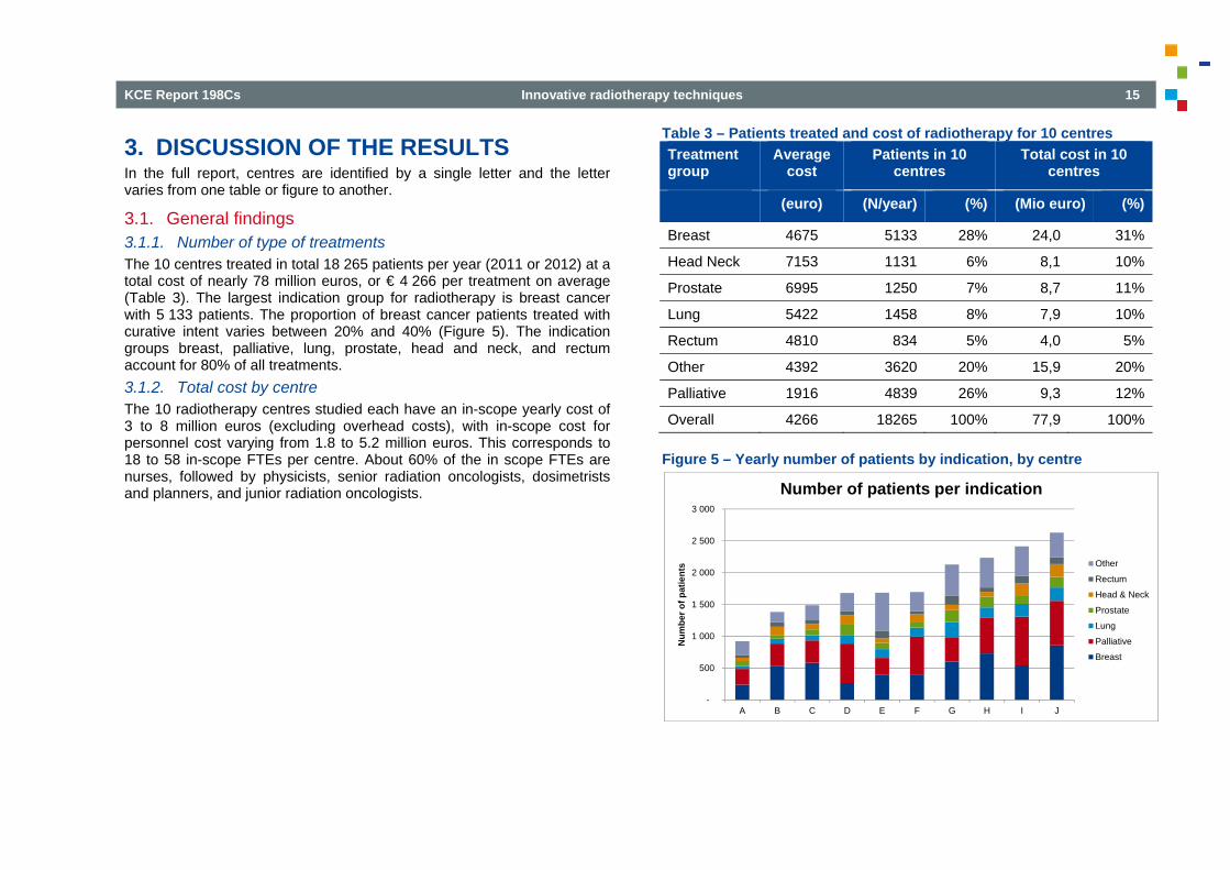
3. DISCUSSION OF THE RESULTS In the full report, centres are identified by a single letter and the letter varies from one table or figure to another.
3.1. General findings 3.1.1. Number of type of treatments The 10 centres treated in total 18 265 patients per year (2011 or 2012) at a total cost of nearly 78 million euros, or € 4 266 per treatment on average (Table 3). The largest indication group for radiotherapy is breast cancer with 5 133 patients. The proportion of breast cancer patients treated with curative intent varies between 20% and 40% (Figure 5). The indication groups breast, palliative, lung, prostate, head and neck, and rectum account for 80% of all treatments. 3.1.2. Total cost by centre The 10 radiotherapy centres studied each have an in-scope yearly cost of 3 to 8 million euros (excluding overhead costs), with in-scope cost for personnel cost varying from 1.8 to 5.2 million euros. This corresponds to 18 to 58 in-scope FTEs per centre. About 60% of the in scope FTEs are nurses, followed by physicists, senior radiation oncologists, dosimetrists and planners, and junior radiation oncologists.
Table 3 – Patients treated and cost of radiotherapy for 10 centres Treatment group
Average cost
Patients in 10 centres
Total cost in 10 centres
(euro) (N/year) (%) (Mio euro) (%)
Breast 4675 5133 28% 24,0 31%
Head Neck 7153 1131 6% 8,1 10%
Prostate 6995 1250 7% 8,7 11%
Lung 5422 1458 8% 7,9 10%
Rectum 4810 834 5% 4,0 5%
Other 4392 3620 20% 15,9 20%
Palliative 1916 4839 26% 9,3 12%
Overall 4266 18265 100% 77,9 100%
Figure 5 – Yearly number of patients by indication, by centre
-
500
1 000
1 500
2 000
2 500
3 000
A B C D E F G H I J
Num
ber o
f pat
ient
s
Number of patients per indication
Other
Rectum
Head & Neck
Prostate
Lung
Palliative
Breast
16 Innovative radiotherapy techniques KCE Report 198Cs
3.1.3. Cost structure The cost of personnel is 50% higher than the equipment cost in radiotherapy departments
An important finding is that despite the high cost of the equipment used in radiotherapy, the personnel cost is higher than the equipment cost in all centres. On average personnel cost represents 41% of total treatment cost, while equipment cost only represents 27% of the treatment cost. Direct and indirect material costs are relatively insignificant. Overhead accounts for about a third of the total cost per treatment.
Treatment delivery is the most expensive part of the process
Only personnel and equipment costs can be linked to a particular part of the process. Therefore, material and overhead costs are not included in the percentages presented in this section. Depending on the centre, the “treatment delivery” accounts for 57% to 68% of total equipment and personnel costs with the overall average being 63%. This was to be expected as the most expensive pieces of equipment (linear accelerators) are used during this phase. Treatment delivery will typically make up a larger part of the total cost of standard fractionation schemes compared with hypofractionation schemes. However, larger radiation doses delivered per fraction add to the complexity of treatment delivery (more imaging, more QA, more use of IMRT or 4D techniques, more highly qualified personnel present for more time) and longer time slots blocked for the activity. After “Treatment delivery” the most expensive phases of the process are “First patient contact”, “Simulation” and “Planning”. “First patient contact” is expensive mostly because it takes up physician time. “Simulation” on the other hand, is expensive due to the cost of the simulator. The cost of “Planning” is high because it can be very time consuming, depending on the degree of software automation. 3.1.4. Process validation Practices and resource use vary within and between centres
The 10 centres show important variations in preparation and delivery of radiotherapy. Resource use and practices vary within and between centres. The collected data demonstrate sensitivity of costs to these
differences. As up to date internationally accepted quality criteria for radiotherapy may not exist, it is difficult to judge which practices and resource use reflect best practice. Resource use may differ in terms of personnel mix, personnel load and equipment. Tasks are not always performed by the same type of personnel across the centres. At some centres and for some treatments, physicians are present during the complete treatment delivery session, whilst at other centres and for other treatments, they are only present for part of the activity. If we would compare the Belgian situation to the situation in the Netherlands, the differences might even be greater, as e.g. dosimetrists in the Netherlands have a broader role. Further research is needed to determine which option is economically most efficient while assuring high quality radiotherapy. 3.1.5. Efficiency During this project a large amount of data on the inputs and outputs of radiotherapy were gathered. This data collection served primarily to estimate the cost of treatment, in function of indication, technique, fractionation scheme and type of equipment. This data could however also be used to investigate other determinants of costs, such as efficiency and quality delivered. As the study was not designed for this purpose, only exploratory analyses were conducted. The sample size (n=10) is also a limitation for statistical analyses. Economies of scale? First, we looked at average treatment cost in function of the overall treatment volume (both in terms of number of patients and fractions) at the centre. No clear relationship was observed. Despite the variation between the volume of the centres, one should keep in mind that the centre with the lowest number of patients still treated nearly 1000 patients per year. This could be the cause of the apparent lack of relation between the volume of a centre and it’s average treatment cost. Indeed, it has been reported that centres with less than 1 000 patients per year could significantly benefit from volume driven efficiency gains, but that the room for improvement is much smaller in larger centres.18 Second we checked the impact of overall patient volume on FTEs (by personnel type) per patient treated. There is a tendency towards economies of scale for nurses and physicists.
KCE Report 198Cs Innovative radiotherapy techniques 17
Personnel efficiency
We used average data for personnel and physician cost, thus reducing variation and facilitating a comparison of their efficiency. However, measurement of personnel efficiency is complex as more personnel for an equal output does not necessarily mean lower efficiency. More personnel may imply more quality, more patient information, more mutual consultations, or stricter adherence to the personnel norms etcetera. Less personnel may on the other hand be due to understaffing because of an unfilled position. A good way of measuring personnel efficiency is through an idle time analysis, measuring and analysing waiting and other unproductive times. Our analysis however did not include such elements. Nevertheless, we could make a number of observations. A first observation is that the presented personnel cost figures seem to be sensitive to the learning curve. For instance, at some centres, the physician is present during the whole treatment session for the innovative technique(s), whilst for routine techniques this is rarely the case. It is clear that personnel costs of innovative techniques may decrease once the centre progresses further on the learning curve. The presented personnel cost figures also appear highly sensitive to the type of equipment used. Personnel time needed for fully automated planning systems for instance is considerably lower than for systems requiring more user interventions. High cost sophisticated systems on this other hand may take longer for treatment delivery, further increasing costs, When one examines personnel efficiency, it should thus take into account the type of equipment used as well.
Equipment efficiency
Departments that work with a fully compatible machine park have an advantage in terms of equipment efficiency over centres who have invested in different brands that are not always compatible. Occupancy rates were estimated for all equipment but not presented in the report as they were based on the same opening hours for all centres and some centres expressed their concerns that the data were therefore not correct. We opted not to adapt the occupancy rate to the individual opening hours of each centre as this would not provide comparable data either. When longer opening hours are combined with higher volume, they may lead to
more intensive and thus more efficient use of the equipment. However, longer opening hours may also boost personnel costs as personnel outside the shifts is more expensive and as extra personnel may be needed to ensure the overlap between the shifts. Therefore, it is not easy to determine which centre uses its equipment in the most efficient way. Merely looking at equipment costs does not provide an answer to this question either as low equipment costs may not only be due to high occupancy rate but also to old equipment. Satellite centres It was not investigated whether smaller satellite centres are less efficient than their main or larger centres. In case the satellite centres were included in the analyses, they were treated in conjunction with the head centre. On one hand it can be expected that the occupancy rate of equipment in small satellite centres is lower than in larger centres as there are fewer patients, on the other hand often only one type of equipment is installed in the satellite which guarantees use of the available equipment. There may be both economies of scale and diseconomies of diversification. Quality of care should not be forgotten either: is less choice in equipment a limitation to deliver the best possible care or not? 3.1.6. Limitations Out of scope activities in university centres The proportion of the personnel cost that was considered out of scope was much larger at university centres compared with non-university centres. In university centres this out of scope time will likely consist of time spent on teaching and research, including clinical research overlapping with the treatments included in this cost study. However, there are important variations for the fraction “out of scope” between university centres (20% to 44%). We left it to the centres to define the time spent by each personnel type on out of scope activities, without providing specific guidance on where to draw the cut-off line between clinical research and patient care. In hindsight, such guidance might have reduced the between centre variation. Of course, time measurements of all activities of each individual, linked to specific machine use time would have been a possible solution, but difficult to implement within the time and budgetary constraints of this project. A possible consequence of the approach we used is that in centres with a high fraction “out of scope”, the real treatment cost may have been
18 Innovative radiotherapy techniques KCE Report 198Cs
underestimated. On the other hand, it can be assumed that educating junior radiotherapy specialists in university centres increased the time spent per activity and thus the cost.
Importance of overhead allocation rules
The results are sensitive to the way the overhead is calculated and allocated in the department. A hospital-wide overhead rate was applied. This percentage was calculated for hospital interventions independent of the discipline. It is not known to what extent the overhead differs between medical specialties, e.g. with a low versus high equipment cost (e.g. geriatrics versus radiotherapy). Two scenario’s of overhead allocation were analysed. Other scenario’s are also possible. For instance, it would be possible to take into account not only the number of fractions but also the average duration per fraction. For stereotactic treatments with a low number of fractions, the average length of a fraction is indeed longer and this implies larger overhead costs.
Importance of fixed versus actual equipment lifetime
We based the number of useful years of the equipment on the actual lifetime with a minimum of 10 years. Centres tend to have a mix of linear accelerators with a varying actual lifetime that only rarely exceeds 10 years. For some accelerators however the age was up to 16 years. What would have been the impact on the treatment cost of introducing a fixed lifetime of 10 years for the equipment? We analysed this and the effect was a mean increase in cost of only 1.2%. The increase was 0 to 1% in 7 centres, 2% in 2 centres and 5% in a single centre with an exceptionally “old” set of linear accelerators.
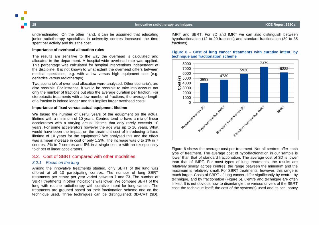
3.2. Cost of SBRT compared with other modalities 3.2.1. Focus on the lung Among the innovative treatments studied, only SBRT of the lung was offered at all 10 participating centres. The number of lung SBRT treatments per centre per year varied between 7 and 73. The number of SBRT treatments in other indications was lower. We compare SBRT of the lung with routine radiotherapy with curative intent for lung cancer. The treatments are grouped based on their fractionation scheme and on the technique used. Three techniques can be distinguished: 3D-CRT (3D),
IMRT and SBRT. For 3D and IMRT we can also distinguish between hypofractionation (12 to 20 fractions) and standard fractionation (30 to 35 fractions).
Figure 6 – Cost of lung cancer treatments with curative intent, by technique and fractionation scheme
Figure 6 shows the average cost per treatment. Not all centres offer each type of treatment. The average cost of hypofractionation in our sample is lower than that of standard fractionation. The average cost of 3D is lower than that of IMRT. For most types of lung treatments, the results are relatively similar across centres: the range between the minimum and the maximum is relatively small. For SBRT treatments, however, this range is much larger. Costs of SBRT of lung cancer differ significantly by centre, by technique, and by fractionation (Figure 5). Centre and technique are often linked. It is not obvious how to disentangle the various drivers of the SBRT cost: the technique itself; the cost of the system(s) used and its occupancy
39934730
5920
73796222
010002000300040005000600070008000
Cos
t (€)
KCE Report 198Cs Innovative radiotherapy techniques 19
level; the duration of specific activities and the personnel present, in particular senior radiation oncologists and physicists.
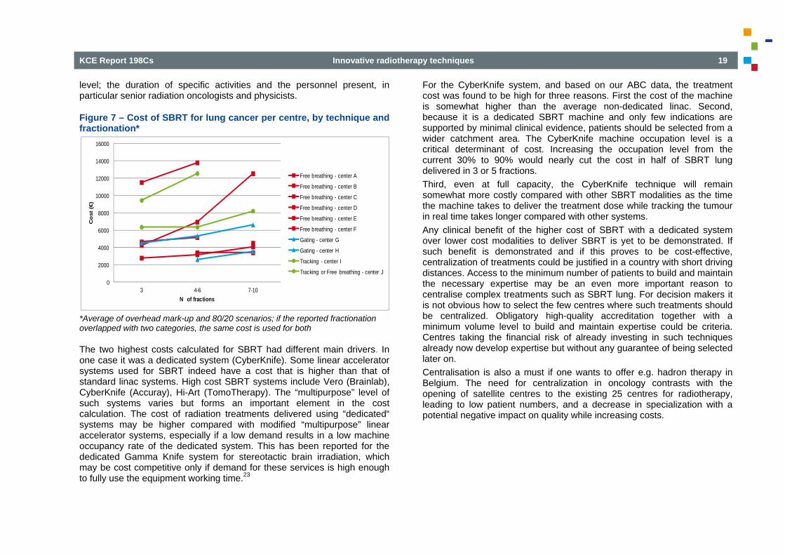
Figure 7 – Cost of SBRT for lung cancer per centre, by technique and fractionation*
*Average of overhead mark-up and 80/20 scenarios; if the reported fractionation overlapped with two categories, the same cost is used for both
The two highest costs calculated for SBRT had different main drivers. In one case it was a dedicated system (CyberKnife). Some linear accelerator systems used for SBRT indeed have a cost that is higher than that of standard linac systems. High cost SBRT systems include Vero (Brainlab), CyberKnife (Accuray), Hi-Art (TomoTherapy). The “multipurpose” level of such systems varies but forms an important element in the cost calculation. The cost of radiation treatments delivered using “dedicated“ systems may be higher compared with modified “multipurpose” linear accelerator systems, especially if a low demand results in a low machine occupancy rate of the dedicated system. This has been reported for the dedicated Gamma Knife system for stereotactic brain irradiation, which may be cost competitive only if demand for these services is high enough to fully use the equipment working time.23
For the CyberKnife system, and based on our ABC data, the treatment cost was found to be high for three reasons. First the cost of the machine is somewhat higher than the average non-dedicated linac. Second, because it is a dedicated SBRT machine and only few indications are supported by minimal clinical evidence, patients should be selected from a wider catchment area. The CyberKnife machine occupation level is a critical determinant of cost. Increasing the occupation level from the current 30% to 90% would nearly cut the cost in half of SBRT lung delivered in 3 or 5 fractions. Third, even at full capacity, the CyberKnife technique will remain somewhat more costly compared with other SBRT modalities as the time the machine takes to deliver the treatment dose while tracking the tumour in real time takes longer compared with other systems. Any clinical benefit of the higher cost of SBRT with a dedicated system over lower cost modalities to deliver SBRT is yet to be demonstrated. If such benefit is demonstrated and if this proves to be cost-effective, centralization of treatments could be justified in a country with short driving distances. Access to the minimum number of patients to build and maintain the necessary expertise may be an even more important reason to centralise complex treatments such as SBRT lung. For decision makers it is not obvious how to select the few centres where such treatments should be centralized. Obligatory high-quality accreditation together with a minimum volume level to build and maintain expertise could be criteria. Centres taking the financial risk of already investing in such techniques already now develop expertise but without any guarantee of being selected later on. Centralisation is also a must if one wants to offer e.g. hadron therapy in Belgium. The need for centralization in oncology contrasts with the opening of satellite centres to the existing 25 centres for radiotherapy, leading to low patient numbers, and a decrease in specialization with a potential negative impact on quality while increasing costs.
0
2000
4000
6000
8000
10000
12000
14000
16000
3 4-6 7-10
Co
st (
€)
N of fractions
Free breathing - center A
Free breathing - center B
Free breathing - center C
Free breathing - center D
Free breathing - center E
Free breathing - center F
Gating - center G
Gating - center H
Tracking - center I
Tracking or Free breathing - center J
20 Innovative radiotherapy techniques KCE Report 198Cs
In the second centre with a high cost for SBRT there was a high level of presence of the senior radiation oncologist during lengthy treatment sessions, most probably reflecting an early phase in the learning curve. In both cases costs are expected to drop considerably with increasing volume and experience, respectively. So these costs should not be considered a good basis for reimbursement or research funding in the long term. The lowest costs for SBRT were found in centres where the time measures for SBRT and IMRT activities were rather similar, as well as the systems used to deliver the irradiation. Low costs driven by a low cost of a set of old linear accelerators or driven by understaffing are not a good basis for reimbursement either. The average cost of SBRT of the lung (€ 6 222) is very similar to the cost of standard fractioned 3D-CRT of the lung. For a given technique and centre, SBRT of the lung delivered in 3 fractions is less costly compared with SBRT delivered in 7 to 10 fractions. The differences in cost between centres was remarkable and deserve further study, especially as no such major differences were present for more standard treatments. 3.2.2. SBRT of spinal, liver, pancreas, bone and oligometastases The average cost for SBRT of liver (3 to 10 fractions) and pancreas (10 fractions) were € 5 586 (based on 5 centres) and € 5 341 (2 centres) respectively. This cost is only slightly higher than the overall average of € 4 927 found for the 6 centres that perform treatment of pancreatic cancer in 25 to 30 fractions with 3D-CRT or IMRT. The cost for SBRT spinal was € 3 573 for 3 fractions, obtained in a single centre only and concerning very few patients. Costs for SBRT of bone were obtained in 2 centres and concern very few patients. The cost varies from € 2 185 for a single fraction to € 8 916 euro for 3 fractions delivered using SBRT with tracking. Costs for SBRT of oligometastatic disease were obtained in 2 centres, but only a single centre reported to treat over 10 patients per year. The fractionation was 3, 5 and 10 in one centre and 10 in the second centre. The costs were € 3 342 for 3 fractions, € 4 012 for 5 fractions and from € 5 076 for 10 fractions.
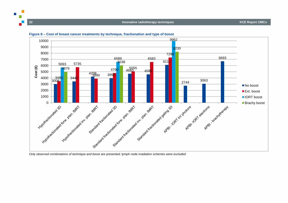
3.3. Breast cancer, focus on APBI and IORT boost When selecting the centres for the cost study, we succeeded to include the three Belgian centres that showed some activity in APBI, but mainly use IORT as a boost. We analysed costs of the different forms of primary breast cancer radiation therapies with curative intent, with the innovation focus on APBI and IORT boost techniques. Schemes including specific irradiation of lymphatic nodes were excluded. Figure 8 shows the average cost of breast irradiation treatments by technique and fractionation scheme.
The cost of MeV electrons and kV photon IORT APBI is rather similar and about half the cost of APBI using brachytherapy.
The average cost of intraoperative single fraction APBI delivered as MeV electrons using a mobile linear accelerator system (Mobetron) was € 3 063 and € 2 744 if delivered as kV photons (Intrabeam). The entire procedure of APBI with MeV electrons has been reported to last for about 15 to 20 minutes.5 Unnecessary radiation to the underlying normal tissue is avoided by mobilizing the mammary gland during surgery and placing a lead plate for shielding on its dorsal surface. This shielding is not needed for APBI with kV photons. As discussed above, our calculated costs do not include any additional cost of the surgical department. This will depend on the extra time, if any, the surgery will take and should consider the cost of the nurses, the surgeon, the anaesthesiologist, the anatomical pathologist and the (shielded) operating room. Often, IORT can be performed while the surgeon has to wait for the pathology result of the sentinel node, thus not extending the surgery time. Based on the KCE cost manual the direct costs of the operating theatre, anaesthesia and sterilization department can be estimated: € 156 per hour and per nurse present. This cost covers medical equipment, staff except for physicians and drugs, pharmaceutical products and consumables.20 The cost of a general surgeon, anaesthesiologist and anatomical pathologist is respectively € 363, € 441 and € 458 per half day. On top of the direct costs excluding physicians, the general overhead rate of 56.6% applies. Based on these considerations we can deduce that the additional cost, if any, is relatively low compared to the cost of the radiotherapy itself.
KCE Report 198Cs Innovative radiotherapy techniques 21
We found a relatively high cost of over € 6 693 for APBI using HDR interstitial multicatheter brachytherapy delivered in 8 fractions of 15-20 minutes over 4 days, starting about 6 weeks after surgery. The cost was measured in a centre where the number of patients treated this way is low. This means that the equipment is not fully utilized and that the resulting cost per patient is very high. All other things being equal, if the number of patients undergoing APBI by brachytherapy in that centre were to increase fourfold, the brachytherapy equipment utilization would still be low and the cost of APBI by brachytherapy would be reduced to € 5 633. The cost is also high because highly qualified specialists (radiation oncologist and physicist) are present during the eight treatment sessions.. Brachytherapy for breast cancer in Belgium is mainly used as a boost after hypo- or standard fractionated WBI. This boost is delivered a few weeks after the end of the WBI. Both HDR and pulsed dose rate (PDR) techniques are used. Placement of 5 up to 12 catheters can be done by the senior radiation oncologist using local anesthesia or with assistance of a surgeon and anesthesiologist under general anesthesia. A correct placement may be guided by clips left in place during the breast surgery. After simulation the patient is placed in a shielded room. HDR brachytherapy using an 0.6 x 3.5 mm Iridium rod and a machine for remote
afterloading is performed in 15-20 minutes. The 10-12 Curie source of 192Ir costs € 5 500 (excl VAT) and needs to be replaced 4 times a year. PDR brachytherapy uses a 1 Curie source with afterloader delivers a total dose of 15 Gy in pulses of 10 minutes per hours for 24 hours. The patient is hospitalized for 1 or 2 nights in a dedicated room. Removing the catheters does not require a general anesthesia. Hospitals invoice isotopes in various ways as described in detail in KCE report no. 79.22 It was not always possible to calculate the exact additional cost of delivering a boost to a specific WBI schedule. Some centres systematically perform a boost so that if we compare the average cost of treatments with boost to the average cost of treatments without boost, we are comparing averages that are based on different sets of centres. Taking this remark into account, the cost of an external beam radiotherapy boost is about € 500 to € 1 500. This additional cost is much lower than the cost of a brachytherapy boost (€ 2 000 to € 2 500, plus isotope and possibly an additional general anesthesia and hospitalisation), an IORT kV boost (2000 € to € 2500, plus extension of surgery) or an IORT MeV boost (€ 2 500 to € 4 000, plus extension of surgery).
22 Innovative radiotherapy techniques KCE Report 198Cs
Figure 8 – Cost of breast cancer treatments by technique, fractionation and type of boost
Only observed combinations of technique and boost are presented; lymph node irradiation schemes were excluded
30083440
4208 39554683 4588
6111
2744 3063
6693
3483
5735
38904790 5055
65837298
56936589
9962
4979
6038
8230
0
1000
2000
3000
4000
5000
6000
7000
8000
9000
10000C
ost (
€)
No boost
Ext. boost
IORT boost
Brachy boost
KCE Report 198Cs Innovative radiotherapy techniques 23
3.4. Cost of other common treatments The IMRT costs we report here are clearly higher for prostate cancer (€ 7 278 for 33-40 fractions) and head and neck cancer (€ 8 237 for 30-35 fractions) than for IMRT for rectum cancer (€ 4 889 for 25-28 fractions) and standard fractionated WBI (€ 4 587 for 25 fractions). The cost difference can in part be explained by a different fractionation. IMRT, as an innovative intervention, was the subject of KCE report no 62 published in 2007.24 No cost study was performed for IMRT at that time. However, the sum of fee for service (article 18), investment costs (A3), operational departmental and point lump sums (B3) was € 5 288 euro for IMRT in 2003. This estimate of the financing is however incomplete as will be discussed in the next section. In comparison, the cost of a course of 25-28 fractions of 3D-CRT for rectum carcinoma is € 4 465. Palliative treatments on average cost less compared with radiotherapy with curative intent: € 1 032(1 fraction), € 1 686 (5 fractions) and € 2 655 (10 to 13 fractions).
3.5. Costs, financing and budget considerations Breast radiotherapy at large accounts for 28% of the patients treated and 31% of the total radiotherapy costs. The number of patients receiving radiotherapy as a palliative treatment is similar (26%) but these treatments account for only 12% of the total costs. Cost comparison of APBI and SBRT with standard modalities The average costs of APBI (except using brachytherapy) is lower compared with existing radiotherapy modalities, while IORT as a boost has a higher cost compared with other boost modalities. For lung and pancreas cancer, the cost of SBRT is only slightly higher compared with existing treatments. SBRT is proposed for oligometastatic disease, including vertebral metastases, and liver metastases. Some of these metastases are currently treated with (lower cost) palliative intent radiotherapy, some are currently not treated with radiotherapy. A budget increase will be needed if these additional indications are to be treated with SBRT.
Matching costs and financing The large overhead rate and the complexity of hospital financing hamper a one-to-one comparison of cost with the current complex financing structure. As there are no legal allocation keys for allocating the general hospital financing components A1, A2, B1, B4 financing to radiotherapy, no perfect one-to-one match is possible between the presented cost and the financing. The A3 financing of about € 90 000 per year per linear accelerator is much lower than the real cost. We observed that the cost (including VAT) per linear accelerator, with or without a limited or extended service contract, varies from less than € 250 000 euro per year (if in use for more than 10 years) to over € 500 000 euro per year (for the more expensive linear accelerators with extended service contract). Note that the VAT level of radiotherapy devices is 21% whereas it is only 6% for medicines. It should also be noted that specific financing of linear accelerators, as is the case in Belgium, creates a disadvantage for APBI techniques that are not based on this technique. Impact of financing policy on practice The large variety in treatments administered (in terms of fractionation and dose), treatment strategies used and equipment choices often leads to a large variety in cost results for a single indication. The design of an optimal financing structure poses a big challenge for the national health care payer. Previous research suggests that reimbursement policies influence radiotherapy practice.25, 26 This has also been reported for the IMRT of breast cancer in the US.27 Financing should therefore be guided by the real costs of the treatment, as calculated in this study, and designed to encourage the most cost-effective treatment schemes. One should take care no incentives are created to adjust the technique or fractionation scheme in function of the billing codes instead of the patient’s needs.
24 Innovative radiotherapy techniques KCE Report 198Cs
3.6. Introducing innovations in health care As mentioned in the introduction radiotherapy equipment has a low regulatory barrier (CE mark) to enter the European market. At market entry there is most often no clear clinical indication that is supported by clinical evidence.1, 2 Furthermore, new sophisticated radiotherapy machines have a high investment cost. Costs of interventions (or better their coverage by the health care payer) are an important input for cost-effectiveness analyses. Effectiveness is often derived from RCT based efficacy data, but in case of novel radiotherapy treatments such data will not be available in the near future. Government and health care payers have to make sure these costly RCTs are conducted, as they provide the high level evidence needed for decision making. Evidence generation outside of a clinical trial context may also be important but is a concept that is not straightforward to implement. The initial diagnosis of cancer and the treatment plan proposed by the multidisciplinary oncology consultation (MOC) is registered at the Belgian cancer registry. Also an additional MOC may take place each time an important oncology management decision is made. Some radiation oncologists felt that the participation to the MOC is underfinanced. This report does not provide an answer to this question as this activity was out of the scope of this study. The functioning and financing of the MOC meetings is however the subject of another ongoing KCE project. Another activity, reportedly underfinanced, is the long term patient follow-up visit. Tariffs for follow-up visits are lower for a radiation oncologist compared with a medical oncologist, creating a financing-driven shift of this activity out of the radiotherapy department in some hospitals. Patients referred from other hospitals most often receive follow up after radiotherapy at the referring hospital. This hampers the registration of endpoints such as local disease progression, which is not always the subject of a new MOC that would trigger an additional registration. In addition, long term side-effects of the radiotherapy are often not communicated back to the radiotherapist.
The indications that are allowed to receive research financing should either be supported by a minimum level of evidence or be the subject of a clinical trial. Defining those indications should ideally start from a systematic review of the literature, which should be updated as evidence develops during the research financing period. In the ideal world activity based costing and the outcome registration should also cover other treatment modalities that may compete with radiotherapy.
KCE Report 198Cs Innovative radiotherapy techniques 25
REFERENCES
1. Hulstaert F, Neyt M, Vinck I, Stordeur S, Mirjana H, Stefan S, et al. The pre-market clinical evaluation of innovative high-risk medical devices. Health Services Research (HSR). Brussels: Belgian Health Care Knowledge Centre (KCE); 2011. KCE Reports 158C (D/2011/10.273/31) Available from: https://kce.fgov.be/sites/default/files/page_documents/kce_158c_innovative_high-risk_medical_devices_0.pdf
2. Hulstaert F, Neyt M, Vinck I, Stordeur S, Huic M, Sauerland S, et al. Pre-market clinical evaluations of innovative high-risk medical devices in Europe. Int J Technol Assess Health Care. 2012;28(3):278-84.
3. Beitsch PD, Shaitelman SF, Vicini FA. Accelerated partial breast irradiation. J Surg Oncol. 2011;103(4):362-8.
4. Njeh C, Saunders M, Langton C. Accelerated partial breast irradiation using external beam conformal radiation therapy: A review. Crit Rev Oncol Hematol. 2012;81(1):1-20.
5. Njeh CF, Saunders MW, Langton CM. Accelerated Partial Breast Irradiation (APBI): A review of available techniques. Radiat Oncol. 2010;5:90.
6. Offersen BV, Overgaard M, Kroman N, Overgaard J. Accelerated partial breast irradiation as part of breast conserving therapy of early breast carcinoma: a systematic review. Radiother Oncol. 2009;90(1):1-13.
7. Nairz O, Sedlmayer F. Re: Accelerated partial breast irradiation as part of breast conserving therapy of early breast carcinoma: a systematic review. Radiother Oncol. 2010;94(3):388-9; author reply 90.
8. Smith BD, Arthur DW, Buchholz TA, Haffty BG, Hahn CA, Hardenbergh PH, et al. Accelerated partial breast irradiation consensus statement from the American Society for Radiation Oncology (ASTRO). Int J Radiat Oncol Biol Phys. 2009;74(4):987-1001.
9. Tipton KN, Sullivan N, Bruening W, Inamdar R, Launders J, Uhl S, et al. Stereotactic Body Radiation Therapy. AHRQ Technical Brief. 2011;6.
26 Innovative radiotherapy techniques KCE Report 198Cs
10. Noël G, Moty-Monnereau C. Radiothérapie extracranienne en conditions stéréotaxiques. Paris: HAS (Haute Autorité de santé); 2006. Available from: http://www.has-sante.fr/portail/upload/docs/application/pdf/rapport_stereo_cyber.pdf
11. National Health Service UK National Radiotherapy Implementation Group Report. Stereotactic Body Radiotherapy. Clinical Review of the Evidence for SBRT [NHS;2010. Available from: http://ncat.nhs.uk/sites/default/files/clinical%20evidence%20review%20Dec%2010%20-%20Final%20J11.pdf
12. Chi A, Liao Z, Nguyen NP, Xu J, Stea B, Komaki R. Systemic review of the patterns of failure following stereotactic body radiation therapy in early-stage non-small-cell lung cancer: clinical implications. Radiother Oncol. 2010;94(1):1-11.
13. Palma D, Visser O, Lagerwaard FJ, Belderbos J, Slotman BJ, Senan S. Impact of introducing stereotactic lung radiotherapy for elderly patients with stage I non-small-cell lung cancer: a population-based time-trend analysis. J Clin Oncol. 2010;28(35):5153-9.
14. Palma D, Lagerwaard F, Rodrigues G, Haasbeek C, Senan S. Curative treatment of Stage I non-small-cell lung cancer in patients with severe COPD: stereotactic radiotherapy outcomes and systematic review. Int J Radiat Oncol Biol Phys. 2012;82(3):1149-56.
15. Arcangeli S, Scorsetti M, Alongi F. Will SBRT replace conventional radiotherapy in patients with low-intermediate risk prostate cancer? A review. Crit Rev Oncol Hematol. 2012;84(1):101-8.
16. Kaplan RS, Anderson SR. Time-driven activity-based costing. Harvard Business Review. 2004.
17. Lievens Y, Slotman BJ. Radiotherapy cost-calculation and its impact on capacity planning. Expert Rev Pharmacoecon Outcomes Res. 2003;3(4):497-507.
18. Lievens Y, van den Bogaert W, Kesteloot K. Activity-based costing: a practical model for cost calculation in radiotherapy. Int J Radiat Oncol Biol Phys. 2003;57(2):522-35.
19. Van de Werf E, Verstraete J, Lievens Y. The cost of radiotherapy in a decade of technology evolution. Radiother Oncol. 2012;102(1):148-53.
20. Swartenbroekx N, Obyn C, Guillaume P, Lona M, Cleemput I. Manual for cost-based pricing of hospital interventions. Health Technology Assessment (HTA). Brussels: Belgian Health Care Knowledge Centre (KCE); 2012 05/2012. KCE Reports 178C (D/2012/10.273/31) Available from: https://kce.fgov.be/sites/default/files/page_documents/KCE_178C_manual_pricing_hospital_interventions.pdf
21. Jorissen A, Lefebvre C, Popleu A, Roodhooft F, Van Liedekerke C, Walraevens G. Cost Accounting. Antwerpen: Standaard Uitgeverij; 1998.
22. Huybrechts M, Cleemput I, Devriese S, Hulstaert F. [Radioisotopes Reimbursement in Belgium]. Health Services Research (HSR). Brussels: Belgian Health Care Knowledge Centre (KCE); 2008. KCE Reports 79 Available from: https://kce.fgov.be/publication/report/radioisotopes-reimbursement-in-belgium
23. Griffiths A, Marinovich L, Barton MB, Lord SJ. Cost analysis of Gamma Knife stereotactic radiosurgery. Int J Technol Assess Health Care. 2007;23(4):488-94.
24. Van den Steen D, Hulstaert F, Camberlin C. Intensiteitsgemoduleerde Radiotherapie (IMRT). Brussel: Federaal Kenniscentrum voor de Gezondheidszorg (KCE); 2007. Health Technology Assessment (HTA) KCE reports 62A (D2007/10.273/32)
25. Lievens Y, Kesteloot K, Rijnders A, Kutcher G, Van den Bogaert W. Differences in palliative radiotherapy for bone metastases within Western European countries. Radiother Oncol. 2000;56(3):297-303.
26. Lievens Y, Van den Bogaert W, Rijnders A, Kutcher G, Kesteloot K. Palliative radiotherapy practice within Western European countries: impact of the radiotherapy financing system? Radiother Oncol. 2000;56(3):289-95.
27. Smith BD, Pan IW, Shih YC, Smith GL, Harris JR, Punglia R, et al. Adoption of intensity-modulated radiation therapy for breast cancer in the United States. J Natl Cancer Inst. 2011;103(10):798-809.
KCE Report 198Cs Innovative radiotherapy techniques 27
RECOMMENDATIONSa
To the Minister of Social Affairs and Public Health, the insurance committee and the Technisch Geneeskundige Raad - Conseil Technique Médical of the RIZIV – INAMI and the Federal Public Service for Health • The introduction of innovative radiotherapy treatments to health insurance should
happen: o On the condition of a financing based on real costs, calculated by an acknowledged
and relevant costing method, such as time-driven activity-based costing; o With a limitation to indications selected on the basis of the available evidence on
effectiveness and safety and in function of the need of the patient and the cost-effectiveness of the technique;
o Or in the context of clinical studies in those indications for which the effectiveness or safety is still insufficiently proven.
• Considering the required expertise, multidisciplinarity and cost-effectiveness, further centralisation of (complex or innovative) radiotherapy techniques is recommended, paying attention to an appropriate referral of patients and long term follow-up of effectiveness and safety (e.g. centralised oncologic patient files accessible by all involved clinicians). This can only be secured in a statistically sufficient reliable way when the centre also treats a suficient number of patients with these techniques.
To the colleges of physicians for radiotherapy and oncology, the Federal Agency for Nuclear Control, the Cancer Registry Foundation • The treatment with complex or innovative radiotherapy should be integrated in an
oncologic quality system, as recommended in KCE report 152, with evaluation based on a customised system of audit, certification or accreditation.
• Registration of innovative radiotherapy along with their indications and technical characteristics is recommended to map the diffusion and to monitor the safety and effectiveness. The latter can however not replace the necessary clinical studies – stimuli from government for execution of more clinical studies is recommended.
a The KCE has sole responsibility for the recommendations