Embed Size (px)
Citation preview
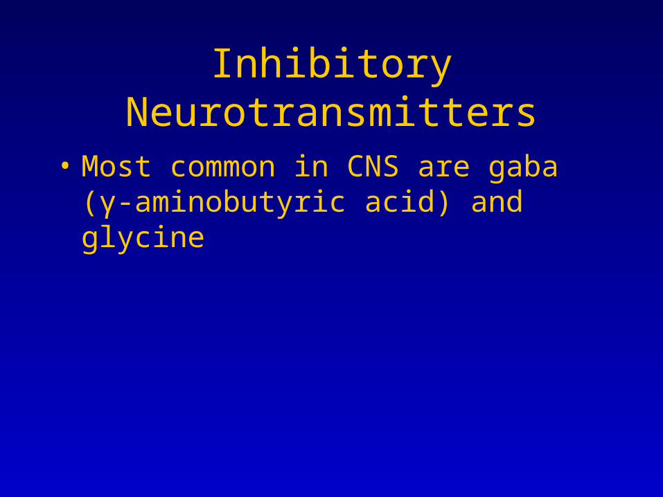
Inhibitory Neurotransmitters
• Most common in CNS are gaba (γ-aminobutyric acid) and glycine
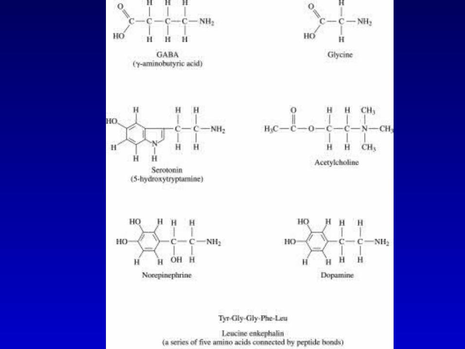
Glycine
• Made from serine
• Used in spinal cord neurons at about 50% of inhibitory synapses
• The other 50% use gaba
Strychnine
• Rat poison that blocks inhibitory neurotransmitter and cause excitation in CNS
• Blocks glycine receptor that leads to membrane depolarizations and over activity and death due to seizures
Gaba
• Most frequently used inhibitory NT in brain & spinal cord. 1/3 of synapses use gaba
• Precursors are glucose, pyruvate and glutamine• GAD= glutamic acid decarboxylase converts
glutamine to gaba• Requires vitamin B6 derivative for GAD activity,
so B6 deficiency can cause gaba deficiency• Noted when infant formula lacked B6=fatal
seizures
Re-Uptake Mechanisms
• Specific Transport proteins on presynaptic membrane to reuptake intact NT
• Re-uptake into glial cell
• Diffusion
• Degradation by enzymes and re-uptake of metabolite
Gaba Removal
• High affinity gaba transporters
• Degradative enzymes are mitochondrial
Multiple Isoforms
• Each subunit can be encoded by any one of several gene or mRNA products to make 3 subtypes of receptors A and C are ionchannels, B is a metabotropic receptor– GABA receptor
• 6 alpha subunit isoforms
• 3 beta subunit isoforms
• 2 gamma subunit isoforms
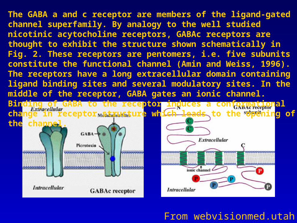
From webvisionmed.utah
The GABA a and c receptor are members of the ligand-gated channel superfamily. By analogy to the well studied nicotinic acytocholine receptors, GABAc receptors are thought to exhibit the structure shown schematically in Fig. 2. These receptors are pentomers, i.e. five subunits constitute the functional channel (Amin and Weiss, 1996). The receptors have a long extracellular domain containing ligand binding sites and several modulatory sites. In the middle of the receptor, GABA gates an ionic channel. Binding of GABA to the receptor induces a conformational change in receptor structure which leads to the opening of the channel.
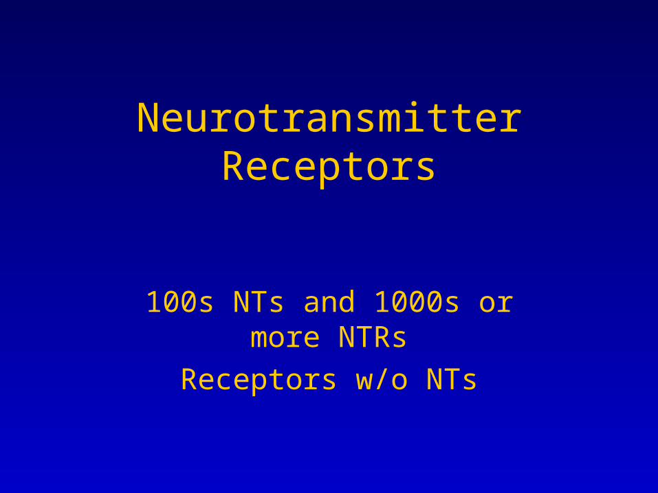
Neurotransmitter Receptors
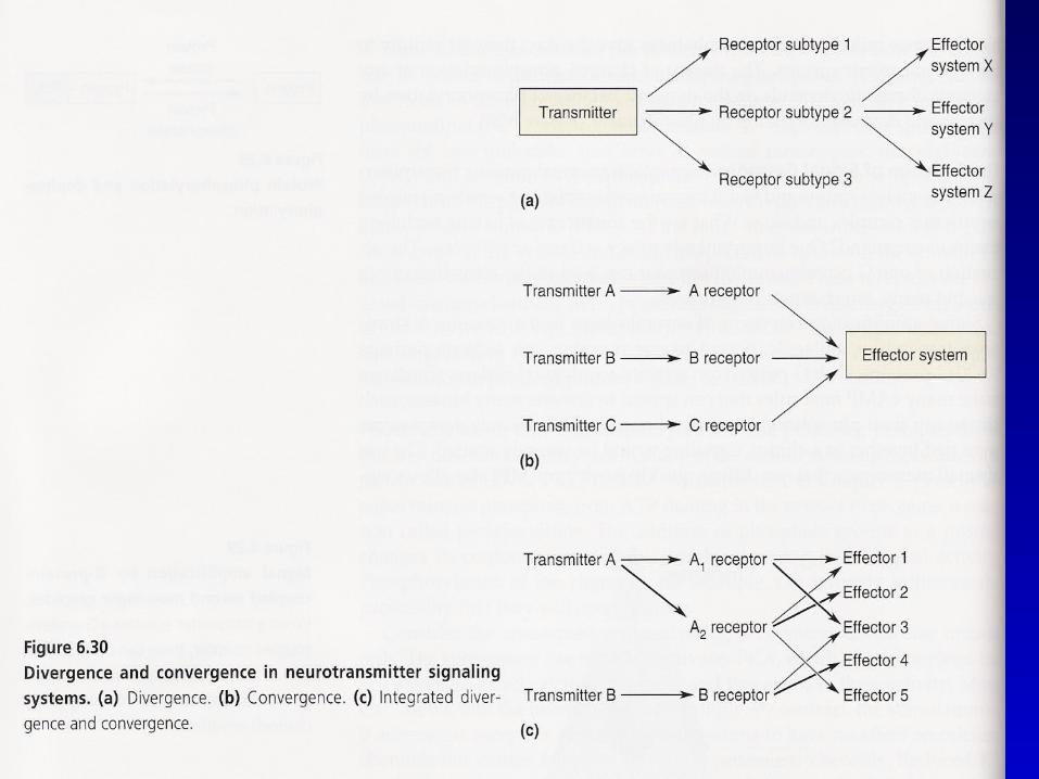
100s NTs and 1000s or more NTRs
Receptors w/o NTs
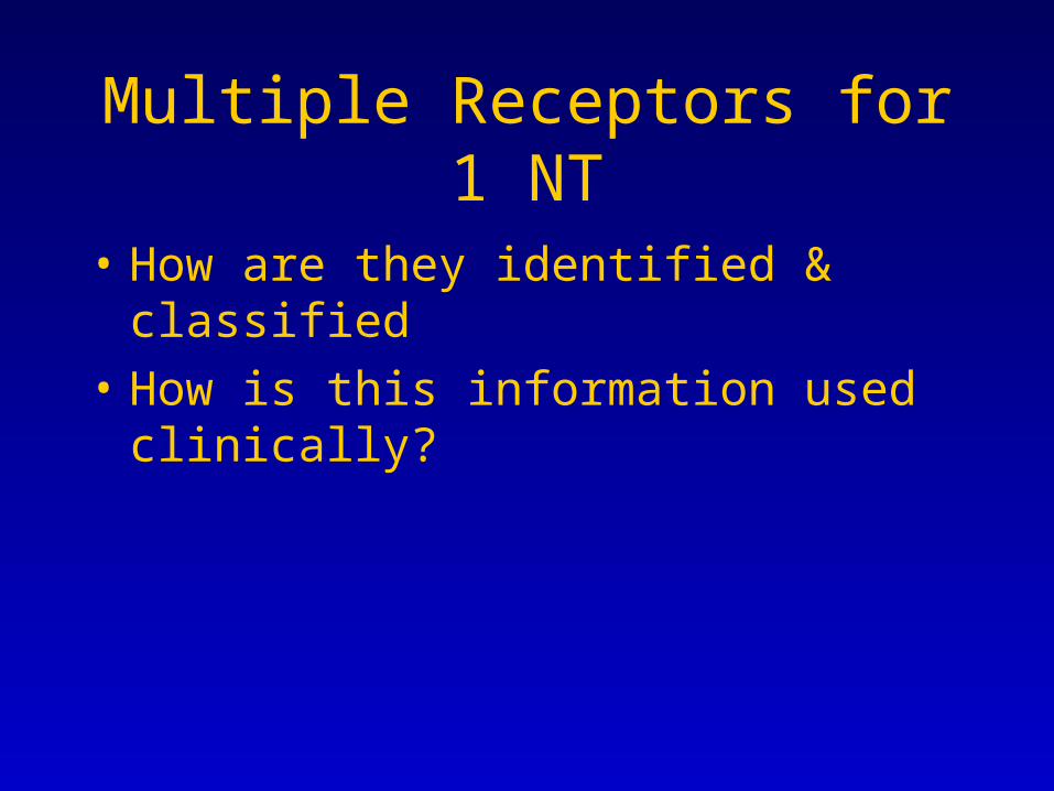
Multiple Receptors for 1 NT
• How are they identified & classified
• How is this information used clinically?
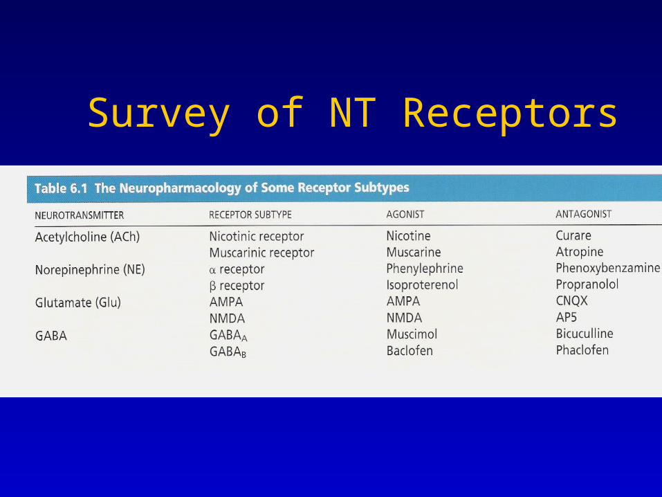
Survey of NT Receptors
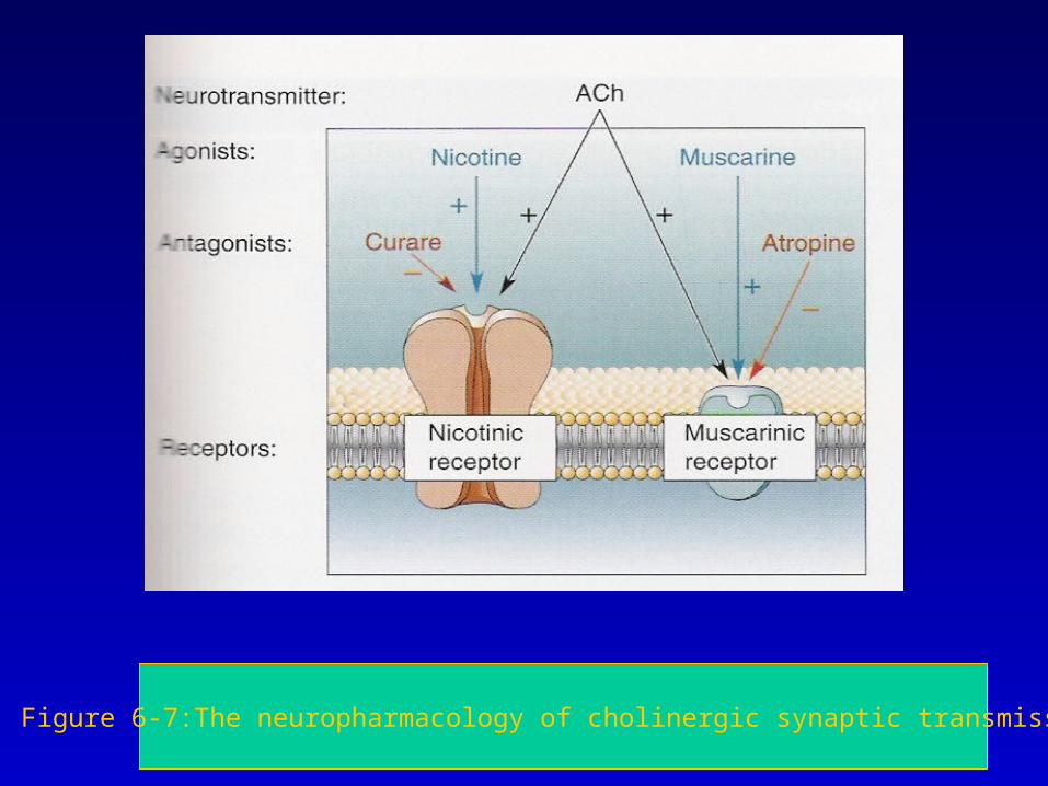
Figure 6-7:The neuropharmacology of cholinergic synaptic transmission
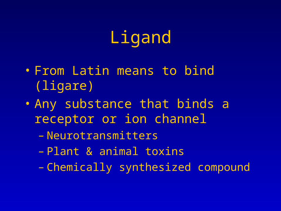
Ligand
• From Latin means to bind (ligare)
• Any substance that binds a receptor or ion channel– Neurotransmitters– Plant & animal toxins– Chemically synthesized compound
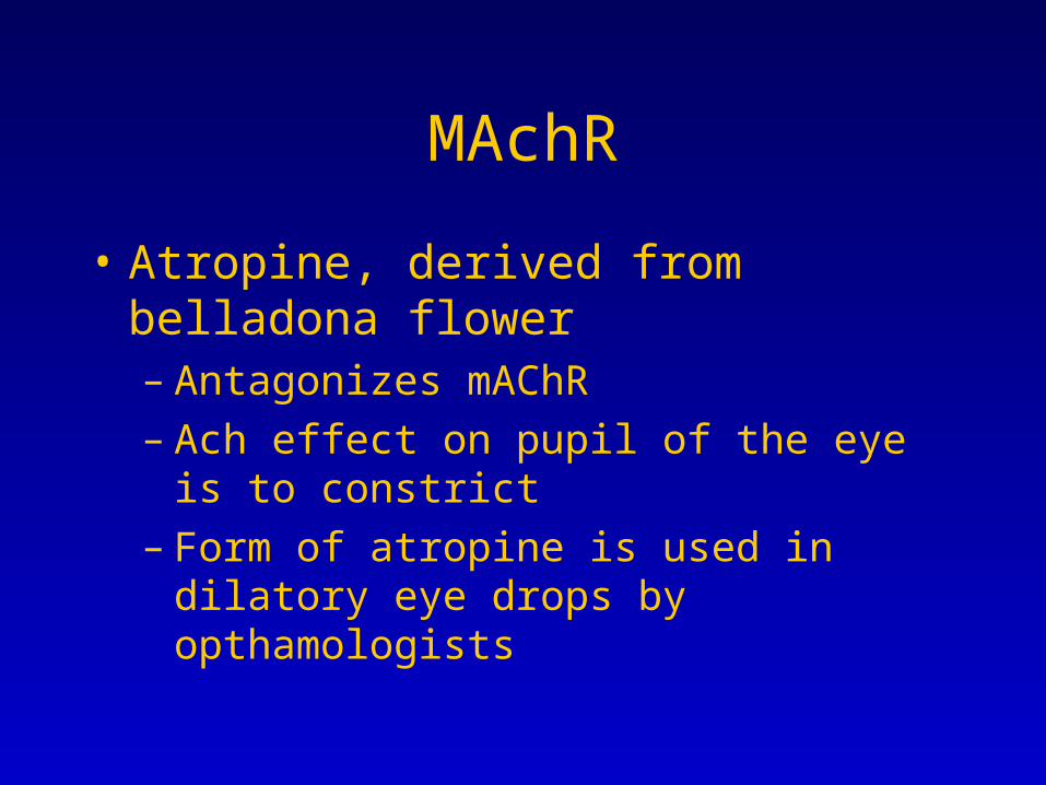
MAchR
• Atropine, derived from belladona flower– Antagonizes mAChR– Ach effect on pupil of the eye is to constrict– Form of atropine is used in dilatory eye drops
by opthamologists
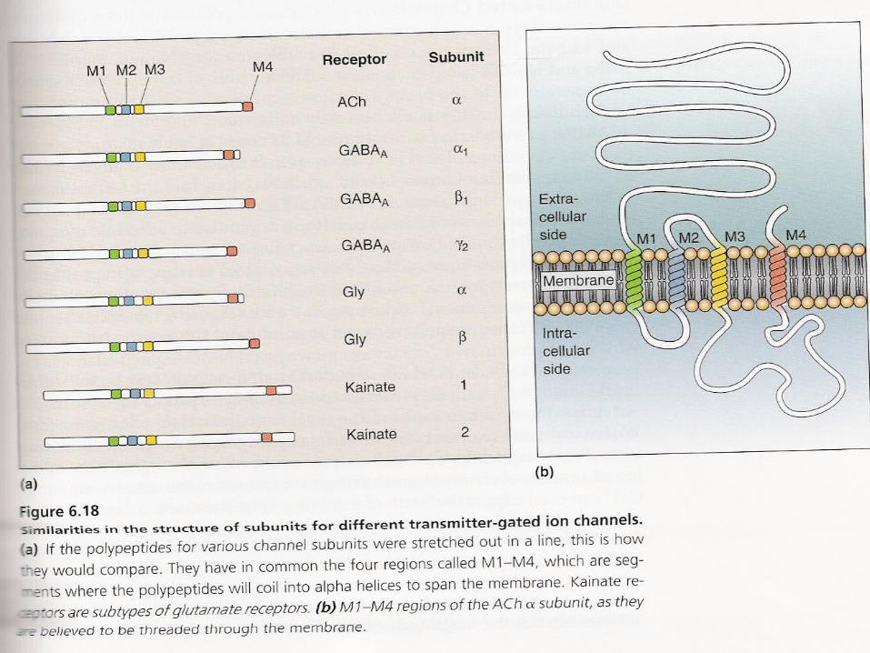

Basic Structure of NT gated ion channel
• 100 nm long-barely extends the width of pm• Subunits arranged to form a barrel with
interior pore through pm• All subunits have common domains named
M1-M4 that are transmembrane• M1-4 form hyophobic alpha helices• Most are composed of 4-5 subunits
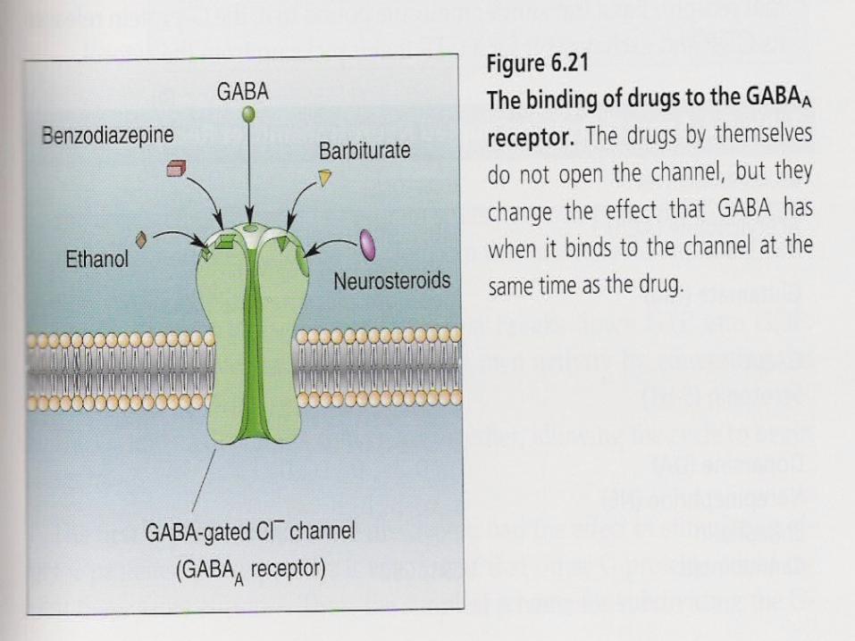
Drugs that bind GABAa Receptors
• Barbituate– Phenobarbital = anesthetic
• Benzodiazapines = tranquilizers– valium
• Binds GABAa subtype with gamma subunit
Effect of Drugs on GABA channels
• In the absence of GABA, drugs have no effect
• In the presence of GABA, drugs can– Increase frequency of
opening/benzodiazepines– Increase duration of opening/barbituates
Pharmacology of VALIUM
• Valium=benzodiazapines bind to gaba gated chloride channel
• Cause the channel to stay open longer==less brain activity=calmer state of mind
• Used as anti-convulsant drugs
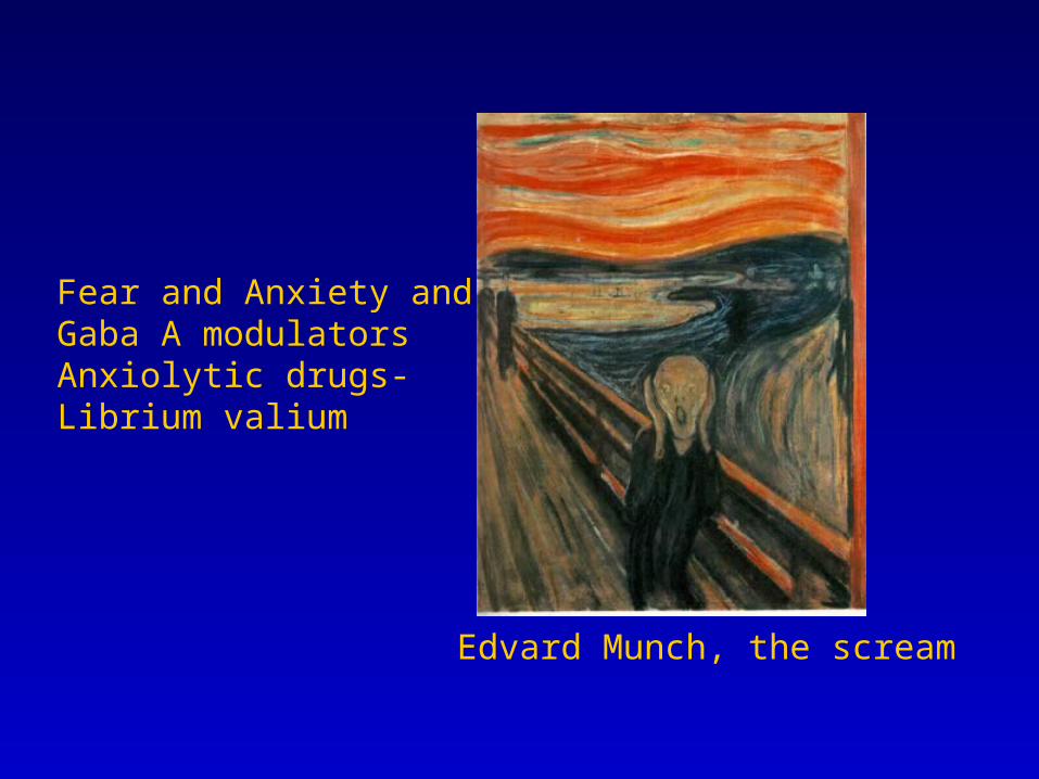
Edvard Munch, the scream
Fear and Anxiety and Gaba A modulatorsAnxiolytic drugs-Librium valium

Thebrainmcgill.ca
Synapses-GabaAnxietyReceptorBy bindingBenzodiazepineBackThree typesMetabotropic vs ionotropicBack to anxiety NTNot the only RapheHormonal brain

Neuromodulation
Metabotropic Ion Channels
Neurotransmitters bind Receptors that are not ion channels
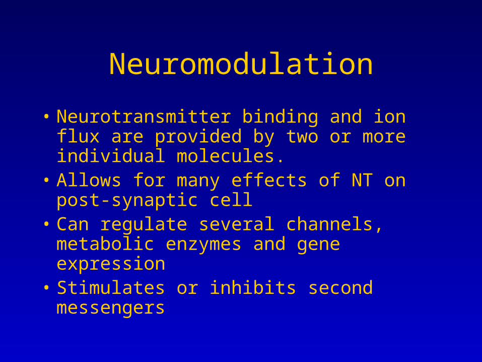
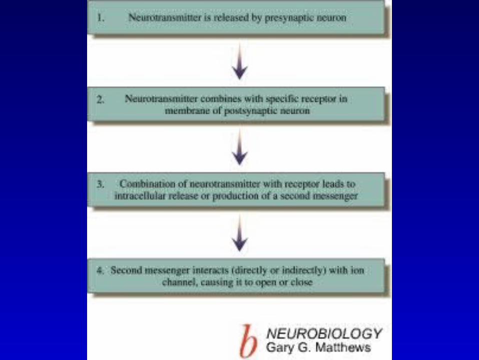
Neuromodulation
• Neurotransmitter binding and ion flux are provided by two or more individual molecules.
• Allows for many effects of NT on post-synaptic cell
• Can regulate several channels, metabolic enzymes and gene expression
• Stimulates or inhibits second messengers
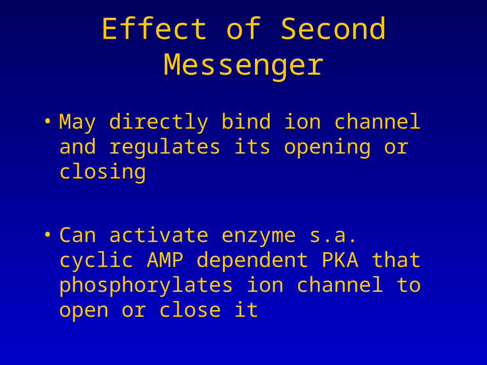
Effect of Second Messenger
• May directly bind ion channel and regulates its opening or closing
• Can activate enzyme s.a. cyclic AMP dependent PKA that phosphorylates ion channel to open or close it
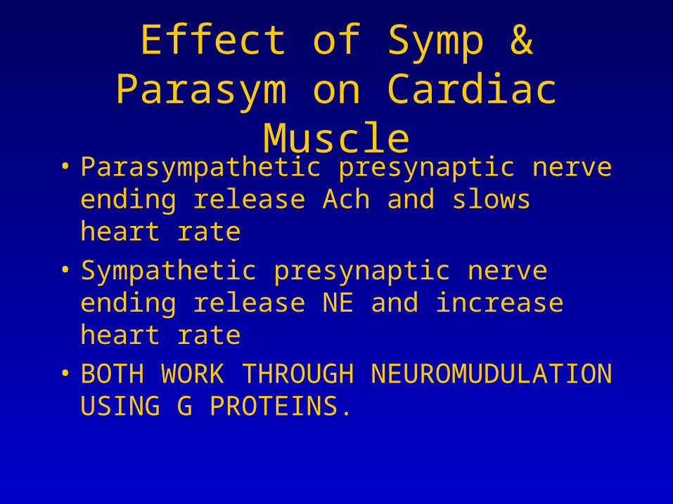
Effect of Symp & Parasym on Cardiac Muscle
• Parasympathetic presynaptic nerve ending release Ach and slows heart rate
• Sympathetic presynaptic nerve ending release NE and increase heart rate
• BOTH WORK THROUGH NEUROMUDULATION USING G PROTEINS.
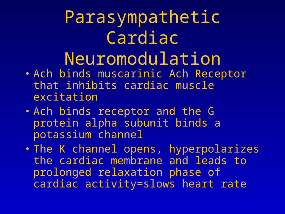
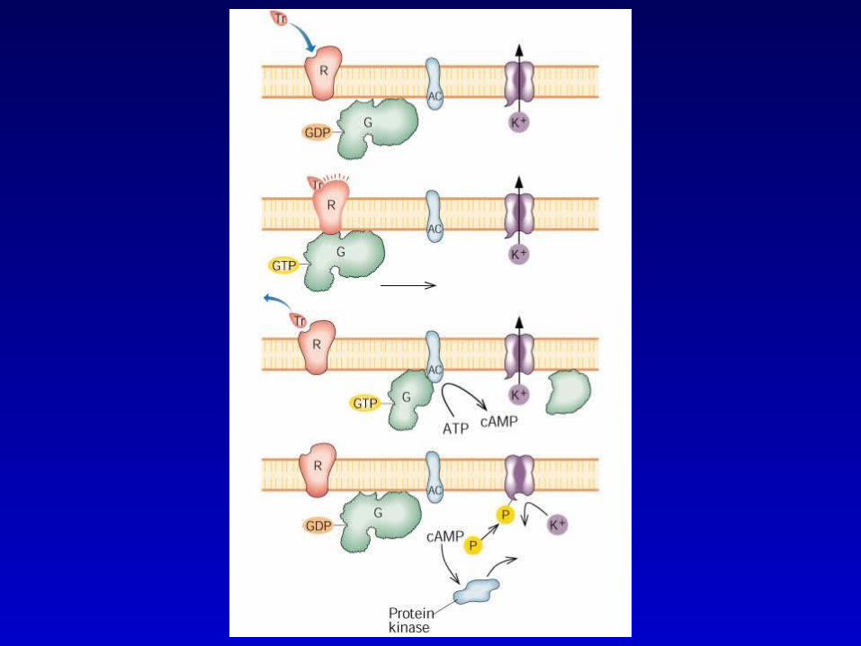
Parasympathetic Cardiac Neuromodulation
• Ach binds muscarinic Ach Receptor that inhibits cardiac muscle excitation
• Ach binds receptor and the G protein alpha subunit binds a potassium channel
• The K channel opens, hyperpolarizes the cardiac membrane and leads to prolonged relaxation phase of cardiac activity=slows heart rate

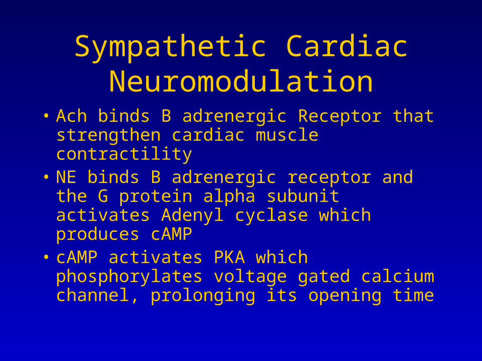
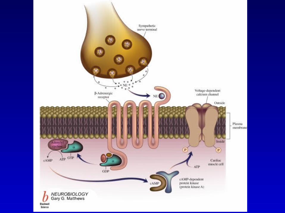
Sympathetic Cardiac Neuromodulation
• Ach binds B adrenergic Receptor that strengthen cardiac muscle contractility
• NE binds B adrenergic receptor and the G protein alpha subunit activates Adenyl cyclase which produces cAMP
• cAMP activates PKA which phosphorylates voltage gated calcium channel, prolonging its opening time
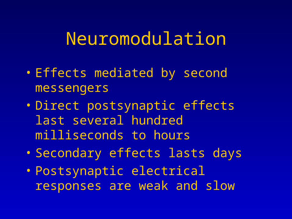
Neuromodulation
• Effects mediated by second messengers
• Direct postsynaptic effects last several hundred milliseconds to hours
• Secondary effects lasts days
• Postsynaptic electrical responses are weak and slow
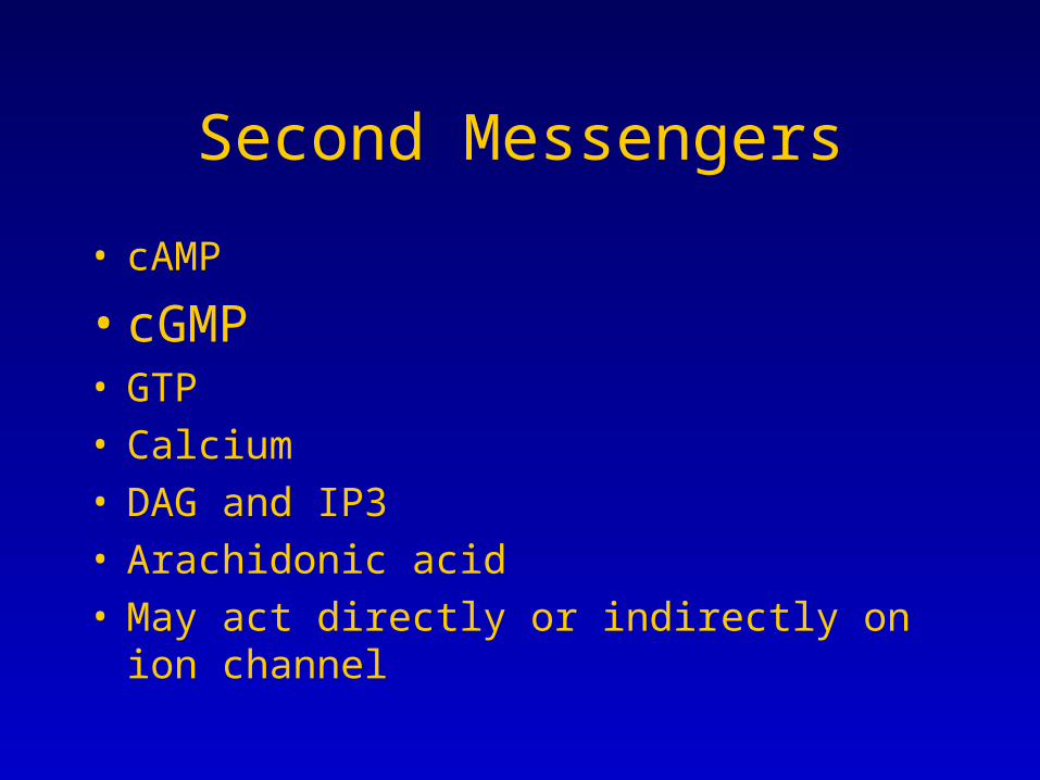
Second Messengers
• cAMP
• cGMP• GTP• Calcium• DAG and IP3• Arachidonic acid• May act directly or indirectly on ion channel
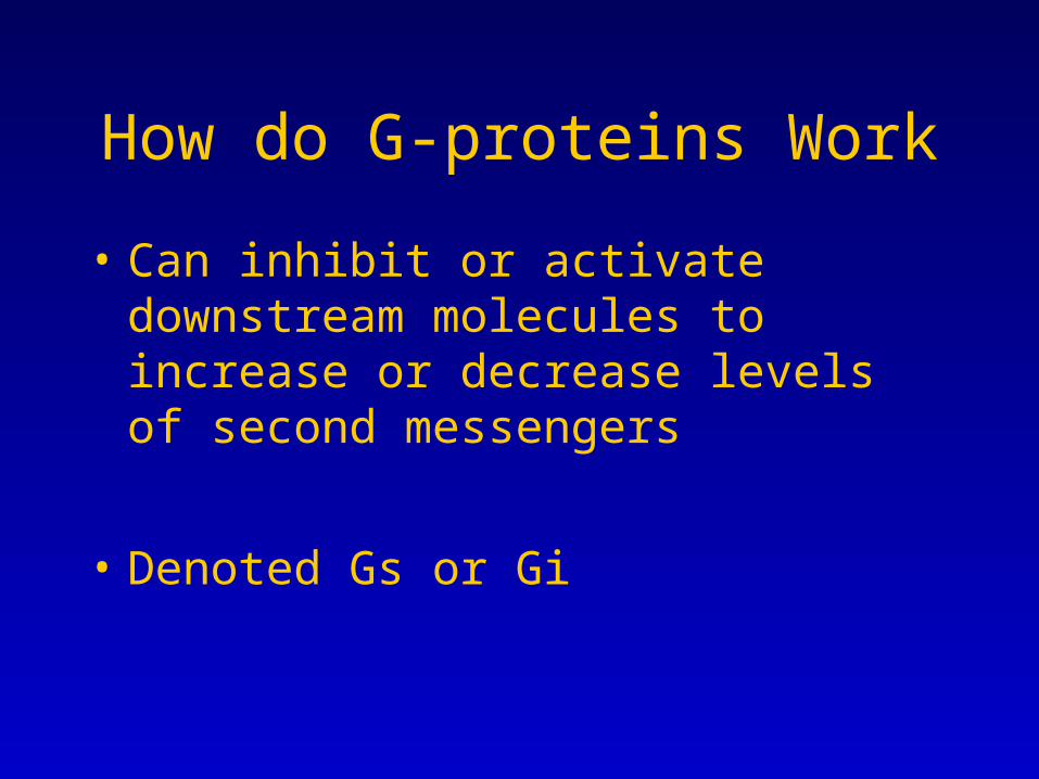
How do G-proteins Work
• Can inhibit or activate downstream molecules to increase or decrease levels of second messengers
• Denoted Gs or Gi
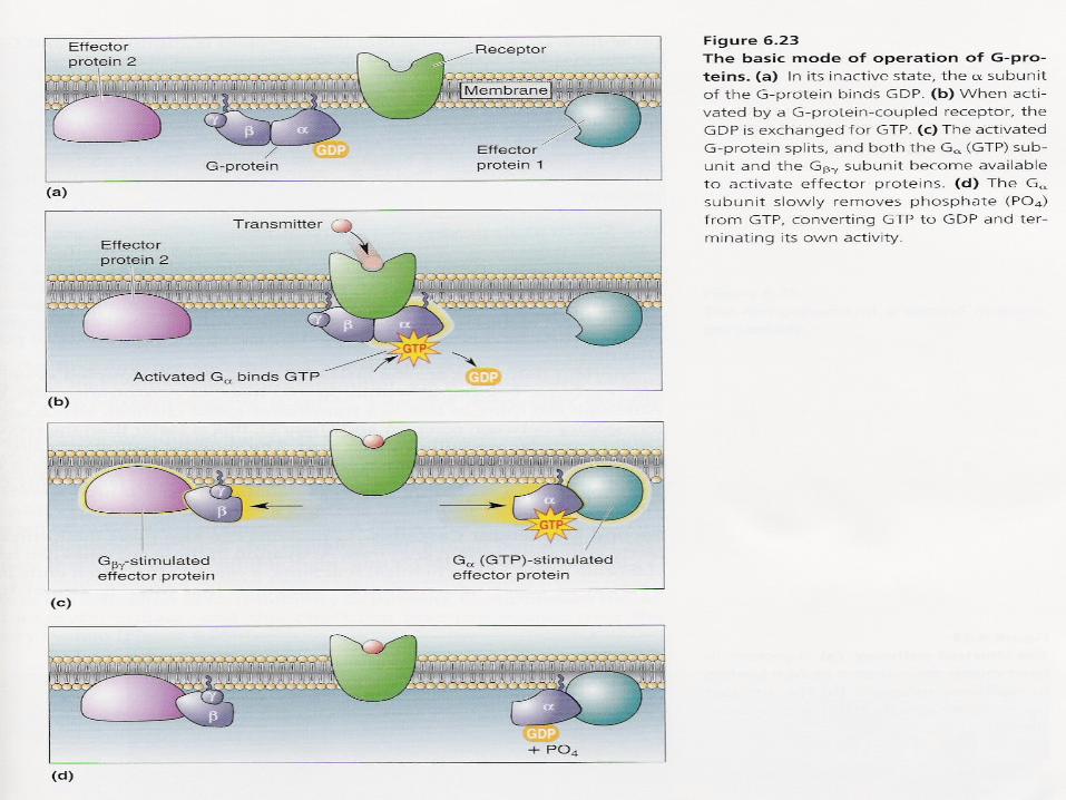

G-proteins Link Channel Activity with NT
• GTP-binding protein cycles between off state (GDP bound) and on state (GTP- bound)
• NT receptors catalyzes replacement of GDP with GTP.
• Activated G-protein activates adenylyl cyclase that produces cAMP
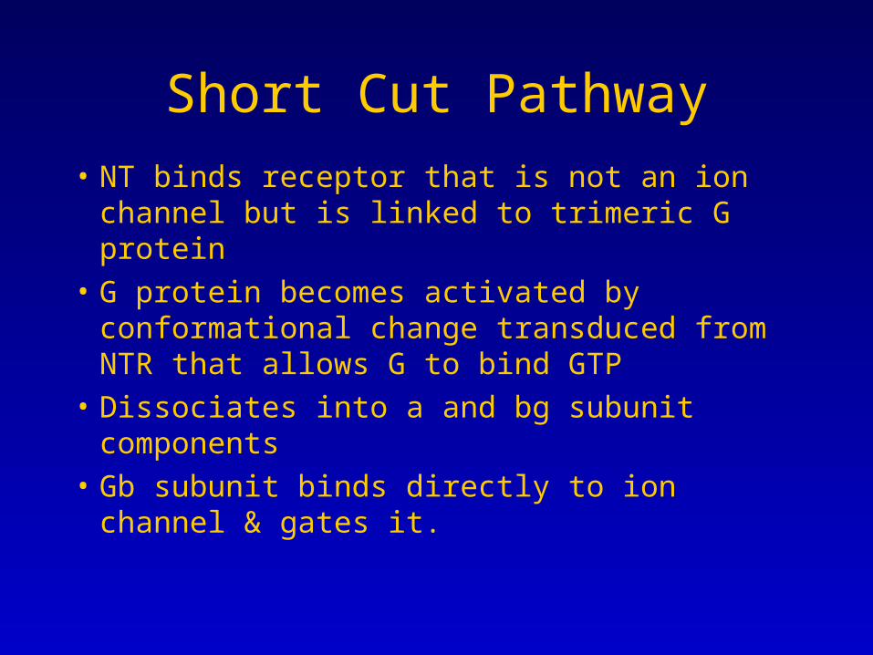
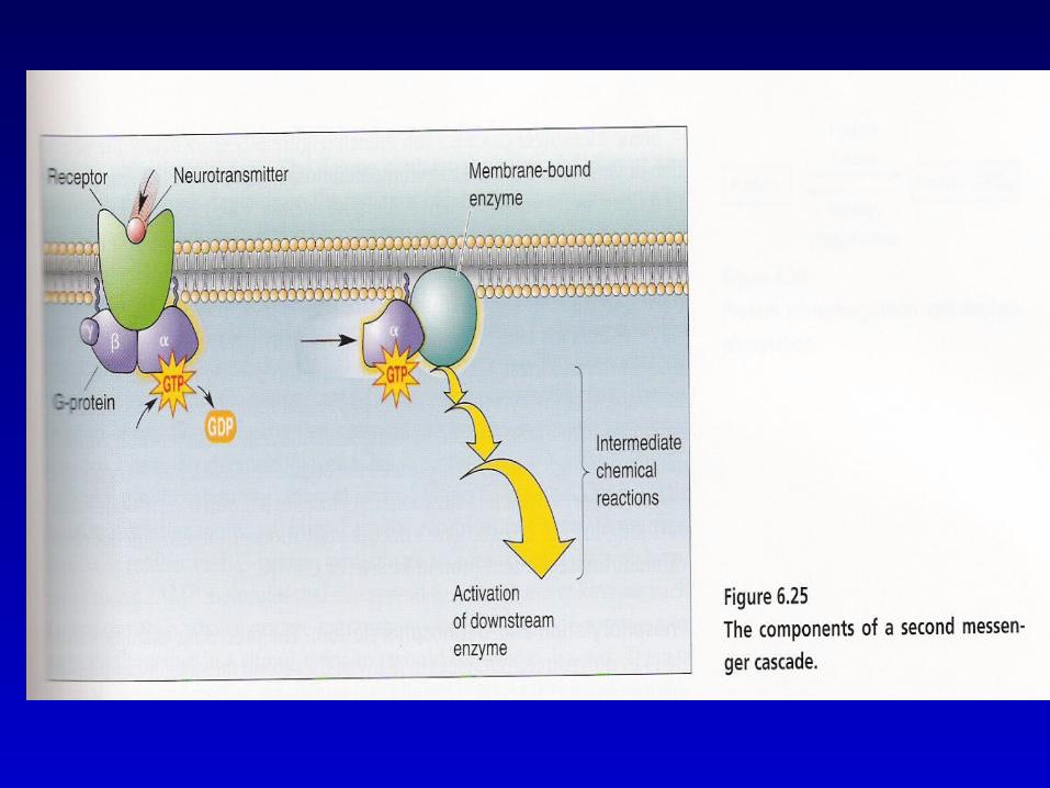
Short Cut Pathway
• NT binds receptor that is not an ion channel but is linked to trimeric G protein
• G protein becomes activated by conformational change transduced from NTR that allows G to bind GTP
• Dissociates into a and bg subunit components• Gb subunit binds directly to ion channel &
gates it.

Long-Term Effects
• Ion channel can be regulated by G-protein directly and by second messengers and enzymes s.a. PKA.
• So the effect of neurotransmitter binding can have prolonged effects on ion channel activity.
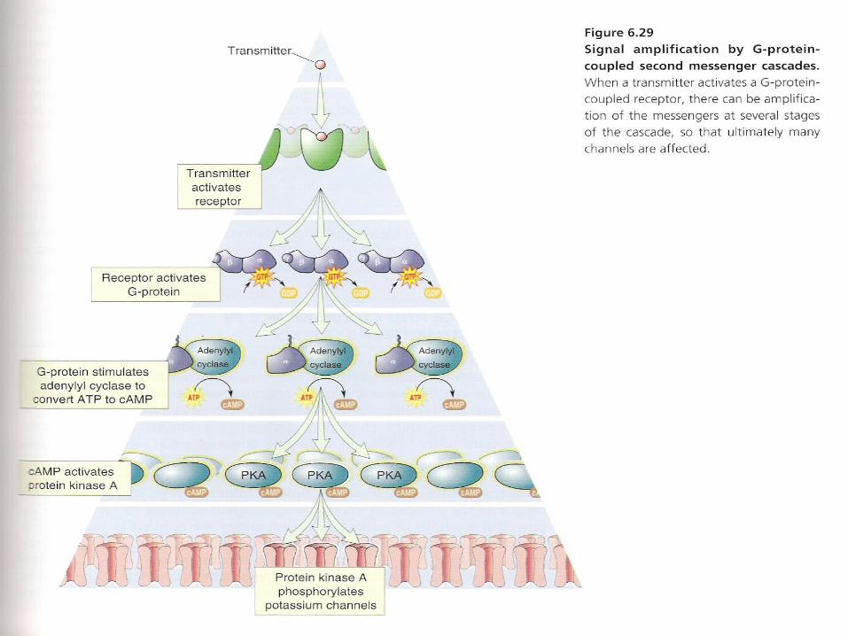
• An activated NT receptor can bind to many G proteins so signal is amplified
• 700 types of G proteins

G Protein Linked to Adenylyl Cyclase
• Causes formation of cAMP and activation of PKA
• PKA phosphorylates serine and threonine residues on target proteins
Regulation of G protein
• Some NT bind to both Gi and Gs linked receptors
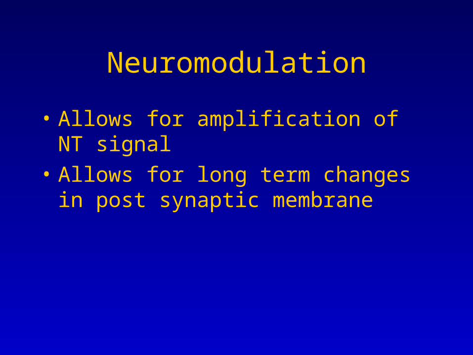
Neuromodulation
• Allows for amplification of NT signal
• Allows for long term changes in post synaptic membrane

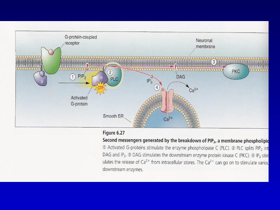
G Proteins linked to Phospholipase
• Phospholipase cleaves PIP2 in the membrane
• Generates to metabolic products each are second messengers
• IP3 and DAG• IP3 causes calcium release from
intracellular stores• DAG activate PKC


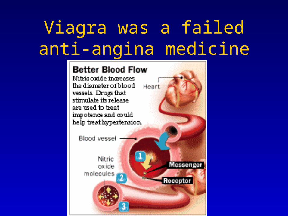
Nitric Oxide (NO)
• Made from arginine by NO synthase
• Chemical exist gas form
• Released from post-synaptic terminal without vesicles and acts on presynaptic terminal = retrograde communication aka retrograde messenger
• Lifetime is 5-10 min
NO relaxes Blood Vessels
• NO production is induced by ACh released from parasympathetic nerve endings onto endothelial cells in blood vessels
• mAchR activation leads to activation of NO synthase & production of NO
• NO diffuses into underlying smooth muscle cells

NO Effect
• Binds iron co-factor in the active site of guanylyl cyclase– Enzyme that forms cGMP for GTP
• Leads to increase cGMP
• cGMP leads to smooth muscle relaxation
• Increasing diameter of blood vessel and enhances blood flow
Viagra inhibits PDE5• Phosphodiesterase degrades cGMP into
GMP • Viagra inhibits PDE5 isoform expressed
primarily in penis• End result is prolonged increased levels of
cGMP in smooth muscle • allows increased blood flow into the
cavernous tissue of the penis thereby generating an erection

Viagra™ started life as a medicine intended to treat angina pectoris.Alfred Nobel - an explosives manufacturer - suffered from angina. In 1890 he was prescribed nitroglycerine (called trinitrin) to relieve the pain of angina attacks. It is still used today.Over 100 years later, the work of Robert Furchgott, Louis Ignarro and Ferid Murad showed that nitric oxide (NO) was an important signalling molecule in the cardiovascular system. It is released from nerve endings and cells lining the walls of blood vessels. The effect is to make the blood vessel relax, or dilate. It is also involved in the prevention of blood clots. In 1998, they received the Nobel Prize for Physiology. The Nobel prizes were set up by the same Alfred Nobel who had been treated with nitroglycerine
Viagra was a failed anti-angina medicine
9 types of PDE
• PDE5 is expressed in erectile tissue of penis and in the retina
Giles Brindley and Drug Therapy for ED Modern drug therapy for ED was advanced enormously in 1983 when British physiologist Giles Brindley, Ph.D. dropped his trousers and demonstrated to a shocked AUA audience his phentolamine-induced erection. The drug Brindley injected into his penis was a non-specific vasodilator, an alpha-blocking agent, and the mechanism of action was clearly corporal smooth muscle relaxation. The effect that Brindley discovered, established the fundamentals for the later development of specific, safe, orally-effective drug therapies, ie, PDE-5 inhibitors. Approved in 1998
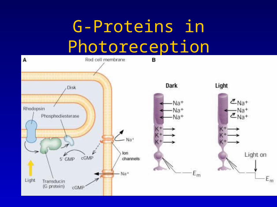
G-Proteins in Photoreception