Embed Size (px)
Citation preview
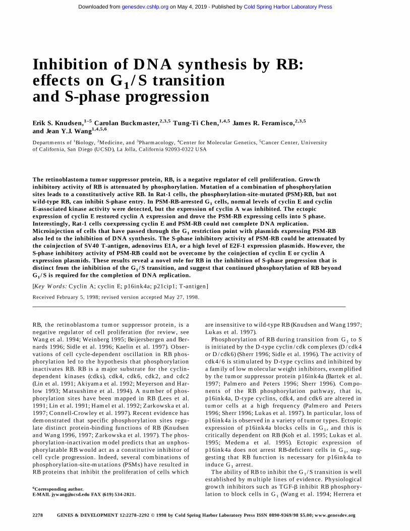
Inhibition of DNA synthesis by RB:effects on G1/S transitionand S-phase progressionErik S. Knudsen,1–5 Carolan Buckmaster,2,3,5 Tung-Ti Chen,1,4,5 James R. Feramisco,2,3,5
and Jean Y.J. Wang1,4,5,6
Departments of 1Biology, 2Medicine, and 3Pharmacology, 4Center for Molecular Genetics, 5Cancer Center, Universityof California, San Diego (UCSD), La Jolla, California 92093-0322 USA
The retinoblastoma tumor suppressor protein, RB, is a negative regulator of cell proliferation. Growthinhibitory activity of RB is attenuated by phosphorylation. Mutation of a combination of phosphorylationsites leads to a constitutively active RB. In Rat-1 cells, the phosphorylation-site-mutated (PSM)-RB, but notwild-type RB, can inhibit S-phase entry. In PSM-RB-arrested G1 cells, normal levels of cyclin E and cyclinE-associated kinase activity were detected, but the expression of cyclin A was inhibited. The ectopicexpression of cyclin E restored cyclin A expression and drove the PSM-RB expressing cells into S phase.Interestingly, Rat-1 cells coexpressing cyclin E and PSM-RB could not complete DNA replication.Microinjection of cells that have passed through the G1 restriction point with plasmids expressing PSM-RBalso led to the inhibition of DNA synthesis. The S-phase inhibitory activity of PSM-RB could be attenuated bythe coinjection of SV40 T-antigen, adenovirus E1A, or a high level of E2F-1 expression plasmids. However, theS-phase inhibitory activity of PSM-RB could not be overcome by the coinjection of cyclin E or cyclin Aexpression plasmids. These results reveal a novel role for RB in the inhibition of S-phase progression that isdistinct from the inhibition of the G1/S transition, and suggest that continued phosphorylation of RB beyondG1/S is required for the completion of DNA replication.
[Key Words: Cyclin A; cyclin E; p16ink4a; p21cip1; T-antigen]
Received February 5, 1998; revised version accepted May 27, 1998.
RB, the retinoblastoma tumor suppressor protein, is anegative regulator of cell proliferation (for review, seeWang et al. 1994; Weinberg 1995; Beijersbergen and Ber-nards 1996; Sidle et al. 1996; Kaelin et al. 1997). Obser-vations of cell cycle-dependent oscillation in RB phos-phorylation led to the hypothesis that phosphorylationinactivates RB. RB is a major substrate for the cyclin-dependent kinases (cdks), cdk4, cdk6, cdk2, and cdc2(Lin et al. 1991; Akiyama et al. 1992; Meyerson and Har-low 1993; Matsushime et al. 1994). A number of phos-phorylation sites have been mapped in RB (Lees et al.1991; Lin et al. 1991; Hamel et al. 1992; Zarkowska et al.1997; Connell-Crowley et al. 1997). Recent evidence hasdemonstrated that specific phosphorylation sites regu-late distinct protein-binding functions of RB (Knudsenand Wang 1996, 1997; Zarkowska et al. 1997). The phos-phorylation-inactivation model predicts that an unphos-phorylatable RB would act as a constitutive inhibitor ofcell cycle progression. Indeed, several combinations ofphosphorylation-site-mutations (PSMs) have resulted inRB proteins that inhibit the proliferation of cells which
are insensitive to wild-type RB (Knudsen and Wang 1997;Lukas et al. 1997).
Phosphorylation of RB during transition from G1 to Sis initiated by the D-type cyclin/cdk complexes (D/cdk4or D/cdk6) (Sherr 1996; Sidle et al. 1996). The activity ofcdk4/6 is stimulated by D-type cyclins and inhibited bya family of low molecular weight inhibitors, exemplifiedby the tumor suppressor protein p16ink4a (Bartek et al.1997; Palmero and Peters 1996; Sherr 1996). Compo-nents of the RB phosphorylation pathway, that is,p16ink4a, D-type cyclins, cdk4, and cdk6 are altered intumor cells at a high frequency (Palmero and Peters1996; Sherr 1996; Lukas et al. 1997). In particular, loss ofp16ink4a is observed in a variety of tumor types. Ectopicexpression of p16ink4a blocks cells in G1, and this iscritically dependent on RB (Koh et al. 1995; Lukas et al.1995; Medema et al. 1995). Ectopic expression ofp16ink4a does not arrest RB-deficient cells in G1, sug-gesting that RB function is necessary for p16ink4a toinduce G1 arrest.
The ability of RB to inhibit the G1/S transition is wellestablished by multiple lines of evidence. Physiologicalgrowth inhibitors such as TGF-b inhibit RB phosphory-lation to block cells in G1 (Wang et al. 1994; Herrera et
6Corresponding author.E-MAIL [email protected] FAX (619) 534-2821.
2278 GENES & DEVELOPMENT 12:2278–2292 © 1998 by Cold Spring Harbor Laboratory Press ISSN 0890-9369/98 $5.00; www.genesdev.org
Cold Spring Harbor Laboratory Press on May 4, 2019 - Published by genesdev.cshlp.orgDownloaded from
al. 1996a). Embryo fibroblasts derived from RB-deficientmice have a shortened G1 phase and a weakened re-sponse to anti-mitogenic signals (Herrera et al. 1996a,b).Ectopic expression of wild-type RB arrests some RB-deficient tumor cells in G1 (Hamel et al. 1993; Wang etal. 1994). A major mechanism by which RB inhibits theG1/S transition is through the repression of E2F-regu-lated genes, among which are cyclin E and cyclin A(Ohtani et al. 1995; Weinberg 1995; Geng et al. 1996;Kaelin 1997; Sidle et al. 1996). Both cyclin E and cyclin Acan bind to and activate cdk2, and cdk2 activity is rate-limiting for S-phase entry (Del Sal et al. 1996; Sherr1996). Using a tetracycline-regulated promoter,Resnitzky et al. have shown that overproduction of cy-clin E or cyclin A in Rat-1 cells shortens the G1 interval(Resnitzky and Reed 1995; Resnitzky et al. 1995). In RB-deficient mouse embryo fibroblasts, the timing of cyclinE expression is advanced, in keeping with the faster pro-gression of these cells from quiescence into S phase (Her-rera et al. 1996a; Hurford et al. 1997). Thus, RB-mediatedrepression of cyclin E expression has been hypothesizedto underlie its G1/S inhibitory activity.
Previously, we have reported the construction of twoPSM-RB proteins that can inhibit the proliferation ofRat-1 cells (Knudsen and Wang 1997). In the context offull-length RB, mutation of nine phosphorylation sites inPSM.9I-RB is required to generate a constitutively activegrowth suppressor (Kundsen and Wang 1997). In the con-text of an amino-terminal deleted RB, which is com-monly known as the large pocket (LP; Qin et al. 1992),mutation of seven phosphorylation sites (PSM.7-LP) issufficient to block the inactivation of RB (Knudsen andWang 1997). In this study we compared the effect ofPSM-RB and p16ink4a on cell cycle progression in Rat-1cells. Although both p16ink4a and PSM-RB induce G1
arrest, they do so at different execution points along theG1/S transition. Our study also uncovered an unex-pected inhibitory effect of PSM-RB on S-phase progres-sion, which was not observed with p16ink4a. Our find-ings show that RB can inhibit both G1/S transition andS-phase progression, and provide an explanation for thecontinued hyperphosphorylation of RB throughout the Sphase of the cell cycle.
Results
PSM-RB arrests Rat-1 cells in G1
Ectopic expression of WT-RB, or the previously de-scribed pD34-phosphorylation site mutant RB, does notinhibit the growth of Rat-1 cells (Knudsen and Wang1997; Lukas et al. 1997; Alevizopoulos et al. 1997). How-ever, PSM.9I-RB, PSM.7LP (Knudsen and Wang 1997)and the RBDcdk (Lukas et al. 1997) can block Rat-1 cellcycle progression. Because p16ink4a and RB are in thesame G1-inhibitory pathway, we compared the G1-inhibitory activity of PSM.7-LP to that of p16ink4a.Rat-1 cells were transiently transfected with vector, WT-LP, PSM.7-LP or p16ink4a expression plasmids and aplasmid encoding the green fluorescent protein (GFP).
Cell cycle progression of the transfected, GFP-positive,cells was measured by BrdU-incorporation. With vectorand WT-LP transfected cells, between 60% and 70% in-corporated BrdU (Fig. 1A). In contrast, only 5% of thecells transfected with p16ink4a or PSM.7-LP incorpo-rated BrdU (Fig. 1A).
To examine the long-term effect, cells were cotrans-fected with a plasmid expressing puromycin resistance,and either vector, WT-LP, PSM.7-LP, or p16ink4a expres-sion plasmids. Transfected cells were subjected to selec-tion with high concentrations of puromycin, throughwhich all untransfected cells were killed within 72 hr(not shown). Labeling of puromycin-selected cells with[3H]thymidine showed robust incorporation with vectoror WT-LP-transfected cells, but virtually no incorpora-tion with PSM.7-LP-transfected cells (not shown). Fol-lowing 7 days of culture, both vector and WT-LP-trans-fected cells grew into colonies, staining darkly with crys-tal violet (Fig. 1B). In contrast, PSM.7-LP and p16ink4asuppressed colony formation. Microscopic viewingshowed the presence of single, flat cells in either PSM.7-LP or p16ink4a-transfected cultures (Fig. 1B). The mor-phologies of the PSM.7-LP and p16ink4a-arrested cellswere indistinguishable from one another. These resultsare consistent with the role of RB as a negative regulatorof cell proliferation.
RB, p107, and p130 are hyperphosphorylatedin PSM-RB-arrested Rat-1 cells
The expression and phosphorylation status of the LP pro-teins was examined after 72 hr of puromycin selection.The WT-LP protein was predominantly hyperphosphory-lated, which was consistent with the continued growthof these transfected cells (Fig. 1C, lane 2). The PSM.7-LPprotein migrated during SDS-PAGE as a doublet (Fig. 1C,lanes 3,4), indicating that the remaining three cdk sitesare phosphorylated (Knudsen and Wang 1997). Therefore,RB kinase activity does not appear to be inhibited in thePSM.7-LP-arrested cells. Although PSM.7-LP from cellsarrested by its own expression migrated as a doublet (Fig.1C, lane 3,4), it migrated as a single band with the coex-pression of p16ink4a (Fig. 1C, lane 5). These observationssuggest that cdk4/6 is active in PSM.7-LP-arrested cellsbut not in p16ink4a-arrested cells.
We also examined the phosphorylation status of p107and p130 in the PSM-RB-arrested cells (Fig. 1D,E). Inasynchronously growing Rat-1 cells, p107 and p130 werehyperphosphorylated and migrated slower than the p107and p130 of quiescent cells (Fig. 1D, cf. lanes 1 and 2).The electrophoretic mobilities of p107 and p130 in thevector, WT-LP- or PSM.7-LP-transfected cells were simi-lar to that of proliferating Rat-1 cells (Fig. 1D, lanes 3–5).Overproduction of p16ink4a has been shown to inhibitthe phosphorylation of p107 and p130 (Del Sal et al.1996; Sherr 1996; Alevizopoulos et al. 1997). The p107protein from p16ink4a-arrested Rat-1 cells migratedfaster than that from PSM.7-LP-arrested cells (Fig. 1E, cf.lanes 3 and 6 to lanes 2 and 5). Therefore, p16ink4a in-hibits the phosphorylation of p107 and p130, whereas
RB inhibits S phase
GENES & DEVELOPMENT 2279
Cold Spring Harbor Laboratory Press on May 4, 2019 - Published by genesdev.cshlp.orgDownloaded from
PSM-RB does not interfere with these phosphorylationevents.
Cyclin A expression is suppressed in PSM-RB-arrestedRat-1 cells
We also examined the effect of PSM-RB on the expres-sion and activity of several cdks, cyclins, and their in-hibitors. The levels of cyclin D1, cyclin E, cdk2, p21cip1,and p27kip1 in PSM.7-LP-expressing cells were similarto those found in proliferating Rat-1 cells and vector- orWT-LP-transfected cells (Fig. 2A,B). In contrast, the lev-els of cyclin A, cyclin B, and cdc2 were reduced inPSM.7-LP-transfected cells (Fig. 2A,B). Consistent withthe normal levels of cyclin E, cdk2, p21cip1, andp27kip1, we found that cyclin E-associated kinase activ-ity was not inhibited by PSM.7-LP (Fig. 2C, aCyclin E).The total cdk2 activity was reduced in PSM.7-LP-arrested cells (Fig. 2C, aCdk2); this is likely a result ofthe inhibition of cyclin A expression.
To determine whether cyclin A was down-regulated atthe RNA level, quantitative RT–PCR was used to mea-
sure the level of cyclin A mRNA. In Rat-1 cells trans-fected with vector and selected with puromycin, thelevel of cyclin A mRNA was similar to that of asynchro-nously growing cells (Fig. 2D). In contrast, in cells trans-fected with PSM.7-LP, the level of cyclin A mRNA wasreduced to that of quiescent cells (Fig. 2D). Therefore,the expression of cyclin A mRNA is inhibited in PSM-RBarrested cells. Together, these results suggest that cellstransfected with PSM-RB are predominantly blocked inlate G1 prior to the expression of cyclin A.
We have shown previously that the ectopic expressionof cyclin A can override the wild-type RB-mediated ar-rest of SAOS-2 cells (Knudsen and Wang 1996), or thev-Abl-mediated arrested of the NIH-3T3 subclone N-3T3cells (Chen et al. 1996). Therefore, we attempted to over-ride the G1-inhibitory effect of PSM-RB with the ectopicexpression of cyclin A. When transfected into Rat-1cells, the cyclin A expression plasmid did not lead to anincrease in the overall levels of cyclin A protein (notshown). The inability of the cells to overproduce cyclinA in these cotransfection experiments could be attribut-able to an induction of cell death, as the cyclin A-trans-
Figure 1. PSM-RB arrests cell growth with-out inhibiting the phosphorylation of p107/p130. (A) Rat-1 cells were cotransfected witha GFP expression plasmid (1.0 µg) and theindicated plasmids (5.0 µg). BrdU was added24 hr post-transfection for a total labelingperiod of 20 hr. Cells were stained with anti-BrdU antibody, and the percentage of GFP-positive cells exhibiting BrdU incorporationwas determined by indirect immunofluores-cence. The average and deviation valuesshown are from two independent experi-ments with at least 200 GFP-positive cellscounted per experiment. (B) Rat-1 cells werecotransfected with a puromycin-resistanceplasmid (0.5 µg) and the indicated expressionplasmids (5.0 µg). Transfected cells were se-lected with puromycin for 7 days and thenstained with crystal violet. Pictures weretaken at 20× magnification with a phase-contrast microscope. (C) Rat-1 cells were co-transfected with a puromycin-resistanceplasmid (0.5 µg) and vector (lane 1), WT-LP(lane 2), or PSM.7-LP (lane 3) expression plas-mids (5.0 µg). Alternatively, PSM.7-LP (2.5µg) and either vector (lane 4) or p16ink4a(lane 5) expression plasmids (2.5 µg) were co-transfected with a puromycin-resistanceplasmid (0.5 µg). Transfected cells were se-lected with puromycin for 72 hr and thenlysed. Total protein (15 µg) was resolved bySDS-PAGE and the transfected RB proteinwas detected by immunoblotting with anti-RB antibody. (ppLP) Hyperphosphorylated large-pocket fragment of RB; (pLP) hypophos-phorylated large-pocket fragment. (D) Rat-1 cells were cotransfected with a puromycin-resistance plasmid (0.5 µg) and vector (lane 3),WT-LP (lane 4), or PSM.7-LP (lane 5) plasmids (5.0 µg). Transfected cells were selected with puromycin for 72 hr and then harvested.As controls, quiescent (lane 1) or asynchronously growing (lane 2) Rat-1 cells were also harvested. Total protein (15 µg) was resolvedby SDS-PAGE and the endogenous p107 or p130 proteins were detected by immunoblotting with the respective antibodies as indicated.(E) Rat-1 cells were cotransfected with a puromycin-resistance plasmid (0.5 µg) and vector (lanes 1,4), PSM.7-LP (lanes 2,5), or p16ink4a(lanes 3,6) plasmids (5.0 µg). Transfected cells were selected with puromycin for 72 hr and then harvested. Total protein (15 µg) wasresolved by SDS-PAGE, and the endogenous p107 proteins were detected by immunoblotting.
Knudsen et al.
2280 GENES & DEVELOPMENT
Cold Spring Harbor Laboratory Press on May 4, 2019 - Published by genesdev.cshlp.orgDownloaded from
fected cells were found to exhibit condensed nuclei (notshown). Because the ectopic expression of cyclin A couldnot be achieved, we were unable to conclude whethercyclin A expression could rescue the PSM-RB-mediatedG1 arrest in Rat-1 cells.
Cyclin E expression partially rescues the negativeeffect of PSM-RB on DNA synthesis
The ectopic expression of cyclin E abrogated the PSM-RB-induced G1 arrest. This was detected as a decrease inthe G1 content and a concomitant increase in S phase byFACS analysis (Fig. 3A). However, cells transfected withcyclin E plus PSM.7-LP exhibited a peculiar profile ofS-phase DNA content with no increase in G2 cells (Fig.3A). This is in direct contrast to cells transfected withcyclin E plus p16ink4a, wherein cyclin E expressioncaused a return to a cell cycle profile similar to that ofnontransfected (CD20-negative) cells (Fig. 3A).
Because cyclin A is required for DNA synthesis(Hunter and Pines 1994), we determined whether the ex-pression of cyclin E could rescue the negative effect ofPSM.7-LP on the expression of cyclin A. Following 72 hrof selection with puromycin, cells coexpressing cyclin Eand PSM.7-LP had cyclin A protein at a level comparableto that of proliferating cells (Fig. 3B). Nevertheless, thesetransfected cells remained as single flat cells through theselection period, similar to those arising from the trans-fection of PSM.7-LP alone (not shown). The expression ofSV40 large T-antigen (T-Ag) with PSM-RB also rescuedthe expression of cyclin A (Fig. 3B), and these cotrans-
fected cells resumed proliferation, leading to the forma-tion of colonies (not shown). Therefore, the ectopic ex-pression of cyclin E overcame the inhibitory effect of RBon cyclin A expression and drove cells into S phase.However, cyclin E did not overcome the growth arrestactivity of PSM-RB, whereas T-Ag completely abrogatedthe PSM-RB-induced growth arrest.
We also examined the incorporation of BrdU by cellstransfected with cyclin E plus PSM-RB. Cells transfectedwith PSM.7-LP did not incorporate BrdU (Fig. 4A). Whencotransfected with cyclin E, a punctate BrdU stainingpattern was observed (Fig. 4A). The same result was ob-tained when the full-length PSM.9I-RB was used in theexperiment. Again, PSM.9I-RB blocked BrdU incorpora-tion, which was partially rescued by the coexpression ofcyclin E. To determine quantitatively the amount ofBrdU incorporation, a digital CCD camera was used tocompare the intensity of BrdU signal, and these analysesshowed that the intensity of punctate staining was ∼25%of the BrdU incorporation observed in homogeneouslylabeled nuclei (data not shown). This result was consis-tent with the FACS profile (Fig. 3A) and suggested thatcells cotransfected with cyclin E and PSM-RB initiatedDNA synthesis but incorporated BrdU at a significantlyreduced rate.
Quantitation of cells with homogenous (positive),punctate, or negative BrdU staining was determined forseveral different transfections (Fig. 4B). Coexpression ofcyclin E with p16ink4a resulted in strong homogenousBrdU staining at a level comparable to that of vector-transfected cells (Fig. 4B). The punctate BrdU staining
Figure 2. PSM-RB inhibits cyclin A expression inRat-1 cells. (A) Rat-1 cells were cotransfected witha puromycin-resistance plasmid (0.5 µg) and vector(lane 3), WT-LP (lane 4), or PSM.7-LP (lane 5) ex-pression plasmids (5.0 µg). Transfected cells wereselected with puromycin for 72 hr and then har-vested. As controls, quiescent (lane 1) or asynchro-nously growing (lane 2) Rat-1 cells were also har-vested. Total protein (15 µg) was resolved by SDS-PAGE, and the endogenous cyclin D1, cyclin E,cyclin A, and cyclin B proteins were detected byimmunoblotting with the respective antibodies. (B)Rat-1 cells were cotransfected with a puromycin-
resistance plasmid and the indicated expression plasmids, selected, andharvested as in A. Total protein (15 µg) was resolved by SDS-PAGE, andthe endogenous cdk2, cdc2, p21cip1, and p27kip1 proteins were detectedby immunoblotting with the respective antibodies. (C) Rat-1 cells werecotransfected with a puromycin-resistance plasmid and the indicatedexpression plasmids, selected, and harvested as in A. Twenty micro-grams of total protein was utilized in in vitro kinase reactions withhistone H1 as a substrate. Cdk/cyclin complexes were recovered by im-munoprecipitation with the indicated antibodies against cyclin E, cyclinA, cdk2, or cdc2 and protein A–Sepharose. A nonspecific rabbit anti-mouse antibody was utilized as a negative control. Incorporation of 32Pinto histone H1 was visualized by autoradiography. (D) Rat-1 cells werecotransfected with a puromycin-resistance plasmid and the indicated
expression plasmids, selected, and harvested as in A. Quiescent and asynchronous growing Rat-1 cells were used as controls. TotalRNA was prepared. Quantitative RT-PCR was performed with 1–16 ng of total RNA as template. The level of cyclin A PCR productwas normalized to an internal control PCR with primers that amplify the sequence of GAPDH (see Materials and Methods). Relativecyclin A mRNA level from five independent PCR reactions is shown, with the level found in asynchronous growing cells set to 100%.
RB inhibits S phase
GENES & DEVELOPMENT 2281
Cold Spring Harbor Laboratory Press on May 4, 2019 - Published by genesdev.cshlp.orgDownloaded from
was not observed when cyclin E was coexpressed withWT-LP (Fig. 4B). In contrast to cyclin E, coexpression ofT-Ag with PSM-RB resulted in strong, homogenous BrdUstaining (Fig. 4B). Thus, T-Ag but not cyclin E can com-pletely overcome the negative effect of PSM-RB on DNAsynthesis.
To determine whether the reduced BrdU incorporationwas the result of a limitation on the amount of cyclin E,we carried out microinjection experiments with cellssynchronized in early G1 (Fig. 4C). Identical to the resultobserved with transfection, microinjection of PSM.7-LPblocked the incorporation of BrdU, while coinjection
with PSM.7-LP and cyclin E gave rise to punctate BrdUstaining (Fig. 4C). The punctate staining was observed atboth 1:1 and 1:4 ratios of PSM-RB to cyclin E (Fig. 4C),indicating that a limitation on cyclin E is not likely to beresponsible for the retardation in S-phase progression.
To determine whether the reduced BrdU incorporationrepresented a block in S phase or a slower rate of DNAsynthesis, cells were labeled for up to 40 hr and the per-centage of cells exhibiting positive, punctate, or negativeBrdU staining was determined (Fig. 4D). We found thatafter 40 hr of continuous labeling, punctate staining per-sisted in cells expressing PSM.7-LP and cyclin E (closedred squares). There was a gradual decline in punctatestaining and a concomitant increase in the percentage ofcells exhibiting homogenous BrdU labeling, suggesting aslow progression through S phase. In contrast, cells co-expressing p16ink4a plus cyclin E (open red squares) orPSM-RB plus T-Ag (closed green diamonds) showed BrdUlabeling similar to vector transfected cells over the ex-perimental time course (Fig. 4D). Following extended pe-riods of culture, those cells that coexpressed p16ink4aand cyclin E gave rise to microcolonies of two to eightadjoining GFP-positive cells, indicating completion ofthe cell cycle (not shown). However, cells coexpressingPSM.7-LP and cyclin E remained as single GFP-positivecells, suggesting that they did not undergo cell divisionduring the course of the experiment (not shown). Takentogether, the results presented in Figures 3 and 4 showthat increased expression of cyclin E can drive PSM-RB-arrested cells into S phase but DNA synthesis is retardedin the presence of PSM-RB.
PSM-RB inhibits DNA synthesis when expressedin S-phase-committed cells
To demonstrate directly that PSM-RB can inhibit DNAsynthesis, we performed microinjection experimentswith cells that have passed through the G1 restrictionpoint (Fig. 5). The DNA polymerase inhibitor aphidicolinwas used to reversibly arrest asynchronous Rat-1 cells inS phase. After the removal of aphidicolin, these cellscompleted DNA replication and entered G2 phase within6 hr, as determined by flow cytometry (not shown).Aphidicolin-treated Rat-1 cells were arrested beyond theG1 restriction point, because they completed DNA rep-lication after release in the presence or absence of serum(Fig. 5A, flow cytometry not shown). Furthermore,aphidicolin-arrested cells contained a high level of cyclinA protein and hyperphosphorylated RB (Fig. 5B). Theseaphidicolin-arrested cells were coinjected with the ex-pression plasmids of interest and GFP. The accumula-tion of plasmid-encoded proteins was allowed to occurover 16 hr in the presence of aphidicolin. Cells were thenreleased from the aphidicolin block, and the effect of theplasmid-encoded proteins was measured by monitoringof BrdU incorporation in the GFP-positive cells (Fig. 5C).
Coinjection of vector or the defective RB mutantC706F-LP did not inhibit DNA synthesis, as 70%–90%of GFP-positive cells stained positive for BrdU (Fig.5C,D). This failure to inhibit S-phase progression oc-
Figure 3. Ectopic expression of cyclin E overcomes the PSM-RB G1 arrest. (A) Rat-1 cells were cotransfected with a CD20expression plasmid (1 µg) and expression plasmids encoding theindicated proteins (PSM.7-LP or p16ink4a, 5 µg; cyclin E, 10 µg).Cells were harvested 48 hr post-transfection by trypsinization,stained with FITC anti-CD20, fixed in ethanol, and then stainedwith propidium iodide. The DNA content of at least 2500CD20-positive, or the bulk CD20-negative, cells was deter-mined by use of a FACScan cytometer with Cellfit software.Representative histograms and the cell cycle distributions fromtwo independent experiments are shown. (B) Cells cotrans-fected with a puromycin-resistance plasmid (0.4 µg) and theindicated expression plasmids: 2.0 µg of PSM.7-LP plus 4.0 µgeach of vector (lanes 1,4), cyclin E (lane 2), or SV40 T-Ag (lane 5),and vector alone (lane 3), were selected with puromycin for 72hr and then harvested. From these selected cells, 15 µg of totalprotein was resolved by SDS-PAGE and cyclin A protein wasdetected by immunoblotting.
Knudsen et al.
2282 GENES & DEVELOPMENT
Cold Spring Harbor Laboratory Press on May 4, 2019 - Published by genesdev.cshlp.orgDownloaded from
Figure 4. Progression of cyclin E-induced DNA synthesis is inhibited byPSM-RB. (A) Rat-1 cells were cotransfected with a GFP expression plasmid(0.4 µg) and the indicated expression plasmids (2.0 µg of PSM.7-LP, PSM.9I-RB or p16ink4a; 4.0 µg of cyclin E or vector). BrdU was added at 24 hrpost-transfection and cells were labeled for 18 hr. Cells were fixed andstained with anti-BrdU antibody (red). Photographs were taken at 63× mag-nification. The lower four panels were printed in blow-up to better visual-ize the punctate BrdU labeling. (Arrows) GFP-positive (green) cells. (B)Rat-1 cells were cotransfected with GFP and the indicated expression plas-mids as described in A. BrdU was added 24 hr post-transfection, and cellswere labeled for 15 hr. The percentage of GFP-positive cells exhibitinghomogenous (positive, solid bars), punctate (bottom open bars), or no (nega-
tive, top open bars) BrdU labeling was determined. Values are the average from two independent experiments with at least 200GFP-positive cells counted per experiment. (C) Rat-1 cells were made quiescent by culturing in serum-free media for 72 hr. These cellswere stimulated with 10% serum for 2 hr and then microinjected with the indicated plasmids at the following concentrations: GFP,100 ng/µl; PSM.7-LP, 50 ng/µl; cyclin E, 50 or 200 ng/µl. Immediately following injection, BrdU was added, cells were labeled for 18hr, and then fixed and stained for BrdU incorporation. The percentage of productively injected (GFP-positive) cells exhibiting homog-enous (positive), punctate, or no (negative) BrdU-labeling was determined (see B for explanation). Values shown are the average fromtwo independent experiments with at least 50 injected cells counted per experiment. Representative photographs taken at 63×magnification are shown. (Arrowheads) GFP-positive cells. (D) Rat-1 cells were cotransfected GFP and the indicated expressionplasmids as described in A. BrdU was added 24 hr post-transfection. Cells were collected at 15, 18, 21, 24, and 40 hr after BrdU additionand processed for indirect immunofluorescence detection of BrdU incorporation. The percentage of GFP-positive cells exhibitinghomogenous (positive), punctate, or no (negative) BrdU labeling was determined for each time point. Values shown are the averagefrom two independent experiments with at least 200 GFP-positive cells counted per experiment. Note that punctate staining was onlyobserved with the combined transfection of PSM.7-LP and cyclin E.
RB inhibits S phase
Cold Spring Harbor Laboratory Press on May 4, 2019 - Published by genesdev.cshlp.orgDownloaded from
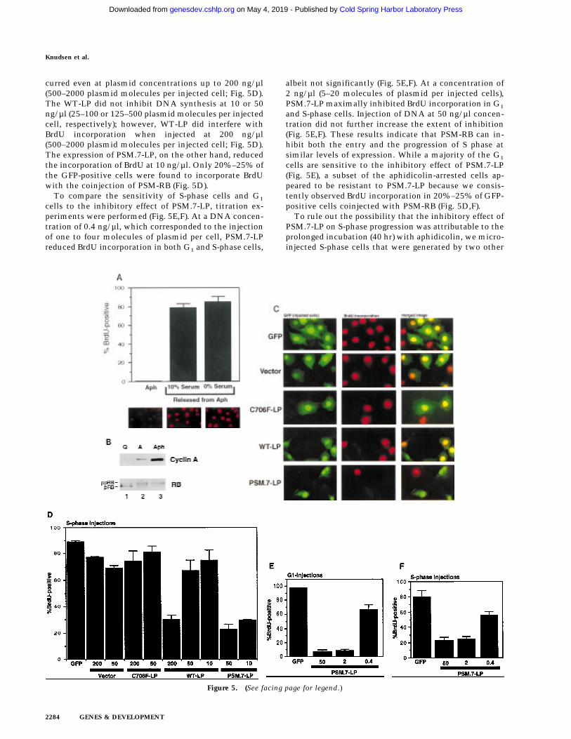
curred even at plasmid concentrations up to 200 ng/µl(500–2000 plasmid molecules per injected cell; Fig. 5D).The WT-LP did not inhibit DNA synthesis at 10 or 50ng/µl (25–100 or 125–500 plasmid molecules per injectedcell, respectively); however, WT-LP did interfere withBrdU incorporation when injected at 200 ng/µl(500–2000 plasmid molecules per injected cell; Fig. 5D).The expression of PSM.7-LP, on the other hand, reducedthe incorporation of BrdU at 10 ng/µl. Only 20%–25% ofthe GFP-positive cells were found to incorporate BrdUwith the coinjection of PSM-RB (Fig. 5D).
To compare the sensitivity of S-phase cells and G1
cells to the inhibitory effect of PSM.7-LP, titration ex-periments were performed (Fig. 5E,F). At a DNA concen-tration of 0.4 ng/µl, which corresponded to the injectionof one to four molecules of plasmid per cell, PSM.7-LPreduced BrdU incorporation in both G1 and S-phase cells,
albeit not significantly (Fig. 5E,F). At a concentration of2 ng/µl (5–20 molecules of plasmid per injected cells),PSM.7-LP maximally inhibited BrdU incorporation in G1
and S-phase cells. Injection of DNA at 50 ng/µl concen-tration did not further increase the extent of inhibition(Fig. 5E,F). These results indicate that PSM-RB can in-hibit both the entry and the progression of S phase atsimilar levels of expression. While a majority of the G1
cells are sensitive to the inhibitory effect of PSM.7-LP(Fig. 5E), a subset of the aphidicolin-arrested cells ap-peared to be resistant to PSM.7-LP because we consis-tently observed BrdU incorporation in 20%–25% of GFP-positive cells coinjected with PSM-RB (Fig. 5D,F).
To rule out the possibility that the inhibitory effect ofPSM.7-LP on S-phase progression was attributable to theprolonged incubation (40 hr) with aphidicolin, we micro-injected S-phase cells that were generated by two other
Figure 5. (See facing page for legend.)
Knudsen et al.
2284 GENES & DEVELOPMENT
Cold Spring Harbor Laboratory Press on May 4, 2019 - Published by genesdev.cshlp.orgDownloaded from
protocols with shorter periods of treatment (20 hr) or noaphidicolin treatment.
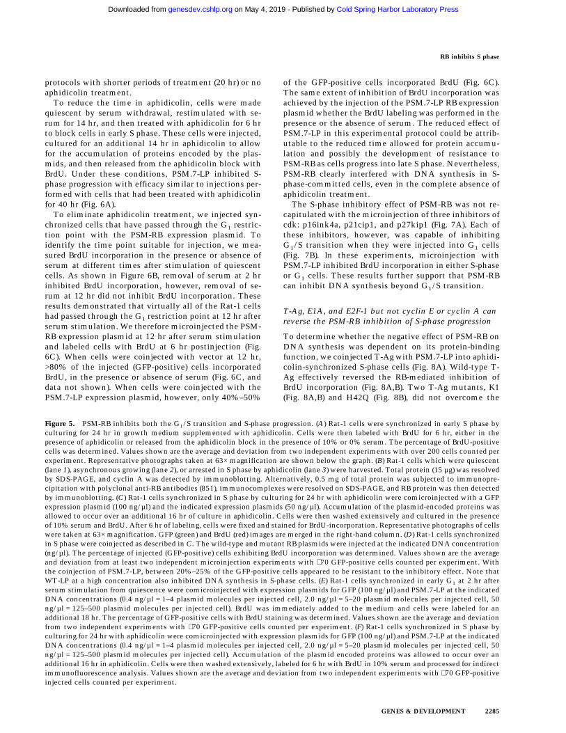
To reduce the time in aphidicolin, cells were madequiescent by serum withdrawal, restimulated with se-rum for 14 hr, and then treated with aphidicolin for 6 hrto block cells in early S phase. These cells were injected,cultured for an additional 14 hr in aphidicolin to allowfor the accumulation of proteins encoded by the plas-mids, and then released from the aphidicolin block withBrdU. Under these conditions, PSM.7-LP inhibited S-phase progression with efficacy similar to injections per-formed with cells that had been treated with aphidicolinfor 40 hr (Fig. 6A).
To eliminate aphidicolin treatment, we injected syn-chronized cells that have passed through the G1 restric-tion point with the PSM-RB expression plasmid. Toidentify the time point suitable for injection, we mea-sured BrdU incorporation in the presence or absence ofserum at different times after stimulation of quiescentcells. As shown in Figure 6B, removal of serum at 2 hrinhibited BrdU incorporation, however, removal of se-rum at 12 hr did not inhibit BrdU incorporation. Theseresults demonstrated that virtually all of the Rat-1 cellshad passed through the G1 restriction point at 12 hr afterserum stimulation. We therefore microinjected the PSM-RB expression plasmid at 12 hr after serum stimulationand labeled cells with BrdU at 6 hr postinjection (Fig.6C). When cells were coinjected with vector at 12 hr,>80% of the injected (GFP-positive) cells incorporatedBrdU, in the presence or absence of serum (Fig. 6C, anddata not shown). When cells were coinjected with thePSM.7-LP expression plasmid, however, only 40%–50%
of the GFP-positive cells incorporated BrdU (Fig. 6C).The same extent of inhibition of BrdU incorporation wasachieved by the injection of the PSM.7-LP RB expressionplasmid whether the BrdU labeling was performed in thepresence or the absence of serum. The reduced effect ofPSM.7-LP in this experimental protocol could be attrib-utable to the reduced time allowed for protein accumu-lation and possibly the development of resistance toPSM-RB as cells progress into late S phase. Nevertheless,PSM-RB clearly interfered with DNA synthesis in S-phase-committed cells, even in the complete absence ofaphidicolin treatment.
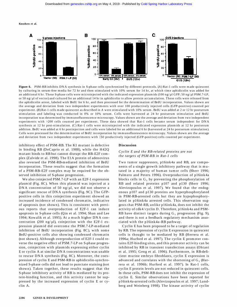
The S-phase inhibitory effect of PSM-RB was not re-capitulated with the microinjection of three inhibitors ofcdk: p16ink4a, p21cip1, and p27kip1 (Fig. 7A). Each ofthese inhibitors, however, was capable of inhibitingG1/S transition when they were injected into G1 cells(Fig. 7B). In these experiments, microinjection withPSM.7-LP inhibited BrdU incorporation in either S-phaseor G1 cells. These results further support that PSM-RBcan inhibit DNA synthesis beyond G1/S transition.
T-Ag, E1A, and E2F-1 but not cyclin E or cyclin A canreverse the PSM-RB inhibition of S-phase progression
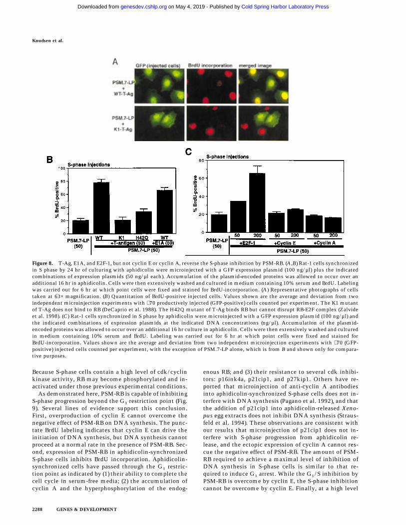
To determine whether the negative effect of PSM-RB onDNA synthesis was dependent on its protein-bindingfunction, we coinjected T-Ag with PSM.7-LP into aphidi-colin-synchronized S-phase cells (Fig. 8A). Wild-type T-Ag effectively reversed the RB-mediated inhibition ofBrdU incorporation (Fig. 8A,B). Two T-Ag mutants, K1(Fig. 8A,B) and H42Q (Fig. 8B), did not overcome the
Figure 5. PSM-RB inhibits both the G1/S transition and S-phase progression. (A) Rat-1 cells were synchronized in early S phase byculturing for 24 hr in growth medium supplemented with aphidicolin. Cells were then labeled with BrdU for 6 hr, either in thepresence of aphidicolin or released from the aphidicolin block in the presence of 10% or 0% serum. The percentage of BrdU-positivecells was determined. Values shown are the average and deviation from two independent experiments with over 200 cells counted perexperiment. Representative photographs taken at 63× magnification are shown below the graph. (B) Rat-1 cells which were quiescent(lane 1), asynchronous growing (lane 2), or arrested in S phase by aphidicolin (lane 3) were harvested. Total protein (15 µg) was resolvedby SDS-PAGE, and cyclin A was detected by immunoblotting. Alternatively, 0.5 mg of total protein was subjected to immunopre-cipitation with polyclonal anti-RB antibodies (851), immunocomplexes were resolved on SDS-PAGE, and RB protein was then detectedby immunoblotting. (C) Rat-1 cells synchronized in S phase by culturing for 24 hr with aphidicolin were comicroinjected with a GFPexpression plasmid (100 ng/µl) and the indicated expression plasmids (50 ng/µl). Accumulation of the plasmid-encoded proteins wasallowed to occur over an additional 16 hr of culture in aphidicolin. Cells were then washed extensively and cultured in the presenceof 10% serum and BrdU. After 6 hr of labeling, cells were fixed and stained for BrdU-incorporation. Representative photographs of cellswere taken at 63× magnification. GFP (green) and BrdU (red) images are merged in the right-hand column. (D) Rat-1 cells synchronizedin S phase were coinjected as described in C. The wild-type and mutant RB plasmids were injected at the indicated DNA concentration(ng/µl). The percentage of injected (GFP-positive) cells exhibiting BrdU incorporation was determined. Values shown are the averageand deviation from at least two independent microinjection experiments with ∼70 GFP-positive cells counted per experiment. Withthe coinjection of PSM.7-LP, between 20%–25% of the GFP-positive cells appeared to be resistant to the inhibitory effect. Note thatWT-LP at a high concentration also inhibited DNA synthesis in S-phase cells. (E) Rat-1 cells synchronized in early G1 at 2 hr afterserum stimulation from quiescence were comicroinjected with expression plasmids for GFP (100 ng/µl) and PSM.7-LP at the indicatedDNA concentrations (0.4 ng/µl = 1–4 plasmid molecules per injected cell, 2.0 ng/µl = 5–20 plasmid molecules per injected cell, 50ng/µl = 125–500 plasmid molecules per injected cell). BrdU was immediately added to the medium and cells were labeled for anadditional 18 hr. The percentage of GFP-positive cells with BrdU staining was determined. Values shown are the average and deviationfrom two independent experiments with ∼70 GFP-positive cells counted per experiment. (F) Rat-1 cells synchronized in S phase byculturing for 24 hr with aphidicolin were comicroinjected with expression plasmids for GFP (100 ng/µl) and PSM.7-LP at the indicatedDNA concentrations (0.4 ng/µl = 1–4 plasmid molecules per injected cell, 2.0 ng/µl = 5–20 plasmid molecules per injected cell, 50ng/µl = 125–500 plasmid molecules per injected cell). Accumulation of the plasmid encoded proteins was allowed to occur over anadditional 16 hr in aphidicolin. Cells were then washed extensively, labeled for 6 hr with BrdU in 10% serum and processed for indirectimmunofluorescence analysis. Values shown are the average and deviation from two independent experiments with ∼70 GFP-positiveinjected cells counted per experiment.
RB inhibits S phase
GENES & DEVELOPMENT 2285
Cold Spring Harbor Laboratory Press on May 4, 2019 - Published by genesdev.cshlp.orgDownloaded from
inhibitory effect of PSM-RB. The K1 mutant is defectivein binding RB (DeCaprio et al. 1988), while the H42Qmutant binds to RB but cannot disrupt the RB–E2F com-plex (Zalvide et al. 1998). The E1A protein of adenovirusalso reversed the PSM-RB-mediated inhibition of BrdUincorporation. These results suggest that the formationof a PSM-RB–E2F complex may be required for the ob-served inhibition of S-phase progression.
We also coinjected PSM.7-LP with an E2F-1 expressionplasmid (Fig. 8C). When this plasmid was injected at aDNA concentration of 50 ng/µl, we did not observe asignificant rescue of DNA synthesis (Fig. 9C). The GFP-positive cells in this coinjection experiment showed anincreased incidence of condensed chromatin, indicativeof apoptosis (not shown). This is consistent with previ-ous reports that overproduction of E2F-1 can induceapoptosis in S-phase cells (Qin et al. 1994; Shan and Lee1994; Kowalik et al. 1995). At a much higher DNA con-centration (200 ng/µl), coinjection with the E2F-1 ex-pression plasmid did overcome the PSM.7-LP-mediatedinhibition of BrdU incorporation (Fig. 8C), with someBrdU-positive cells also showing condensed chromatin(not shown). Although overproduction of E2F-1 could re-verse the negative effect of PSM.7-LP on S-phase progres-sion, coinjection with plasmids expressing either cyclinE or cyclin A at similar DNA concentrations was unableto rescue DNA synthesis (Fig. 8C). Moreover, the coex-pression of cyclin E and PSM-RB in aphidicolin-synchro-nized S-phase cells did not lead to punctate staining (notshown). Taken together, these results suggest that theS-phase inhibitory activity of RB is mediated by its pro-tein-binding function, and this activity cannot be sup-pressed by the increased expression of cyclin E or cy-clin A.
Discussion
Cyclin E and the RB-related proteins are notthe targets of PSM-RB in Rat-1 cells
Two tumor suppressors, p16ink4a and RB, are compo-nents of a single growth inhibitory pathway that is mu-tated in a majority of human tumor cells (Sherr 1996;Palmero and Peters 1996). Overproduction of p16ink4ablocks cells in G1 by preventing the phosphorylation ofRB and related proteins p107 and p130 (Sherr 1996;Alevizopoulos et al. 1997). We found that the endog-enous p107 and p130 proteins are hyperphosphorylatedin PSM-RB-arrested cells but they are hypophosphory-lated in p16ink4a arrested cells. This observation sug-gests that PSM-RB, unlike p16ink4a, does not inhibit theactivity of cdk4/cyclin D. Therefore, p16ink4a and PSM-RB have distinct targets during G1 progression (Fig. 9),and there is not a feedback regulatory mechanism asso-ciated with the p16ink4a–RB pathway.
Cyclin E has been proposed to be a target of regulationby RB. The repression of cyclin E expression in quiescentcells is thought to be mediated by RB (Herrera et al.1996a; Hurford et al. 1997). The cyclin E promoter con-tains E2F-binding sites, and this promoter activity can beinhibited by RB in transient transfection assays (Ohtaniet al. 1995; Geng et al. 1996). Furthermore, in RB-defi-cient murine embryo fibroblasts, cyclin E expression isadvanced and correlates with the shortening of G1 (Her-rera et al. 1996b; Hurford et al. 1997). In Rat-1 cells,cyclin E protein levels are not reduced in quiescent cells.In these cells, PSM-RB does not inhibit the expression ofcyclin E. Similar observations have been reported forp16ink4a-arrested cells (Alevizopoulos et al. 1997; Lund-berg and Weinberg 1998). The kinase activity of cyclin
Figure 6. PSM-RB inhibits DNA synthesis in S-phase cells synchronized by different protocols. (A) Rat-1 cells were made quiescentby culturing in serum-free media for 72 hr and then stimulated with 10% serum for 14 hr, at which time aphidicolin was added foran additional 6 hr. These S-phase cells were microinjected with the indicated expression plasmids (100 ng/µl GFP; 50 ng/µl PSM.7-LP,or 50 ng/µl of vector) and cultured for an additional 14 hr in aphidicolin to allow protein accumulation. These cells were released fromthe aphidicolin arrest, labeled with BrdU for 6 hr, and then processed for the determination of BrdU incorporation. Values shown arethe average and deviation from two independent experiments with over 100 productively injected cells (GFP-positive) counted perexperiment. (B) Rat-1 cells made quiescent as described in A were stimulated with 10% serum. BrdU was added at 2 or 12 hr postserumstimulation and labeling was conducted in 0% or 10% serum. Cells were harvested at 24 hr postserum stimulation and BrdUincorporation was determined by immunofluorescence microscopy. Values shown are the average and deviation from two independentexperiments with >200 cells counted per experiment. These data showed that Rat-1 cells became serum independent for DNAsynthesis at 12 hr post-stimulation. (C) Rat-1 cells were microinjected with the indicated expression plasmids at 12 hr postserumaddition. BrdU was added at 6 hr postinjection and cells were labeled for an additional 6 hr (harvested at 24 hr postserum stimulation).Cells were processed for the determination of BrdU incorporation by immunofluorescence microscopy. Values shown are the averageand deviation from two independent experiments with ∼50 productively injected (GFP-positive) cells counted per experiment.
Knudsen et al.
2286 GENES & DEVELOPMENT
Cold Spring Harbor Laboratory Press on May 4, 2019 - Published by genesdev.cshlp.orgDownloaded from
E–cdk2 is inhibited as Rat-1 cells exit the cell cycle(Resnitzky and Reed 1995). However, PSM-RB does notinhibit cyclin E/cdk2 kinase activity, most likely be-cause PSM-RB does not influence the levels of cdk-in-hibitors such as p21cip1 and p27kip1. Thus, cyclin Eexpression and cyclin E-associated kinase activity arenot the targets of PSM-RB in Rat-1 cells.
PSM-RB causes repression of cyclin A expressionthat can be relieved by cyclin E
The phosphorylation of RB during G1 progression coin-
cides with the traversal of the G1 restriction point, be-yond which cells are committed to DNA synthesis andbecome insensitive to positive and negative growth sig-nals (Weinberg 1995; Del Sal et al. 1996). The observa-tion that PSM-RB can induce G1 arrest supports the hy-pothesis that unphosphorylated RB prevents entry into Sphase. Our results, together with those reported byLukas et al. (1997), suggest that the overall activity ofcdk2 is rate-limiting for S-phase entry and unphosphory-lated RB interferes with the accumulation of active cdk2complexes. In Rat-1 cells, PSM-RB achieves this goal bysuppressing the expression of cyclin A (Fig. 9).
Cyclin A mRNA is down-regulated in quiescent Rat-1cells and stimulated in late G1 (Resnitzky and Reed1995; Resnitzky et al. 1995). The cyclin A promoter alsocontains E2F-binding sites that have been implicated inpromoter repression (DeGregori et al. 1996; Slansky et al.1996). A PSM-RB–E2F complex may directly bind to andrepress the cyclin A promoter in PSM-RB-arrested cells.We have shown that the coexpression of SV40 T-Ag canovercome the PSM-RB-mediated inhibition of cyclin Aexpression (Fig. 9). This is consistent with the idea thata PSM-RB–E2F complex causes the repression of cyclin Apromoter.
It is interesting to find that overproduction of cyclin Ecan also activate the expression of cyclin A in cells ex-pressing PSM-RB (Fig. 9). Lukas et al. (1997) have shownthat cyclin E cannot induce the E2F transactivating func-tion in cells expressing an unphosphorylatable RB pro-tein (RBDcdk, which is similar but not identical to PSM-RB). Thus, the activation of E2F activity is not respon-sible for the observed cyclin A expression. There are twopossible mechanisms by which the overproduction of cy-clin E may relieve the repressive function of PSM-RB.First, increased cyclin E kinase activity may inactivatethe DNA-binding function of E2F and thus remove thePSM-RB–E2F complex from the cyclin A promoter. Al-ternatively, cyclin E kinase may disrupt the transcrip-tion repression function of PSM-RB. Three recent reportshave shown that RB can recruit a histone deacetylase torepress transcription (Brehm et al. 1998; Luo et al. 1998;Magnaghi-Jaulin et al. 1998). The cyclin A promotercould conceivably be derepressed by a direct inhibitionof the histone deacetylase activity through the increasedexpression of cyclin E.
PSM-RB can inhibit DNA synthesis beyond the G1
restriction point
The previous model predicts that once a threshold levelof cdk2 activity is achieved and DNA synthesis has beeninitiated, then RB would have no further effect on cellcycle progression (Weinberg 1995). This notion is basedon two lines of evidence: First, microinjection of RB pro-tein into G1 SAOS-2 cells can inhibit DNA synthesis,but no inhibition was observed when it was injected intoS-phase SAOS-2 cells (Goodrich et al. 1991). Second,overproduction of cyclin E can overcome the effect ofp16ink4a and RBDcdk and drive the completion of thecell cycle (Alevizopoulos et al. 1997; Lukas et al. 1997).
Figure 7. S-phase inhibitory effect of PSM-RB is not observedwith cdk inhibitors. (A) Rat-1 cells arrested in S-phase by cul-turing for 24 hr with aphidicolin were microinjected with theindicated expression plasmids (GFP, 100 ng/µl; p16ink4a,p21cip1, p27kip1, or PSM,7-LP, 50 ng/µl). Following 16 hr morein aphidicolin to allow the accumulation of the plasmid-en-coded protein, cells were released from the aphidicolin blockand labeled with BrdU for 6 hr. Cells were then fixed and stainedfor BrdU incorporation. The percentage of GFP-positive cellswith BrdU staining was determined. Values shown are the av-erage and deviation from two independent experiments with∼70 GFP-positive cells counted per experiment. (B) Rat-1 cellswere synchronized in early G1 by a 2-hr stimulation of quies-cent cells with 10% serum. These early G1 cells were microin-jected with the indicated expression plasmids (GFP, 100 ng/µl;p16ink4a, p21cip1, p27kip1, or PSM,7-LP, 50 ng/µl). BrdU wasimmediately added to the media, cells were labeled continu-ously for 18 hr and then processed for immunofluorescenceanalysis. Values shown are the average and deviation from twoindependent experiments with ∼70 productively injected cells(GFP-positive) counted per experiment.
RB inhibits S phase
GENES & DEVELOPMENT 2287
Cold Spring Harbor Laboratory Press on May 4, 2019 - Published by genesdev.cshlp.orgDownloaded from
Because S-phase cells contain a high level of cdk/cyclinkinase activity, RB may become phosphorylated and in-activated under those previous experimental conditions.
As demonstrated here, PSM-RB is capable of inhibitingS-phase progression beyond the G1 restriction point (Fig.9). Several lines of evidence support this conclusion.First, overproduction of cyclin E cannot overcome thenegative effect of PSM-RB on DNA synthesis. The punc-tate BrdU labeling indicates that cyclin E can drive theinitiation of DNA synthesis, but DNA synthesis cannotproceed at a normal rate in the presence of PSM-RB. Sec-ond, expression of PSM-RB in aphidicolin-synchronizedS-phase cells inhibits BrdU incorporation. Aphidicolin-synchronized cells have passed through the G1 restric-tion point as indicated by (1) their ability to complete thecell cycle in serum-free media; (2) the accumulation ofcyclin A and the hyperphosphorylation of the endog-
enous RB; and (3) their resistance to several cdk inhibi-tors: p16ink4a, p21cip1, and p27kip1. Others have re-ported that microinjection of anti-cyclin A antibodiesinto aphidicolin-synchronized S-phase cells does not in-terfere with DNA synthesis (Pagano et al. 1992), and thatthe addition of p21cip1 into aphidicolin-released Xeno-pus egg extracts does not inhibit DNA synthesis (Straus-feld et al. 1994). These observations are consistent withour results that microinjection of p21cip1 does not in-terfere with S-phase progression from aphidicolin re-lease, and the ectopic expression of cyclin A cannot res-cue the negative effect of PSM-RB. The amount of PSM-RB required to achieve a maximal level of inhibition ofDNA synthesis in S-phase cells is similar to that re-quired to induce G1 arrest. While the G1/S inhibition byPSM-RB is overcome by cyclin E, the S-phase inhibitioncannot be overcome by cyclin E. Finally, at a high level
Figure 8. T-Ag, E1A, and E2F-1, but not cyclin E or cyclin A, reverse the S-phase inhibition by PSM-RB. (A,B) Rat-1 cells synchronizedin S phase by 24 hr of culturing with aphidicolin were microinjected with a GFP expression plasmid (100 ng/µl) plus the indicatedcombinations of expression plasmids (50 ng/µl each). Accumulation of the plasmid-encoded proteins was allowed to occur over anadditional 16 hr in aphidicolin. Cells were then extensively washed and cultured in medium containing 10% serum and BrdU. Labelingwas carried out for 6 hr at which point cells were fixed and stained for BrdU-incorporation. (A) Representative photographs of cellstaken at 63× magnification. (B) Quantitation of BrdU-positive injected cells. Values shown are the average and deviation from twoindependent microinjection experiments with ∼70 productively injected (GFP-positive) cells counted per experiment. The K1 mutantof T-Ag does not bind to RB (DeCaprio et al. 1988). The H42Q mutant of T-Ag binds RB but cannot disrupt RB-E2F complex (Zalvideet al. 1998). (C) Rat-1 cells synchronized in S phase by aphidicolin were microinjected with a GFP expression plasmid (100 ng/µl) andthe indicated combinations of expression plasmids at the indicated DNA concentrations (ng/µl). Accumulation of the plasmid-encoded proteins was allowed to occur over an additional 16 hr culture in aphidicolin. Cells were then extensively washed and culturedin medium containing 10% serum and BrdU. Labeling was carried out for 6 hr at which point cells were fixed and stained forBrdU-incorporation. Values shown are the average and deviation from two independent microinjection experiments with ∼70 (GFP-positive) injected cells counted per experiment, with the exception of PSM.7-LP alone, which is from B and shown only for compara-tive purposes.
Knudsen et al.
2288 GENES & DEVELOPMENT
Cold Spring Harbor Laboratory Press on May 4, 2019 - Published by genesdev.cshlp.orgDownloaded from
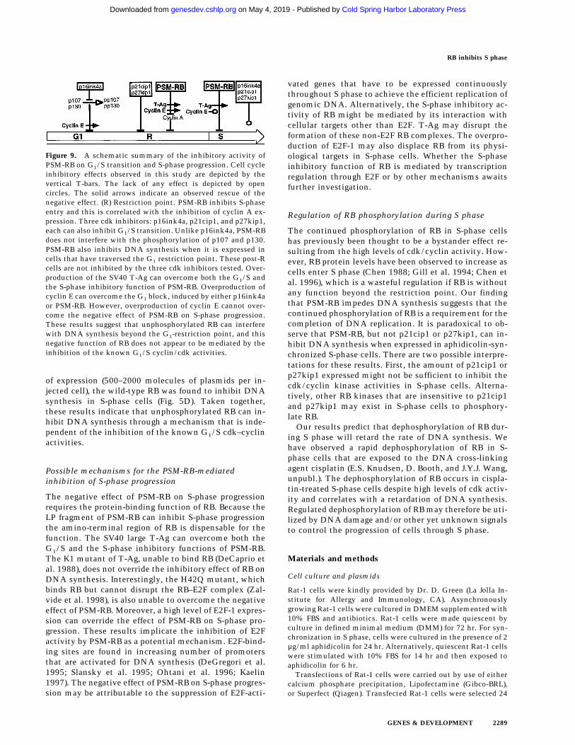
of expression (500–2000 molecules of plasmids per in-jected cell), the wild-type RB was found to inhibit DNAsynthesis in S-phase cells (Fig. 5D). Taken together,these results indicate that unphosphorylated RB can in-hibit DNA synthesis through a mechanism that is inde-pendent of the inhibition of the known G1/S cdk–cyclinactivities.
Possible mechanisms for the PSM-RB-mediatedinhibition of S-phase progression
The negative effect of PSM-RB on S-phase progressionrequires the protein-binding function of RB. Because theLP fragment of PSM-RB can inhibit S-phase progressionthe amino-terminal region of RB is dispensable for thefunction. The SV40 large T-Ag can overcome both theG1/S and the S-phase inhibitory functions of PSM-RB.The K1 mutant of T-Ag, unable to bind RB (DeCaprio etal. 1988), does not override the inhibitory effect of RB onDNA synthesis. Interestingly, the H42Q mutant, whichbinds RB but cannot disrupt the RB–E2F complex (Zal-vide et al. 1998), is also unable to overcome the negativeeffect of PSM-RB. Moreover, a high level of E2F-1 expres-sion can override the effect of PSM-RB on S-phase pro-gression. These results implicate the inhibition of E2Factivity by PSM-RB as a potential mechanism. E2F-bind-ing sites are found in increasing number of promotersthat are activated for DNA synthesis (DeGregori et al.1995; Slansky et al. 1995; Ohtani et al. 1996; Kaelin1997). The negative effect of PSM-RB on S-phase progres-sion may be attributable to the suppression of E2F-acti-
vated genes that have to be expressed continuouslythroughout S phase to achieve the efficient replication ofgenomic DNA. Alternatively, the S-phase inhibitory ac-tivity of RB might be mediated by its interaction withcellular targets other than E2F. T-Ag may disrupt theformation of these non-E2F RB complexes. The overpro-duction of E2F-1 may also displace RB from its physi-ological targets in S-phase cells. Whether the S-phaseinhibitory function of RB is mediated by transcriptionregulation through E2F or by other mechanisms awaitsfurther investigation.
Regulation of RB phosphorylation during S phase
The continued phosphorylation of RB in S-phase cellshas previously been thought to be a bystander effect re-sulting from the high levels of cdk/cyclin activity. How-ever, RB protein levels have been observed to increase ascells enter S phase (Chen 1988; Gill et al. 1994; Chen etal. 1996), which is a wasteful regulation if RB is withoutany function beyond the restriction point. Our findingthat PSM-RB impedes DNA synthesis suggests that thecontinued phosphorylation of RB is a requirement for thecompletion of DNA replication. It is paradoxical to ob-serve that PSM-RB, but not p21cip1 or p27kip1, can in-hibit DNA synthesis when expressed in aphidicolin-syn-chronized S-phase cells. There are two possible interpre-tations for these results. First, the amount of p21cip1 orp27kip1 expressed might not be sufficient to inhibit thecdk/cyclin kinase activities in S-phase cells. Alterna-tively, other RB kinases that are insensitive to p21cip1and p27kip1 may exist in S-phase cells to phosphory-late RB.
Our results predict that dephosphorylation of RB dur-ing S phase will retard the rate of DNA synthesis. Wehave observed a rapid dephosphorylation of RB in S-phase cells that are exposed to the DNA cross-linkingagent cisplatin (E.S. Knudsen, D. Booth, and J.Y.J. Wang,unpubl.). The dephosphorylation of RB occurs in cispla-tin-treated S-phase cells despite high levels of cdk activ-ity and correlates with a retardation of DNA synthesis.Regulated dephosphorylation of RB may therefore be uti-lized by DNA damage and/or other yet unknown signalsto control the progression of cells through S phase.
Materials and methods
Cell culture and plasmids
Rat-1 cells were kindly provided by Dr. D. Green (La Jolla In-stitute for Allergy and Immunology, CA). Asynchronouslygrowing Rat-1 cells were cultured in DMEM supplemented with10% FBS and antibiotics. Rat-1 cells were made quiescent byculture in defined minimal medium (DMM) for 72 hr. For syn-chronization in S phase, cells were cultured in the presence of 2µg/ml aphidicolin for 24 hr. Alternatively, quiescent Rat-1 cellswere stimulated with 10% FBS for 14 hr and then exposed toaphidicolin for 6 hr.
Transfections of Rat-1 cells were carried out by use of eithercalcium phosphate precipitation, Lipofectamine (Gibco-BRL),or Superfect (Qiagen). Transfected Rat-1 cells were selected 24
Figure 9. A schematic summary of the inhibitory activity ofPSM-RB on G1/S transition and S-phase progression. Cell cycleinhibitory effects observed in this study are depicted by thevertical T-bars. The lack of any effect is depicted by opencircles. The solid arrows indicate an observed rescue of thenegative effect. (R) Restriction point. PSM-RB inhibits S-phaseentry and this is correlated with the inhibition of cyclin A ex-pression. Three cdk inhibitors: p16ink4a, p21cip1, and p27kip1,each can also inhibit G1/S transition. Unlike p16ink4a, PSM-RBdoes not interfere with the phosphorylation of p107 and p130.PSM-RB also inhibits DNA synthesis when it is expressed incells that have traversed the G1 restriction point. These post-Rcells are not inhibited by the three cdk inhibitors tested. Over-production of the SV40 T-Ag can overcome both the G1/S andthe S-phase inhibitory function of PSM-RB. Overproduction ofcyclin E can overcome the G1 block, induced by either p16ink4aor PSM-RB. However, overproduction of cyclin E cannot over-come the negative effect of PSM-RB on S-phase progression.These results suggest that unphosphorylated RB can interferewith DNA synthesis beyond the G1-restriction point, and thisnegative function of RB does not appear to be mediated by theinhibition of the known G1/S cyclin/cdk activities.
RB inhibits S phase
GENES & DEVELOPMENT 2289
Cold Spring Harbor Laboratory Press on May 4, 2019 - Published by genesdev.cshlp.orgDownloaded from
hr post-transfection with 2.5 µg/ml puromycin (Sigma) for 72hr, and either harvested for analysis of protein or cultured for anadditional 96 hr and stained with crystal violet as describedpreviously (Qin et al. 1992).
The WT-LP, C706F-LP, PSM.7-LP, PSM.9I-RB, and CMV-NEO vectors have been described previously (Knudsen andWang 1997). The puromycin-resistance plasmid, pBABE-PUROwas kindly provided by Dr. H. Land (Imperial Cancer ResearchFund, London, UK). The p16ink4a expression plasmid has beendescribed previously (Knudsen and Wang 1997). The cyclin Eand cyclin A plasmids have been described previously (Knudsenand Wang 1996, 1997). The T-Ag expression plasmids were ob-tained from Dr. J. DeCaprio (Dana-Farber Cancer Institute, Bos-ton, MA) and Dr. S. Subramani (UCSD). The GFP-expressionplasmid, Green Lantern, is commercially available (GIBCO-BRL).
Microinjection, immunofluorescence, and flow cytometry
Cells were microinjected with glass capillary needles, madewith a Kopf vertical pipette puller. The plasmids were coin-jected at the concentration of 100 ng/µl GFP and 0.4–200 ng/µlof the effector plasmid. Injections were carried out with an Ep-pendorf microinjector. All plasmids utilized for microinjectionwere doubly purified to remove any impurities. Plasmids wereoriginally purified either by cesium chloride banding or QiagenMaxi-Prep kits. The resulting plasmid DNA (50 µg) was thenrepurified over a Qiagen Midi-column. Plasmid concentrationwas determined photometrically, and the integrity of all plas-mids was determined by visual analysis following agarose gelelectrophoresis. Injected cells were grown on acid-washed glasscoverslips. The approximate number of plasmid molecules in-troduced into the injected cells was determined on the basis ofpreviously reported injection volumes (Capecchi 1980; Huanget al. 1996). For the plasmids utilized in this study the approxi-mate number of plasmid molecules per injected cell is given asfollow: 0.4 ng/µl = 1–4 plasmid molecules per injected cells; 2.0ng/µl = 5–20 plasmid molecules per injected cell; 10 ng/µl = 25–100 plasmid molecules per injected cell; 50 ng/µl = 125–500plasmid molecules per injected cell; 100 ng/µl = 250–1000 plas-mid molecules per injected cell; 200 ng/µl = 500–2000 plasmidmolecules per injected cells.
Several different culture conditions were used for the injec-tion of S-phase cells. First, Rat-1 cells that had been cultured for24 hr in aphidicolin-containing media were used. The injectedcells were cultured for an additional 16 hr in aphidicolin toallow for the accumulation of the plasmid-encoded protein.Cells were washed extensively to remove the aphidicolin andBrdU was added immediately to the medium. Cells were thenfixed following a 6 hr labeling period. Alternatively, Rat-1 cellswere rendered quiescent via culture in DMM for 72 hr. Cellswere stimulated with serum for 14 hr and then cultured in thepresence of aphidicolin for and additional 6 hr. These S-phasecells were subjected to microinjection and then cultured an ad-ditional 14 hr in aphidicolin. These cells were then releasedfrom the aphidicolin arrest in the presence of BrdU, fixed, andstained following 6 hr of labeling. Finally, quiescent cells thathad been serum stimulated for 12 hr were subjected to micro-injection, and BrdU was added to the cells 6 hr post-injection.Cells were fixed after 6 hr of labeling and then stained for theincorporation of BrdU.
For the injection of early G1 cells, Rat-1 cells that had beenrendered quiescent by culture in DMM for 72 hr were releasedinto medium containing 10% FBS for 2 hr and then microin-jected. BrdU was added immediately following injection. Cellswere then fixed after 18 hr in the presence of BrdU.
Following microinjection and labeling with BrdU to measureDNA synthesis, cells were fixed in 3.7% formaldehyde in PBSfor 15 min and then permeabilized in 0.3% Triton X-100 in PBSfor 10 min. Primary antibody staining (Accurate Scientific, 1:500 dilution) was carried out in PBS supplemented with 5 mg/ml BSA and 0.5% NP-40, 10 mM MgCl2, and 100 U/µl DNase 1for 1 hr at 37°C in a humidified chamber. The coverslips werethen washed in PBS. Secondary antibody (Jackson Laboratories,1:100 dilution) was diluted and incubated in PBS supplementedwith 5mg/ml BSA and 0.5% NP-40. Coverslips were washedagain in PBS, and mounted on glass slides.
Fluorescent microscopy was performed with a Zeiss AxiophotMicroscope with either 40× or 63× objective lenses. Photo-graphs of stained cells were recorded with a Hamamatsu CCDcamera. Digital images were printed on a Mitsubishi color sub-limation printer.
For flow cytometry, cells were transfected with a CD20 cellsurface marker and the indicated expression plasmids. Forty-eight hours post-transfection, cells were harvested by trypsin-ization (trypsin does not effect the staining for CD20). Cellswere stained for the expression of CD20 with an FITC conju-gated anti-CD20 antibody (PharMingen). These cells were thenfixed in 80% ethanol and stained with propidium iodide. TheDNA content of CD20-positive and negative cells was deter-mined by use of a FACSCAN cytometer equipped with cellfitsoftware (Becton-Dickinson).
Quantitative RT–PCR
Total cellular RNA was isolated with Trizol reagent (GIBCO-BRL) according to manufacturer’s instruction. First-strandcDNAs were synthesized from 2 µg of DNase I-treated totalRNA by use of SuperScript II reverse transcriptase (Life Tech-nology, GIBCO-BRL) and random antisense primers accordingto the manufacturer’s instruction. Diluted cDNAs were sub-jected to PCR amplifications with two sets of primers specificfor glyceraldehyde 3-phosphate dehydrogenase (GAPDH) (GGT-CATCAATGGGAAACCCA TCAC and TGATGGCATGGAC-TGTGGTCATGA) and cyclin A (AGACCCTGCATTTG-GCTGTG and ACAAACTCTGCTACTTCTGG). In all PCR re-actions, the GAPDH primers were added to the reaction mix-ture four cycles later than the cyclin A primers. PCR-amplifiedproducts were separated on a 1% agarose gel and analyzed bySouthern blotting with 32P-labeled GAPDH and cyclin AcDNAs as probes. The expression levels of GAPDH and cyclinA were quantitated with a PhosphoImager (Molecular Dynam-ics) from five PCR reactions that were performed in the linearrange. The relative expression levels of cyclin A were normal-ized to the expression levels of GAPDH from the given PCRreaction, and the cyclin A levels from asynchronous growingcells were arbitrarily set to 100%.
Antibodies, immunoblotting, and kinase reactions
Cyclin D1 was detected with a polyclonal antibody kindly pro-vided by Dr. S.I. Reed (Scripps Research Institute, La Jolla, CA).Cyclin E was detected with monoclonal antibody from PharM-ingen. Cyclin A was detected with a polyclonal antibody kindlyprovided by Dr. T. Hunter (Salk Institute, La Jolla, CA). CyclinB was detected with a monoclonal antibody from PharMingen.Cdk2 protein was detected with polyclonal antibody providedby Dr. M. Pagano (Mitotix, Cambridge, MA) or from Santa CruzScientific. The Cdc2 and RB antibodies have been previouslydescribed (Welch and Wang 1993). p107 and p130 antibodieswere obtained from Santa Cruz Scientific. The p21cip1 antibodywas from PharMingen, and p27kip1 antibody was kindly pro-
Knudsen et al.
2290 GENES & DEVELOPMENT
Cold Spring Harbor Laboratory Press on May 4, 2019 - Published by genesdev.cshlp.orgDownloaded from
vided by Dr. J. Roberts (Fred Hutchinson Cancer Research Cen-ter). Immunoblotting was carried out by use of standard proce-dures and Immobilon P membranes.
Kinase reactions were carried out essentially as described(Knudsen and Wang 1996), with the exception that only 20 µg oftotal cell lysate from the indicated cells was utilized per kinasereaction. The kinase complexes were isolated via immunopre-cipitation with the indicated antibodies and protein A–Sepha-rose and then used to phosphorylate histone H1 as a substrate.The nonspecific rabbit anti-mouse antibody was obtained fromCappell.
Acknowledgments
We thank Dr. Karen E. Knudsen for critical reading of the manu-script and also thank all the members of the Wang and Feram-isco laboratories for thoughtful discussions and/or technical as-sistance. We are also grateful to Dr. Tony Hunter, Dr. James A.DeCaprio, Dr. Steven I. Reed, Dr. Jim Roberts, Dr. WebsterCavenee, and Dr. Doug Green for helpful discussion and/or pro-vision of reagents. This work was supported by a grant toJ.Y.J.W. (CA58320) from the National Institutes of Health(NIH), and a grant to J.R.F. from the California Tobacco RelatedDisease Research Program. E.S.K. is supported by a traininggrant to the UCSD Cancer Center from National Cancer Insti-tute–NIH (T32CA09290).
The publication costs of this article were defrayed in part bypayment of page charges. This article must therefore be herebymarked ‘‘advertisement’’ in accordance with 18 USC section1734 solely to indicate this fact.
References
Akiyama, T., T. Ohuchi, S. Sumida, K. Matsumoto, and K.Toyoshima. 1992. Phosphorylation of the retinoblastomaprotein by cdk2. Proc. Natl. Acad. Sci. 89: 7900–7904.
Alevizopoulos, K., J. Vlach, S. Hennecke, and B. Amati. 1997.Cyclin E and c-Myc promote cell proliferation in the pres-ence of p16(INK4a) and of hypophosphorylated retinoblasto-ma family proteins. EMBO J. 16: 5322–5333.
Bartek, J., J. Bartkova, and J. Lukas. 1997. The retinoblastomaprotein pathway in cell cycle control and cancer. Exp. CellRes. 237: 1–6.
Beijersbergen, R.L. and R. Bernards. 1996. Cell cycle regulationby the retinoblastoma family of growth inhibitor proteins.Biochim. Biophys. Acta. 1287: 103–120.
Brehm, A., E.A. Miska, D.J. McCance, J.L. Reid, A.J. Bannister,and T. Kouzarides. 1998. Retinoblastoma protein recruitshistone deacetylase to repress transcription. Nature391: 597–601.
Capecchi, M.R. 1980. High efficiency transformation by directmicroinjection of DNA into cultured mammalian cells. Cell2: 479–488.
Chen, P.L., P. Scully, J.Y. Shew, J.Y.J. Wang, and W.H. Lee. 1988.Phosphorylation of the retinoblastoma gene product ismodulated during the cell cycle and cellular differentiation.Cell 58: 1193–1198.
Chen, Y., E.S. Knudsen, and J.Y.J. Wang. 1996. Cells arrested inG1 by the v-Abl tyrosine kinase do not express cyclin Adespite the hyperphosphorylation of RB. J. Biol. Chem.271: 19637–19640.
Connell-Crowley, L., J.W. Harper, and D.W. Goodrich. 1997.Cyclin D1/Cdk4 regulates retinoblastoma protein-mediatedcell cycle arrest by site-specific phosphorylation. Mol. Biol.Cell 8: 287–301.
DeCaprio, J.A., J.W. Ludlow, J. Figge, J.Y. Shew, C.M. Huang,W.H. Lee, E. Marsilio, E. Paucha, and D.M. Livingston. 1988.SV40 large tumor antigen forms a specific complex with theproduct of the retinoblastoma susceptibility gene. Cell54: 275–283.
DeGregori, J., T. Kowalik, and J.R. Nevins. 1995. Cellular tar-gets for activation by the E2F1 transcription factor includeDNA synthesis- and G1/S-regulatory genes. Mol. Cell. Biol.15: 4215–4224.
DelSal, G., M. Loda, and M. Pagano. 1996. Cell cycle and cancer:Critical events at the G1 restriction point. Crit. Rev. Onco-genesis 7: 127–142.
Geng, Y., E.N. Eaton, M. Picon, J.M. Roberts, A.S. Lundberg, A.Gifford, C. Sardet, and R.A. Weinberg. 1996. Regulation ofcyclin E transcription by E2Fs and retinoblastoma protein.Oncogene 12: 1173–1180.
Gill, R.M., P.A. Hamel, J. Zhe, E. Zacksenhaus, B.L. Gallie, andR.A. Phillips. 1994. Characterization of the human RB1 pro-moter and of elements involved in transcriptional regula-tion. Cell Growth Differ. 5: 467–474.
Goodrich, D.W., N.P. Wang, Y.W. Qian, E.Y.H.P. Lee, and W.H.Lee. 1991. The retinoblastoma gene product regulates pro-gression through the G1 phase of the cell cycle. Cell 67: 293–302.
Hamel, P.A., R.M. Gill, R.A. Phillips, and B.L. Gallie. 1992.Regions controlling hyperphosphorylation and conforma-tion of the retinoblastoma gene product are independent ofdomains required for transcriptional repression. Oncogene7: 693–701.
Hamel, P.A., R.A. Phillips, M. Muncaster, and B.L. Gallie. 1993.Speculations on the roles of RB1 in tissue-specific differen-tiation, tumor initiation, and tumor progression. FASEB J.7: 846–854.
Herrera, R.E., T.P. Makela, and R.A. Weinberg. 1996a. TGFbeta-induced growth inhibition in primary fibroblasts re-quires the retinoblastoma protein. Mol. Biol. Cell 7: 1335–1342.
Herrera, R.E., V.P. Sah, B.O. Williams, T.P. Makela, R.A. Wein-berg, and T. Jacks. 1996b. Altered cell cycle kinetics, geneexpression, and G1 restriction point regulation in Rb-defi-cient fibroblasts. Mol. Cell. Biol. 16: 2402–2407.
Huang, L.C., K.C. Clarkin, and G.M. Wahl. 1996. Sensitivityand selectivity of the DNA damage sensor responsible foractivating p53-dependent G1 arrest. Proc. Natl. Acad. Sci.93: 4827–4832.
Hunter, T. and J. Pines. 1994. Cyclins and cancer. II: Cyclin Dand CDK inhibitors come of age. Cell 79: 573–582.
Hurford, R.K., Jr., D. Cobrinik, M.H. Lee, and N. Dyson. 1997.pRB and p107/p130 are required for the regulated expressionof different sets of E2F responsive genes. Genes & Dev.11: 1447–1463.
Kaelin, W.G., Jr. 1997. Recent insights into the functions of theretinoblastoma susceptibility gene product. Cancer Invest.15: 243–254.
Knudsen, E.S. and J.Y.J. Wang. 1996. Differential regulation ofretinoblastoma protein function by specific Cdk phosphory-lation sites. J. Biol. Chem. 271: 8313–8320.
———. 1997. Dual mechanisms for the inhibition of E2F bind-ing to RB by cyclin-dependent kinase-mediated RB phos-phorylation. Mol. Cell. Biol. 17: 5771–5783.
Koh, J., G.H. Enders, B.D. Dynlacht, and E. Harlow. 1995. Tu-mour-derived p16 alleles encoding proteins defective in cell-cycle inhibition. Nature 375: 506–510.
Kowalik, T.F., J. DeGregori, J.K. Schwarz, and J.R. Nevins. 1995.E2F1 overexpression in quiescent fibroblasts leads to induc-tion of cellular DNA synthesis and apoptosis. J. Virol.
RB inhibits S phase
GENES & DEVELOPMENT 2291
Cold Spring Harbor Laboratory Press on May 4, 2019 - Published by genesdev.cshlp.orgDownloaded from

69: 2491–2500.Lees, J.A., K.J. Buchkovich, D.R. Marshak, C.W. Anderson, and
E. Harlow. 1991. The retinoblastoma protein is phosphory-lated on multiple sites by human cdc2. EMBO J. 10: 4279–4290.
Lin, B.T., S. Gruenwald, A.O. Morla, W.H. Lee, and J.Y.J. Wang.1991. Retinoblastoma cancer suppressor gene product is asubstrate of the cell cycle regulator cdc2 kinase. EMBO J.10: 857–864.
Lukas, J., D. Parry, L. Aagaard, D.J. Mann, J. Bartkova, M.Strauss, G. Peters, and J. Bartek. 1995. Retinoblastoma-pro-tein-dependent cell-cycle inhibition by the tumour suppres-sor p16. Nature 375: 503–506.
Lukas, J., T. Herzinger, K. Hansen, M.C. Moroni, D. Resnitzky,K. Helin, S.I. Reed, and J. Bartek. 1997. Cyclin E-inducedS-phase without activation of the pRb/E2F pathway. Genes& Dev. 11: 1479–1492.
Lundberg, A.S. and R.A. Weinberg. 1998. Functional inactiva-tion of the retinoblastoma protein requires sequential modi-fication by at least two distinct cyclin-cdk complexes. Mol.Cell. Biol. 18: 753–761.
Luo, R.X., A.A. Postigo, and D.C. Dean. 1998. Rb interacts withhistone deacetylase to repress transcription. Cell 92: 463–473.
Magnaghi-Jaulin, L., R. Groisman, I. Naguibneva, P. Robin, S.Lorain, J.P. Le Villain, F. Troalen, D. Trouche, and A. Harel-Bellan. 1998. Retinoblastoma protein represses transcriptionby recruiting a histone deacetylase. Nature 391: 601–605.
Matsushime, H., D.E. Quelle, S.A. Shurtleff, M. Shibuya, C.J.Sherr, and J.Y. Kato. 1994. D-type cyclin-dependent kinaseactivity in mammalian cells. Mol. Cell. Biol. 14: 2066–2076.
Medema, R.H., R.E. Herrera, F. Lam, and R.A. Weinberg. 1995.Growth suppression by p16ink4 requires functional retino-blastoma protein. Proc. Natl. Acad. Sci. 92: 6289–6293.
Meyerson, M. and E. Harlow. 1994. Identification of G1 kinaseactivity for cdk6, a novel cyclin D partner. Mol. Cell. Biol.14: 2077–2086.
Ohtani, K., J. DeGregori, and J.R. Nevins. 1995. Regulation ofthe cyclin E gene by transcription factor E2F1. Proc. Natl.Acad. Sci. 92: 12146–12150.
Ohtani, K., J. DeGregori, G. Leone, D.R. Herendeen, T.J. Kelly,and J.R. Nevins. 1996. Expression of the HsOrc1 gene, a hu-man ORC1 homolog, is regulated by cell proliferation via theE2F transcription factor. Mol. Cell. Biol. 16: 6977–6984.
Pagano, M., R. Pepperkok, F. Verde, W. Ansorge, and G. Draetta.1992. Cyclin A is required at two points in the human cellcycle. EMBO J. 11: 961–971.
Palmero, I. and G. Peters. 1996. Perturbation of cell cycle regu-lators in human cancer. Cancer Surv. 27: 351–367.
Qin, X.Q., T. Chittenden, D.M. Livingston, and W.G. Kaelin, Jr.1992. Identification of a growth suppression domain withinthe retinoblastoma gene product. Genes & Dev. 6: 953–964.
Qin, X.Q., D.M. Livingston, W.G. Kaelin, Jr., and P.D. Adams.1994. Deregulated transcription factor E2F-1 expressionleads to S-phase entry and p53-mediated apoptosis. Proc.Natl. Acad. Sci. 91: 10918–10922.
Resnitzky, D. and S.I. Reed. 1995. Different roles for cyclins D1and E in regulation of the G1-to-S transition. Mol. Cell. Biol.15: 3463–3469.
Resnitzky, D., L. Hengst, and S.I. Reed. 1995. Cyclin A-associ-ated kinase activity is rate limiting for entrance into S-phaseand is negatively regulated in G1 by p27Kip1. Mol. Cell. Biol.15: 4347–4352.
Shan, B. and W.H. Lee. 1994. Deregulated expression of E2F-1induces S-phase entry and leads to apoptosis. Mol. Cell. Biol.14: 8166–8173.
Sherr, C.J. 1996. Cancer cell cycles. Science 274: 1672–1677.Sidle, A., C. Palaty, P. Dirks, O. Wiggan, M. Kiess, R.M. Gill,
A.K. Wong, and P.A. Hamel. 1996. Activity of the retinoblas-toma family proteins, pRB, p107, and p130, during cellularproliferation and differentiation. Crit. Rev. Biochem. Mol.Biol. 31: 237–271.
Slansky, J.E. and P.J. Farnham. 1996. Introduction to the E2Ffamily: Protein structure and gene regulation. Curr. Top. Mi-crobiol. Immunol. 208: 1–30.
Strausfeld, U.P., M. Howell, R. Rempel, J.L. Maller, T. Hunt,and J.J. Blow. 1994. Cip1 blocks the initiation of DNA rep-lication in Xenopus extracts by inhibition of cyclin-depen-dent kinases. Curr. Biol. 4: 876–883.
Wang, J.Y.J., E.S. Knudsen, and P.J. Welch. 1994. The retinoblas-toma tumor suppressor protein. Adv. Cancer Res. 64: 25–85.
Weinberg, R.A. 1995. The retinoblastoma protein and cell cyclecontrol. Cell 81: 323–330.
Welch, P.J. and J.Y.J. Wang. 1993. A C-terminal protein-bindingdomain in the retinoblastoma protein regulates nuclear c-Abl tyrosine kinase in the cell cycle. Cell 75: 779–790.
Zalvide, J., H. Stubdal, and J.A. DeCaprio. 1998. The J domain ofsimian virus 40 large T antigen is required to functionallyinactivate RB family proteins. Mol. Cell. Biol.18: 1408–1415.
Zarkowska, T. and S. Mittnacht. 1997. Differential phosphory-lation of the retinoblastoma protein by G(1)/S cyclin-depen-dent kinases. J. Biol. Chem. 272: 12738–12746.
Knudsen et al.
2292 GENES & DEVELOPMENT
Cold Spring Harbor Laboratory Press on May 4, 2019 - Published by genesdev.cshlp.orgDownloaded from
10.1101/gad.12.15.2278Access the most recent version at doi: 12:1998, Genes Dev.
Erik S. Knudsen, Carolan Buckmaster, Tung-Ti Chen, et al.
progression S-phase/S transition and1Inhibition of DNA synthesis by RB: effects on G
References
http://genesdev.cshlp.org/content/12/15/2278.full.html#ref-list-1
This article cites 54 articles, 28 of which can be accessed free at:
License
ServiceEmail Alerting
click here.right corner of the article or
Receive free email alerts when new articles cite this article - sign up in the box at the top
Cold Spring Harbor Laboratory Press
Cold Spring Harbor Laboratory Press on May 4, 2019 - Published by genesdev.cshlp.orgDownloaded from