Embed Size (px)
Citation preview
1
VERKSAMHETSOMRÅDE KARDIOLOGI
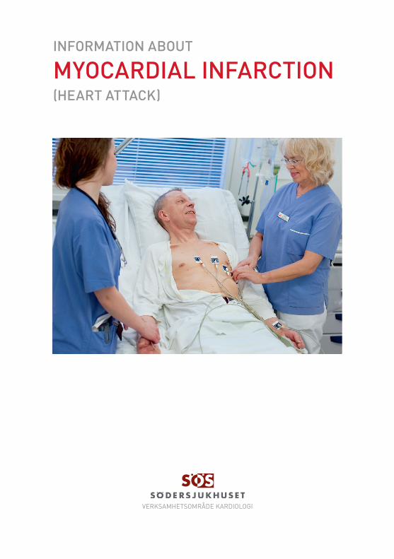
INFORMATION ABOUT
(HEART ATTACK)
MYOCARDIAL INFARCTION

2
What is a myocardial infarction?
Myocardial infarction (MI) means that part of the heart muscle suddenly loses its blood supply. Without prompt treatment, this can lead to damage to the affected part of the heart. An MI is part of a range or disorders called ’acute coronary syndrome’ (ACS).
Understanding the heart and coronary arteriesThe heart is mainly made of special muscle. The hearts left side pumps blood into arteries (blood vessels) to the head and body whereas the hearts right side pumps blood to the lungs.
Like any other muscle, the heart muscle needs a good blood supply. The coronary arteries take blood to the heart muscle. There are two main coronary arteries (left/right) that branch off from the aorta. (The aorta is the large artery which takes oxygen-rich blood from the left heart chamber to the body.) The main coronary arteries divide into smaller branches which take blood to all parts of the heart muscle.
What happens when you have a myocardial infarction?If you have an MI, a coronary artery or one of its smaller branches is suddenly blocked. The part of the heart muscle supplied by this artery loses its blood (and oxygen) supply bringing it at risk of dying unless the blockage is quickly removed (the word ’infarction’ means death of some tissue). If one of the main coronary arteries is blocked, a large part of the heart muscle is threatened. If a smaller branch artery is blocked, a smaller amount of heart muscle is affected. In people who survive an MI, the part of the heart muscle that dies (’infarcts’) is replaced by scar tissue over the next few weeks to months which can cause future problems such as heart failure or arrhythmia.

3
What causes myocardial infarction?
Thrombosis - the most common causeThe common cause of an MI is a blood clot (thrombosis) that forms inside a coronary artery, or one of its branches and blocks the blood flow to a part of the heart.
Blood clots do not usually form in normal arteries. However, a clot may form if there is some atheroma within the lining of the artery. Atheroma is the technical term for fatty patches or ’plaques’ (This is similar to water pipes that get ’furred up’). Plaques of atheroma may gradually form over a number of years in one or more places in the coronary arteries. Each plaque has an outer firm shell with a soft inner fatty core.
What happens is that a ’crack’ (’plaque rupture’) develops in the outer shell of the atheroma plaque. This exposes the softer inner core of the plaque to blood and can trigger the clotting mechanism in the blood to form a blood clot. Therefore, a build up of atheroma is the root problem that leads to most cases of MI.
Treatment with a procedure called angioplasty (see below) can break up the clot and restore blood flow through the artery.
If treatment is given quickly enough this prevents damage to the heart muscle, or limits the extent of the damage.

4
Who has a myocardial infarction?Acute coronary syndrome (ACS) is the most common reason for hospitalisation in Sweden. 50% of patients admitted with this diagnosis develop an MI (25300 patients 2006). Most MIs occur in people over 50, and become more common with increasing age.
Sometimes younger people are affected. An MI is more common for men than women. An MI may occur in people known to have heart disease such as angina. It can also happen ’out of the blue’ in people with no previous symptoms of heart disease. (Atheroma often develops without any symptoms at first.).
People with risk factors such as smoking, diabetes, hyperlipide-mia etc (see below) have a higher risk of MI.
What are the symptoms of a myocardial infarction?Severe chest pain is the usual main symptom. The pain may also travel up into your jaw, and down your left arm, or down both arms. You may also sweat, feel sick, and feel faint. The pain may be similar to angina, but it is usually more severe and lasts longer. (Angina usually goes off after a few minutes. MI pain usually lasts more than 15 minutes - sometimes several hours.)
A small MI occasionally happens without causing pain (a ’silent MI’). It may be truly pain-free, or sometimes the pain is mild and you may think it is just heartburn or ’wind’.
Some people collapse and die suddenly if they have a large or severe MI.
What should I do if I suspect I am having a myocardial infarction?Call for an ambulance immediately (112). Use nitroglycerine tablets or spray if available (see below for the reason for this), repetitively if necessary. Treatment is commenced straight away and you will normally be admitted to hospital for observation, diagnosis and further treatment.How is myocardial infarction diagnosed and assessed?Many people develop chest pains that are not due to an MI.

5
For example, you can have quite bad chest pains with heartburn, gallbladder problems, or with pains from conditions of the mus-cles or bones in the chest wall. However, tests can usually confirm MI. These are:• A tracing of the electrical flow in the heart called ECG (electrocardiogram). There are typical changes to the norm pattern of the heart tracing if you have an MI. However, it is possible to have a normal ECG even if you have had an MI. • Blood tests. A blood test that measures a biomarker or enzymes called troponin is the usual test that confirms an MI. This chemical is present in heart muscle cells and damage to heart muscle cells releases troponin into the bloodstream. The blood level of troponin increases within 3-12 hours from the onset of chest pain, peaks at 24-48 hours, and returns to a normal level over 5-14 days.
A rough idea as to the severity of the MI (the amount of heart mus-cle that is damaged) can be gauged by the degree of abnormality of the ECG and the level of troponin in the blood. Your heart tracing will be monitored for a few days to check on the heart rhythm. Various blood tests will be done to check on your general wellbeing.
Other tests may be done in some cases. This may be to clarify the diagnosis (if the diagnosis is not certain) or to diagnose compli-cations such as heart failure if this is suspected. For example, an echocardiogram (an ultrasound scan of the heart) may be done.
Also, before discharge from hospital, you may be advised to have tests to assess the severity of atheroma in the coronary arteries. For example, an ECG taken whilst you exercise on a treadmill or bike (’exercise-ECG’). Or, angiography of the coronary arteries. In this test a dye (’contrast agent’) is injected into the coronary arte-ries. The dye can be seen by special X-ray equipment. This shows up the structure of the arteries (like a road map) and can show the location and severity of any atheroma.

6
Treatment for myocardial infarction.The following is a ’typical’ situation and mentions the common treatments offered. Each case is different and treatments may vary depending on your situation.
Aspirin and other antiplatelet drugsAs soon as possible after an MI is suspected you will be given a dose of aspirin. Aspirin (Trombyl ®) reduces the ’stickiness’ of platelets. Platelets are tiny particles in the blood that trigger the blood to clot. It is the platelets that become stuck onto a patch of atheroma inside an artery that go on to form the clot (thrombosis) of an MI. Another antiplatelet drug called clopidogrel (Plavix ®) is also usually given as soon as possible. This works in a different way to aspirin and adds to the action of reducing platelet sticki-ness.
NitroglycerineNitroglycerine principal action is vasodilatation – widening of the blood vessels which leeds to the subsiding of chest pain and the decrease of blood pressure.
Pain reliefA strong pain killer like morphine given by injection into a vein will ease the pain. This is sometimes associated with sickness and anti-sickness medication is therefore used frequently in combina-tion.
Treatment to restore blood flow in the blocked coronary arteryThe part of the heart muscle without blood and oxygen supply does not die immediately. If blood flow is restored within a few hours, much of the heart muscle that would have been damaged permanently will survive. This is why an MI is a medical emer-gency, and treatment is given urgently. The quicker the blood flow is restored, the better the outlook (’time is muscle’). There are two treatments that can be done to restore blood flow back through the blocked artery.

7
Emergency angioplasty is, ideally, the best treatment if it is avai-lable and can be done within a few hours of symptoms starting. In this procedure a tiny wire with a balloon at the end is put into a large artery in the groin or arm. It is then passed up to the heart and into the blocked section of a coronary artery using special x-ray guidance. The balloon is blown up inside the blocked part of the artery to open it wide again. A metal device (’stent’) may be left in the widened section of the artery. A stent is a wire mesh tube which gives support to the artery and helps to keep the artery widened.
This treatment usually works well to restore blood flow and gre-atly improve the outlook. The most crucial factor is the quickness in which treatment is given after symptoms have developed.
BetablockersBeta-blockers (eg Metoprolol, Seloken ®)’block’ the action of certain hormones such as adrenaline. These hormones increase the rate and force of the heartbeat. Beta-blockers have some pro-tective effect on the heart muscle, lower the blood pressure and heart rate and they also help to prevent abnormal heart rhythms from developing.
Injections of heparin or a similar drugThese are usually given until angioplasty is performed or for a few days to help prevent further blood clots if a conservative strategy is chosen. After angioplasty these agents are usually discontinued.

8
Treatment after a myocardial infarctionOnce you have had an MI, you will normally be advised to take regular medication for the rest of your life. Briefly, the following four drugs are commonly prescribed to prevent a further MI, and to help prevent complications.• Aspirin ((Trombyl ®): to reduce the ’stickiness’ of platelets in the blood which helps to prevent blood clots forming. • Clopidogrel (Plavix ®): in addition to aspirin, especially after angioplasty with stenting. Treatment duration: 6-12 months depending on stent type.• A beta-blocker (Metoprolol/Seloken ®): to slow the heart rate, lower blood pressure and to reduce the chance of abnormal heart rhythms developing. • An ACE inhibitor or angiotensin converting enzyme-inhibitor (Ramipril/Triatec®): ACE inhibitors have a number of actions including lowering the blood pressure, reducing the risk of structural changes in the damaged heart muscle and having a protective effect concerning future events. • A statin drug (Simvastatin ®): to lower the level of cholesterol and other blood lipids in your blood. This helps to prevent the build-up of atheroma and therefore reduces the risk in the future.
Many people recover well from an MI and have no complications. Before discharge from hospital it is common for a doctor or nurse to advise you how to reduce any risk factors (see below). This ad-vice aims to reduce your risk of a future MI as much as possible.
Other drugs or treatments may be needed if you develop compli-cations. For example, treatments for heart failure or atrial fibrilla-tion.

9
How serious is a myocardial infarction?This often depends on the amount of heart muscle that is damaged. In many cases only a small part of the heart muscle is damaged which heals as a small patch of scar tissue. The heart can usually function normally with a small patch of scar tissue.
A larger MI is more likely to be life-threatening or cause compli-cations. Some possible complications that may occur after an MI include the following.• Heart failure. If a large area of the heart muscle is damaged, then the pumping ability of the heart may be reduced. Less blood than usual is then pumped around the body, especially when extra blood is needed when you exercise. Symptoms such as breathlessness, tiredness, and swollen ankles may develop. Mild heart failure can often be treated with medication. Severe heart failure can be more a more serious condition.• A further MI may happen sometime in the future. This is more likely if the coronary arteries are badly affected with atheroma, or further build up of atheroma continues. If the risk of this is thought to be high then surgery may be advised to bypass or widen severely narrowed coronary arteries by further angi oplastic procedures with stenting.
The most crucial time is during the first day or so. If no complica-tions arise, and you are well after a couple weeks, then you have a good chance of making a full recovery. A main objective then is to get back into normal life, and to minimise the risk of a further MI.

10
After having a myocardial infarctionAfter recovering from an MI, it is natural to wonder if there are any ’dos and don’ts’. In the past, well-meaning but bad advice to ”rest and take it easy from now on” caused some people to become over-anxious about their hearts. Some people gave up their jobs, hobbies, and any activity that caused exertion for fear of ’straining the heart’.
However, quite the opposite is true for most people who recover from an MI. Regular exercise and getting back to normal work and life is usually advised. Much can be done to reduce the risk of a further MI.

11
Can myocardial infarction be prevented?Everybody has a risk of developing atheroma which can lead to an MI. However, certain ’risk factors’ increase the risk and include:
Preventable or treatable risk factors: • smoking • hypertension (high blood pressure) • high cholesterol level • lack of exercise • a poor diet • obesity • excess alcohol
Diabetes. The increased risk of heart disease is reduced by good control of the blood sugar level, and reducing blood pressure if it is high.
Risk factors that are fixed and you cannot change: • a family history of heart disease or a stroke that occurred in a father or brother aged below 55, or in a mother or sister aged below 65 • being male. • ethnic group. If you can reduce any risk factors, it reduces your risk of having an MI (or of having a further MI if you have already had one). Some risk factors are fixed and you cannot change them. Ho wever, if you have a fixed risk factor, you may want to make extra effort to reduce preventable risk factors such as smoking or lack of exercise.
12
VERKSAMHETSOMRÅDE KARDIOLOGI
SJUKHUSBACKEN 10, 118 83 STOCKHOLM,TEL 08-616 10 00
SL-BUSS 3, 4, 74, 164 PENDELTÅG STOCKHOLM SÖDRA
WWW.SODERSJUKHUSET.SE Pro
duce
rad:
Nov
embe
r 20
09 F
otog
rupp
en S
ÖS
Myocardial infarction (MI) is usually caused by a blood clot that stops blood flow in a heart (coronary) artery. Call for an ambulance imme-diately if you develop severe chest pain.
Treatment with an emergency procedure (PCI) to restore blood flow through the blocked artery are usually done as soon as possible to prevent damage to the heart muscle. Other treatments help to ease the pain and prevent complications. Reducing risk factors can help to prevent an MI.
Further sources of information and helpThis information brochure is based on a text by Patient UK (www.patient.co.uk/showdoc/23068792/ ).
Further information can be found at:
British Heart Foundation 14 Fitzhardinge Street, London, W1H 4DHTel (Heart Help Line): 08450 70 8070 Web: www.bhf.org.uk