Embed Size (px)
Citation preview
1
Inflammation and Repair
(Fall 2017)
Alan Burns, PhD
Room 2168
Outline – 2 Lectures1) Inflammation
a) Define
2) Acute Inflammation
a) Vascular events
b) Cellular events
c) Mediators of inflammation
d) Possible outcomes of inflammation
3) Chronic Inflammation
a) Cellular events
b) Granulomatous inflammation
4) Patterns of Acute and Chronic Inflammation
5) Clinical picture
6) Repair by healing, scar formation and fibrosis
a) Granulation tissue
b) Clinical wound healing2

3
1. Inflammation
• Inflammatio (L) - “to set on fire”
• “-itis” e.g., appendicitis, pancreatitis, meningitis, arthritis
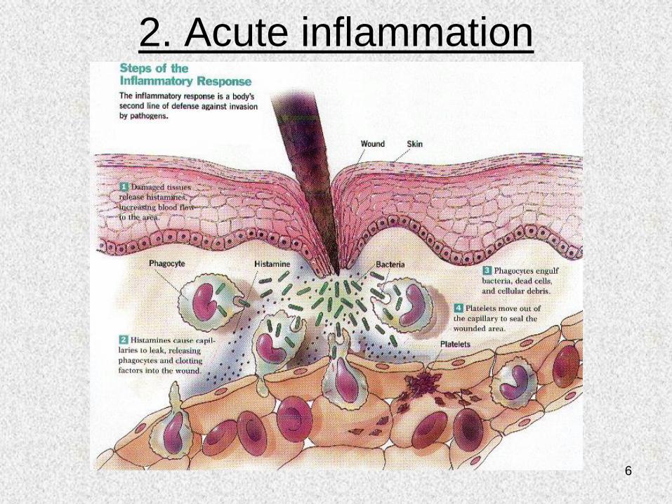
Inflammation – what is it?
• provoked response to tissue injury
• chemical agents
• cold, heat
• trauma
• invasion of microbes
• destroys, dilutes or contains the injurious agent
• induces repair
• protective response
• can be potentially harmful (e.g., arthritis)
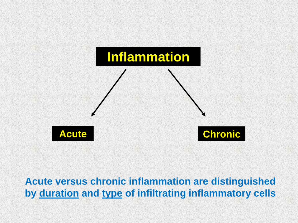
Inflammation
Acute Chronic
Acute versus chronic inflammation are distinguished
by duration and type of infiltrating inflammatory cells
6
2. Acute inflammation
7
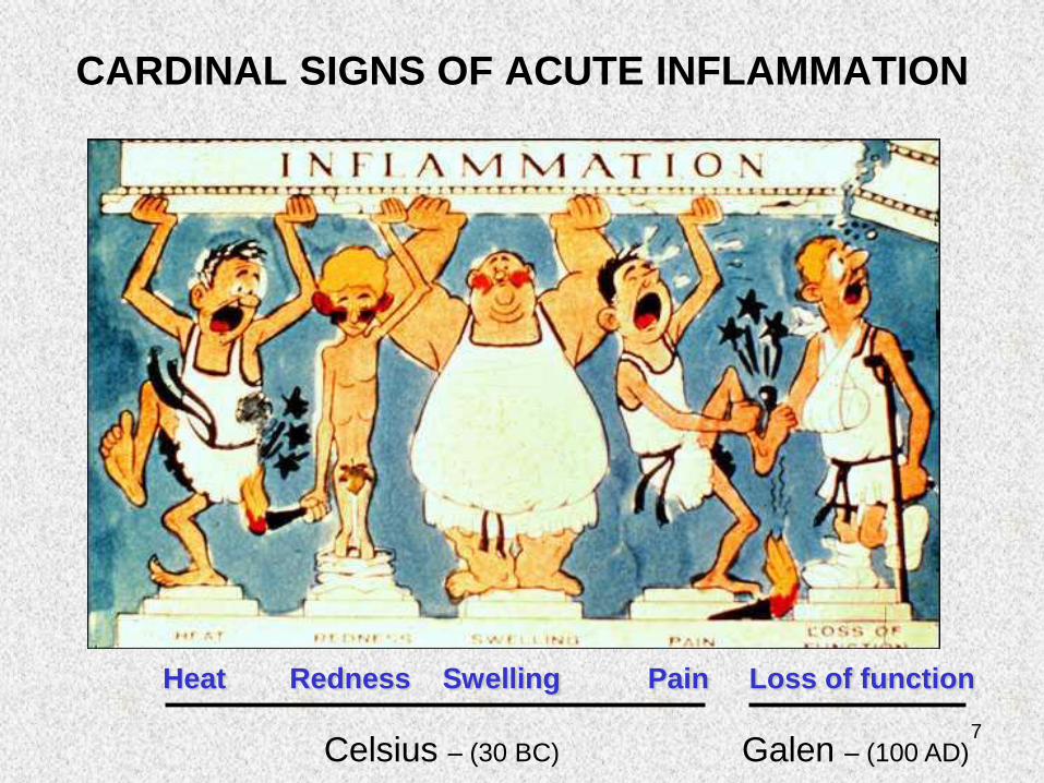
CARDINAL SIGNS OF ACUTE INFLAMMATION
Heat Redness Swelling Pain Loss of function
Celsius – (30 BC) Galen – (100 AD)
8
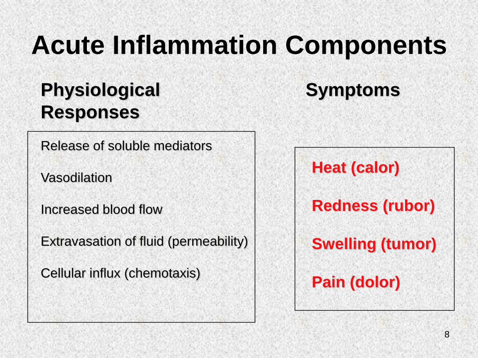
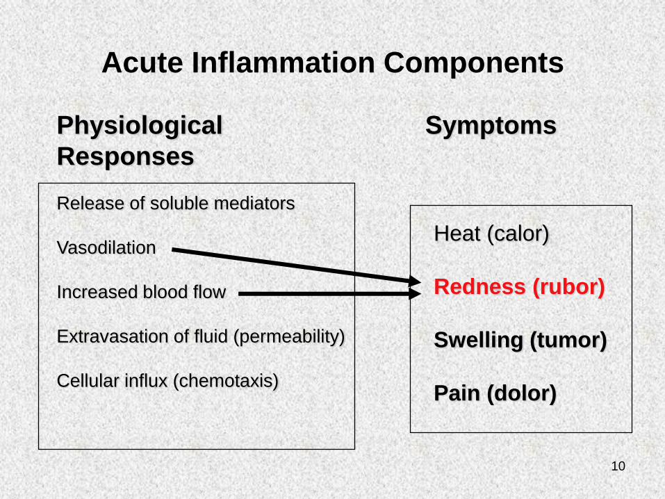
Acute Inflammation Components
Release of soluble mediators
Vasodilation
Increased blood flow
Extravasation of fluid (permeability)
Cellular influx (chemotaxis)
Heat (calor)
Redness (rubor)
Swelling (tumor)
Pain (dolor)
Physiological Symptoms
Responses
9
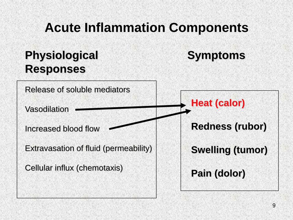
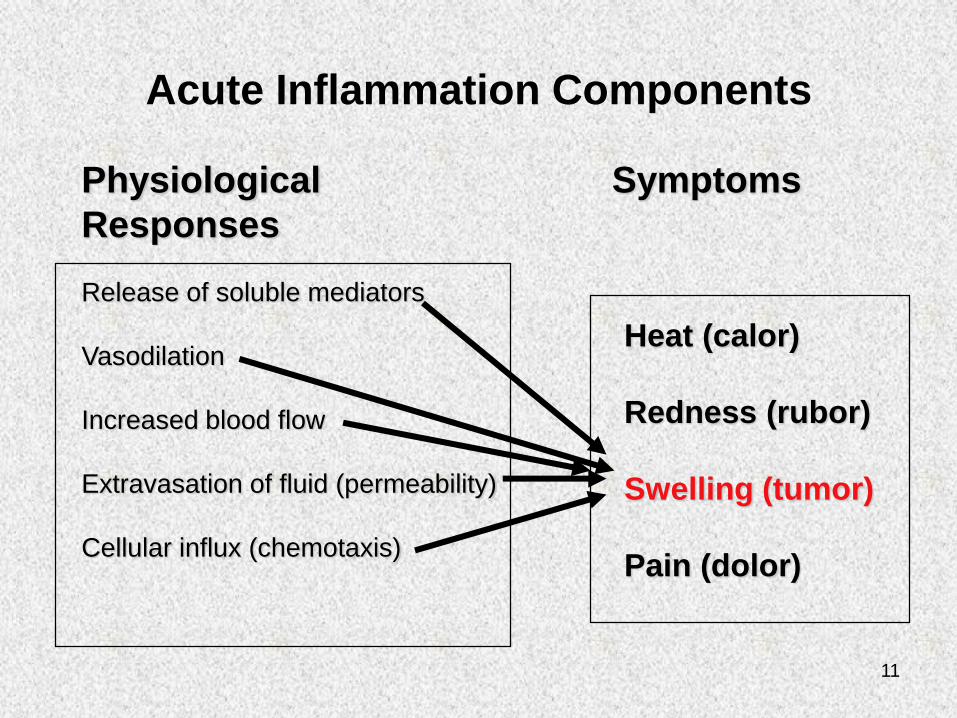
Acute Inflammation Components
Physiological Symptoms
Responses
Release of soluble mediators
Vasodilation
Increased blood flow
Extravasation of fluid (permeability)
Cellular influx (chemotaxis)
Heat (calor)
Redness (rubor)
Swelling (tumor)
Pain (dolor)
10
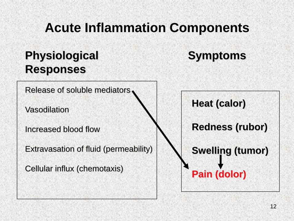
Acute Inflammation Components
Physiological Symptoms
Responses
Release of soluble mediators
Vasodilation
Increased blood flow
Extravasation of fluid (permeability)
Cellular influx (chemotaxis)
Heat (calor)
Redness (rubor)
Swelling (tumor)
Pain (dolor)
11
Acute Inflammation Components
Physiological Symptoms
Responses
Release of soluble mediators
Vasodilation
Increased blood flow
Extravasation of fluid (permeability)
Cellular influx (chemotaxis)
Heat (calor)
Redness (rubor)
Swelling (tumor)
Pain (dolor)
12
Acute Inflammation Components
Physiological Symptoms
Responses
Release of soluble mediators
Vasodilation
Increased blood flow
Extravasation of fluid (permeability)
Cellular influx (chemotaxis)
Heat (calor)
Redness (rubor)
Swelling (tumor)
Pain (dolor)
13
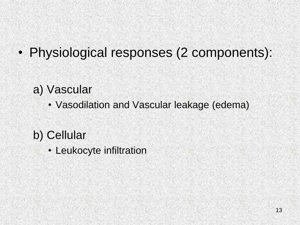
• Physiological responses (2 components):
a) Vascular
• Vasodilation and Vascular leakage (edema)
b) Cellular
• Leukocyte infiltration
14
2a) Vascular Events
• First, there may be a brief (few seconds)
arteriolar vasoconstriction (e.g., blanching after
a burn or scrape) mediated by autonomic nerves
or direct injury to arteriolar smooth muscle wall
• Second, smooth muscle relaxes and
vasodilation follows
– This vasodilatory smooth muscle response is termed
active hyperemia*
*increase in organ blood flow
15
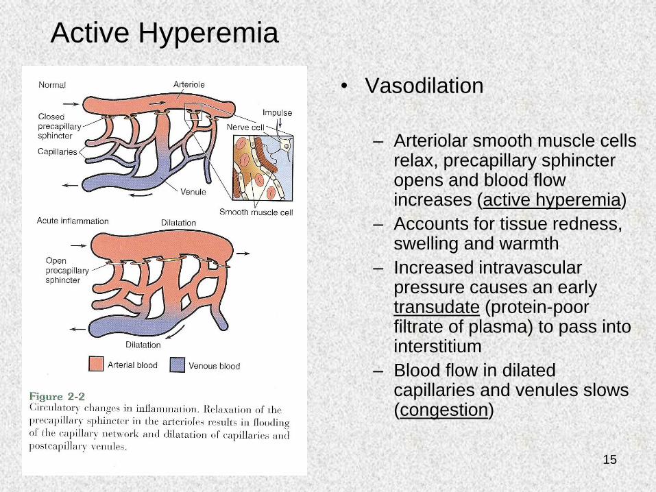
Active Hyperemia
• Vasodilation
– Arteriolar smooth muscle cells relax, precapillary sphincter opens and blood flow increases (active hyperemia)
– Accounts for tissue redness, swelling and warmth
– Increased intravascular pressure causes an early transudate (protein-poor filtrate of plasma) to pass into interstitium
– Blood flow in dilated capillaries and venules slows (congestion)
16
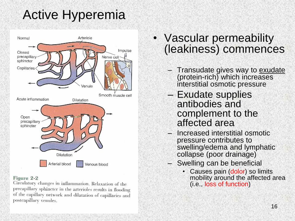
Active Hyperemia
• Vascular permeability (leakiness) commences
– Transudate gives way to exudate(protein-rich) which increases interstitial osmotic pressure
– Exudate supplies antibodies and complement to the affected area
– Increased interstitial osmotic pressure contributes to swelling/edema and lymphatic collapse (poor drainage)
– Swelling can be beneficial• Causes pain (dolor) so limits
mobility around the affected area (i.e., loss of function)
17
Vascular leakage• Five mechanisms known to cause vascular
leakiness and all or any combination of these
events may occur in response to a given
stimulus
1) Histamines, bradykinins, leukotrienes cause an early,
brief (15 – 30 min.) immediate transient response in
the form of reversible endothelial cell contraction that
widens intercellular gaps of venules (not arterioles or
capillaries)
18
Vascular leakage
2) Cytokine mediators (TNF, IL-1) induce:
– reversible endothelial cell junction retraction through
cytoskeleton reorganization (4 – 6 hrs post injury,
lasting 24 hrs or more)
3) Severe injuries may cause:
– immediate direct endothelial cell damage (necrosis,
detachment) making them leaky until they are
repaired (immediate sustained response)
– delayed damage as in thermal or UV injury (sunburn)
or some bacterial toxins (delayed prolonged leakage)
19
Vascular leakage
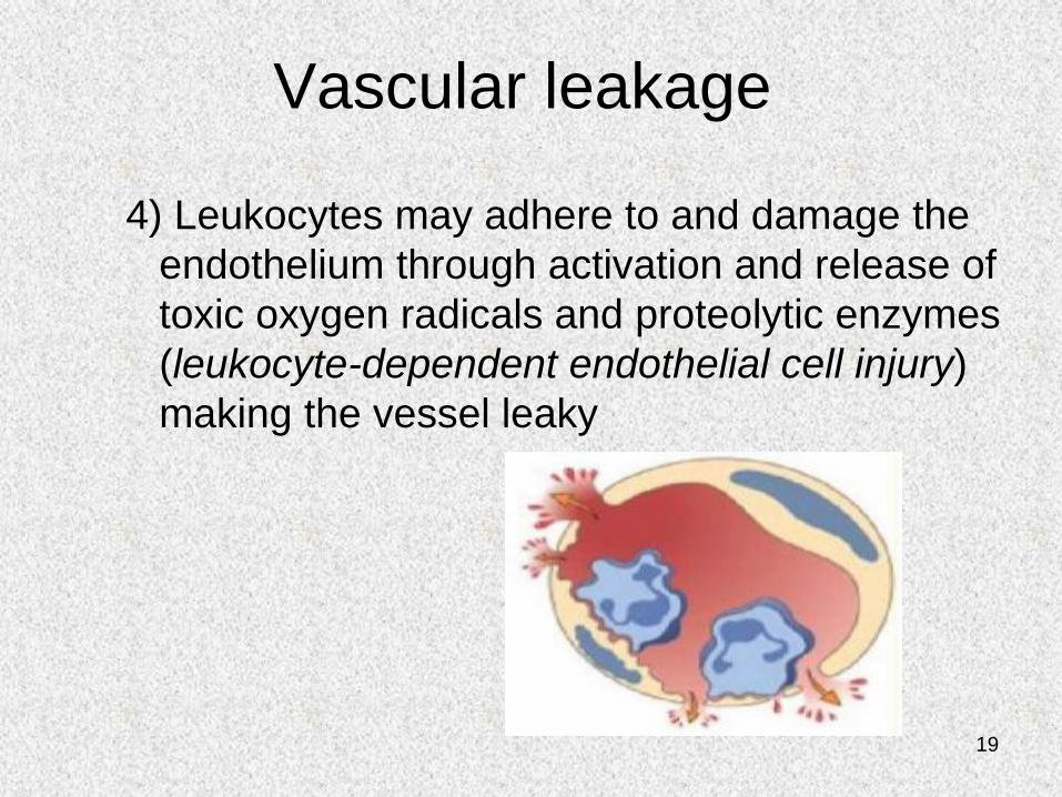
4) Leukocytes may adhere to and damage the
endothelium through activation and release of
toxic oxygen radicals and proteolytic enzymes
(leukocyte-dependent endothelial cell injury)
making the vessel leaky
20
Vascular leakage
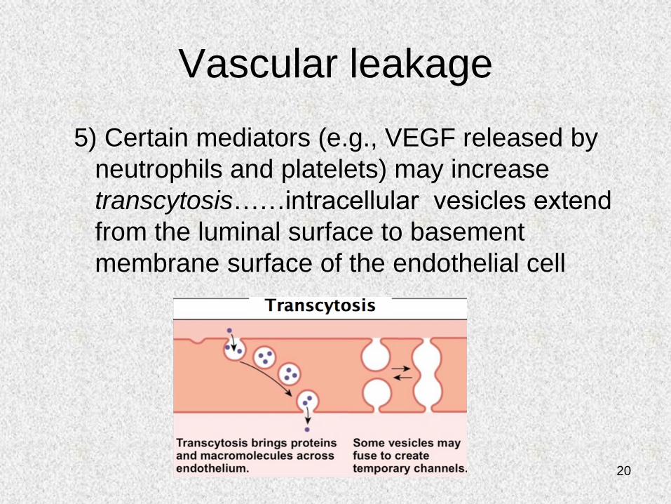
5) Certain mediators (e.g., VEGF released by
neutrophils and platelets) may increase
transcytosis……intracellular vesicles extend
from the luminal surface to basement
membrane surface of the endothelial cell
21
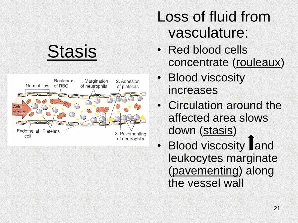
Stasis
Loss of fluid from vasculature:
• Red blood cells concentrate (rouleaux)
• Blood viscosity increases
• Circulation around the affected area slows down (stasis)
• Blood viscosity and leukocytes marginate (pavementing) along the vessel wall
22
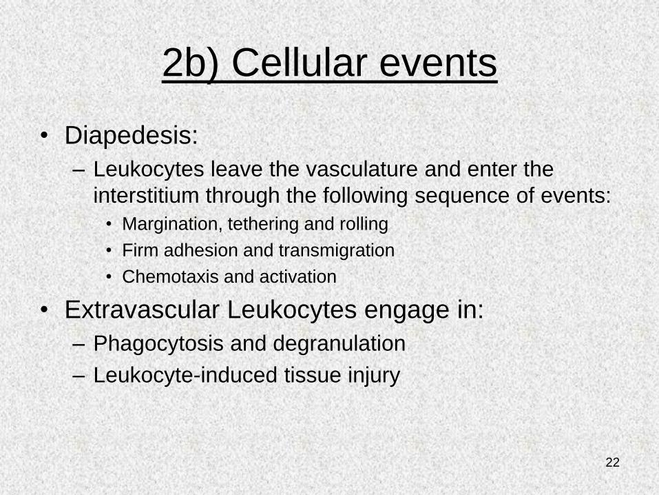
2b) Cellular events
• Diapedesis:
– Leukocytes leave the vasculature and enter the
interstitium through the following sequence of events:
• Margination, tethering and rolling
• Firm adhesion and transmigration
• Chemotaxis and activation
• Extravascular Leukocytes engage in:
– Phagocytosis and degranulation
– Leukocyte-induced tissue injury
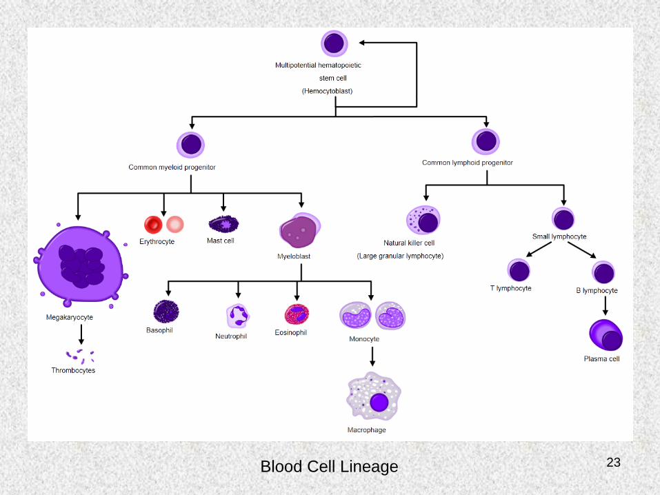
23Blood Cell Lineage
24
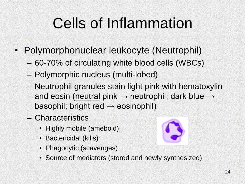
Cells of Inflammation
• Polymorphonuclear leukocyte (Neutrophil)
– 60-70% of circulating white blood cells (WBCs)
– Polymorphic nucleus (multi-lobed)
– Neutrophil granules stain light pink with hematoxylin
and eosin (neutral pink → neutrophil; dark blue →
basophil; bright red → eosinophil)
– Characteristics
• Highly mobile (ameboid)
• Bactericidal (kills)
• Phagocytic (scavenges)
• Source of mediators (stored and newly synthesized)
25
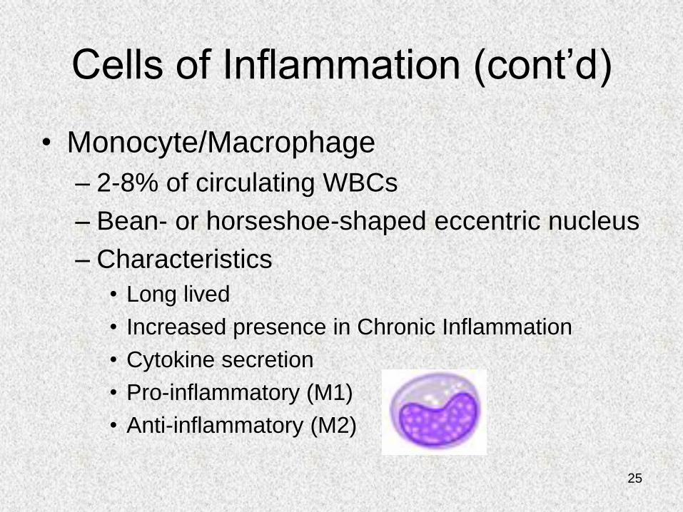
Cells of Inflammation (cont’d)
• Monocyte/Macrophage
– 2-8% of circulating WBCs
– Bean- or horseshoe-shaped eccentric nucleus
– Characteristics
• Long lived
• Increased presence in Chronic Inflammation
• Cytokine secretion
• Pro-inflammatory (M1)
• Anti-inflammatory (M2)
26
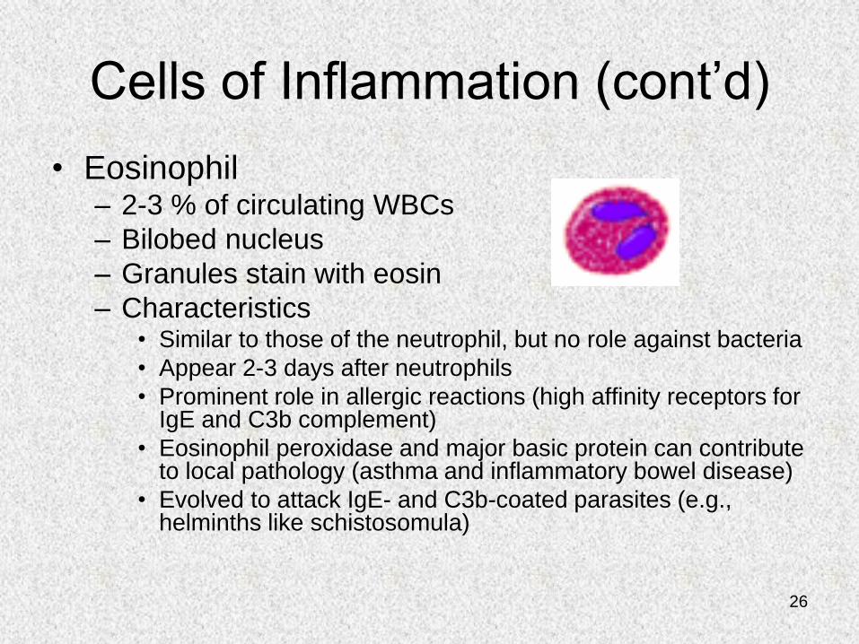
Cells of Inflammation (cont’d)
• Eosinophil– 2-3 % of circulating WBCs
– Bilobed nucleus
– Granules stain with eosin
– Characteristics• Similar to those of the neutrophil, but no role against bacteria
• Appear 2-3 days after neutrophils
• Prominent role in allergic reactions (high affinity receptors for IgE and C3b complement)
• Eosinophil peroxidase and major basic protein can contribute to local pathology (asthma and inflammatory bowel disease)
• Evolved to attack IgE- and C3b-coated parasites (e.g., helminths like schistosomula)
27
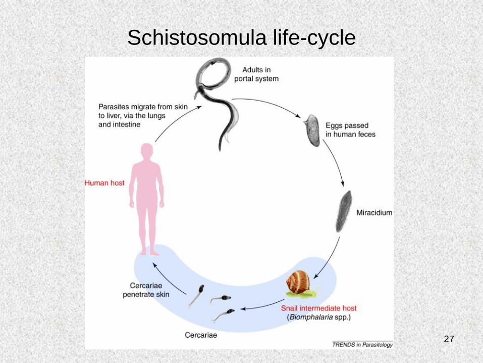
Schistosomula life-cycle
28
Cells of Inflammation (cont’d)
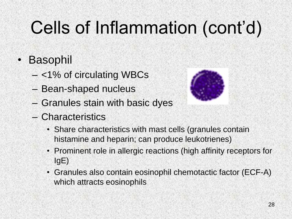
• Basophil
– <1% of circulating WBCs
– Bean-shaped nucleus
– Granules stain with basic dyes
– Characteristics
• Share characteristics with mast cells (granules contain
histamine and heparin; can produce leukotrienes)
• Prominent role in allergic reactions (high affinity receptors for
IgE)
• Granules also contain eosinophil chemotactic factor (ECF-A)
which attracts eosinophils
29
Cells of Inflammation (cont’d)
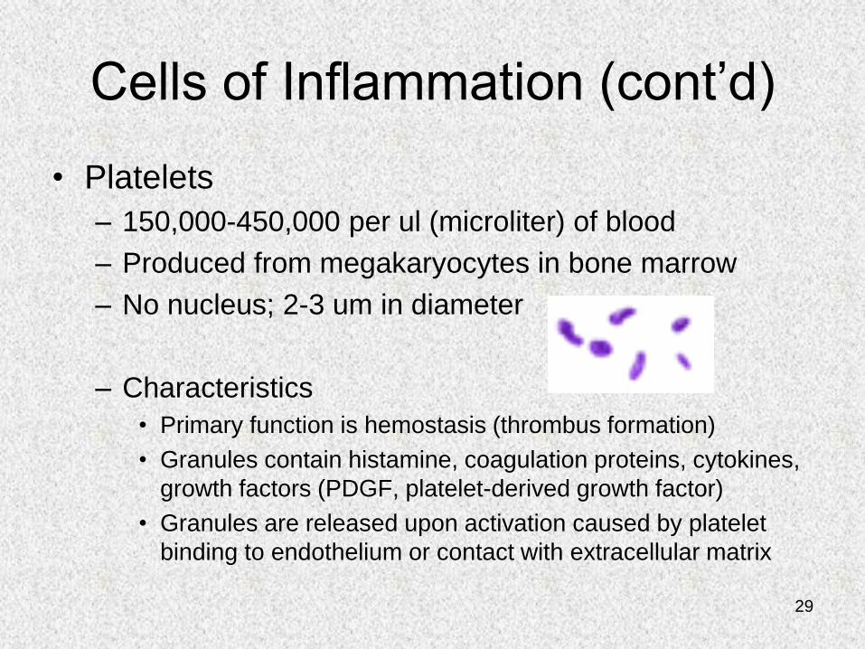
• Platelets
– 150,000-450,000 per ul (microliter) of blood
– Produced from megakaryocytes in bone marrow
– No nucleus; 2-3 um in diameter
– Characteristics
• Primary function is hemostasis (thrombus formation)
• Granules contain histamine, coagulation proteins, cytokines,
growth factors (PDGF, platelet-derived growth factor)
• Granules are released upon activation caused by platelet
binding to endothelium or contact with extracellular matrix
30
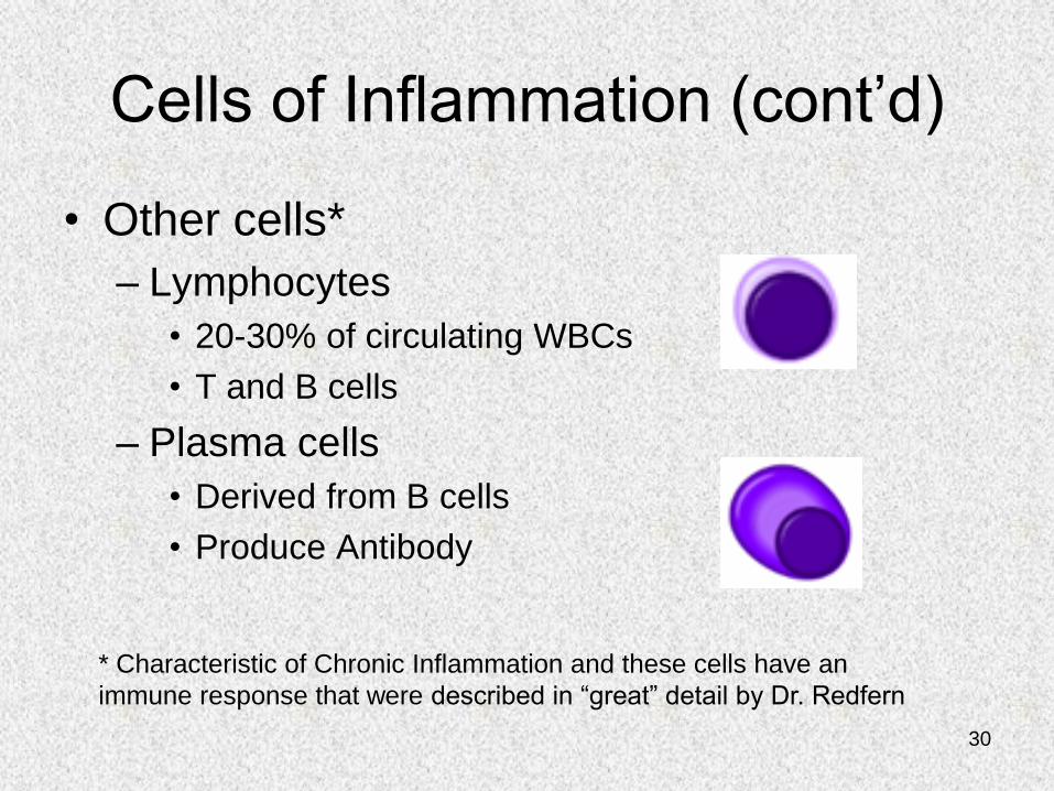
Cells of Inflammation (cont’d)
• Other cells*
– Lymphocytes
• 20-30% of circulating WBCs
• T and B cells
– Plasma cells
• Derived from B cells
• Produce Antibody
* Characteristic of Chronic Inflammation and these cells have an
immune response that were described in “great” detail by Dr. Redfern

31
Leukocyte Recruitment at Inflammatory Sites
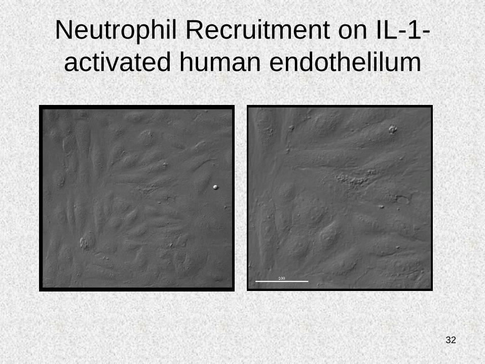
32
Neutrophil Recruitment on IL-1-
activated human endothelilum
33
Chemotaxis
• Leukocytes follow chemical gradient to site of
injury (chemotaxis)
– Soluble bacterial products
– Complement components (C5a)
– Chemokines (e.g., IL-8)
– LTB4 (AA metabolite)
• Chemotactic agents bind surface receptors
inducing calcium mobilization and assembly of
cytoskeletal contractile elements……i.e.,
leukocyte activation
34
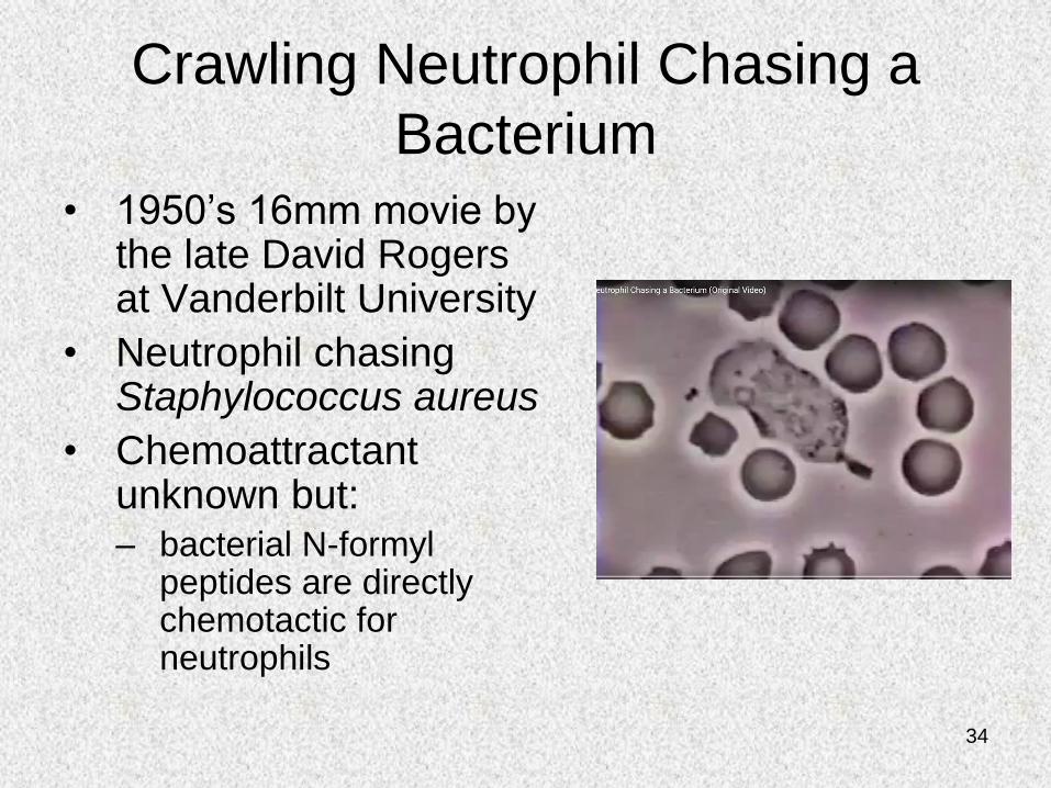
Crawling Neutrophil Chasing a
Bacterium
• 1950’s 16mm movie by the late David Rogers at Vanderbilt University
• Neutrophil chasing Staphylococcus aureus
• Chemoattractant unknown but:– bacterial N-formyl
peptides are directly chemotactic for neutrophils
35
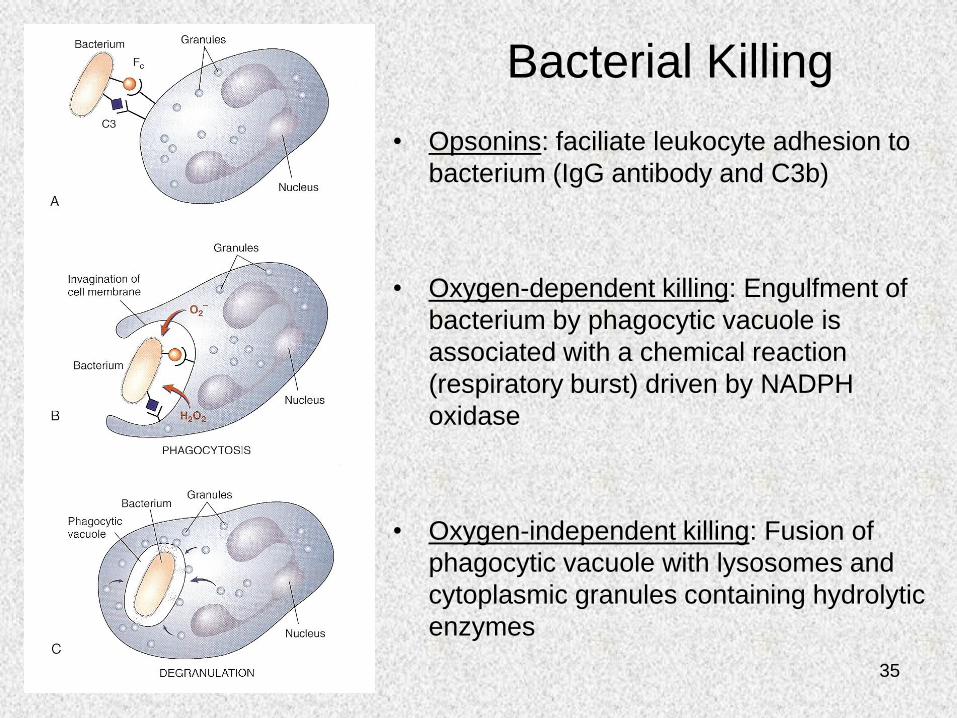
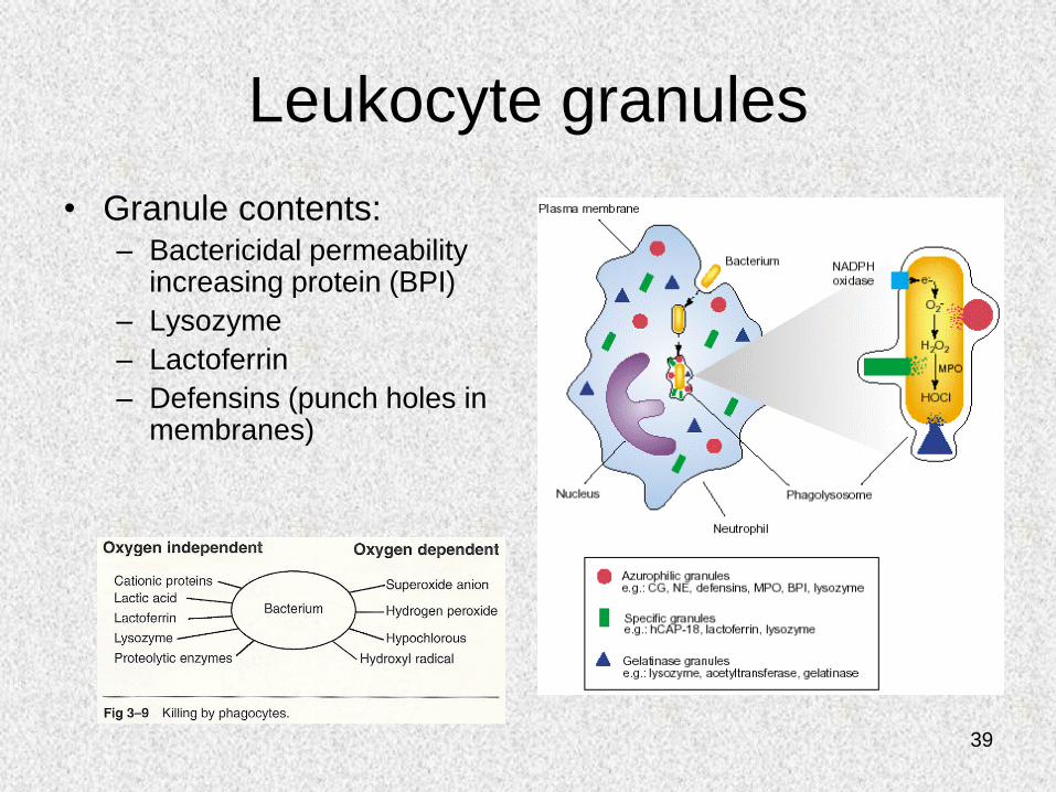
Bacterial Killing
• Opsonins: faciliate leukocyte adhesion to
bacterium (IgG antibody and C3b)
• Oxygen-dependent killing: Engulfment of
bacterium by phagocytic vacuole is
associated with a chemical reaction
(respiratory burst) driven by NADPH
oxidase
• Oxygen-independent killing: Fusion of
phagocytic vacuole with lysosomes and
cytoplasmic granules containing hydrolytic
enzymes
36
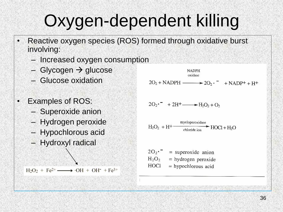
Oxygen-dependent killing• Reactive oxygen species (ROS) formed through oxidative burst
involving:
– Increased oxygen consumption
– Glycogen glucose
– Glucose oxidation
• Examples of ROS:
– Superoxide anion
– Hydrogen peroxide
– Hypochlorous acid
– Hydroxyl radical
.OH = hydroxyl radical
radical
37
Reactive oxygen species
• Hydrogen peroxide alone is insufficient
• Myeloperoxidase (MPO), contained in neutrophil azurophilic granules, converts hydrogen peroxide to hypochlorous acid (HOCl- ), an oxidant/antimicrobial agent
38
Degradation and Clean-up
• Reactive end-products only active within
phagolysosome
• Dead microorganisms degraded by
lysosomal acid hydrolases
• Hydrogen peroxide broken down to water
and oxygen by catalase
39
Leukocyte granules
• Granule contents:– Bactericidal permeability
increasing protein (BPI)
– Lysozyme
– Lactoferrin
– Defensins (punch holes in membranes)
40
Leukocyte-induced tissue injury
• Destructive enzymes may enter
extracellular space in event of:
– Premature degranulation
– Frustrated phagocytosis (large, flat)
– Membranolytic substances (urate crystals)
– Persistent leukocyte activation (rheumatoid
arthritis, emphysema)
41
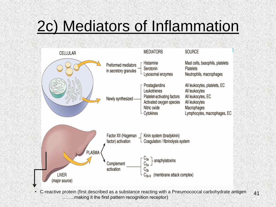
2c) Mediators of Inflammation
• C-reactive protein (first described as a substance reacting with a Pneumococcal carbohydrate antigen
……..making it the first pattern recognition receptor)
42
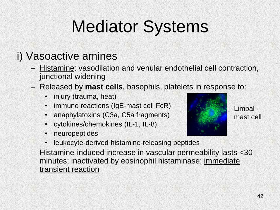
Mediator Systems
i) Vasoactive amines– Histamine: vasodilation and venular endothelial cell contraction,
junctional widening
– Released by mast cells, basophils, platelets in response to:
• injury (trauma, heat)
• immune reactions (IgE-mast cell FcR)
• anaphylatoxins (C3a, C5a fragments)
• cytokines/chemokines (IL-1, IL-8)
• neuropeptides
• leukocyte-derived histamine-releasing peptides
– Histamine-induced increase in vascular permeability lasts <30 minutes; inactivated by eosinophil histaminase; immediate transient reaction
Limbal
mast cell
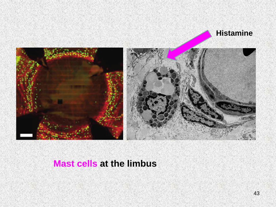
43
Venule
Mast cells at the limbus
Histamine
44
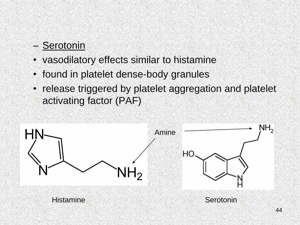
– Serotonin
• vasodilatory effects similar to histamine
• found in platelet dense-body granules
• release triggered by platelet aggregation and platelet
activating factor (PAF)
Histamine
SerotoninHistamine
Amine
45
Mediator Systems
ii) Plasma Protein Systems
• Clotting (coagulation) factors
• Complement cascade
46
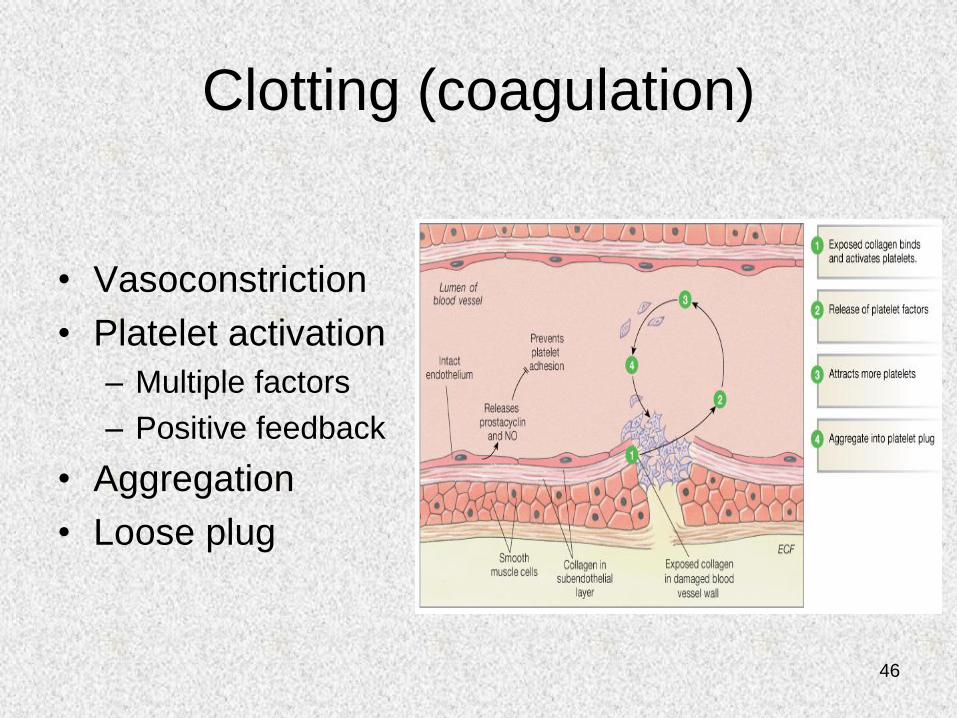
Clotting (coagulation)
• Vasoconstriction
• Platelet activation
– Multiple factors
– Positive feedback
• Aggregation
• Loose plug
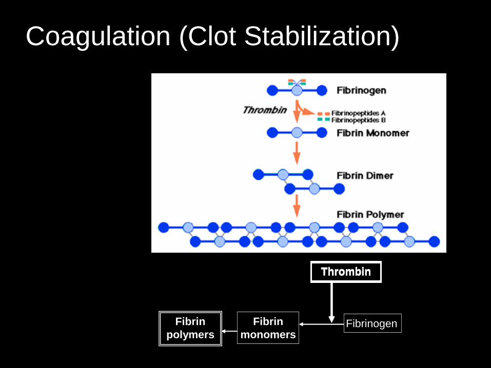
FibrinogenFibrin
monomers
Fibrin
polymers
ThrombinThrombin
Coagulation (Clot Stabilization)
48
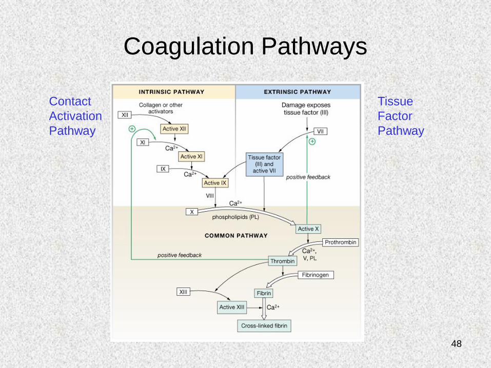
Coagulation Pathways
Contact
Activation
Pathway
Tissue
Factor
Pathway
49
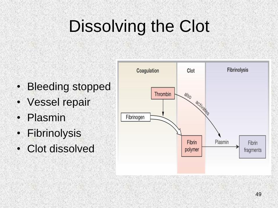
Dissolving the Clot
• Bleeding stopped
• Vessel repair
• Plasmin
• Fibrinolysis
• Clot dissolved
50
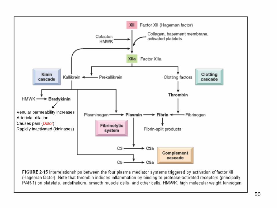
Venular permeability increases
Arteriolar dilation
Causes pain (Dolor)
Rapidly inactivated (kininases)
51
Complement system ~20 interactive
plasma and cell membrane components
• “Complements” antibody killing of bacteria
• However, antibodies are not necessary for
complement activation and bacterial killing
52
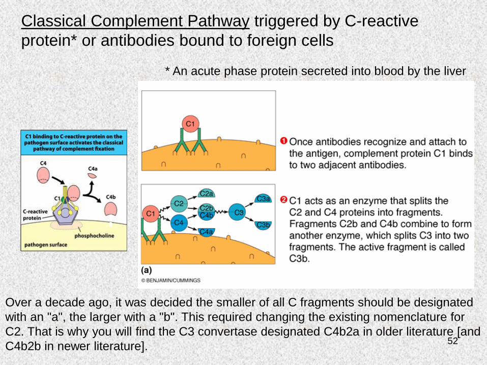
Classical Complement Pathway triggered by C-reactive
protein* or antibodies bound to foreign cells
* An acute phase protein secreted into blood by the liver
Over a decade ago, it was decided the smaller of all C fragments should be designated
with an "a", the larger with a "b". This required changing the existing nomenclature for
C2. That is why you will find the C3 convertase designated C4b2a in older literature [and
C4b2b in newer literature].
53
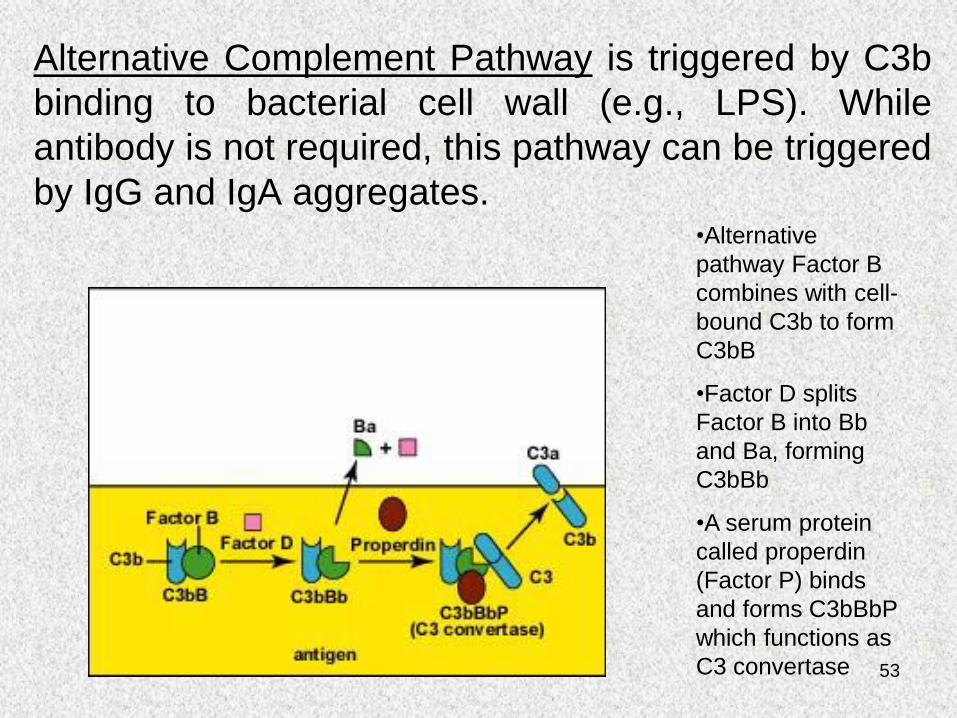
Alternative Complement Pathway is triggered by C3b
binding to bacterial cell wall (e.g., LPS). While
antibody is not required, this pathway can be triggered
by IgG and IgA aggregates.•Alternative
pathway Factor B
combines with cell-
bound C3b to form
C3bB
•Factor D splits
Factor B into Bb
and Ba, forming
C3bBb
•A serum protein
called properdin
(Factor P) binds
and forms C3bBbP
which functions as
C3 convertase
54
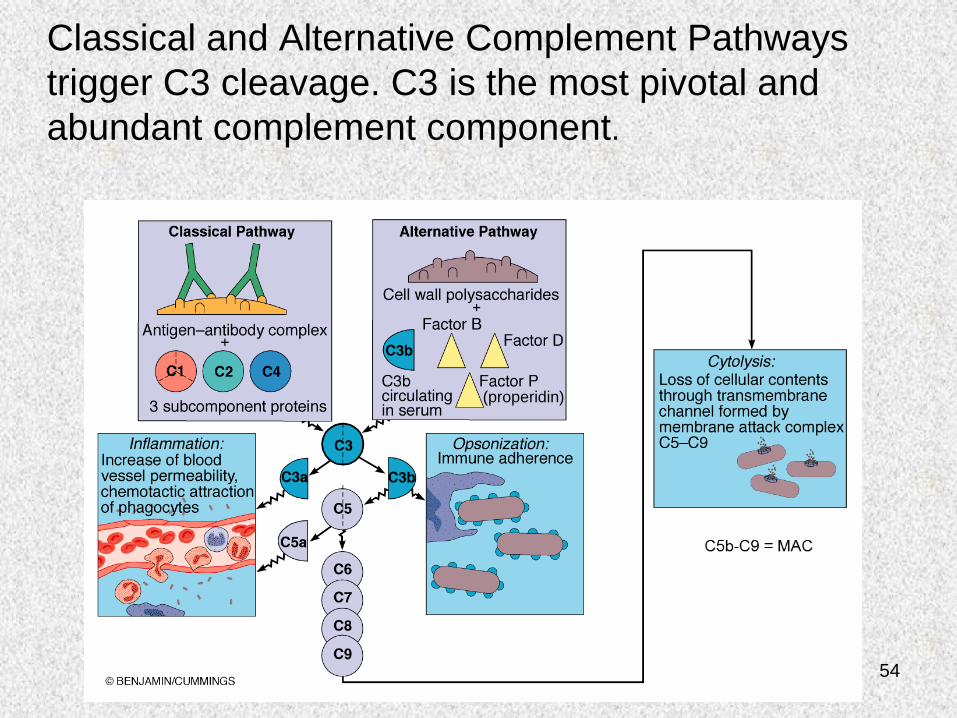
Classical and Alternative Complement Pathways
trigger C3 cleavage. C3 is the most pivotal and
abundant complement component.
(properidin)
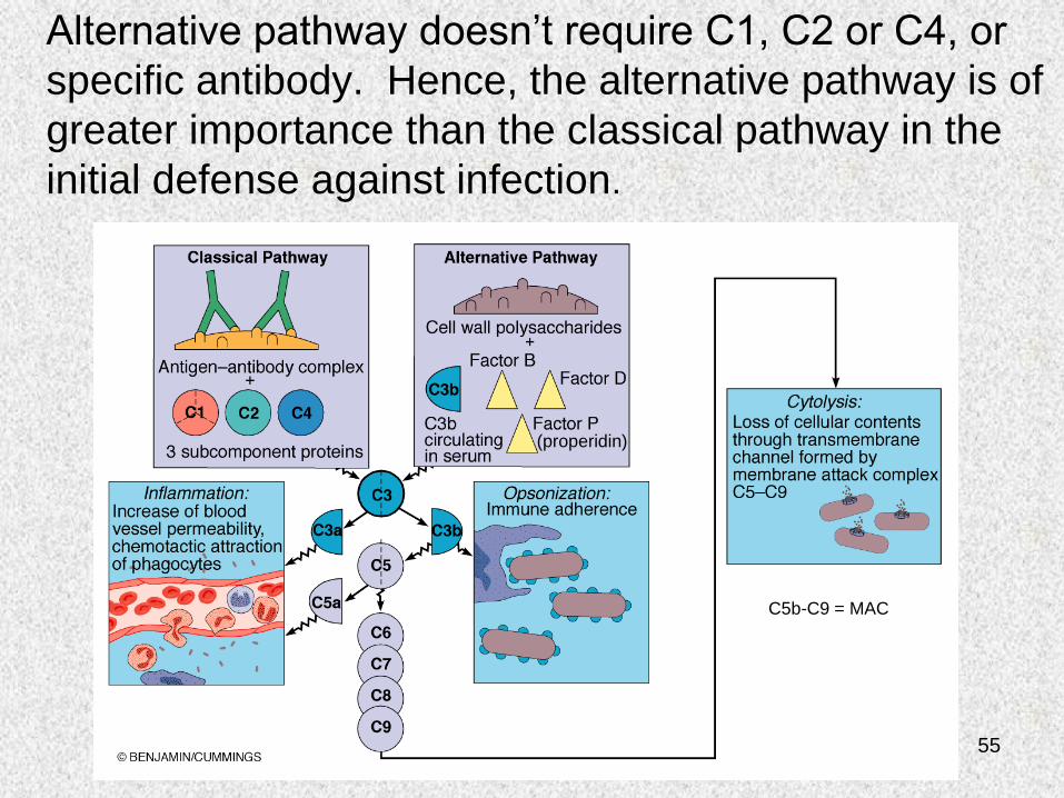
55
Alternative pathway doesn’t require C1, C2 or C4, or
specific antibody. Hence, the alternative pathway is of
greater importance than the classical pathway in the
initial defense against infection.
(properidin)
C5b-C9 = MAC
56
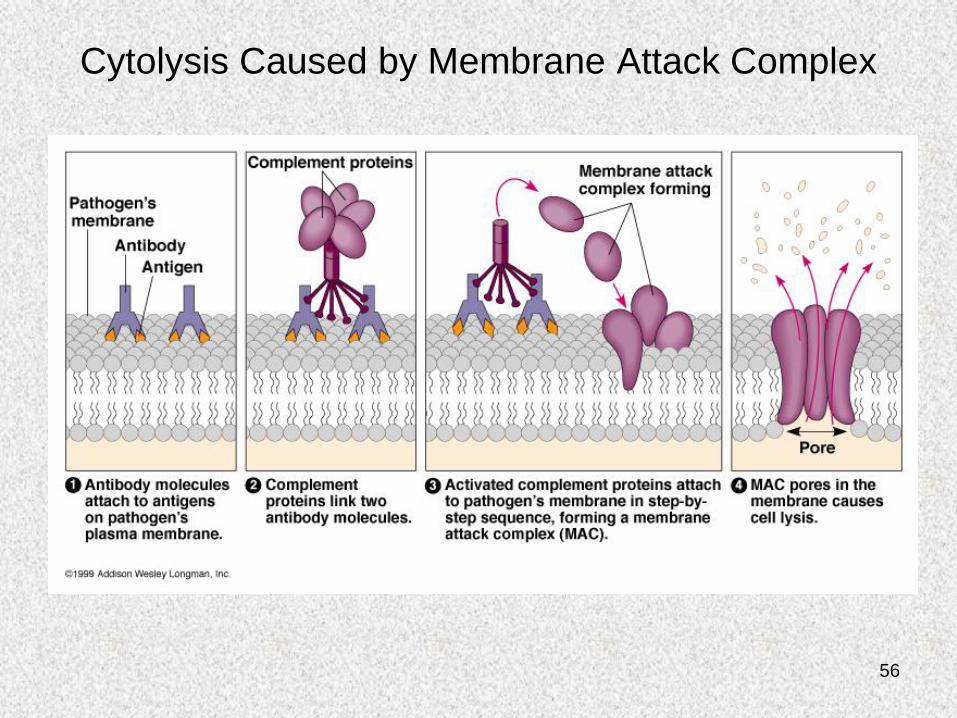
Cytolysis Caused by Membrane Attack Complex
57
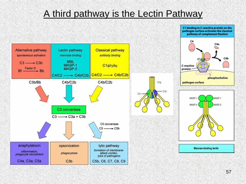
A third pathway is the Lectin Pathway
58
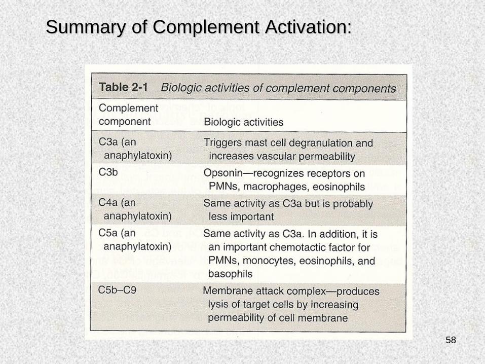
Summary of Complement Activation:
59
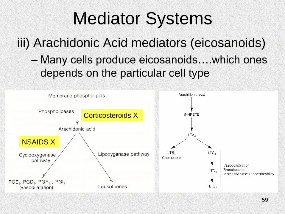
Mediator Systems
iii) Arachidonic Acid mediators (eicosanoids)
– Many cells produce eicosanoids….which ones
depends on the particular cell type
Corticosteroids X
NSAIDS X
60
Mediator Systems
iv) Platelet activating factor, PAF
– Like the eicosanoids, PAF is derived from membrane
phospholipids by the action of phospholipase A2
– Generated by mast cells, platelets, endothelial cells
and leukocytes
– Causes vasodilation and increases vascular
permeability
– Enhances arachidonic acid metabolism in leukocytes
leading to increased motility, degranulation, and free
radical formation
61
Mediator Systems
v) Cytokines and Chemokines– Protein cell products that act as a message to other
cells, telling them how to behave.
– Released by many cell types
– IL-1 and TNF activate endothelium and cause fever and lethargy
– IFN- activates macrophages/neutrophils, boosting their killing ability
– IL-8 (chemokine) is chemotactic for neutrophils
– IL-6, IL-8, TNF, IL-1 increase acute phase protein production; C-reactive protein (opsonin) and mannan binding lectin (opsonin) activate complement
62
Mediator Systems
vi) Phagocyte Products
– Leak from PMNs and macrophages after demise, attempts at phagocytosis, etc.
– Acid proteases (normally within lysosomes)
– Neutral proteases such as elastase and collagenase are destructive in ECM
– Counteracted by serum and ECM anti-proteases
63
Mediator Systems
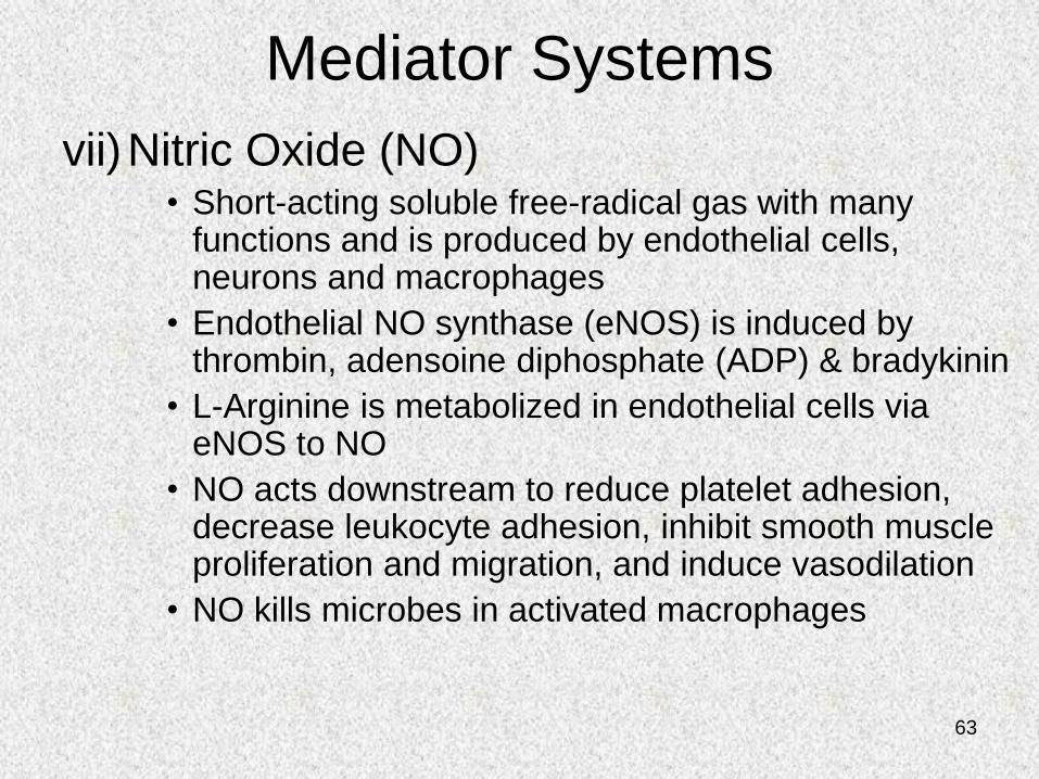
vii)Nitric Oxide (NO)• Short-acting soluble free-radical gas with many
functions and is produced by endothelial cells, neurons and macrophages
• Endothelial NO synthase (eNOS) is induced by thrombin, adensoine diphosphate (ADP) & bradykinin
• L-Arginine is metabolized in endothelial cells via eNOS to NO
• NO acts downstream to reduce platelet adhesion, decrease leukocyte adhesion, inhibit smooth muscle proliferation and migration, and induce vasodilation
• NO kills microbes in activated macrophages
64
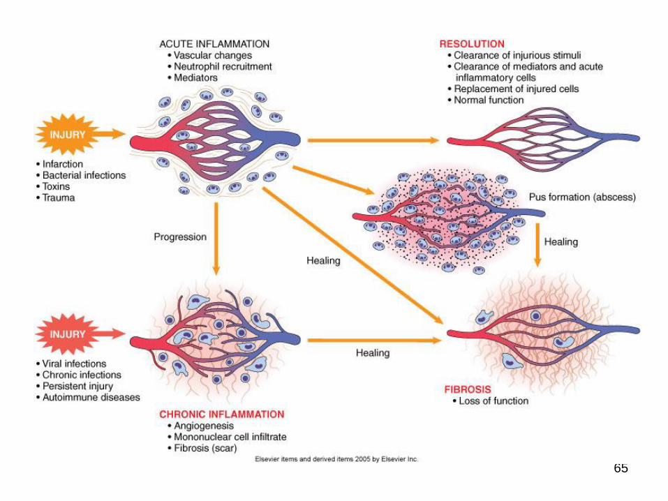
2d) Possible outcomes of acute
inflammation
• Complete resolution
• Scarring (fibrosis)
• Abscess formation
• Progression to chronic inflammation
65
66
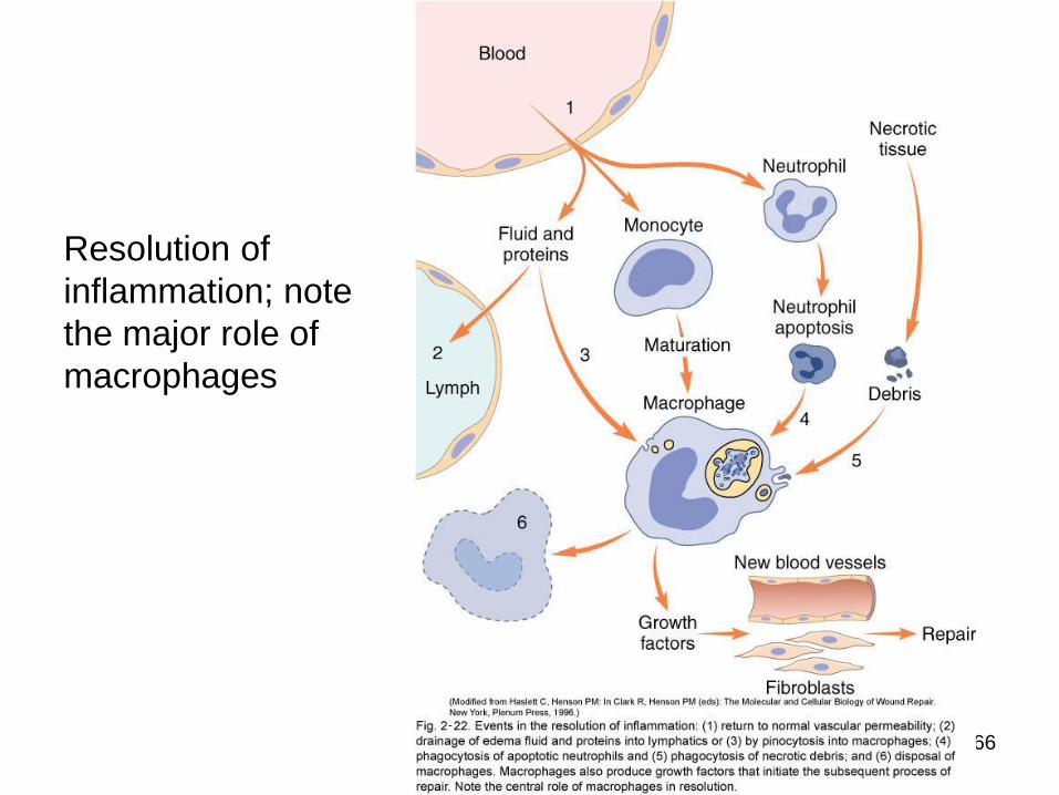
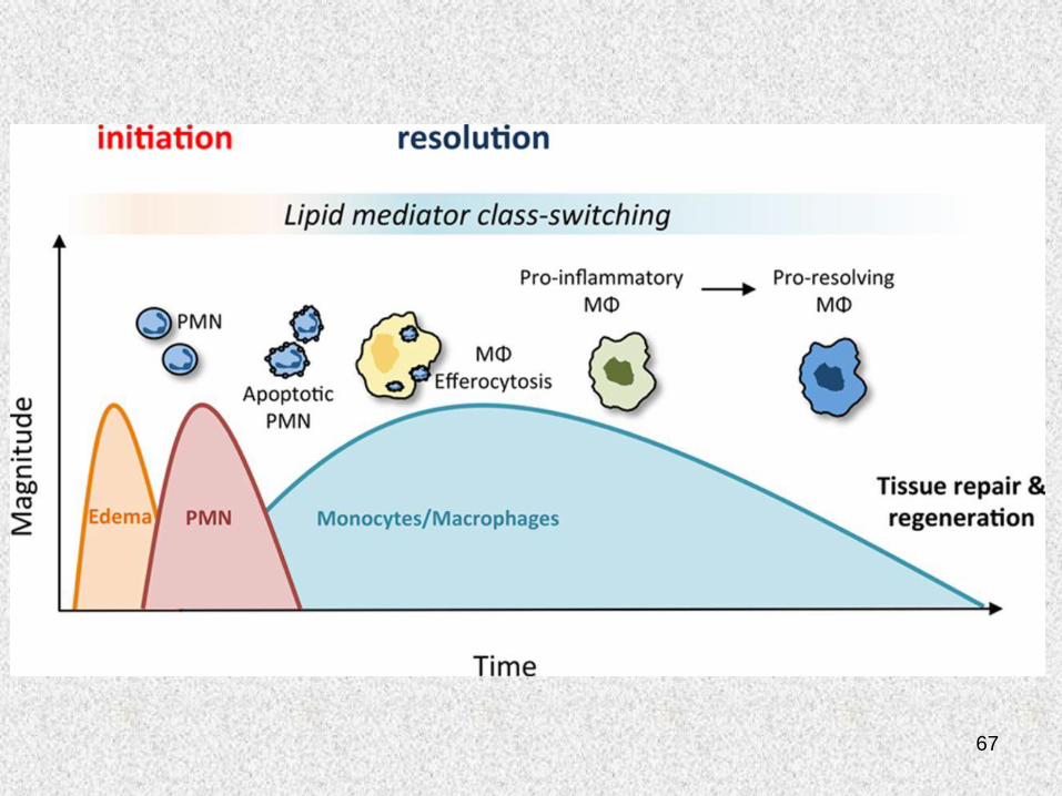
Resolution of
inflammation; note
the major role of
macrophages
67
68
69
3. Chronic inflammation
70
• Chronic inflammation is a prolonged process (weeks-months-years) in which three processes are occurring simultaneously:
1) Active inflammation• Lymphocyte, macrophage, plasma cell (mononuclear cell)
infiltration
2) Tissue destruction by inflammatory cells
3) Tissue healing (repair & fibrosis)• Attempts at repair with fibrosis and angiogenesis (new vessel
formation)
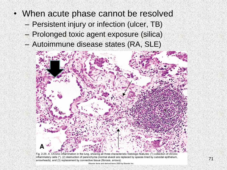
71
• When acute phase cannot be resolved– Persistent injury or infection (ulcer, TB)
– Prolonged toxic agent exposure (silica)
– Autoimmune disease states (RA, SLE)
72
3a) Chronic Inflammatory Cells
• Macrophages
– Scattered throughout the body (microglia, Kupffer cells, sinus histiocytes, alveolar macrophages, etc.)
– Circulate as monocytes and reach site of injury within 24 – 48 hrs and transform into macrophages
– Become activated by T cell-derived cytokines, endotoxin, and mediators of inflammation
73
3a) cont’d
• T and B lymphocytes
– Antigen-activated (delivered via macrophages and dendritic cells)
– Release macrophage-activating cytokines (in turn, macrophages release lymphocyte-activating cytokines until inflammatory stimulus is removed)
• Plasma cells
– Terminally differentiated B cells
– Produce antibodies
74
3a) cont’d
• Eosinophils
– Found especially at sites of parasitic infection,
or at allergic (IgE-mediated) sites
75
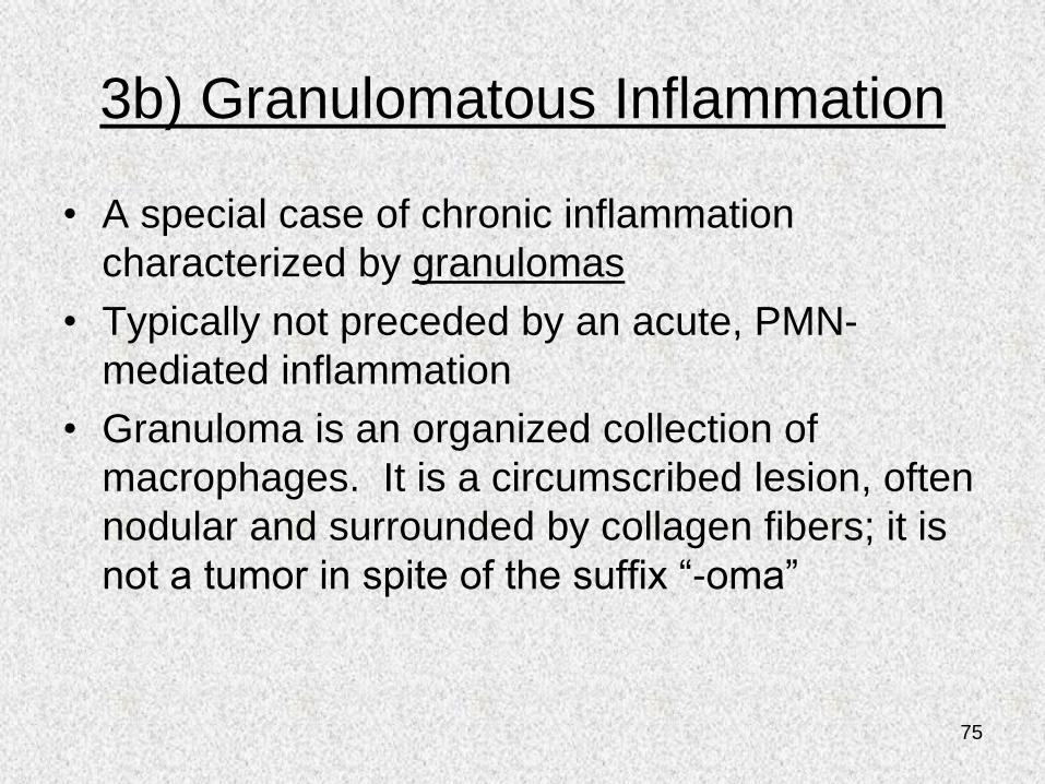
3b) Granulomatous Inflammation
• A special case of chronic inflammation
characterized by granulomas
• Typically not preceded by an acute, PMN-
mediated inflammation
• Granuloma is an organized collection of
macrophages. It is a circumscribed lesion, often
nodular and surrounded by collagen fibers; it is
not a tumor in spite of the suffix “-oma”
76
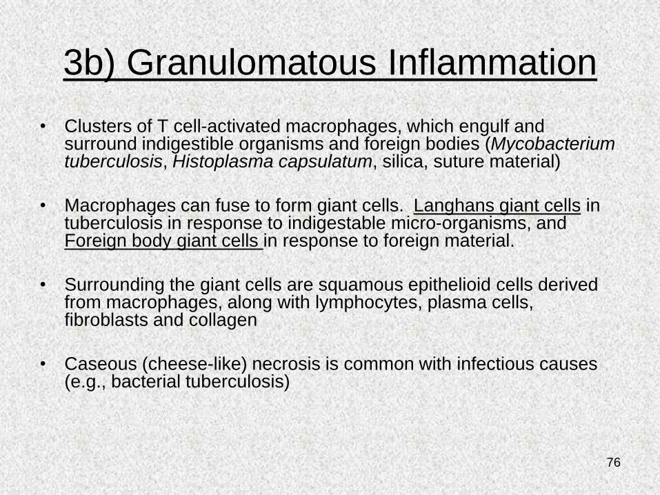
3b) Granulomatous Inflammation
• Clusters of T cell-activated macrophages, which engulf and surround indigestible organisms and foreign bodies (Mycobacterium tuberculosis, Histoplasma capsulatum, silica, suture material)
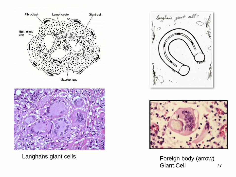
• Macrophages can fuse to form giant cells. Langhans giant cells in tuberculosis in response to indigestable micro-organisms, and Foreign body giant cells in response to foreign material.
• Surrounding the giant cells are squamous epithelioid cells derived from macrophages, along with lymphocytes, plasma cells, fibroblasts and collagen
• Caseous (cheese-like) necrosis is common with infectious causes (e.g., bacterial tuberculosis)
77
Foreign body (arrow)
Giant Cell
Langhans giant cells
78
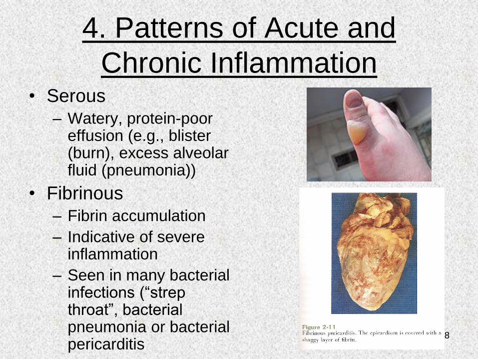
4. Patterns of Acute and
Chronic Inflammation• Serous
– Watery, protein-poor effusion (e.g., blister (burn), excess alveolar fluid (pneumonia))
• Fibrinous – Fibrin accumulation
– Indicative of severe inflammation
– Seen in many bacterial infections (“strep throat”, bacterial pneumonia or bacterial pericarditis
79
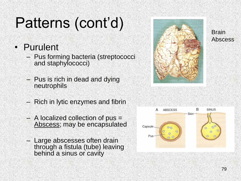
Patterns (cont’d)
• Purulent– Pus forming bacteria (streptococci
and staphylococci)
– Pus is rich in dead and dying neutrophils
– Rich in lytic enzymes and fibrin
– A localized collection of pus = Abscess; may be encapsulated
– Large abscesses often drain through a fistula (tube) leaving behind a sinus or cavity
Brain
Abscess
80
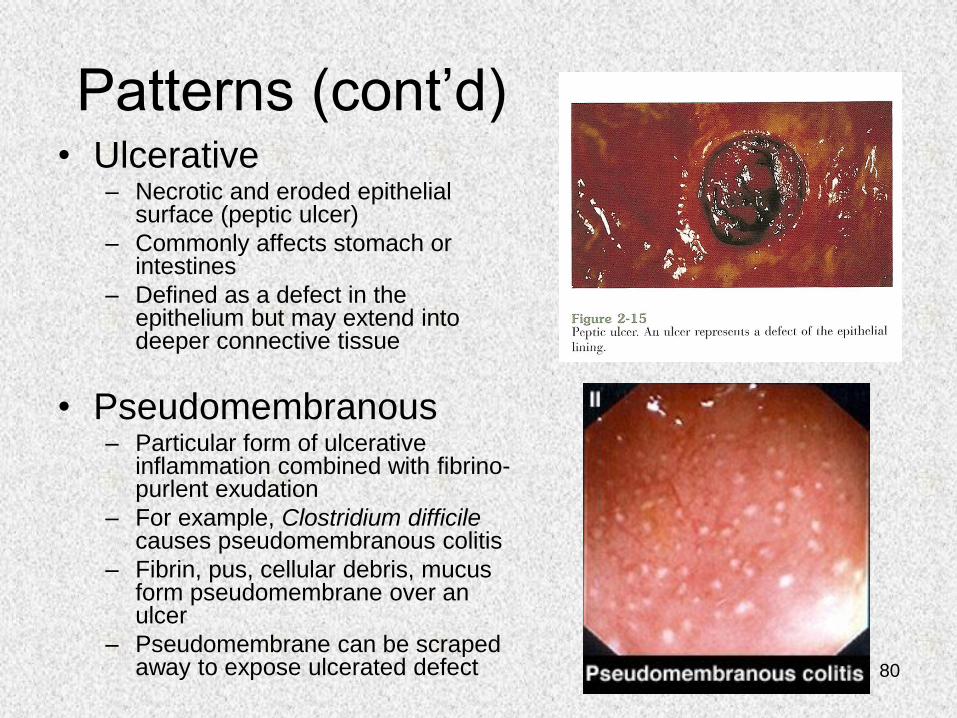
Patterns (cont’d)• Ulcerative
– Necrotic and eroded epithelial surface (peptic ulcer)
– Commonly affects stomach or intestines
– Defined as a defect in the epithelium but may extend into deeper connective tissue
• Pseudomembranous– Particular form of ulcerative
inflammation combined with fibrino-purlent exudation
– For example, Clostridium difficilecauses pseudomembranous colitis
– Fibrin, pus, cellular debris, mucus form pseudomembrane over an ulcer
– Pseudomembrane can be scraped away to expose ulcerated defect
81
5. Clinical Picture
• Fever– IL-1 and TNF are endogenous pyrogens whose
effects on the thermoregulatory centers in the hypothalmus are mediated by prostaglandins
– Recall……Prostaglandin synthesis is blocked by NSAIDS (e.g., aspirin) which inhibit cyclooxygenase
– Other acute-phase reactions include:
• Anorexia (fever leads to loss of appetite and decreased food consumption) and skeletal muscle protein degradation
• Hypotension and sleepiness (late fever phase)
82
Clinical Picture (cont’d)
• Leukocytosis
– Elevated white blood cell count
• Normal=4000-10,000 cells/ul
• Leukocytosis=15,000-20,000 cells/ul
– Bacterial infection (neutrophilia)
– Parasitic infection (eosinophilia)
– Viral infection (lymphocytosis)
83
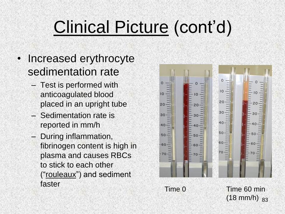
Clinical Picture (cont’d)
• Increased erythrocyte
sedimentation rate– Test is performed with
anticoagulated blood
placed in an upright tube
– Sedimentation rate is
reported in mm/h
– During inflammation,
fibrinogen content is high in
plasma and causes RBCs
to stick to each other
(“rouleaux”) and sediment
fasterTime 0 Time 60 min
(18 mm/h)
84
6. Repair by Healing, Scar
Formation and Fibrosis1. After injury tissues may regenerate or heal.
2. Regeneration involves restitution of tissue identical to that lost by injury, healing is a fibroproliferative response that “patches” a tissue defect.
3. Some tissues are capable of healing (bone after a fracture, epithelium after a superficial wound), for most tissues repair is accomplished by ECM deposition resulting in a scar.
4. In chronic inflammation both tissue injury and repair involve ECM deposition as fibrosis (an abnormal deposition of connective tissue).
85
The sequence of healing:
1. An inflammatory response to eliminate the
initial stimulus and initiate ECM deposition.
2. Proliferation & migration of parenchymal and
connective tissue cells.
3. Formation of granulation tissue
4. Synthesis of ECM proteins.
5. Tissue remodeling
6. Wound contraction and development of wound
strength.
86
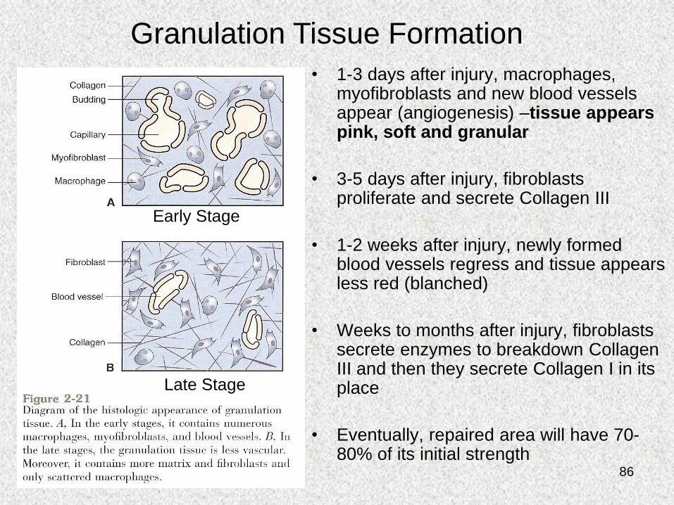
• 1-3 days after injury, macrophages, myofibroblasts and new blood vessels appear (angiogenesis) –tissue appears pink, soft and granular
• 3-5 days after injury, fibroblasts proliferate and secrete Collagen III
• 1-2 weeks after injury, newly formed blood vessels regress and tissue appears less red (blanched)
• Weeks to months after injury, fibroblasts secrete enzymes to breakdown Collagen III and then they secrete Collagen I in its place
• Eventually, repaired area will have 70-80% of its initial strength
Early Stage
Late Stage
Granulation Tissue Formation
87
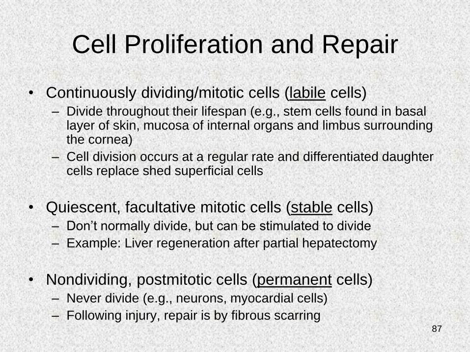
Cell Proliferation and Repair
• Continuously dividing/mitotic cells (labile cells)– Divide throughout their lifespan (e.g., stem cells found in basal
layer of skin, mucosa of internal organs and limbus surrounding the cornea)
– Cell division occurs at a regular rate and differentiated daughter cells replace shed superficial cells
• Quiescent, facultative mitotic cells (stable cells)– Don’t normally divide, but can be stimulated to divide
– Example: Liver regeneration after partial hepatectomy
• Nondividing, postmitotic cells (permanent cells)– Never divide (e.g., neurons, myocardial cells)
– Following injury, repair is by fibrous scarring
88
Repair by Regeneration or Scar
Formation?
• Regeneration– Labile (stem) cells and quiescent (stable) cells
– If injury was small, of short duration and if cells can regrow
• Scar Formation– Postmitotic (permanent) cells
– If the injury was large and cells cannot grow
* Often repair involves both regeneration and scar formation
89
6b) Clinical Wound Healing
• First Intention/Primary Healing
• Second Intention/Secondary Healing
90
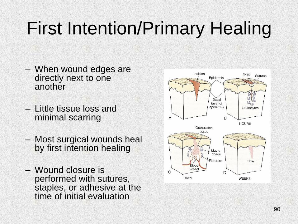
First Intention/Primary Healing
– When wound edges are directly next to one another
– Little tissue loss and minimal scarring
– Most surgical wounds heal by first intention healing
– Wound closure is performed with sutures, staples, or adhesive at the time of initial evaluation
91
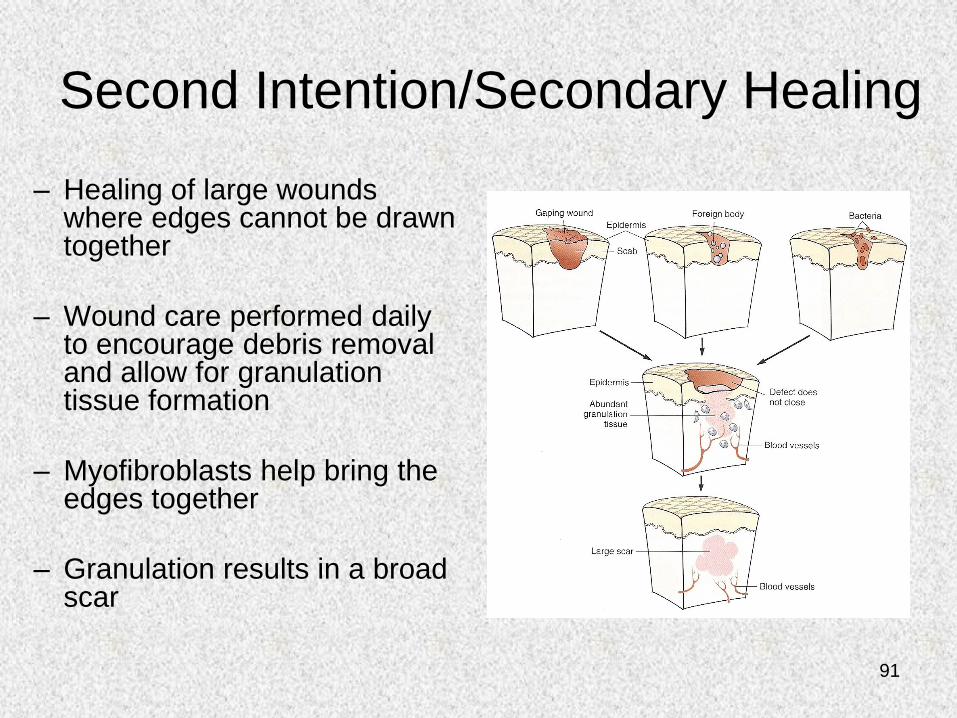
Second Intention/Secondary Healing
– Healing of large wounds where edges cannot be drawn together
– Wound care performed daily to encourage debris removal and allow for granulation tissue formation
– Myofibroblasts help bring the edges together
– Granulation results in a broad scar

![Skin Inflammation, [Acute, Suppurative, Chronic, Chronic ... · Skin – Inflammation, [Acute, Suppurative, Chronic, Chronic Active, Granulomatous] presence of mononuclear cells (lymphocytes,](https://img.dokumen.tips/doc/110x75/5f0eb0c97e708231d44075f1/skin-inflammation-acute-suppurative-chronic-chronic-skin-a-inflammation.jpg)