Embed Size (px)
Citation preview
1
Inflammationand Chemokines
Robert BeattyMCB150
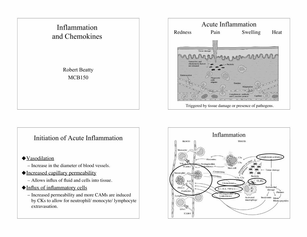
Acute InflammationRedness Pain Swelling Heat
Triggered by tissue damage or presence of pathogens.
Initiation of Acute Inflammation
Vasodilation– Increase in the diameter of blood vessels.
Increased capillary permeability– Allows influx of fluid and cells into tissue.
Influx of inflammatory cells– Increased permeability and more CAMs are induced
by CKs to allow for neutrophil/ monocyte/ lymphocyteextravasation.
Inflammation
TLRs
2
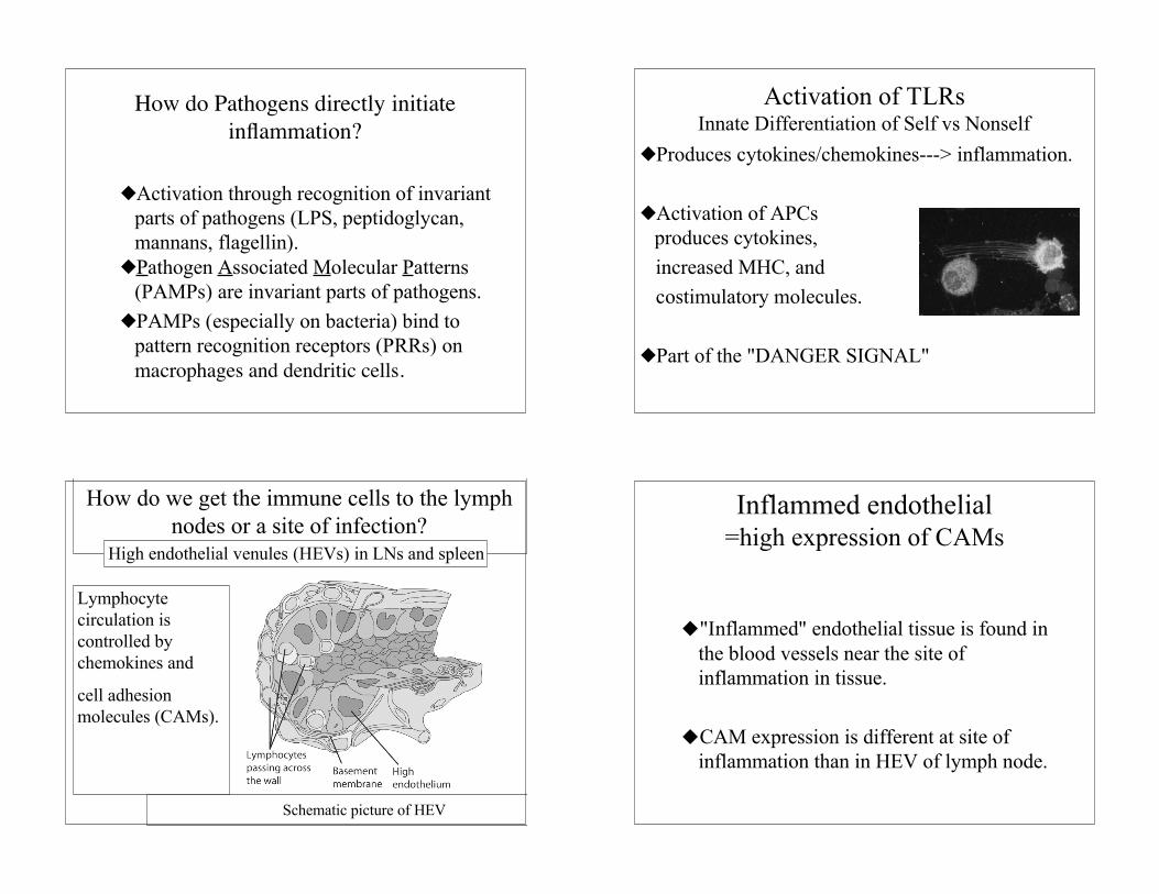
How do Pathogens directly initiateinflammation?
Activation through recognition of invariantparts of pathogens (LPS, peptidoglycan,mannans, flagellin).Pathogen Associated Molecular Patterns
(PAMPs) are invariant parts of pathogens.PAMPs (especially on bacteria) bind to
pattern recognition receptors (PRRs) onmacrophages and dendritic cells.
Activation of TLRsInnate Differentiation of Self vs Nonself
Produces cytokines/chemokines---> inflammation.
Activation of APCs produces cytokines,
increased MHC, and costimulatory molecules.
Part of the "DANGER SIGNAL"
How do we get the immune cells to the lymphnodes or a site of infection?
Schematic picture of HEV
Lymphocytecirculation iscontrolled bychemokines and
cell adhesionmolecules (CAMs).
High endothelial venules (HEVs) in LNs and spleen
Inflammed endothelial=high expression of CAMs
"Inflammed" endothelial tissue is found inthe blood vessels near the site ofinflammation in tissue.
CAM expression is different at site ofinflammation than in HEV of lymph node.
3
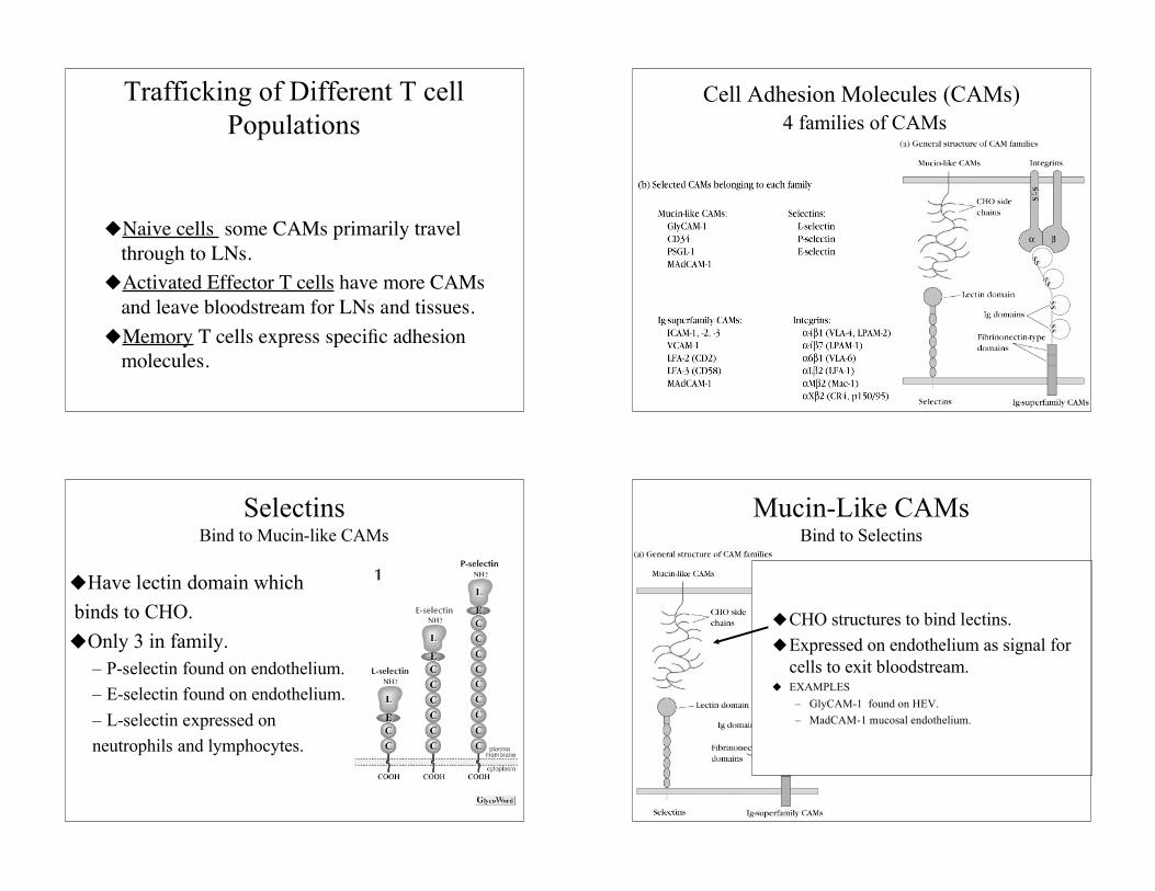
Trafficking of Different T cellPopulations
Naive cells some CAMs primarily travelthrough to LNs.
Activated Effector T cells have more CAMsand leave bloodstream for LNs and tissues.
Memory T cells express specific adhesionmolecules.
Cell Adhesion Molecules (CAMs) 4 families of CAMs
.
SelectinsBind to Mucin-like CAMs
Have lectin domain which binds to CHO.Only 3 in family.
– P-selectin found on endothelium.– E-selectin found on endothelium.– L-selectin expressed onneutrophils and lymphocytes.
Mucin-Like CAMsBind to Selectins
CHO structures to bind lectins.Expressed on endothelium as signal for
cells to exit bloodstream. EXAMPLES
– GlyCAM-1 found on HEV.– MadCAM-1 mucosal endothelium.
4
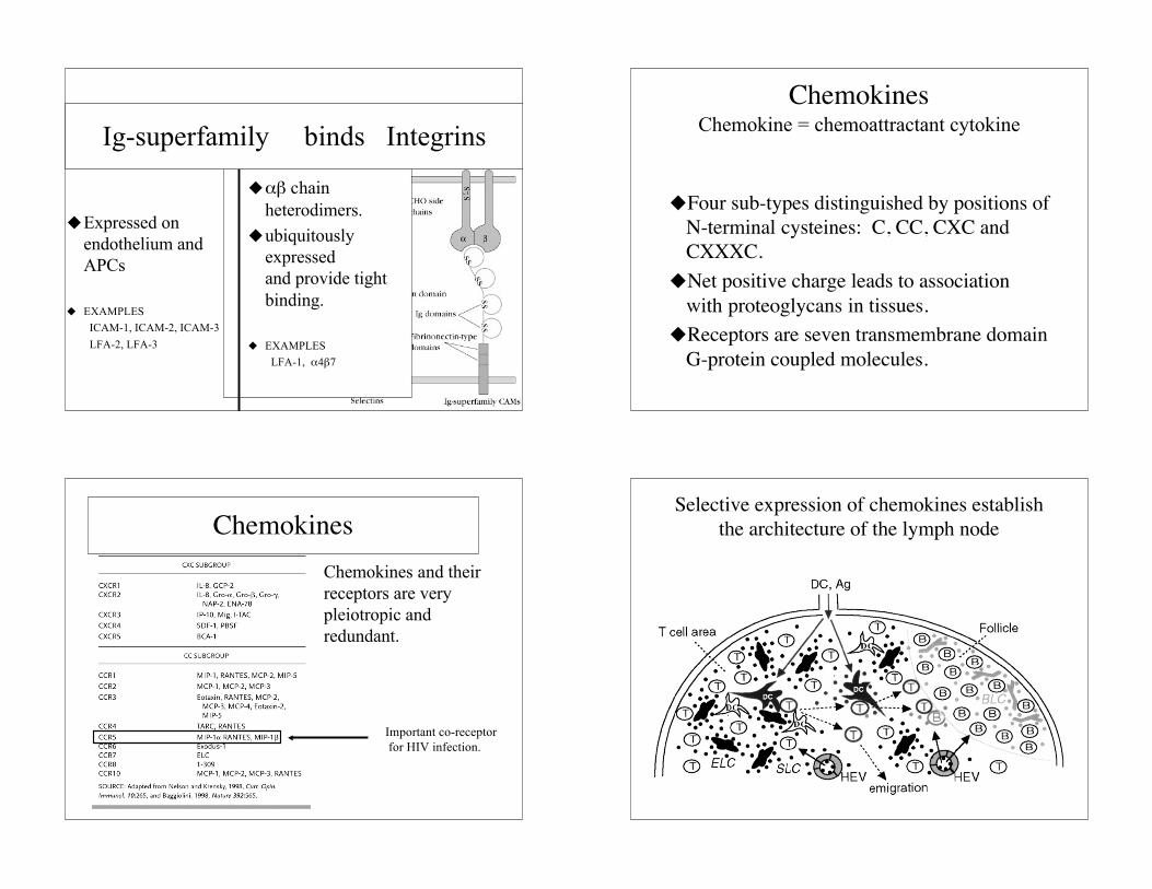
Expressed onendothelium andAPCs
EXAMPLESICAM-1, ICAM-2, ICAM-3LFA-2, LFA-3
αβ chainheterodimers.
ubiquitouslyexpressed and provide tightbinding.
EXAMPLESLFA-1, α4β7
Ig-superfamily binds IntegrinsChemokines
Chemokine = chemoattractant cytokine
Four sub-types distinguished by positions ofN-terminal cysteines: C, CC, CXC andCXXXC.
Net positive charge leads to associationwith proteoglycans in tissues.
Receptors are seven transmembrane domainG-protein coupled molecules.
Chemokines and theirreceptors are verypleiotropic andredundant.
Important co-receptor for HIV infection.
ChemokinesSelective expression of chemokines establish
the architecture of the lymph node
5
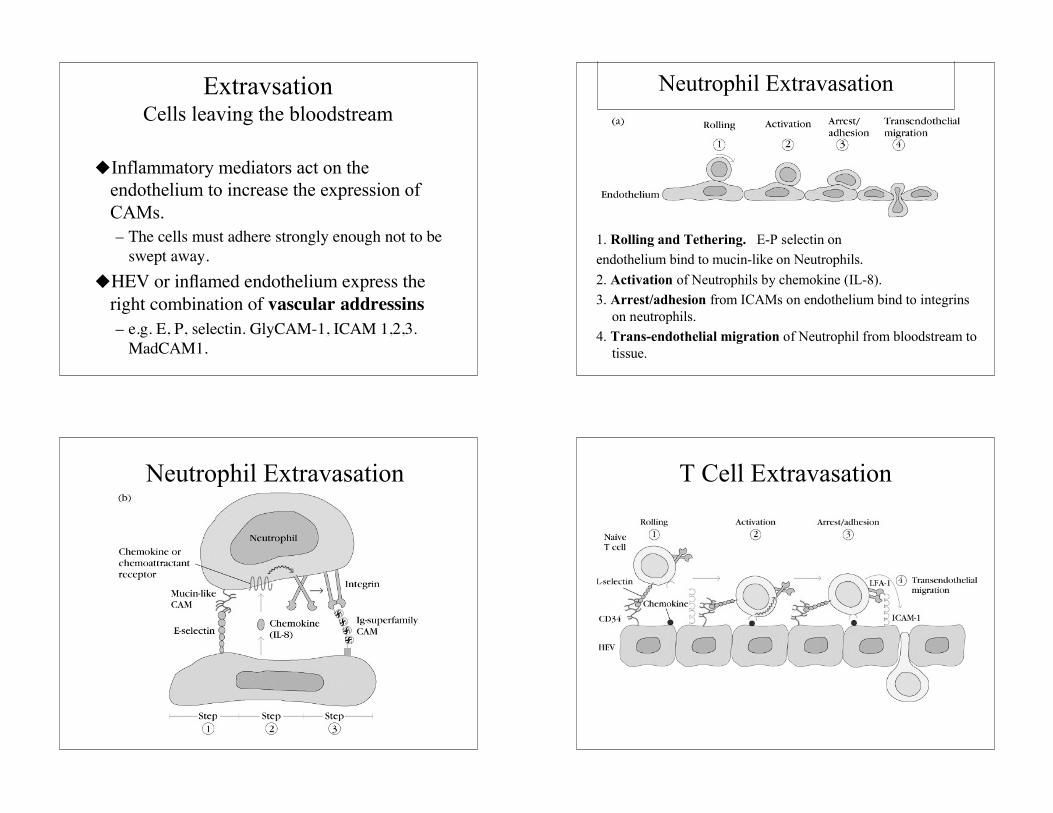
ExtravsationCells leaving the bloodstream
Inflammatory mediators act on theendothelium to increase the expression ofCAMs.– The cells must adhere strongly enough not to be
swept away.HEV or inflamed endothelium express the
right combination of vascular addressins– e.g. E, P, selectin. GlyCAM-1, ICAM 1,2,3.
MadCAM1.
1. Rolling and Tethering. E-P selectin onendothelium bind to mucin-like on Neutrophils.2. Activation of Neutrophils by chemokine (IL-8).3. Arrest/adhesion from ICAMs on endothelium bind to integrins
on neutrophils.4. Trans-endothelial migration of Neutrophil from bloodstream to
tissue.
Neutrophil Extravasation
Neutrophil Extravasation T Cell Extravasation
6
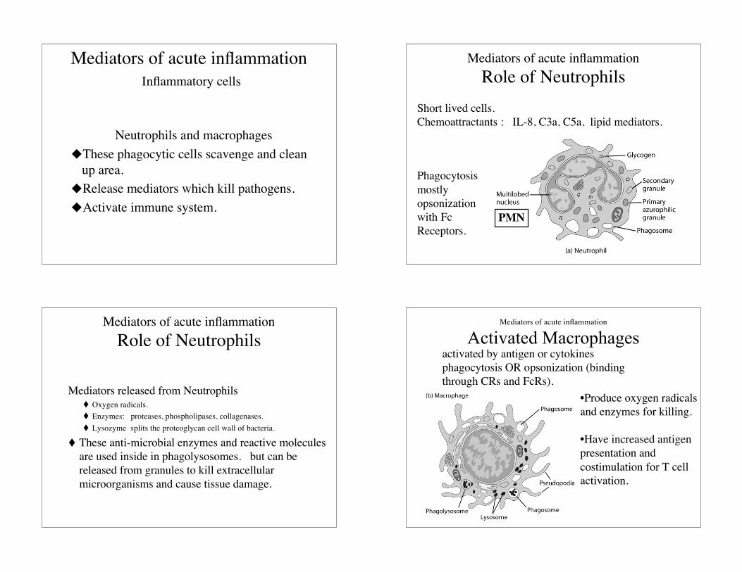
Mediators of acute inflammation Inflammatory cells
Neutrophils and macrophagesThese phagocytic cells scavenge and clean
up area.Release mediators which kill pathogens.Activate immune system.
Mediators of acute inflammationRole of Neutrophils
Phagocytosismostlyopsonizationwith FcReceptors.
Short lived cells.Chemoattractants : IL-8, C3a, C5a, lipid mediators.
PMN
Mediators of acute inflammationRole of Neutrophils
Mediators released from Neutrophils Oxygen radicals. Enzymes: proteases, phospholipases, collagenases. Lysozyme splits the proteoglycan cell wall of bacteria.
These anti-microbial enzymes and reactive moleculesare used inside in phagolysosomes. but can bereleased from granules to kill extracellularmicroorganisms and cause tissue damage.
Mediators of acute inflammation
Activated Macrophagesactivated by antigen or cytokinesphagocytosis OR opsonization (bindingthrough CRs and FcRs).
•Produce oxygen radicalsand enzymes for killing.
•Have increased antigenpresentation andcostimulation for T cellactivation.
7
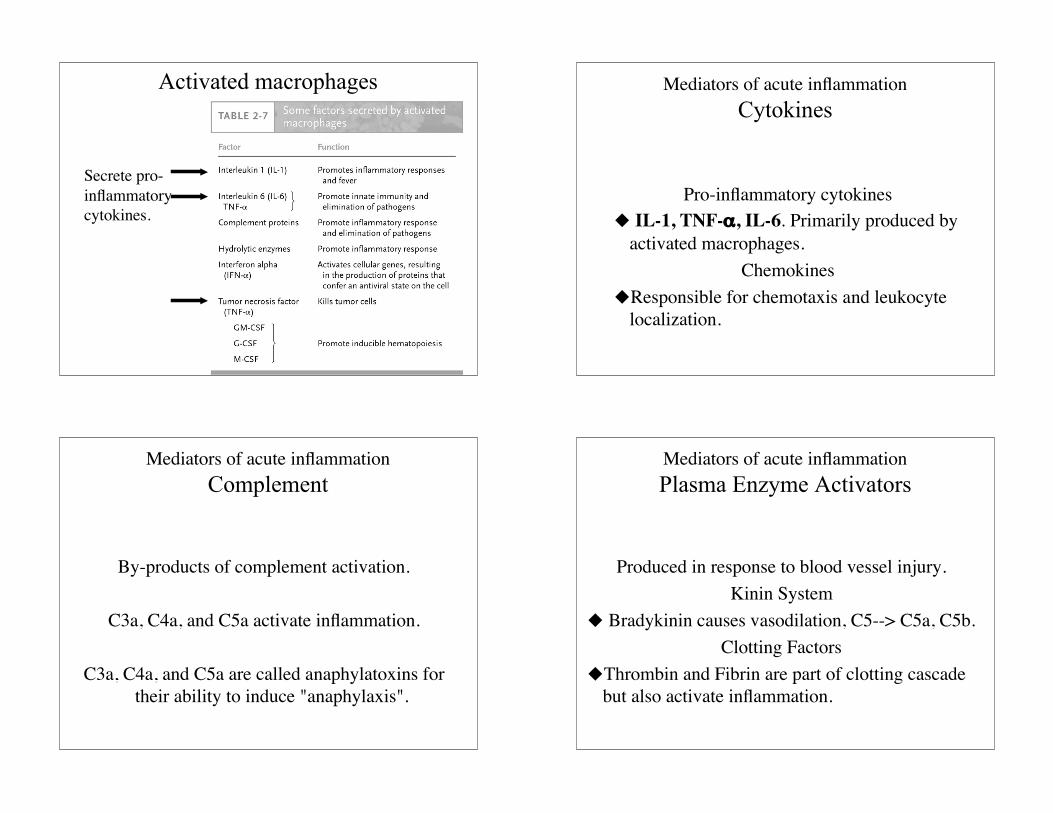
Activated macrophages
Secrete pro-inflammatorycytokines.
Mediators of acute inflammationCytokines
Pro-inflammatory cytokines IL-1, TNF-α, IL-6. Primarily produced by
activated macrophages.Chemokines
Responsible for chemotaxis and leukocytelocalization.
Mediators of acute inflammationComplement
By-products of complement activation.
C3a, C4a, and C5a activate inflammation.
C3a, C4a, and C5a are called anaphylatoxins fortheir ability to induce "anaphylaxis".
Mediators of acute inflammationPlasma Enzyme Activators
Produced in response to blood vessel injury.Kinin System
Bradykinin causes vasodilation, C5--> C5a, C5b.Clotting Factors
Thrombin and Fibrin are part of clotting cascadebut also activate inflammation.
8
Mediators of acute inflammation
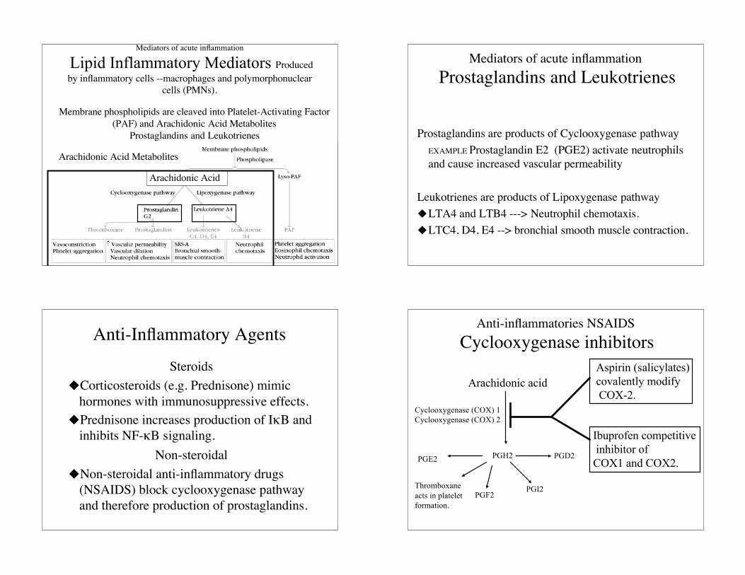
Lipid Inflammatory Mediators Producedby inflammatory cells --macrophages and polymorphonuclear
cells (PMNs).
Arachidonic Acid
Arachidonic Acid Metabolites
Membrane phospholipids are cleaved into Platelet-Activating Factor(PAF) and Arachidonic Acid Metabolites
Prostaglandins and Leukotrienes
Mediators of acute inflammation Prostaglandins and Leukotrienes
Prostaglandins are products of Cyclooxygenase pathwayEXAMPLE Prostaglandin E2 (PGE2) activate neutrophilsand cause increased vascular permeability
Leukotrienes are products of Lipoxygenase pathwayLTA4 and LTB4 ---> Neutrophil chemotaxis.LTC4, D4, E4 --> bronchial smooth muscle contraction.
Anti-Inflammatory AgentsSteroids
Corticosteroids (e.g. Prednisone) mimichormones with immunosuppressive effects.
Prednisone increases production of IκB andinhibits NF-κB signaling.
Non-steroidalNon-steroidal anti-inflammatory drugs
(NSAIDS) block cyclooxygenase pathwayand therefore production of prostaglandins.
Anti-inflammatories NSAIDS Cyclooxygenase inhibitors
Arachidonic acid
Cyclooxygenase (COX) 1Cyclooxygenase (COX) 2
PGH2PGE2 PGD2
PGF2PGI2Thromboxane
acts in plateletformation.
Aspirin (salicylates)covalently modify COX-2.
Ibuprofen competitive inhibitor of COX1 and COX2.
9
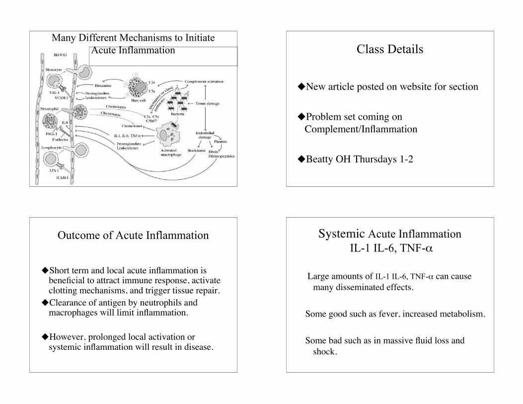
Many Different Mechanisms to InitiateAcute Inflammation Class Details
New article posted on website for section
Problem set coming onComplement/Inflammation
Beatty OH Thursdays 1-2
Outcome of Acute Inflammation
Short term and local acute inflammation isbeneficial to attract immune response, activateclotting mechanisms, and trigger tissue repair.
Clearance of antigen by neutrophils andmacrophages will limit inflammation.
However, prolonged local activation orsystemic inflammation will result in disease.
Systemic Acute Inflammation IL-1 IL-6, TNF-α
Large amounts of IL-1 IL-6, TNF-α can causemany disseminated effects.
Some good such as fever, increased metabolism.
Some bad such as in massive fluid loss andshock.
10
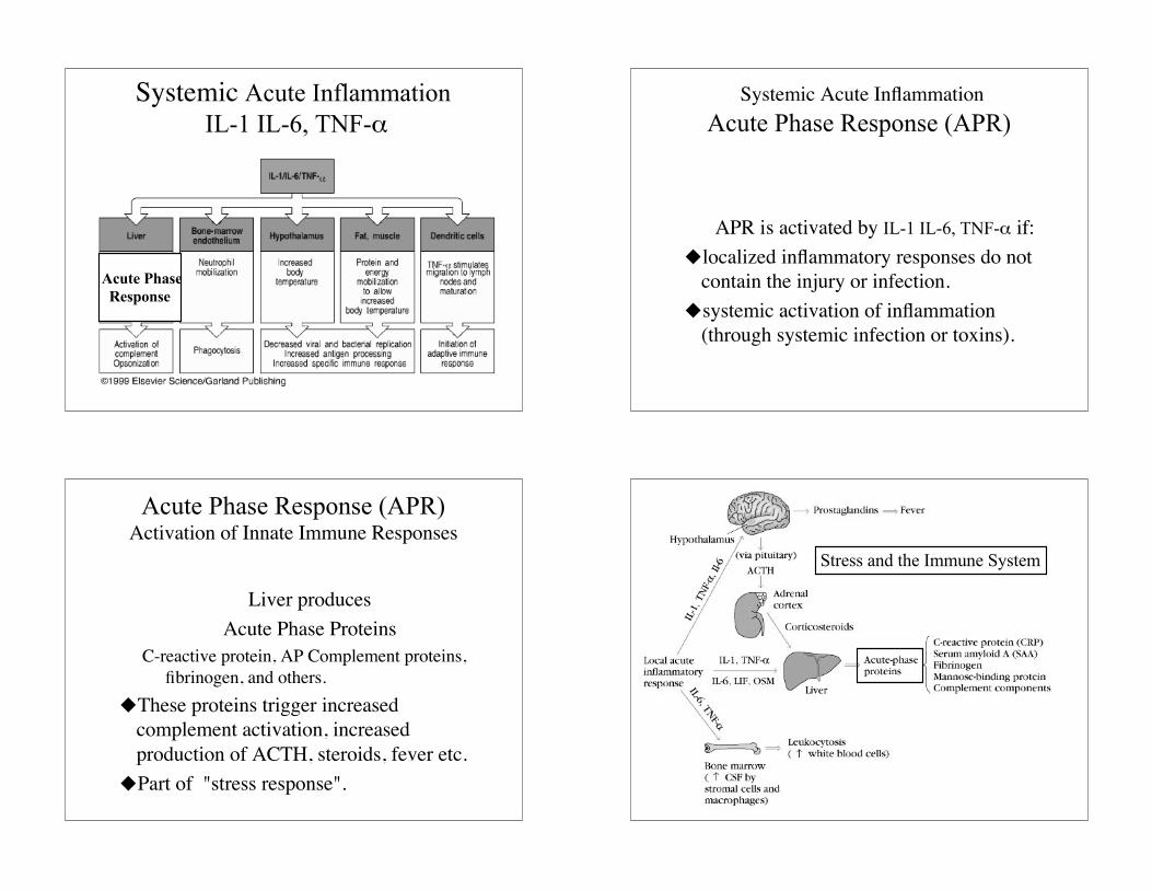
Systemic Acute Inflammation IL-1 IL-6, TNF-α
Acute Phase Response
Systemic Acute InflammationAcute Phase Response (APR)
APR is activated by IL-1 IL-6, TNF-α if:localized inflammatory responses do not
contain the injury or infection.systemic activation of inflammation
(through systemic infection or toxins).
Acute Phase Response (APR)Activation of Innate Immune Responses
Liver producesAcute Phase Proteins
C-reactive protein, AP Complement proteins,fibrinogen, and others.
These proteins trigger increasedcomplement activation, increasedproduction of ACTH, steroids, fever etc.
Part of "stress response".
Stress and the Immune System
11
Stress and the Immune ResponseImmune Central Nervous System
CNS can activate APR in the liver.– IL-1, IL-6, TNF-α, Oncostatin-M, Leukemia
inhibitory factor, produced by CNS can ALLactivate production of acute phase proteins.
Acute phase proteins and cytokines canactivate hormonal pathways and CNS.
Effects of "Stress" onImmune Response
Stress hormones (e.g. glucocorticoids) canbe made by the CNS and haveimmunosuppressive effects (inhibit Th1).
Stress hormones and cytokines made byCNS can act on endothelium to initiateinflammatory cascade.
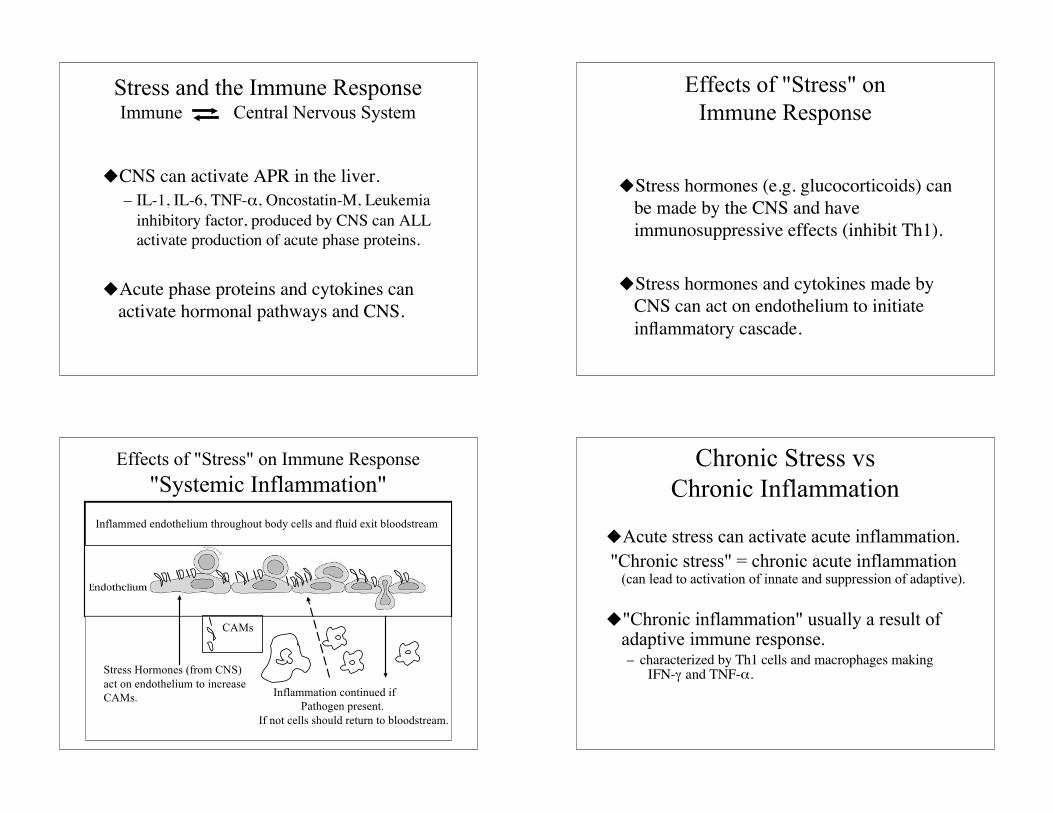
Effects of "Stress" on Immune Response"Systemic Inflammation"
Stress Hormones (from CNS)act on endothelium to increaseCAMs. Inflammation continued if
Pathogen present. If not cells should return to bloodstream.
CAMs
Inflammed endothelium throughout body cells and fluid exit bloodstream
Chronic Stress vsChronic Inflammation
Acute stress can activate acute inflammation. "Chronic stress" = chronic acute inflammation
(can lead to activation of innate and suppression of adaptive).
"Chronic inflammation" usually a result ofadaptive immune response.– characterized by Th1 cells and macrophages making
IFN-γ and TNF-α.
12
Adaptive Immune ResponsesLeading to Chronic Inflammation
Infectious agent persisting.Autoimmunity.Cancer. Tissue damage or an adaptive
immune response to tumor can result inchronic inflammation in area surroundingtumor.
Delayed type hypersensitivity (DTH).