Embed Size (px)
Citation preview
INFLAMMATION
Acute And Chronic
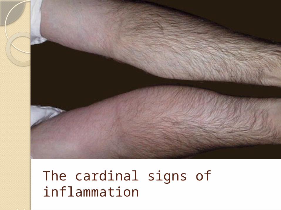
The cardinal signs of inflammation
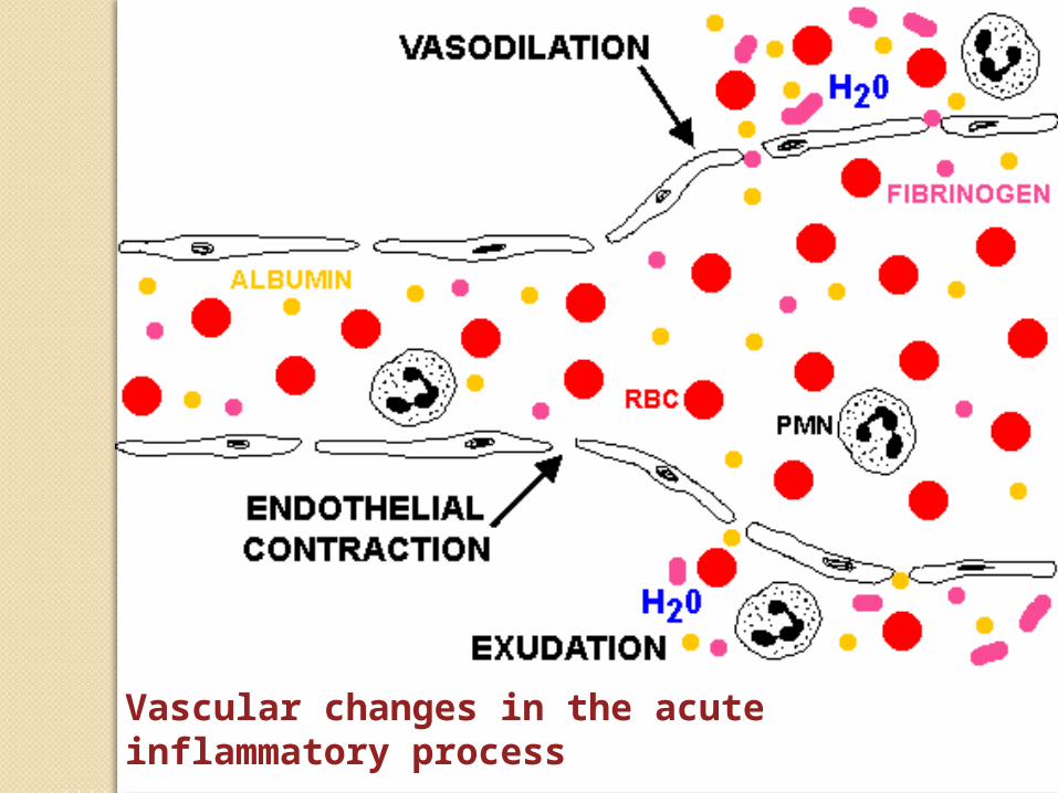
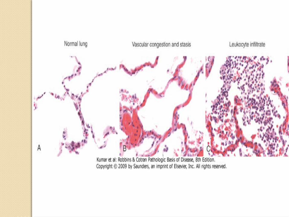
Vascular changes in the acute inflammatory process
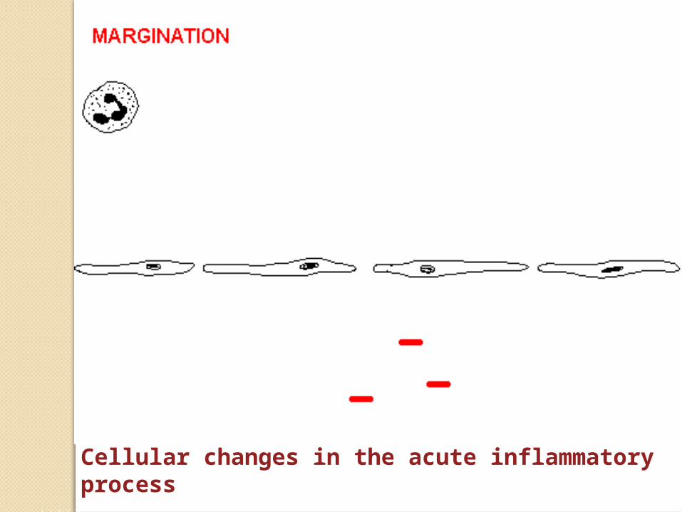
Cellular changes in the acute inflammatory process
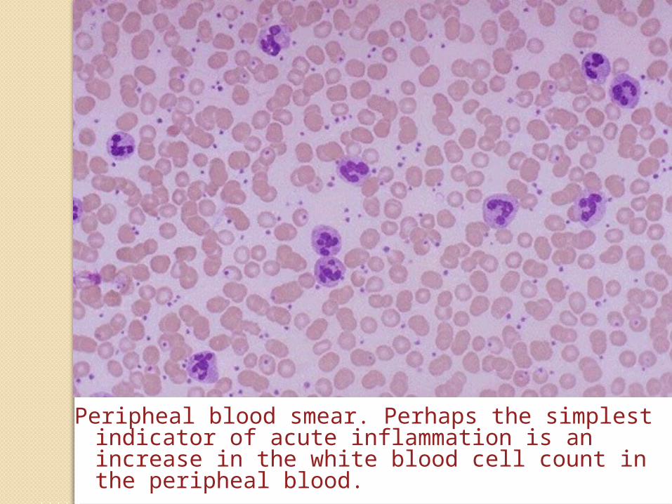
Peripheal blood smear. Perhaps the simplest indicator of acute inflammation is an increase in the white blood cell count in the peripheal blood.
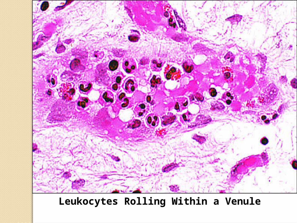
Leukocytes Rolling Within a Venule
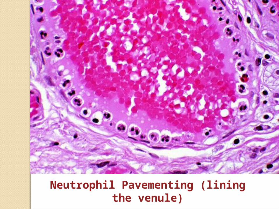
Neutrophil Pavementing (lining the venule)
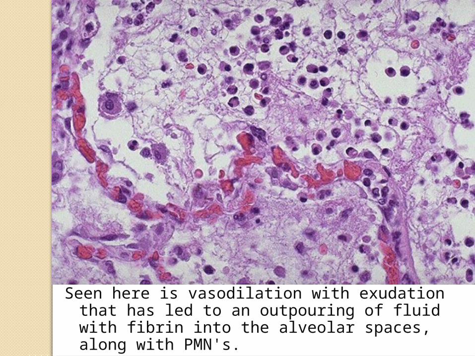
Seen here is vasodilation with exudation that has led to an outpouring of fluid with fibrin into the alveolar spaces, along with PMN's.
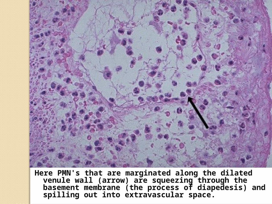
Here PMN's that are marginated along the dilated venule wall (arrow) are squeezing through the basement membrane (the process of diapedesis) and spilling out into extravascular space.
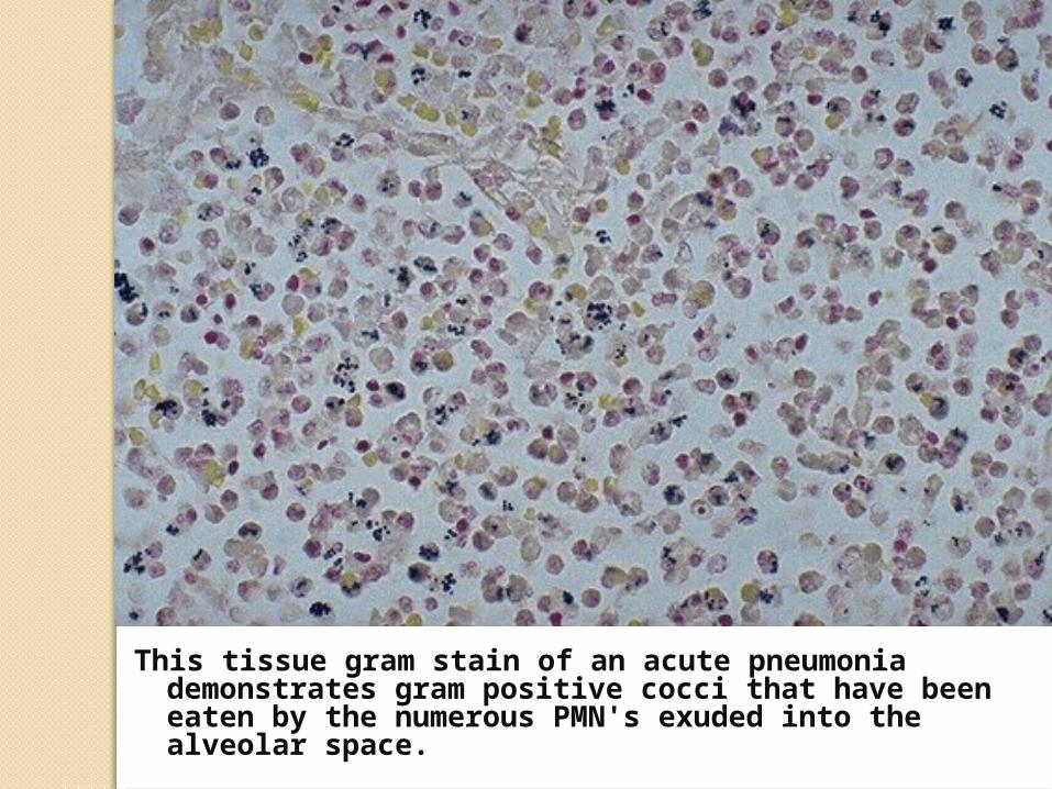
This tissue gram stain of an acute pneumonia demonstrates gram positive cocci that have been eaten by the numerous PMN's exuded into the alveolar space.
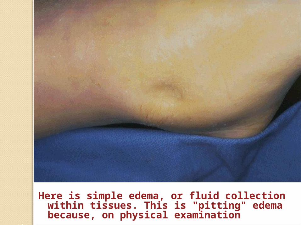
Here is simple edema, or fluid collection within tissues. This is "pitting" edema because, on physical examination
Morphologic Patterns of Acute Inflammation
Serous InflammationFibrinous InflammationPurulent InflammationUlcer
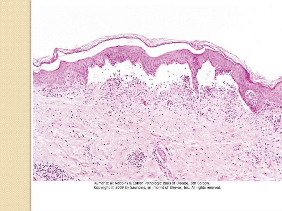
1- Serous Inflammation
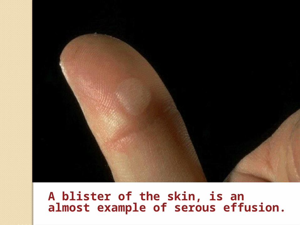
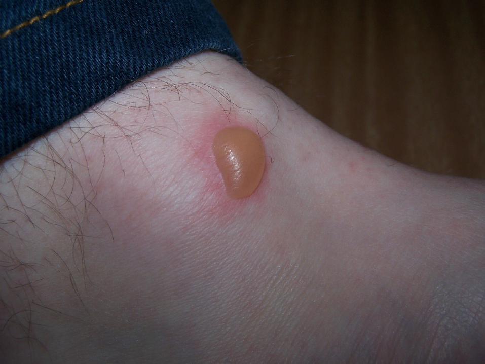
A blister of the skin, is an almost example of serous effusion.
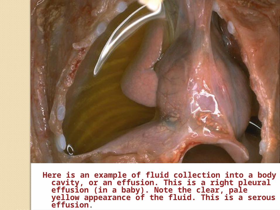
Here is an example of fluid collection into a body cavity, or an effusion. This is a right pleural effusion (in a baby). Note the clear, pale yellow appearance of the fluid. This is a serous effusion.
2- Fibrinous Inflammation
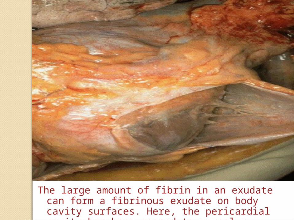
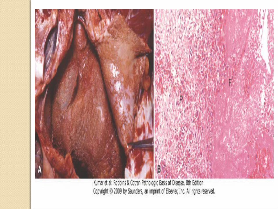
The large amount of fibrin in an exudate can form a fibrinous exudate on body cavity surfaces. Here, the pericardial cavity has been opened to reveal a fibrinous pericarditis
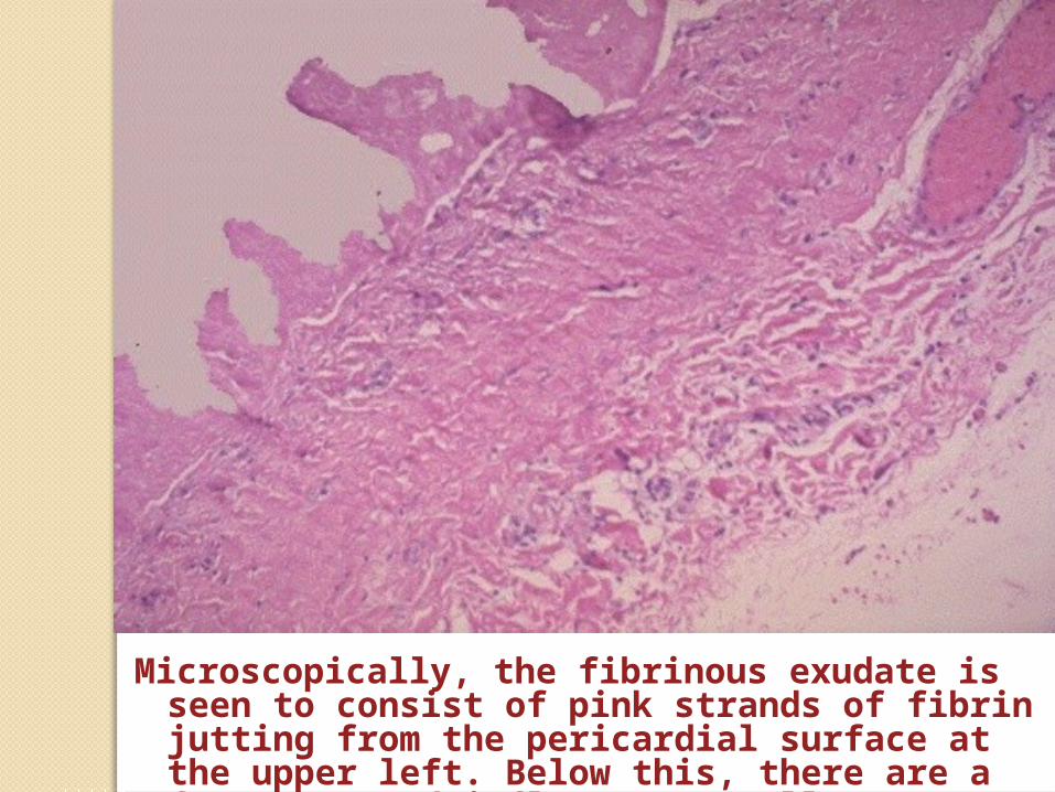
Microscopically, the fibrinous exudate is seen to consist of pink strands of fibrin jutting from the pericardial surface at the upper left. Below this, there are a few scattered inflammatory cells.
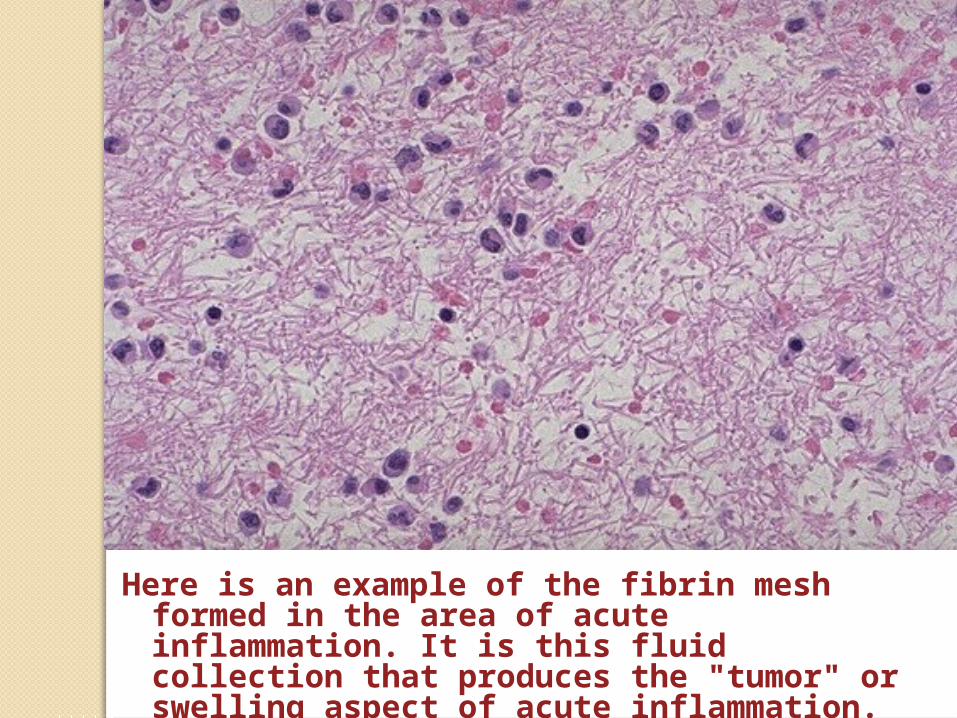
Here is an example of the fibrin mesh formed in the area of acute inflammation. It is this fluid collection that produces the "tumor" or swelling aspect of acute inflammation.
3- Purulent Inflammation
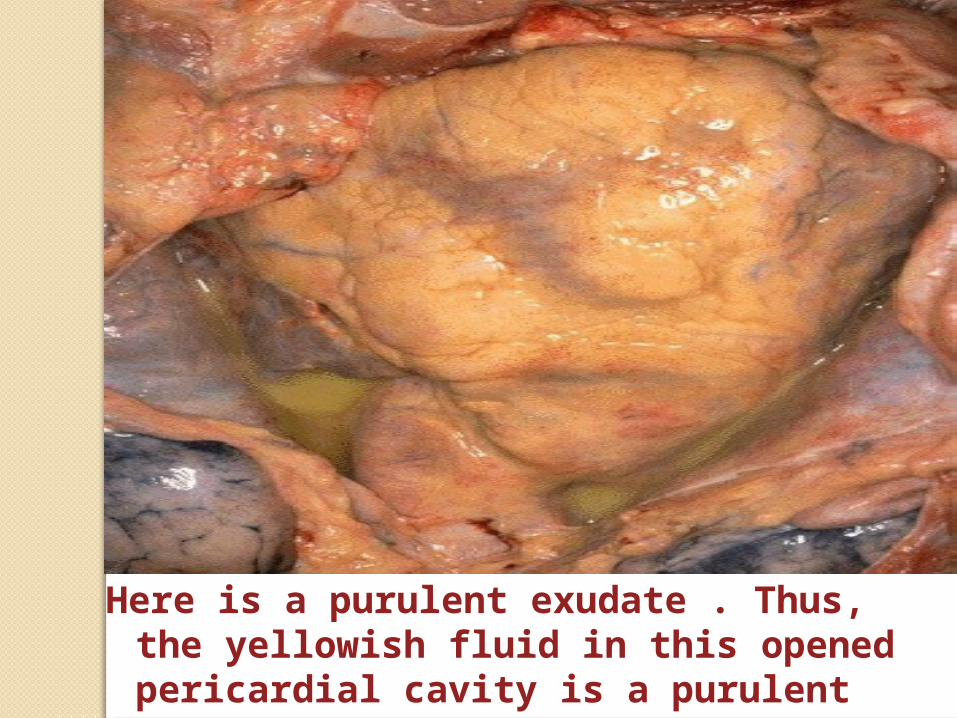
Here is a purulent exudate . Thus, the yellowish fluid in this opened pericardial cavity is a purulent exudate.
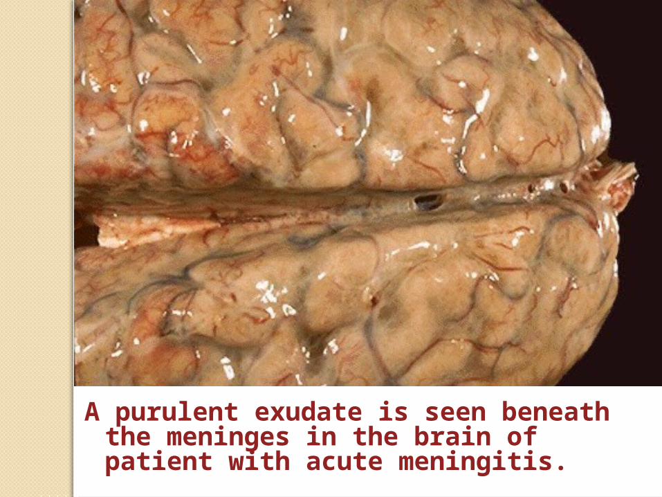
A purulent exudate is seen beneath the meninges in the brain of patient with acute meningitis.
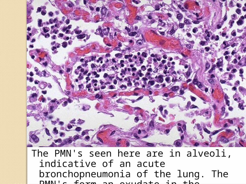
The PMN's seen here are in alveoli, indicative of an acute bronchopneumonia of the lung. The PMN's form an exudate in the alveoli.
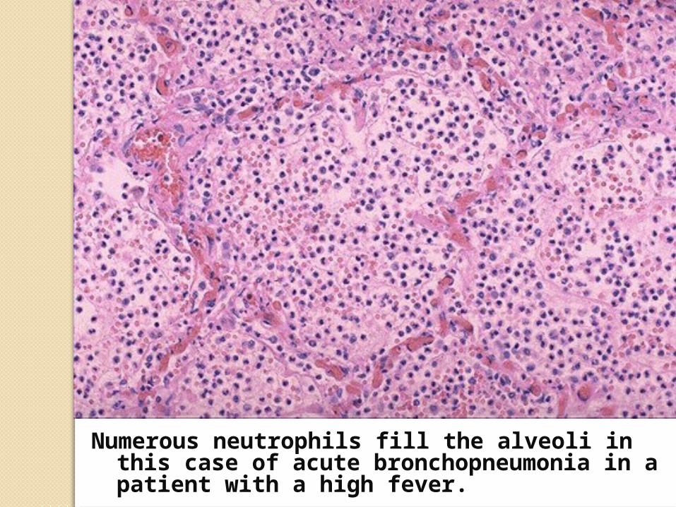
Numerous neutrophils fill the alveoli in this case of acute bronchopneumonia in a patient with a high fever.
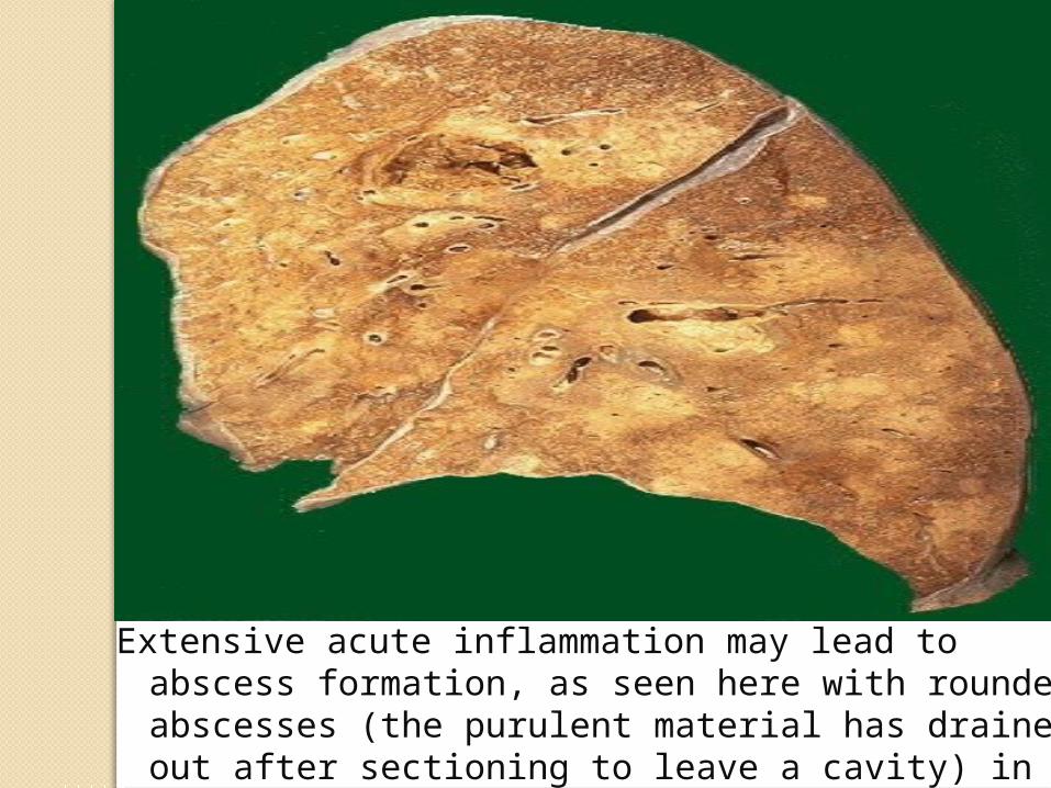
Extensive acute inflammation may lead to abscess formation, as seen here with rounded abscesses (the purulent material has drained out after sectioning to leave a cavity) in upper lobe.
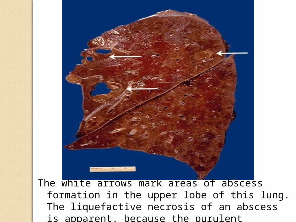
The white arrows mark areas of abscess formation in the upper lobe of this lung. The liquefactive necrosis of an abscess is apparent, because the purulent contents are draining out to leave a cavity.
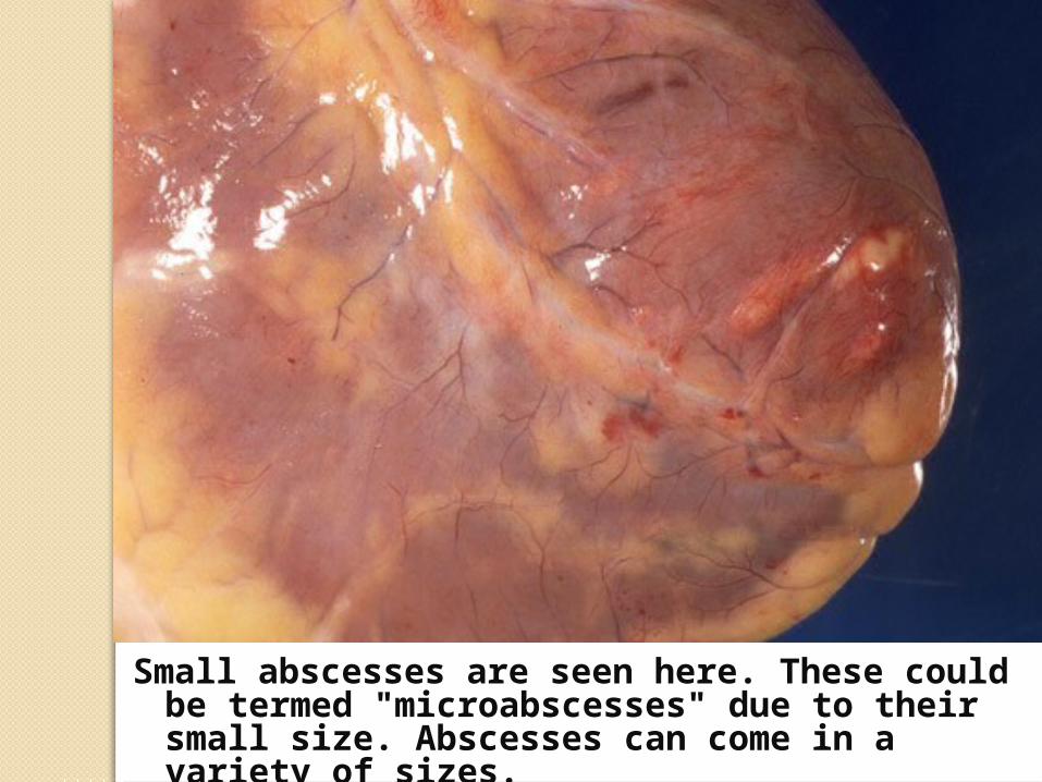
Small abscesses are seen here. These could be termed "microabscesses" due to their small size. Abscesses can come in a variety of sizes.
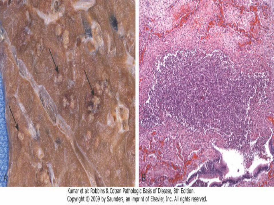
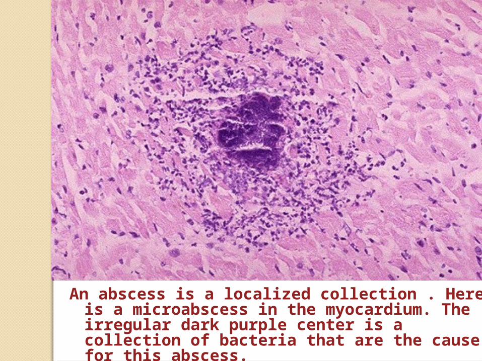
An abscess is a localized collection . Here is a microabscess in the myocardium. The irregular dark purple center is a collection of bacteria that are the cause for this abscess.

4- Ulcer
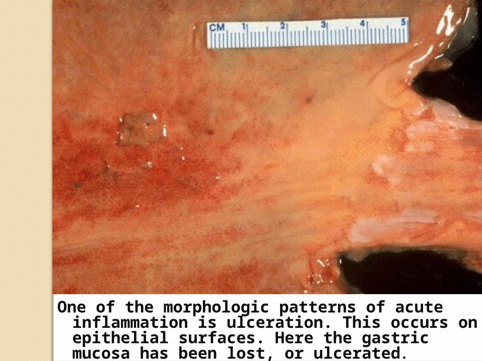
One of the morphologic patterns of acute inflammation is ulceration. This occurs on epithelial surfaces. Here the gastric mucosa has been lost, or ulcerated.
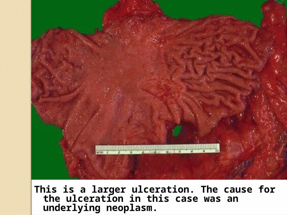
This is a larger ulceration. The cause for the ulceration in this case was an underlying neoplasm.
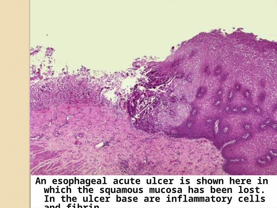
An esophageal acute ulcer is shown here in which the squamous mucosa has been lost. In the ulcer base are inflammatory cells and fibrin.
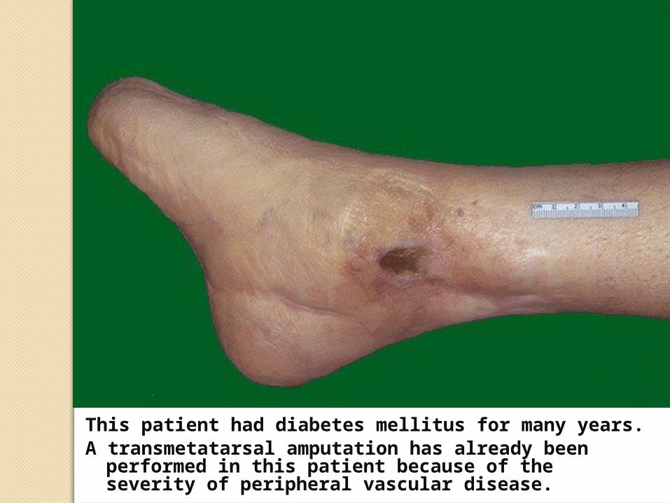
This patient had diabetes mellitus for many years. A transmetatarsal amputation has already been
performed in this patient because of the severity of peripheral vascular disease.
Chronic Inflammation
Chronic Nonspecific Inflammation
Granulomatous Inflammation
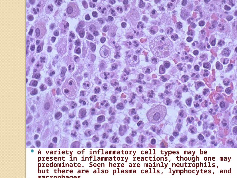
A variety of inflammatory cell types may be present in inflammatory reactions, though one may predominate. Seen here are mainly neutrophils, but there are also plasma cells, lymphocytes, and macrophages
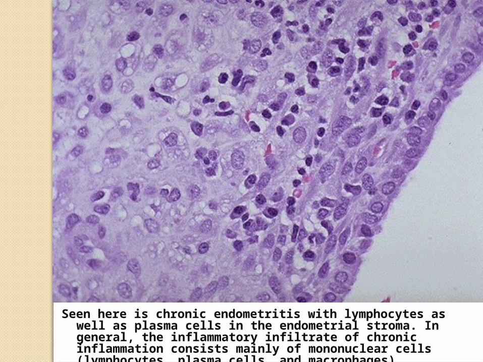
Seen here is chronic endometritis with lymphocytes as well as plasma cells in the endometrial stroma. In general, the inflammatory infiltrate of chronic inflammation consists mainly of mononuclear cells (lymphocytes, plasma cells, and macrophages).
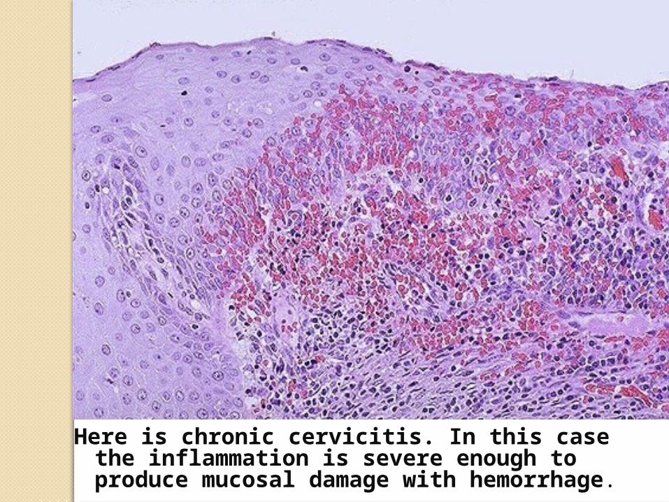
Here is chronic cervicitis. In this case the inflammation is severe enough to produce mucosal damage with hemorrhage.

Here, chronic inflammation of the bronchi has led to dilation and scarring with increased tan to white collagenous tissue.

The focal nature of granulomatous inflammation is demonstrated in this microscopic section of lung in which there are granulomas in the parenchyma. This is why the chest radiograph with tuberculosis or other granulomatous diseases is often described as "reticulonodular". A biopsy could miss such lesions from sampling error, too.

Here are two pulmonary granulomas. Granulomatous inflammation typically consists of mixtures of cells including epithelioid macrophages, giant cells, lymphocytes, plasma cells, and fibroblasts.

Langhans type giant cells are a "committee" of epithelioid macrophages. Seen here are two Langhans type giant cells in which the nuclei are lined up around the periphery of the cell.

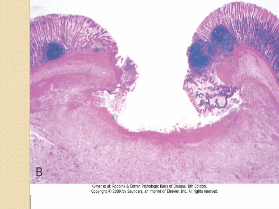
This is a caseating granuloma. Epithelioid cells surround a central area of necrosis that appears irregular, amorphous, and pink. Grossly, areas of caseation appear cheese-like.


![Inflammation and Introduction to Wound Healing...cardinal signs of acute inflammation: » Rubor (erythema [redness]): vasodilatation, increased blood flow » Tumor (swelling): extravascular](https://img.dokumen.tips/doc/110x75/612a2196391bc8275d5c2bab/inflammation-and-introduction-to-wound-healing-cardinal-signs-of-acute-inflammation.jpg)