Embed Size (px)
Citation preview
41
Turkish Journal of Trauma & Emergency Surgery
Original Article Klinik Çalışma
Ulus Travma Acil Cerrahi Derg 2013;19 (1):41-44
Inferior glenohumeral dislocation (luxatio erecta humeri): report of six cases and review of the literature
İnferior omuz çıkığı (luksasyo erekta): Altı olgu sunumu ve literatürün gözden geçirilmesi
Ahmet İMERCİ,1 Yalçın GÖLCÜK,2 Sabri Gökhan UĞUR,3 Hüseyin Tamer URSAVAŞ,4 Ahmet SAVRAN,4 Levent SÜRER5
1Department of Orthopaedics and Traumatology, Erzurum Palandoken State Hospital, Erzurum; 2Department of Emergency, Bitlis State Hospital, Bitlis; 3Department of Orthopaedics and Traumatology, Karaman State Hospital, Karaman; 4Department of Orthopaedics and Traumatology, Izmir Tepecik Training and Research Hospital, Izmir; 5Department of Orthopaedics and
Traumatology, Acibadem Bodrum Hospital, Mugla, Turkey.
1Erzurum Palandöken Devlet Hastanesi, Ortopedi ve Travmatoloji Kliniği, Erzurum; 2Bitlis Devlet Hastanesi, Acil Servis, Bitlis;
3Karaman Devlet Hastanesi, Ortopedi ve Travmatoloji Kliniği, Karaman;4İzmir Tepecik Eğitim ve Araştırma Hastanesi, Ortopedi ve Travmatoloji Kliniği, İzmir; 5Acıbadem Bodrum Hastanesi, Ortopedi ve Travmatoloji
Kliniği, Muğla.
Correspondence (İletişim): Ahmet İmerci, M.D. Erzurum Palandöken Devlet Hastanesi, Ortopedi ve Travmatoloji Kliniği, 25000 Erzurum, Turkey.Tel: +90 - 442 - 235 50 80 e-mail (e-posta): [email protected]
BACKGROUNDInferior shoulder dislocation, also referred to as luxatio erecta, is a rare type of shoulder dislocation. Its incidence is about 1 in 200 (0.5%) among all shoulder dislocations. The objective of this study was to review six cases of inferior shoulder dislocation, including their clinical and radiologi-cal presentation, management, and final outcome.METHODSFour males and two females, a total of six patients, with the diagnosis of inferior shoulder dislocation were treated between 2007 and 2010. Our purpose is to present our ex-perience in the treatment of these patients together with the parallel research available in the literature.RESULTSConstant score was used to evaluate shoulder function. Pain, position, daily activities, range of motion, and strength scores were noted. All patients had good to excel-lent results with full functional recovery within two years after closed reduction and shoulder rehabilitation.
CONCLUSIONDoctors should be familiar with the occurrence of this in-frequent condition and should prevent possible complica-tions that might result from early reductions by using cor-rect maneuvers in lieu of ordinary reduction techniques.Key Words: Closed reduction; emergency; inferior dislocation; luxatio erecta; shoulder; trauma.
AMAÇİnferior omuz çıkığı, ayrıca luksasyo erekta olarak adlandı-rıp omuz çıkığının nadir görülen bir tipidir. Görülme sıklığı tüm omuz çıkık arasındaki yaklaşık 200’de 1’dir (%0,5). Bu çalışmanın amacı, inferior omuz çıkığının klinik ve rad-yolojik olarak sunumu, tedavisi ve nihai sonucun 6 olgu ile gözden geçirilmesidir.GEREÇ VE YÖNTEMDört erkek ve iki kadın, toplam 6 hasta aşağı omuz çıkığı tanısı ile 2007 ve 2010 yılları arasında tedavi edildi. Tedavi ettiğimiz bu hastalar nedeniyle, bizim amacımız kaynaklar paralelinde deneyimimizi paylaşmaktır.
BULGULARHastaların omuz fonksiyonlarını değerlendirmek için Cons-tant omuz skorlaması kullanılarak ağrı, pozisyon, günlük yaşam aktiviteleri, eklem hareket açıklıkları ve güçü kayde-dildi. Hastaların tamamında kapalı redüksiyon ve rehabili-tasyon ile tedavi sonrası 2 yıl içinde tam fonksiyonel iyileş-me sağlanarak mükemmel ya da iyi sonuç alındı.
SONUÇDoktorlar bu nadir durumun oluşumuna alışık olmalı ve doğru redüksiyon teknikleri dışında yapılan erken redük-siyonlar neticesinde oluşabilecek olası komplikasyonları önlemeleri gerekir.Anahtar Sözcükler: Kapalı redüksiyon; acil; inferior çıkık; luksasyo erekta; omuz; travma.
doi: 10.5505/tjtes.2013.35305
Ulus Travma Acil Cerrahi Derg
The shoulder joint is the joint where dislocations occur most frequently.[1] Multi-directional mobility of the shoulder joint, its anatomic structure and frequent exposition to traumas result in the more frequent oc-currence of dislocations.[2] Forward and backward dislocations are observed at rates of 95% and 4-5%, respectively, in patients with shoulder dislocations.[1,3] Downward dislocation (luxatio erecta - LE), with an occurrence rate of 0.5% among all shoulder dislo-cations, on the other hand, is a traumatic case that is observed quite rarely, which generally occurs during hyperabduction type trauma of the arm. In such cases, it is usually observed that the inferior capsule of the joint is torn.[4,5] For the formation of the LE, a great amount of force is required; thus, many other injuries can be seen together.[6]
The current study presents six cases of downward dislocation of the glenohumeral joint that were treated with closed reduction.
MATERIALS AND METHODSFour males and two females, a total of six pa-
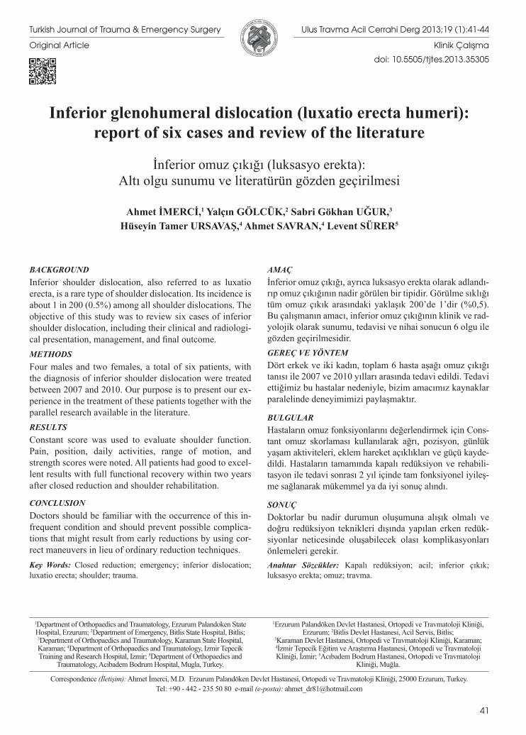
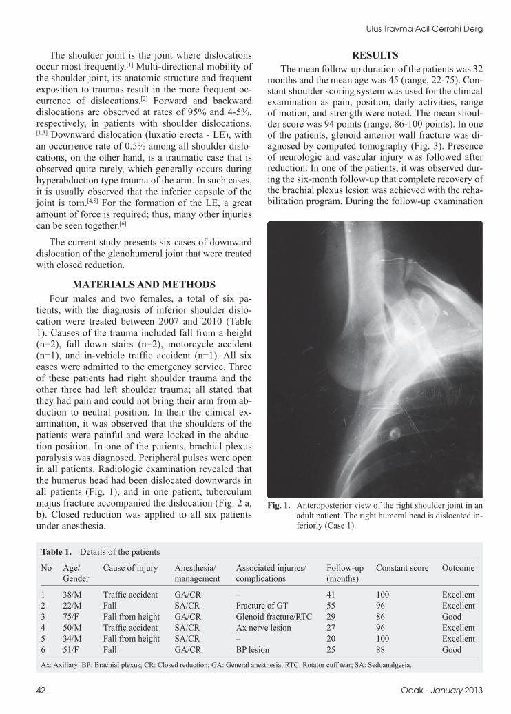
tients, with the diagnosis of inferior shoulder dislo-cation were treated between 2007 and 2010 (Table 1). Causes of the trauma included fall from a height (n=2), fall down stairs (n=2), motorcycle accident (n=1), and in-vehicle traffic accident (n=1). All six cases were admitted to the emergency service. Three of these patients had right shoulder trauma and the other three had left shoulder trauma; all stated that they had pain and could not bring their arm from ab-duction to neutral position. In their the clinical ex-amination, it was observed that the shoulders of the patients were painful and were locked in the abduc-tion position. In one of the patients, brachial plexus paralysis was diagnosed. Peripheral pulses were open in all patients. Radiologic examination revealed that the humerus head had been dislocated downwards in all patients (Fig. 1), and in one patient, tuberculum majus fracture accompanied the dislocation (Fig. 2 a, b). Closed reduction was applied to all six patients under anesthesia.
RESULTSThe mean follow-up duration of the patients was 32
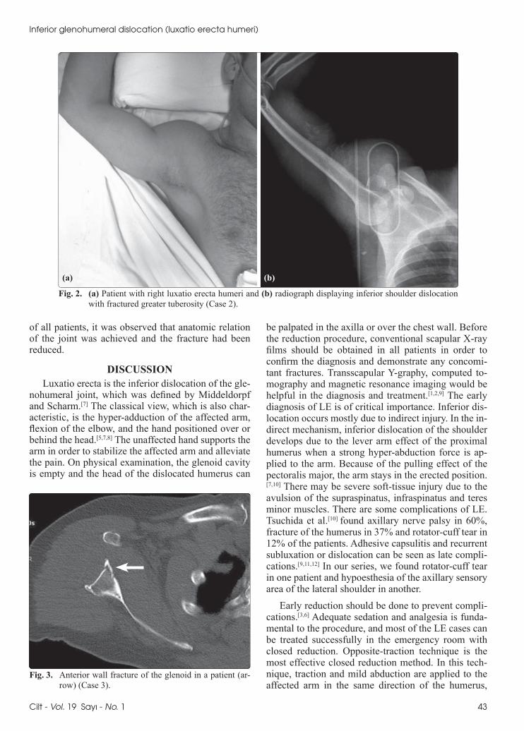
months and the mean age was 45 (range, 22-75). Con-stant shoulder scoring system was used for the clinical examination as pain, position, daily activities, range of motion, and strength were noted. The mean shoul-der score was 94 points (range, 86-100 points). In one of the patients, glenoid anterior wall fracture was di-agnosed by computed tomography (Fig. 3). Presence of neurologic and vascular injury was followed after reduction. In one of the patients, it was observed dur-ing the six-month follow-up that complete recovery of the brachial plexus lesion was achieved with the reha-bilitation program. During the follow-up examination
42 Ocak - January 2013
Table 1. Details of the patients
No Age/ Cause of injury Anesthesia/ Associated injuries/ Follow-up Constant score Outcome Gender management complications (months)
1 38/M Traffic accident GA/CR – 41 100 Excellent2 22/M Fall SA/CR Fracture of GT 55 96 Excellent3 75/F Fall from height GA/CR Glenoid fracture/RTC 29 86 Good4 50/M Traffic accident SA/CR Ax nerve lesion 27 96 Excellent5 34/M Fall from height SA/CR – 20 100 Excellent6 51/F Fall GA/CR BP lesion 25 88 Good
Ax: Axillary; BP: Brachial plexus; CR: Closed reduction; GA: General anesthesia; RTC: Rotator cuff tear; SA: Sedoanalgesia.
Fig. 1. Anteroposterior view of the right shoulder joint in an adult patient. The right humeral head is dislocated in-feriorly (Case 1).
Inferior glenohumeral dislocation (luxatio erecta humeri)
of all patients, it was observed that anatomic relation of the joint was achieved and the fracture had been reduced.
DISCUSSIONLuxatio erecta is the inferior dislocation of the gle-
nohumeral joint, which was defined by Middeldorpf and Scharm.[7] The classical view, which is also char-acteristic, is the hyper-adduction of the affected arm, flexion of the elbow, and the hand positioned over or behind the head.[5,7,8] The unaffected hand supports the arm in order to stabilize the affected arm and alleviate the pain. On physical examination, the glenoid cavity is empty and the head of the dislocated humerus can
be palpated in the axilla or over the chest wall. Before the reduction procedure, conventional scapular X-ray films should be obtained in all patients in order to confirm the diagnosis and demonstrate any concomi-tant fractures. Transscapular Y-graphy, computed to-mography and magnetic resonance imaging would be helpful in the diagnosis and treatment.[1,2,9] The early diagnosis of LE is of critical importance. Inferior dis-location occurs mostly due to indirect injury. In the in-direct mechanism, inferior dislocation of the shoulder develops due to the lever arm effect of the proximal humerus when a strong hyper-abduction force is ap-plied to the arm. Because of the pulling effect of the pectoralis major, the arm stays in the erected position.[7,10] There may be severe soft-tissue injury due to the avulsion of the supraspinatus, infraspinatus and teres minor muscles. There are some complications of LE. Tsuchida et al.[10] found axillary nerve palsy in 60%, fracture of the humerus in 37% and rotator-cuff tear in 12% of the patients. Adhesive capsulitis and recurrent subluxation or dislocation can be seen as late compli-cations.[9,11,12] In our series, we found rotator-cuff tear in one patient and hypoesthesia of the axillary sensory area of the lateral shoulder in another.
Early reduction should be done to prevent compli-cations.[3,6] Adequate sedation and analgesia is funda-mental to the procedure, and most of the LE cases can be treated successfully in the emergency room with closed reduction. Opposite-traction technique is the most effective closed reduction method. In this tech-nique, traction and mild abduction are applied to the affected arm in the same direction of the humerus,
Cilt - Vol. 19 Sayı - No. 1 43
Fig. 2. (a) Patient with right luxatio erecta humeri and (b) radiograph displaying inferior shoulder dislocation with fractured greater tuberosity (Case 2).
(a) (b)
Fig. 3. Anterior wall fracture of the glenoid in a patient (ar-row) (Case 3).
Ulus Travma Acil Cerrahi Derg
occurrence mechanism and clinical presentation. Doc-tors should be familiar with the occurrence of this in-frequent condition and should prevent possible com-plications that might result from early reductions by using correct maneuvers in lieu of ordinary reduction techniques.
Conflict-of-interest issues regarding the authorship or article: None declared.
REFERENCES1. Rockwood CA, Wirth MA. Subluxations and dislocations
about the glenohumeral joint. In: Rockwood CA, Green DP, Bucholz RW, editors. Fractures in adults. Philadelphia: Lip-pincott-Raven; 1996. p. 1193-39.
2. Yamamoto T, Yoshiya S, Kurosaka M, Nagira K, Nabeshima Y. Luxatio erecta (inferior dislocation of the shoulder): a re-port of 5 cases and a review of the literature. Am J Orthop (Belle Mead NJ) 2003;32:601-3.
3. Sahin N, Oztürk A, Ozkan Y, Atıcı T, Ozkaya G. A compari-son of the scapular manipulation and Kocher’s technique for acute anterior dislocation of the shoulder. Eklem Hastalik Cerrahisi 2011;22:28-32.
4. Mallon WJ, Bassett FH 3rd, Goldner RD. Luxatio erecta: the inferior glenohumeral dislocation. J Orthop Trauma 1990;4:19-24.
5. Yanturali S, Aksay E, Holliman CJ, Duman O, Ozen YK. Luxatio erecta: clinical presentation and management in the emergency department. J Emerg Med 2005;29:85-9.
6. Matsumato K, Ohara A. Yamamoto K,Takigami I, Naganawa T. Luxatio erecta (inferior dislocation of the shoulder): A re-port of two cases and a review of the literature. Injury Extra 2005;36:450-3.
7. Karaoglu S, Guney A, Ozturk M, Kekec Z. Bilateral luxatio erecta humeri. Arch Orthop Trauma Surg 2003;123:308-10.
8. Mesa M, Carpintero P, Carpintero J. Bilateral luxatio erecta humeri. Acta Orthop Belg 1996;62:116-9.
9. Groh GI, Wirth MA, Rockwood CA Jr. Results of treatment of luxatio erecta (inferior shoulder dislocation). J Shoulder Elbow Surg 2010;19:423-6.
10. Tsuchida T, Yang K, Kimura Y, Taniwaki M, Ishigaki S, Itoi E. Luxatio erecta of bilateral shoulders. J Shoulder Elbow Surg 2001;10:595-7.
11. Musmeci E, Gaspari D, Sandri A, Regis D, Bartolozzi P. Bi-lateral luxatio erecta humeri associated with a unilateral bra-chial plexus and bilateral rotator cuff injuries: a case report. J Orthop Trauma 2008;22:498-500.
12. Wang KC, Hsu KY, Shih CH. Brachial plexus injury with erect dislocation of the shoulder. Orthop Rev 1992;21:1345-7.
13. Camarda L, Martorana U, D’Arienzo M. A case of bilateral luxatio erecta. J Orthop Traumatol 2009;10:97-9.
14. Durukan P, Yıldız M, Barik A, Kaya N, Yılmaz E. Inferior glenohumeral dislokasyon (Luxatio Erecta): İki olgu sunu-mu. Türkiye Acil Tıp Dergisi 2005;5:142-4.
15. Ebrahimzadeh MH, Fattahi A. Inferior glenohumeral dislo-cation (luxatio erecta humeri), report of two cases. Eur J Or-thop Surg Traumatol 2006;16:30-2.
16. Féry A, Sommelet J. Erect dislocation of the shoulder (luxa-tio erecta humeri). General review apropos of 10 cases. [Ar-ticle in French] Int Orthop 1987;11:95-103. [Abstract]
while opposite-directional traction is performed with a rounded sheet.[1,2,12,13] Neurovascular examination and follow-up radiographs are important to exclude iatro-genic fractures after reduction. Successfully reduced cases should be immobilized by using arm-body ban-dage. If the reduction is unsuccessful, it should be re-peated under anesthesia. The standard closed reduction of LE is contraindicated in neck and shaft fractures of the humerus and in the case of any suspicion of ma-jor vascular injury. In these cases, open reduction with surgery is indicated.[2,9,14] Since LE occurs after high-energy trauma, a complete systemic examination must be done in order not to miss any other organ or system injuries. The prognosis is excellent in most of the non-complicated LE cases.[2,4,7,9,15]
Although closed reduction is usually successful without difficulty, failures do occur, usually secondary to entrapment of the humeral head in the torn inferior joint capsule. If this occurs, operative treatment with open reduction is the treatment of choice.[1,7,10] Addi-tionally, if displacement of the tuberculum majus is more than 5 mm after reduction, surgery would be in-dicated. If the fracture involves more than 25% of the glenoid cavity, then surgery would also be indicated as instability may occur.[1]
In a study of 16 consecutive patients with 18 shoul-der dislocations, initial treatment of closed reduction failed in four patients, and they were surgically treat-ed; recurrent instability of the injured shoulder devel-oped in six patients, who were treated with a capsular reconstruction. The mean follow-up was nine years. Eighty-three percent of the patients had good to excel-lent treatment outcomes, and none of the associated neurovascular injuries affected final outcomes.[9] In their meta-analysis of 80 cases, Mallon et al.[4] found that 80% of patients sustained a fracture of the greater tuberosity or a rotator cuff tear, and 60% had some degree of neurologic compromise. Typically, however, these injuries resolved within one year. Our study re-sults support those of Groh et al.[9] and Mallon et al.[4] Almost all patients achieved good strength and motion with non¬operative management, and associated neu-rologic and associated injury did not affect the final outcomes. There was no direct association between age and comorbidities sustained during the injuries. None of our patients needed surgical intervention, and 100% of the patients had excellent or good outcome. Post-traumatic frozen shoulder is common and leads to a poor functional result.[16] Post-traumatic frozen shoulder did not develop in any of our patients.
In conclusion, in this series, all dislocations were reduced with close reduction technique, and none of the patients developed recurrent instability. LE is a rare form of shoulder dislocation due to its specific
44 Ocak - January 2013