Embed Size (px)
Citation preview

Infection Control Tool Kit on
Emerging Infectious Disease Outbreaks
ACKNOWLEDGEMENTS
This tool kit was developed with the contributions of the team members of the Infectious Disease Centre @ Princess
Margaret Hospital of Hong Kong on behalf of the Western Pacific Region of World Health Organisation.
The centre was built by the Hong Kong government under the Hospital Authority to provide infectious disease (ID) service for
the community and to address ID surge capacity after SARS outbreak in 2003. One third of the SARS patients in HK were
treated at Princess Margaret Hospital and left the team with valuable experience in the management of infectious diseases.
The HAIDC is built for the management of major outbreaks of both emerging and re-emerging infectious diseases and to
ensure that the public and staff will be offered the best possible protection.
The comprehensive chapters on epidemiological and infection control principles were developed by the authors of the
Course Book (Basic Module) of the Hospital Authority Infectious Disease Simulation Training Center (Version 1.1, 2009) :
Dr. Chow Chun Bong
Dr. Buckley Tom
Dr. Ng Tak Keung
Dr. Lam Hoi Shiu
Dr. Tong Chak Kwan
Dr. Tsang Tak Yin
Dr. Tsang Kay Yan
Ms Leung Fung Yee
Ms Tang Wai Chun
The tool kit with full set of Information, Education and Communication (IEC) materials was further developed with the
contribution of Infection Control Team of Princess Margaret Hospital and the following team members:
Dr. Tong Wah Kun
Ms. May Lee
Ms. Ho Oi Man
Ms. Fong Kit Sum

Contents
Introduction 1
Objectives 1
Use of the Tool Kit 1
Section A - Best Defense Strategy – Good Basic Infection Control Practice
Pathogenicity and host defense mechanisms 2-7
Transmission of Infection in health care setting 8-13
Isolation of the patient 14-17
Disinfection and sterilization 18-26
Environmental cleanliness 27-29
Specimen collection and handling 30-37
Handling of infectious diarrhea 38-40
IC Tool Kit for Section A 41
Behaviour Change
Cough Etiquette Poster 42
Hand Hygiene Pocket Leaflet 43-44
Hand Hygiene Poster – New 45
Flu Vaccine Promotion Poster 46
Environmental Cleaning
Procedure Guide on Dilution of Sodium Hypochlorite Solution 47
Cleansing Item Color Coding System 48
Swan-neck sealing method for clinical waste 49

Contents
Section B - Specific Response to selected situations
Severe Acute Respiratory Syndrome 50-61
Avian Influenza 62-70
IC Tool Kit for Section B 71
PPE Guide
N95 Respirator Wearing Photo Guide 72-73
PPE Donning Stepwise Photo Guide 74
PPE Removal Stepwise Photo Guide 75
PPE Donning & Removal Assessment Checklist 76
PPE Photo Guide for Avian Influenza 77
Full Face shield Application Poster 78
Patient Handling
Summary of Transmission-based Precaution 79-81
Procedure Guide on Admission of infectious case in AED/Outpatient Settings 82-84
Action Checklist for Admission of Patient with Avian Influenza 85-86
Action Checklist for Admission of Patient with Influenza A (H1N1) 87-90
Action Checklist for Acute Gastroenteritis Outbreak Management 91
Infection Control Patrol System Action Checklist 92-93
High Risk Procedure
Photo Guide on Suctioning 94
Procedure Guide on Nasopharyngeal Aspiration Collection 95-98
Procedure Guide on Sputum Induction 99-102
Procedure Guide on CPR 103-106
Photo Procedure Guide on Throat & Nasal Swab Specimen Collection 107

Introduction
There is no arguing that Infection Control has an integral role in the provision of a safe health care environment for both
patients and health care workers, in particular when there is outbreak of lethal emerging or re-emerging infectious diseases.
Lack of adherence to safe practice or inadvertent exposure to pathogens, including bacteria and viruses, in the health care
environment can lead to significant morbidity and mortality in patients and health care workers alike.
Objectives
This infection control tool kit is designed for use in low-resource countries and in particular health facility level with emphasis
on the preparedness and “know how” skills in handling emerging and re-emerging infectious disease outbreaks. It aims to
provide tools for application as practice guide and monitoring framework which are user friendly, practical and
evidence-based.
Use of the toolkit
The tool kit is developed in 2 sections :
Section A - Best Defense Strategy – Good Practice of Basics Infection Control Measures;
Section B – Specific Response to Selected Situation
with “Preparedness” and “Response” as the respective philosophical underpinnings to life-threatening emerging and
re-merging infectious disease outbreaks. Major epidemiological and infection control principles are provided under each key
area of focus on top of checklists, photo guides and posters. For checklist, it consists of groups of questions easily answered
by yes/no/NA responses. The checklist can be used as preparedness exercise (i.e. drills or simulation training) or
performance monitoring tool with its intended design to highlight all important and vital steps to ensure patient and staff
safety and public health protection. Completing the checklist helps identify areas in which existing practices or resources are
generally satisfactory or where there are issues and weaknesses that should be addressed and corrected with immediate
attention.
The strategies outlined in the tool kit by no means are the only way of managing emerging infectious diseases and outbreaks.
Users are advised to refer to their own context and make appropriate modifications whilst confronting problems like
resources limitations. Although the content of the tool kit is believed to be accurate at time of release, users are advised to
check updates in relation to emerging infectious disease threats or changes in practice or research.
1

SECTION A
Best Defense Strategy – Good Basic Infection Control Practice
1. Pathogenicity and host defense mechanisms
Pathogen
A pathogen is a microbe that has the ability to cause host tissue injury. The host damage can be as a result of direct
microbial activity or arise from the host immune response. This definition encompasses classical pathogens and
opportunistic pathogens. Opportunistic pathogens form part of a group that target susceptible groups in the general
population, e.g., old people and people with immune function disorders.
Pathogenicity
The pathogenicity of an organism is defined as the ability to cause host damage. Virulence is defined as the level of
pathogenicity. Thus, a pathogen has greater virulence if its capacity to cause host damage is high.
Sources and causes of virulence:
• Adhesins: Bacteria possess special abilities to adhere to linings so that any flushing of the system will not cause their
excretion. For example: E coli bacteria have special adhesins as part of its fimbriae (pili) so they can adhere to the
bladder epithelium even though bladder emptying flushes out the system.
• Toxins: Toxins are released by bacterial that can directly harm the host tissue or trigger destructive host tissue
mechanisms. There are only a few toxins:
o A-B toxins
o pore-forming toxins (form pores in host cells therefore lysis of cell occurs)
o proteolytic toxins (breakdown proteins)
• Invasiveness: Microorganisms can invade the body in several ways and this is a complex mechanism
• Intracellular survival: Pathogens can survive within the host cell with only a few mechanisms.
• Evasion of the immune response: The longer the pathogen stays inside a host, the more damage it can cause. Evading
the immune response is the most important factor in terms of determining the virulence of the microorganism.
Pathogens can vary their antigenicity, or evade complement activation by masking themselves, or degrade the immune
components by enzyme release. Another example is the development of a capsule around the bacterial cell wall, so that
phagocytosis cannot occur.
General principles of microbial pathogenesis
Pathogenic organisms typically interact directly with the host, and if they are capable of finding a niche, invading skin or
mucous membranes, disseminating and evading host defenses and then changes in host physiology result that translates
into symptoms.
Colonization (Table 1): Ligand-specific adherence to host receptors
� Invasion: Penetration of skin, mucosa or other epithelial membranes to reach the circulation or specific target organ or
cell type
2

� Multiplication: Depends on preferred niche of the organism and its growth rate; slow or rapid; intracellular or extracellular
� Dissemination: Spread locally or disseminate widely
There are exceptions with some micro-organisms, e.g. S aureus, Group A streptococci, producing toxins or even super
antigens which have a profound effect on the immune system.
Table 1. Bacteria commonly found on the surfaces of the human body
Bacterium Skin Conjunc-
tiva
Nose Pharynx Mouth Lower
GI
Anterior
urethra
Vagina
Staphylococcus epidermidis
and other coagulase-negative
staphylococci
++ + ++ ++ ++ + ++ ++
Staphylococcus aureus + +/- + + + ++ +/- +
Streptococcus mitis, S.
salivarius, S. mutans (viridans
group)
++ ++ +/- + +
Enterococci +/- + ++ + +
Streptococcus pneumoniae +/- +/- + + +/-
Streptococcus pyogenes
(group A)
+/- +/- + + +/- +/-
Neisseria spp. + + ++ + + +
Neisseria meningitidis + ++ + +
Enterobacteriaceae (coliform) +/- +/- +/- + ++ + +
Pseudomonas aeruginosa +/- +/- + +/-
Haemophilus influenzae +/- + + +
Bacteroides spp. ++ + +/-
Lactobacillus spp. + ++ ++ ++
Clostridium spp. +/- ++
Corynebacteria (Diphtheroid) ++ + ++ + + + + +
Mycobacteria + +/- +/- + +
Actinomycetes + +
Spirochetes except
Treponema pallidum (syphilis)
+ ++ ++
Mycoplasma + + + +/- +
3

First line of defense
External and mechanical barriers and associated predisposing risk factors to infections (primary infections at the portal of
entry can result in bacteraemia and/or secondary infections at the other body sites by haematogenous or contiguous
spread):
• Skin � skin and soft tissue infection
o Trauma and burns
o Surgery
o Dryness
o Underlying skin problems, e.g. dermatitis, eczema, psoriasis
o Intravascular catheter insertion
o Drainage tube insertion
• Mucous membranes � mucositis
o Nasogastric tubes
o Nasotracheal tubes
o Endotracheal tubes
• Cilia � pneumonia
o Smoking
o Preceding infections by certain viruses and bacteria
• Normal flora (microbial antagonism) � gastroenteritis, selection for Clostridium difficile and multi-drug resistant
organism (MDRO)
o Previous administration of broad spectrum antibiotics
• Gag and cough reflexes � pneumonia
o Unconsciousness
• Gastrointestinal tract peristalsis � pneumonia
o Peritonitis and other intraabdominal infections causing ileus
• Secretions (saliva, sweat, GI secretions, including gastric acid, and vaginal secretions, interferon) � pneumonia,
gastroenteritis
o Gastrectomy
o Antacids
Second line of defense
Non-specific inflammatory response: phagocytosis (neutrophils, monocytes)
Specific immune responses: two subdivisions exist
• Cell-mediated
o Lymphokines enhance the function of macrophages
o Cytotoxic T cells directly kill viral infected cells
• Humoral
o Antibodies by B lymphocytes in conjunction with helper T cells
� Act as an opsonin to enhance phagocytic process
� Prevention of adherence factors
� Neutralize the toxins produced by the invading bacteria
4

� Induce complement activation and the inflammatory response
� Cause cell lysis
� Inhibit motility
� Inhibit metabolic pathways and growth of the microorganism
Risk factors:
• Malnutrition
• Extremes of age
• Inherited and acquired immunodeficiencies
• Chronic disease
• Immunosuppressive therapy
• Surgery
• Inadequate immunization
Table 2. Selected pathogens associated with immunodeficiency diseases
Pathogen History Host defense
affected
Clinical examples
Pneumocystis carinii,
Cryptococcus neoformans,
herpesviruses
Disseminated infections,
opportunistic infections,
persistent viral infections
T cells Severe combined
immunodeficiency, AIDS
Haemophilus influenzae,
Streptococcus pneumoniae,
Giardia lamblia, Campylobacter
spp. enteroviruses
Recurrent respiratory
infections with encapsulated
organisms, chronic diarrhoea,
aseptic meningitis
B cells Common variable
immunodeficiency, X-linked
agammaglobulinaemia
Staphylococcus aureus,
Burkholderia cepacia, Serratia
marcescens, Aspergillus spp.
Nocardia spp.
Gingivitis, aphthous ulcers,
recurrent pyogenic infections,
delayed umbilical stump
separation
Phagocytes Chronic granulomatous
disease, Chédiak-Higashi
syndrome, leukocyte
adhesion deficiency
Neisseria spp. Recurrent bacteraemia,
recurrent meningitis
Complement Late complement
component deficiency
Staphylococcus aureus, H.
influenzae, S. pneumoniae,
Candida albicans
Eczema, kyphoscoliosis,
pathologic fractures,
pulmonary and cutaneous
infections, mucocutaneous
candidiasis
T cells,
phagocytes
Hyperimmunoglobulin
E-recurrent infections
(Job’s syndrome)
5

Signs and symptoms of infections (may overlap with those of inflammation due to other causes e.g. autoimmune disease):
• Systemic
o Fever
o Chills
o Myalgia
o Headache
o Anorexia
• Peripheral
o Redness
o Heat
o Pain
o Swelling
o Loss of function
• Consequences
o Refractory hypotension
o Multi-organ failure
6

References
1. Damani NN. Manual of infection control 2nd ed. London: Greenwich Medical Media, 2003.
2. Holland SM, Gallin JI. Evaluation of the patient with suspected immunodeficiency. In: Mandell GL, Bennet JE, Dolin R
(ed). Principles and practice of infectious diseases 6th ed. Philadelphia: Elsevier, 2005:149-160.
3. Opal SM, Keusch G. Host responses to infection. In: Cohen J, Powderly WG. (ed). Infectious diseases 2nd ed.
Philadelphia: Mosby, 2004: 31-52.
7

2. Transmission of infection in health care setting
Patients are at higher risk of developing infection in hospital because of underlying medical diseases, invasive procedures
and medical devices, surgical operations, immunosuppressive therapy, or they may be related to transplantation procedures.
These lower the defense mechanism against invasion of not only virulent pathogens but also low-grade pathogens such as
coagulase negative staphylococcus in implant infection and catheter related blood stream infection. Nosocomial infection
prolongs length of stay and increases morbidity and mortality. To control and prevent hospital acquired infection, it is
important to understand the transmission of pathogens which result in colonization and infection of patients.
Chain of transmission
There are 6 essential components in the chain that result in infection: causative agent, reservoir, portal of exit, mode of
transmission, portal of entry and susceptible host.
1. Agents: causing infection includes viruses, bacteria, fungi, parasites and even unconventional agents - prions. They
can be part of the patient’s own flora causing endogenous infection or may be acquired from other sources leading to
exogenous infection.
2. Reservoir: human subjects, animals, inanimate environment, contaminated equipment and device and solution can
serve as the reservoir where the agents survive and even multiply e.g. chronic hepatitis B in infected or carrier patients.
Pseudomonas and Legionella persist in tap water, sink and cooling tower systems. Asymptomatic carriers may pose a
risk of transmission because they often are not recognized by staff to take precautionary action.
8

The survival of some pathogens in the environment is listed in the following tables
Table 1. Persistence of clinically relevant bacteria on dry inanimate surfaces.
Type of bacteria Duration of persistence (range)
Acinetobacter spp 3 days – 5 months
Bordetella pertussis 3 – 5 days
Campylobacter jejuni Up to 6 days
Clostridium difficile (spores) 5 months
Chlamydia pneumoniae ≤ 30 hours
Chlamydia psittaci 15 days
Corynebacterium diphtheriae 7 days – 6 months
Corynebacterium pseudotuberculosis 1 – 8 days
Escherichia coli 1.5 hours – 16 months
Enterococcus spp. including VRE and VSE 5 days – 4 months
Haemophilus influenzae 12 days
Heliobacter pylori ≤ 90 minutes
Klebsiella spp 2 hours – > 30 months
Listeria spp 1 day - months
Mycobacterium bovis > 2 months
Mycobacterium tuberculosis 1 day to 4 months
Neisseria gonorrhoeae 1-3 days
Proteus vulgaris 1-2 days
Pseudomonas aeruginosa 6 hours – 16 months
Salmonella typhi 6 hours – 4 weeks
Salmonella typhimurium 10 days – 4.2 years
Salmonella spp 1 day
Serratia marcescens 3 days – 2 months
Shigella spp 2 days – 5 months
Staphylococcus aureus, including MRSA 7 days – 7 months
Streptococcus pneumoniae 1 – 20 days
Streptococcus pyogenes 3 days – 6.5 months
Vibrio cholerae 1 – 7 days
9

Table 2. Persistence of clinically relevant fungi on dry inanimate surfaces.
Type of fungus Duration of persistence (range)
Candida albicans 1-120 days
Candida parapsilosis 14 days
Torulopsis glabrata 102-150 days
Table 3. Persistence of clinically relevant viruses on dry inanimate surfaces.
Type of virus Duration of persistence (range)
Adenovirus 7 days – 3 months
Astrovirus 7 – 90 days
Coronavirus 3 hours
SARS associated virus 72 – 96 hours
Coxsackie virus > 2 weeks
Cytomegalovirus 8 hours
Echovirus 7 days
HAV 2 hours – 60 days
HBV > 1 week
HIV > 7 days
Herpes simplex virus, type 1 and 2 4.5 hours – 8 weeks
Influenza virus 1 – 2 days
Norovirus and feline calici virus 8 hours – 7 days
Papillomavirus > 7 days
Papovavirus 8 days
Parvovirus > 1 year
Poliovirus type 1 4 hours - < 8 days
Poliovirus type 2 1 day – 8 weeks
Pseudorabies virus ≥ 7 days
Respiratory syncytial virus Up to 6 hours
Rhinovirus 2 hours – 7 days
Rotavirus 6 – 60 days
Vacciniavirus 3 weeks - > 20 weeks
3. Port of exit: Common portals of exit associated with human reservoirs include the respiratory, genitourinary, and
gastrointestinal tracts, the skin and mucous membranes and the placenta (transmission from mother to fetus).
4. Mode of transmission: The microorganism can be acquired by inhalation (through respiratory tract), ingestion
(through gastrointestinal tract), inoculation (through accidental sharp injury or bites), contact (during sexual intercourse)
and transplacental transmission (microbes may cross placenta from the mother to fetus). Some can spread by using
10

more than one transmission route.
� Contact transmission: Contact is the most common mode of transmission of infection in the health care
settings. It may be subdivided into direct contact and indirect contact
Direct contact. Refers to person-to-person spread of organisms through actual physical contact. It occurs in many
daily healthcare activities either by ‘clean’ tasks such as bathing, temperature taking, patient lifting, dressing change,
insertion and changing of drip sets or by dirty tasks such as handling patient excreta, suction through contaminated
hands of health care workers.
Hand hygiene is the most effective way to prevent transmission by the contact route as promoted by WHO at
5 critical moments
Indirect contact: Occurs when a susceptible person comes in contact with a contaminated object e.g. endoscopes,
respiratory equipment. Thorough cleaning, disinfection, and sterilization are essential in the health care setting to
prevent nosocomial infection acquired from contaminated items and equipment.
11

� Droplet transmission: Results from contact with contaminated respiratory secretions. A person with a
droplet-spread infection coughs, sneezes, or talks, releasing infected secretions that spread through the air to
the oral or nasal mucous membranes of a person nearby. Microbes in droplet nuclei (mucus droplets) can travel
up to 3 feet (1 meter). Droplet transmission differs from airborne transmission in that the droplets don’t remain
suspended in the air but settle on surfaces. Examples of diseases spread by droplets include respiratory viral
infection e.g. influenza, parainfluenza, RSV and bacterial e.g. whooping cough, meningococcal infection.
� Airborne transmission: Airborne transmission occurs when fine microbial particles or dust particles containing
pathogens remain suspended in the air for a prolonged period, and then are spread widely by air currents and
inhaled. The tiny particles remain suspended in the air for several hours and may cause infection when a
susceptible person inhales them. Examples of diseases spread by the airborne include pulmonary tuberculosis,
varicella, and measles.
� Direct inoculation and transplacental transmission: Sharps injury and placental transmission may result in
transmission and congenital infection respectively with blood borne pathogens such as HIV, HBV, and HCV.
Contaminated intravenous infusions have caused outbreaks of iatrogenic bacteraemia.
5. Portal of entry. The portal of entry is the path by which an infectious agent invades a susceptible host. Usually, this
path is the same as the portal of exit. In addition, each invasive device, e.g. intravenous line, creates an additional
portal of entry into a patient’s body thus increasing the chance of developing an infection and contaminated
neurosurgical instruments in CJD diseases.
6. Susceptible host. The final link in the chain of infection is the susceptible host. The human body has many defense
mechanisms for resisting the entry and multiplication of pathogens. Examples include an intact skin, an active
coughing reflex, appropriate stomach acidity, innate defense mechanisms composed of white blood cells,
macrophages, and complement and finally an adaptive immune response involving humoral and cellular immune
responses. When these defense mechanisms are weakened and interrupted due to invasive devices, procedures,
surgery, trauma, malignancy, chemotherapy in immunocompromised persons and in persons at extreme of age,
infective agents of even low virulence are likely to invade the body and cause an infection.
12

Further reading
1. WHO Guidelines on Hand Hygiene in Health Care 2009
http://whqlibdoc.who.int/publications/2009/9789241597906_eng.pdf
2. How long do nosocomial pathogens persist on the inanimate surfaces? A systemic review
http://www.biomedcentral.com/1471-2334/6/130
13

3. Isolation of the patient
Isolation
Refers to the precautions that are taken in the hospital to prevent the spread of an infectious agent from an infected or
colonized patient to susceptible persons, based on our current understanding of the way infections can transmit.
There are two tiers of isolation precautions:
• Standard Precautions – applied to the care of all patients in all health care settings, regardless of the suspected or
confirmed presence of an infectious agent.
• Transmission-Based Precautions – applied to patients who are known or suspected to be infected or colonized with
infectious agents, including certain epidemiologically important pathogens, which require additional control measures to
effectively prevent transmission.
There are three categories of Transmission-Based Precautions: Contact Precautions, Droplet Precautions and Airborne
Precautions. They are used when the route(s) of transmission is (are) not completely interrupted using Standard
Precautions alone. For some diseases that have multiple routes of transmission (e.g. SARS, HSI), more than one
Transmission-Based Precautions category may be used. When used either singly or in combination, they are always used
in addition to Standard Precautions.
Patient placement
Appropriate patient placement is a significant component of isolation precautions. A single room is important to prevent
direct- or indirect-contact transmission when the source patient has poor personal hygiene, contaminates the environment or
cannot be expected to assist in maintaining infection control precautions to limit transmission of microorganism.
It is very important to consider the epidemiology and mode of transmission of the infecting pathogen and the patient
population being served in determining patient placement.
1. Contact Precautions
• In acute care hospitals, place patients in a single-patient room when available.
• When single-patient rooms are in short supply, apply the following principles for making decisions on patient placement
o Prioritize patients with conditions that may facilitate transmission (e.g. uncontained drainage, stool incontinence) for
single-patient room placement.
o Cohort patients who are infected or colonized with the same pathogen and are suitable roommates
o If it becomes necessary to place a patient who requires Contact Precautions in a room with a patient who is not
infected or colonized with the same infectious agent;
� Avoid placing patients on Contact Precautions in the same room with patients who have conditions that may
increase the risk of adverse outcome from infection or that may facilitate transmission (e.g. patients who are
immunocompromised or have open wounds).
� Ensure that patients are physically separated (> 3 feet apart) from each other. Draw the privacy curtain
between beds to minimize opportunities for direct contact.
� Change protective attire and perform hand hygiene between contact with patients in the same room,
14

regardless of whether one or both patients are on Contact Precautions.
2. Droplet Precautions
• In acute care hospitals, place patients in a single-patient room when available.
• When single-patient rooms are in short supply, apply the following principles for making decisions on patient placement
o Prioritize patients who have excessive cough and sputum production for single-room placement.
o Cohort patients who are infected with the same pathogen and are suitable roommates.
o If it becomes necessary to place a patient who requires Droplet Precautions in a room with a patient who does not
have the same infection.
� Avoid placing patients on Droplet Precautions in the same room with patients who have conditions that may
increase the risk of adverse outcome from infection or that may facilitate transmission (e.g. those who are
immunocompromised, have or have anticipated prolonged length of stay).
� Ensure that patients are physically separated (> 3 feet apart) from each other. Draw the privacy curtain
between beds to minimize opportunities for close contact.
� Change protective attire and perform hand hygiene after contact with patients in the same room, regardless of
whether one or both patients are on Droplet Precautions.
3. Airborne Precautions
• In acute care hospitals and long-term care settings, place patients who require Airborne Precautions in an Airborne
Infection Isolation Room (AIIR) that has been constructed in accordance with current guidelines.
o Provide at least 6 (existing facility) or 12 (new construction/renovation) air changes per hour.
o Direct exhaust of air to the outside. If it is not possible to exhaust air from an AIIR directly to the outside, the air
may be returned to the air-handling system or adjacent spaces if all air is directed through HEPA filters
o Whenever an AIIR is in use for a patient on Airborne Precautions, monitor air pressure daily with visual indicators
(e.g. smoke tubes, flutter strips), regardless of the presence of differential pressure sensing devices (e.g.
manometers)
o Keep the AIIR door closed when not required for entry and exit.
• When an AIIR is not available, transfer the patient to a facility that has an available AIIR.
• In the event of an outbreak or exposure involving large numbers of patients who require Airborne Precautions:
o Consult infection control professionals before patient placement to determine the safety of an alternative room that
does not meet engineering requirements for an AIIR.
o Cohort patients who are presumed to have the same infection in areas of the facility that are away from other
patients, especially patients who are at increased risk for infection (e.g. immunocompromised patients).
o Use temporary portable solutions (e.g. exhaust fan) to create a negative pressure environment in the converted
area of the facility. Discharge air directly to the outside, away from people and air intakes, or direct all the air
through HEPA filters before it is introduced to other air spaces.
15

Protective Environments (PE) for Immunocompromised Patients
Patients who have congenital primary immune deficiencies or acquired diseases are at increased risk for numerous types of
infection. Guidelines for preventing infections in certain groups of immunocompromised patient have been published.
Evidence supports placing allogeneic hematopoietic stem cell transplant (HSCT) patients in a Protective Environment.
A Protective Environment is designed to minimize fungal spore counts in air by maintaining:
• filtration of incoming air by using central or point-of-use HEPA filters
• directed room air flow
• positive room air pressure of 2.5 kPa relative to the corridor
• well-sealed rooms
• 12 ACH
Patients should be maintained in a PE room except for required diagnostic or therapeutic procedures that cannot be
performed in the room, e.g. radiology, operating theatre. Respiratory protection e.g. N95 respirator should be provided for
the patient when leaving PE during periods of construction.
Psychological Effect of Isolation
When isolation of patients is necessary, efforts must be made to counteract possible adverse effects on patients, such as
anxiety, depression and other mood disturbances, perceptions of stigma, reduced contact with clinical staff and increases in
preventable adverse events; in order to improve acceptance by the patients and adherence by health care providers.
Psychological support for the patient in isolation comes in many forms, such as allowing an individual to express feelings
about the constraints of isolation and providing information about the purpose of isolation.
16
References
1. CDC. Guidelines for Isolation Precautions: Preventing Transmission of Infectious Agents in Healthcare Settings 2007.
2. CDC. Guidelines for Environmental Infection Control in Health-Care Facilities. Recommendations of CDC and the
Healthcare Infection Control Practices Advisory Committee (HICPAC). MMWR 2003; 52(RR10);1-42.
3. CDC. Guidelines for Preventing Health-Care-Association Pneumonia, 2003. Recommendations of CDC and the
Healthcare Infection Control Practices Advisory Committee (HICPAC). MMWR 2004; 53(RR3):1-40.
4. CDC. Guidelines for preventing opportunistic infections among hematopoietic stem cell transplant recipients.
Recommendations of CDC, the Infectious Disease Society of America, and the America Society of Blood and Marrow
Transplantation. MMWR 2000; 49(RR10);1-125.
17

4. Disinfection and sterilization
Each year many surgical procedures and invasive medical procedures are performed that involve contact of instruments and
devices with patient’s sterile tissue or mucous membranes. There is risk of person to person transmission of pathogens via
contaminated devices if they are not properly disinfected or sterilised e.g. tuberculosis via contaminated bronchoscopes. In
recent years it has been acknowledged that a contaminated inanimate environment may play a role in the outbreak and
persistence of multi-drug resistant bacteria e.g. MRSA, VRE and clostridium difficile. Hand hygiene with alcohol hand rub as
promoted by the WHO is an important measure in breaking the chain of transmission in hospitals.
Microorganisms vary in their resistance to disinfection process with prions being resistant to all routine
disinfection/sterilisation process as shown below:
18

Definition of terms
• Sterilization describes a process that destroys or eliminates all forms of microbial life and is carried out in health-care
facilities by physical or chemical methods
• Disinfection describes a process that eliminates many pathogenic microorganism except bacterial spores on inanimate
objects
o High-level disinfectants - will kill all microorganisms except large numbers of bacterial spores
o Intermediate-level disinfectants - might be cidal for mycobacteria, vegetative bacteria, most viruses, and most fungi
but do not necessarily kill bacterial spores
o Low-level disinfectants - can kill most vegetative bacteria, some fungi, and some viruses in a practical period of time
(<10 minutes)
• Antiseptics
o Disinfectant that can be applied to living tissues to destroy transient and permanent skin flora.
o Hand hygiene (alcohol hand rub of visibly clean hands or hand washing with antiseptic soap after dirty procedure or
visiting toilet) is probably the most important infection control measure as promoted by WHO
o Commonly used sterilization / high level disinfectant and antiseptics are listed in tables 1-4
• Decontamination removes pathogenic microorganisms from objects so they are safe to handle, use, or discard.
• Cleaning is the removal of visible soil (e.g. organic and inorganic material) from objects and surfaces and
normally is accomplished manually or mechanically using water with detergents or enzymatic products. It is
essential before high-level disinfection and sterilization because inorganic and organic materials that remain
on the surfaces of instruments interfere with the effectiveness of these processes
o Disinfection can be achieved using physical or chemical method.
o Moist heat is the most effective and controllable method for heat tolerable items
Temperature ℃℃℃℃ Time
Sterilization by steam 134 3 mins
121 15 mins
Instrument boiler 100 10 mins
Automated washer disinfector 71 3 mins
80 1 min
90 12 secs
Linen 65 10 mins
71 3 mins
19

Factors that affect the efficacy of both disinfection and sterilization include prior cleaning of the object; organic and inorganic
load present; type and level of microbial contamination; concentration of and exposure time to the germicide; physical nature
of the object (e.g. crevices, hinges, and lumens); presence of biofilms; temperature and pH of the disinfection process; and
in some cases, relative humidity of the sterilization process.
Spauling has devised a rational approach to the disinfection and sterilization of patient care items and equipment which are
classified into 3 risk categories
1. Critical items
Those that enter the sterile tissue or vascular systems should be sterile because of high risk of infection if such items
are contaminated by any microorganism including spores. They include surgical instruments like cardiac and urinary
catheters, implants and ultrasound probes. They should be purchased as sterile or be sterilized by steam sterilization if
possible, or if heat sensitive, by ethylene oxide or hydrogen peroxide plasma or liquid chemical sterilant if other methods
are unsuitable.
2. Semi-critical items
Those that come in contact with mucous membranes or non-intact skin. Respiratory therapy and anaesthetic equipment,
some endoscopes, laryngoscope blades, oesophageal manometry probes, vaginal and rectal probes. They should be
free from all microorganisms; however, small numbers of bacterial spores are permissible. Intact mucous membranes
generally are resistant to infection by common bacterial spores but susceptible to other organisms, such as bacteria,
mycobacteria, and viruses.
They minimally require high-level disinfection using chemical disinfectants e.g. glutaraldehyde, ortho-phthaladehyde,
peracetic acid with hydrogen peroxide using concentration and contact time as recommended by the manufacturer.
Endoscopes should be rinsed and flushed using sterile water after high-level disinfection to prevent contamination with
organisms in tap water e.g. nontuberculous mycobacteria, Legionella, or Pseudomonas. Alternatively, filtered water
(0.2µ filter) rinse should be followed by an alcohol rinse and forced air-drying.
3. Non-critical items
Those that come in contact with intact skin but not mucous membrane. Intact skin is the effective barrier to most
microorganisms. Sterility of items in contact with skin is not critical. Examples of noncritical items are bedpans, blood
pressure cuffs, bed rails linen, furniture and floors. They may be decontaminated with low level disinfectant where they
are used and do not need to be transported to central processing area. Nevertheless if they are contaminated, they
could potentially contribute to secondary transmission by contaminating the hands of healthcare workers and medical
equipment. Recent data have revived the discussion on the importance of environment contamination in the spread and
persistence of hospital problem pathogens such as MRSA, VRE, clostridium difficile and acinetobacter.
20

Decontamination of equipment used on patients with Creutzfeldt-Jakob disease
These unconventional agents are extremely resistant to physical and chemical agents. It is advisable to use a single use
instrument in high infectivity tissues such as brain, spinal cord, anterior eye surgery and in case of variant CJD procedure
involving lymphoid tissue in patients with known or suspected CJD or related disorder and at-risk patients (those who are
asymptomatic but have a clinical or family history that place them at risk). Instruments used on at-risk patients that involve
low/no infectivity tissue should be thoroughly cleaned and sterilized or disinfected using physical or chemical process e.g.
134 0C for 18 minutes, immersion in 20,000 ppm sodium hypochlorite for 1 hour or 2M sodium hydroxide for 1 hour, and for
histological specimen’s immersion in 96% formic acid for 1 hour).
21

Table 1. Characteristics of selected chemicals used as high-level disinfectants or chemical sterilants
HP (7.5%) PA (0.2%) Glut (≥2.0%) OPA (0.55%) HP/PA
(7.35%/0.23%)
HLD Claim 30m @ 200C NA 20-90m @
200-25
0C
15m @ 200C,
5m @ 250C in AER
15m @ 200C
Sterilization claim 6h @ 200C 12m @ 50-56
0C 10h @ 20
0-25
0C None 3h @ 20
0C
Activation No No Yes No No
Reuse life1 21 d Single use 14-30 d 14 d 14 d
Shelf life stability2 2 y 6 mo 2 y 2 y 2 y
Disposal
restrictions
None None Local3 Local
3 None
Materials
compatibility
Good Good Excellent Excellent No data
Monitor MEC4 Yes (6%) No Yes (1.5% or
higher)
Yes (0.3% OPA) No
Safety Serious eye
damage (eye
glasses)
Serious eye and
skin damage
(conc soln)5
Respiratory Eye irritant, stains
skin
Eye damage
Processing Manual or
automated
Automated Manual or
automated
Manual or automated Automated
Organic material
resistance
Yes Yes Yes Yes Yes
OSHA exposure
limit
1 ppm TWA None None6 None HP-1 ppm TWA
Cost profile (per
cycle)7
+ (manual)
++ (automated)
+++ (automated) + (manual)
++ (automated)
++ (manual) ++ (manual)
Abbreviations: HLD = high level disinfectant; HP = hydrogen peroxide; PA = peracetic acid; OPA = ortho-phthalaldehyde; m =
minutes; h = hours; NA = not applicable; TWA = time weighted average.
1 Number of days a product can be reused as determined by re-use protocol
2 time a product can remain in storage (unused)
3 MEC=minimum effective concentration is the lowest concentration of active ingredients at which the product is effective
22

Table 2. Comparison of various method of sterilization
Sterilization
method
Advantages Disadvantages
Steam Nontoxic to patient
Cycle easy to control and monitor
Rapidly microcidal
Least affected by organic/inorganic soils among
sterilization processes listed
Rapid cycle time
Penetrates medical packing, device lumens
Deleterious for heat sensitive instruments
Microsurgical instruments damaged by repeated
exposure
May leave instruments wet, causing them to rust
Potential for burns
Hydrogen
peroxide
gas plasma
Safe for environment
Leaves no toxic residuals
Cycle time is 28-75 minutes and no aeration
necessary
Use for heat and moisture sensitive items since
process temperature < 500C
Simple to operate, install and monitor
Compatible with most medical devices
Only requires electrical outlet
Cellulose (paper), linens and liquids cannot be
processed
Sterilization chamber size large
Some endoscopes or medical devices with long or
narrow lumens may not be able to be processed
Requires synthetic packaging & special container
tray
Hydrogen peroxide may be toxic at levels >
1ppmTWA
100%
Ethylene
oxide (ETO)
Penetrates packaging materials, device lumens
Single-dose cartridge and negative-pressure
chamber minimizes the potential for gas leak and
ETO exposure
Simple to operate and monitor
Compatible with most medical materials
Requires aeration time to remove ETO residue
Sterilization chamber size large
ETO is toxic, a carcinogen, and flammable
ETO cartridges cannot be stored in flammable liquid
storage cabinet
Lengthy cycle/aeration time
ETO
Mixtures
Penetrates medical packing and many plastics
Compatible with most medical materials
Cycle easy to control and monitor
Potential hazards to staff and patients
Lengthy cycle/aeration time
ETO is toxic, a carcinogen, and flammable
Peracetic
acid
Rapid cycle time (30-45 minutes)
Low temperature (50-550C liquid immersion
sterilization)
Environmental friendly by-products
Sterilant flows through endoscope which facilitates
salt, protein and microbe removal
Point-of-use system, no sterile storage
Biological indicator may not be suitable for routine
monitoring
Use of immersible instruments only
Some material incompatibility
Small number or single instruments processed in a
cycle
Potential for serious eye and skin damage (conc
solution) with contact
23

Table 3. Antimicrobial activities and summary of properties of disinfectants
Disinfectant Antimicrobial activity Other properties
Bacteria Spores Viruses Stability
Myco-
bacteria Enveloped Non
enveloped
Inactivation
by organic
matter
Corrosive/
damaging
Irritant/
sensitizing
Alcohol
60-70%
+++ +++ - ++ ++ Yes Yes Slight No
Chlorine
releasing
agents
(0.5-1.0%)
+++ +++ +++ +++ +++ No Yes Yes Yes
Clear
soluble
phenolics
(1-2%)
+++ ++ - ++ + Yes No Slight Yes
Glutaral-
dehyde (2%)
+++ +++ +++ +++ +++ Moderate No No Yes
Peracetic
acid
(0.2-0.35%)
+++ +++ +++ +++ +++ No No Slight Slight
Peroxygen
compounds
(3-6%)
+++ ± ± +++ ± Moderate Yes Slight No
Table 4. Antimicrobial activities of antiseptics
24

Group Gram positive
bacteria
Gram negative
bacteria
Mycobacteria Fungi Viruses Speed of
action
Alcohols +++ +++ +++ +++ +++ Fast
Chlorhexidine
(2% and 4% aq)
+++ +++ + + +++ Intermediate
Hexachlorophane
(3% aq)
+++ ++ + + + Intermediate
Iodine compounds +++ +++ +++ ++ +++ Intermediate
Iodophors +++ +++ + ++ ++ Intermediate
Phenol derivatives +++ + + + + Intermediate
Triclosan +++ ++ + - +++ Intermediate
Quaternary
ammonium
compounds
+ ++ - - + Slow
25
Further reading
1. Guideline for Disinfection and Sterilization in Healthcare Facilities, 2008 CDC
http://www.cdc.gov/ncidod/dhqp/pdf/guidelines/Disinfection_Nov_2008.pdf
2. Infection Control for Creutzfeldt-Jakob Disease (CJD) HA guideline
http://ha.home/visitor/view_content.asp?parent_id=8564&content_id=4673&language=ENG&visit_mode=
3. WHO guidelines on Hand hygiene in health care 2009
http://whqlibdoc.who.int/publications/2009/9789241597906_eng.pdf
26

5. Environmental cleanliness
Environment surfaces have been considered as a source of pathogens that can cause hospital infection for many years.
Many pathogens are able to survive on inanimate surfaces for weeks and up to months. Controlling and limiting the spread of
hospital infection has become one of the most challenging aspects of health care epidemiology. Environmental cleaning on a
regular basis has been recommended so as to prevent the spread of organisms. Further intense environmental cleaning
should be implemented during an outbreak. A clean environment provides the right setting for good patient care and good
infection control.
How to achieve hospital cleanliness
• Establish a system that meets appropriate standards of cleanliness
• Institute audit mechanisms
Practical steps
• Develop a color code system to differentiate clean and dirty areas in hospital. Separate cleansing of utensils and
equipment so as to prevent cross contamination is recommended. The system can provide clarity for cleaning staff and
reinforce the clean and dirty concept and emphasize a high standard of hygiene.
• Provide a well-written cleaning manual for cleaning staff. The manual should include the rules of cleaning, cleaning
tasks, equipment required, safety factors and assessment and method. Staff should consistently follow the instructions
to prepare and to perform the cleaning task. Contingency procedure such as the handling of the spillage of infectious
material must be established. Regular drill should be run to train staff to recognize the importance of the procedure and
the identification of lapses so as to promote a safe working environment. Nowadays, most cleaning services are
contracted-out and as a result the cleaning service may face problems of high staff turnover, unstable quality of cleaning
standard, and inflexibility of cleaning roles. These factors could all have a detrimental effect on the cleaning standard
within a hospital. The standardized manual facilitates staff to cope with the change in their working location.
• CDC recommends that ‘Wet-dust horizontal surfaces regularly (e.g. daily, 3 times per week) using clean cloths
wetted with disinfectant’ as being adequate for cleaning surfaces. The first priority in the selection of material for
cleaning should be that it is disposable or made of microfiber material. An increase in cleaning frequency is dictated by
whether or not an area is a high touching area. The wipe method follows a figure of eight pattern to enhance the
cleaning standards.
• Subjective cleaning audit utilizing criteria such as no visible dirt, no smell, and no stain. The ‘Essence of
Care-Benchmarks for Care Environment’ published by the Department of Health includes indicators of best practice for
a clean environment as a reference. A more scientific approach is to use adenosine triphosphate (ATP) bioluminescence
swabbing as a detection tool for effective hospital cleaning. A presence of ATP after cleaning and disinfection is an
indicator of poor cleaning.
• Regular assessment of the compliance to housekeeping procedures should be advocated. An audit tool and schedule
with a multi-disciplinary team is highly recommended by NHS. Remedial work and education should focus on the
weaknesses found in the audit report.
27

NHS proposed an action plan ‘Towards cleaner hospitals and lower rates of infection’ so as to focus on the challenges of a
clean environment. A team is advocated with patient representatives to assess hospital cleanliness from a patient
perspective. Patients expect a hospital to be clean as measured in star ratings, similar to a clean hotel. Patient satisfaction is
a key indicator for evaluation. Change requires the efforts of patients, nurses and all health care workers to achieve success.
A more aggressive approach to achieve a clean environment is to empower patients with more knowledge and to encourage
them to demand the highest standards of hygiene. The external pressure of a high standard cleanliness within hospitals
provides the momentum to change.
28

References
1. C. J. Griffith, R.A. Cooper, J. Gilmore, C. Davies and M. Lewis. 2000. An evaluation of hospital cleaning regimes and
standards. Journal of Hospital Infection. 45, 19–28
2. Towards cleaner hospitals and lower rates of infection - A summary of action. 2004. Department of Health.
3. Evaluation of ATP bioluminescence swabbing as a monitoring and training tool for effective hospital cleaning. 2007.
Department of Health.
4. A. Al-Hamad & S. Maxwell. 2008 How clean is clean? Proposed methods for hospital cleaning assessment. Journal of
Hospital Infection. 70, 328-334
5. Deep Clean Good Practice Guidance. 2008. NHS. Department of Health.
6. Going Further Faster II: applying the learning to reduce HCAI and improve cleanliness. 2008. NHS. Department of
Health.
7. Colour coding hospital cleaning materials and equipment. 2007. NHS. Department of Health.
8. Cleaning standards for Victoria n public hospitals. Revised 2005. online available: www.health.vic.gov.au/cleaning
29

6. Specimen collection and handling
Key messages:
To know:
• What is the right kind of laboratory investigation
• What is the right way to order the test
• What is the right kind of specimen
• What is the right container
• What is the right time to collect the specimen
• What is the right way to collect the specimen
• What is the safe way to handle the specimen
Principles:
The right investigation
• Don’t forget the importance of taking a relevant history, performing proper physical examination and interpreting
non-specific lab results e.g. CXR, CBP, L/RFT in guiding the ordering of specific microbiological investigation.
• Consider that the syndrome or disease might not be related to infection, e.g. fever caused by autoimmune disease,
malignancy, drug reaction, disturbance of body temperature regulation at the central nervous system.
• Know the indication of each test to be ordered which could differ greatly for particular syndrome. However, a standard
battery of tests for a generic syndrome is not recommended as each patient’s presentation could be very different.
Overwhelming non-specific findings by extensive non-targeted investigation could be very confusing in making a
diagnosis and cause a delay in proper treatment, or even mislead the physician to an incorrect diagnosis. Ask the
microbiologist or Infectious Disease physician for any queries.
• Know the limitation of each test to be ordered, so that correct interpretation of the results can guide clinical management.
Some investigations don’t give a clear answer indicating an active infection but rather indicate past exposure. Again, ask
the microbiologist or Infectious Disease physician for any queries.
• Know how each test result would affect clinical management. If it really makes no difference, don’t order it. Don’t do it for
the sake of “completeness” or “convenience”. It’s not worth such waste of resources and time.
• Know the availability and turn around time of the test to be ordered. For some special tests, there may be specific
charges to the department or the patient.
30

The right way to order
Most tests can now be ordered electronically, which also provides additional points-to-note and instructions for certain tests.
Some require prior arrangement with the microbiologist or lab. The clinician can also consult the electronic copy of laboratory
user guide at the CMS station. It is important to provide the following information with accuracy for accurate specimen
processing and result interpretation:
• Patient’s double identifiers
• Patient’s location
• Relevant clinical information, including current antimicrobial consumption or any particular antibiotic for susceptibility
testing
• Date and time of collection
• Nature of the specimen and the body site from which it is taken.
The right specimen
Know the best body site for a specimen to be collected for optimal yield with meaningful results. Some specimens are not fit
for testing at all. Ask the laboratory or microbiologist, or consult the laboratory user guide for any queries.
The right container
The laboratory should specify the type of specimen container for each test which maybe subject to different testing protocol
and instrumentation. All specimens for culture require sterile containers to prevent overgrowth of contaminants affecting
result interpretation.
The right time to collect
The time of collection could affect the yield depending on the body site sampled and time of clinical presentation. Always try
to collect samples for culture before administration or change of any antimicrobial agents, especially if such treatment delay
won’t cause harm to the patient. Serological diagnosis often requires sampling of acute and convalescent clotted blood,
10-14 days or longer apart, to note any significant change of antibody titre since the presence of IgM might be a false
positive. Do ask the microbiologist for any queries.
The right way to collect
Good technique, including aseptic technique, is vital to sample representative or clinically significant pathogens or their
biomarkers, especially for culture of specimens taken from body sites which are easily contaminated from microflora (refer to
Table 1 in the chapter “Host Defense Mechanisms”). This is important when cautious interpretation is required to differentiate
colonization or contamination from infection or disease. Taking sufficient quantity of material is another rule of thumb for
optimal yield.
31

The safe way to handle
Safety and decontamination procedures protect the specimen collector and colleagues, laboratory personnel, and the patient
from risks associated with specimen collection. Standard precautions always apply for all patients and their specimens to
reduce the risk of transmission from both recognized and unrecognized sources of infection. Transmission-based
precautions must be used for patients known or suspected to be infected with highly transmissible or epidemiologically
important pathogens which can spread by airborne or droplet transmission or by contact with dry skin or contaminated
surfaces (please refer to the chapter “Isolation of Patient”). Points for specimen management are highlighted as below:
• Wear gloves, gowns, and where appropriate, masks and/or goggles or face shield when collecting specimens (use
particulate respirator for aerosol-generating procedures, e.g. collecting nasopharyngeal aspirate, performing
bronchoscopy).
• Collect the specimen in a watertight and leak-proof primary containers and transport the containers within a sealable,
watertight secondary container (e.g. zip-lock plastic bag with a separate compartment for paperwork, large centrifuge
tubes with screw caps), which is then put into an outer container or packaging box with biohazard warning label for
transport. During transportation specific PPE is not necessary but good hand hygiene must be observed.
• Never transport syringes with needles to the lab. Instead, remove the needle with a protective device, recap the syringe,
and place it in a sealable, leak-proof plastic bag.
• Do not transport leaking specimen containers to the lab.
• Disinfection of work areas and decontamination of spills of blood or infectious body fluids is generally achieved by
10,000 ppm of sodium hypochlorite or 1 in 5 dilution of household bleach such as Clorox after covering the spill with
cloth or paper towels. Appropriate PPE (gloves, disposable gown, face and eye protection) should be worn by staff
doing the clean-up procedure.
32

Table 1. Specimens usually required for microbiological investigation for specific syndromes
Acute syndrome Possible diseases/pathogens
(examples)
Specimens Lab studies
Diarrhoea V. cholerae, enterotoxigenic E. coli,
Giardia, Cryptosporidium, Shigella,
Salmonella, Campylobacter, amoeba,
enterohaemorrhagic E. coli, Clostridium
difficile, rotavirus, norovirus
Faeces, duodenal
aspirate
Culture, C. difficile toxin,
PCR, antigen detection,
ova & cysts
Haemorrhagic fever Dengue, yellow fever, Rift Valley, Crimean
Congo, tick-borne flaviviruses, Lassa,
arenaviruses, Ebola, Marburg,
hantaviruses, malaria, relapsing fever
Blood (EDTA,
clotted),
post-mortem
specimens
Viral culture, antigen
detection, antibodies, PCR,
malaria smear (thick & thin)
Jaundice Yellow fever, hepatitis A-E, Leptospira Blood (clotted,
EDTA), liver biopsy
Antibodies, antigen
detection, PCR, viral culture
Neurological Poliomyelitis, GBS, HSV, VZV, measles,
mumps, influenza, Japanese encephalitis,
enterovirus, CMV, JC virus, LCMV, rabies,
pneumococcus, meningococcus, H.
influenzae, Group B streptococcus, E. coli,
Listeria, Leptospira, syphilis, Nocardia,
TB, Cryptococcus, Aspergillus,
zygomycetes, Strongyloides,
Angiostrongylus, Toxoplasma,
neurocysticercosis, amoeba, malaria
CSF, blood (EDTA,
clotted, smear,
culture), throat
swab, faeces,
post-mortem
specimens
Gram stain, Indian ink,
bacterial antigen,
cryptococcal antigen,
galactomannan, bacterial,
fungal & viral culture,
PCR/nucleic acid
amplification, antibodies,
microscopy
Respiratory Bacterial pneumonia including TB and
Legionnaires’ disease, respiratory viruses,
pertussis, hantavirus, diphtheria,
streptococcal pharyngitis, anthrax, plague,
dimorphic fungi, aspergillosis, PCP
Sputum, blood
(culture, clotted),
urine,
nasopharyngeal
aspirate / swab,
throat swab
Gram stain, acid-fast stain,
bacterial, fungal and viral
culture, urine antigen,
PCR/nucleic acid
amplification, antibodies,
galactomannan, cytology
Dermatological VZV, enteroviruses, measles, rubella,
parvovirus B19, dengue, typhus, typhoid,
Scarlet fever, meningococcus,
gonococcus, syphilis, Candida, Penicillium
marneffei, anthrax
Vesicular fluid,
blood (clotted,
culture), lesion
swab
Gram stain, bacterial, fungal
culture, antigen detection
(DIF), antibodies, PCR,
galactomannan
Ophthalmological Chlamydia trachomatis, adenovirus, HSV,
gonococcus, amoeba, Fusarium and other
mould
Conjunctival swab,
blood (clotted),
throat swab,
contact lens case &
fluid
Gram stain, antigen (DIF) &
antibody detection, nucleic
acid amplification, viral,
fungal, amoebic culture
33

Specimen collection for specific disease (please refer to the corresponding documents in the References for
detailed practical guidance):
• Respiratory disease outbreak of unknown aetiology
o Nasopharyngeal swab / aspirate, sputum, tracheal aspirate, broncho-alveolar lavage fluid, pleural fluid
o Acute and convalescent sera
o Whole blood for culture
o Whole blood plasma for PCR
o Fixed tissues from all major organs, non-fixed tissues from lung and upper airway
o Urine, stool
• Avian influenza A (H5N1)
o Throat swab, lower respiratory samples
o Acute and convalescent sera
• Anthrax
o Blood
o Serum
o Skin lesion exudates (vesicular fluid under the eschar is better than a piece of the eschar)
o Stool, rectal swab
o Sputum, nasal swab, pleural fluid
o CSF
• Botulism
o Stool, enema, gastric aspirate, vomitus
o Serum
o Tissue
• Plague
o Bubo fluid, tissue
o Blood
o Serum
o Bronchial wash, sputum, throat swab
o CSF
• Smallpox
o Lesion fluid or crusts
o Respiratory secretions
o Blood
o Tissue
34
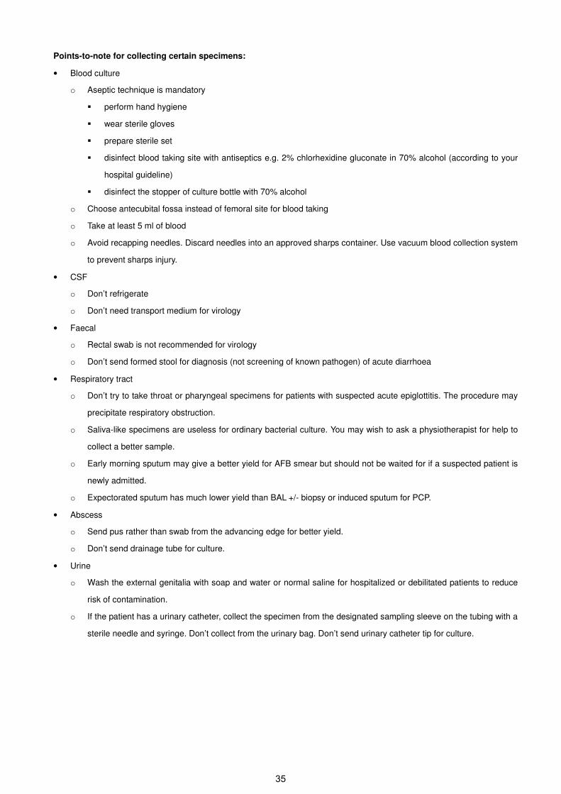
Points-to-note for collecting certain specimens:
• Blood culture
o Aseptic technique is mandatory
� perform hand hygiene
� wear sterile gloves
� prepare sterile set
� disinfect blood taking site with antiseptics e.g. 2% chlorhexidine gluconate in 70% alcohol (according to your
hospital guideline)
� disinfect the stopper of culture bottle with 70% alcohol
o Choose antecubital fossa instead of femoral site for blood taking
o Take at least 5 ml of blood
o Avoid recapping needles. Discard needles into an approved sharps container. Use vacuum blood collection system
to prevent sharps injury.
• CSF
o Don’t refrigerate
o Don’t need transport medium for virology
• Faecal
o Rectal swab is not recommended for virology
o Don’t send formed stool for diagnosis (not screening of known pathogen) of acute diarrhoea
• Respiratory tract
o Don’t try to take throat or pharyngeal specimens for patients with suspected acute epiglottitis. The procedure may
precipitate respiratory obstruction.
o Saliva-like specimens are useless for ordinary bacterial culture. You may wish to ask a physiotherapist for help to
collect a better sample.
o Early morning sputum may give a better yield for AFB smear but should not be waited for if a suspected patient is
newly admitted.
o Expectorated sputum has much lower yield than BAL +/- biopsy or induced sputum for PCP.
• Abscess
o Send pus rather than swab from the advancing edge for better yield.
o Don’t send drainage tube for culture.
• Urine
o Wash the external genitalia with soap and water or normal saline for hospitalized or debilitated patients to reduce
risk of contamination.
o If the patient has a urinary catheter, collect the specimen from the designated sampling sleeve on the tubing with a
sterile needle and syringe. Don’t collect from the urinary bag. Don’t send urinary catheter tip for culture.
35

References
1. Basic diagnostic testing protocols for level A laboratories for the presumptive identification of Bacillus anthracis. CDC
(USA), American Society for Microbiology, and Association of Public Health Laboratories. 3/18/02.
http://www.asm.org/ASM/files/LEFTMARGINHEADERLIST/DOWNLOADFILENAME/0000000521/bacillusanthracispr
otocol[1].pdf
2. Collecting, preserving and shipping specimens for the diagnosis of avian influenza A(H5N1) virus infection: Guide for
field operations. WHO. October 2006.
http://www.who.int/csr/resources/publications/surveillance/MainTextEPR_ARO_2006 1.pdf
3. Gill VJ, Fedorko DP, Witebsky FG. The clinician and the microbiology laboratory. In: Mandell GL, Bennet JE, Dolin R
(ed). Principles and practice of infectious diseases.6th edn. Philadelphia: Elsevier, 2005:203-241.
4. Sentinel laboratory guidelines for suspected agents of bioterrorism: botulinum toxin. American Society for Microbiology.
Revised 7.30.03.
http://www.asm.org/ASM/files/LeftMarginHeaderList/DOWNLOADFILENAME/000000000522/Botulism.pdf
5. Specimen collection guidelines. CDC (USA) website: Emergency and Preparedness Response. How to investigate
unexplained respiratory disease outbreaks. Specimen collection and handling.
http://emergency.cdc.gov/urdo/pdf/SpecCollectionGuidelines.pdf
6. Smallpox fact sheet – Information for lab workers. CDC (USA)
http://emergency.cdc.gov/agent/smallpox/vaccination/pdf/vaccinia-specimen-collection.pdf
7. Specimen selection. CDC (USA) website: Emergency and Preparedness Response.
http://emergency.cdc.gov/documents/PPTResponse/table2specimenselection.pdf
8. Specimen collection and transport guidelines for suspect smallpox cases. CDC (USA) website: Emergency and
Preparedness Response. http://emergency.cdc.gov/agent/smallpox/response-plan/files/guide-d.pdf
9. Sentinel laboratory guidelines for suspected agents of bioterrorism: Yersinia pestis. American Society for Microbiology.
15 August 2005.
http://www.asm.org/ASM/files/LeftMarginHeaderList/DOWNLOADFILENAME/000000000524/Ypestis81505.pdf
10. Sentinel laboratory guidelines for suspected agents of bioterrorism: unknown viruses. American Society for
Microbiology. Final 10/10/03.
http://www.asm.org/ASM/files/LEFTMARGINHEADERLIST/DOWNLOADFILENAME/0000001160/ukvirusesgl.pdf
11. Recommended BSL for BT agents (Appendixes B & C). In: Sentinel laboratory guidelines for suspected agents of
bioterrorism: Clinical laboratory bioterrorism readiness plan. American Society for Microbiology. Revised 10 August 06.
http://www.asm.org/ASM/files/LeftMarginHeaderList/DOWNLOADFILENAME/000000001204/BTtemplateRevised8-10
-6.pdf
12. Guidelines for the collection of clinical specimens during field investigation of outbreaks. WHO.
http://www.who.int/csr/resources/publications/surveillance/whocdscsredc2004.pdf
13. Sentinel laboratory guidelines for suspected agents of bioterrorism and emerging infectious diseases: avian influenza
A H5N1. American Society for Microbiology. 15 December 2006.
http://www.asm.org/ASM/files/LeftMarginHeaderList/DOWNLOADFILENAME/000000002283/AvianInfluenza11-2008.
14. Guideline on transport of clinical specimens and infectious substances. Hospital Authority. Revised 1 July 2006.
http://ha.home/ho/ps/Guidelinesontransportofclinicalspecimens.pdf
36

15. Miller JM, Krisher K, Holmes HT. General principles of specimen collection and handling. In: Murray PR, Baron EJ,
Jorgensen JH, Landry ML, Pfaller MA (ed). Manual of Clinical Microbiology 9th edn. Washington, D.C.: ASM Press,
2007:43-54.
16. Infection prevention and control of epidemic- and pandemic-prone acute respiratory diseases in health care. WHO
June 2007 http://www.who.int/csr/resources/publications/swineflu/WHOCEPR2007_6/en/index.html
17. Guideline for taking blood for culture. Princess Margaret Hospital. Revised 03/2009.
http://pmh.home:8000/ict/FrontPage/index.html
18. Laboratory user guide 2009. Princess Margaret Hospital.
http://kwcpath.home/labguide/Laboratory%20User%20Guide%202009.pdf
19. Wilson J. Infection control in clinical practice. 2nd ed. London: Baillière Tindall, 2001.
37

7. Handling of infectious diarrhoea
Diarrhoeal disease accounts for approximately 2 million deaths annually in children under the age of five years. Disease and
death caused by diarrhoea is a global problem but is especially prevalent in developing countries where parents often fail to
recognize the danger signs and children simply die through rapid loss of fluids and undernourishment through lack of food.
Definition
Diarrhoea is the passage of loose or liquid stools more frequently than is normal for the individual. It is usually a symptom of
gastrointestinal infection. Depending on the type of infection, the diarrhoea may be watery (e.g. in cholera) or passed with
blood (e.g. in dysentery).
Aetiology
As a symptom of infection, diarrhoea is caused by bacterial, viral, parasitic organisms.
• Bacterial: Salmonella, Shigella, Campylobacter, and Escherichia Coli, Vibrio parahaemolyticus, Vibrio cholera,
Clostridium difficile and cholera. Contaminated food and water can result in several types of infectious diarrhoea.
• Viral: Norovirus, Rotavirus, enteric Adenoviruses and Hepatitis A/E. Rotavirus is the most significant viral pathogen
causing diarrhoea, particularly in children.
• Parasites: Giardia lamblia, Entamoeba histolytica and Cryptosporidium. Parasites enter the body through food and
water and take up residence in the digestive system.
Clinical presentation
Most are self-limiting and besides acute diarrhoea, symptoms may include nausea and vomiting, loss of appetite, and
abdominal pain. As dehydration worsens, symptoms progress from thirst, restlessness, decreased skin turgor and sunken
eyes to decreased consciousness, rapid and feeble pulse and low or undetectable pulse. Dehydration and hypovolemic
shock are the primary causes of death.
The outcome will be fatal in some susceptible hosts such as infants, elderly people and the immunocompromised. It is often
a coinfection with other diseases, such as malaria and HIV, and is frequently a comorbidity factor in deaths resulting from
these diseases.
Treatment
Replacement of fluids and electrolytes is the cornerstone of supportive care for patients with diarrhoea and greatly reduces
mortality due to dehydration. While rehydration counters fluid loss it neither stops the symptoms nor kills the pathogens
responsible for the illness. There are very few treatments for specific diarrhoeal pathogens. Antibiotics, which are commonly
prescribed, are ineffective against many pathogens and indiscriminate use contributes to the development of resistant
organisms.
Laboratory confirmation
Most cases of acute diarrhoea never need diagnosis. The most useful diagnostic tool is stool culture and examination for
parasites. The earlier cultures are performed, the greater the chance of obtaining a positive result. For viral gastroenteritis,
the diagnostic methods are electron microscopy, enzyme immunoassay, polymerase chain reaction and serology.
38

Mode of transmission
Transmission of an infectious agent resulting in diarrhoea is usually directly or indirectly from an infected or colonized person
to a susceptible host often on the hands.
• Predominantly fecal-oral route. It may be spread from
o Person to person, aggravated by poor personal hygiene.
o Contracted from contaminated foods if prepared or stored in unhygienic conditions. Water may contaminate food
during irrigation and fish and seafood from polluted water may contribute to the disease.
o Water contaminated with human faeces. Animal faeces also contain microorganisms that can cause diarrhoea.
• Pathogen with low infective doses, long shedding period and prolonged survival in the environment enhance the
infectiveness of the illness accounting for outbreak.
• Person to person transmission is a major determinant of transmission in hospital and institutional outbreaks.
• Preventative measures include
o Contaminated food and water sources should be identified and eliminated
o Food hygiene should be maintained e.g. shellfish should be cooked thoroughly
o Contact precautions should be instituted to reduce the transmission of organisms from an infected or colonized host
through direct or indirect contact. This involves:
� Patient placement - isolation of suspected cases.
� Gloving – change gloves after contact with infective material. Remove gloves before leaving the isolation room.
� Hand washing – with an antibacterial agent or an alcohol based hand rub after removing gloves. Do not touch
potentially contaminated surfaces or items before leaving the room.
� Gowns and protective apparel – use a clean, nonsterile disposable gown. Remove before leaving the room. Do
not touch potentially contaminated surfaces or items before leaving the room.
� Patient transport – limited to essential transport.
� Patient care equipment – reserve non-critical patient care equipment. Clean and disinfect equipment regularly.
o Care of the diaper host
� Handle infective diarrhoea with contact precautions
� Clean up vomitus immediately with contact precautions
� Prevent further contaminated areas by cleansing with an effective disinfectant
o Environmental hygiene. Good hygienic housekeeping and pest control.
o Carriers e.g. food handlers should be educated regarding personal hygiene i.e. hand hygiene should be reinforced,
to prevent spread.
Clinical practice on handling of diapers
• Adopt a clean to dirty conceptual framework
• Dispose of used diapers in a proper and timely fashion
• Observe hand hygiene after removal of gloves
• Monitor personal hygiene and self-discipline among staff and visitors
• Staff should discard excreta via bedpan macerators to avoid further contamination or spillage incidents
39

References
1. 2007 Guideline for isolation precautions: preventing transmission of infectious agents in health care settings. Health
Care Infection Control Practices Advisory Committee.
2. Bala Hota. 2004. Contamination, Disinfection, and Cross-Colonization: Are Hospital Surfaces Reservoirs for
Nosocomial Infection? CID. 39 (15 October)
3. Food safety. CDC. Online available: http://www.cdc.gov/foodsafety/
4. Water related diseases. Online available: http://www.who.int/water_sanitation_health/diseases/diarrhoea/en/
40

No Page
1 42
2 43-44
3 45
4 46
1 47
2 48
3 49
Environmental Cleaning
Behaviour Change
Remarks : C = Checklist , G = Guideline, L = Leaflet , P = Poster , PG = Photo Guide
PG
P
Procedure Guide on Dilution of Sodium Hypochlorite solution
Swan-neck sealing method for clinical waste
Cleansing Item Color Coding System P
Index
Content Format
Cough Etiquette Poster P
Section A : Basic Infection Control Tools
Hand Hygiene Pocket Leaflet
Hand Hygiene Poster - New
Flu Vaccine Promotion Poster
L
P
P
41

• Cover the nose/mouth
when coughing or sneezing
• Use tissue paper to contain
respiratory secretions and
dispose in the waste receptacle
Respiratory Hygiene and Respiratory Hygiene and
Cough EtiquetteCough Etiquette
• Perform hand hygiene if
contact respiratory
secretions and
contaminated objects
• Put on a surgical mask if
there is respiratory
symptoms
42

43

44

PREVENT NOSOCOMIAL INFECTION PLEASEPREVENT NOSOCOMIAL INFECTION PLEASE WASH YOURWASH YOUR HANDHANDS!S!
Palm to palm Palm to palm, fingers interlaced
Rotate clasped fingers in palm (vice versa)
Rotate thumb in clasped palm (vice versa)
�������� ���� ����
�������� ����
Rotational rub wrist with palm
(vice versa) (Optional)
Palm rubs over dorsum
(vice versa)
Fingers interlocked, rub
backs of fingers to opposing palms (vice versa)
45

Care Your Care Your
FamilyFamily
Have you vaccinated yet ?Have you vaccinated yet ?
FLUFLU
Protect YourselfProtect Yourself
Safeguard Safeguard
PatientPatient
46

Rev. Nov 2003
Dilution of Hypochloride commonly used in clinical area
Ratio Dilution
Photo Guide Applications Time (Mins.) Effective ppm
1part of hypochloride in 9 parts of clean water
1:9
e.g. 30 ml hypochloride in 270ml clean water
30ml hypochloride 270ml clean water
- Accidental splash of blood, leaking of body fluid
- Mop with dry cloth, then covered with hypochloride soaked cloth or absorbent
- Clear up ultimately
Soak for 5 Mins 5000ppm
1part of hypochloride in 49 parts of clean water
1:49
e.g. 100ml hypochloride in 4900ml clean water
100ml hypochloride 4900ml clean water
- Bed frame, chair, mattress, wheel chair, stretcher
- Vomit: Immediate clear up, then
disinfect - Mop
Soak for 30 Mins 1000ppm
1part of hypochloride in 99 parts of clean water
1:99
e.g. 100ml hypochloride in 9900ml clean water
100ml hypochloride 9900ml clean water
- Bed pan、Urinal
Soak for 10 Mins 500ppm
1part of hypochloride in 249 parts of clean water
1:249
e.g. 16ml hypochloride in 3984ml clean water
16ml hypochloride 4900ml clean water
- Medication Cup、Toys
Soak for 10 Mins 200ppm
* Choice of Hypochloride - Clorox/Hypo 6 ppm = parts per million
47

Office/Nurse Station/
Other Non-clinical AreasPantry Sluice Room
Patient
Areas
Transmission
Based
Precaution
Droplet Precaution
Disposable
Gloves
Cleansing
ClothDisposable Cloth or
Immediate Laundry
Treatment after use
Cleansing
Mope
Staff's Toilet/
Changing Room
Patient's Toilet/
Changing RoomUtensils/
Location
Cleansing Utensils Color Code System
Drying
Mope
48

紅色醫療廢物袋的紅色醫療廢物袋的紅色醫療廢物袋的紅色醫療廢物袋的「「「「鵝頸結鵝頸結鵝頸結鵝頸結」」」」封口方法封口方法封口方法封口方法
“Swan-neck” method of sealing red bag for clinical waste
紅色廢物袋在3/4滿時就要封妥Seal clinical waste red bag when no more than75% full
中間扭曲後對摺Twist firmly then double over
緊握著已扭曲的部份Hold the twist firmly
把索膠帶套著紅色廢物袋的頸部Pass the “gun tie” over neck of the Red Bag
把索膠帶拉緊形成有效的封口Tighten the seal manually to create an effective seal
1 2 3
4 5 6
將廢物標籤緊繫於紅色袋的封口上The Medical waste label should tied securely to the neck of the red
bag
BIOHAZARD病房醫院
49
SECTION B – Specific Response to selected situations
In this section, instead of adopting a disease model which can be non-exhaustive and unpredictable with new emergence of
novel infectious disease, based on the unique experience of the tool kit development team and the intention to bring about
enlightenment to handle other unknown situations, only SARS and Avian Influenza H5N1 were selected as exemplars to
demonstrate comprehensive approach that should be adopted for “Preparedness” and “Response” to specific outbreak
situations.
However, tool kit was developed so as to cover every detail practical areas as far as possible so as to reduce risk accordingly.
In dealing with other situations, the principles of transmission-based precautions shall always be applied appropriately.
There are three categories of Transmission-Based Precautions: Contact Precautions, Droplet Precautions and Airborne
Precautions. They are used when the route(s) of transmission is (are) not completely interrupted using Standard
Precautions alone. For some diseases that have multiple routes of transmission (e.g. SARS, HSI), more than one
Transmission-Based Precautions category may be used. When used either singly or in combination, they are always used
in addition to Standard Precautions.
Once again, the strategies outlined in the tool kit by no means are the only way of managing emerging infectious diseases
and outbreaks. Users are advised to refer to their own context and make appropriate modifications whilst confronting
problems like resources limitations. Although the content of the tool kit is believed to be accurate at time of release, users are
advised to check updates in relation to emerging infectious disease threats or changes in practice or research.
1. Severe Acute Respiratory Syndrome
Introduction
Severe acute respiratory syndrome (SARS) is a serious form of respiratory failure, caused by a coronavirus,
(SARS-associated coronavirus (SARS-CoV)) which was first isolated in 2003. Infection with the SARS virus resulted in acute
respiratory distress (severe breathing difficulty) and sometimes death. The world was unprepared for the emergence of
SARS and the lack of preparedness resulted in considerable confusion, heightened public concern and increased
transmission rates. It was a dramatic example of how quickly world travel can spread a disease but also an example of how
quickly a networked health system can respond to an emerging threat.
Background information
This contagious respiratory infection was first described on February 26, 2003 and was identified as a new disease by World
Health Organization (WHO) physician, Dr. Carlo Urbani. He diagnosed it in a 48-year-old businessman who had traveled
from the Guangdong province of China, through Hong Kong, to Hanoi, Vietnam. The businessman died from the illness as
did Dr. Urbani on March 29, 2003, at the age of 46.
In the meantime, SARS was spreading, and within 6 weeks of its discovery, it had travelled throughout many parts of Asia,
North America and Europe. According to the World Health Organization (WHO), a total of 8,098 people worldwide became
sick with SARS during the 2003 outbreak. The heaviest burden of illness was felt in China, Hong Kong, Taiwan, Singapore,
Canada and Vietnam.1,2
50

The morbidity, mortality and speed and ease of transmission associated with SARS led to the WHO declaring SARS as a
global health threat, resulting in the issuance of an unprecedented travel advisory. Daily WHO updates tracked the spread of
SARS as it was not clear whether it would become a global pandemic, or would settle into a less aggressive pattern. By June
2003, the epidemic had subsided but even as the number of new cases dwindled, and travel advisories began to be lifted,
the sober truth remained: every new case had the potential to spark another outbreak.
Clinical experience
Approximately 23% to 32% of patients with SARS became critically ill.3, 4, 5, 6
Acute Lung Injury (ALI) was the most common
severe organ dysfunction and occurred in 16% of all patients with SARS and 80% of critically ill patients with SARS. Virtually
all patients with ALI required mechanical ventilation. The case fatality rate worldwide was 9.6% but among those with a
SARS-related critical illness 50% died.5,6
Mortality was highest amongst elderly patients especially those with co morbidities
while children were relatively protected from the illness.4 There was no vertical transmission documented in pregnant
women.7
Symptoms
The hallmark symptoms were fever greater than 38.0 degrees C4,8
and cough, difficulty breathing, or other respiratory
symptoms. Symptoms in the order of how commonly they appeared include:
• Fever
• Chills and shaking
• Muscle aches
• Cough
• Headache
Less common symptoms include (also in order):
• Dizziness
• Productive cough (sputum)
• Sore throat
• Runny nose
• Nausea and vomiting
• Diarrhoea
Lower respiratory symptoms usually began 2 to 7 days after symptom onset but these could be the initial presenting
symptoms in up to 30% of patients. Gastrointestinal symptoms occurred with variable frequency but included nausea,
diarrhoea and vomiting. Tachycardia and tachypnoea were also extremely common. The median time from exposure to
symptom onset was approximately one week (1,4).
Two thirds of patients had pulmonary infiltrates on their initial chest X-ray and virtually all had infiltrates by one week.
Laboratory abnormalities4,9
included
• Blood clotting tests
• Blood chemistries
o ALT and CPK were sometimes elevated
51

o Elevated lactate dehydrogenase
o Sodium and potassium were sometimes low
• Chest X-ray or chest CT scan
• Complete blood count (CBC)
o Low white blood cell (WBC) count
o Moderate lymphopenia
• Low platelet count
Confirmatory laboratory testing involved viral serology and/or detection of viral RNA by nucleic acid amplification from
samples of serum, nasopharyngeal secretions, urine or stool. Specific tests included the PCR test for SARS virus, antibody
tests for SARS, and direct SARS virus isolation. All current tests have some limitations. Negative test results could not
reliably exclude the SARS virus until more than 28 days after symptom onset.
Nosocomial transmission from patients to health care workers was a prominent and worrisome feature of the SARS
outbreaks. In Hong Kong, almost 25% of the total number of SARS cases comprised health care workers.10
While in
Singapore and Toronto health care workers accounted for approximately half the cases, 20% of whom became critically ill.4,6
There were concerns that airway management and mechanical ventilation strategies may have contributed to the high
nosocomial transmission rate.
Lung pathology in SARS
Diffuse alveolar damage with varying degrees of organisation was the predominant pathology in the lungs of SARS
patients.11
Several phases have been recognized
• Acute phase: evidence of hyaline membranes, interstitial and alveolar oedema, mild interstitial infiltrates of
inflammatory cells and vascular congestion.
• Organizing phase: evidence of interstitial and airspace fibroblast proliferation, and type II cell hyperplasia.
Specific treatment for SARS
Antiviral agents for SARS
During the outbreak in 2003 many different interventions were used in management, including antivirals, like ribavirin, IFN
and protease inhibitors, as well as immunomodulatory agents, especially systemic corticosteroids. The uncertain natural
history of untreated SARS and the uncontrolled nature of these interventions meant that no drug intervention was of proven
benefit or prophylactic value. Despite progress in the search for new antivirals there is still a need for effective antiviral drugs
to prevent and treat SARS. Predictive correlations between in vitro activity and antiviral effects have not been validated
either in animal models or humans infected with SARS.
Community acquired pneumonia and SARS
During the SARS epidemic patients with new lung infiltrates were initially treated with antibiotics consistent with published
current guidelines on the management of community-acquired pneumonia.12
Patients with SARS in the ICU should be
treated for drug resistant and atypical pathogens. Pseudomonas aeruginosa should be covered if patients have risk factors
such as structural lung damage, recent hospitalization, high dose steroids, malnutrition, or recent antibiotics.
52

Clinical management of severe SARS
Patients with SARS who developed hypoxaemic respiratory failure (16%) met the current definition of Acute Lung Injury (ALI)
and Acute Respiratory Distress Syndrome (ARDS) .13
These include refractory hypoxaemia, bilateral diffuse pulmonary
infiltrates on chest X-ray and a PaO2/FIO2 ratio of less than 300.The recommended clinical management of severe SARS is
the same as for patients with ALI/ARDS as the clinical and pathological manifestations of lung injury from severe SARS
cannot be distinguished from ALI or ARDS.
Mechanical ventilation of severe SARS
Survival in patients with ALI/ARDS is improved by a adopting a strategy of ventilation with low tidal volumes (6ml/kg
predicted body weight).14
The mode of ventilation in patients with SARS is controversial. Many clinicians believe that the
risks of intubation for nosocomial transmission are significant and favour noninvasive mechanical ventilation. On the other
hand many physicians favour early intubation under relatively controlled conditions and that noninvasive ventilation may
itself be associated with nosocomial transmission to health care workers. Furthermore, while noninvasive ventilation might
be considered as a modality for short-term improvement in hypoxaemic respiratory failure, SARS is generally a disease
requiring longer term support as this form of lung injury is not rapidly reversible.
Corticosteroids in severe SARS
During the SARS epidemic many patients were treated with either pulse steroids or very high doses of corticosteroids. The
resultant small studies that were published were not randomized controlled trials, which is not surprising considering the
rapid emergence of SARS in a rapidly short time period. Anecdotally, several cases of more rapid clearing of chest X-rays
have been reported and one case series reported more rapid resolution of fever, respiratory symptoms and chest X-rays but
no differences in survival.15
Indications varied but corticosteroids were generally reserved for patients with SARS and persistent fever, worsening
hypoxaemia, worsening dyspnoea and progression of lung pathology on chest X-ray. Pulse steroids were generally
administered at doses of 500-1000mg of methylprednisolone intravenously for three days but often longer. Many patients
also received low dose steroids as recommended in the Surviving Sepsis Campaign Guidelines.16
However in patients with early ALI/ARDS or sepsis pulse steroids or high dose steroids do not appear to improve
survival17,18,19
and are not recommended.20
A large study by the ARDS Clinical Trials Network does not support the use of
steroids for late or unresolving ALI or ARDS.21
There is also increasing evidence of long-term morbidity – muscle weakness
and neuropathy.18,22,23,24
Adjuvant strategies for severe SARS
The morbidity and mortality in critically ill patients is decreased by several adjuvant strategies as recommended in the most
recent Surviving Sepsis Campaign guidelines.25
These include
• Deep vein thrombosis prophylaxis. Depending on the risk factors present this can be based on pharmacologic or
physical methods.26, 27, 28, 29
• Stress ulcer prophylaxis. H2 receptor inhibitors are efficacious.30,31
• Sedation protocols in critically ill mechanically ventilated patients.32,33,34
53

• Avoidance of neuromuscular blockers because of prolonged neuromuscular weakness and paralysis, especially when
used in conjunction with steroids.35,36,37
• Semirecumbent position with head of the bed elevated to 450
in mechanically ventilated patients (38-42) to limit
aspiration risk and prevent the development of ventilator–associated pneumonia.
Preparation of the health care worker and the health care system
There have been many publications describing the outbreak of SARS in 2003.3,4,5,6,43
Specific recommendations concerning
clinical issues, infection control, expansion of facilities and research associated with SARS have also been
published.44,45,46,47
Organizational planning
SARS highlighted the need for a high level of vigilance and alertness throughout all parts of the healthcare system.
Contingency planning prior to an outbreak and providing a leadership structure within a hospital and within individual
departments is essential. The framework of response to infectious disease outbreaks within the Hospital Authority begins
with watchfulness and surveillance of abnormal patterns of infections, which is part of the everyday risk management culture
within the practice of medicine. When an abnormal pattern of infectious disease is detected a swift assessment by the
hospital infection control team on the significance of the infection, risk of hospital spread, availability of existing knowledge
and guidance to treatment and control, and potential threat to the community enables the hospital to take appropriate actions
to manage and control the outbreak. When it is considered that the outbreak poses a significant risk to the hospital system
the Hospital Authority initiates a contingency response with a clear command structure. In addition the Government has its
own alert and response system for combating infectious diseases on both a local and regional basis.
Within the specialty of intensive care a culture has developed where everyone recognizes that their work may have wider
public health implications and that an illness in one patient may have consequences for the whole ICU. Many of Hong Kong’s
ICUs have upgraded their isolation facilities and may have a separate ICU specifically for the admission of SARS cases or
other infectious diseases patients. Cohorting and isolating appropriate patients is part of the everyday management of an
ICU. In addition, all ICUs have contingency plans in place in the event of SARS or Highly Pathogenic Avian Influenza
occurring in Hong Kong. These focus on staff deployment and training, hardware procurement, and surge capacity.
The CDC has provided guidelines to help healthcare facilities prepare for the outbreak of an infectious disease.45
These
emphasize the importance of
• Identified leadership structure
• Delegation of responsibility
o Sustaining the work force
o Access controls
o Communication
o Education and training
o Infection control procedures
o Psychosocial support
o Data collection
o Clinical management protocols
54

Consideration also needs to be given to the following areas:
• Should SARS patients be cared for in one or several institutions
• Should SARS patients be cared for in separate ICUs
• Transportation of non-SARS patients to other facilities or ICUs
• Transportation of potentially contagious SARS patients within a healthcare facility
• Mobilization of manpower as staff may be lost due to fear, quarantine and illness
• Ensuring adequate and advance supplies of consumables and durable materials and equipment
• Clinical and infectious disease training for all healthcare workers
• Communication strategies both horizontally and vertically. Intensive care leadership will need to communicate with the
hospital administration, infectious disease specialists, infection control teams, SARS ward physicians, Accident and
Emergency specialists, the laboratory, and public health officials.
• Identifying psychosocial support for health care workers. Despair, depression and a sense of isolation were common
reactions in health care workers during the SARS epidemic. Some health care workers were viewed as being high risk
themselves by the community because of their work with SARS patients. Health care workers also had concerns for
their family members during SARS not just in terms of the risk to their families but also concerning childcare, economic
support and the provision of appropriate accommodation.
Infection control precautions in the ICU
Effective protection of health care workers is of utmost importance and is also crucial in breaking the chain of transmission.
In the ICU, the risk of transmission is high as viral shedding peaks during the second week of the illness15
when admission is
usually required. Droplet spread can be increased by aerosol-generating interventions, for example, nebulisers, intubation
and bronchoscopy.
Six key strategies are crucial as infection control measures in the ICU to prevent nosocomial transmission of SARS.
� Dilution and removal of contaminants through the use of negative pressure isolation rooms
� Use of personal protective equipment, e.g. gowns, gloves, eye, face and respiratory protection
� Hand hygiene
� Environmental cleaning and disinfection
� Source control measures aimed at containing the patient’s secretions
� Limiting contact with patients
Considerable time should be spent on increasing awareness, education and vigilance of all health care workers with regards
an infectious diseases outbreak and the standards required for personal protection. Training extends from the most senior to
the most junior of staff. All hospital staff should undergo yearly training and refresher courses in infection control. Staff
within ICU should undergo yearly N95 mask fit testing.
On a day-to-day basis, maintenance of a high level of vigilance should be paramount. All staff should change into ICU attire
before entering the ICU. In addition, surgical masks and jackets (not white coats) should be standard uniform. All non-ICU
staff entering the ICU should wear surgical masks and colour coded gowns to reflect their status. Hand washing programs
should be regularly monitored and reinforced. Non-clinical areas previously located within an ICU, such as storerooms,
55

library, seminar room, doctors on call rooms, offices, and kitchens should be moved out of the immediate vicinity of the
clinical areas. The only exceptions are the relative’s interview room and an emergency shower and toilet. There should be
no eating or drinking within the ICU environs.
Monitoring and policing the system takes considerable effort. Each day an assigned registered nurse should act as the
monitor. Infectious control infringements should be identified and staff education programs conducted on a regular basis.
All patients should be assessed as to their risk of infectious disease prior to admission. Suspect cases are identified, isolated
and screened until proven negative either for SARS or HPAI. Personal protective equipment should be readily available at
the entrance and at nominated sites within the ICU.
Procedures involving the airway should be viewed as placing staff at risk. Full personal protective equipment including N95
mask and face shield should be routinely worn before and during endotracheal intubation, flexible bronchoscopy, and
percutaneous tracheostomy. In-line, closed endotracheal suction system should be routine. Nebulizers should not be used
in ICU and BiPAP only under strict guidelines. Patients receiving BiPAP should be isolated and the level of PPE raised.
Training programs on manual handling of bodily secretions should be regularly conducted.
56

Table 1. Summary of appropriate standards and infection control precautions necessary when caring for SARS patients44,46
Airborne infection isolation room
Negative pressure isolation rooms: equipped with ante-chamber
Wash basins with antibacterial soap and/or alcohol-based hand rub Avoid disruption of negative pressure barrier
Stock individual rooms with supplies including resuscitation trolleys Personal protective equipment (PPE)
Basic: gown, gloves, eye protection (i.e. goggles or face shield), and respiratory protection (i.e. N95 mask)
During aerosol generating procedures
N 95 is the minimum acceptable level of respiratory protection Provide written instructions to healthcare workers as to how to put on and remove PPE
Avoid touching face and surfaces
Monitor PPE use: concept of a ‘policeman’
Hand hygiene
Ensure availability of hand hygiene products (i.e. sinks with antibacterial soap, disposable towels and
alcohol-based handrub.
Environmental cleaning and disinfection
Assigned staff to clean SARS environment
Clean horizontal and frequently touched surfaces at least daily
Equipment and procedures
Oxygen therapy
Avoid nebulized humidity e.g. Venturi mask without humidification Avoid bag-valve mask ventilation
Use a two person technique to ensure a tight fit to the face Intubation
Performed by the most experienced clinician available Mechanical ventilators
Submicron filters on exhalation outlet of mechanical ventilator Non-invasive ventilation
Not recommended Bronchoscopy
Avoid if possible Patient transportation
Avoid unless essential
Visitors and personnel
Limit to essential staff only
Staff education
Provide training on infection control procedures
Provide training on donning and removal of PPE
Emphasize importance of vigilance
Emphasize importance of alerting supervisors when breaches occur
57

Prognosis
As the first wave of SARS began to subside, the death rate proved to have been about 14 to 15 percent of those diagnosed.
In people over age 65, the death rate was higher than 50 percent. Many more were sick enough to require mechanical
ventilation and more still required hospitalization in intensive care units.
Intensive public health policies are proving to be effective in controlling outbreaks. Many nations stopped the SARS epidemic
within their own countries. All nations must be vigilant, however, to keep this disease under control. Viruses in the
coronavirus family are known for their ability to change (mutate) in order to better spread among humans.
Conclusion
In the short term the 2003 SARS epidemic was devastating to the people and economy of Hong Kong. In the longer term it
provided Hong Kong an opportunity to prepare for future infectious disease outbreaks. Many lessons have been learned
and constant revision and refinement of new policies, procedures, protocols and contingency plans is ongoing. Within ICU,
the epidemic has resulted in a change in ICU culture with far greater emphasis on the detection, surveillance, prevention and
management of infectious diseases. Protection of health care workers is paramount.
International experts in infectious disease and epidemiologists consider it likely that there will be another outbreak of a
seriously transmissible respiratory pathogen. Clinicians, hospital administrators and government officials across the world
have addressed organizational issues within health care systems and issues concerning health care workers so that it might
be better able to handle the next outbreak. As a result it is hoped that there will be less confusion, decreased public concern
and decreased transmission rates.
58

References
1. World Health Organization. Summary of probable SARS cases with onset of illness from 1 November to 31 July 2003
(revised 26 September 2003) Available from URL: http://www.who.int/csr/SARS/country/table2003_09_23/en/
2. WHO. Consensus document on the Epidemiology of Severe Acute Respiratory Syndrome. Available online at:
http://www.who.int/csr/SARS/en/WHOconsensus.pdf
3. Gomersall CD, Joynt GM, Lam P, Li T, Yap F, Lam D, Buckley T, Sung JJY, Hui D, Antonio GE, Leung P. Short-term
outcome of critically ill patients with severe acute respiratory syndrome. Int Care Med 2004; 30; 381-387.
4. Booth CM, Matukas LM, Tomlinson GA, et al. Clinical features and short-term outcomes of 144 patients with SARS in
the greater Toronto area. JAMA 2003; 289(21):2801-9
5. Fowler RA, Lapinsky SE, Hallett D, et al. Critically ill patients with severe acute respiratory syndrome. JAMA 2003;
290:367-373
6. Lew TWK, Kwek T-K, Tai D, et al. Acute respiratory distress syndrome in critically ill patients with severe acute
respiratory distress syndrome. JAMA 2003; 290:374-380
7. Shek CC, Ng PC, Fung GPG et al. Infants born to mothers with severe acute respiratory distress syndrome. Pediatrics
2003; 112:e254-e256.
8. Jernigan JA, Low DE, Helfand RF. Combining clinical and epidemiologic features for early recognition of SARS. Emerg
Infect Dis 2004; 10:327-333.
9. Lee N, Hui D, Wu A, Chan P, Cameron P, Joynt GM et al. A major outbreak of severe acute respiratory syndrome in
Hong Kong. N Engl J Med 2003; 348:1986-94.
10. Buckley T. Severe acute respiratory syndrome (SARS) in Hong Kong’s ICUs: the lessons learned. Critical Care and
Resuscitation 2005; 7:130-132.
11. Franks TJ, Chong PY, Chui P, et al. Lung pathology of severe acute respiratory syndrome (SARS): a study of 8
autopsy cases from Singapore. Hum Path 2003; 34:743-748.
12. Niederman MS, Mandell LA, Anzueto A, et al. Guidelines for the management of adults with community acquired
pneumonia. Diagnosis, assessment of severity, antimicrobial therapy and prevention. Am J Respir Crit Care Med 2001;
163:1730-1754
13. Bernard GR, Artigas A, Brigham KL, et al. The American-European Consensus Conference on ARDS. Definitions,
mechanisms, relevant outcomes, and clinical trial coordination. Am J Respir Crit Care Med 1994; 149: 818-824.
14. The Acute Respiratory Distress Syndrome Network. Ventilation with lower tidal volumes as compared with traditional
tidal volumes for acute lung injury and the acute respiratory distress syndrome. N Engl J Med 2000; 342: 1301-1308.
15. Peiris JS, Chu CM, Cheng VC, et al. Clinical progression and viral load in a community outbreak of
coronavirus-associated SARS pneumonia: a prospective study. Lancet 2003; 361:1767-1772
16. Dellinger RP, Carlet JM, Masur H, et al. Surviving Sepsis Campaign guidelines for management of severe sepsis and
septic shock. Crit Care Med 2004; 32:858-873.
17. Bernard GR, Luce JM, Sprung CL, et al. High dose corticosteroids in patients with the adult respiratory distress
syndrome. N Engl J Med 1987; 317: 1566-1570.
18. Bone RC, Fisher CJ Jr, Clemmer TP, et al. Early methylprednisolone treatment for septic syndrome and the adult
respiratory distress syndrome. Chest 1987; 92: 1032-1036.
19. Luce JM, Montgomery AB, Marks JD, et al. Ineffectiveness of high dose methylprednisolone in preventing
parenchymal lung injury and improving mortality in patients with septic shock. Am Rev Respir Dis 1988; 138:62-68.
59

20. Thompson BT. Glucocorticoids and acute lung injury [review]. Crit Care Med 2003; 31:S253-S257.
21. National Heart, Lung, and Blood Institute. NHLBI ARDSnet website. Available online at: http://www.ardsnet.org
22. Weigelt JA, Norcross JF, Borman KR, et al. Early steroid therapy for respiratory failure. Arch Surg 1985; 120:536-540.
23. Herridge MS, Cheung AM, Tansey CM, et al. One-year outcomes in survivors of the acute respiratory distress
syndrome. N Engl J Med 2003; 348:683-693.
24. De Jonghe B, Sharshar T, Lefaucheur J-P, et al. Paresis acquired in the intensive care unit: a prospective multicenter
study. JAMA 2002; 288:2859-2867.
25. Dellinger RP, Levy MM, Carlet JM, et al. Surviving Sepsis Campaign: International guidelines for management of
severe sepsis and septic shock: 2008. Intensive Care Med 2008; 34:17-60.
26. Cade JF. High risk of the critically ill for venous thromboembolism. Crit Care Med 1982; 10:448-450.
27. Belch JJ, Lowe GD, Ward AG, et al. Prevention of deep vein thrombosis in medical patients by low-dose heparin. Scott
Med J 1981; 26: 115-117.
28. Samama MM, Cohen AT Darmon JY, et al. A comparison of enoxaparin with placebo for the prevention of venous
thromboembolism in acutely ill medical patients. N Eng J Med 1999; 341:793-800.
29. Kapoor M, Kupfer Y, Tessler S. Subcutaneous heparin prophylaxis significantly reduces the incidence of venous
thromboembolic events in the critically ill. Crit Care Med 1999; 27 (12,Suppl) A69.
30. Cook D, Guyatt G, Marshall J, et al. A comparison of sucralfate and ranitidine for the prevention of upper
gastrointestinal bleeding in patients requiring mechanical ventilation. N Engl J Med 1998; 338:791-797.
31. Cook DJ, Reeve BK, Guyatt GH, et al. Stress ulcer prophylaxis in critically ill patients: resolving discordant
meta-analyses. JAMA 1996; 275:308-314.
32. Kollef MH, Levy NT, Ahrens TS, et al. The use of continuous IV sedation is associated with prolongation of mechanical
ventilation. Chest 1998; 114:541-548.
33. Kress JP, Pohlman AS, O’Connor MF, et al. Daily interruption of sedative infusions in critically ill patients undergoing
mechanical ventilation. N Eng J Med 2000; 342:1471-1477.
34. Brook AD, Ahrens TS, Schaiff R, et al. Effect of nursing-implemented sedation protocol on the duration of mechanical
ventilation. Crit Care Med 1999; 27:2609-2615.
35. Giostra E, Magistris MR, Pizzolato G, et al. neuromuscular disorder in intensive care unit patients treated with
pancuronium bromide: occurrence in a cluster group of seven patients and two sporadic cases, with electrophysiologic
and histologic examination. Chest 1994; 106:210-220.
36. Brandom BW, Yellon RF, Lloyd ME, et al. Recovery from doxacurium infusion administered to produce immobility for
more than four days in pediatric patients in the intensive care unit. Anesth Analg 1997; 84:307-314.
37. Leatherman JW, Fluegel WL, David WS, et al. Muscle weakness in mechanically ventilated patients with severe
asthma. Am J Respir Crit Care Med 1996; 153: 1686-1690.
38. Antonelli M, Conti G, Rocco M, et al. A comparison of non-invasive positive-pressure ventilation and conventional
mechanical ventilation in patients with acute respiratory failure. N Eng J Med 1998; 339:429-435.
39. Torres A, Serra-Batlles J, Ros E, et al. Pulmonary aspiration of gastric contents in patients receiving mechanical
ventilation: the effect of body position. Ann Intern Med 1992; 116:540-543.
40. Ibanez J, Penafiel A, Marse P, et al. Gastro-esophageal reflux in intubated patients receiving enteral nutrition: Effect of
supine and semirecumbent positions. J Parenter Enteral Nutr 2000; 24:103-106.
41. Orozco-Levi M, Torres A, Ferrer M, et al. Semirecumbent position protects from pulmonary aspiration but not
completely from gastroesophageal reflux in mechanically ventilated patients. Am J Respir Crit Care Med 1995;
60

152:1387-1390.
42. Drakulovic MB, Torres A, Bauer TT, et al. Supine body position as a risk factor for nosocomial pneumonia in
mechanically ventilated patients: a randomized trial. Lancet 1999; 354:1851-1858
43. Buckley TA. Retrospective analysis of critically ill patients with severe acute respiratory syndrome admitted to an
intensive care unit. Critical Care 2004, 8; S20.
44. Gomersall CD, Tai DYH, Loo S, et al. Expanding ICU facilities in an epidemic: recommendations based on experience
from the SARS epidemic in Hong Kong and Singapore. Intensive Care Med 2006; 32:1004-1013.
45. Rubinson L, Nuzzo JB, Talmor DS, O’Toole T, Kramer BR, Inglesby TV, et al. Augmentation of hospital critical care
capacity after bioterrorist attacks or epidemics: Recommendations of the Working Group on Emergency Mass Critical
Care. Crit Care Med 2005; 33: E1-13
46. Centers for Disease Control and Public Health Guidance for Community-Level Preparedness and Response to Severe
acute respiratory syndrome (SARS) Available online at: http://www.cdc.gov.ncidod/sars/guidance/I/index.htm
47. Levy MM, Baylor MS, Bernard GR, et al. Clinical issues and research in respiratory failure associated with severe
acute respiratory syndrome. Am J Respir Crit Care Med 2005; 171:518-526.
61

2. Avian Influenza
Virology of Avian Influenza (H5, H7, H9)
• Influenza viruses are enveloped RNA viruses that have 8 segmented genomes.
• There are 2 major antigenic glycoproteins embedded in their surface membrane, the hemagglutinin (HA) and
neuraminidase (NA).
• H5 and H7 avian influenza viruses are highly pathogenic avian influenza (HPAI) that can cause sudden onset of rapidly
progressive fatal diseases in birds and poultry.
• Highly pathogenic H5N1 viruses replicate in ducks’ respiratory and intestinal tracts and are excreted in large amount in
respiratory secretions and in faeces for 11 days and some for 17 days and longer. Infected ducks can remain
asymptomatic.
• Avian influenza can infect birds, poultry, cats, leopards and tigers but generally do not affect human beings. Under
exceptional situations, e.g. close contact at high concentration with avian virus, human can be infected.
• Studies found that the hemagglutinin (HA) gene sequence of H5N1 coded multiple basic amino acids, which inserts
adjacent to a cleavage site. This change enabled avian influenza to be cleaved by intracellular proteases, resulting in
widespread systemic infections and multi-organ involvement.
• H5N1 virus infects the macrophages and alveolar epithelial cells. It is a potent cytokine inducer (especially TNF-α and
interferon-β). It prompts unregulated cytokine production in macrophages. This may contribute to the unusual severity of
H5N1 diseases and tissue damages in infected host. A higher response of CCL3, CCR1 and CCR5 in adult
macrophages than in neonatal macrophages is observed.
Current Situation
• Since December 2003, outbreaks of H5 avian influenza among poultry had been reported in many Southeast Asian and
European countries.
• As of 30 May 2009, a total of 429 confirmed human cases of H5N1 avian influenza infection with 262 deaths have been
reported in Azerbaijan, Bangladesh, Egypt, Iraq, Laos, Myanmar, Nigeria, Cambodia, China, Indonesia, Pakistan,
Turkey, Djibouti, Thailand and Vietnam. Most of these cases have been linked to direct contact with diseased birds.
• WHO reported that the overall case fatality of human avian influenza A (H5N1) infection is 61%; it is highest among
persons 10 to 19 years of age and lowest among persons 50 years of age or older.
• H7 and H9 influenza A became statutorily notifiable diseases in addition to H5 diseases since 31 December 2004.
• Human cases of Avian influenza in Hong Kong since 1999: Five cases of H9N2 noted without any confirmed human
H5N1 cases.
CLINICAL FEATURES
Incubation period
The incubation period of avian influenza A (H5N1) may be longer than other known human influenza virus. In 1997, most
cases occurred within 2 to 4 days after exposure. In clusters in which limited, human-to-human transmission has probably
occurred, the incubation period ranged from 3 to 9 days. 6,34
Infectivity and human to human transmission
• Direct avian-to-human H5N1 virus transmission is the predominant means of human infection. Handling of sick or dead
62

poultry during the week before the onset of illness is the most commonly recognized risk factor. As of the date of this
document, no sustained efficient human-to-human transmission of avian influenza A is known to have occurred, and
there is no evidence to suggest airborne transmission from humans to humans27
. But limited, nonsustained
human-to-human transmission has probably occurred during very close, unprotected contact with a severely ill patient.
Respiratory secretions and all bodily fluids, including faeces, should be considered potentially infectious.30
• The following are considered risk factors for acquiring H5 infections
o Exposure to live infected poultry one week before onset of illness7-8
o Close physical contact with H5 infected patients9
o Health care workers10
o Laboratory staff handling H5 virus
Special clinical features
• Most patients with influenza A (H5N1) have initial symptoms of high fever (typically a temperature of more than 38oC)
and an influenza-like illness with lower respiratory tract symptoms20
. However, absence of lower respiratory symptoms
among children has been reported more frequently since 2005. Early consultation and antiviral therapy may have
altered the clinical course of these illnesses.34, 35
• Gastrointestinal symptoms (including diarrhoea, abdominal pain, and vomiting) are evident in 50% of the 1997 Hong
Kong H5N1 avian flu cohort11-12
and in 70% of the 2003- 2004 Vietnam/ Thailand cohort13
. Watery diarrhoea may
precede respiratory manifestation by up to one week21
• Isolated encephalopathic illness or diarrhoea without apparent respiratory symptoms have also been reported22
.
• Conjunctivitis is observed in 87.6% H7N7 infected persons.14
• Symptoms of H9N2 infection are generally mild and self limiting.15
• Leukopenia, lymphopenia, mild-to-moderate thrombocytopenia, and elevated levels of aminotransferases are common
but not universal. Other reported abnormalities include elevated levels of creatine phosphokinase, hypoalbuminemia,
and increased d-dimer levels and changes indicative of disseminated intravascular coagulopathy. 34, 35
Clinical progression and mortality
• Patients with severe H5N1 diseases may develop primary viral pneumonia with subsequent progression to respiratory
failure with manifestation of Adult Respiratory Distress Syndrome (ARDS). The median time from onset of illness to
ARDS was reported to be 6 days in Thailand.20
• Multi-organ failure with signs of renal dysfunction and sometimes cardiac compromise has been common. Other
complications included ventilator-associated pneumonia, pulmonary hemorrhage, pneumothorax, pancytopenia, Reye’s
syndrome, and sepsis syndrome without documented bacteraemia.20
• The median day for mortality is 9-10 days after symptoms onset (ranges from day 6 – day 30).
• Risk factors predicting severe diseases and poor prognosis include old age, delayed presentation to hospital, early
presence of pneumonia, leucopenia and lymphopenia as well as increased levels of lactate dehydrogenase at
presentation. 11,12,20,33
63

DIAGNOSIS
Radiological Features
• Radiographic changes include diffuse, multifocal, or patchy infiltrates; interstitial infiltrates; and segmental or lobular
consolidation with air-bronchograms.
• Radiographic abnormalities were present a median of 7 days after the onset of fever in one study.23
• There are no specific or diagnostic radiological features except rapid and dramatic worsening of chest X-ray.
Laboratory Diagnosis for Avian Influenza20,33,34,35,48
• Detection of viral RNA by means of conventional or real time reverse-transcriptase polymerase chain reaction remains
the best method for the initial diagnosis of influenza A (H5N1) infection.
• Unlike other influenza A infection, H5N1 viruses can replicate efficiently only in cells in the lower region of the respiratory
tract where the avian receptors are prevalent. Accordingly, oropharyngeal swab specimens and lower respiratory tract
specimens (e.g. bronchoalveolar larvage or tracheal aspirates, if available) are the preferred specimens for testing as
the diagnostic yields are higher. Nasal or nasopharyngeal swab specimens are acceptable. Other non-respiratory
specimens, e.g. stool, CSF for viral culture should be taken as clinically indicated.
• Detection of H5N1 viral RNA in faeces or blood may provide prognostic information though it has lower diagnostic
sensitivity than those from respiratory specimens.
• Specimens collected within the first 3 days of illness onset are more likely to detect H5N1 influenza virus. Negative
result in single respiratory specimen do not rule out influenza A (H5N1) virus infection. Repeated collection of multiple
specimen types is recommended.
• WHO recommends against the use of rapid antigen tests for the detection of human infections of avian influenza
because:
o Poor clinical sensitivity for the detection of influenza A (H5N1) virus
o Unable to differentiate between human and avian subtypes of influenza A viruses.
o A negative rapid test result does not exclude human infection with avian influenza viruses.
• The turnaround time of these tests are as follows:
Immunoflourescence
assay
RT-PCR Virus
culture
Serology
Turnaround time *
< 24 hours 4-6 hours 2-10 days <24 hours**
* based on working day, and when specimens were received
**requires paired sera of 10-14 days interval to confirm the diagnosis
Immunoflourescence assay
Immunofluorescence assay (IFA) can be used for the detection of virus in either clinical specimens or cell culture using
H5-specific monoclonal antibody
Virus Culture
• Specimen inoculated into Mardin-Darby Canine Kidney cells to produce cytopathic effects within.
• Sub-typing can be done subsequently by immunofluorescence staining with H5 specific monoclonal antibodies or
haemagglutination-inhibition test or using H5 specific RT-PCR.
64
Paired serology 48
• Detection of anti-H5 antibodies is essential for epidemiological investigations and may provide retrospective diagnostic
confirmation in patients.
• Microneutralization assay are the most reliable methods for the measurement of highly pathogenic avian influenza A
specific antibody. But they are labour intensive and require biosafety 3 facilities.
• To detect any significant 4-fold rise or more or single titre of 1:80 or more in convalescent phase samples are considered
to be diagnostic.
ANTIVIRAL THERAPY
• In Hong Kong, antivirals available for the treatment and/or prophylaxis of influenza A include the M2 inhibitors
(amantadine) and the neuraminidase inhibitors (zanamivir and oseltamivir).
• Laboratories in WHO influenza network showed most recent H5N1 strains are resistant against M2 inhibitors
(Amantadine and Rimantadine).17
• WHO advises against the use of M2 inhibitors because of risk of increasing the selective pressure for resistant strains
with pandemic potential.17
• Oseltamivir remains the primary antiviral agent of choice for the treatment of A(H5N1) virus infections. Though there is
no clinical controlled trial on the optimal dose and duration of therapy for H5N1 infected patients, observational evidence
suggests that early oseltamivir administration may be associated with reduced mortality in patients. 36,37
• In contrast to uncomplicated seasonal influenza, A(H5N1) disease is associated with higher levels and more sustained
viral replication.33
Oseltamivir treatment is warranted for patients with influenza A(H5N1) virus infection presenting to
clinical care at a later stage of illness. The optimal treatment regimen of oseltamivir should therefore be guided by the
clinical course of the disease in the patient.
• Early prescription with modified regimen of oseltamivir for adults, including a two-fold higher dosage (150 mg BD),
longer duration (a total of 10 days) and combination therapy with amantadine or rimantadine 100 mg BD (unless there is
good reason to believe that the virus is likely to be amantadine resistant) has been recommended by some experts as
an alternative.37,38
No adverse pharmacologic interactions have been shown in humans. 40
• For infants <1 year of age, consult paediatric infectious disease specialists for advice on off-label use of oseltamivir with
informed consent in this age group.
• For children aged 1-12 years old, the modified regimen of oseltamivir treatment should be 4mg/kg BD for 10 days.
Consultation with paediatric infectious disease specialists is necessary in case this regimen is considered. For ease of
administration :
o ≤15 kg 60mg BD x 10 days
o >15 - 23 kg 90mg BD x 10 days
o >23 - 40kg 120mg BD x 10 days
o >40 kg 150mg BD x 10 days
• Whereas, the dosage of amantadine / rimantadine for children should be as follows: Children 1-9 years and Children
≥10 years who weigh <40 kg: 5 mg/kg/day in 2 divided doses (maximum dose: 150 mg/day). Children ≥10 years who
weigh ≥40 kg: 100 mg twice daily.
• FDA advises clinicians to closely monitor patients taking oseltamivir for signs of abnormal behaviour, in particular, if a
dose higher than the standard recommended dose of oseltamivir is used. Recent study from Beijing showed that
Chinese and Japanese may be at higher risk of neuropsychiatric adverse events following oseltamivir administration, as
65

a result of a single nucleotide polymorphism in the human cytosolic sialidase gene being over-represented in these 2
populations49
. Hence, if neuropsychiatric symptoms occur, the risks and benefits of continuing treatment should be
evaluated.
• The oral bioavailability of oseltamivir in patients with severe diarrhoea or gastrointestinal dysfunction related to influenza
A (H5N1) virus infection or those in whom the drug has been administered extemporaneously (e.g. by means of a
nasogastric tube) is uncertain. 30,37,39
• During oseltamivir therapy, the emergence of highly resistant variants with an H274Y neuraminidase mutation may be
associated with a fatal outcome.38
Infection by influenza A (H5N1) viruses containing an N294S mutation that causes a
reduction in oseltamivir susceptibility by a factor of 12 to 15 times was reported to be present in two Egyptian patients
with fatal disease before therapy41
, and avian influenza A (H5N1) viruses with reduced susceptibility to neuraminidase
inhibitors are occasionally detected.42
• Zanamivir is active against oseltamivir-resistant variants with N1 neuraminidase mutations at H274Y or N294S, the
value of inhaled zanamivir has not been studied in human influenza A (H5N1) disease. Suboptimal delivery to sites of
infection in patients with pneumonic or extrapulmonary disease is a concern.30,43
• Recent study from Hong Kong showed that mice infected by the H5N1 virus show significant improvements in survival
rate (P = 0.02), survival time (P < 0.02), and inflammatory markers (P < 0.01) if they are treated, at 48 hours after viral
challenge, with a triple combination of zanamivir, celecoxib, and mesalazine, when compared with zanamivir alone.50
INFECTION CONTROL MEASURES
• Standard precautions incorporated with respiratory hygiene and cough etiquette should be applied to ALL patients at
ALL times, including those who have avian influenza. In addition to standard precautions, transmission-based
precautions which include droplet, contact and airborne infection isolation precautions should be adopted based on risk
assessment.
• Apply standard, droplet and contact precautions for routine patient care of suspected or confirmed AI patients. During
aerosol-generating procedures in A (H5N1) virus-infected patients, health-care workers should wear eye protection,
gowns, gloves and particulate respirators that are at least as effective as the NIOSH-certified N95, EUFFP2 or
equivalent. Besides, procedure should be undertaken in an airborne precaution room.
• Negative pressure isolation room with adequate ventilation (>12 air changes per hour) is recommended if available.
• Such practice should be implemented at time of admission and continued until 7 days after resolution of fever for adult
persons (> 12 years of age), and 21 days after onset of illness for children (≤ 12 years of age) as young children can
shed virus at higher titres for up to 21 days
• HEPA filter should be attached to the expiratory ports of ventilators, and a closed tracheal suctioning system used for
aspiration of respiratory secretions to reduce generation and spread of infectious aerosols. To minimize risk of
nosocomial infection, maintain adequate medical ward ventilation during application of oxygen therapy or noninvasive
positive pressure ventilation (NPPV). If NPPV is to be used, a closed system with a head helmet and expiratory port
HEPA filter is recommended whenever possible.
• Limit the number of health care workers with direct contact with patient and limit access to the environment of patients.
• Take proper infection control precautions and personal protective equipment (PPE) when collecting the specimens.
• Those caring for infected patients should monitor temperature twice daily and report any influenza like illness for 10
days after the last possible AI exposure.
66

• If unwell for any reason, health care workers should not be involved in direct patient care. Health care workers with fever
(temperature ≥ 38°C) and patient contact should undergo appropriate diagnostic testing. If an alternative cause is not
identified, they should be treated immediately with oseltamivir on the assumption of influenza infection.
• Those with a possible exposure to infectious aerosols, secretions, or other body fluids or excretions because of a lapse
in aseptic technique should be considered for post-exposure chemoprophylaxis with oseltamivir as soon as possible for
7 to 10 days. The duration of post-exposure prophylaxis may be extended to 14 days if continuous spread in the source
environment occurs.
• Adults and adolescents 13 years of age or above: 75mg QD x 7-10 days after last known exposure.
• Children 1-13 years old : (Body weight Duration after last known exposure)
o ≤15 kg (30 mg QD x 7-10 days)
o >15 - 23 kg (45 mg QD x 7-10 days)
o >23 - 40kg (60 mg QD x 7-10 days)
o > 40kg (75 mg QD x 7-10 days)
• At the emergency response level, pre-exposure prophylaxis for the health care workers involving direct patient care
should be considered.
H5N1 VACCINE
• Current influenza vaccine against circulating H3N2 and H1N1 subtypes is not effective against H5N1 and other types of
avian influenza virus.
• Vaccination with the seasonal inactivated influenza vaccine is to reduce the risk of coinfection and potential
reassortment between avian and circulating human strains.
• Multiple vaccines are currently under study and one non-adjuvanted H5N1 vaccine has been approved by the FDA in
April 200744
. A major problem with the development of an effective vaccine against avian influenza (and H5, in particular)
has been poor immunogenicity in humans45,46
.
• Non-adjuvanted H5N1 vaccine is intended for use in adults from 18 to 65 years of age and is given as two doses one
month apart. The vaccine was well-tolerated; the most common side effects were pain at the injection site, headache,
malaise, and muscle pain. The vaccine was safe, immunogenicity was modest.
• Adjuvanted vaccines are associated with better overall immune responses despite a lower dose of antigen to be given.
Some of these "antigen-sparing" approaches have used subvirion versus whole virion vaccine design. Up till now, a two
doses regimen of an adjuvanted H5N1 (1194-clade 1) vaccine, at 3.8µg, is available and conferred significant antigen
sparing as well as cross clade (to cover clade 2) neutralising antibody response51
.
67

References
1. WHO. Laboratory study of H5N1 viruses in domestic ducks: main findings. 29 October 2004.
2. WHO Avian influenza and the significance of transmission to humans. 15 January 2004.
3. Chen S, Lee KH, Steinhauer DA, et al. Structure of the Hemagglutinin Precursor Cleavage Site, a Determinant of
Influenza Pathogenicity and the Origin of the Labile Conformation. Cell; 95: 409–417
4. Cheung CY, Poon LLM, Lau AS, et al. Induction of pro-inflammatory cytokines in human macrophages by influenza A
(H5N1) virus: mechanism for the unusual severity of human disease? Lancet 2002; 360: 1831- 1837
5. Nicholson KG, Wood JM, Zambon M. Influenza. Lancet 2003; 363: 1733-1745
6. Ungchusak K, Auewarakul P, Sowll, S, et al. Probable person to person transmission of avian influenza A (H5N1). N
Engl J Med 2005; 352:333-40
7. Mounts AW, Kwong H, Izurieta HS, et al. Case control study of risk factors for avian influenza A9 (H5N1) disease,
Hong Kong, 1997. J Infect Dis 1999; 180: 505-8
8. Bridges CB, Lim W, Primmer JH, et al. Risk of influenza A (H5N1) Infection among poultry workers Hong Kong 1997-
1998. J Infect Dis 2002; 185: 1005-10.
9. Katz JM, Lim W, Bridges CB, et al. Antibody response in individuals infected with avian influenza A (H5N1) viruses and
detection of anti- H5 antibody among household and social contacts. J Infect Dis 1999; 180: 1763-70
10. Bridges CB, Katz JM, Seto WH, et al. Risk of influenza A (H5N1) infection among health care workers exposed to
patients with influenza A (H5N1), Hong Kong. J Infect Dis 2000; 181: 344-8
11. Chan PKS. Outbreak of avian influenza A (H5N1) virus infection in Hong Kong 1997. CID 2002; 34 (supp 2); s58- s64
12. Yuen KY, Chan PKS, Peiris M, et al. Clinical features and rapid viral diagnosis of human disease associated with avian
influenza A H5N1 Lancet 1998; 351: 467-471
13. Hien TH, Liem NT, Dung NT, et al. Avian influenza A (H5N1) in 10 patients in Vietnam. New Engl J Med 2004; 350:
1179-1188
14. Fouchier RAM, Schneeberger PM, Rozendaal FW, et al. Avian influenza A virus (H7N7) associated with human
conjunctivitis and a fatal case of acute respiratory distress syndrome. Proc Natl Acad Sci 2004; 101:1356-61
15. Peiris M, Yuen KY, Leung CW, et al. Human infection with influenza H9N2. Lancet 1999; 354: 916-7
16. CDC. Role of Laboratory diagnosis. Accessible at http://www.cdc.gov/flu/professionals/labdiagnosis.htm
17. WHO. Avian influenza A (H5N1)- update 22: First data on patients from Vietnam, clinical data from Hong Kong 1997,
susceptibility of H5N1 viruses to antiviral drugs. Accessible at http://www.who.int/csr/don/2004_02_12a/en/print.html
18. WHO. WHO interim guidelines on clinical management of humans infected by influenza A (H5N1). 2 March 2004.
Accessible at http://www.who.int/csr/disease/avian_influenza/guidelines/clinicalmanage/en/
19. Belshe RB. The origins of pandemic influenza – lessons from the 1918 virus. N Engl J Med 2005; 353:2210-11
20. The Writing Committee of the World Health Organization Consultation on Human Influenza A/H5. Avian influenza A
(H5N1) infection in humans. N Engl J Med 2005; 353:1374-85
21. Apisarnthanarak A, Kitphati R, Thongphubeth K, et al. Atypical avian influenza (H5N1). Emerg Infect Dis 2004;
10:1321-4
22. de Jong MD, Bach VC, Phan TQ. Fatal avian influenza A (H5N1) in a child presenting with diarrhea followed by coma.
N Engl J Med. 2005; 352:686-91
23. Chotpitayasunondh T, Ungchusak K, Hanshaoworakul W. Human Disease from Influenza A (H5N1), Thailand, 2004.
Emerg Infect Dis 2005; 11:201-9
68

24. WHO. WHO recommendations on the use of rapid testing for influenza diagnosis. July 2005.
25. WHO. WHO guidelines for the collection of human specimens for laboratory diagnosis of avian influenza infection. 12
January 2005.
26. WHO. Recommended laboratory tests to identify avian influenza A virus in specimens from humans. June 2005.
27. WHO. Influenza A (H5N1): WHO interim infection control guidelines for health care facilities (updated in May 2007).
28. WHO. Availability of H5N1 prototype strains for influenza pandemic vaccine development. February 2005.
29. WHO. Avian influenza frequently asked questions. Updated 3 November 2005.
30. Abdel-Ghafar AN, Chotpitayasunondh T, Gao Z, et al. Update on avian influenza A (H5N1) virus infection in humans.
N Engl J Med 2008 Jan 17; 358(3):261-73
31. World Health Organization. Avian influenza - situation in Indonesia - update 16. 2006 (Accessed June 16, 2008, at
http://www.who.int/csr/don/2006_05_31/en/print.html)
32. Idem. Update: WHO-confirmed human cases of avian influenza A (H5N1) infection, 25 November 2003-24 November
2006. Wkly Epidemiol Rec 2007; 82:41-8. (Accessed June 16, 2007, at http://www.who.int/wer/2007/wer8206.pdf .
33. de Jong M, Simmons CP, Thanh TT, et al. Fatal outcome of human influenza A (H5N1) is associated with high viral
load and hypercytokinemia. Nat Med 2006; 12:1203-7
34. Kandun IN, Wibisono H, Sedyaningsih ER, et al. Three Indonesian clusters of H5N1 virus infection in 2005. N Engl J
Med 2006; 355:2186-94
35. Oner AF, Bay A, Arslan S, et al. Avian influenza A (H5N1) infection in eastern Turkey in 2006. N Engl J Med 2006;
355:2179-85.
36. World Health Organization. WHO rapid advice guidelines on pharmacological management of humans infected with
avian influenza A (H5N1) virus. 2006.
37. World Health Organization. Clinical management of human infection with avian influenza A (H5N1) virus. 2007.
38. de Jong MD, Thanh TT, Khanh TH, et al. Oseltamivir resistance during treatment of influenza A (H5N1) infection. N
Engl J Med 2005; 353:2667-72
39. Ilyushina NA, Hoffmann E, Solomon R, Webster RG, Govorkova EA. Amantadine-oseltamivir combination therapy for
H5N1 influenza virus infection in mice. Antivir Ther 2007; 12:363-70
40. Morrison D, Roy S, Rayner C, et al. A randomized, crossover study to evaluate the pharmacokinetics of amantadine
and oseltamivir administered alone and in combination. PLoS Clin Trials (in press).
41. Saad MD, Boynton BR, Earhart KC, et al. Detection of oseltamivir resistance mutation N294S in humans with influenza
A H5N1. In: Program and abstracts of the Options for the Control of Influenza Conference, Toronto, June 17-23,
2007:228. Abstract
42. Hurt AC, Selleck P, Komadina N, Shaw R, Brown L, Barr IG. Susceptibility of highly pathogenic A(H5N1) avian
influenza viruses to the neuraminidase inhibitors and adamantanes. Antiviral Res 2007; 73:228-31
43. Le QM, Kiso M, Someya K, et al. Avian flu: isolation of drug-resistant H5N1 virus. Nature 2005; 437:1108.
44. FDA News. FDA approves first US vaccine for humans against the avian influenza virus H5N1.
http://www.fda.gov/bbs/topics/NEWS/2007/NEW01611.html
45. Stephenson I, Nicholson KG, Glück R, et al Safety and antigenicity of whole virus and subunit influenza A/Hong
Kong/1073/99 (H9N2) vaccine in healthy adults: phase I randomised trial. Lancet. 2003 Dec 13; 362(9400):1959-66.
46. Stephenson I, Nicholson KG, Colegate A, et al. Boosting immunity to influenza H5N1 with MF59-adjuvanted H5N3
A/Duck/Singapore/97 vaccine in a primed human population Vaccine 2003; 21(15):1687-93.
47. The Scientific Committee on Emerging and Zoonotic Diseases of the Centre for Health Protection has issued
69

guidelines on antiviral use for novel influenza treatment and prophylaxis. available at
http://www.chp.gov.hk/guidelinehp.asp?lang=en&id=30&pid=13
48. World Health Organization. Collecting, preserving and shipping specimens for the diagnosis of avian influenza A
(H5N1) virus infection: guide for field operations.
49. Li CY, Yu Q, Ye ZQ, et al. A nonsynonymous SNP in human cytosolic sialidase in a small Asian population results in
reduced enzyme activity: potential link with severe adverse reactions to oseltamivir. Cell Res 2007; 17(4):357-62
50. Zheng BJ, Chan KW, Lin YP, Zhao GY, Chan C, Zhang HJ, et al. Delayed antiviral plus immunomodulator treatment
still reduces mortality in mice infected by high inoculum of influenza A/H5N1 virus. Proc Natl Acad Sci. 2008 Jun 3.
51. Leroux-Roels I, Borkowski A, Vanwolleghem T, et al. Lancet 2007; 307(9587):580-9
70

No Page
1 72-73
2 74
3 75
4 76
5 77
6 78
1 79-81
2 82-84
3 85-86
4 87-90
5 91
6 92-93
1 94
2 95-98
3 99-102
4 103-106
5 107
C
Remarks : C = Checklist , G = Guideline, L = Leaflet , P = Poster , PG = Photo Guide
Procedure Guide on CPR C
Photo Procedure Guide on Throat & Nasal Swab Specimen Collection PG
Procedure Guide on Sputum Induction C
Action Checklist for Acute Gastroenteritis Outbreak Management C
Infection Control Patrol System Action Checklist C
Photo Guide on Suctioning PG
High Risk Procedure
Procedure Guide on Nasopharyngeal Aspiration Collection
Action Checklist for Admission of Patient with Avian Influenza C
Action Checklist for Admission of Patient with Influenza A (H1N1) C
Procedure Guide on Admission of infectious case in AED/Outpatient Settings G
Summary of Transmission-based Precaution G
PPE Photo Guide for Avian Influenza PG
Full Face shield Application Poster P
PPE Removal Stepwise Photo Guide PG
PPE Donning & Removal Assessment Checklist C
IndexSection B : Tools for Specific response to selected Situation
PPE Guide
Patient Handling
Content Format
N95 Respirator Wearing Photo Guide PG
PPE Donning Stepwise Photo Guide PG
71

N95 Respirator Wearing Guide
Applicable to 3MTM
9210(N95),,,, 1862 (FFP2),,,, 1870(FFP2/N95) Folding Type Respirator
Al l these models have water resistant properties
1. Staff should wear N95 respi ra tor in des ignated area, p lace both secur ing
s t raps (upper and lower) to outward s ide as shown in photo.
2. Pre-mould the nose br idge meta l bar wi th s l ight curvatu re, then spread out
the resp i ra to r to cup shape.
3. Put t ing on the resp i ra tor – meta l bar (Upper ) s ide should be on top and
res t ing on nose br idge a rea, the other ( lower ) s ide should cover ch in snugly
wi th lower secur ing s t rap pu l led over to pos ter io r neck be low ear lobe leve l .
4. Pul l the upper secur ing s t rap over to upper occ ip i ta l area. Tidy both secur ing
s t raps and avoid twis t ing, f ine ly ad jus t resp i ra to r pos i t ion to maximize
comfor t and f i t t ing.
5. Bi-manual l y p lace f inger t ips on the r idge of nose br idge, apply even and
gent le pressure g radual l y outward to mode meta l bar accord ing to nose and
face contour.
� Caution ::::Don’ t ever t ry to pinch the metal bar by only one hand.
This could cause sharp angle formation of metal bar and lead to
leakage thus causing respirator fa i lure.
6. Posit ive Pressure Seal Check: Bi-manual l y and gent ly conceal resp i ra to r,
b low a i r ou t in tent ional l y. I f leakage of a i r out v ia r im of resp i ra tor is fe l t
(usual ly warm), readjustment o f secur ing s t raps is ind icated.
Negative Pressure Seal Check: Bi -manual l y and gent l y conceal resp i ra tor,
suck a i r in in tent ional ly. S l ight co l lapse of resp i ra tor is normal and ind icates
proper wear ing . I f leakage of a i r in v ia r im of resp i ra tor is fe l t (usual ly coo l ) ,
readjustment o f secur ing s t raps is ind icated.
Caution Never enter isolat ion or high r isk area before respirator is properly worn!
72

N95 Respirator Wearing Guide
Appl icable to 3MTM
1860,,,, 3MTM
1860S;;;;Gerson 2735、、、、 2735S & 2737
3MTM
1860、、、、 1860S;;;;Gerson 2735S & 2735 have water resistant propert ies
Gerson 2737 does not have water resistant properties
1. Staff should wear N95 respi ra tor in des ignated area, p lace hand through
both secur ing s t raps (upper and lower) as shown in photo.
2. Put t ing on the resp i ra tor – meta l bar (Upper ) s ide should be on top and
res t ing on nose br idge a rea and the lower s ide should cover ch in snugly.
3. Pul l the lower secur ing s t rap over to poster ior neck be low ear lobe leve l .
4. Pul l the upper secur ing s t rap over to upper occ ip i ta l area. Tidy bo th
secur ing s t raps and avoid twis t ing, f ine ly ad jus t resp i ra tor pos i t ion to
maximize comfor t and f i t t ing.
5. Bi -manual l y p lace f inger t ips on the r idge of nose br idge, apply even and
gent le pressure g radual l y outward to mode meta l bar accord ing to nose and
face contour.
� Caution ::::Don’ t ever t ry to pinch the metal bar by only one hand.
This could cause sharp angle formation of metal bar and lead to
leakage thus causing respirator fa i lure
6. Posit ive Pressure Seal Check: Bi-manual l y and gent ly conceal resp i ra to r,
b low a i r ou t in tent ional l y. I f leakage of a i r out v ia r im of resp i ra tor is fe l t
(usual ly warm), readjustment o f secur ing s t raps is ind icated.
Negative Pressure Seal Check : Bi-manual l y and gent ly conceal resp i ra tor,
suck a i r in in tent ional ly. S l ight co l lapse of resp i ra tor is normal and ind icates
proper wear ing. I f leakage of a i r in v ia r im of resp i ra tor is fe l t (usual l y cool ) ,
readjustment o f secur ing s t raps is ind icated.
Caution Never enter isolat ion or high r isk area before respirator is properly worn!
73

Gowning Area
Protective Measures Step by Step
Put on N95 Mask
Put on Cap
Put on Full Face-shield
Put on Gown
Put on Gloves
Wash Hands with Antiseptics Antiseptics
Wash Hands with Antiseptics
Wash Hands with Antiseptics
IN
Antiseptics
Antiseptics
74

PPE Removing Area
Protective Measures Step by Step
Remove Gloves
Remove Gown
Remove Full Face-shield
Remove Cap
Remove N95 Mask
Put on Surgical Mask
OUT
Wash Hands with Antiseptics
Wash Hands with Antiseptics
Wash Hands with Antiseptics
Wash Hands with Antiseptics
Antiseptics
Antiseptics
Antiseptics
Antiseptics
75

PPE Donning & Removal Assessment Checklist Name of Staff: Assessment Date︰︰︰︰
Department: Assessor︰︰︰︰
PPE Donning Assessment In
Step Action Action Requirement Pass Fail Remarks
1 Wash hands OR Use
Alcohol Based Handrub
Wash hands or hand rub five times in each step
(according to hand hygiene photo guide)
2 Wear N95 Respirator Perform Fit Test
(According to the wearing method of N95 Respirator)
3 Wash hands OR Use
Alcohol Based Handrub
Wash hands or hand rub five times in each step
(according to hand hygiene photo guide)
4 Put on Disposal Cap Cover all hair and both ears
5 Put on Full Face-shield Wear on forehead
6 Put on Disposal Gown
Cover the whole body, well tie the string &
cannot touch the floor
(string tie on the side of gown)
7 Wash hands OR Use
Alcohol Based Handrub
Wash hands or hand rub five times in each step
(according to hand hygiene photo guide)
8 Put on Latex Gloves Put on gentlely and wrap both sleeves
PPE Removal Assessment Out
Step Action Action Requirement Pass Fail Remarks
1 Remove Latex Gloves Put down gentlely & dispose into waste bin
2 Wash hands OR Use
Alcohol Based Handrub
Wash hands or hand rub five times in each step
(according to hand hygiene photo guide)
3 Remove Disposal Gown
Loose the string of body first, & then the string of
neck, remove gown gentlely and dispose into waste
bin
4 Wash hands OR Use
Alcohol Based Handrub
Wash hands or handrub five times in each step
(according to hand hygiene photo guide)
5 Remove Full
Face-shield
Motion gentlely: put down forward &
dispose into waste bin
6 Remove Disposal Cap Motion gentlely: put down backward &
dispose into waste bin
7 Wash hands OR Use
Alcohol Based Handrub
Wash hands or hand rub five times in each step
(according to hand hygiene photo guide)
8 Remove N95 Respirator Motion gentlely: hold by hand to remove &
dispose into waste bin
9 Wash hands OR Use
Alcohol Based Handrub
Wash hands or hand rub five times in each step
(according to hand hygiene photo guide)
10 Put on Surgical Mask Cover nose & mouth tightly
76

PERSONAL PROTECTIVE EQUIPMENT (Avian Flu) Photo Guide
Standard precautions for ALL patients
Transmission based precautions as indicatedTransmission based precautions as indicatedTransmission based precautions as indicatedTransmission based precautions as indicated
** based on risk assessment** based on risk assessment** based on risk assessment** based on risk assessment
Surg Other activities, no anticipated patient contact
1. Enter isolation
room (No patient
contact)
** N95 Respirator or Surgical Mask
Surgical Mask
** N95 Respirator or Surgical Mask
Disposable Gown Full Face Shield or Goggles
Surgical Mask
2. Close patient
contact
(<1 meter)
3. Procedures with
high risk of
generating
aerosols (e.g.
NPA, BiPAP,
Resuscitation,
Intubation,
Nebulizer,
Suction, and
Brochoscopy)
and requiring
prolong close
contact with
affected patients
N95 Respirator
Disposable Gown Disposable cap
Latex Gloves Full Face Shield or Goggles
** Surgical Mask or N95 Mask
Disposable Gown Latex Gloves
Full Face Shied or Goggles
Other Patient Areas Risk Assessment
Risk Item
4. Other activities, no
anticipated patient
contact Surgical mask
Surgical mask
High Risk Patient Areas
77

Put on your face shield
Put on your Put on your
face shieldface shield
Protect Mucous MembraneProtect Mucous Membrane
Avoid SplashAvoid Splash
Wear Face ShieldWear Face Shield
78

Summary of Transmission-based Precaution Summary of Recommended Airborne Precautions
Requirements Airborne Precautions
Single Room
Yes Door closed It is recommended that single patient rooms be fitted with ensuite facilities. In the advent of no ensuite facilities, a toilet and bathroom should be dedicated for individual patient use.
Negative Pressure*
Yes, if available otherwise single room with door closed and window open
Hand Hygiene
Yes
Hand cleaning with soap and water or water-free alcohol based skin cleanser
Gloves
Standard Precautions
Use to protect for anticipated contact with blood and body substances
Gown/Apron Standard Precautions
Use to protect where soiling or splashing are likely
Mask Yes, N95 or P2 Mask (perform fit check each time a mask is worn to ensure it fits the face firmly with no gaps between the mask and the wearers face according to manufacturer instructions prior to entering room)
Remove mask after leaving patient room
Protective eyewear
Standard Precautions Use to protect eyes if splash likely or where aerosol may be generated
Special Handling of Equipment
Standard Precautions Avoid contaminating environmental surfaces and equipment with used gloves
Transport of Patients
Surgical mask for patient when they leave the room1,3
Patients on oxygen therapy must be changed to nasal prongs and have a surgical mask over the top of the nasal prongs for transport (if medical condition allows). Advise transport staff of level of precautions to be maintained. Respiratory hygiene for coughing and sneezing patients suspected of having an infectious respiratory illness. Notify area receiving patient.
Alert Respiratory hygiene for coughing patients
Visitors to patient room must also wear P2 or N95 mask and perform hand hygiene
Signage of room indicating precautions to be applied
Patient Medical Records must not be taken into the room.
Room Cleaning Refer to local cleaning policy. May require additional cleaning with a disinfectant agent depending on the organism. Consult with infection control professional.
* Where there are competing patient priorities for a negative pressure room, infection control and infectious diseases/microbiology staff must be consulted.
79

Summary of Recommended Droplet Precautions
Requirements Droplet Precautions
Single Room
Yes, or Cohort with patient with same pathogen (in consultation with infection control professional, or infectious diseases physician). It is recommended that single patient rooms be fitted with ensuite facilities. In the advent of no ensuite facilities, a toilet and bathroom should be dedicated for individual or cohort patient use.
Negative Pressure
No special air handling or ventilation required
Hand Hygiene
Yes
Hand cleaning with soap and water or water-free alcohol based skin cleanser
Gloves
Standard Precautions
Use to protect for anticipated contact with blood and body substances
Gown/Apron
Standard Precautions
Use to protect where soiling or splashing are likely
Mask Yes
Surgical Mask
Remove mask after leaving patients room
Protective eyewear
Yes
Special Handling of Equipment
Standard Precautions Avoid contaminating environmental surfaces and equipment with used gloves
Transport of Patients
Respiratory hygiene for coughing and sneezing patients suspected of having an infectious respiratory illness.
Surgical mask for patient when they leave the room.
Patients on oxygen therapy must be changed to nasal prongs and have a surgical mask over the top of the nasal prongs for transport (if medical condition allows).
Advise transport staff of level of precautions to be maintained. Notify area receiving the patient.
Alert When cohorting patients, they require minimum of one metre of patient separation. Visitors to patient room must wear a fluid resistant surgical mask and protective eyewear (if unable to maintain 1 meter distance) and perform hand hygiene. Patient Medical Records must not be taken into the room. Signage of room.
Room Cleaning
Refer to local cleaning policy. May require additional cleaning with a disinfectant agent depending on organism. Consult with infection control professional.
80

Summary of Recommended Contact Precautions
Requirements
Contact Precautions
Single Room
Yes, or Cohort with patient with same pathogen in consultation with infection control
Negative Pressure
No special air handling or ventilation required
Hand Hygiene
Yes Hand cleaning with soap or antiseptic and water, or water-free alcohol based skin cleanser
Gloves
Yes, If there is direct contact with the patient or their environment.
Gown/Apron Yes, If there is direct contact with the patient or their environment.
Mask
Standard Precautions Use to protect face if splash or aerosol likely
Protective eyewear
Standard Precautions Use to protect eyes if splash likely or where aerosol may be generated
Special Handling of Equipment
Single use or if reusable, process before next patient Avoid contaminating environmental surfaces and equipment with used gloves
Transport of Patients
Respiratory hygiene for coughing and sneezing patients suspected of having an infectious respiratory illness. Notify the area receiving patient. Advise transport staff of level of precautions to be maintained.
Alert
Remove gloves and gown/apron and perform hand hygiene on leaving the room. Patient Medical Records must not be taken into the room. Signage of room.
Room Cleaning
Refer to local cleaning policy. May require additional cleaning with a disinfectant agent depending on organism. Consult with infection control professional.
81

Guidance on Admission of an Infectious Attendant in
Accident and Emergency Unit / Out-patient Clinic Setting
Objectives :
1. To enhance the clinical staff on the infection control concept in the working environment
2. To familiarize clinical staff on the implementation of the infection control concept into daily
practice with limited resources
3. To build up confidence and team spirit among all staff to tackle the outbreak of emergence
and reemergence of infectious diseases
4. To provide framework for hands-on practice to enforce the infection control practices
Accident and Emergency Unit / Out-patient Clinic
1. Triage of attendant:
– According to the sign and symptom of various infectious disease
– According to the epidemiological link
i. Fever
ii. Travel history
iii. Occupational history
iv. Contact history of the related infective agents
v. Clustering of people with same sign and symptom
2. Cohorting identified infectious attendants with the epidemiological link in order to prevent
spread / outbreak of infectious agents
– Identify specific area / room for the containment of these infectious attendants
– Environmental preparation of the containment area or room:
i. Ensuite toilet and hand basin in the isolation area or room (if possible)
ii. Set up hand hygiene sanitary and personal protective equipment (PPE) station for
health care worker(HCW) to donning and removal of PPE (Refer to PPE Station Set
Up illustration)
iii. Separate the areas for donning and removal of PPE
iv. Post up poster of donning and removal of PPE in the related areas to remind the
clinical staff on steps of the procedure. ( Refer to Step for donning of PPE & Steps for
removal of PPE)
v. Post up poster of handwashing and alcoholic handrubbing near the hand basin to
remind clinical staff on the steps for hand hygiene. (Refer to Steps for Handwashing
and alcoholic handrubbing)
82

– Assign specific health care workers to take care the infectious attendants with appropriate
personal protective equipment and related infection control practice as according to the
route of transmission of the infective agents(Refer to the Summary of Recommended
Standard Precautions & Summary of Transmission-based Precaution)
– Identify the equipment that used by the infective attendant with specific label
– Identify the cleansing utensil for use in the cohorting area or isolation room with specific
label
– Separate the reusable equipments, consumables and cleansing utensil for the infectious
patients from the non-infectious one.
3. History taking
– Sign and symptom of infectious diseases, e.g. fever, rash, blister, vesicles, headache,
oral flush, face and body flushing
– Epidemiological linkage of the related infectious diseases
i. Fever
ii. Travel history
iii. Occupational history
iv. Contact history of the related infective agents
v. Clustering of people with same sign and symptom
4. Transportation of the identified infectious attendant
– Assign specific health care workers with donning of appropriate personal protective
equipment to transport the infectious attendant to ward or in-patient areas
– Assist the infectious attendant to put on surgical mask to prevent spread of infective agent
during transportation.
– Identify the specific route for the transportation of the infectious attendant to the designated
place
– Put all the documents, folders, equipments and consumables of the infectious attendants
into the plastic bags before transportation
– Disinfect the transported stretcher / wheelchair and equipments immediately after
transportation
5. Disinfection of the used equipments and terminal disinfection
– Wear appropriate PPE to protect the performing clinical staff to prevent exposure, splash
and splatter during cleansing
– Use 1:49 Sodium Hypochloride solution or appropriate disinfectant solution to disinfect the
reusable equipments and consumables which used by the infectious attendants
– Use 1: 249 Sodium Hypochloride solution to disinfect the feeding utensil of the infectious
attendant
83

– Chlorine bleach/Clorox preparation: prepare fresh each day and changed when it becomes
dirty or following use (Refer to Dilution of Chlorine bleach/Clorox Photo Guide)
– Remove the PPE immediately after the disinfection by the performing staff
6. Terminal disinfection / Environmental disinfection
– Discard the single used consumables and clinical waste in plastic leakage preventable bag
( Indicated for incineration if feasible)
– Discard all sharps to the puncture- proof container with proper covered and send for
incineration
– Limit agitation when handling soiled linen and disposing the waste to prevent gross
contamination.
– Securely closed or tier up the container or bag used for containing waste and soiled linen
with alert marking before removal.
– Contain the heavily soiled linen with blood or other fluids by leak proof bags to prevent
spread of infectious agent.
– Use 1:49 Sodium Hypochloride solution to mop and disinfect the cohorting area for
containment of the infectious attendants with the cleansing principle – from clean to dirty &
from top to bottom.
– Ventilate the cohorting area for one hour before open for the subsequent cohorting of
another infectious attendants
– Limit traffic to the cohorting area during cleansing and ventilating period with indicated
signage
– The performing staff should perform handwashing and donning of PPE before the
procedure and remove PPE immediately after disinfection
84

Checklist on Admission of Patient with Avian Influenza (AI) Purposes:
1. To standardize the admission procedure on handling of Avian Influenza cases 2. To prevent spreading of infection in clinical setting for patients and staff 3. To maintain a clean and safe environment
Method: Use below checklist as a tool for staff to monitor and manage the essential patient activities
Date / Time : ___________/___________ Ward in-charge: _____________________ Staff Nurse: _______________________ Health Care Supporting Staff: ____________________ Ward cleansing staff: _______________
Phases
Responsible person
Admission Procedure
√√√√ column
Ward in-charge 1. To obtain information regarding the patient’s condition, i.e. general condition, status for resp., Cardiovascular system, hydration condition To alert all ward staff for receiving a highly suspected avian influenza case To designate an isolation room for receiving case
To assign designated carers (to minimize handling staff number)
�
�
�
�
Ward in-charge / Designated Staff Nurse
2. Check isolation room ventilation To check the negative pressure (if applicable) To ensure natural / assisted ventilation is satisfactory in case negative room is not
available (i.e. opened windows assisted with uni-directional fanning or exhaust fanning) To enter time log sheet for the process of AI patient admission
�
�
�
Staff Nurse 3. To put up appropriate isolation precaution signage To prepare medical equipments, admission documents and necessary consumables marked for designated patient use only
To prepare the admission tray To prepare the laboratory form and equipment for collection of nasopharyngeal
aspirate specimen
�
�
�
�
Cleansing Staff 4. To prepare patient isolation room with
• garbage bin with plastic bag
• linen bin with laundry bag To prepare anteroom of isolation room with
• garbage bin with red plastic bag
• linen bin with laundry bag
• container for goggles (if goggle instead of full face shield is used)
�
�
�
�
�
Preparation for admission
Patient receiving team
5. To gown up with appropriate and prepare for admission �
Cleansing Staff 6. To disinfect the patient transporting stretcher / wheel chair thoroughly with large alcohol wipe To transport the stretcher / wheel chair out of anteroom/isolation room after disinfection
To remove all PPE in anteroom/ designated PPE Removal area
�
�
�
7. To remove all the dirty linen and refuse inside the ante/isolation room with full PPE To remove all PPE in anteroom / designated PPE removal area
�
�
8. To set up all alarm limits for medical equipments which used by the patient To obtain history and perform physical assessment To communicate with colleagues outside by available means and complete the admission procedure To assists doctor in physical examination and ensures patient safety To remind the importance of respiratory hygiene to patient and relatives
�
�
�
�
�
�
Procedure upon patient admission
Designated Staff Nurse
9. To obtain detail patient and family history from the accompanied relative in safe environment (i.e. cold zone / non-clinical area)
To educate and assist relative to remove PPE (if indicated)
�
�
85

Health Care Supporting Staff
10. To collect the contaminated private clothing of the patient and put into the translucent plastic bag for proper treatment (dispose if infectious risk cannot be eliminated)
�
All staff working inside the patient room
11. To discard all disposable items into the garbage bin after use �
Health Care Supporting Staff
12. To disinfect all instrument and medical equipments moved out from the isolation room by large alcohol wipe / 1:49 Sodium Hypochloride solution
�
Staff nurse 13. To complete ward and room entry log sheet To prepare supply of carry additional equipments for patient physical examination and treatment To act as patrol nurse to monitor the infection control compliance for both staff and relatives
�
�
�
Admission procedure inside patient room
Cleansing Staff 14. To clear up garbage bins and soiled linen bins in patient room and anteroom if 2/3 full noted
�
(*** Remarks : role delineation can be modified subject to user’s actual staffing level) Observers’ name: ________________________ Comments/Recommendations:_____________________________________________________________ ______________________________________________________________________________________ _______________________________________________________________________________________ _______________________________________________________________________________________
86

Patient Care ChecklistNew influenza A (H1N1) June 2009
Replaces: 15 May 2009Expires: December 2009.
This checklist is intended for use by hospital staff treating anyone with a medically suspected or confirmed case of new influenza A (H1N1) per local definition. This checklist highlights areas of care critical for the management of new influenza A (H1N1).It is not intended to replace routine care.
UPON ARRIVAL TO CLINICAL SETTING/TRIAGE Direct patient with flu-like symptoms to designated waiting area Provide instruction and materials to patient on respiratory
hygiene/cough etiquette Put medical/surgical mask on patient if available and
tolerable to patient
BEFORE PATIENT TRANSPORT/TRANSFER Put medical/surgical mask on patient if available and
tolerable to patient
BEFORE ENTERING DESIGNATED AREA (isolation room or cohort)
Put on medical/surgical mask Clean hands The above applies to visitors also
BEFORE DISCHARGE OF CONFIRMED OR SUSPECTED CASE
Provide instruction and materials to patient/caregiver on respiratory hygiene/cough etiquette
Provide advice on home isolation, infection control and limiting social contact
Record patient address and telephone number
AFTER DISCHARGE Dispose of or clean and disinfect dedicated patient equipment as
per local protocol Change and launder linen without shaking Clean surfaces as per local protocol Dispose of viral-contaminated waste as clinical waste
BEFORE LEAVING DESIGNATED AREA (isolation room or cohort)
Remove any personal protective equipment (gloves, gown, mask, eye protection)
Dispose of disposable items as per local protocol Clean hands Clean and disinfect dedicated patient equipment and personal
equipment that has been in contact with patient Dispose of viral-contaminated waste as clinical waste
The above applies to visitors also
BEFORE PATIENT ENTRY TO DESIGNATED AREA (isolation room or cohort)
Post restricted entry and infection control signs Provide dedicated patient equipment if available Ensure at least 1 metre (3.3 feet) between patients in cohort
area Ensure local protocol for frequent linen and surface cleaning in
place
INITIAL AND ONGOING PATIENT MANAGEMENTSupportive therapy for new influenza A (H1N1) patient as for any influenza patient including:
Give oxygen to maintain oxygen saturation above 90% or if respiratory rate is elevated (when oxygen saturation monitor not available)
Give paracetamol/acetaminophen if considering an antipyretic for patients less than 18 years old
Give appropriate antibiotic if evidence of secondary bacterial infection (e.g. pneumonia)
Consider alternative or additional diagnoses Decide on need for antivirals* (oseltamivir or zanamivir),
considering contra-indications and drug interactions
UPON INITIAL ASSESSMENT Record respiratory rate over one full minute and oxygen
saturation if possible If respiratory rate is high or oxygen saturation is below 90%
alert senior care staff for action# Record history, including flu-like symptoms, date of onset, travel,
contact with people who have flu-like symptoms, co-morbidities
Consider specialized diagnostic tests (e.g. RT-PCR) Use medical/surgical mask, eye protection, gloves when taking
respiratory samples Label specimen correctly and send as per local regulations with
biohazard precautions Consider alternative or additional diagnoses Report suspected case to local authority
This checklist is not intended to be comprehensive.
Additions and modifications to fit local practice are encouraged.
*# See instructions on the back side for additional information and references. Equipment on this checklist is recommended if available.
BEFORE EVERY PATIENT CONTACT Put on medical/surgical mask Clean hands Put on eye protection, gown and gloves if there is
risk of exposure to body fluids/splashes Clean and disinfect personal/dedicated patient
equipment between patients Change gloves (if applicable) and clean hands
between patients IF USING AEROSOL-GENERATING PROCEDURES ALSO (e.g. intubation, bronchoscopy, CPR, suction)
Allow entry of essential staff only Put on gown Put on particulate respirator
(e.g. EU FFP2, US NIOSH-certified N95) if available Put on eye protection, and then put on gloves Perform planned procedure in an adequately ventilated room
87

ABOUT THIS CHECKLISTThe WHO Patient Care Checklist: new influenza A (H1N1) is intended for use by hospital staff treating a patient with a medically suspected or confirmed case of new influenza A (H1N1). This checklist combines two aspects of care: i) clinical management of the individual patient and ii) infection control measures to limit the spread of new influenza A (H1N1).
WHO Patient Safety Checklists are practical and easy-to-use tools that highlight critical actions to be taken at vulnerable moments of care. They are produced in a format that can be referred to readily and repeatedly by staff to help ensure that all essential actions are performed. WHO Patient Safety Checklists are not comprehensive protocols and are not intended to replace routine care.
How to use the checklist
Staff can use this checklist in a variety of ways - ticking the boxes is optional. The objective is to ensure that no critical patient care items are missed during or immediately following care. The checklist can be: - used as part of the patient’s clinical record; - reproduced as wall posters for hospitals or clinics; or - printed up as cards for staff members to carry around with them.
Parts of the checklist can also be extracted for use in any of these formats.
This checklist does not replace clinical guidance or clinical judgment. Its users should also familiarize themselves with the relevant WHO guidance documents referenced below, which were used in the development of the checklist.
Local modification
The WHO Patient Care Checklist: new influenza A (H1N1) may be reformatted or revised to accommodate local practice. Facilities and individuals are cautioned, however, against making the checklist too complex.
Related guidance
Guidance relating to infection control:
Infection prevention and control in health care in providing care for confirmed or suspected A (H1N1) swine influenza patients Interim guidance (Publication date: 29 April 2009) http://www.who.int/csr/resources/publications/infection_control/en/index.html
Infection prevention and control of epidemic- and pandemic-prone acute respiratory diseases in health care. WHO Interim Guidelines (Publication date: June 2007) http://www.who.int/csr/resources/publications/WHO_CD_EPR_2007_6/en/
Guidance relating to clinical management:
Clinical management of human infection with new Influenza A (H1N1) virus (Publication date: 21 May 2009) http://www.who.int/entity/csr/resources/publications/swineflu/clinical_managementH1N1_21_May_2009.pdf
*Currently there are a lack of data on the clinical effectiveness of antivirals for this disease. Antiviral drugs are to be used according to national pandemic influenza preparedness plans. If antivirals are prescribed, oseltamivir or zanamivir should be used for influenza A (H1N1) patients because of increased risk of the resistance with other antivirals. Where antiviral drugs are available for treatment, clinicians should make decisions based on assessment of the individual patient’s risk. Risks versus benefits should also be evaluated on a case-by-case basis.
Such guidance may be updated as the situation evolves. For the most up-to-date guidance on the checklist and other documents, refer to the WHO web site (www.who.int) or contact your WHO country office.
GLOSSARY OF SELECTED CHECKLIST TERMSClean hands: Hands can be cleaned either by handwashing with soap and water or by handrubbing with an alcohol-based handrub formulation. The preferred technique while caring for suspected or confirmed cases of new influenza A (H1N1) is handrubbing, unless hands are visibly soiled. Hands must be cleaned at five key moments: 1) before touching a patient; 2) before clean/aseptic procedure; 3) after body fluid exposure risk; 4) after touching a patient; and 5) after touching patient surroundings. Designated area (isolation room / cohort): The placing of patients either colonized or infected with the same pathogen in one designated area. It is specifically used when single or isolation rooms are not available. It allows for identified health-care workers to provide care to these specific patients with the aim of trying to prevent spread of infection to others. Patients with confirmed infection should ideally be in a separate cohort to those with suspected infection. Cough etiquette: Health-care workers, patients and family members should cover mouth and nose (e.g. with a tissue) when coughing or sneezing. If a tissue is used, discard it in a bin with a lid and then clean hands. Cough etiquette should be communicated to patients through posters and leaflets. Separate waiting area: Waiting area for symptomatic persons should be separate from general waiting area. This can be a separate part of the general waiting area as long as there is at least one metre (3.3 feet) distance between the designated area and the regular waiting area. Maintain at least one metre between symptomatic patients within this designated area. Eye protection: This can either be an eye visor, goggles, or a face shield. Conventional eye glasses are not designed to protect against splashes to eye mucosa and should not be used as eye protection. Flu-like symptoms: Fever, cough, headache, muscle and joint pain, sore throat, runny nose, and sometimes vomiting and diarrhoea. Gown: A clean, non-sterile long-sleeved gown. Infection control guidance to patient/caregiver on discharge: If patient still symptomatic or if patient less than one year old (Patients less than one year old may continue to be infectious for three weeks after cessation of symptoms): • Patient quarantined: the sick person should be placed in a separate room and should have limited social contact. • Instruction on cough etiquette. • All persons in the household should perform hand hygiene frequently and after every contact with the sick person. • The caregiver should wear the best available protection to prevent exposure to respiratory secretions, and avoid contact with
body fluids or contaminated items; minimize close (less than 1 metre) and face-to-face contact with the patient; perform hand hygiene when indicated.
Medical/surgical masks: Procedure or surgical masks to protect the wearer’s nose and mouth from inadvertent exposures (e.g. splashes). Particulate respirator: A special type of fit-tested mask with the capacity to filter particles to protect against inhaling infectious aerosols (e.g. EU FFP2 and US NIOSH-certified N95). Respiratory hygiene: See cough etiquette
#RESPIRATORY RATE (reference for high values):
AGE RESPIRATORY RATE
<2 months ≥60/minute
2–11 months ≥50/minute
1–5 years ≥40/minute
>5–12 years ≥30/minute
≥13 years ≥20/minute
CHECKLIST DEVELOPMENT PROCESSIn response to the pandemic threat by a new influenza A (H1N1) strain, the checklist development process began on 30 April 2009. The checklist development group in the WHO Patient Safety Programme collaborated with technical experts in WHO Health Security and Environment. They consulted experts in three areas: i) infection control, ii) clinical management of pandemic-prone Influenza, and iii) health care checklists. The design and content of the checklist were developed iteratively through successive rounds of consultation. Clinical teams in a number of settings tested its clarity and usability. Its use in clinical practice will be the subject of ongoing evaluation.
88

Action List for HSI Admissions
Action Monitor
UPON ARRIVAL TO CLINICAL SETTING
Direct patient with flu-like symptoms to designated waiting area Nurse
Provide instruction and materials to patient on respiratory
hygiene/cough etiquette
Nurse
Put medical/surgical mask on patient if available and
tolerable to patient
Nurse
Inform respective authorities Nurse
UPON INITIAL ASSESSMENT
Record respiratory rate over one full minute and oxygen
saturation if possible
Nurse
If respiratory rate is high or oxygen saturation is below 90%
alert senior care staff for action
Nurse
Record history, including flu-like symptoms, date of onset,
travel, contact with people who have flu-like symptoms,
co-morbidities
Nurse
Take Nasopharyngeal Aspirate / Nasal & Throat Swabs Nurse
Use medical/surgical mask, eye protection, gloves when
taking respiratory samples
Nurse
Label specimen correctly and send as per guideline
with biohazard precautions
Nurse
Ensure specimen taken is of good quality Nurse
Consider alternative or additional diagnoses Doctor
Report suspected case to respective authorities Doctor
Consider Chest X Ray Doctor
Consider other tests Doctor
Report to Health Care setting Infection Control personnel Doctor
INITIAL AND ONGOING PATIENT MANAGEMENT
Supportive therapy for new influenza A (H1N1) patient as for any
influenza patient including:
Give oxygen to maintain oxygen saturation above 90% or if
respiratory rate is elevated (when oxygen saturation monitor
not available)
Doctor
Give paracetamol /acetaminophen if considering an
antipyretic for patients less than 18 years old
Doctor
Give appropriate antibiotic if evidence of secondary bacterial
infection (e.g. pneumonia)
Doctor
Consider alternative or additional diagnoses
Consider other tests
Doctor
Decide on need for antivirals* (oseltamivir or zanamivir),
considering contra-indications and drug interactions
Doctor
Update clinical condition
Doctor
89

BEFORE PATIENT TRANSPORT/TRANSFER
Put medical/surgical mask on patient if available and
tolerable to patient
BEFORE DISCHARGE OF CONFIRMED OR
SUSPECTED CASE
Check virology results & make sure clinically fit for discharge Doctor & Nurse
Provide instruction and materials to patient/caregiver on
respiratory hygiene/cough etiquette
Nurse
Provide advice on home isolation, infection control and
limiting social contact
Nurse
Record patient address and telephone number for probable
contact tracing
Nurse
AFTER DISCHARGE
Dispose of or clean and disinfect dedicated patient
equipment as per local protocol
Supporting
Staff
Change and launder linen without shaking Supporting
Staff
Clean surfaces as per local protocol Supporting
Staff
Dispose of viral-contaminated waste as clinical waste Supporting
Staff
The action guide may be updated and modified should circumstances so required.
90

Checklist on handling of acute gastroenteritis cases
during enteric viral/bacterial infection epidemics
Purposes:
1. To standardize the infection control practices on handling of acute gastroenteritis cases
2. To prevent spreading of infection in clinical setting for patients and staff
3. To maintain a clean and safe environment
Method: Use below checklist as a tool for staff to monitor and manage the essential patient activities
Action taken Recommended infection control measures in general ward
Yes No
• Prompt isolation of patient to � single room � corner bed � cohort
• Take contact precaution until GE symptom have subsided
• Minimize number of staff contact
• Pay attention to patients with symptoms of diarrhea and vomiting
• Restrict patient & staff movement from affected area to unaffected area
• Designated equipment
• Designated toilet facilities
• Remind patient to wash hands after toileting
• Provide vomiting bag / bowl at bedside
• Pay attention to patient with feeding tube
• Alert private food safety and remove any exposed food e.g. fruit, cake or juice etc
• Proper procedure on handling of excreta / change of napkins should be strictly observed ( No
double gloving )
• Restrict visiting for affected area
• Prompt and wide area cleansing and disinfection; clean affected areas of 2 meters around with
1000 ppm Sodium Hypochloride Solution
• Terminal disinfection after discharge or transfer out, including change of curtain
• Wear PPE before environmental cleansing /assist patient in vomiting
• Be alert and look for any other patient / staff has symptoms
• Staff with diarrhea and vomiting symptoms should seek medical advice and exclusion of symptomatic
staff from work until 48 hrs after symptom recovery
• Hand hygiene before and after patient care are essential (after toileting and changing diapers AND
before eating or preparing food.)
91
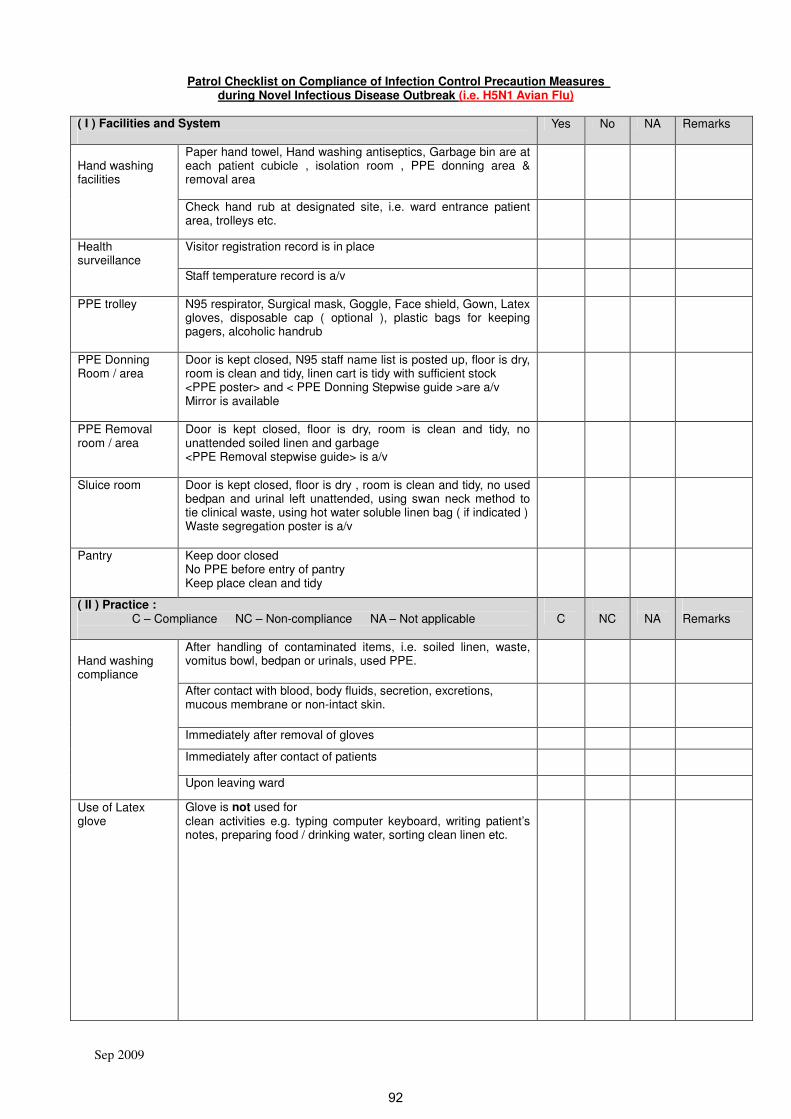
Sep 2009
Patrol Checklist on Compliance of Infection Control Precaution Measures during Novel Infectious Disease Outbreak (i.e. H5N1 Avian Flu)
( I ) Facilities and System
Yes No NA Remarks
Paper hand towel, Hand washing antiseptics, Garbage bin are at each patient cubicle , isolation room , PPE donning area & removal area
Hand washing facilities
Check hand rub at designated site, i.e. ward entrance patient area, trolleys etc.
Visitor registration record is in place Health surveillance
Staff temperature record is a/v
PPE trolley N95 respirator, Surgical mask, Goggle, Face shield, Gown, Latex gloves, disposable cap ( optional ), plastic bags for keeping pagers, alcoholic handrub
PPE Donning Room / area
Door is kept closed, N95 staff name list is posted up, floor is dry, room is clean and tidy, linen cart is tidy with sufficient stock <PPE poster> and < PPE Donning Stepwise guide >are a/v Mirror is available
PPE Removal room / area
Door is kept closed, floor is dry, room is clean and tidy, no unattended soiled linen and garbage <PPE Removal stepwise guide> is a/v
Sluice room Door is kept closed, floor is dry , room is clean and tidy, no used bedpan and urinal left unattended, using swan neck method to tie clinical waste, using hot water soluble linen bag ( if indicated ) Waste segregation poster is a/v
Pantry Keep door closed No PPE before entry of pantry Keep place clean and tidy
( II ) Practice : C – Compliance NC – Non-compliance NA – Not applicable
C
NC
NA
Remarks
After handling of contaminated items, i.e. soiled linen, waste, vomitus bowl, bedpan or urinals, used PPE.
After contact with blood, body fluids, secretion, excretions, mucous membrane or non-intact skin.
Immediately after removal of gloves
Hand washing compliance
Immediately after contact of patients
Upon leaving ward
Use of Latex glove
Glove is not used for clean activities e.g. typing computer keyboard, writing patient’s notes, preparing food / drinking water, sorting clean linen etc.
92

Sep 2009
( II ) Practice : C – Compliance NC – Non-compliance NA – Not applicable
C
NC
NA
Remarks
Use of Latex glove
Glove is used for � obnoxious procedures or procedures anticipated contacts
with blood, body fluids, secretion, excretions, mucous membrane and non-intact skin, e.g. suction, handling urinals and bedpans, changing napkin, draining urine, direct contact of wound.
� Should not be washed or disinfected with alcohol-based solutions and reuse.
� Gloves should not be used between patients or move from “dirty” to “clean” body sites in the same patient.
� When touching patients require contact isolation
Mask Worn when caring / visiting patient. ( N95 respirator for airborne transmission diseases & High risk procedures / surgical mask for droplet transmission diseases )
Gown ( Disposable / Linen gown )
When caring patient with direct close contact
Eye protection During high risk procedure such as taking NPA, suctioning, resuscitation
PPE Removal PPE must be removed & wash hands upon leaving the isolation room / cubicle ( Designated area ).
( III ) Duty of Patrol staff
Y N NA Remarks
Checking Staff has put on PPE in right steps and method
PPE item is sufficient
Staff was not wearing jewelry on duty
Assisting Staff to put on PPE if required
Staff to transfer items from clean zone to dirty zone
Enhancing Staff to adhere to infection control measures
Staff to wash hands properly
Log Sheet Record mobile staff in log sheet
Reporting Staff lapse practices
Additional comments and recommendations:
Ward : ___________________ Date : ______________________
Auditor : ___________________
93

Photo Guide on Suctioning Procedure (Special Points to Tackle “Before” & “After”)
A. Wear appropriate PPE after risk assessment 1. N95 mask / Surgical mask, 2. Full Face Shield, 3. Disposable cap, 4. Disposable gown, 5. Latex glove,
B. Secure tubing:
1. To ensure the tubing is long enough to reach patient. 2. To check the tubing is connected with L shape
connector 3. To ensure the system is secured.
Befo
re p
rocedure
C. Test the suction force is within desirable range.
D. Keep on PPE:
Rinsing the tubing with sterile water
E. Disposing the tubing properly
After
pro
cedure
隔離區要隔離區要隔離區要隔離區要
Protective M easures Step by Step
除手套除手套除手套除手套 Remove Gloves
除保護袍除保護袍除保護袍除保護袍
R emo ve Gown
除全面罩除全面罩除全面罩除全面罩 R emo ve Full Face-shield
除帽除帽除帽除帽
R emo ve C ap
除除除除 N95 口罩口罩口罩口罩
R emo ve N95 M ask
戴外科口罩戴外科口罩戴外科口罩戴外科口罩
Put on Surgical M ask
出出出出
OUT 洗手洗手洗手洗手 Wash Hands w ith Antiseptics 消毒液 洗手洗手洗手洗手 Wash Hands w ith Antiseptics 消毒液 洗手洗手洗手洗手 Wash Hands w ith Antiseptics 消毒液 洗手洗手洗手洗手 Wash Hands w ith Antiseptics 消毒液
F. Degowning according to the infection control guideline
94
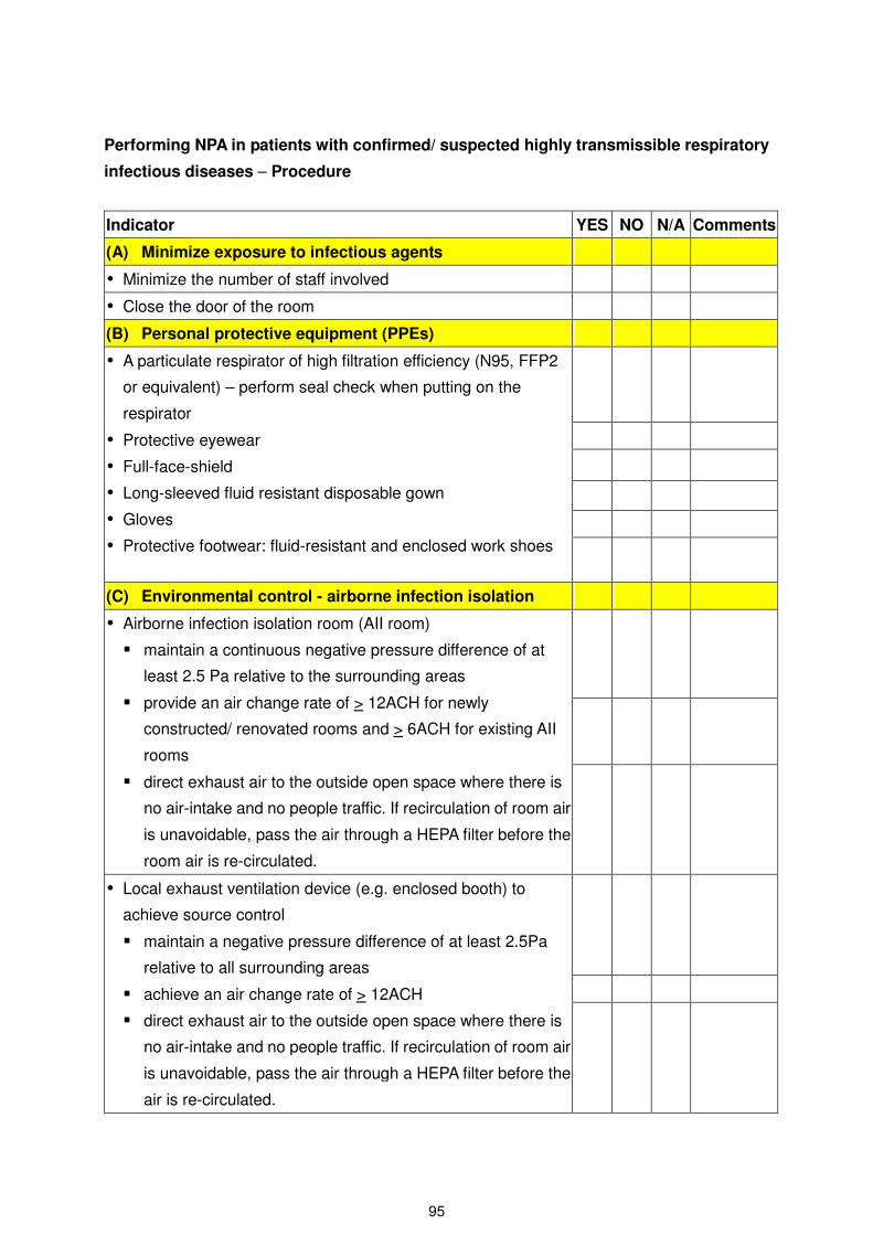
Performing NPA in patients with confirmed/ suspected highly transmissible respiratory
infectious diseases – Procedure
Indicator YES NO N/A Comments
(A) Minimize exposure to infectious agents
� Minimize the number of staff involved
� Close the door of the room
(B) Personal protective equipment (PPEs)
� A particulate respirator of high filtration efficiency (N95, FFP2
or equivalent) – perform seal check when putting on the
respirator
� Protective eyewear
� Full-face-shield
� Long-sleeved fluid resistant disposable gown
� Gloves
� Protective footwear: fluid-resistant and enclosed work shoes
(C) Environmental control - airborne infection isolation
� Airborne infection isolation room (AII room)
� maintain a continuous negative pressure difference of at
least 2.5 Pa relative to the surrounding areas
� provide an air change rate of > 12ACH for newly
constructed/ renovated rooms and > 6ACH for existing AII
rooms
� direct exhaust air to the outside open space where there is
no air-intake and no people traffic. If recirculation of room air
is unavoidable, pass the air through a HEPA filter before the
room air is re-circulated.
� Local exhaust ventilation device (e.g. enclosed booth) to
achieve source control
� maintain a negative pressure difference of at least 2.5Pa
relative to all surrounding areas
� achieve an air change rate of > 12ACH
� direct exhaust air to the outside open space where there is
no air-intake and no people traffic. If recirculation of room air
is unavoidable, pass the air through a HEPA filter before the
air is re-circulated.
95

� Single room with exhaust system to increase ACH and to
create a negative pressure
� Keep room door closed with room air exhausted directly to
outside open space where there is no air-intake and no
people traffic
� Adequately-ventilated single room
� Keep the room door closed with windows opened to open
space where there is no air intake duct and no people traffic
� May consider using a portable, industrial-grade HEPA filter to
provide air cleaning if an AII room or an enclosed booth that is
engineered for airborne isolation is not available
� Position the HEPA filter appropriately such that all room air
passes through the filter
- Consult engineer regarding the appropriate placement of
the filter
(D) Preparation
Preparation of equipment
� Specimen label, a bottle of transport medium and a mucus
extractor
� Water-proof and leak-proof specimen bag labeled ‘biohazard’
� Open the bag and stand it on a firm flat surface away from
the patient’s immediate environment, preferably at the room
entrance, with the upper rim of the bag folded slightly back
so that the bag is made wide open to avoid causing
contamination to the outside of the bag.
� Suction equipment
Preparation of nurse
� Put on personal protective equipment – see ‘B’
Preparation of the patient
� Verify the personal identification particulars on the specimen
label with the patient or against the patient’s identification
bracelet
� Affix the specimen label to the mucus extractor
� Explain the procedure to the patient to allay fear and to gain
cooperation, instruct the patient not to struggle during the
procedure
� Arrange the patient in a sitting up position, with the head tilted
96

slightly backward
� Cover the patient’s mouth with a surgical mask
� Remove gloves and perform hand hygiene
(E) Performing NPA
� Put on gloves
� Remove the cap of the bottle of viral transport medium
� Turn on the suction
� Connect the mucus extractor to the wall suction tubing
� Estimate the length of suction catheter to be inserted into the
nostril – measures approximately from the base of the nostril to
the earlobe
� Position yourself by the patient’s side and avoid facing the
patient directly
� Insert the suction catheter into nostril until it reaches the
posterior pharyngeal wall
� Apply suction and rotate the suction catheter
� Do not apply suction continuously for more than 15 seconds
� After obtaining the specimen, suction the whole viral transport
medium into the specimen bottle of the mucus extractor
� Detach the mucus extractor from the suction tubing
� Detach the suction catheter from the specimen bottle
� Minimize cross contamination on disposing of the suction
catheter: with the gloved-hand still holding the suction catheter,
turn that glove inside out so that it wraps the used suction
catheter inside it
� Discard the suction catheter and the glove into a leak-proof,
water-proof and sturdy waste bag printed or tagged with
‘biohazard’ labeling.
� Remove the glove from the other hand and discard the glove
� Perform hand-hygiene
� Put on gloves
� Cap the specimen bottle securely
� Shake the specimen bottle gently to mix the specimen with the
transport medium
� Drop the specimen upright gently into the prepared specimen
bag
(F) De-gowning
� Wash hands immediately after the procedure
97

� Remove PPEs in the designated gown-down area
� Remove PPEs gently and carefully to avoid causing
contamination to self and to the environment.
(G) Packaging and transport of specimen
� Close the opening of the specimen bag securely
� Do not put the laboratory request form together with the
specimen into the same compartment. If the specimen bag
carries a separate compartment for laboratory request form,
put the laboratory request form into that designated
compartment.
� Send the specimen to the laboratory immediately
� Make arrangement with laboratory staff to receive the
specimen prior to sending out the specimen
� If the specimen cannot be sent to the laboratory immediately,
keep the specimen in the appropriate storage condition.
(H) Cleaning and disinfection, and disposal of wastes
� Reusable equipments are cleaned and disinfected before
putting back for common use
� Reusable respiratory therapy equipments (e.g. suction bottle,
suction tubing ) are disinfected using at least high level
disinfection
� The specimen transport box is disinfected after use
� Disposable used equipments and wastes are disposed of in
water-proof, leak-proof and sturdy waste bags printed or
tagged with biohazard labeling at the point of use
� The environment is cleaned and disinfected immediately after
the procedure
(I) Record
� The staff who has performed the procedure is required to enter
his/her name and contact number on the designated record
� The record is filed and kept in a binder for a specified period of
time before disposal
98
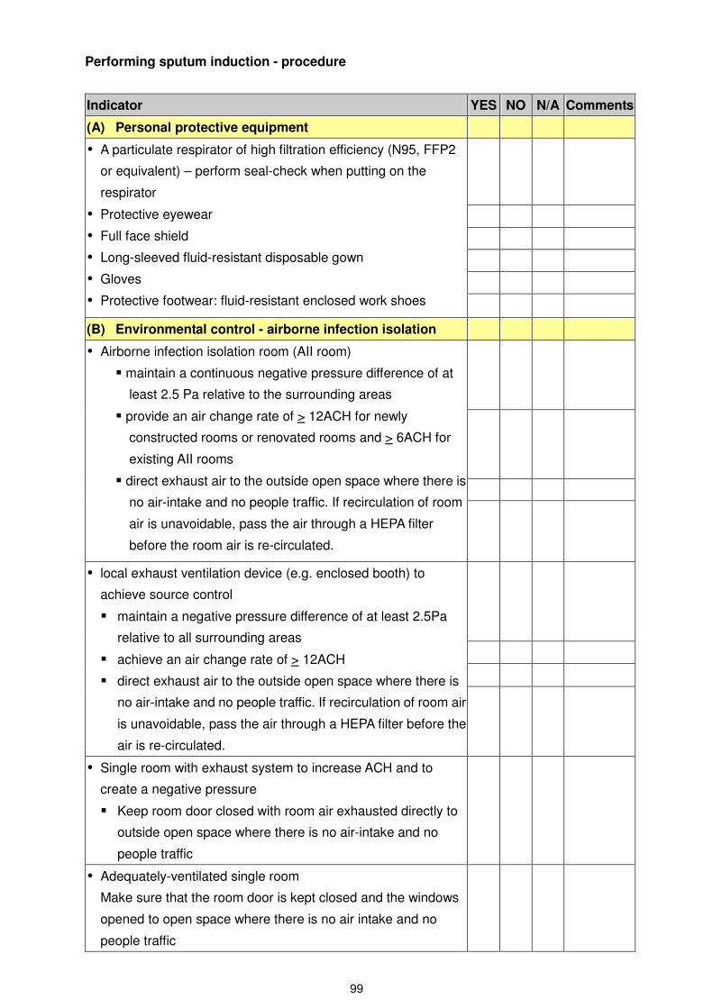
Performing sputum induction - procedure
Indicator YES NO N/A Comments
(A) Personal protective equipment
� A particulate respirator of high filtration efficiency (N95, FFP2
or equivalent) – perform seal-check when putting on the
respirator
� Protective eyewear
� Full face shield
� Long-sleeved fluid-resistant disposable gown
� Gloves
� Protective footwear: fluid-resistant enclosed work shoes
(B) Environmental control - airborne infection isolation
� Airborne infection isolation room (AII room)
� maintain a continuous negative pressure difference of at
least 2.5 Pa relative to the surrounding areas
� provide an air change rate of > 12ACH for newly
constructed rooms or renovated rooms and > 6ACH for
existing AII rooms
� direct exhaust air to the outside open space where there is
no air-intake and no people traffic. If recirculation of room
air is unavoidable, pass the air through a HEPA filter
before the room air is re-circulated.
� local exhaust ventilation device (e.g. enclosed booth) to
achieve source control
� maintain a negative pressure difference of at least 2.5Pa
relative to all surrounding areas
� achieve an air change rate of > 12ACH
� direct exhaust air to the outside open space where there is
no air-intake and no people traffic. If recirculation of room air
is unavoidable, pass the air through a HEPA filter before the
air is re-circulated.
� Single room with exhaust system to increase ACH and to
create a negative pressure
� Keep room door closed with room air exhausted directly to
outside open space where there is no air-intake and no
people traffic
� Adequately-ventilated single room
Make sure that the room door is kept closed and the windows
opened to open space where there is no air intake and no
people traffic
99

� May consider using a portable, industrial-grade HEPA filter to
provide air cleaning if an AII room or an enclosed booth that is
engineered for airborne isolation is not available
� Position the HEPA filter appropriately such that all room air
passes through the filter
- Consult engineer regarding the appropriate placement of
the filter
(C) Minimize exposure to infectious agents
� Put up ‘airborne’ and ‘contact’ precaution signages on the room
door
� Hang up a ‘No Entry: sputum induction in progress’ sign on the
room door, until the procedure is finished and 99% of the
airborne contaminants has been removed, the duration of
which would be subject to factors such as the ACH and size of
the room.
� Staff who are not involved in the procedure are not allowed to
enter the room
� Keep the room door closed
� Minimize entry and exit during the procedure
(D) Preparation
Preparation of the patient
� See that explanation has been given to the patient
� See that the patient has been fasted for the duration required
Preparation of equipment
� Nebulizer for sputum induction
� Bronchodilator puffer
� Specimen labels and specimen bottles
� Check the personal identification particulars on the
specimen label with the patient or against the patient’s
identification bracelet
� Affix the specimen label to the specimen bottle
� A water-proof and leak-proof specimen bag labeled
‘biohazard’,
� Open the bag and stand it on a firm flat surface away from
the patient’s immediate environment, preferably at the
entrance of the room, with the upper rim of the bag folded
slightly back so that opening of the bag is made wide open
to avoid causing contamination to the outside of the bag
� An empty bottle/ emesis bowl for saliva
� Peak-expiratory flow meter
(E) Procedure
100

Instructions to patient
� How to use the nebulizer
� Take deep breaths while inhaling the mist
� Expectorate the induced sputum into the specimen bottles
provided
� Cough deliberately and vigorously if spontaneous coughing
does not occur
� Cover the mouth with tissue paper during coughing
� Spit saliva into the empty bottle / emesis bowl provided
� Stay inside the booth or room, and to remain there until all
specimens have been saved and coughing has stopped
� Perform hand hygiene after the procedure
� Put on a surgical mask before leaving the room
� Call for assistance with the call bell provided should that be
required
Essential steps
� Take baseline peak expiratory flow rate and vital signs (blood
pressure, pulse rate, respiratory rate and SpO2 )
� Administer Salbutamol puff as prescribed
� Measure peak expiratory flow rate after administration of
Salbutamol
� Administer nebulized hypertonic sodium chloride solution
� Measure peak expiratory flow rate and vital signs immediately
and at specified intervals
� Chest physiotherapy may be employed to enhance the
production of sputum
(F) Observation during the procedure
� Observe the condition of the patient closely during the
procedure and watch out for signs and symptoms of respiratory
distress
� Continuous pulse oximetry monitoring
� Peak flow rate at the specified time intervals
� Respiratory rate, blood pressure and pulse rate at the
specified time intervals
(G) Packaging and transport of specimen
� Put on gloves
� Cap the specimen bottle securely
� Drop the specimen into the prepared specimen bag
� Remove gloves
� Perform hand-hygiene
101
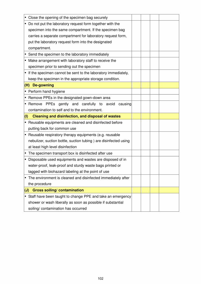
� Close the opening of the specimen bag securely
� Do not put the laboratory request form together with the
specimen into the same compartment. If the specimen bag
carries a separate compartment for laboratory request form,
put the laboratory request form into the designated
compartment.
� Send the specimen to the laboratory immediately
� Make arrangement with laboratory staff to receive the
specimen prior to sending out the specimen
� If the specimen cannot be sent to the laboratory immediately,
keep the specimen in the appropriate storage condition.
(H) De-gowning
� Perform hand hygiene
� Remove PPEs in the designated gown-down area
� Remove PPEs gently and carefully to avoid causing
contamination to self and to the environment.
(I) Cleaning and disinfection, and disposal of wastes
� Reusable equipments are cleaned and disinfected before
putting back for common use
� Reusable respiratory therapy equipments (e.g. reusable
nebulizer, suction bottle, suction tubing ) are disinfected using
at least high level disinfection
� The specimen transport box is disinfected after use
� Disposable used equipments and wastes are disposed of in
water-proof, leak-proof and sturdy waste bags printed or
tagged with biohazard labeling at the point of use
� The environment is cleaned and disinfected immediately after
the procedure
(J) Gross soiling/ contamination
� Staff have been taught to change PPE and take an emergency
shower or wash liberally as soon as possible if substantial
soiling/ contamination has occurred
102

Performing cardio-pulmonary resuscitation in patients with confirmed/ suspected
highly infectious respiratory diseases - Procedure
Indicator YES NO N/A Comments
(A) Specific infection control aspects
Minimize exposure of staff and environment to infectious
agents
� Minimize the number of staff involved
� Minimize generation of aerosols
� minimize the duration of manual bagging via the face mask
� avoid open suctioning of airway secretions
� Close the door of the room or pull close the curtains of the bed
Personal protective equipment
� A particulate respirator of high filtration efficiency: N95, FFP2
or a suitable alternative – perform seal check when putting on
the respirator
� Protective eyewear
� Full face shield
� Long-sleeved fluid-resistant disposable gown
� Gloves
� Protective footwear: fluid resistant and enclosed work shoes
Manual resuscitator – bag-valve-filter-mask unit (BVFM unit)
� Attach a high efficiency bacterial-viral filter to the patient end of
the BVM unit between the valve of the resuscitator bag and the
face-mask
� Filtration efficiency of the bacterial/viral filter
� Viral: >99.999%
� Bacterial: >99.9999%
Cardio-pulmonary resuscitation procedure
1) The first responder
� confirm unresponsiveness
� Shout for help
� Open airway
� Confirm absence of respiration and pulselessness
� Note the time of cardiac arrest
� Be sure already on full PPEs
2) The first responder
� Start external cardiac compression immediately at a rate of
100bpm
3) The second responder
� Call doctor/ central resuscitation team
� Mobilize manpower required to carry out the resuscitation
103

� Move the crash cart to the patient’s bedside
� Put on full PPEs
4) When two/ more staff members are available in full PPEs,
commence manual bagging with BVFM unit and continue with
external cardiac compression.
� Attaches ECG electrodes once the crash cart is taken to the
bedside.
� Authorized personnel can deliver defibrillation if it is a VF/
pulseless VT, followed by immediate external cardiac
compression.
� Perform CPR using a compression: ventilation ratio of 30:2
(before endotracheal intubation). Deliver each breath over 1
second. Stop chest compression only when the breath is
being delivered.
� Holds the BVFM unit tightly against the patient’s face with
both hands to achieve a tight seal between the face and the
mask
� Depending on the number of staff available, go to either
scenario 1 or scenario 2.
5) Scenario 1 – CPR when more than two staff are available
� One staff performs external cardiac compression
� One staff holds the BVFM unit tightly against the patient’s
face with both hands
� One staff squeezes the resuscitator bag gently with one
hand
Scenario 2 – CPR when only 2 staff are available
� One staff holds the BVFM unit tightly against the patient’s
face with both hands
� One staff performs both external cardiac compression and
squeezing of the resuscitator bag at a compression:
ventilation ratio of 30:2
Endotracheal intubation
� Prepare the necessary equipment before start
� If it is anticipated to be a difficult intubation
� Call in anaesthetist or intensivist
� Prepare equipment for difficult intubation
� During intubation, if the patient starts to regain consciousness
and struggle, the doctor may consider giving the patient
sedation +/- muscle relaxant (patient struggling will increase
the risk of generation of aerosols).
� After the endotracheal tube is inserted, inflate the endotracheal
104

tube cuff adequately to ensure there is no leak
� Connect the BVFM unit (with the high-efficiency bacterial/ viral
filter attached to it) to the endotracheal tube
� Ascertain correct position of the endotracheal tube (e.g. by
stethoscope or ETCO2 detection device if one is available )
� Fix the endotracheal tube securely in place
� Performing CPR after intubation
� Ventilate at 8-10 breaths per minute and continue to
perform external cardiac compressions at a rate of at least
100bpm.
� No need to synchronize external cardiac compression with
ventilation.
� Monitor the condition of the patient (blood pressure, ECG and
SpO2) throughout the whole procedure.
� Put used reusable equipment into a receiver right at the point
of use to minimize cross-contamination
Connecting the patient to the ventilator
� Before attaching the ventilator circuit to the endotracheal tube,
make sure that the doctor has set the ventilator settings and
the ventilator has been put on ‘Standby’ mode.
� Reactivate the ventilator after connecting the patient to the
ventilator
The ventilator circuit
� High efficiency bacterial/viral filter – attach to the expiratory
port of the ventilator
� Gas scavenging system – attach to the exhaust port of the
ventilator (if available)
� High efficiency heat and moisture bacterial/viral filter – attach
to the patient end of the ventilator circuit (preferable to using
heated water reservoir)
� If a closed-suction catheter is available, first attach the
closed-suction catheter to endotracheal tube, followed by
attaching the high-efficiency bacterial/viral filter to the side
connection port of the closed-suction catheter
� Closed-suction catheter – attach to the patient end of the
ventilator circuit
� Disposable ventilator tubings (preferable to reusable one)
De-gowning
� Wash hands immediately after CPR
� Remove PPEs in the designated gown-down area
� Remove PPEs gently and carefully to avoid causing
105

contamination to self and to the environment.
Emergency decontamination
� Staff change PPEs and take an emergency shower or wash
liberally as soon as possible if substantial soiling/
contamination has occurred
� Senior staff mobilize manpower to relief contaminated staff for
changing of PPEs or for emergency shower/ wash should that
require.
Cleaning and disinfection, and disposal of wastes
� The receiver of used equipment is put into a plastic bag before
removing from the isolation area
� Reusable equipments are cleaned and disinfected before
putting back for common use.
� Respiratory therapy equipments are disinfected with at least
high level disinfection
� Disposable used instruments and wastes are disposed of as
clinical wastes at the point of use
� Sharps are disposed of in sharps box at the point of use
(attached to the crash cart)
� The immediate environment is cleaned and disinfected
immediately after the procedure
(B) Crash cart
� Used items are replaced at the earliest possible convenience
� Replaced items are arranged according to the standardized
layout as set by the facility
(C) Documentation
The CPR procedure
� The resuscitation procedure and the events prior to the
cardio-pulmonary arrest are documented in both the CPR
record and the patient’s clinical notes.
� The facility requires that the CPR record be faxed or sent to the
facility CPR subcommittee for record and evaluation.
Log of staff involved in the CPR procedure
� Staff who have participated in the CPR procedure are required
to enter their name and contact number on the designated
record
� The records are filed in a binder and are kept there for a
specified period before discarding
106

Procedures for Taking Throat & Nasal Swab
Prepare Equipments - Sterile Swabs x 2 - Specimen bottle with viral transport medium (Viral TM
Bottle) - Appropriate PMH microbiology laboratory form & DH2542 - Zip-lock specimen carrier plastic bag
Sterile Swab & Viral TM Bottle
Procedures for taking Throat & Nasal Swab
Prepare Patient • Explain the procedures to patient (additional assistance may be
required for paediatric or uncooperative cases). • Cover patient’s mouth with surgical mask. • Ask patient to sit up with head tilted slightly backward.
Specimen Taking • Check expiry date for sterile items before use. • Staff put on appropriate PPE and wash hands. • To take nasal swab, a dry sterile swab is inserted into the nostril,
parallel to the palate, and left in place for a few seconds. • It is then slowly withdrawn with a rotating motion. • Specimens from both nostrils are obtained with the same swab. • The tip of the nasal swab is put into a TM bottle containing 2–3 ml
of virus transport medium and the applicator stick is broken off. • To take throat swab, both tonsils and the posterior pharynx are
swabbed vigorously. And the tip of the throat swab is placed into the
same TM bottle as described above. • Screw up the specimen container tightly. • Use appropriate laboratory form and zip-lock specimen carrier
plastic bag. • Remove contaminated PPE with care, and wash hands.
Aftercare • Observe patient for any complications, e.g., nasal bleeding, blood
stained sputum or desaturation. • Send out the specimen within 24 hours of collection according to
hospital guidelines. • After office hour, preferably store specimen at 4°C.
Nasal Swab
Throat Swab
Tips of both swabs broken
into ONE TM Bottle
107