Embed Size (px)
Citation preview
1
INDUCTION OF INFLAMMASOME DEPENDENT PYROPTOSIS BY CARBON
BLACK NANOPARTICLES* Anna C. Reisetter
1, Larissa V. Stebounova
2, Jonas Baltrusaitis
2, Linda Powers
1, Amit Gupta
1,
Vicki H. Grassian2 and Martha M. Monick
1
From Department of Medicine1 and Department of Chemistry
2
University of Iowa, Iowa City, IA
Running Title: Nanoparticles induce pyroptosis
Address Correspondence to: Martha M. Monick, PhD, Division of Pulmonary, Critical Care, and
Occupational Medicine, Room 100, EMRB, University of Iowa, Iowa City, IA 52242, Phone: (319) 335-
7590, Fax: (319) 335-6530, Email: [email protected]
Inhalation of nanoparticles has been
implicated in respiratory morbidity and
mortality. In particular, carbon black
nanoparticles are found in many different
environmental exposures. Macrophages take up
inhaled nanoparticles and respond via release
of inflammatory mediators and in some cases,
cell death. Based on new data, we propose that
exposure of macrophages (both a macrophage
cell line and primary human alveolar
macrophages) to carbon black nanoparticles
induces pyroptosis, an inflammasome-
dependent form of cell death. Exposure of
macrophages to carbon black nanoparticles
resulted in inflammasome activation as defined
by cleavage of caspase 1 to its active form and
downstream IL-1β release. The cell death that
occurred with carbon black nanoparticle
exposure was identified as pyroptosis by the
protective effect of a caspase 1 inhibitor and a
pyroptosis inhibitor. These data demonstrate
carbon black nanoparticle exposure activates
caspase 1, increases IL-1β release after LPS
priming and induces the proinflammatory cell
death, pyroptosis. The identification of
pyroptosis as a cellular response to carbon
nanoparticle exposure is novel, and relates to
environmental and health impacts of carbon-
based particulates.
Macrophages are critical regulators of local
immune homeostasis. They are highly adaptive
components of the innate immune system and
respond in diverse ways to pathogens and other
potential danger signals (1-3). In the lung, the
alveolar macrophage is the first line of defense
against environmental exposures. Alveolar
macrophages phagocytose particulate matter,
release inflammatory cytokines, and interact with
other cells and molecules through the expression
of surface receptors. One way in which an
immune response is generated in alveolar
macrophages is through the phagocytosis of
deposited particles within the respiratory tract (4).
The nanoparticle industry has expanded
substantially in recent years. A variety of
engineered carbon nanoparticles are used in
consumer products such as car tires, rubber, and
printer toner cartridges (5). Nanoparticles are also
being used as novel means of drug delivery.
Additionally, carbonaceous nanoparticles are
present as an environmental contaminant.
Combustion processes are a significant source of
carbon nanoparticles. Elemental carbon-based
nanoparticles with a diameter of less than 100 nm
are a major part of diesel exhaust and ambient
pollution (6).
Particulate ambient pollution is known to cause
adverse health effects in susceptible individuals,
and aggravates existing respiratory conditions
such as asthma and COPD (7). Even moderate
levels of ambient air particulates are known to
induce acute adverse health effects such as
mortality in heart and lung diseases and chronic
lung morbidity (8). Ultrafine particles are unique
in their ability to bypass mucociliary clearance
mechanisms and penetrate into deeper regions of
the respiratory tract (9-12). Although bulk
elemental carbon is considered chemically inert,
(as in diamond and graphite), seemingly inert
substances have been shown to elicit an
inflammatory response when exposure occurs with
nanoscale particles compared to an equivalent
mass dose of larger particles (11-15). Carbon
black (CB) nanoparticles can cause cytotoxic
injury, increase levels of proinflammatory
chemokines and inhibit cell growth (16).
There are several explanations for this
increased toxicity, including the increased surface
http://www.jbc.org/cgi/doi/10.1074/jbc.M111.238519The latest version is at JBC Papers in Press. Published on April 27, 2011 as Manuscript M111.238519
Copyright 2011 by The American Society for Biochemistry and Molecular Biology, Inc.
by guest on February 7, 2019http://w
ww
.jbc.org/D
ownloaded from
2
area of nanoparticles (10,12,14,17-22). In a
previous study, acute adverse effects of different
types of carbonaceous nanoparticles instilled in
mice strongly correlated with particle size and
surface area (23). A surface area threshold of ~20
cm2 was defined for acute lung inflammation in
mice, below which no inflammatory responses
were observed (23). CB nanoparticles showed
higher surface reactivity compared with a similar
dose of larger particles (24). CB nanoparticles
have also been shown to induce oxidative stress in
alveolar macrophages, and it is believed that this
capacity for oxidation may be mediated by particle
surface functionality (19,25-29). A recent study
showed that the oxidative potency of CB
nanoparticles correlates with their surface area and
inflammatory responses (30). A possible
mechanism for CB nanoparticles particle-related
inflammation involves direct and indirect reactive
oxygen species (ROS) generation by particle-cell
interactions, which in turn activates redox-
sensitive transcription of proinflammatory genes
(30). ROS have been implicated as the cause of
significant inflammation and, in some cases, cell
death (31).
One possible outcome of macrophage exposure
to nanoparticles is cell death. Cell death may be
categorized according to several characteristics
including non-inflammatory or pro-inflammatory,
and accidental or programmed. Apoptosis,
perhaps the best characterized of these
mechanisms, is a programmed and non-
inflammatory process. It is characterized by
distinctive DNA cleavage, as well as activation of
the executioner caspases, 3 and 9 (31,32). In
contrast to apoptosis, necrosis is defined as an
accidental and pro-inflammatory form of cell
death, in which the plasma membrane loses its
integrity, allowing rapid fluid influx, leading to
cell swelling and lysis (32-34). Pyroptosis is a
recently described mechanism of cell death,
sharing unique characteristics with both necrosis
and apoptosis (32-36). It is defined by its
dependence on inflammasome activation and
caspase 1 activity. Inflammasomes, which can
differ in their subunit composition, have been
shown to activate caspase 1, which, in the setting
of a microbial stimulus, activates the
proinflammatory cytokines IL-1 β and IL-18 (37)
(33,34). Like apoptosis, pyroptosis is a form of
programmed cell death. But unlike apoptosis,
pyroptosis is characterized by loss of membrane
integrity. This is due to caspase 1-dependent
insertion of a pore into the membrane, leading to
fluid influx, cell swelling and lysis (38).
Pyroptosis ultimately leads to release of cellular
contents and inflammation (32-34,36,38,39).
The recent expansion of the nanotechnology
industry, as well as the continually growing
sources of combustion derived pollution, warrants
investigation into the potential health effects of
these nanoparticles. In this study, we examined
the effect of CB nanoparticles on the
inflammasome and pyroptosis. The data show that
macrophage exposure to 20 ± 6 nm CB
nanoparticles induces caspase 1 activation and IL-
1 β release and the pro-inflammatory form of cell
death, pyroptosis.
Experimental Procedures
Source of manufactured nanomaterial. TiO2
and carbon black (CB) nanoparticles were
purchased from Degussa, GmbH (Düsseldorf,
Germany). Manufacturer’s stated average
diameters of titanium dioxide (TiO2) nanoparticles
(Degussa P25) and CB nanoparticles (Degussa
Printex 90) are 21 nm and 14 nm, respectively.
The nanoparticles were used as received from the
manufacturer without modification.
Bulk characterization of nanoparticles. Powder
X-ray diffraction (XRD) was used to identify
crystalline phases of the sample. XRD was
performed using a Bruker D-5000 q – q
diffractometer with Kevex-sensitive detector
(Madison, WI). High resolution transmission
electron microscopy (HRTEM) (JEOL JEM-
2100F, Japan) operating at 200 kV was used to
image the nanoparticles and measure their
diameters to compare the average diameter to the
manufacturer’s specifications. Samples for TEM
analysis were deposited from methanol
suspensions onto Cu grids. Dynamic light
scattering (DLS) (Beckman Coulter Delsa Nano C,
Brea, CA) was used to measure hydrodynamic
diameter of the nanoparticle aggregates in reduced
serum media (OptiMEM, Invitrogen) which was
used as a cell culture media in the cytotoxicity
experiments. Inductively coupled plasma optical
emission spectroscopy (ICP-OES) analysis was
performed to check for metal impurities in CB
nanoparticles. The nanoparticles were digested in
by guest on February 7, 2019http://w
ww
.jbc.org/D
ownloaded from
3
concentrated nitric acid at 90ºC prior to the ICP
analysis. The digested solutions were filtered and
centrifuged for 30 minutes at 14,000 rpm in order
to remove nanoparticles and aggregates that were
not dissolved. The final solutions were analyzed
by ICP-OES (Varian 720 ES, Walnut Creek, CA).
Surface characterization of nanoparticles.
Surface area and surface composition of the TiO2
and CB nanoparticles were examined. Surface area
measurements of powdered samples were
performed on an automated multipoint BET
surface area apparatus (Quantachrome Nova
4200e, Boynton Beach, FL) using nitrogen gas as
the adsorbent. Samples were degassed at 100ºC for
24 hours under vacuum before the analysis.
Surface area of TiO2 and CB nanoparticles was
calculated using 7-point BET method. X-ray
photoelectron spectroscopy (XPS) was used to
probe the surface chemical composition
characteristics of the powdered samples (Ultra-
Axis DLD. Kratos, Manchester, UK). The system
has been described before (40).
Demonstration of intracellur CB
nanoparticles (TEM). Samples were fixed
overnight with 2.5% glutaraldehyde in 0.1 M
cacodylate buffer. Post fixation was carried
out for 1 hour at room temperature with a
buffered 1% osmium tetroxide solution
reduced with 1.5% potassium ferrocyanide.
Samples were en bloc stained using 2.5%
uranyl acetate. Cells were then rinsed and
dehydrated. Infiltration of Spurr’s epoxy resin
and acetone were carried out over several days
to 100% resin and cured overnight in a 70oC
oven. Sections of 100 nm thickness were cut
using a Leica EM UC6 ultramicrotome. Grids
were then counterstained with 5% uranyl acetate
for 12 minutes and Reynold’s lead citrate for 5
minutes. Samples were imaged using a JEOL
1230 transmission electron microscope.
Cell culture. RAW264.7 cells were obtained
from ATCC (#TIB-71) and maintained in D-MEM
with 10 % fetal bovine serum and gentamycin, 40
μg /ml. Cells were sub-cultured every two to three
days. Experiments were run in 6 well Costar
tissue culture plates, 96 well assay plates or
coverslip chamber slides.
Human alveolar macrophages. To obtain
normal human alveolar macrophages, subjects
were recruited who were nonsmokers with no
underlying medical conditions and on no
medications other than possible birth control.
After informed consent was obtained, subjects
underwent standard flexible bronchoscopy.
Bronchoalveolar lavage was performed by
instilling 20 ml of normal saline into a tertiary
bronchus up to five times in three different lung
segments. The first collection out of five was
discarded for possible contamination from upper
airway secretions or by lidocaine, which is used to
locally anesthetize the subject during the
procedure. The remaining lavage was transported
to the laboratory where fluid was filtered through
sterile gauze and centrifuged at 200 x g for 5 min
to pellet cellular material. Cells were washed twice
in PBS and finally re-suspended in RPMI plus
Glutamax for cell culture. Cyto-prep slides were
also made with the cells, and were stained with
Wright stain. Slides were microscopically
examined to ensure that greater than 95% of the
cells were macrophages (41-43).
The cells were then placed in culture and
exposed to CB nanoparticles. All procedures and
protocols described in this communication were
approved by the University of Iowa Institutional
Review Board. Written informed consent was
obtained and all clinical investigation has been
conducted according to the principles expressed in
the Declaration of Helsinki.
Whole cell protein isolation. Whole cell protein
was obtained by lysing the cells on ice for 20
minutes in 200 l of lysis buffer (0.05 M Tris pH
7.4, 0.15 M NaCl, 1% NP-40, with added protease
and phosphatase inhibitors: 1 protease minitab
(Roche Biochemicals)/10 ml and 100 ul 100X
phosphatase inhibitor cocktail (Calbiochem)/10
ml. The lysates were sonicated for 20 seconds,
kept at 4o for 30 minutes, spun at 15,000 g for 10
minutes and the supernatant saved. Protein
determinations were made using the Bradford
Protein assay from Bio-Rad. Cell lysates were
stored at –70o until use.
Cell supernatant protein isolation. To isolate
proteins from cell supernatants, macrophages were
cultured in Opti-MEM® from Invitrogen to allow
for reduced serum culture. Cell supernatant
protein was obtained by concentrating the
supernatants in Amicon p10 filter tubes, spun at
3,000 g for 30 minutes. Protein determinations
were made using the Bradford Protein assay from
by guest on February 7, 2019http://w
ww
.jbc.org/D
ownloaded from
4
Bio-Rad. Concentrated supernatants were stored
at –70o until use.
Western analysis. Western analysis for the
presence of active caspase 1 was performed on
whole cell proteins and concentrated supernatants
from RAW cell experiments. 30 g of protein was
mixed 1:1 with 2x sample buffer (20% glycerol,
4% SDS, 10% -mercaptoethanol, 0.05%
bromophenol blue and 1.25 M Tris pH 6.8, all
chemicals from Sigma Chemical Co.) heated to
95o for 5 minutes and loaded onto a 10% SDS-
PAGE gel and run at 100 V for 90 minutes. Cell
proteins were transferred to PVDF (Bio-Rad
Hercules, CA) by semi-dry transfer (BioRad).
Equal loading of the protein groups on the blots
was evaluated using Ponceaus S, a staining
solution designed for staining proteins on PVDF
membranes or by stripping and reprobing with
antibodies to beta actin or GAPDH. The PVDF
was dried and then incubated with the primary
antibody overnight in 5% milk. The blots were
washed x4 with TTBS and incubated for 1 hour
with horseradish-peroxidase conjugated anti-
rabbit or mouse IgG antibody. Immunoreactive
bands were developed using a chemiluminescent
substrate (ECL Plus, Amersham, Arlington
Heights, IL). An autoradiograph was obtained,
with exposure times of 10 seconds to 2 minutes.
IL-1 β release. For these studies, RAW cells
were cultured in standard medium for 24 hours
with and without LPS (10 ng/ml). After the
culture period, the supernatants were harvested
and stored at –70o until assayed. The amount of
IL-1 β in the supernatant was measured by ELISA
(R & D Systems, Minneapolis, MN).
Cell survival analysis. For analysis of cell
survival, macrophages were cultured in 96 well
tissue culture plates. Following incubations with
nanoparticles, plasma membrane integrity was
assayed by two methods (LDH release and
PrestoBlue assay). Triplicate cultures were
performed on all experiments. LDH released into
the supernatant was monitored using CytoTox-
ONE™ Homogeneous Membrane Integrity Assay
which measures LDH release via a coupled
fluorescent assay (Promega). PrestoBlue Cell
Viability Assay was done following the
manufacturers protocol.
Whole cell DNA Isolation/DNA analysis. For
DNA analysis, cells were cultured in 100 mm
tissue culture plates. Following incubations with
particles, whole cells were harvested in 500 μl
PBS and DNA was isolated (DNeasy Blood and
Tissue Kit, Qiagen, Valencia, CA). DNA
concentration was measured and equal
concentrations were loaded and run on a SYBR
Green gel (E-Gel 2% with SYBR Safe,
Invitrogen). The gel was visualized with
ultraviolet light, and samples were examined for
laddering.
Quantitative RT-PCR. Total RNA was isolated
using the RNAqueous – 4PCR kit (Ambion Inc.,
Austin, TX) following the manufacturer’s
instructions. RNA quality and quantity were
assessed with Experion automated electrophoresis
system (Bio-Rad Laboratories, Hercules, CA)
using the Experion RNA StdSens Analysis Kit
according to the manufacturer’s protocol. RNA
quality was considered adequate for use if the
28S/18S ratio was >1.2 and the RNA Quality
Indicator (RQI) was >7. Total RNA (300 ng) was
reverse-transcribed to cDNA using iScript cDNA
Synthesis kit (Bio-Rad) following the
manufacturer’s instructions. PCR reactions were
performed as previously described (44).
Specificity of the amplification was confirmed
using melting curve analysis. Data were collected
and recorded by CFX Manager Software and
expressed as a function of threshold cycle (CT).
The relative quantity of the gene of interest was
then normalized to relative quantity of HPRT (
CT). The sample mRNA abundance was calculated
by the formula 2-( CT)
.
Specific primer sets used are as follows (5' to 3'):
IL-1 β (IL-1 β fw, 5’- CTCCAGGGACAGGAT
ATGGA-3’; IL-1 β rev, 5’- TTCTGCTTGAGAG
GTGCTGA-3’); (IL-18 fw, 5’- ACAGCTTCGG
GAAGAGGAAAGGAA-3’; IL-18 rev, 5’- TGTC
TTCTACTGGTTCAGCAGCCA-3’); TNFα
(TNFα fw, 5’- AGGACACCATGAGCACTGAA
AGCA-3’; TNFα rev, 5’-TTGAGGGTTTGC
TACAACATGGGC-3’).
RESULTS
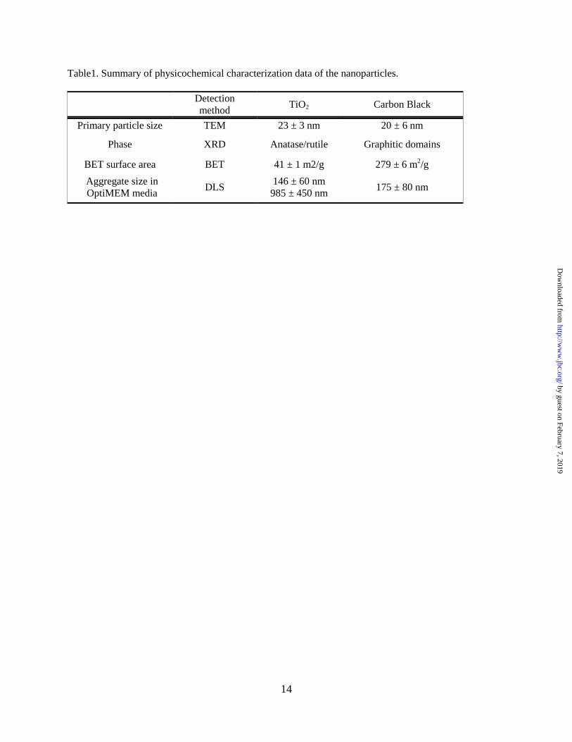
Nanoparticle characterization. The results of
particle characterization are summarized in Table
1. Primary particle diameters obtained from the
TEM images (Figure 1A) indicate that TiO2
nanoparticles have diameter of 23 ± 3 nm and CB
nanoparticles have diameter of 20 ± 6 nm (Table
1). For the CB nanoparticles, this result is close to
by guest on February 7, 2019http://w
ww
.jbc.org/D
ownloaded from
5
the manufacturer’s specified diameter of 14 nm.
The aggregate size distributions of TiO2 and CB
nanoparticles were measured using DLS in
OptiMEM media. Both types of nanoparticles
form aggregates in the cell culture media. TiO2
nanoparticles have bimodal size distribution with
peaks at 146 ± 60 nm and 985 ± 450 nm with a
larger size distribution dominating over a smaller
size distribution. CB nanoparticles have aggregate
sizes of 175 ± 80 nm (Table 1). The XRD analysis
of TiO2 nanoparticles indicates that there are
anatase and rutile crystalline phases present in the
sample (45). A diffraction pattern for CB
nanoparticles is similar to the literature reports for
CB (46) and indicates crystalline phases of
graphitic domains. XPS analysis of TiO2
nanoparticles indicates 33, 61 and 6% of Ti, O and
C, respectively, showing close to stoichiometric
Ti:O ratio and residual hydrocarbon and hydroxyl
groups present on the surface of the sample
(Figure 1B). High resolution XPS analysis of CB
nanoparticles shows the presence of carbon at
285.0 eV with traces of oxygen at 530.0 eV
(Figure 1B). ICP-OES analysis of CB
nanoparticles digested in concentrated nitric acid
revealed small amounts of metal impurities such
as Ca (0.02%) and K (0.04%) present in the
sample. The BET specific surface areas of TiO2
and CB nanoparticles calculated using the 7-point
BET method are 41 ± 1 m2/g and 279 ± 6 m
2/g,
respectively (Table 1). CB is considered to be a
microporous material. The external surface area of
CB nanoparticles calculated using the Halsey
method (47) is 176 ± 9 m2/g.
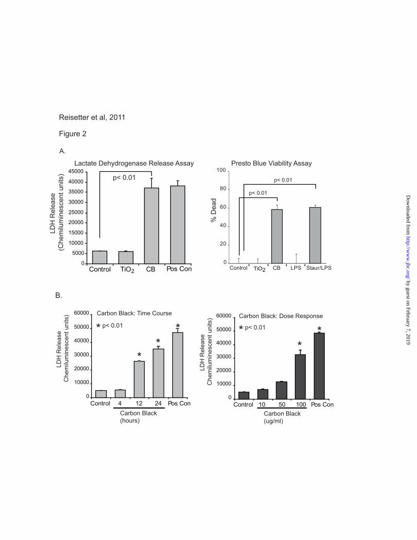
CB nanoparticle exposure induces macrophage
cell death (RAW264.7 cells). To evaluate the
effect of nanoparticles on macrophage viability,
RAW264.7 cells were seeded into 96 well tissue
culture plates and cultured for 24 hours with TiO2
(30 μg/cm2) or CB nanoparticles (30 μg/cm
2). At
the end of the culture period, one group was
treated with ATP as a positive control for cell
death (31,38,45-47). Samples were assayed for
LDH release. Figure 2A shows that CB
nanoparticles, and not TiO2 nanoparticles, induce
LDH release from macrophages. To confirm cell
death with an alternative assay, a PrestoBlue cell
viability reagent was used. PrestoBlue analyzes
cell death by determining the level of reducing
activity associated with living cells. RAW264.7
cells were seeded into 96 well tissue culture plates
and cultured for 24 hours in the same conditions.
As with the LDH release assay, the PrestoBlue
analysis showed cell killing with exposure to CB
nanoparticles and not TiO2 nanoparticles. To further characterize the cell death caused by
CB nanoparticle exposure, both time and dose
response experiments were performed. Figure 2B
demonstrates an increase in LDH release by 12
hours that continues increasing through 24 hours
of particle exposure. Optimal effects were seen at
a dose of 30 μg/cm2. As a composite, these data
show that CB nanoparticles, and not TiO2
nanoparticles, decrease plasma membrane
integrity leading to cell death.
Nanoparticle-induced plasma membrane
disruption is characterized by an increase in cell
size. The LDH and PrestoBlue assays suggest that
exposure to CB nanoparticles disrupts the plasma
membrane. To determine whether this reduced
plasma membrane integrity affected cell size, cell
size was quantified after exposure to CB
nanoparticles. In the first experiment (Figure 3A),
cells were exposed to CB nanoparticles and
images were obtained immediately (15 minutes)
and again after 24 hours. Cell size was determined
by drawing circles around representative cells
from 10 fields and calculating area using Image J
software. Figure 3A shows an increase in cell size
after exposure to CB nanoparticles (mean of 162.7
± 17.57 mm2 for control cells compared to 276.9 ±
39.99 mm2 for CB nanoparticle exposed
macrophages). To analyze volume changes,
macrophages were loaded with a fluorescent
tracer, cell tracker green CMFDA (Invitrogen).
Figure 3B shows an increase in volume with CB
nanoparticle exposure. To confirm that the
macrophages were internalizing the particles,
transmission electron microscopy was used to
analyze particle exposed macrophages. Figure 3C
demonstrates that CB nanoparticles are taken up
by macrophages and appear to localize both in the
cytosol and in membrane bound vesicles.
CB nanoparticles activate the inflammasome
(caspase 1 and IL-1 β release). Both necrosis and
pyroptosis are characterized by LDH release and
loss of plasma membrane integrity. We asked if
CB nanoparticle exposure was activating the
inflammasome (central to the pyroptosis form of
cell death). The inflammasome’s two primary
activation markers, caspase 1 activity and IL-1 β
release, were measured. Cells were primed for 3
by guest on February 7, 2019http://w
ww
.jbc.org/D
ownloaded from
6
hours with LPS (10 ng/ml). After priming, media
was replaced, and fresh LPS was added, along
with CB and TiO2 nanoparticles for an additional 6
hours as previously described (48). Western
analysis was performed using whole cell lysates,
as well as concentrated supernatants. Figure 4A
shows that in RAW264.7 cells, significantly more
of the 20 kD cleaved caspase 1 was present in both
the lysates and supernatants of cells exposed to CB
nanoparticles as opposed to those exposed to TiO2
nanoparticles. Recent studies have defined the
release of active caspase 1 as a valid measure of
inflammasome activity (49-53).
To determine if caspase 1 activation also
occurred in a relevant human primary cell, human
alveolar macrophages were cultured in the same
conditions. Cell lysates and supernatants were
harvested and Western analysis performed for
active caspase 1. Figure 4B shows that in human
alveolar macrophages, CB nanoparticle exposure
caused activation of caspase 1 as demonstrated by
the presence of the 20 kD cleaved caspase 1 in
both cell lysates and supernatants. To confirm the
caspase 1 activation, IL-1 β release was measured
using an ELISA. Figure 4C demonstrates that
with LPS priming both RAW264.7 cells and
human alveolar macrophages increase IL-1 β
release with CB nanoparticle exposure (p<0.01).
Figure 4C shows that CB alone does not induce
IL-1 protein release from macrophages. To
further examine the role of LPS priming in the CB
augmentation of IL-1release, we examined LPS
or CB exposed cells for IL-1, IL-18 and
TNFmRNAs. We found that LPS, but not CB,
induced transcript up regulation (Figure 4D). This
supports our conclusion that CB alone activates
the inflammasome leading to cell death
(pyroptosis), while CB plus a microbial stimulus
leads to both pyroptosis and IL-1 release.
To confirm that the increase in IL-1β release
with CB nanoparticles was due to caspase 1
activation, an experiment was performed using the
caspase 1 inhibitor, YVAD. Figure 4E shows that
the increase in IL-1 β release with CB
nanoparticles is blocked in the YVAD exposed
cells. As a composite, these data suggest that CB
nanoparticle exposure in macrophages activates
the inflammasome as shown by caspase 1
activation and IL-1β release.
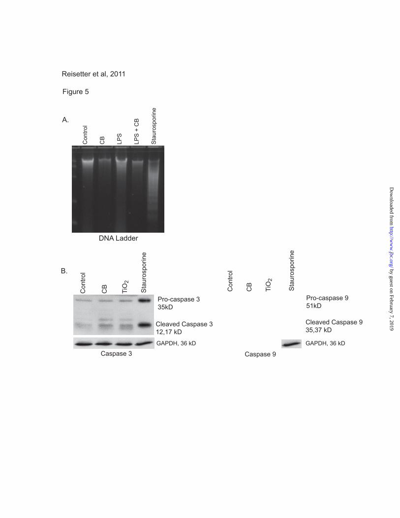
CB nanoparticle-induced cell death is not due
to apoptosis. Because the CB nanoparticle-
induced cell death is characterized by loss of
plasma membrane integrity, it is unlikely that it is
an apoptotic process. However, to confirm that
apoptosis was not involved we examined the effect
of CB nanoparticles on apoptosis. Macrophages
were exposed to CB or TiO2 nanoparticles for 16-
24 hours. Staurosporine was used as a positive
control for apoptosis. Following the incubation
period, DNA was isolated from whole cell lysates.
The DNA concentration was measured, and equal
amounts were run on a SYBR green gel and
visualized using ultraviolet light. Figure 5A
shows that, while the cells exposed to
staurosporine show distinctive DNA laddering
characteristic of apoptosis, none of the CB
nanoparticle exposed cells show DNA laddering.
In a second set of experiments, macrophages from
CB nanoparticle exposed cells were lysed, proteins
isolated and Western analysis performed for
activation of two caspases linked to apoptosis,
caspase 3 and caspase 9 (31). Figure 5B shows
that caspases 3 and 9 were activated in cells
exposed to staurosporine but not in those exposed
to CB nanoparticles. Taken in combination with
the LDH and PrestoBlue assays, this demonstrates
that CB nanoparticles induce non-apoptotic cell
death.
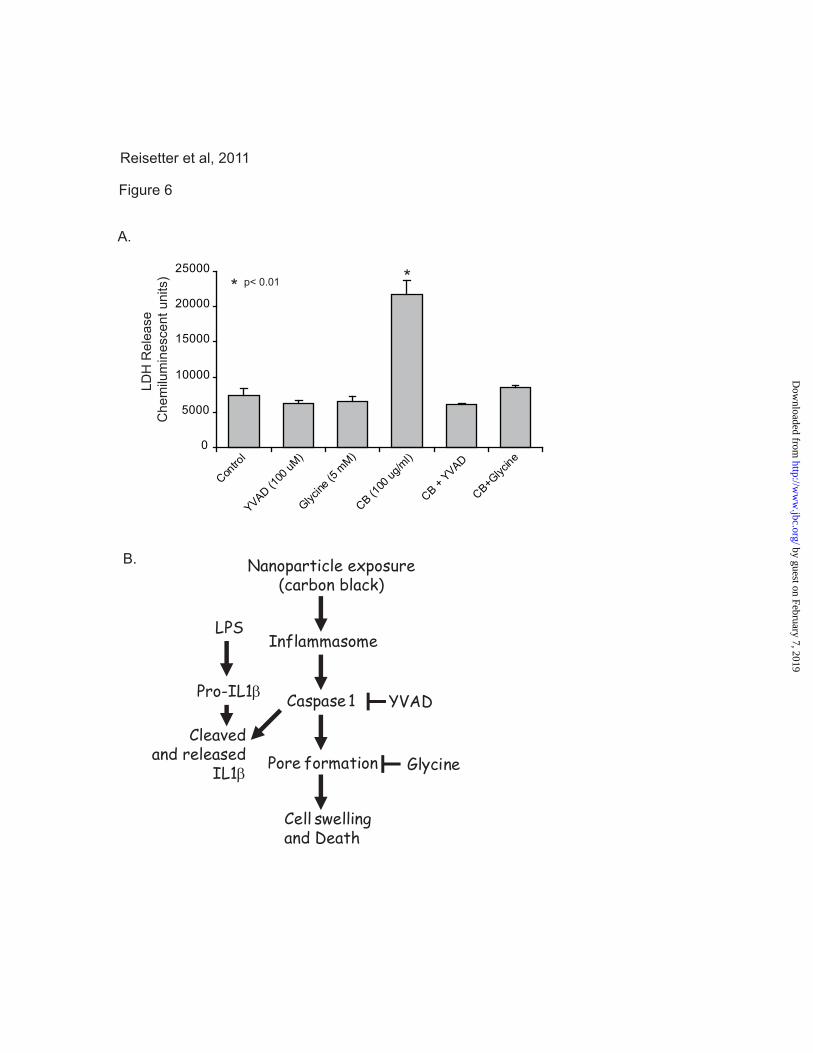
Inhibition of caspase 1 blocks CB
nanoparticle-induced cell death in macrophages.
In light of the observation that CB nanoparticles
activate the inflammasome and induce cell death
in macrophages, we next sought to characterize the
specific mechanism of cell death. The cell death
modality pyroptosis is characterized by caspase 1
activation and the subsequent opening of cell
membrane pores, resulting in an influx of
extracellular fluid and eventual cell lysis (38). To
confirm that the observed non-apoptotic cell death
was pyroptosis, we examined the effects of a
caspase 1 inhibitor (YVAD) and a pyroptosis
inhibitor (glycine) on RAW264.7 cells exposed to
CB nanoparticles. RAW264.7 cells were seeded
into 96 well tissue culture plates and cultured for
24 hours with CB nanoparticles (30 μg/cm2), CB
nanoparticles in the presence of YVAD, or CB
nanoparticles in the presence of glycine. At the
end of the culture period, all samples were assayed
for LDH release. Figure 6A shows that, as
previously demonstrated, CB nanoparticle exposed
by guest on February 7, 2019http://w
ww
.jbc.org/D
ownloaded from
7
cells showed decreased membrane integrity as
evidenced by LDH release. Both YVAD and
glycine attenuated the effects of CB nanoparticle-
induced LDH release. Cells exposed to CB
nanoparticles in the presence of either of these
inhibitors released near control levels of LDH.
DISCUSSION
In this study, we found that CB nanoparticles
induced cell death in macrophages, and that this
occurred in the absence of any detectable
transition metals. These data show that following
phagocytosis of the CB nanoparticles,
macrophages increased in size - the opposite of the
cellular condensation associated with apoptosis.
Macrophage exposure to CB nanoparticles led to
inflammasome activation, as characterized by
caspase 1 activation and IL-1 β release. The
identification of the cell death undergone by
exposed cells as pyroptosis was confirmed by the
inhibiting effects of both a caspase 1 inhibitor and
a pyroptosis inhibitor on CB nanoparticle-induced
cell death (Figure 6B).
A daily dose of CB in the working environment
can be as large as 120 g/kg person, assuming a
threshold limiting value for respirable carbon
black of 2.5 mg/m3
(54). Additionally, an eight-
hour average amount of elemental carbon detected
on a heavily traveled roadway in Harlem was 6.2
µg/m3 (55), which corresponds to the alveoli
burden of 7 µg, assuming a respiratory volume of
0.7 m3 per hour and 0.2 alveolar deposition
fraction for 20 nm particles based on human
deposition model (56). In the current study, 30
µg/cm2 of CB nanoparticles were applied to cell
culture wells. Although it is difficult to compare
these doses with the mass per mass and mass per
volume concentrations discussed above, it is
within the mass range that is observed in
occupational and environmental settings but does
represent a substantial dose for respirable carbon
black.
Pyroptosis, a pro-inflammatory form of cell
death, proceeds through the activation of the
inflammasome, leading to cleavage of caspase 1
into its active form. Once activated, caspase 1
cleaves the pro-inflammatory cytokines IL-1 β and
IL-18 into their active forms, allowing for their
release into the extra-cellular environment.
Although indicative of caspase 1 activation, the
release of these inflammatory cytokines is not
required for caspase 1 activation or pyroptosis.
The production of pro-IL-1 β and pro-IL-18 is
mediated by toll like receptors (TLRs). The
release of active IL-1 β and IL-18 associated with
pyroptosis in previous reports (36,57-65) has all
included TLR stimulatory effects of microbial
antigens. Thus, while priming cells with LPS
prior to nanoparticle exposure allows for an
additional confirmation of caspase 1 activation,
inflammasome activation can occur separately
from LPS priming. This is supported by previous
reports, showing that caspase 1 induced cell death
may proceed independently of IL-1 β and IL-18
secretion when a microbial stimulus is not present
(38,66).
Pyroptosis is characterized by a loss of
membrane integrity (32-34,38). While the exact
mechanism of this remains unknown, it has been
demonstrated that membrane pore formation
occurs, leading to cell swelling, and necrosis-like
lysis. Our data supports pyroptosis after CB
nanoparticle exposure with evidence of cell
swelling, caspase 1 activation and cell death.
There have been a number of inflammasomes
characterized with different subunits. Although
this study does not specify which inflammasome
CB nanoparticles activate, it has been shown that
particulate matter including silica, asbestos, MSU,
cholesterol crystals (37,67,68), and aluminum
adjuvants (69) induce a caspase 1 dependent
inflammatory response mediated by the NALP3
inflammasome (31,37,70-72). As such, these data
suggest that CB nanoparticles activate the NALP3
inflammasome as well, though ongoing studies
will further characterize the CB nanoparticle
inflammasome. To the best of our knowledge, this
is the first instance in which nanoparticles have
been implicated in inducing pyroptosis (31).
Several mechanisms of inflammasome
activation have been proposed, including the
generation of ROS (13,18,73-78), potassium efflux
(50), cathepsin B (79), and phagosomal
destabilization(28,48,72,80). Disintegration of the
cellular membrane by CB nanoparticles can cause
the production of ROS. Aam and Fonnum showed
that low doses of CB nanoparticles activate rat
alveolar macrophages to produce ROS (28). They
suggested that the ERK MAPK pathway
participates in intracellular signaling leading to the
ROS generation. Hornung et al demonstrated that
by guest on February 7, 2019http://w
ww
.jbc.org/D
ownloaded from
8
the NALP3 inflammasome activation induced by
silica crystals and aluminum salts could be
replicated via sterile lysosomal damage,
implicating intracellular pH or cathepsin B activity
in inflammasome activation (48). Even
nanoparticles made from materials that are
considered inert in bulk form have been found to
induce pulmonary inflammation when exposure
occurs with nanoscale particles. Although this
study did not find TiO2 nanoparticles to be
inflammasome activating in macrophages, Yazdi
et al showed that in human keratinocytes, nano-
TiO2 activates the NALP3 inflammasome and
induces IL-1 β (51). Any or all of the potential
mechanisms discussed may apply to CB
nanoparticles’ mechanism of inflammasome
activation. Further investigation into the ROS
generated by alveolar macrophages in response to
CB nanoparticle exposure is warranted.
The present study shows that macrophage
exposure to CB nanoparticles activates the
inflammasome leading to pyroptosis. CB merits
further investigation into its mechanisms of
inflammation modulation (increased IL-1
release) and pyroptosis. As a primary component
in ambient pollution and diesel exhaust, and a
component of toners in printers used in office
buildings worldwide, CB nanoparticles are a
critical target for study. A better understanding of
their mechanism of inflammasome activation may
allow us to appropriately regulate potential health
hazards.
REFERENCES
1. Opitz, B., van Laak, V., Eitel, J., and Suttorp, N. (2010) Am J Respir Crit Care Med 181,
1294-1309
2. Harada, R. N., and Repine, J. E. (1985) Chest 87, 247-252
3. Twigg, H. L., 3rd. (2004) Seminars in respiratory and critical care medicine 25, 21-31
4. Sibille, Y., and Reynolds, H. Y. (1990) Am Rev Respir Dis 141, 471-501
5. (2010) Overview on Promising Nanomaterials for Industrial Applications. Nanoroad
SME European project
6. Moller, W., Brown, D. M., Kreyling, W. G., and Stone, V. (2005) Part Fibre Toxicol 2, 7
7. Hussain, S., Thomassen, L. C., Ferecatu, I., Borot, M. C., Andreau, K., Martens, J. A.,
Fleury, J., Baeza-Squiban, A., Marano, F., and Boland, S. (2010) Part Fibre Toxicol 7, 10
8. Lundborg, M., Johard, U., Lastbom, L., Gerde, P., and Camner, P. (2001) Environ Res
86, 244-253
9. Lippmann, M., Yeates, D. B., and Albert, R. E. (1980) British journal of industrial
medicine 37, 337-362
10. Oberdorster, G. (1994) Ann Occup Hyg 38, 601-615, 421-602
11. Ferin, J., Oberdorster, G., and Penney, D. P. (1992) Am J Respir Cell Mol Biol 6, 535-542
12. Oberdorster, G., Ferin, J., and Lehnert, B. E. (1994) Environ Health Perspect 102 Suppl
5, 173-179
13. Beck-Speier, I., Dayal, N., Karg, E., Maier, K. L., Schumann, G., Schulz, H., Semmler,
M., Takenaka, S., Stettmaier, K., Bors, W., Ghio, A., Samet, J. M., and Heyder, J. (2005)
Free Radic Biol Med 38, 1080-1092
14. Stone, V., Shaw, J., Brown, D. M., Macnee, W., Faux, S. P., and Donaldson, K. (1998)
Toxicol In Vitro 12, 649-659
15. Renwick, L. C., Brown, D., Clouter, A., and Donaldson, K. (2004) Occup Environ Med
61, 442-447
16. Yamawaki, H., Iwai, N. (2006) Circulation Journal 70, 129-140
by guest on February 7, 2019http://w
ww
.jbc.org/D
ownloaded from
9
17. Beck-Speier, I., Dayal, N., Karg, E., Maier, K. L., Roth, C., Ziesenis, A., and Heyder, J.
(2001) Environ Health Perspect 109 Suppl 4, 613-618
18. Hussain, S., Boland, S., Baeza-Squiban, A., Hamel, R., Thomassen, L. C., Martens, J. A.,
Billon-Galland, M. A., Fleury-Feith, J., Moisan, F., Pairon, J. C., and Marano, F. (2009)
Toxicology 260, 142-149
19. Koike, E., and Kobayashi, T. (2006) Chemosphere 65, 946-951
20. Mauderly, J. L., Snipes, M. B., Barr, E. B., Belinsky, S. A., Bond, J. A., Brooks, A. L.,
Chang, I. Y., Cheng, Y. S., Gillett, N. A., Griffith, W. C., and et al. (1994) Research
report (Health Effects Institute), 1-75; discussion 77-97
21. Moss, O. R., and Wong, V. A. (2006) Inhalation toxicology 18, 711-716
22. Oberdorster, G., Oberdorster, E., and Oberdorster, J. (2005) Environ Health Perspect
113, 823-839
23. Stoeger, T., Reinhard, C., Takenaka, S., Schroeppel, A., Karg, E., Ritter, B., Heyder, J.,
and Schulz, H. (2006) Environmental Health Perspectives 114, 328-333
24. Brown, D. M., Dickson, C., Duncan, P., Al-Attili, F., Stone, V. (2010) Nanotechnology
21, 215104/215101-215104/215109
25. Barlow, P. G., Clouter-Baker, A., Donaldson, K., Maccallum, J., and Stone, V. (2005)
Part Fibre Toxicol 2, 11
26. Ma, J. Y., and Ma, J. K. (2002) J Environ Sci Health C Environ Carcinog Ecotoxicol Rev
20, 117-147
27. Ito, T., Ikeda, M., Yamasaki, H., Sagai, M., and Tomita, T. (2000) Environ Toxicol
Pharmacol 9, 1-8
28. Aam, B. B., Fonnum, F. . (2007) Archives of toxicology 81, 441-446
29. Dick, C. A. J., Brown, D. M., Donaldson, K., Stone, V. (2003) Inhalation toxicology 15,
39-52
30. Stoeger, T., Takenaka, S., Frankenberger, B., Ritter, B., Karg, E., Maier, K., Schulz, H.,
Schmid, O. (2009) Environmental Health Perspectives 117, 54-60
31. Martinon, F., Mayor, A., and Tschopp, J. (2009) Annu Rev Immunol 27, 229-265
32. Fink, S. L., and Cookson, B. T. (2005) Infection and immunity 73, 1907-1916
33. Kepp, O., Galluzzi, L., Zitvogel, L., and Kroemer, G. European journal of immunology
40, 627-630
34. Bergsbaken, T., Fink, S. L., and Cookson, B. T. (2009) Nat Rev Microbiol 7, 99-109
35. Schroder, K., and Tschopp, J. Cell 140, 821-832
36. Suzuki, T., Franchi, L., Toma, C., Ashida, H., Ogawa, M., Yoshikawa, Y., Mimuro, H.,
Inohara, N., Sasakawa, C., and Nunez, G. (2007) PLoS pathogens 3, e111
37. Cassel, S. L., Joly, S., and Sutterwala, F. S. (2009) Seminars in immunology 21, 194-198
38. Fink, S. L., and Cookson, B. T. (2006) Cell Microbiol 8, 1812-1825
39. Miao, E. A., Leaf, I. A., Treuting, P. M., Mao, D. P., Dors, M., Sarkar, A., Warren, S. E.,
Wewers, M. D., and Aderem, A. Nature immunology 11, 1136-1142
40. Baltrusaitis, J., Usher, C. R., and Grassian, V. H. (2007) Phys Chem Chem Phys 9, 3011-
3024
41. Monick, M. M., Powers, L. S., Barrett, C. W., Hinde, S., Ashare, A., Groskreutz, D. J.,
Nyunoya, T., Coleman, M., Spitz, D. R., and Hunninghake, G. W. (2008) J Immunol 180,
7485-7496
by guest on February 7, 2019http://w
ww
.jbc.org/D
ownloaded from
10
42. Monick, M. M., Powers, L. S., Gross, T. J., Flaherty, D. M., Barrett, C. W., and
Hunninghake, G. W. (2006) J Immunol 177, 1636-1645
43. Monick, M. M., Powers, L. S., Walters, K., Lovan, N., Zhang, M., Gerke, A., Hansdottir,
S., and Hunninghake, G. W. (2010) J Immunol 185, 5425-5435
44. Hansdottir, S., and Monick, M. M. (2011) Vitam Horm 86, 217-237
45. Brough, D., and Rothwell, N. J. (2007) J Cell Sci 120, 772-781
46. Perregaux, D., and Gabel, C. A. (1994) The Journal of biological chemistry 269, 15195-
15203
47. Walev, I., Reske, K., Palmer, M., Valeva, A., and Bhakdi, S. (1995) Embo J 14, 1607-
1614
48. Hornung, V., Bauernfeind, F., Halle, A., Samstad, E. O., Kono, H., Rock, K. L.,
Fitzgerald, K. A., and Latz, E. (2008) Nature immunology 9, 847-856
49. Dostert, C., Petrilli, V., Van Bruggen, R., Steele, C., Mossman, B. T., and Tschopp, J.
(2008) Science (New York, N.Y 320, 674-677
50. Petrilli, V., Papin, S., Dostert, C., Mayor, A., Martinon, F., and Tschopp, J. (2007) Cell
death and differentiation 14, 1583-1589
51. Yazdi, A. S., Guarda, G., Riteau, N., Drexler, S. K., Tardivel, A., Couillin, I., and
Tschopp, J. Proceedings of the National Academy of Sciences of the United States of
America 107, 19449-19454
52. Pazar, B., Ea, H. K., Narayan, S., Kolly, L., Bagnoud, N., Chobaz, V., Roger, T., Liote,
F., So, A., and Busso, N. (2011) Journal of immunology 186, 2495-2502
53. Guarda, G., Zenger, M., Yazdi, A. S., Schroder, K., Ferrero, I., Menu, P., Tardivel, A.,
Mattmann, C., and Tschopp, J. (2011) Journal of immunology 186, 2529-2534
54. Vesterdal, L. K., Folkmann, J. K., Jacobsen, N. R., Sheykhzade, M., Wallin, H., Loft, S.,
and Moller, P. (2010) Particle and Fibre Toxicology 7, 33
55. Kinney, P. L., Aggarwal, M., Northridge, M. E., Janssen, N. A., and Shepard, P. (2000)
Environmental Health Perspectives 108, 213-218
56. Cassee, F. R., Freijer, J.I., Subramaniam, R., Asgharian, B., Miller, F.J., van Bree, L., and
Rombout, P.J.A. . (1999) Bilthoven, the Netherlands: Dutch National Institute of Public
Health and Environment (RIVM)
57. Miao, E. A., Leaf, I. A., Treuting, P. M., Mao, D. P., Dors, M., Sarkar, A., Warren, S. E.,
Wewers, M. D., and Aderem, A. (2010) Nature immunology 11, 1136-1142
58. Sauer, J. D., Witte, C. E., Zemansky, J., Hanson, B., Lauer, P., and Portnoy, D. A. (2010)
Cell Host Microbe 7, 412-419
59. Silveira, T. N., and Zamboni, D. S. (2010) Infection and immunity 78, 1403-1413
60. Ngai, S., Batty, S., Liao, K. C., and Mogridge, J. (2010) Febs J 277, 119-127
61. Whitfield, N. N., Byrne, B. G., and Swanson, M. S. (2010) Infection and immunity 78,
423-432
62. Rupper, A. C., and Cardelli, J. A. (2008) Infection and immunity 76, 2304-2315
63. Fink, S. L., Bergsbaken, T., and Cookson, B. T. (2008) Proceedings of the National
Academy of Sciences of the United States of America 105, 4312-4317
64. Bergsbaken, T., and Cookson, B. T. (2007) PLoS pathogens 3, e161
65. Fink, S. L., and Cookson, B. T. (2007) Cell Microbiol 9, 2562-2570
66. Monack, D. M., Navarre, W. W., and Falkow, S. (2001) Microbes Infect 3, 1201-1212
67. Duewell, P., Kono, H., Rayner, K. J., Sirois, C. M., Vladimer, G., Bauernfeind, F. G.,
Abela, G. S., Franchi, L., Nunez, G., Schnurr, M., Espevik, T., Lien, E., Fitzgerald, K. A.,
by guest on February 7, 2019http://w
ww
.jbc.org/D
ownloaded from
11
Rock, K. L., Moore, K. J., Wright, S. D., Hornung, V., and Latz, E. Nature 464, 1357-
1361
68. Cassel, S. L., Eisenbarth, S. C., Iyer, S. S., Sadler, J. J., Colegio, O. R., Tephly, L. A.,
Carter, A. B., Rothman, P. B., Flavell, R. A., and Sutterwala, F. S. (2008) Proceedings of
the National Academy of Sciences of the United States of America 105, 9035-9040
69. Eisenbarth, S. C., Colegio, O. R., O'Connor, W., Sutterwala, F. S., and Flavell, R. A.
(2008) Nature 453, 1122-1126
70. Martinon, F., Gaide, O., Petrilli, V., Mayor, A., and Tschopp, J. (2007) Seminars in
immunopathology 29, 213-229
71. Martinon, F., Petrilli, V., Mayor, A., Tardivel, A., and Tschopp, J. (2006) Nature 440,
237-241
72. Chen, G. Y., and Nunez, G. Nat Rev Immunol 10, 826-837
73. Zhou, R., Yazdi, A. S., Menu, P., and Tschopp, J. (2011) Nature 469, 221-225
74. Tschopp, J., and Schroder, K. (2010) Nat Rev Immunol 10, 210-215
75. Slane, B. G., Aykin-Burns, N., Smith, B. J., Kalen, A. L., Goswami, P. C., Domann, F.
E., and Spitz, D. R. (2006) Cancer Res 66, 7615-7620
76. Hu, Y., Mao, K., Zeng, Y., Chen, S., Tao, Z., Yang, C., Sun, S., Wu, X., Meng, G., and
Sun, B. (2010) Journal of immunology 185, 7699-7705
77. Hussain, S., Thomassen, L. C., Ferecatu, I., Borot, M. C., Andreau, K., Martens, J. A.,
Fleury, J., Baeza-Squiban, A., Marano, F., and Boland, S. (2010) Particle and Fibre
Toxicology 7, 10
78. Val, S., Hussain, S., Boland, S., Hamel, R., Baeza-Squiban, A., and Marano, F. (2009)
Inhalation toxicology 21 Suppl 1, 115-122
79. Davis, M. J., and Swanson, J. A. Journal of leukocyte biology 88, 813-822
80. Cassel, S. L., and Sutterwala, F. S. European journal of immunology 40, 607-611
FOOTNOTES
*This manuscript was supported by NIH R01 HL079901, NIH RO1 R01HL96625 and NCRR 3 UL1
RR024979 to MM and R01HL96625 to VHG. This publication was made possible by Grant Number
UL1RR024979 from the National Center for Research Resources (NCRR), a part of the National
Institutes of Health (NIH). Its contents are solely the responsibility of the authors and do not necessarily
represent the official views of the CTSA or NIH.
The abbreviations used are: BET, Brunauer-Emmett-Teller; CB, carbon black; CMFDA, 5-
chloromethylfluorescein diacetate; COPD, chronic obstructive pulmonary disease; DLS,
dynamic light scattering; ELISA, enzyme-linked immunosorbant assay; ICP-OES, inductively
coupled plasma optical emission spectroscopy; IL-1 β, interleukin 1 β; LDH, lactate
dehydrogenase; LPS, lipopolysaccharide; NALP3, NACHT domain, leucine-rich-repeat (LRR)
domain, and pyrin domain (PYD)-containing protein 3; ROS, reactive oxygen species; TEM,
transmission electron microscopy; TTBS, tris buffered saline; XPS, x-ray photoelectron
spectroscopy; XRD, x-ray diffraction; YVAD, benzyloxycarbonyl-Tyr-Val-Ala-Asp(OMe)-
fluoromethylketone.
by guest on February 7, 2019http://w
ww
.jbc.org/D
ownloaded from
12
FIGURE LEGENDS
Figure 1. Nanoparticle characterization. A. High resolution TEM images of CB and TiO2
nanoparticles. The scale bar in both images is 20 nm. Both nanoparticles form aggregates when in
solution. Particles were sonicated for 1 hour and vortexed vigorously prior to cell exposure. Primary
particle diameters were obtained from the images. B. Surface composition of TiO2 and CB nanoparticles
was measured using X-ray photoelectron spectroscopy.
Figure 2. CB nanoparticles induce macrophage cell death. A. RAW264.7 cells were exposed to CB
(30 μg/cm2) or TiO2 (30 μg/cm
2) nanoparticles for 24 hours. One group was treated with ATP as a
positive control for LDH release. At the end of the culture period, LDH analysis was performed to
determine cell viability. The experiment was repeated using identical conditions. A PrestoBlue cell
viability assay was performed as indicated by the manufacturer. Significance was determined using
nonpaired Student’s t test. B. RAW264.7 cells were exposed to CB nanoparticles (30 μg/cm2) for 4, 12,
and 24 hours. ATP was used as a positive control for LDH release. At the end of the culture period, an
LDH assay was performed to determine cell viability. Additionally, RAW264.7 cells were exposed to CB
nanoparticles at 3 μg/cm2, 15 μg/cm
2, or 30 μg/cm
2 for 24 hours. An LDH assay was performed.
Significance was determined using nonpaired Student’s t test.
Figure 3. CB nanoparticles cause an increase in macrophage cell size. A. Bright field images of CB
nanoparticle exposed cells were obtained 15 minutes after exposure and again after 24 hours. Circles
were drawn around representative cells from 10 fields and then used to calculate cellular area. B.
Fluorescent images of cells were obtained before CB nanoparticle exposure and again, 24 hours after
exposure. Cells were loaded with cell tracker green CMFDA. Average cellular volume was calculated
from the level of fluorescence per cell. C. Transmission electron microscopy images of control and CB
nanoparticle exposed cells.
Figure 4. CB nanoparticles activate the inflammasome. A. Activation of caspase 1 in RAW264.7
cells. 3x10⁶ RAW264.7 cells per well were seeded into culture plates. Cells were primed for 3 hours
with LPS (10 ng/ml). After 3 hours, media was aspirated off of cells and replaced with fresh. LPS was
re-added, and CB (30 μg/cm2) or TiO2 (30 μg/cm
2) nanoparticles were added for an additional 6 hours.
After the culture period, whole cell lysates were harvested in Western Lysis Buffer. Supernatants were
concentrated and proteins analyzed. Western analysis for caspase 1 was performed on cellular lysates and
concentrated supernatants. B. 3x10⁶ human alveolar macrophages per well were seeded into culture
plates. Cells were primed and cultured as detailed above. Western analysis for caspase 1 was performed
on cellular lysates and concentrated supernatants. Beta actin and Ponceau S stain demonstrate equal
loading. C. 1x10⁶ RAW264.7 cells per well were seeded into culture plates. Cells were cultured as
above, except for an extended second incubation time (16-24 hours). Supernatants were harvested and
IL-1 levels measured by ELISA. Significance was determined using nonpaired Student’s t test. D.
Expression of IL-1 β, IL-18, and TNFα mRNA in human alveolar macrophages exposed to LPS or CB
nanoparticles. 3x10⁶ human alveolar macrophages per well were seeded into culture plates. Cells were
incubated with CB nanoparticles (30 μg/cm2) or LPS (10 ng/ml) for 4 hours. RNA was isolated and qRT-
PCR performed. * p< 0.01 by Student’s t-test (compared to control). E. 1x10⁶ RAW264.7 cells per well
were seeded into culture plates. Cells were primed for 3 hours with LPS (10 ng/ml). For the second
incubation period, LPS was re-added and CB nanoparticles (30 μg/cm2), TiO2 nanoparticles (30 μg/cm
2),
YVAD (100 μM), and ATP were added for an additional 24 hours. Supernatants were harvested and IL-
1 levels measured by ELISA.
Figure 5. Nanoparticle-induced cell death is not apoptosis. A. 5x10⁶ RAW264.7 cells were seeded
into 100 mm culture dishes and incubated with LPS (10 ng/ml), CB nanoparticles (30 μg/cm2), or both
by guest on February 7, 2019http://w
ww
.jbc.org/D
ownloaded from
13
for 24 hours. One group was exposed to staurosporine (1 μM) as a positive control for apoptosis.
Following the culture period, DNA was isolated. DNA concentration was measured and equal
concentrations were loaded and run on a SYBR Green gel. The gel was visualized with ultraviolet light,
and samples were examined for laddering. B. 3x10⁶ RAW264.7 cells per well were seeded into culture
plates. Cells were exposed to CB nanoparticles (30 μg/cm2), TiO2 nanoparticles (30 μg/cm
2), or
staurosporine (1 μM) for 24 hours. Proteins were isolated and Western analysis for the apoptosis-
associated caspases 3 and 9 was performed.
Figure 6. Blocking caspase 1 activation protects macrophages from CB nanoparticles toxicity. A.
RAW264.7 cells were exposed to CB nanoparticles (30 μg/cm2), CB nanoparticles in combination with
the pyroptosis inhibitor glycine (5 mM), CB nanoparticles in combination with the caspase 1 inhibitor
YVAD (100 μM), or TiO2 nanoparticles (30 μg/cm2) for 24 hours. At the end of the culture period, LDH
analysis was performed to determine cell viability. B. The diagram summarizes the CB nanoparticle
induced pathway to inflammasome activation and pyroptosis, as supported by this study.
by guest on February 7, 2019http://w
ww
.jbc.org/D
ownloaded from
14
Table1. Summary of physicochemical characterization data of the nanoparticles.
Detection
method TiO2 Carbon Black
Primary particle size TEM 23 ± 3 nm 20 ± 6 nm
Phase XRD Anatase/rutile Graphitic domains
BET surface area BET 41 ± 1 m2/g 279 ± 6 m2/g
Aggregate size in
OptiMEM media DLS
146 ± 60 nm
985 ± 450 nm 175 ± 80 nm
by guest on February 7, 2019http://w
ww
.jbc.org/D
ownloaded from
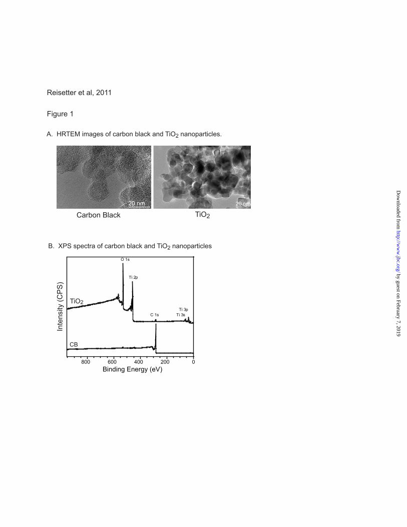
Figure 1
Reisetter et al, 2011
A. HRTEM images of carbon black and TiO2 nanoparticles.
Binding Energy (eV)0200400600800
O 1s
C 1s
Ti 2p
Ti 3pTi 3s
B. XPS spectra of carbon black and TiO2 nanoparticles
Inte
nsity
(CP
S)
CB
TiO2
Carbon Black TiO2
by guest on February 7, 2019http://w
ww
.jbc.org/D
ownloaded from
LDH
Rel
ease
C
hem
ilum
ines
cent
uni
ts)
LDH
Rel
ease
(C
hem
ilum
ines
cent
uni
ts)
LDH
Rel
ease
C
hem
ilum
ines
cent
uni
ts)
Figure 2
Reisetter et al, 2011
0
10000
20000
30000
40000
50000
60000
Control 10 50 100 Pos Con0
10000
20000
30000
40000
50000
60000 Carbon Black: Time Course Carbon Black: Dose Response
Carbon Black(ug/ml)
Control 4 12 24 Pos Con
Pos Con
Carbon Black(hours)
0
5000
10000
15000
20000
25000
30000
35000
40000
45000
Control TiO2 CB
p< 0.01
**
**
** p< 0.01 * p< 0.01
A.
Lactate Dehydrogenase Release Assay Presto Blue Viability Assay
B.
% D
ead
p< 0.01
p< 0.01
0
20
40
60
80
100
Control TiO2 CB LPS Staur/LPS
by guest on February 7, 2019http://w
ww
.jbc.org/D
ownloaded from
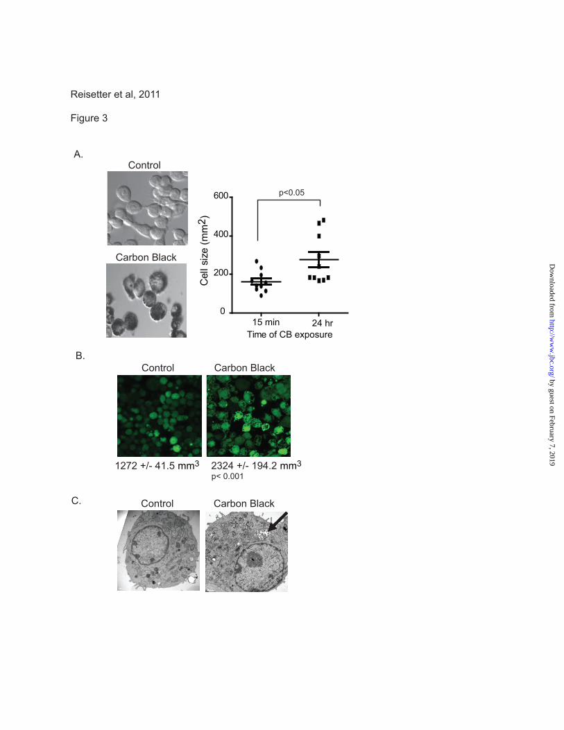
Figure 3
Reisetter et al, 2011
A.
B.
C.
1272 +/- 41.5 mm3 2324 +/- 194.2 mm3p< 0.001
Control Carbon Black
Control
Carbon Black
Control Carbon Black
15 min 24 hr0
200
400
600
Time of CB exposure
Cel
l siz
e (m
m2 )
p<0.05
by guest on February 7, 2019http://w
ww
.jbc.org/D
ownloaded from
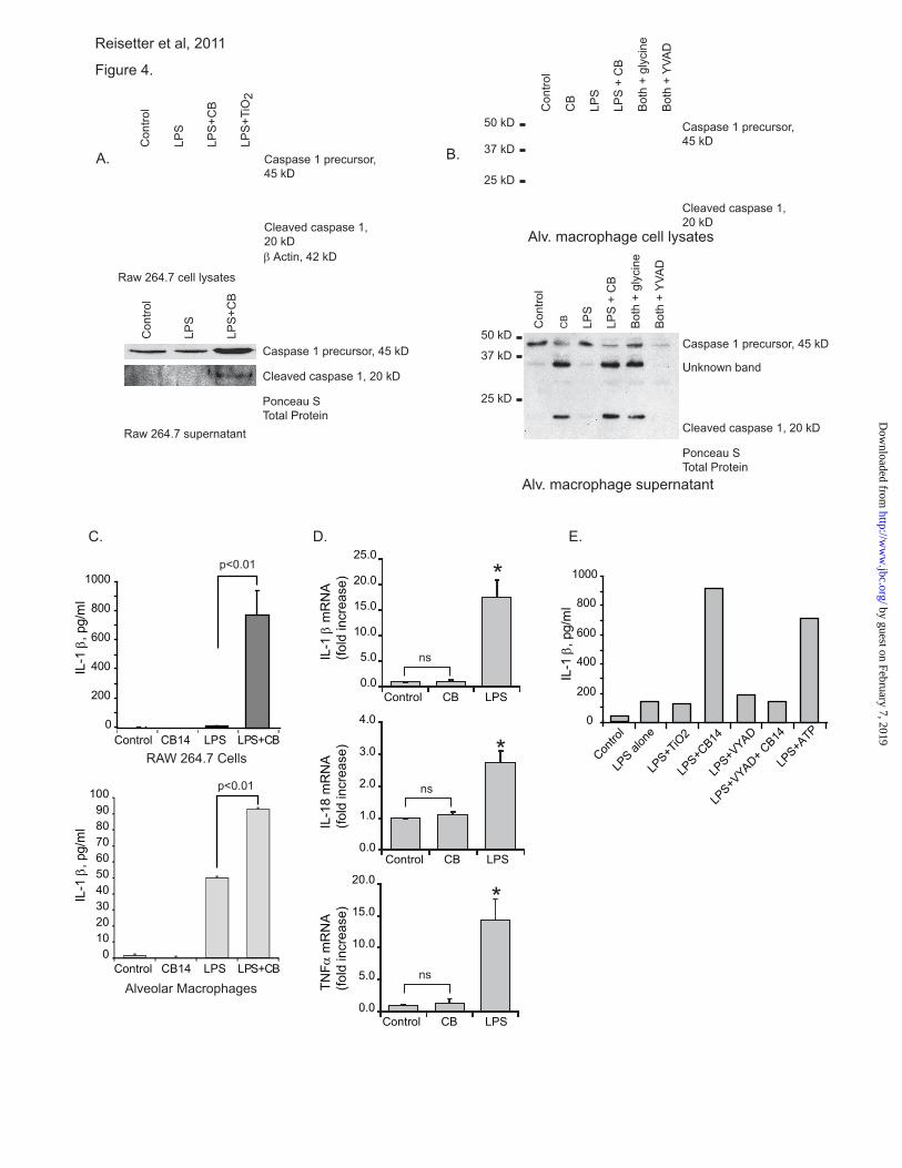
Figure 4.
Reisetter et al, 2011
B.
C. E.
Con
trol
LPS
LPS
+CB
Con
trol
LPS
LPS
+CB
LPS
+TiO
2
Con
trol
CB LP
S
LPS
+ C
B
Bot
h +
glyc
ine
Bot
h +
YVA
D
Con
trol
CB
LPS
LPS
+ C
B
Bot
h +
glyc
ine
Bot
h +
YVA
D
Ponceau S Total Protein
Ponceau S Total Protein
Cleaved caspase 1, 20 kD
IL-1
β, p
g/m
lIL
-1 β
, pg/
ml
0
200
400
600
800
1000
LPS+CB
A.
p<0.01
p<0.01
Caspase 1 precursor, 45 kD
Cleaved caspase 1, 20 kD
Cleaved caspase 1, 20 kD
Caspase 1 precursor, 45 kD
Caspase 1 precursor, 45 kD
Cleaved caspase 1, 20 kD
Caspase 1 precursor, 45 kD
β Actin, 42 kD
Raw 264.7 supernatant
Alv. macrophage supernatant
Raw 264.7 cell lysates
Alv. macrophage cell lysates
50 kD
37 kD
25 kD
50 kD
37 kD
25 kD
Unknown band
RAW 264.7 Cells
Alveolar Macrophages
0102030405060708090
100
Control CB14 LPS
LPS+CB Control CB14 LPS
0
200
400
600
800
1000
LPS al
one
Contro
l
LPS+T
iO2
LPS+C
B14
LPS+V
YAD
LPS+V
YAD+ CB14
LPS+A
TP
IL-1
β, p
g/m
l
IL-1
β m
RN
A(fo
ld in
crea
se)
IL-1
8 m
RN
A(fo
ld in
crea
se)
TNFα
mR
NA
(fold
incr
ease
)
D.
Control CB LPS
Control CB LPS
Control CB LPS
ns
ns
ns
*
*
*
0.0
5.0
10.0
15.0
20.0
25.0
0.0
1.0
2.0
3.0
4.0
0.0
5.0
10.0
15.0
20.0
by guest on February 7, 2019http://w
ww
.jbc.org/D
ownloaded from
Con
trol
CB
LPS
LPS
+ C
B
Sta
uros
porin
e
Con
trol
CB
TiO
2
Sta
uros
porin
e
Con
trol
CB
TiO
2
Sta
uros
porin
ePro-caspase 3 35kD
Cleaved Caspase 312,17 kD
Pro-caspase 951kD
Cleaved Caspase 9 35,37 kD
Caspase 3 Caspase 9
DNA Ladder
Figure 5
Reisetter et al, 2011
A.
B.
GAPDH, 36 kDGAPDH, 36 kD
by guest on February 7, 2019http://w
ww
.jbc.org/D
ownloaded from
0
5000
10000
15000
20000
25000
Contro
l
YVAD (100
uM)
Glycine
(5 m
M)
CB (100
ug/m
l)
CB + YVAD
CB+Glyc
ine
LDH
Rel
ease
C
hem
ilum
ines
cent
uni
ts)
A.
B.
Figure 6
Reisetter et al, 2011
** p< 0.01
Nanoparticle exposure(carbon black)
Inflammasome
Caspase 1
Pore formation
Cell swellingand Death
LPS
YVAD
Glycine
Pro-IL1β
Cleavedand released
IL1β
by guest on February 7, 2019http://w
ww
.jbc.org/D
ownloaded from
Vicki H. Grassian and Martha M. MonickAnna C. Reisetter, Larissa V. Stebounova, Jonas Baltrusaitis, Linda Powers, Amit Gupta,
Induction of inflammasome dependent pyroptosis by carbon black nanoparticles
published online April 27, 2011J. Biol. Chem.
10.1074/jbc.M111.238519Access the most updated version of this article at doi:
Alerts:
When a correction for this article is posted•
When this article is cited•
to choose from all of JBC's e-mail alertsClick here
by guest on February 7, 2019http://w
ww
.jbc.org/D
ownloaded from