Embed Size (px)
Citation preview
Page 1
Ultrasound of the Thyroid and Parathyroid
Indications for Thyroid US
enlarged gland
palpable nodule
history childhood XRT or other high risk category
incidental nodule identified while imaging the neck
neck pain
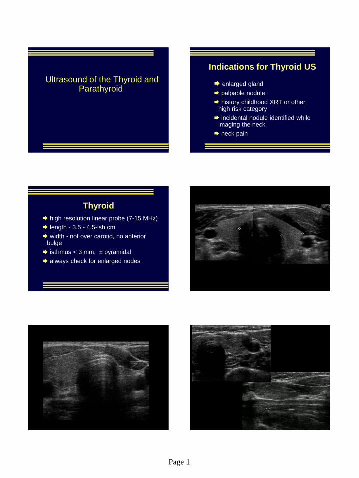
Thyroid
high resolution linear probe (7-15 MHz)
length - 3.5 - 4.5-ish cm
width - not over carotid, no anterior bulge
isthmus < 3 mm, ± pyramidal
always check for enlarged nodes
Normal thyroid trv
Anderson agenesis left lobe thyroid
esoph nml trv
Page 2
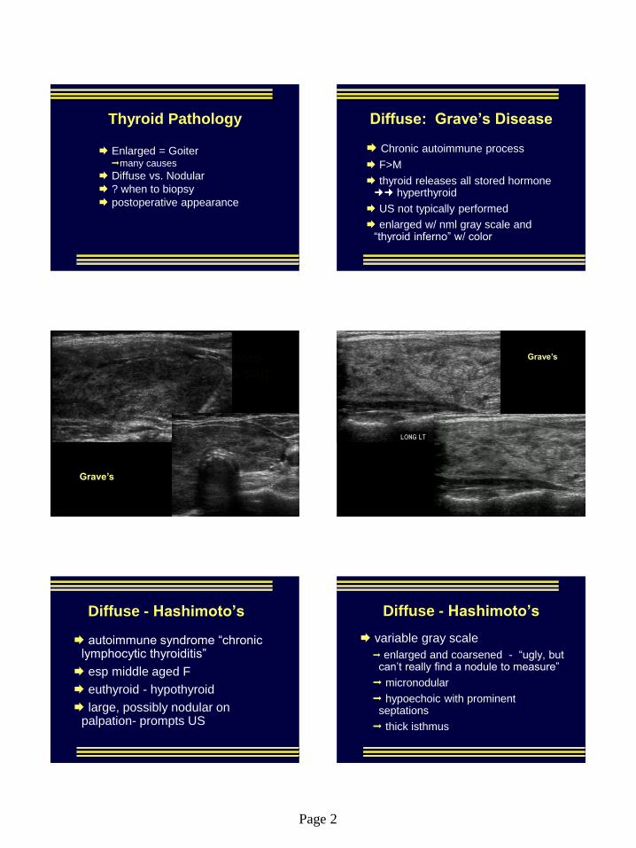
Thyroid Pathology
Enlarged = Goiter many causes
Diffuse vs. Nodular
? when to biopsy
postoperative appearance
Diffuse: Grave’s Disease
Chronic autoimmune process
F>M
thyroid releases all stored hormone hyperthyroid
US not typically performed
enlarged w/ nml gray scale and “thyroid inferno” w/ color
klevanov known Graves looks like hashimotos sag
Grave’s
Graves hypervascular and heterogeneous
Grave’s
Diffuse - Hashimoto’s
autoimmune syndrome “chronic lymphocytic thyroiditis”
esp middle aged F
euthyroid - hypothyroid
large, possibly nodular on palpation- prompts US
Diffuse - Hashimoto’s
variable gray scale
enlarged and coarsened - “ugly, but can’t really find a nodule to measure”
micronodular
hypoechoic with prominent septations
thick isthmus
Page 3
Lorocco typical hashimoto's sag L.jpg
Newcomb micronodular Hahs Spinosa micronodular Hashimotos mild.
2 different
patients
Caaarrolo suusp nodule in Hashimotos
Try not to overcall nodules
If really convincing, biopsy
Hatcher long Hashimotos now growing low grade lymphoma Sag.jpg
Hatcher long Hashimotos now growing low grade lymphoma Sag.jpg
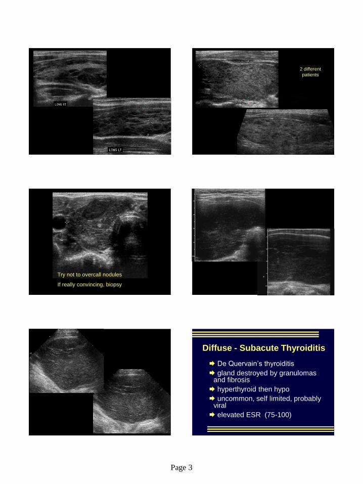
Diffuse - Subacute Thyroiditis
De Quervain’s thyroiditis
gland destroyed by granulomas and fibrosis
hyperthyroid then hypo
uncommon, self limited, probably viral
elevated ESR (75-100)
Page 4
Diffuse - Subacute Thyroiditis
nonspecific US, but tender
diffusely heterogeneous, or ill-defined patchy hypovascular areas that disappear at F/U
hypocellular (fibrosis) if bx done but can mimic atypical or suspicious
adenopathy common
Mountain subacute thyroiditis ESR 75 Trv R.jpg
Painful
ESR 75
Lundblad subacute thyroiditis
8/01
Lundblad subacute thryoiditis recovered
10/01
Pay attention to history
Suggest ESR
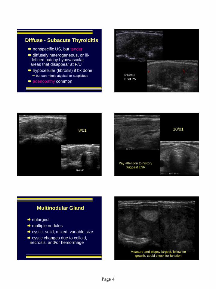
Multinodular Gland
enlarged
multiple nodules
cystic, solid, mixed, variable size
cystic changes due to colloid, necrosis, and/or hemorrhage
MNG Sperber sag
Measure and biopsy largest, follow for
growth, could check for function
Page 5
Focal Disease - Nodules
Incredibly common
4 - 7 % people have palpable nodules
50 - 70% people > 60 years at US and autopsy
palpation found only 20% of nodules seen at US (Chernobyl)
US critical
Thyroid Nodules - Background
More common in women than men
Increasing prevalence with increasing age
Most grow slowly over time
5-10% are malignant
which 10%????
Colloid crystals ring down Middle aged woman with palpable nodule
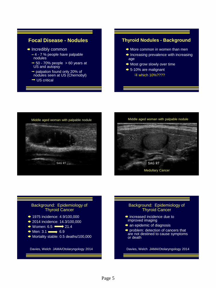
Kpodo medullary thyroid ca
Medullary Cancer
Middle aged woman with palpable nodule
Background: Epidemiology of Thyroid Cancer
1975 incidence: 4.9/100,000
2014 incidence: 14.3/100,000
Women: 6.5 21.4
Men: 3.1 6.9
Mortality stable: 0.5 deaths/100,000
Davies, Welch JAMA/Otolaryngology 2014
Background: Epidemiology of Thyroid Cancer
increased incidence due to improved imaging
an epidemic of diagnosis
problem: detection of cancers that are not destined to cause symptoms or death
Davies, Welch JAMA/Otolaryngology 2014
Page 6
Background: Thyroid Nodules
The challenge is to reassure the majority of patients who have benign nodular disease, and diagnose the “aggressive” malignant minority.
Clark Papillary macdonald pap
BWH Thyroid Nodule Clinic 1995-2003
1,985 patients / 3,483 nodules.
cancer: 14.9% (295 pts)
US Characteristics:
885 Nodules in 729 patients
10.8 % malignant
*Frates et al J Clin Endocrin Metab 2006; 91: 3411-3417
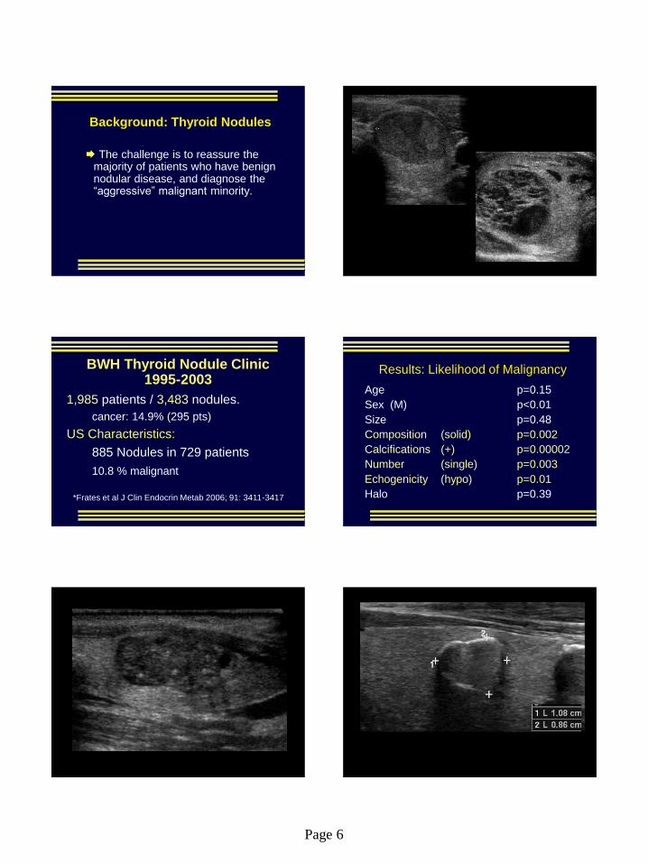
Age p=0.15
Sex (M) p<0.01
Size p=0.48
Composition (solid) p=0.002
Calcifications (+) p=0.00002
Number (single) p=0.003
Echogenicity (hypo) p=0.01
Halo p=0.39
Results: Likelihood of Malignancy
Krouskas papillary CA
Graichen rim calcifcation positive papillary sag.jpg
Page 7
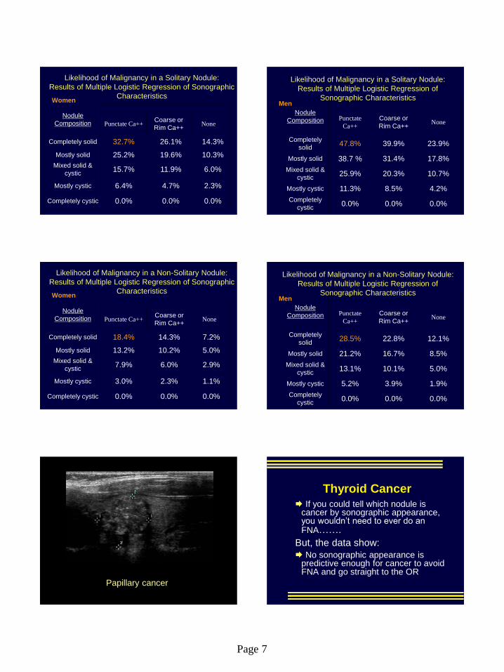
Likelihood of Malignancy in a Solitary Nodule:
Results of Multiple Logistic Regression of Sonographic
Characteristics
Nodule
Composition Punctate Ca++ Coarse or
Rim Ca++ None
Completely solid 32.7% 26.1% 14.3%
Mostly solid 25.2% 19.6% 10.3%
Mixed solid &
cystic 15.7% 11.9% 6.0%
Mostly cystic 6.4% 4.7% 2.3%
Completely cystic 0.0% 0.0% 0.0%
Women
Likelihood of Malignancy in a Solitary Nodule:
Results of Multiple Logistic Regression of
Sonographic Characteristics
Nodule
Composition Punctate
Ca++
Coarse or
Rim Ca++ None
Completely
solid 47.8% 39.9% 23.9%
Mostly solid 38.7 % 31.4% 17.8%
Mixed solid &
cystic 25.9% 20.3% 10.7%
Mostly cystic 11.3% 8.5% 4.2%
Completely
cystic 0.0% 0.0% 0.0%
Men
Likelihood of Malignancy in a Non-Solitary Nodule:
Results of Multiple Logistic Regression of Sonographic
Characteristics
Nodule
Composition Punctate Ca++ Coarse or
Rim Ca++ None
Completely solid 18.4% 14.3% 7.2%
Mostly solid 13.2% 10.2% 5.0%
Mixed solid &
cystic 7.9% 6.0% 2.9%
Mostly cystic 3.0% 2.3% 1.1%
Completely cystic 0.0% 0.0% 0.0%
Women
Likelihood of Malignancy in a Non-Solitary Nodule:
Results of Multiple Logistic Regression of
Sonographic Characteristics
Nodule
Composition Punctate
Ca++
Coarse or
Rim Ca++ None
Completely
solid 28.5% 22.8% 12.1%
Mostly solid 21.2% 16.7% 8.5%
Mixed solid &
cystic 13.1% 10.1% 5.0%
Mostly cystic 5.2% 3.9% 1.9%
Completely
cystic 0.0% 0.0% 0.0%
Men
Winnick papillary sag
Papillary cancer
Thyroid Cancer
If you could tell which nodule is cancer by sonographic appearance, you wouldn’t need to ever do an FNA…….
But, the data show:
No sonographic appearance is predictive enough for cancer to avoid FNA and go straight to the OR
Page 8
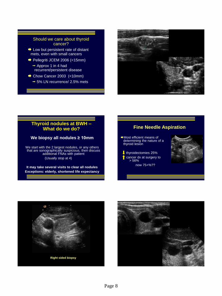
Should we care about thyroid cancer?
Low but persistent rate of distant mets, even with small cancers
Pellegriti JCEM 2006 (<15mm)
Approx 1 in 4 had recurrent/persistent disease
Chow Cancer 2003 (<10mm)
5% LN recurrence/ 2.5% mets
Harwell tiny papillary presented w + node. Voorhees calcified IJ node at presentation
Thyroid nodules at BWH –
What do we do?
We biopsy all nodules ≥ 10mm
We start with the 2 largest nodules, or any others that are sonographically suspicious, then discuss
additional FNAs with patient
(Usually stop at 4)
It may take several visits to clear all nodules
Exceptions: elderly, shortened life expectancy
Fine Needle Aspiration
Most efficient means of determining the nature of a thyroid lesion
thyroidectomies 25%
cancer dx at surgery to > 56%
now 75+%??
Thyroid biopsy R
Right sided biopsy
singer FNA mural nodule L.avi
Page 9
Crowley Hurthle cell hematoma
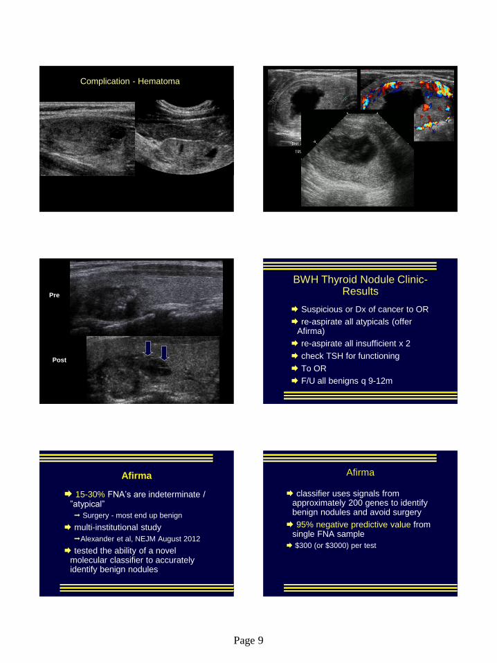
Complication - Hematoma Wedlich thyroid pumping
vessel during bxg
adler papillary cancer hematoma in gland post FNA.jpg
Pre
Post
BWH Thyroid Nodule Clinic- Results
Suspicious or Dx of cancer to OR
re-aspirate all atypicals (offer Afirma)
re-aspirate all insufficient x 2
check TSH for functioning
To OR
F/U all benigns q 9-12m
Afirma
15-30% FNA’s are indeterminate / ”atypical”
Surgery - most end up benign
multi-institutional study
Alexander et al, NEJM August 2012
tested the ability of a novel molecular classifier to accurately identify benign nodules
Afirma
classifier uses signals from approximately 200 genes to identify benign nodules and avoid surgery
95% negative predictive value from single FNA sample
$300 (or $3000) per test
Page 10
SRU Consensus Conference Recommendations
Management of Nodule found at US
Criteria for FNA change as size changes
High risk criteria - FNA smaller size
Low risk criteria - can wait for growth
Frates et al Radiology 2005;237:794-800
A. Solitary Nodule (Only a single nodule that is 1 cm in maximum diameter)
Ultrasound Features Recommendations
Microcalcifications Strongly consider US-FNA if 1 cm
Solid (or almost entirely solid) or
Coarse calcifications Strongly consider US-FNA if 1.5 cm
Mixed solid and cystic or
Almost entirely cystic with a solid mural
component
Consider US-FNA if 2 cm
None of the above but
Grown substantially since prior US Consider US-FNA
Almost entirely cystic and
None of the above characteristics and
No substantial growth (or no prior US)
US-FNA likely unnecessary
B. Multiple Nodules (At least two nodules 1 cm in
maximum diameter)
Recommendation: Consider US-FNA of one or more
nodules; selection to be prioritized based on the previously
stated criteria, in the order listed above
1 Fine needle aspiration is likely unnecessary in
diffusely enlarged glands with multiple nodules of
similar sonographic appearances without intervening
normal parenchyma. 2 The presence of abnormal lymph nodes overrides the
sonographic features of the thyroid nodule(s) and should
prompt US-FNA or biopsy of the lymph node and/or an
ipsilateral thyroid nodule.
What to do?
SRU consensus (ACE guidelines)
BWH system Every nodule > 10 mm
Repeat if >15% growth/year
Insufficient data for “pattern approach” at present
TIRADS is coming!
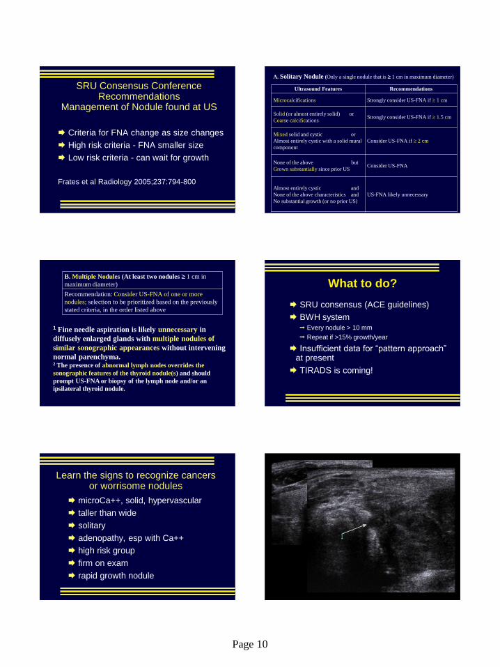
Learn the signs to recognize cancers or worrisome nodules
microCa++, solid, hypervascular
taller than wide
solitary
adenopathy, esp with Ca++
high risk group
firm on exam
rapid growth nodule
Albertson anaplastic thyroid Trv.jpg
Page 11
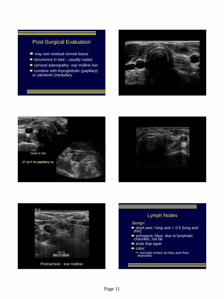
Post-Surgical Evaluation
may see residual normal tissue
recurrence in bed – usually nodes
cervical adenopathy- esp midline low
combine with thyroglobulin (papillary) or calcitonin (medullary
Frechette beautiful post op neck trv
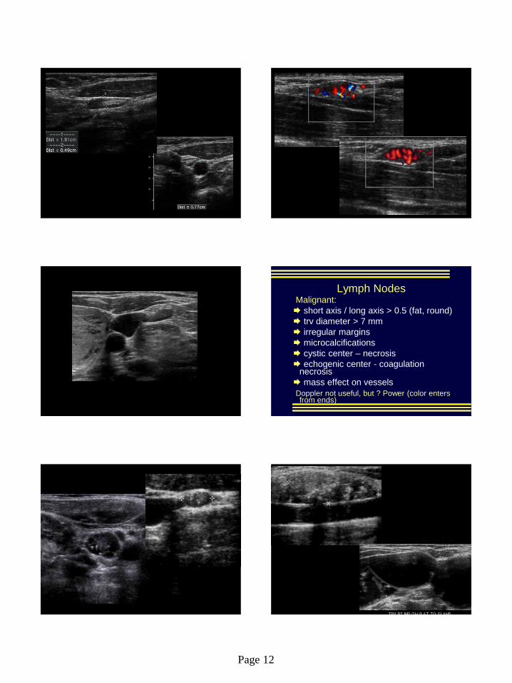
Canido 17yo recurrent papillary right bed
17 yo F hx papillary ca
Diglioia met papillary invading esoph and recurrent laryngeal nerve.jpg
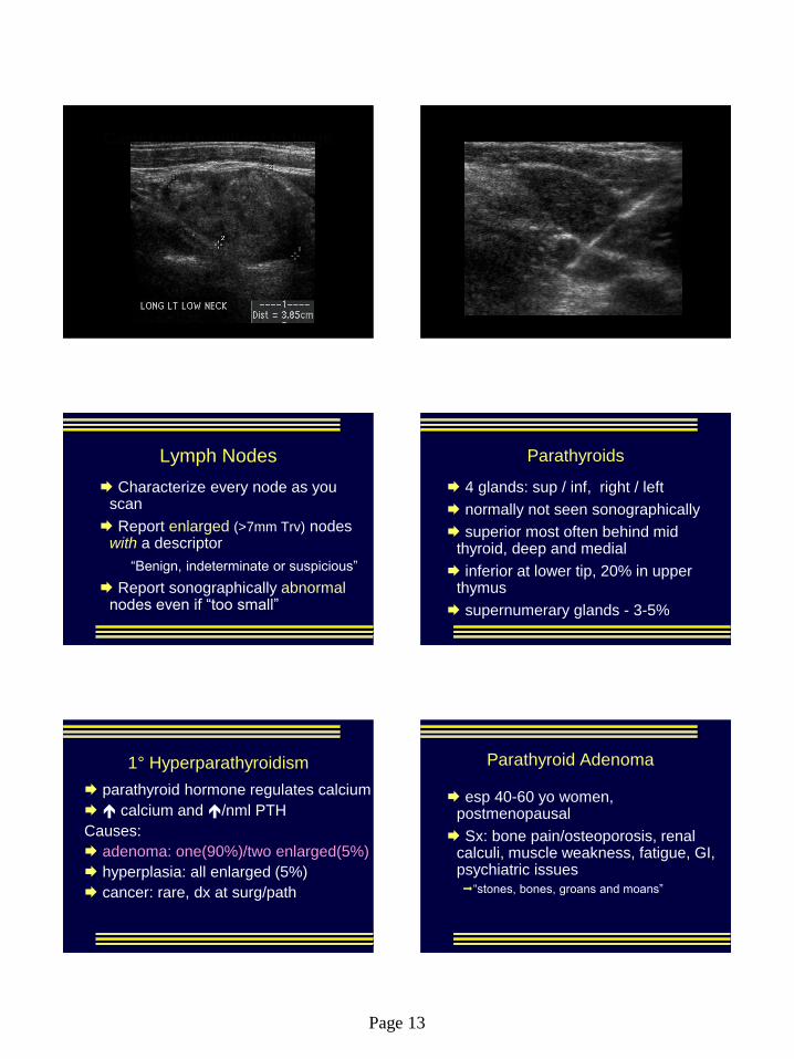
Grillo recurrent papillary
Pretracheal - low midline
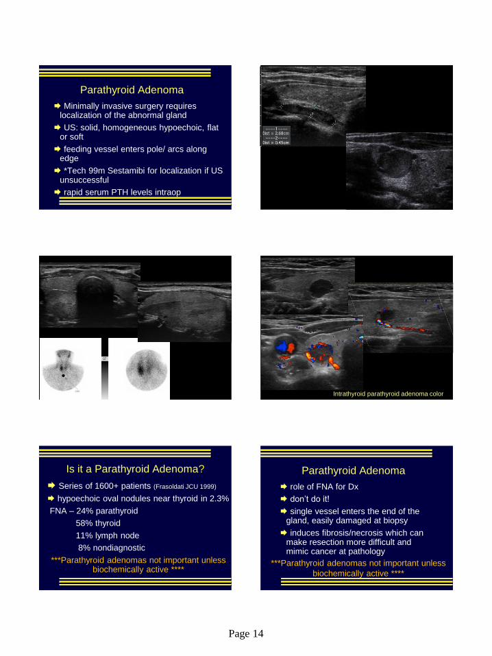
Lymph Nodes
Benign:
short axis / long axis < 0.5 (long and thin)
echogenic hilus- due to lymphatic channels, not fat
ends that taper
color: normally enters at hilus and then
branches
Page 12
Philben enlarged benign node enlarged for 2y trv.jpg Larocco enlarged lat node post pap color.jpg
Tsegai mobile benign lymph node.avi
Lymph Nodes Malignant:
short axis / long axis > 0.5 (fat, round)
trv diameter > 7 mm
irregular margins
microcalcifications
cystic center – necrosis
echogenic center - coagulation necrosis
mass effect on vessels
Doppler not useful, but ? Power (color enters from ends)
Grillo papillary in node cohen, Badler papillary cancer met node
Tomasso cystic metastatic papillary CA Meuchner pap 8 years out nodes
Page 13
Carter met papillary to huge neck node
Keene great FNA enlarged high R node.avi
Lymph Nodes
Characterize every node as you scan
Report enlarged (>7mm Trv) nodes with a descriptor
“Benign, indeterminate or suspicious”
Report sonographically abnormal nodes even if “too small”
Parathyroids
4 glands: sup / inf, right / left
normally not seen sonographically
superior most often behind mid thyroid, deep and medial
inferior at lower tip, 20% in upper thymus
supernumerary glands - 3-5%
1° Hyperparathyroidism
parathyroid hormone regulates calcium
calcium and /nml PTH
Causes:
adenoma: one(90%)/two enlarged(5%)
hyperplasia: all enlarged (5%)
cancer: rare, dx at surg/path
Parathyroid Adenoma
esp 40-60 yo women, postmenopausal
Sx: bone pain/osteoporosis, renal calculi, muscle weakness, fatigue, GI, psychiatric issues “stones, bones, groans and moans”
Page 14
Parathyroid Adenoma
Minimally invasive surgery requires localization of the abnormal gland
US: solid, homogeneous hypoechoic, flat or soft
feeding vessel enters pole/ arcs along edge
*Tech 99m Sestamibi for localization if US unsuccessful
rapid serum PTH levels intraop
Bayles intrathyroidal parathyroid PTH 3000 Eglittis adenoma
Rickabaugh huge right parathyroid adenoma isoechoic
Volk 2 parathyroid adenomas path proven
Intrathyroid parathyroid adenoma color
Is it a Parathyroid Adenoma?
Series of 1600+ patients (Frasoldati JCU 1999)
hypoechoic oval nodules near thyroid in 2.3%
FNA – 24% parathyroid
58% thyroid
11% lymph node
8% nondiagnostic
***Parathyroid adenomas not important unless biochemically active ****
Parathyroid Adenoma
role of FNA for Dx
don’t do it!
single vessel enters the end of the gland, easily damaged at biopsy
induces fibrosis/necrosis which can make resection more difficult and mimic cancer at pathology
***Parathyroid adenomas not important unless
biochemically active ****
Page 15
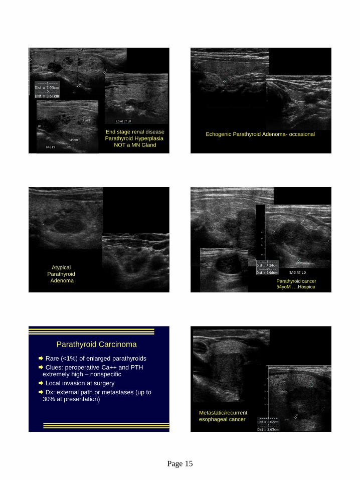
Campbell ESRD parathyroid hyperplasia called MNG Sag Rt.jpg
End stage renal disease
Parathyroid Hyperplasia
NOT a MN Gland
Bauchman echogenic parathyroid adenoma
Echogenic Parathyroid Adenoma- occasional
Credle atypical parathryoid adenoma US
Atypical
Parathyroid
Adenoma
Krickl parathyroid cancer 54M mets to liver
Parathyroid cancer
54yoM ….Hospice
Parathyroid Carcinoma
Rare (<1%) of enlarged parathyroids
Clues: peroperative Ca++ and PTH extremely high – nonspecific
Local invasion at surgery
Dx: external path or metastases (up to 30% at presentation)
Kelley metastatic esophageal into thyroid Trv.jpg
Metastatic/recurrent
esophageal cancer
Page 16
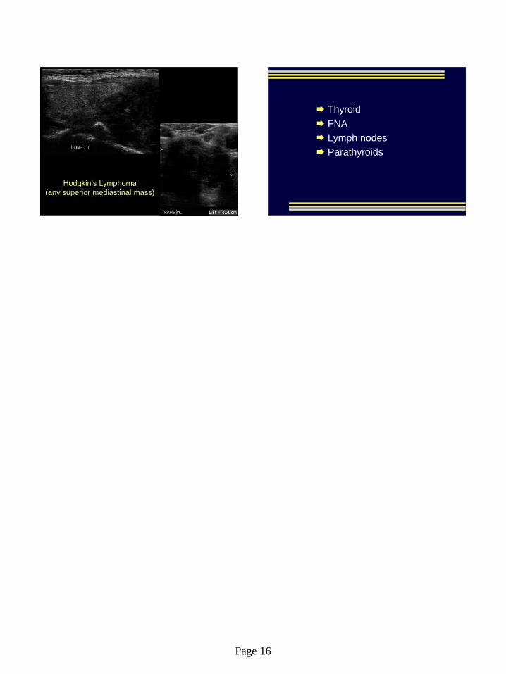
Dechiaro ? papillary Hodg lymphoma sag L.
Hodgkin’s Lymphoma
(any superior mediastinal mass)
Thyroid
FNA
Lymph nodes
Parathyroids


![Papillary thyroid carcinoma coexists with undifferentiated ... · Papillary thyroid carcinoma (PTC) is the commonest thyroid carcinoma worldwide [1], while undifferentiated thyroid](https://img.dokumen.tips/doc/110x75/605714f9a806da25134f71a8/papillary-thyroid-carcinoma-coexists-with-undifferentiated-papillary-thyroid.jpg)