Embed Size (px)
Citation preview
RESEARCH ARTICLE SPECIAL COLLECTION: TRANSLATIONAL IMPACT OF RAT
Increased trabecular bone and improved biomechanics in anosteocalcin-null rat model created by CRISPR/Cas9 technologyLaura J. Lambert*,1, Anil K. Challa*,1, Aidi Niu2, Lihua Zhou2, Janusz Tucholski2, Maria S. Johnson3,Tim R. Nagy3, Alan W. Eberhardt4, Patrick N. Estep4, Robert A. Kesterson1 and Jayleen M. Grams2,5,‡
ABSTRACTOsteocalcin, also known as bone γ-carboxyglutamate protein (Bglap), isexpressed by osteoblasts and is commonly used as a clinical marker ofbone turnover. A mouse model of osteocalcin deficiency has implicatedosteocalcin as amediator of changes to the skeleton, endocrine system,reproductive organs and central nervous system. However, differencesbetweenmouse and human osteocalcin at both the genome and proteinlevels have challenged the validity of extrapolating findings from theosteocalcin-deficient mouse model to human disease. The ratosteocalcin (Bglap) gene locus shares greater synteny with that ofhumans. To further examine the role of osteocalcin in disease, wecreated a ratmodel with complete loss of osteocalcin using theCRISPR/Cas9 system. Rat osteocalcin was modified by injection of CRISPR/Cas9 mRNA into the pronuclei of fertilized single cell Sprague-Dawleyembryos, and animals were bred to homozygosity and compoundheterozygosity for the mutant alleles. Dual-energy X-ray absorptiometry(DXA), glucose tolerance testing (GTT), insulin tolerance testing (ITT),microcomputed tomography (µCT), and a three-point breakbiomechanical assay were performed on the excised femurs at 5months of age. Complete loss of osteocalcin resulted in bones withsignificantly increased trabecular thickness, densityandvolume.Corticalbone volume and density were not increased in null animals. The boneshad improved functional quality as evidenced by an increase in failureload during the biomechanical stress assay. Differences in glucosehomeostasis were observed between groups, but there were nodifferences in body weight or composition. This rat model of completeloss of osteocalcin provides a platform for further understanding the roleof osteocalcin in disease, and it is a novel model of increased boneformationwith potential utility in osteoporosisandosteoarthritis research.
KEY WORDS: Osteocalcin, Bone strength, Bone structure,Genetic animal models, Osteocalcin knockout
INTRODUCTIONOsteocalcin, also known as bone γ-carboxyglutamate protein(Bglap), is the most abundant noncollagenous protein in bone and
comprises ∼1% of total body protein (McGuigan et al., 2010). Anosteocalcin-deficient mouse model displays increased cortical bonethickness and density, trabecular bone and bone strength (Ducy et al.,1996). This osteocalcin-deficient mouse model generated muchinterest outside of bone formation due to additional metabolic,reproductive and neurological phenotypes (Lee et al., 2007; Ouryet al., 2011, 2013; Karsenty and Oury, 2012). The mice displayedobesity, and decreased insulin sensitivity and glucose tolerance (Leeet al., 2007). Osteocalcin was later shown to act as a regulator of malefertility, resulting in smaller reproductive organs and lower circulatingtestosterone (Oury et al., 2011); and to influence cognition bymodulating neurotransmitter synthesis (Oury et al., 2013).
The contribution of osteocalcin to bone structure and function,metabolism, male fertility and cognition in humans remains to bedetermined (Li et al., 2016). There are considerable differencesbetween the mouse and human osteocalcin gene loci, complicatinginterpretation of the results from the osteocalcin-deficient mousemodel and raising the possibility that data from the mouse modelmight not be pertinent to human disease (Booth et al., 2013, 2014).For example, the mouse osteocalcin gene locus underwent atriplication event resulting in two functional copies of osteocalcinexpressed in bone (Bglap-1 and Bglap-2) and an additional copyexpressed in other non-osteoid tissues (Bglap-3) (Desbois et al.,1994). In the osteocalcin-deficient mouse model, the entire Bglap-2sequence to exon 4 of Bglap-1 was deleted (Ducy et al., 1996). TheBglap-3 gene remained intact. Further confounding interpretation ofdata from the mouse model is the interspersal of a progestin andadiponectin receptor, Paqr6, that also underwent triplication in thegene locus. Two putative Paqr6 genes were included in the deletion tocreate the osteocalcin-deficient animal. Similar to humans, the ratosteocalcin gene locus consists of a single copy of osteocalcin. Likethe human osteocalcin gene, transcription of the rat osteocalcin gene isupregulated by vitamin D, whereas the mouse osteocalcin genes aredownregulated by vitamin D (Kesterson et al., 1993; Arbour et al.,1995; Javed et al., 1999; Booth et al., 2013). This similarity andsynteny between rat and human osteocalcin gene loci suggests that therat might be a more appropriate animal model system to investigateosteocalcin function, particularly as it pertains to relevance in humandisease. In this paper, we address this issue and report the generationand initial characterization of an osteocalcin-null mutant rat model.
RESULTSGeneration of a osteocalcin-null rat using CRISPR/Cas9systemThe high degree of similarity and synteny between the rat andhuman osteocalcin gene loci (Fig. 1A,B) indicated that targeting theosteocalcin gene early in the protein sequence would create a modelanalogous to loss of osteocalcin in humans. Thus, two CRISPRguide RNAs were designed to target exons 1 and 2 of osteocalcin todisrupt the protein early in the amino acid sequence (Fig. 1C). TheReceived 4 March 2016; Accepted 19 July 2016
1Department of Genetics, University of Alabama at Birmingham, Birmingham,AL 35294, USA. 2Department of Surgery, University of Alabama at Birmingham,Birmingham, AL 35294, USA. 3Department of Nutrition Sciences, University ofAlabama at Birmingham, Birmingham, AL 35294, USA. 4Department of BiomedicalEngineering, University of Alabama at Birmingham, Birmingham, AL 35294, USA.5Department of Surgery,BirminghamVAMedicalCenter,Birmingham,AL35233,USA.*These authors contributed equally to the work
‡Author for correspondence ( [email protected])
J.M.G., 0000-0002-6510-6995
This is an Open Access article distributed under the terms of the Creative Commons AttributionLicense (http://creativecommons.org/licenses/by/3.0), which permits unrestricted use,distribution and reproduction in any medium provided that the original work is properly attributed.
1169
© 2016. Published by The Company of Biologists Ltd | Disease Models & Mechanisms (2016) 9, 1169-1179 doi:10.1242/dmm.025247
Disea
seModels&Mechan
isms

CRISPR guide RNA and Cas9 mRNAmixtures were microinjectedinto the pronuclei of 33 fertilized embryos of Sprague-Dawley ratsand transferred to pseudopregnant female rats, of which 21 pupswere born (64%).
Multiple alleles result in complete loss of osteocalcin proteinGenotyping was performed by amplifying a 601-bp fragmentencompassing the CRISPR single guide RNA (sgRNA) target sitesfrom tail genomic DNA samples, followed by a heteroduplexmobility assay (HMA). Based on HMA profiles, the presence ofindels was identified in 12 of 21 (58%) pups (Fig. 2A) born fromthe first set of microinjected embryos. The alleles were confirmedby Sanger sequencing, which revealed frameshift mutations.Multiple alleles were present in many founder animals andindicated mosaicism (Fig. 2B). Founders 1 and 19 were mated toeach other in order to establish germline transmission of mutantalleles including a 7-bp insertion (+7), a compound 2-bp insertionand a 15-bp deletion (+2–15), and a 312-bp deletion (–312). Asecond set of pups born from the CRISPR microinjection yielded
an additional founder animal that transmitted a 240-bp deletion(–240, data not shown). All of these alleles were predicted to causeframeshift mutations resulting in premature stop codons (Fig. 2C).Total loss of osteocalcin protein was demonstrated by complete lossof both γ-carboxylated (Gla-) and uncarboxylated (Glu-) forms ofosteocalcin in the serum of the osteocalcin-null male rats asdetermined by enzyme-linked immunosorbent assay (ELISA)(Fig. 2D). Western blots of tissue samples (brain, heart, lung,bone, gonadal fat, subcutaneous fat, quadriceps muscle, liver,kidney, pancreas and spleen) from wild-type animals demonstratedthat osteocalcin was only expressed in bone (data not shown); therewas no osteocalcin detectable in any of these tissues taken from ahomozygous 312-bp deletion (–312) F2 generation animal asexpected (data not shown). Loss of osteocalcin protein in the nullmutant animals was further confirmed by immunohistochemistryon sectioned femurs (Fig. 2E) and by western blot analyses ofprotein isolated from whole tibiae of animals with compoundheterozygous combinations of these alleles (Fig. 2F), in contrast tothe robust signal from wild-type animals.
Fig. 1. Comparison of the similarity and synteny of osteocalcin between species and CRISPR guide design. (A) Peptide sequence of different species.Gray shading indicates conserved amino acid residues. The asterisk indicates a cleavage site between a prepropeptide and mature protein regions. CRISPRcut sites are indicated by arrows above the sequence. (B) Structure of the genomic locus of human and rat versus mouse osteocalcin. A triplication ofosteocalcin and the interspersed progestin and adiponectin receptor 6 (Paqr6) gene occurred in the mouse but not in the human or rat osteocalcin gene locus.(C) Design of the two CRISPR guide RNAs to exons 1 and 2 of the rat gene.
1170
RESEARCH ARTICLE Disease Models & Mechanisms (2016) 9, 1169-1179 doi:10.1242/dmm.025247
Disea
seModels&Mechan
isms

Loss of osteocalcin does not affect body composition5-month-old wild-type and osteocalcin-null male rats were scannedby dual-energy X-ray absorptiometry (DXA) for total body
composition. Total body weight, the percentage fat mass,percentage lean mass, bone mineral density (BMD) and bonemineral content (BMC)were not significantly different (Fig. 3A–E).
Fig. 2. Identification of founder animals and multiple null alleles with indels. (A) Heteroduplex mobility assay of genomic DNA from F0 animals born frommicroinjection with CRISPR/Cas9. Founder 1 wasmated to founder 19 to establish the osteocalcin colony. L, ladder; C, wild-type control. (B) Mutant alleles +7, +2–15, –240 and –312 transmitted to the F1 generation are indicative of mosaicism in the founder germlines. (C) Predicted translations of mutant alleles. (D) SerumGlu-osteocalcin, and serum Glu- and Gla-osteocalcin levels for wild-type (WT) and osteocalcin-null rats. Results are mean±s.e.m. n=3 for WT and null.****P<0.0001 (two-tailed parametric unpaired t-test). (E) Osteocalcin immunohistochemistry. Black arrows indicate positive (WT, left) and negative (null, right)osteoblasts. (F) Western blot of femurs from compound heterozygous F2 animals demonstrate complete loss of osteocalcin protein.
Fig. 3. DXA analysis of wild-type and osteocalcin-null male rats. Wild-type (WT) and osteocalcin-null male rats were analyzed for (A) weight, P=0.4339,(B) percentage fat mass, P=0.1490, (C) percentage lean mass, P=0.1517, (D) bone mineral density, P=0.5958, (E) bone mineral content, P=0.1215, and (F)gonadal fat pad weight, P=0.2434. n=7 WT, n=8 null. Results are mean±s.e.m. P-values were calculated with a two-tailed parametric unpaired t-test.
1171
RESEARCH ARTICLE Disease Models & Mechanisms (2016) 9, 1169-1179 doi:10.1242/dmm.025247
Disea
seModels&Mechan
isms
Gonadal fat pads were excised from the males of each cohort andweighed. There was no significant difference between the twogroups (Fig. 3F).
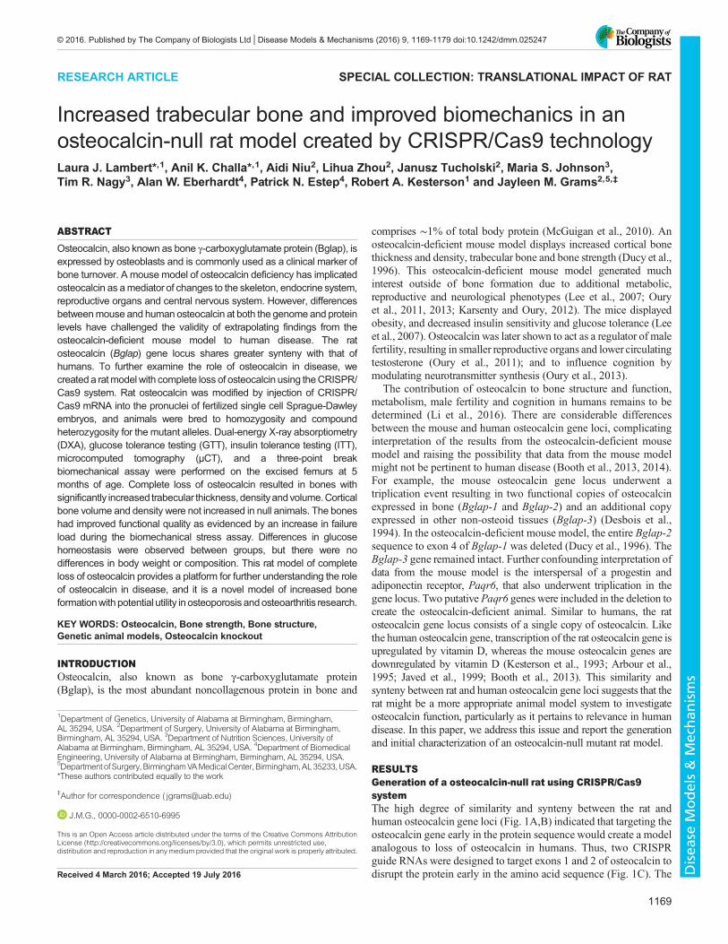
Loss of osteocalcin results in differences in glucose andinsulin toleranceFasting glucose measurements were assessed from fresh tail bloodin 5-month-old male rats of osteocalcin-null and wild-type ratsimmediately prior to challenge with glucose or insulin for glucoseand insulin tolerance tests, respectively. There was no significantdifference between groups for the fasting glucose level (Fig. 4A;P=0.2080); however, at 75 min after insulin injection the groupsbegan to diverge (Fig. 4B,C), indicating an increase in insulin
sensitivity in osteocalcin-null animals (10.04±0.70 mg dl−1 and13.00±0.73 mg dl−1 for null versus WT animals, respectively;mean±s.e.m., P=0.0337). Consistent with an increase in insulinsensitivity, osteocalcin-null animals exhibited significantly lowerblood glucose levels at 30 min post glucose injection (Fig. 4D,E;16.62±0.73 mg dl−1 versus 18.34±1.38 mg dl−1, respectively;P=0.0279).
Loss of osteocalcin affects trabecular bone measurementsOsteocalcin-null and wild-type control animals were sacrificed at5 months of age and the femurs excised for further analysisby microcomputed tomography (μCT). Cortical bone volume(P=0.2585), percentage bone volume (P=0.3378), thickness
Fig. 4. Glucose and insulin tolerance tests in wild-type and osteocalcin-null male rats.Wild-type (WT) and osteocalcin-null male rats were analyzed for (A)fasting glucose levels, P=0.2080, and by (B) an insulin tolerance test and (D) a glucose tolerance test. (C) Quantification of insulin tolerance test results,P=0.0337. (E) Quantification of glucose tolerance test results, P=0.2656. n=3 WT, n=5 null. Results are mean±s.e.m. *P≤0.05, **P≤0.01, ***P≤0.001. P-valueswere calculated with a two-tailed parametric unpaired t-test (C,E) and two-way ANOVA (B,D).
1172
RESEARCH ARTICLE Disease Models & Mechanisms (2016) 9, 1169-1179 doi:10.1242/dmm.025247
Disea
seModels&Mechan
isms

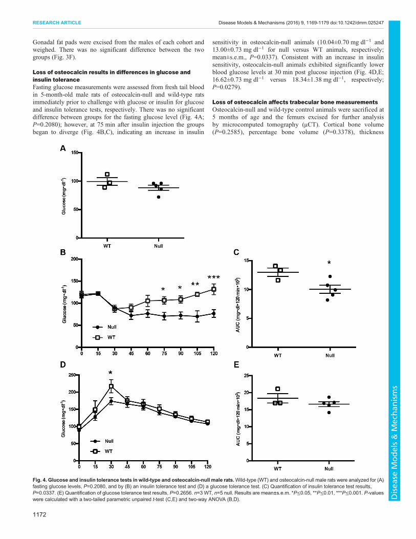
(P=0.2876), density (P=0.0845), and average periostealcircumference (P=0.7865) and endosteal circumference(P=0.4144) were not significantly different between groups(Fig. 5A–G). For trabecular bone, there was an increase in bonevolume in osteocalcin-null rats compared to control animals(4.96±0.35 mm3 and 6.35±0.44 mm3, respectively; P=0.0316)(Fig. 6B). Trabecular bone measurements in null animals alsodiffered significantly from wild-type in percentage bone volume
(P=0.0035), thickness (P=0.0021) and density (P=0.0004)(Fig. 6C–E). Trabecular separation (P=0.2130) and number(P=0.1332) were not different between groups (Fig. 6F,G).
Loss of osteocalcin affects femoral biomechanicsExcised femurs from 5-month-old male wild-type and osteocalcin-null rats were subjected to a three-point break assay to determine thestrength and stiffness of the bone. This assay revealed that there was a
Fig. 5. Cortical µCT from wild-typeand osteocalcin-null male rats.(A) Representative µCT image of ratfemoral cortical bone from a wild-type(WT, left) and an osteocalcin-null (right)rat. (B–G) Quantification of µCT data.(B) Cortical bone volume, P=0.2585,(C) cortical bone volume/tissue volume,P=0.3378, (D) cortical thickness,P=0.2876, (E) cortical density,P=0.0845. n=8 WT, n=8 null.(F) Periosteal circumference, P=0.7865.(G) Endosteal circumference,P=0.4144. n=7 WT, n=8 null. Resultsare mean±s.e.m. P-values werecalculated with a two-tailed parametricunpaired t-test.
1173
RESEARCH ARTICLE Disease Models & Mechanisms (2016) 9, 1169-1179 doi:10.1242/dmm.025247
Disea
seModels&Mechan
isms
significant increase in themaximum force needed to break the femursof osteocalcin-null rats compared to wild-type animals (196.2±7.62 N and 165.5±5.93 N, respectively; mean±s.e.m., P=0.0118)(Fig. 7A). The osteocalcin-null animals also had a significant increasein stiffness (347.6±22.97 N mm−1 versus 274±23.78 N mm−1;P=0.0492) (Fig. 7D). There was no difference in deflection orenergy to Fmax (Fig. 7B,C; P=0.6233 versus 0.8405, respectively).
DISCUSSIONA mouse model of osteocalcin deficiency supports that osteocalcinis a negative regulator of bone formation (Ducy et al., 1996);moreover, the mouse model also implicated osteocalcin as amediator of skeletal modulation of metabolism, male fertility andcognition (Lee et al., 2007; Oury et al., 2011, 2013). The role ofosteocalcin in humans and in human disease remains undetermined
Fig. 6. Trabecular µCT from wild-typeand osteocalcin-null male rats.(A) Representative µCT image of ratfemoral trabecular bone from a wild-type(WT, left) and an osteocalcin-null (right) rat.(B–G) Quantification of µCT data.(B) Trabecular bone volume, P=0.0316,(C) trabecular bone volume/tissue volume,P=0.0035, (D) trabecular thickness,P=0.0021, (E) trabecular density,P=0.0004, (F) trabecular separation,P=0.2130 and (G) trabecular number,P=0.1332. n=7 WT, n=8 null. Results aremean±s.e.m. P-values were calculated witha two-tailed parametric unpaired t-test.*P≤0.05, **P≤0.01, ***P≤0.001.
1174
RESEARCH ARTICLE Disease Models & Mechanisms (2016) 9, 1169-1179 doi:10.1242/dmm.025247
Disea
seModels&Mechan
isms

(Booth et al., 2013, 2014; Li et al., 2016). Owing to considerabledivergence between mouse and human osteocalcin at both thegenomic and protein level, the validity of extrapolating results fromthe osteocalcin-deficient mouse to human disease has beenchallenged (Booth et al., 2013, 2014). Similarity and syntenybetween the rat and human osteocalcin gene locus suggest that therat might be a more appropriate animal model system to investigateosteocalcin function in humans. Here, we report the generation andinitial characterization of an osteocalcin-null mutant rat modelcreated using the CRISPR/Cas9 system. As demonstrated in theosteocalcin-deficient mouse model, complete loss of osteocalcin inthe rat affected bone structure and function, with increasedtrabecular bone and increased bone strength. In contrast to themouse model, our data did not demonstrate increased density of
cortical bone based on µCT. Furthermore, the osteocalcin-null ratmodel did not develop obesity, insulin resistance or glucoseintolerance. However, the data reported here are limited to adult5-month-old animals, and further studies are underway at additionaltime points in males and females to determine whether additionalphenotypes will be detected based on age and/or sex.
Given that the null mutant rat model did not display a strikingmetabolic phenotype as seen in the osteocalcin-deficient mousemodel, the data reported here support that results generated using themouse model might not be translatable to the role of osteocalcin inhumans. The major genomic differences between mice and humansinclude that: (1) osteocalcin is a highly conserved gene; however,the human and mouse genes share only ∼68% identity versus thehuman and rat genes sharing ∼75% identity in the osteocalcin
Fig. 7. Three-point break biomechanical assay.Wild-type (WT) and osteocalcin-null male rats were analyzed for (A) maximum force, P=0.0118, (B) deflection,P=0.6233, (C) Energy to Fmax, P=0.8405, (D) stiffness, P=0.0492. n=6 WT, n=8 null. Results are mean±s.e.m. P-values were calculated with a two-tailedparametric unpaired t-test. *P≤0.05.
1175
RESEARCH ARTICLE Disease Models & Mechanisms (2016) 9, 1169-1179 doi:10.1242/dmm.025247
Disea
seModels&Mechan
isms
coding sequence; (2) the mouse osteocalcin gene locus underwent atriplication event resulting in two functional copies of osteocalcinbeing expressed in bone and an additional copy expressed in non-osteoid tissues (Desbois et al., 1994); both the human and ratosteocalcin gene loci contain a single copy; and (3) vitamin Ddownregulates expression of the mouse osteocalcin genes, whereasit upregulates expression of the human and rat gene (Kesterson et al.,1993; Arbour et al., 1995; Javed et al., 1999; Booth et al., 2013).There are also significant differences between the osteocalcin-deficient mouse model and the null mutant rat model we developed.First, the mouse model presumably still has a functional copy ofBglap-3, whereas the rat model has a complete loss of osteocalcinprotein. Second, the mouse model included deletion of two putativePaqr6 genes, whereas the rat model represents a selective knockoutof osteocalcin only. Although the two deleted Paqr6 genes arelikely to be pseudogenes, it is intriguing to consider that deletion ofa progestin and adiponectin receptor family member might bepredicted to display metabolic, gonadal and cognitive alterations(Michalakis and Segars, 2010; Patel et al., 2012; Pang et al., 2013;Singh et al., 2013). Although off-target effects are described usingCRISPR/Cas9 methodology (Wang et al., 2015), the use of anoutbred strain with several different homozygous null mutant andcompound heterozygous lines that all resulted in a complete loss ofosteocalcin protein provides strong evidence that the observedphenotypes are due solely to loss of osteocalcin gene function.Given that osteocalcin is themost abundant noncollagenous protein
in bone, it might be surprising that complete loss of the protein doesnot result in a more pronounced skeletal phenotype. However, it willalso be important to investigate the phenotype in the axial skeleton andat additional time points. In addition, given that osteocalcin actsprimarily as an extracellular bone matrix protein, this might indicatefunctional redundancy with other bone matrix proteins. Regardless,both the previously published mouse model of osteocalcin deficiencyand the rat model of complete loss of osteocalcin described heredisplay an increase in bone strength, supporting the idea thatosteocalcin functions as a negative regulator of the skeleton.Understanding of the molecular mechanisms involved in promotingbone density has been important to the development of multiple drugtherapies for osteoporosis, such as the capthepsin K inhibitorsOdanacatib (Eisman et al., 2011) and Balicatib (Adami et al., 2006),anti-SOST antibodies (Padhi et al., 2011), anti-Dkk1 antibodies(Hoeppner et al., 2009), and GSK3β and Sfrp1 inhibitors (Kulkarniet al., 2006; Moore et al., 2009). Bisphosphonates, a standardtreatment used in osteoporosis, function to decrease osteoclast-mediated bone resorption (Tsubaki et al., 2014). Previous in vitrostudies have suggested that osteocalcin might recruit osteoclastprecursors and increase subsequent differentiation of these cells intoosteoclasts (Malone et al., 1982;MundyandPoser, 1983; Chenu et al.,1994), suggesting that osteocalcin might be a novel target for treatingosteopenia or osteoporosis.Moreover, previously published data have suggested that
osteocalcin might have relevance to bone disease. For example,loss of Twist expression in the mouse inhibited osteocalcinexpression and animals displayed the premature cranialossification of Saethre–Chotzen syndrome (Yousfi et al., 2001).Second, the osteocalcin promoter contains a cis-acting elementtermed ose2, a binding site for the osteoblast transcriptionalregulator Runx2, which has been implicated in cleidocranialdysplasia in humans and mice (Mundlos et al., 1995; Otto et al.,1997). Finally, a polymorphic marker, D1S3737, is tightly linked tothe human osteocalcin gene. One allele of D1S3737 is significantlyassociated with bone mineral density in postmenopausal women
when compared with controls, suggesting that genetic variation atthe osteocalcin gene locus might predispose some women toosteoporosis (Raymond et al., 1999). Increased bone density, then,might be protective from osteoporosis and fracture prevalence;however, too much of an imbalance can result in skeletal dysplasia,joint replacements, mandible enlargement, bone pain and nervecompression. Thus, osteocalcin might represent a target for thedevelopment of novel therapeutic agents for diseases of bonedysmetabolism. The osteocalcin-null rat model we have developedhere might be pivotal to elucidating the molecular mechanisms bywhich osteocalcin acts on bone remodeling. To this end, futurestudies will focus on primary osteoblast and osteoclast culturemodels to assess the consequences of osteocalcin deletion in the rat.
In conclusion, we report that complete loss of osteocalcin in ratsresults in increases in trabecular bone volume and density and bonestrength. To our knowledge, this is the first genetically targetedallele in rats to produce a bone phenotype. The complete loss ofosteocalcin did not affect body weight or composition. In contrast toresults in the mouse model of osteocalcin deficiency, insulinsensitivity might be increased in the rat model. Given the limitationsof the mouse model, the rat might be a more appropriate animalmodel system to investigate osteocalcin function, particularly as itpertains to relevance in human disease.
MATERIALS AND METHODSUse of animalsAll rats were obtained from Taconic Farms, Inc. (Hudson, NY).Phenotyping assays were conducted in homozygous null and compoundheterozygous animals of multiple combinations of confirmed null alleles.The single copy number of the Bglap genewas demonstrated in the presenceof only two bands in HMAs of heterozygous animals and of only one bandin the homozygous wild-type and null mutant animals. Additionally,following genotyping, all null mutant animals had total loss of protein byserum ELISA, western blot and immunohistochemistry (Fig. 2D–F). Allprocedures using the rat (Rattus norvegicus) in this project were conductedwith the approval of the IACUC and the UAB Animal Resources Program(ARP), with only the requested number of animals needed for completion ofthe project. The ARP has been accredited by AAALAC since 1971. UAB isregistered as a research institution with the USDA and is in full compliancewith the NIH policy on animal welfare as filed with the Office for Protectionfrom Research Risks on April 16, 1979, and reaffirmed on April 1, 1990(#A3255-01).
CRISPR sgRNA design and synthesisCRISPR guide RNAs were designed using the MIT Server to target exons 1and 2 in the rat osteocalcin locus. Rbglap-CRISPR1 (Exon 1, reverse strand)was 5′-CAGAGAGGCAGAATGCAGTCAGG-3′; Rbglap-CRISPR2(Exon 2, reverse strand) was 5′-TTTGTCAGACTCAGAGTCGCTGG-3′[nucleotides in italics represent the protospacer adjacent motif (PAM), thesequence required for CRISPR/Cas9 targeting]. Annealed oligonucleotidesencoding the guideRNA were cloned into a plasmid vector (Hwang et al.,2013) and confirmed by sequencing. Single guide RNAs (sgRNAs) weregenerated using the Ampliscribe T7 RNA transcription kit (Epicenter,Madison, WI) and purified. Cas9 mRNAwas in vitro transcribed using thepCS2-nCas9n plasmid (gift of Wenbiao Chen, Department of MolecularPhysiology and Biophysics, Vanderbilt University, USA; Jao et al., 2013)and SP6 in vitro transcription kit (CellScript Inc., Madison, WI). The finalconcentration of Cas9 mRNA and CRISPR sgRNA in the injection solutionwere 25 ng µl−1 and 50 ng µl−1, respectively.
GonadotropinsFemale Sprague-Dawley rat embryo donors from 3 weeks of age toadulthood were administered 20 IU of PMSG (Sigma, St Louis, MO) at 3days prior to the day taken as conception followed by 30 IU of HCG (Sigma,St Louis, MO) 2 days later to induce superovulation. At 5 days prior to the
1176
RESEARCH ARTICLE Disease Models & Mechanisms (2016) 9, 1169-1179 doi:10.1242/dmm.025247
Disea
seModels&Mechan
isms
day taken as conception recipient Sprague-Dawley female rats over 8 weeksof age were administered 40 IU of LHRHa (Sigma, St Louis, MO) tosynchronize estrous cycles. Donor and recipient females were mated to studand vasectomized Sprague Dawley males, respectively, on one day prior tothe day taken as conception.
Collection of embryosAt day 0.5 post-conception, the synchronized donor female was humanelysacrificed using CO2 followed by cervical dislocation. The animal wasplaced in dorsal recumbency and the abdomen was liberally scrubbed withbetadine. The abdomen was carefully opened to expose the abdominalcavity and the uterine horns were sequentially grasped carefully with bluntforceps to allow tracing to the corresponding ovary. The oviduct wasisolated, excised, and flushed with sterile medium to expose the cumulusmasses containing fertilized embryos. Embryos were cultured in KSOM(Millipore, Darmstadt, Germany) prior to microinjection.
MicroinjectionFertilized embryos were placed in M2 medium (Millipore, Darmstadt,Germany) and covered in embryo-tested mineral oil on a Leitz/LeicaLaborlux S Nomarski DIC microscope (Leica, Wetzlar, Germany).The CRISPR/Cas9 solution was injected directly into the pronuclei usingan injection needle and holding pipette controlled by micromanipulators.
Embryo transferInstruments were sterilized by bead sterilization for 5 s. Anesthesia wasinduced in the recipient rat by placement in a chamber with 1.0–1.5 l min−1
of 5% isoflurane with oxygen as a carrier gas and maintained duringsurgery with 3% isoflurane by placement of the nose of the pre-anesthetized rat in a nose cone after subcutaneous injection of 0.10 ml of0.3 mg ml−1 buprenorphine (Reckitt Benckiser Pharmaceuticals, Inc.,Richmond, VA) and 0.2 ml of 5 mg ml−1carprofen (Zoetis, FlorhamPark, NJ). The lower back of the recipient rat was shaved above the leftuterine horn, and placed on a sterile tissue on the stage of the microscope.The oviduct was exposed through an incision in the abdominal wall. Themanipulated embryos were then injected into the oviduct by gentle pressurethrough the pipette into the ostium of the oviduct. The reproductive tractwas then carefully replaced in the abdomen, the abdomen was closed usingsuture in the body wall, and the skin was closed with a single wound clip.Post-surgical analgesics were provided [subcutaneous injection 0.2 mlcarprofen (5 mg ml−1) after 24 h].
Animal identificationAnimals were identified by cage card, sex and through unique ‘ear tags’consecutively numbered that were affixed at weaning.
BiopsiesTail biopsies were performed when animals were weaned. A 5–7 mmportion of the distal segment of the tail was cut and the remainder cauterizedfor analysis. Genomic DNA was purified from the lysed tail samples.
Identification of foundersFounder animals were identified by PCR using primers flanking the targetloci that amplified a 601-bp fragment in wild-type animals (Rnbglap-genF1,5′-GGCTCAGGCAGTGGATATAAA-3′; Rnbglap-genR1, 5′-CACAAC-TCCTCCCTACCAATATG-3′). Positive samples were confirmed bymodified Sanger sequencing. Heteroduplex formation of amplifiedfragments was facilitated by denaturing the PCR samples at 95°C for10 min and slowly cooling the samples to 4°C over ∼20 min to enablerenaturation. The re-annealed samples, which included homoduplexes andheteroduplexes, were run on 6% polyacrylamide-TBE gels at 100 V for45 min before staining with ethidium bromide to visualize bands under UVlight. Samples showing heteroduplex mobility shifts were cloned into aplasmid vector (pCR 2.1) using the TOPO-TA kit (Thermo Scientific,Waltham, MA). Recombinant plasmids with inserts were isolatedand subjected to Sanger sequencing to obtain sequence information ofmodified alleles.
Genotyping procedureThe following PCR primer sets were used to identify the indel alleles foundin F0, F1 and F2 animals: Rnbglap-genF1, 5′-GGCTCAGGCAGTGGA-TATAAA-3′ and Rnbglap-genR1, 5′-CACAACTCCTCCCTACCAATAT-G-3′ (601 bp); Rnbglap-genF2, 5′-AAGTCCCACACAGCAACTC-3′ andRnbglap-genR2, 5′-CGGAGTCTATTCACCACCTTAC-3′ (474 bp); andRnbglap-genR3, 5′-CTCTCTGGTAGTTTGTCCCTTC-3′ and Rnbglap-genF3, 5′-CACAGCATCCTTTGGGTTTG-3 (329 bp).
Western blottingTissue samples were homogenized in ice-cold T-PER lysis buffer (ThermoScientific, Waltham, MA) with a protease inhibitor tablet (Complete Mini,EDTA-free; Roche Diagnostics, Mannheim, Germany). Proteinconcentration was determined using the BCA assay (Thermo Scientific,Waltham, MA). For immunoblotting, protein was loaded on 15% SDS-PAGE gels and separated by electrophoresis, transferred to Immobilon-PSQ
membrane (Millipore, Darmstadt, Germany) at 100 V for 50 min, andcrosslinked using 2.5% glutaraldehyde for 1 h at room temperature.Membranes were blocked with 5% nonfat dry milk for 1 h at roomtemperature, then incubated overnight with a monoclonal anti-osteocalcinantibody [Abcam, Cambridge, MA, cat. no. ab13420, validated in rat inEsteves, et al. (2013)] diluted 1:2500 at 4°C, followed by incubation withhorseradish-peroxidase-conjugated goat anti-mouse-IgG secondaryantibody. The immunoblots were visualized by chemiluminescence(Thermo Scientific, Waltham, MA).
ImmunohistochemistryImmunohistochemical analyses of osteocalcin was performed as previouslydescribed (Cook et al., 2013). Briefly, excised femurs were submerged in10% neutral buffered formalin (Sigma, St Louis, MO) for fixation beforedecalcification with 10% EDTA and paraffin embedment. 5-μm-thicksections of formalin-fixed paraffin-embedded decalcified femoral tissuesections were deparaffinized in xylene and rehydrated in graded alcohols.For antigen retrieval, slides were immersed and boiled for 20 min in adiluted (1:30), pH 9.0 antigen unmasking solution (Vector Laboratories,Burlingame, CA). Slides were incubated in a horse serum blocking solution(ImmPRESS system, Vector Laboratories) for 1 h followed by incubationwith monoclonal anti-osteocalcin antibody [1:200; Abcam, Cambridge,MA, cat. no. ab13420, validated in rat in Esteves et al. (2013)] in phosphate-buffered saline solution containing 1% bovine serum albumin. Appropriatesecondary antibody (ImmPRESS, Vector Laboratories) was applied andslides were incubated in DAB (3,3′-diaminobenzidine) peroxidase substratesolution (Dako). Each slide was then incubated with Harris hematoxylincounterstain (Fisher Scientific, Waltham, MA). Cells positive forosteocalcin stained brown.
DXAIn vivo body composition of the rats was assessed using the GE LunarProdigy DXA with Small Animal Software (GE, Madison, WI; v.6.10) aspreviously described and validated (Moreau et al., 2001; Bertin et al., 1998).The rats were anesthetized with a constant flow of 4% isoflurane in oxygen.They were then placed in a prostrated position on the DXA and scannedusing the Small Animal Software (v.6.10). Each scan took ∼5 min and theresulting data were analyzed by drawing a region of interest that included theentire rat. Data obtained from this scan included total body fat mass, leanmass, BMD and BMC.
Insulin tolerance test and glucose tolerance testRats were fasted for 4 h prior to the ITT conducted in the afternoon andovernight prior to the GTT conducted in the morning. After the fast, the ratswere weighed and placed in a clear restraint tube which allowed easy accessto the tail for blood sampling. The tip of the tail (1 mm) was cut with ascalpel for the baseline sample. Blood was collected from this same pointthroughout the test, without needing to cut the tail again. Approximately 2 µlof blood per read was analyzed for glucose content using the Alphatrakanalyzer and strips (Zoetis, Florham Park, NJ). Insulin or glucose wasinjected into the peritoneal cavity after the baseline glucose measurement
1177
RESEARCH ARTICLE Disease Models & Mechanisms (2016) 9, 1169-1179 doi:10.1242/dmm.025247
Disea
seModels&Mechan
isms

and blood glucose was measured at the following time points post injection:15, 30, 45, 60, 75, 90, 105 and 120 min. Doses of insulin and glucose weredependent on the glucose status of the animals and were 0.75 IU kg−1 and1.50 mg g−1, respectively.
µCTWild-type littermates and osteocalcin-null rats of 5 months of age weresacrificed and their femurs were dissected. Excised rat femurs werescanned using the Scanco µCT40 desktop cone-beam µCT scanner(Scanco Medical AG, Brüttisellen, Switzerland). The femur was placedinverted in a 20-mm diameter scanning holder and scanned at thefollowing settings: 20-mm resolution, 70 kVp and 114 µA with anintegration time of 200 ms. Scans were automatically reconstructed into2D slices and all slices were analyzed using the µCT Evaluation Program(v.6.5-2, Scanco Medical, Brüttisellen, Switzerland). For the corticalanalysis, the bone was scanned at the midshaft of the bone for a scan of 50slices. The region of interest (ROI) was drawn on every slice and fitted tothe outside of the cortical bone, to include all the bone and marrow. Thethreshold for cortical bone was set at 316 (grayscale value). The 3Dreconstruction was performed using all of the outlined slices. Data wereobtained on bone volume (BV), total volume (TV), BV/TV, bone densityand cortical thickness. An additional analysis was performed using thesoftware to measure the periosteal circumference using the same outlines.For the analysis of endosteal circumferences, a new ROI was drawn andfitted to the inside of the cortical bone. The same analysis was thenperformed as for the periosteal circumference. For the trabecular bone, thescan was started distal to the growth plate and consisted of 312 slices. Theregion of interest started at the point on the scan where the condylesended. From this point, 200 slices were outlined on the inside of thecortical bone, enclosing only the trabecular bone and marrow. Trabecularbone was thresholded at 211 (grayscale value) and the 3D analysisperformed on the 200 slices. Data were obtained on trabecular bonevolume, total volume, thickness, density, separation and number.
Biomechanical strength testingThree-point bending tests were completed using an MTS 858 MiniBionix(MTS Systems Co., Eden Prairie, MN, USA) equipped with a 15,000 N loadcell (calibrated to 1500 N). Upon killing, the femurs were harvested, cleanedof soft tissues, wrapped in saline-moistened gauze and frozen until use. Thebones were placed onto a custom three-point bending apparatus such thatprimary loading occurs in the posterior to anterior direction. The rate ofdeformation was established (typically 0.1–0.5 mm s−1), and data recordedwith a sampling rate of 100 Hz using MTS Basic Testware (MTS SystemsCo., Eden Prairie, MN) with parameters being time, axial force anddeflection. These data were imported into Excel for analysis and themaximum force, maximum bending moment, stiffness (the slope of theforce-displacement curve), and energy to maximum force (area underthe force–displacement curve from the point of contact to the point ofmaximum force) were calculated.
Collection of serumWhole blood was collected into 1.5 ml tubes at the time of killing by cardiacpuncture with a 26 gauge needle. Blood was allowed to clot at roomtemperature for 30 min before centrifugation at 1000 g for 15 min. Serumwas collected into 1.5 ml tubes and stored at −20°C.
Enzyme-linked immunosorbent assayA sandwich-type EIA Rat Osteocalcin High Sensitive EIA Kit (Takara BioInc., Nojihigashi, Japan; cat. no. MK147) was used according tomanufacturer instructions to measure osteocalcin in serum fromosteocalcin-null and wild-type male rats.
Statistical analysesOutcomes were analyzed using GraphPad Prism software (La Jolla, CA) bytwo-tailed parametric unpaired t-tests with a 95% confidence level or as.e.m., or as the area under the curve, with one-way ANOVA, two-wayANOVA or ANCOVA as appropriate. P≤0.05 was considered significant.
Outliers were determined by Grubb’s outlier test (a=0.05) and excludedfrom analyses.
This article is part of a special subject collection ‘Spotlight on Rat: TranslationalImpact’, guest edited by Tim Aitman and Aron Geurts. See related articles in thiscollection at http://dmm.biologists.org/collection/rat-disease-model.
AcknowledgementsThis work was completed with contribution from Larry Johnson, Daniel Kennedy,and the UAB Small Animal Phenotyping Core.
Competing interestsThe authors declare no competing or financial interests.
Author contributionsL.J.L. performed experiments with A.N., wrote the main manuscript text, analyzeddata and prepared all figures. A.K.C. designed the CRISPR targets and genotypingstrategies, prepared CRISPR/Cas9 reagents, and genotyped the founder animals.L.J.Z. and J.T. performed western blot experiments. M.S.J. and T.R.N. collected andinterpreted µCT, DXA, and glucose homeostasis data. A.W.E. and P.N.E. collectedand interpreted biomechanical data. R.A.K. and J.M.G. conceived the project,designed experiments and interpreted data. All authors discussed the results andimplications, commented on the data at all stages, and assisted with manuscriptpreparation.
FundingThis work was supported by the Society for Surgery of the Alimentary Tract; theSociety of University Surgeons Foundation; and the University of Alabama atBirmingham (UAB) Comprehensive Arthritis, Musculoskeletal, Bone, andAutoimmunity Center, the Department of Surgery, and Department of Genetics.Services obtained from the UAB Transgenic and Genetically Engineered ModelSystems Core Facility (R.A.K.) in this publication are supported by awards from theNational Institutes of Health [grant numbers P30CA13148, P30AR048311,P30DK074038, P30DK05336, P60DK079626]. Services obtained from the UABSmall Animal Phenotyping Core (M.S.J. and T.R.N.) are supported by the NationalInstitutes of Health Nutrition and Obesity Research Center [grant numberP30DK056336] and the Diabetes Research Center [grant number P30DK079626].
ReferencesAdami, S., Isaia, G., Luisetto, G., Minisola, S., Sinigaglia, L., Gentilella, R.,
Agnusdei, D., Iori, N. and Nuti, R. (2006). Fracture incidence andcharacterization in patients on osteoporosis treatment: the ICARO study.J. Bone Miner. Res. 21, 1565-1570.
Arbour, N. C., Darwish, H. M. and DeLuca, H. F. (1995). Transcriptional control ofthe osteocalcin gene by 1,25-dihydroxyvitamin D-2 and its 24-epimer in ratosteosarcoma cells. Biochim. Biophys. Acta 1263, 147-153.
Bertin, E., Ruiz, J., Mourot, J., Peiniau, P. and Portha, B. (1998). Evaluation ofdual-energy X-ray absorptiometry for body-composition assessment in rats.J. Nutr. 128, 1550-1554.
Booth, S. L., Centi, A., Smith, S. R. and Gundberg, C. (2013). The role ofosteocalcin in human glucose metabolism: marker or mediator? Nat. Rev.Endocrinol. 9, 43-55.
Booth, S. L., Centi, A. J. and Gundberg, C. (2014). Bone as an endocrine organrelevant to diabetes. Curr. Diab. Rep. 14, 556.
Chenu, C., Colucci, S., Grano, M., Zigrino, P., Barattolo, R., Zambonin, G.,Baldini, N., Vergnaud, P., Delmas, P. D. and Zallone, A. Z. (1994). Osteocalcininduces chemotaxis, secretion of matrix proteins, and calcium-mediatedintracellular signaling in human osteoclast-like cells. J. Cell Biol. 127, 1149-1158.
Cook, L. M., Cao, X., Dowell, A. E., Debies, M. T., Edmonds, M. D., Beck, B. H.,Kesterson, R. A., Desmond, R. A., Frost, A. R., Hurst, D. R. et al. (2013).Ubiquitous Brms1 expression is critical for mammary carcinoma metastasissuppression via promotion of apoptosis. Clin. Exp. Metastasis 29, 315-325.
Desbois, C., Hogue, D. A. and Karsenty, G. (1994). The mouse osteocalcin genecluster contains three genes with two separate spatial and temporal patterns ofexpression. J. Biol. Chem. 269, 1183-1190.
Ducy, P., Desbois, C., Boyce, B., Pinero, G., Story, B., Dunstan, C., Smith, E.,Bonadio, J., Goldstein, S., Gundberg, C. et al. (1996). Increased boneformation in osteocalcin-deficient mice. Nature 382, 448-452.
Eisman, J. A., Bone, H. G., Hosking, D. J., McClung, M. R., Reid, I. R., Rizzoli, R.,Resch, H., Verbruggen, N., Hustad, C. M., DaSilva, C. et al. (2011). Odanacatibin the treatment of postmenopausal women with low bone mineral density: three-year continued therapy and resolution of effect. J. Bone Miner. Res. 26, 242-251.
Esteves, J. C., Marcantonio, E., Jr., de Souza Faloni, A. P., Rocha, F. R.,Marcantonio, R. A., Wilk, K. and Intini, G. (2013). Dynamics of bone healingafter osteotomy with piezosurgery or conventional drilling – histomorphometrical,immunohistochemical, and molecular analysis. J. Transl. Med. 11, 221.
1178
RESEARCH ARTICLE Disease Models & Mechanisms (2016) 9, 1169-1179 doi:10.1242/dmm.025247
Disea
seModels&Mechan
isms
Hoeppner, L. H., Secreto, F. J. and Westendorf, J. J. (2009). Wnt signaling as atherapeutic target for bone diseases. Expert Opin. Ther. Targets 13, 485-496.
Hwang, W. Y., Fu, Y., Reyon, D., Maeder, M. L., Tsai, S. Q., Sander, J. D.,Peterson, R. T., Yeh, J.-R. J. and Joung, J. K. (2013). Efficient in vivo genomeediting using RNA-guided nucleases. Nat. Biotechnol. 31, 227-229.
Jao, L. E., Wente, S. R., Chen, W. (2013). Efficient multiplex biallelic zebrafishgenome editing using a CRISPR nuclease system. Proc. Natl. Acad. Sci. USA110, 13904-13909.
Javed, A., Gutierrez, S., Montecino, M., van Wijnen, A. J., Stein, J. L., Stein,G. S. and Lian, J. B. (1999). Multiple Cbfa/AML sites in the rat osteocalcinpromoter are required for basal and vitamin D-responsive transcription andcontribute to chromatin organization. Mol. Cell. Biol. 19, 7491-7500.
Karsenty, G. and Oury, F. (2012). Biology without walls: the novel endocrinology ofbone. Annu. Rev. Physiol. 74, 87-105.
Kesterson, R. A., Stanley, L., DeMayo, F., Finegold, M. and Pike, J. W. (1993).The human osteocalcin promoter directs bone-specific vitamin D-regulatablegene expression in transgenic mice. Mol. Endocrinol. 7, 462-467.
Kulkarni, N. H., Onyia, J. E., Zeng, Q., Tian, X., Liu, M., Halladay, D. L., Frolik,C. A., Engler, T., Wei, T., Kriauciunas, A. et al. (2006). Orally bioavailable GSK-3alpha/beta dual inhibitor increases markers of cellular differentiation in vitro andbone mass in vivo. J. Bone Miner. Res. 21, 910-920.
Lee, N. K., Sowa, H., Hinoi, E., Ferron, M., Ahn, J. D., Confavreux, C., Dacquin,R., Mee, P. J., McKee, M. D., Jung, D. Y. et al. (2007). Endocrine regulation ofenergy metabolism by the skeleton. Cell 130, 456-469.
Li, J., Zhang, H., Yang, C., Li, Y. and Dai, Z. (2016). An overview of osteocalcinprogress. J. Bone Miner. Metab. 34, 367-379.
Malone, J. D., Teitelbaum, S. L., Griffin, G. L., Senior, R. M. and Kahn, A. J.(1982). Recruitment of osteoclast precursors by purified bone matrix constituents.J. Cell Biol. 92, 227-230.
McGuigan, F., Kumar, J., Ivaska, K. K., Obrant, K. J., Gerdhem, P. andÅkesson,K. (2010). Osteocalcin gene polymorphisms influence concentration of serumosteocalcin and enhance fracture identification. J. Bone Miner. Res. 25,1392-1399.
Michalakis, K. G. and Segars, J. H. (2010). The role of adiponectin in reproduction:from polycystic ovary syndrome to assisted reproduction. Fertil. Steril. 94,1949-1957.
Moore, W. J., Kern, J. C., Bhat, R., Commons, T. J., Fukayama, S., Goljer, I.,Krishnamurthy, G., Magolda, R. L., Nogle, L., Pitts, K. et al. (2009). Modulationof Wnt signaling through inhibition of secreted frizzled-related protein I (sFRP-1)with N-substituted piperidinyl diphenylsulfonyl sulfonamides. J. Med. Chem. 52,105-116.
Moreau, M., Libouban, H., Legrang, E., Basle, M., Audran, M. and Chappard, D.(2001). Lean, fat and bone masses are influenced by orchidectomy in the rat. Adensitometric X-ray absorptiometric study. J. Musculoskelet. Neuronal Interact. 1,209-213.
Mundlos, S., Mulliken, J. B., Abramson, D. L., Warman, M. L., Knoll, J. H. andOlsen, B. R. (1995). Genetic mapping of cleidocranial dysplasia and evidence ofa microdeletion in one family. Hum. Mol. Genet. 4, 71-75.
Mundy, G. R. and Poser, J. W. (1983). Chemotactic activity of the gamma-carboxyglutamic acid containing protein in bone. Calcif. Tissue Res. 35, 164-168.
Otto, F., Thornell, A. P., Crompton, T., Denzel, A., Gilmour, K. C., Rosewell, I. R.,Stamp, G.W., Beddington, R. S., Mundlos, S., Olsen, B. R. et al. (1997). Cbfa1,a candidate gene for cleidocranial dysplasia syndrome, is essential for osteoblastdifferentiation and bone development. Cell 89, 765-771.
Oury, F., Sumara, G., Sumara, O., Ferron, M., Chang, H., Smith, C. E., Hermo, L.,Suarez, S., Roth, B. L., Ducy, P. et al. (2011). Endocrine regulation of malefertility by the skeleton. Cell 144, 796-809.
Oury, F., Ferron, M., Huizhen, W., Confavreux, C., Xu, L., Lacombe, J., Srinivas,P., Chamouni, A., Lugani, F., Lejeune, H. et al. (2013). Osteocalcin regulatesmurine and human fertility through a pancreas-bone-testis axis. J. Clin. Invest.123, 2421-2433.
Padhi, D., Jang, G., Stouch, B., Fang, L. and Posvar, E. (2011). Single-dose,placebo-controlled, randomized study of AMG 785, a sclerostin monoclonalantibody. J. Bone Miner. Res. 26, 19-26.
Pang, Y., Dong, J. and Thomas, P. (2013). Characterization, neurosteroid bindingand brain distribution of human membrane progesterone receptors δ and ɛ (mPRδand mPRɛ) and mPRδ involvement in neurosteroid inhibition of apoptosis.Endocrinology 154, 283-295.
Patel, S. A., Hoehn, K. L., Lawrence, R. T., Sawbridge, L., Talbot, N. A., Tomsig,J. L., Turner, N., Cooney, G. J., Whitehead, J. P., Kraegen, E. W. et al. (2012).Overexpression of the adiponectin receptor AdipoR1 in Rat skeletal muscleamplifies local insulin sensitivity. Endocrinology 153, 5231-5246.
Raymond, M. H., Schutte, B. C., Torner, J. C., Burns, T. L. and Willing, M. C.(1999). Osteocalcin: genetic and physical mapping of the human gene BGLAPand its potential role in postmenopausal osteoporosis. Genomics 60, 210-217.
Singh, M., Su, C. and Ng, S. (2013). Non-genomic mechanisms of progesteroneaction in the brain. Front. Neurosci. 7, 159.
Tsubaki, M., Komai, M., Itoh, T., Imano, M., Sakamoto, K., Shimaoka, H.,Takeda, T., Ogawa, N., Mashimo, K., Fujiwara, D. et al. (2014). Nitrogen-containing bisphosphonates inhibit RANKL- and M-CSF-induced osteoclastformation through the inhibition of ERK1/2 and Akt activation. J. Biomed. Sci.21, 10.
Wang, X., Wang, Y., Wu, X., Wang, J., Wang, Y., Qiu, Z., Chang, T., Huang, H.,Lin, R.-J. and Yee, J.-K. (2015). Unbiased detection of off-target cleavage byCRISPR-Cas9 and TALENs using integrase-defective lentiviral vectors. Nat.Biotechnol. 33, 175-178.
Yousfi, M., Lasmoles, F., Lomri, A., Delannoy, P. and Marie, P. J. (2001).Increased bone formation and decreased osteocalcin expression induced byreduced Twist dosage in Saethre-Chotzen syndrome. J. Clin. Invest. 107,1153-1161.
1179
RESEARCH ARTICLE Disease Models & Mechanisms (2016) 9, 1169-1179 doi:10.1242/dmm.025247
Disea
seModels&Mechan
isms