Embed Size (px)
Citation preview
Marquette Universitye-Publications@Marquette
Master's Theses (2009 -) Dissertations, Theses, and Professional Projects
Incidence of Removable Partial Denture Types inEastern WisconsinDeo K. PunMarquette University
Recommended CitationPun, Deo K., "Incidence of Removable Partial Denture Types in Eastern Wisconsin" (2010). Master's Theses (2009 -). Paper 46.http://epublications.marquette.edu/theses_open/46
INCIDENCE OF REMOVABLE PARTIAL DENTURE TYPES IN EASTERN WISCONSIN
by
Deo K. Pun, DMD, MS
A Thesis submitted to the Faculty of the Graduate School, Marquette University,
in Partial Fulfillment of the Requirements for the Degree of Master of Science
Milwaukee, Wisconsin
May 2010
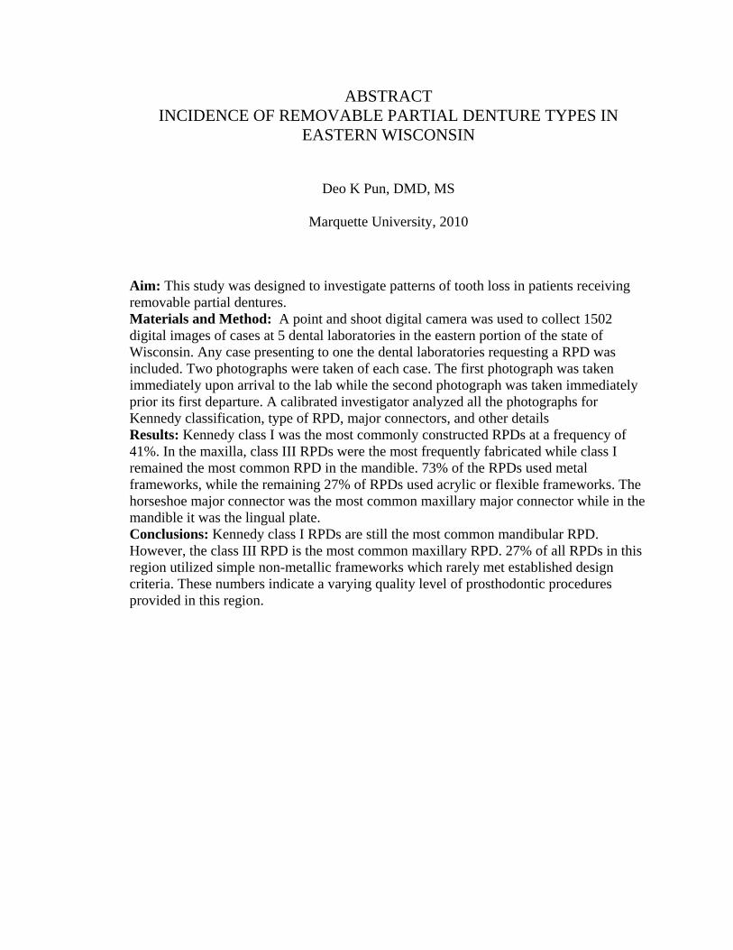
ABSTRACT INCIDENCE OF REMOVABLE PARTIAL DENTURE TYPES IN
EASTERN WISCONSIN
Deo K Pun, DMD, MS
Marquette University, 2010
Aim: This study was designed to investigate patterns of tooth loss in patients receiving removable partial dentures. Materials and Method: A point and shoot digital camera was used to collect 1502 digital images of cases at 5 dental laboratories in the eastern portion of the state of Wisconsin. Any case presenting to one the dental laboratories requesting a RPD was included. Two photographs were taken of each case. The first photograph was taken immediately upon arrival to the lab while the second photograph was taken immediately prior its first departure. A calibrated investigator analyzed all the photographs for Kennedy classification, type of RPD, major connectors, and other details Results: Kennedy class I was the most commonly constructed RPDs at a frequency of 41%. In the maxilla, class III RPDs were the most frequently fabricated while class I remained the most common RPD in the mandible. 73% of the RPDs used metal frameworks, while the remaining 27% of RPDs used acrylic or flexible frameworks. The horseshoe major connector was the most common maxillary major connector while in the mandible it was the lingual plate. Conclusions: Kennedy class I RPDs are still the most common mandibular RPD. However, the class III RPD is the most common maxillary RPD. 27% of all RPDs in this region utilized simple non-metallic frameworks which rarely met established design criteria. These numbers indicate a varying quality level of prosthodontic procedures provided in this region.
i
ACKNOWLEDGEMENTS
Deo K. Pun, DMD, MS
I’m indebted to so many individuals for completion of this thesis project. First of
all, I would like to thank Dr. Michael Waliszewski for suggesting the thesis topic. His
enormous effort in numerous revision of the study, his vision, critical evaluation and
organizational skills are highly appreciated. It was a great opportunity to work with him. I
would like to thank Dr. Kenneth Waliszewski for valuable suggestions and taking time
off from private practice to be in the multiple meetings. I truly appreciate your thought
and ideology. A special thanks to program director Dr. Gerald Ziebert for his guidance
not only in this thesis project but also overall Prosthodontic program. Suggestions from
Dr. Jerry Walker for this study is highly appreciated. I owe a lot to Dr. David Berzins for
his mentorship in Dental Biomaterials and helping me with IRB protocol, and guiding me
with use of Excel. I do like thank Dr. Arthur Hefty and Alexis Dye for their expertise in
the statistical help. Thanks to Dr. Geoffrey Thompson for proof reading and for valuable
input. I would also like to thank Cole Stockheimer, a junior dental student for helping me
in data collection. It would be impossible to complete this study without volunteers in
each of the five dental laboratories. One person was involved in taking photographs in
each laboratories.
I would also like to thank numerous people who were influential indirectly in this
research. I would like to remember my beloved sister Babita Pun who left this world last
year. You have been inspiration to me in my everyday life in whatever I do. Besides, my

ii
parents have been continuously supportive of me especially, my father who is battling
cancer but still fighting strong. This has inspired me to work harder and not to give up.
Finally, I would like to acknowledge my wife Manju Rana-Pun for being supportive all
the time.

iii
TABLE OF CONTENTS
ACKNOWLEDGEMENTS……………………………………………….....i
LIST OF FIGURES...……………………………………………………….iv
LIST OF TABLES.………………………………………………………….v
CHAPTERS
I. INTRODUCTION……………………………………………..1
II. LITERATURE REVIEW……………………………………...4
III. MATERIALS AND METHODS……………………………..21
IV. RESULTS…………………………………………………….32
V. DISCUSSION………………………………………………...50
VI. SUMMARY AND CONCLUSIONS………………………...59
BIBLIOGRAPHY………………………………………………………….62
iv
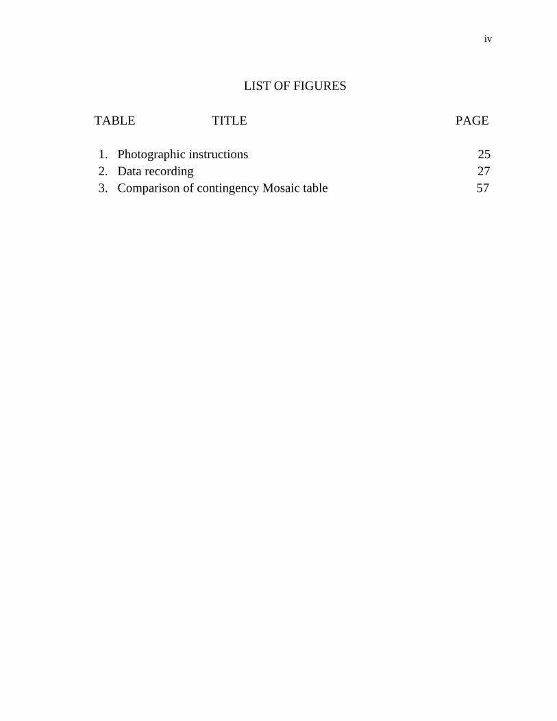
LIST OF FIGURES
TABLE TITLE PAGE 1. Photographic instructions 25 2. Data recording 27 3. Comparison of contingency Mosaic table 57
v
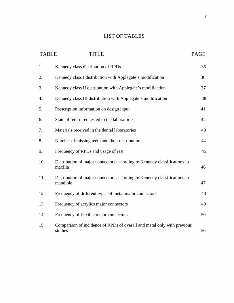
LIST OF TABLES
TABLE TITLE PAGE 1. Kennedy class distribution of RPDs 35
2. Kennedy class I distribution with Applegate’s modification 36
3. Kennedy class II distribution with Applegate’s modification 37
4. Kennedy class III distribution with Applegate’s modification 38
5. Prescription information on design input 41
6. State of return requested to the laboratories 42
7. Materials received in the dental laboratories 43
8. Number of missing teeth and their distribution 44
9. Frequency of RPDs and usage of rest 45
10. Distribution of major connectors according to Kennedy classifications in maxilla 46
11. Distribution of major connectors according to Kennedy classifications in mandible 47
12. Frequency of different types of metal major connectors 48
13. Frequency of acrylics major connectors 49
14. Frequency of flexible major connectors 50
15. Comparison of incidence of RPDs of overall and metal only with previous studies 56

1
CHAPTER I
INTRODUCTION
2
Recent investigations have analyzed trends in demand for prosthodontics in the
United States.1,2 Over 90 years ago Hillyer pointed out that as the edentulous conditions
decrease, the partial will increase.3 Despite decreasing rates of tooth loss the need for
removable prosthodontic treatment remains high.4-6 It appears that a consequence of the
professions improved preventive measures has been an increase in the number of patients
who require partial prosthodontic treatment.7 Likewise, an aging population retaining
more teeth results in an increased number of teeth at risk for disease. Joshi and Manski
evaluated some of the aspects of the more teeth more disease hypothesis.8, 9 Conservative
treatment modalities used to treat partial edentulism, such as dental implants also happen
to be the most expensive. This continues to limit their availability to lower socio-
economic groups in whom the highest rates of tooth loss occur.10-12 It should not be a
surprise then that conventional removable prosthodontic treatment modalities continue to
outnumber implant tooth replacements in general practice.13 Despite its frequency of use,
the removable partial denture (RPD) receives minimal interest in the prosthodontic
literature or at professional meetings.
Multiple RPD classification systems have been presented.14-19 The Kennedy
classification system is currently the system used in two prominent RPD textbooks20,21
and was found to be the most commonly used system according to an older analysis.19
The Kennedy classification system’s main differentiating factor is its ability to quickly
and clearly demonstrate the location of tooth support in relation to edentulous spaces.
Due to its simplicity it does not make judgments regarding condition or position of the
remaining teeth. The principles, concepts, and practices in prosthdontics (PCPP) states
that a classification system should allow immediate visualization of the type of arch
3
being considered as well as immediate differentiation between tooth-borne and tooth-
tissue supported RPDs.22 More recently the American College of Prosthodontists (ACP)
has formulated a prognostic diagnostic index that takes into account the many clinical
factors which may complicate a particular case.23 The ultimate goal of any classification
system includes improved communication and consistency within the profession.
Different classifications may also have associated treatment principles that assist in
treatment standardization and teaching. If classifications are periodically analyzed the
opportunity exists to compare their prevalence longitudinally.

4
CHPATER II
LITERATURE REVIEW
5
Kennedy Classification: existing demographic information
Several attempts to analyze specific trends in the prevalence of various types of
RPDs have been presented. Most of these studies are of European populations.
Anderson investigated the prevalence of the classification of RPDs at Birmingham Dental
Hospital in the United Kingdom.24 Out of a total of 417 RPDs, 208 (49.9%) were
Kennedy Class I, 76 (18.2%) Class II, 101 (24.2%) Class III, and 32 (7.6%) Class IV.
This was the earliest study of Kennedy classification prevalence. As a follow-up,
Anderson’s group reviewed another patient population to evaluate the type of RPD being
worn.25 Out of a total of 483 RPDs made, 302 (62.52%) had metal frameworks, 40
(8.28%) followed the ‘Every’ type of RPD design, and 141 (29.2%) were ‘plate’ RPDs.
The ‘Every’ RPDs were all-acrylic resin frameworks occasionally incorporating retentive
wire clasps. Plate dentures were also all-acrylic resin RPDs. However, while the ‘Every’
dentures consciously avoided covering gingival tissues the ‘plate’ dentures fully covered
the cingulum areas of teeth. Another review at Birmingham hospital found that Class III
RPDs were the most common RPD still in function among patients attending recall.26
This may be due to better survival rates of Class III RPDs or biases introduced during the
recall process. According to the information presented, only 38% of the patients recalled
were examined. More recently, three commercial dental laboratories in the United
Kingdom were surveyed resulting in examination of 80 maxillary and 44 mandibular cast
metal framework RPDs.27 The authors spent one day in each of the laboratories collecting
the information. Kennedy Class III (69%) was the most frequent maxillary RPD while
Class II was the most frequent in the mandibular arch (43%). Interestingly, the study was
6
repeated 10 years later for comparison purposes.28 This time five commercial dental
laboratories were surveyed for a total of 330 prescriptions and frameworks examined.
Kennedy Class III (55%) was still the most common maxillary RPD, though the
frequency decreased. Kennedy Class I was the most common mandibular RPD found.
These differences may have been due to chance or possible changes in tooth loss and
treatment selection.
Derry and Bertram recalled 65 RPDs made in Denmark after 2 years of service.29
Nine of the original RPDs could not be recalled leaving 88% of the original RPDs to be
examined. Of the recalled group, Kennedy Class III was the most frequent followed by
Class I then II. These patients were all treated in a dental school environment and all
received a cast metal frame RPD. Axéll and Öwall investigated the prevalence of RPDs
in Sweden.30 A clinical examination of 20,333 subjects aged 15 and over was conducted
between January 1973 and October 1974. A RPD was found in 6.2% of those examined.
Another study from Sweden reported that 14.5% of examined individuals had a RPD.31 In
this project a color photograph of the RPD was examined for 543 subjects ranging in age
from 41 to 65 years. Information on remaining tooth distribution was also recorded,
although the Kennedy classification was not used. Laine and Murtomaa surveyed 957
people in Finland to determine the frequency of different types of RPDs in people aged
15 and over.32 Trained interviewers asked subjects if they were using removable dentures.
The subjects were then asked to identify whether they were using a cast metal framed
RPD or an acrylic resin RPD with the aid of a series of photographs. 6.3% of those
interviewed claimed to be wearing a RPD. All-acrylic RPDs dominated the distribution
with 70% of all maxillary and 61% of all mandibular. Another Finnish investigation by
7
Tervonen and colleagues sought the prevalence of edentulism and the frequency of
removable dentures among 4 age groups.33 1600 subjects were examined by calibrated
dentists, 400 subjects per age group. The frequency of RPD use varied by age group and
was 3.2% in the 25 year olds, 9.1% in the 35 year olds, 9.5% in 50 year olds, and 7.0% in
the 65 year olds. No distinction was made between RPD type and no discussion or
analysis of tooth distribution was done.
Bergman’s classic long-term follow-up consisted of 33 RPD patients in Sweden.34
2 cases were Class II and a single case was Class III. Of the 30 Class I RPDs, 25 were
mandibular. All patients received cast metal framework RPDs and were placed on a
yearly recall program. Yeung et al described the usage of cast metal frame RPDs in
patients treated at a teaching dental hospital in Hong Kong.35 Patients were recalled for
clinical examination 5-6 years after placement. 100 out of 192 patients were constantly
wearing their RPDs. Half of the prostheses were therefore no longer being worn. No
dental maintenance program had been established. In contrast to some other evaluations,
127 out of 249 RPDs (51%) were Class III. Similar percentages were found in both
arches. A study in Australia by Witherell and Smales recalled RPD patients from the
University of Adelaide for examination in 1978.36 While some were all-acrylic the
majority of cases used cast metal frameworks. 64 out of the 86 patients treated in 1973
attended a recall appointment. Partials made up to the year 1966 were included meaning a
single patient often had multiple RPDs in the total analysis. The authors noted how
prostheses were replaced 4 times or more due to failure during this time frame. A total of
150 RPDs was thereby assessed. A distinct difference between maxillary and mandibular
8
arches was seen. 60 out of 86 mandibular RPDs were Class I. 29 Class II and 21 Class III
maxillary RPDs were found from the total of 64.
Several interesting demographic analyses were also made in Middle-Eastern
populations by Keyf,37 Sadig and Idowu,38 and Al-Dwairi.39 Keyf surveyed 528 RPD
frameworks from 362 patients in Turkey.37 The frequency of Kennedy Class I, II, and III
RPDs were 43%, 38%, and 18% respectively. Surprisingly, they did not find Class IV
RPDs. This was likely in part due to the fact that information was only collected for
metal frameworks. Sadig and Idowu reviewed 650 work authorizations for patients in
Saudi Arabia over the course of one year.38 From these 650 work authorization, 422 total
patient cases were analyzed. Transitional RPDs, and non-conventional RPDs were
excluded. Therefore, only cast metal RPD frameworks were included in the analysis. The
Kennedy classification was recorded for each case using Applegate’s guidelines.18 Class
III RPDs were found to be the most common at 40.8% and Class IV the least at 5.9%. In
the 319 patients with opposing RPDs, maxillary Class III opposing mandibular Class III
was the most common at 22.5%. RPD frequency was also investigated among a
Jordanian population.39 Two hundred laboratory authorization forms from cases treated
by 5th year dental students were collected over a period of 2 years. All-acrylic,
transitional, and complex RPD designs were excluded leaving a total of 350 cast metal
frameworks for analysis. Kennedy Class III with or without modification spaces was the
most frequent at 47% on the maxilla and 45% on the mandible. Class IV was the least
frequent pattern. 150 of the 200 patients had metal frame RPDs fabricated for both
arches. It was also found that opposing Class III RPDs was the most common
combination.
9
Several studies of this topic exist from the USA. Schwalm et al. recalled patients
treated at the University of Washington one or two years after placement of a RPD.40 All
RPDs were made by dental students. Despite the short-term recall period 69 of the 161
patients who received a RPD were unable to be reevaluated. In this setting the Class I
RPD was the most common followed by Class III and II respectively. All patients treated
at this fee-for-service clinic received a cast metal framework RPD. Chandler and Brudvik
followed up this group of patients 8 to 9 years after placement.41 Only 38 patients with 44
RPDs attended the recall. Ten RPDs from this group were no longer being worn. Of those
recalled Class I (55.8%) remained the most prevalent followed by Class II (32.4%) then
Class III (11.8%). Curtis and colleagues surveyed the incidence of various classes of
RPDs fabricated at a single dental laboratory in California.42 Work authorizations of 400
patients were collected over 20 consecutive working days. 327 of the 400 work
authorizations were included in the study. 40% of all RPDs were Kennedy Class I while
33% were Class II, 18% Class III, and 9% Class IV. While Class I RPDs were the most
common mandibular RPD at 49%, Class II RPDs were the most common maxillary RPD
at 38%. Information regarding type of major connectors and retainers used was also
gathered. When this demographic information was compared to data from earlier
analyses there was an increase in the incidence of Kennedy Class II RPDs. Due to the
relatively small number of cases, differences in international treatment philosophies and
other issues, some caution is noted in their conclusions.
Redford’s group estimated denture use among the US non-institutionalized
population 18-74 years of age using data acquired through the NHANES.6 The grand
scope of this project allowed examination of 7,374 subjects. 8% of the total examined
10
subjects were using a RPD. The prevalence increased with age to 22% at 55-64 years.
This finding did not include all-acrylic RPDs. Using five commercial dental laboratories
from different regions of North America Öwall and Taylor investigated the frequency of
different types of RPDs.43 Each laboratory was asked to provide details of 300
consecutive RPDs as they are prepared for shipping out of the laboratory. Details were
recorded by means of 35mm slide photographs with a clear work order number. All
prostheses classifying as RPDs were to be included. The photographs were either with the
framework on the definitive cast or with the completed prosthesis alongside. 1,363 total
case photographs were analyzed. While information regarding number of remaining teeth
and some information regarding tooth distribution was presented; there is no mention of
Kennedy classification. This makes comparison and analysis difficult. 95% of RPDs
analyzed had cast metal frameworks. This percentage seems high considering clinical
realities and general practice. No recent analysis of incidence or prevalence of various
types of RPDs or Kennedy classification could be found for the USA.
Prevalence of RPD framework types: acrylic or metal
The few studies looking at RPD fabrication trends in the USA have looked at data
pertaining to conventional metal framework RPDs. While this simplifies analysis of the
framework design it may not give realistic data in regards to the treatment being provided
to the majority of the population. While valid long-term outcome data is sorely lacking,
all-acrylic resin RPDs continue to be used with great frequency.44,45 It has been
demonstrated in several other countries that all-acrylic resin RPDs are far more common
than cast metal framework RPDs.46-48 Lewandowska et al. reported on RPD frequency in
11
Poland.46 20 dental laboratories were questioned for type of prosthetic appliance being
used. Of the 983 patients who received a RPD, half had one fabricated in both arches for
a total of 1,418. 87% of the RPDs fabricated were all acrylic resin without a metal
framework. It was highlighted that the socialized dental services in Poland provide this
type of RPD for free as an alternative to the more expensive cast metal framework RPD.
Thean surveyed the use of RPDs among 469 private dental practitioners in Singapore.47
Only 172 (37%) responses were received. The study reported that residentially based
practitioners recommended all acrylic resin RPDs for the majority of cases. Few town
practitioners however recommended all acrylic resin RPDs. Due to the suspected high
percentage of all acrylic resin RPDs fabricated by this group of providers it was not
surprising that less than 10% stated that they conducted tooth preparations prior to the
final impression. It was speculated that the frequency of all acrylic resin RPDs would
have been higher if government providers were included in the survey. Radhi collected
131 written prescriptions from five dental laboratories in Kingdom of Bahrain over a
period of 2 months.48 89% of prescriptions requested an all acrylic resin RPD. 109 of
the 131 cases evaluated had the definitive cast made from an irreversible hydrocolloid
impression. These definitive casts were created a minimum of 24 hours after the
impressions were made. Prescriptions for immediate and transitional prostheses were
included in the analysis. Schwarz and Barsby discussed the fact that many of the British
dentists they surveyed were providing all acrylic RPDs for the National Health Service.
They also referenced the dental estimates from the UK which showed a ratio of 1 cast
metal framework RPD for every 7 all acrylic RPDs created.49 Another recent analysis
from the United Kingdom and Ireland was completed by Lynch and Allen.50 While
12
reviewing aspects of RPD education they also reported the frequency of cast metal
framework and all acrylic resin RPDs. A ratio of 3 cast metal frameworks for every 2 all
acrylic resin RPDs was found in the 11 dental schools that responded. These numbers
demonstrate the sometimes significant difference between institutionalized, government
subsidized, and private practice treatment.
These various studies highlight several of the factors which help determine
whether a cast metal framework or all acrylic resin RPD is fabricated. Insurance
reimbursement, capabilities of dental laboratory support, and location and extent of
missing teeth all appear to influence this decision. However, prosthodontic education
may also play a role. While these studies are from other countries, it is suspected that
similar factors play a role in the provision of selected RPDs in the USA. In addition,
newer types of flexible acrylic or vinyl RPDs have received much attention in dental
advertisement over the past decade. No recent peer-reviewed comparison of the
prevalence of these different RPD framework materials could be found for the USA.
Framework concerns: comparison of acrylic versus metal
Without the established design principles and strength of cast metal framework
RPDs it is believed that alternative RPD frameworks have reduced longevity and
significant periodontal consequences. Occasional case reports or clinical tips have been
published describing methods to improve the longevity of the all acrylic resin RPD.
McCartney advocated the addition of wire rest seats to help prevent the tissue recession
and inflammation caused by all acrylic resin RPDs using only soft tissue support.51
Smith and Rymarz blended some of the benefits of cast metal frameworks with the
13
convenience of acrylic resin.52 They recommended casting clasp assemblies only and
then using acrylic resin to join these parts. The clasp assemblies were simply rests and
clasp arms with a loop or beads for purposes of resin retention. It would seem that these
efforts are undertaken due to poor clinical performance of acrylic resin. Studies by
Zalataric,53 Tuominen,54 and Carlson55-57 all investigated periodontal and dental health of
RPD abutment teeth. Nearly all RPDs in these studies were acrylic resin without cast
metal frameworks. It was generally found that teeth in contact with these acrylic
frameworks were more likely to demonstrate dental disease. Zlatarić studied 205 patients
wearing a RPD for a period of 1-10 years.53 They found significant differences in
periodontal health of abutment and non-abutment teeth and concluded that RPD design
plays an important role. Tuominen reported that wearing a RPD significantly increased
the odds of having periodontal pockets and concluded RPDs are a threat to periodontal
tissue.54 Similar to Zlatarić, 92% of subjects were wearing all acrylic resin RPDs with no
mention of maintenance protocol. Wearing of RPDs significantly increased the odds of
having deeper periodontal pockets. The authors therefore suggested that wearing of RPD
is a threat to periodontal tissues. Carlsson published some of the earliest data on the
condition of recalled RPD patients.55-57 Many of these patients had been treated with
complete maxillary dentures opposing an all acrylic resin mandibular RPD. The 13 year
results of treatment confirmed the earlier assessment that all acrylic resin RPDs had a
high incidence of fracture and or need for repair. After admitting that patients often had
selected the all acrylic resin RPD due to economic reasons the authors concluded that it is
poor economy in the long run to choose the cheaper construction.
14
In contrast to the available information regarding poor results of treatment with all
acrylic resin RPDs it is interesting to note that several studies with positive long-term
clinical results utilized cast metal framework RPDs.40,41,58,59 The authors concluded that
there is no direct evidence of well made RPDs significantly contributing to periodontal
disease. Bergman’s 10 year follow up of 23 patients wearing such RPDs yielded fairly
positive results.58 Most of the RPDs were Kennedy Class I. Yearly maintenance visits
and oral hygiene instructions were conducted for all patients. They concluded that if the
RPD was well designed and oral hygiene was maintained, the RPD will not cause dental
disease. Bergman has followed the same patient population up to 25 years.59 13 of the
original RPDs were still functioning. A specific comparison of gingival health was
conducted by Bissada.60 While not covering tissue was the preferred method for
prevention of gingival inflammation in RPD wearers, inflammation was greater when
acrylic contacted the gingival tissue than when metal was used. These projects seem to
confirm a preference for metal framework RPDs in terms of clinical performance and
periodontal health.
Framework handling: laboratory communication and clinical practice
If metal frameworks are preferred one must consider that modern RPD framework
fit continues to be less than ideal.61-65 Since the profession adopted Akers one-piece RPD
casting technique in 1925 dentists began to rely more heavily upon dental laboratories.66
With time it appears that the average practitioner understands less of the RPD framework
creation process than previous generations.67,68 There is an apparent trend that USA
dental school curriculums have reduced time spent on removable prosthodontic
15
education.69 There is concern this decrease in educational focus will have an effect on
the quality of care provided by general practitioners.
The quality of communications with the laboratory has been reviewed
consistently over the past several decades. Sykora and Calikkocaoglu sent identical casts
to multiple commercial dental laboratories in order to compare and analyze designs for an
extracoronal RPD.70 No instructions were given to the laboratory except for a letter
explaining the purpose of the project. 25 different laboratories returned a framework.
According to the authors a remarkable variation in designs was received. While
tendencies were evident for major connectors and certain clasp assemblies, some labs did
not use a surveyor and some designs were unsatisfactory. This finding was similar to that
of McCracken’s survey of design of a mandibular RPD.71 Frantz published two reports
on design variation among dentists.72,73 In the first, 97 dentists with varying experience
were asked to submit their ideal design for a RPD for a Kennedy Class II modification 1
maxillary diagnostic cast. The responses included 96 different designs. In the next
project Frantz had 57 dentists examine a patient with a Kennedy Class II modification 1
maxillary arch prior to prescribing their ideal RPD design. Radiographs and casts were
available for all dentists. Despite the in-depth information, 57 different designs were still
received.
In an effort to see if time had improved consistency of RPD designs in general
practice Basker and Davenport repeated their 1977 survey.27,28 Existing cast metal
frameworks and prescriptions were reviewed at 5 laboratories. They examined 330
prescriptions and frameworks. The study found 40.3% of the prescriptions from the
dentists included the RPD design details. This was double the 21% found with this
16
information in 1977. While improvement was seen the authors noted that 60% of cases
were still being designed by the laboratory. Schwarz and Barsby also investigated
communication and the provision of RPD therapy in the UK.74 They first published
responses given to them by the laboratories themselves. Only 673 out of the 1858
(36.2%) metal framework RPDs were designed by the dentist. However, it was also
interesting to note that only 4.6% of the casts reviewed by the laboratory showed any
evidence of tooth preparation. 1,773 out of 1858 showed no evidence of tooth
preparation. The authors note that most of the specific advantages of a cast metal
framework RPD are lost if the abutment teeth are not appropriately prepared to receive it.
The authors confirmed that there is a divergence between clinical practice and dental
school instruction. Schwarz and Barsby then surveyed 605 practitioners through the
mail.49 It was found that 52% of dentists rarely or never used diagnostic casts. Only 51%
had access to a dental surveyor while 34% of respondents have the diagnostic casts
surveyed. 87% of respondents never or rarely prepared guiding plane surfaces while 56%
rarely or never prepared rest seats. When asked how much time on average was spent in
the provision of a RPD 213 out of 474 (44.9%) respondents said less than 1 hour.
Another 231 out of the 474 (48.7%) averaged between 1 and 2 hours to provide a RPD.
The remaining 30 averaged more than 2 hours. As mentioned previously, the authors
discussed the fact that many of the dentists surveyed were providing all acrylic RPDs for
the National Health Service. They also referenced the dental estimates from the UK
which showed a ratio of 1 cast metal framework RPD for every 7 all acrylic RPDs
created. The article also noted how practitioners are economically unable to spend more
time to provide what is thought to be the standard of care due to the low reimbursement
17
rates for a RPD. Lynch and Allen investigated some aspects of the quality of written
prescriptions and master impressions used for fabrication of prostheses in the United
Kingdom and Ireland.75 When the prescriptions for RPDs were considered alone, 84 of
the 136 final impressions were made with alginate. Surprisingly, the minimum time
between an impression and pouring of the definitive cast was 72 hours. Out of the 136
total RPD prescriptions, 28 were considered to be poorly written while an additional 40
had no written instructions at all. Exactly 50% of cases therefore were designed almost
entirely by the laboratory. These findings correlate with those found by Radhi.48
Similar information has been collected in the USA. Taylor received
questionnaires from 303 out of 488 commercial dental laboratories.76 78% of those
responding designed the majority of RPDs sent to them. That same percentage of
laboratories estimated that less than 10% of the mandibular distal extension definitive
casts were of the altered cast type. The overall conclusion was that practicing dentists
frequently use techniques that require minimum appointment time.
Cotmore et al. surveyed RPD treatment and framework design between two dental
school classes that graduated 7 years apart.77 There was a good response rate of 90% of
the class of the ‘70s and 64% of the class of the 60’s. Some deficiencies in standard of
care protocol were found. In contrast to the findings from Britain, the vast majority of
respondents claim they communicate well with the dental laboratory. It was interesting to
note that significant differences existed in treatment methods and design between the
different classes. The authors indicate that this demonstrates the influence of educational
techniques upon practice. Hardy and Stuart studied prescriptions from 11 dental
laboratories believed to be near Howard University in Washington, D.C.78 Work
18
authorizations, diagnostic casts, and definitive casts or final impressions were examined
for 300 cases. They found 81% of dentists failed to provide adequate prescriptions. In
addition, 57% of the prescriptions lacked any written detail for proposed framework
design. 8% of cases sent the final impressions to the laboratory, in all of these cases the
impression material was irreversible hydrocolloid. Authors also found that 67% of
definitive casts were submitted as unacceptable for prosthesis fabrication due to voids or
bleb in critical areas such as rest seats and retentive areas. Burns surveyed prosthodontic
specialists in an attempt to discern what would be considered the standard of care for
RPD design and fabrication.79 195 responses were collected at the 1987 American college
of Prosthodontists Annual meeting. 66% used stock trays for RPD final impressions
which matched the 66% that used irreversible hydrocolloid for the same. Laboratory
prescriptions were used in a high percentage of communications with the dental
laboratory. In particular, 90% wrote detailed descriptions of the RPD framework. It was
suspected that respondents often used multiple methods of communication thus resulting
in no responses of 100%. 98% of patients were appointed for a separate framework try-in
appointment by this group. 78% of respondents used rigid frameworks with stress
releasing clasp assemblies for distal extension cases.
Clinical outcomes: general population within the USA
With these potential explanations for poor quality prostheses it is a
disappointment that clinical analyses of non-institutionalized treatment in the USA
demonstrate a high frequency of defects. Two fairly recent articles looked at the same
data set from the National Health and National Examination Survey (NHANES III).6,80
19
This survey evaluated 7,374 persons. Examiners were trained and calibrated. Of those
examined, 1306 were using a RPD. Five criteria; integrity, tooth wear, presence of
temporary material or adhesive, stability, and retention were used to gauge the quality of
removable prostheses. A mere 36% of RPDs were free of defects. Frank and colleagues
from the University of Washington studied whether RPDs made in community practices
met the standards of design and fabrication.81 The study involved only Kennedy Class I
and II mandibular RPDs made during a five year period. 91 of 292 eligible patients were
examined, 82 of which yielded usable data. Most subjects (128) declined to be
examined. 8 categories of evaluation regarding design and fabrication were developed
from the PCPP.22 Inter-examiner reliability demonstrated the acceptability of the criteria.
More than half (43/82) of the RPDs met 4 or fewer standards. 43% (35/82) of the RPDs
were rated as clinically acceptable while 46% (38/82) could be made acceptable by
modification. The remaining 9 required replacement. Previous to this study Frank
conducted a survey of patients in the same dental plan and metropolitan area.82 Only
40.3% of the patients were completely satisfied with their distal extension RPD.
Fortunately, only 19.7% expressed some level of overall dissatisfaction with the RPD.
Statement of problem: purpose
Perhaps the poor quality of removable prostheses seen in the general USA
population is in part due to the factors discussed. Further analysis of the type of
prostheses requested, design instructions given, and quality of material provided to the
laboratory would clarify some of the sources of defective RPDs. Considering the
previously mentioned factors, a modern analysis of the incidence of various classes of

20
RPDs would be of interest. It also provides an opportunity to investigate some of the
existing trends in non-institutionalized RPD services. The purpose of this study was to
investigate patterns of tooth loss in patients receiving removable partial dentures (RPD).
In addition, details regarding how this treatment is provided was collected.

21
CHAPTER III
MATERIALS AND METHODS
22
Five regional commercial dental laboratories (Nu-Art Dental Laboratory Inc.
Wauwatosa; Capitol Dental Laboratory Inc., Menomonee Falls; Saber Dental Studio Inc.,
Waukesha; Badger Dental Laboratory Inc., Milwaukee; Lord’s Dental Studio Inc., Green
Bay) located in the state of Wisconsin, USA were chosen for data collection. These
laboratories were selected based upon initial telephone survey of number of RPDs
produced. The laboratories authorized collection of digital images for the study. The
proposed study was approved by the Institutional Review Board (Protocol number HR-
1887) of Marquette University School of Dentistry, Milwaukee, WI, USA.
Data was collected by the laboratories using digital photography. Any case
presenting to the lab requesting fabrication of a RPD was included. For this study, a
removable partial denture was defined as any prosthesis that replaces some teeth in a
partially dentate arch; it can be removed from the mouth and replaced at will.83 Two
photographs of this case would then be taken. The first photograph was taken
immediately upon receipt to the lab. The second photograph was taken immediately prior
to its initial departure from the lab. In this way, a particular case could only be recorded
once. In addition, repairs and relines were thereby excluded.
Images were standardized by use of pre-measured backgrounds. The 12” x 16”
background was placed on the floor or similar open space. The contents of an incoming
lab case box that was to be included were then placed upon this predetermined area. The
contents could therefore include a prescription, impression, a dental cast, or both. If
received articulated, the mounting rings were loosened to allow cast placement on the
background. Hinge-type articulators were opened to allow photography of the casts from
an occlusal direction. Lab prescriptions were placed flat with no creases. Personal
23
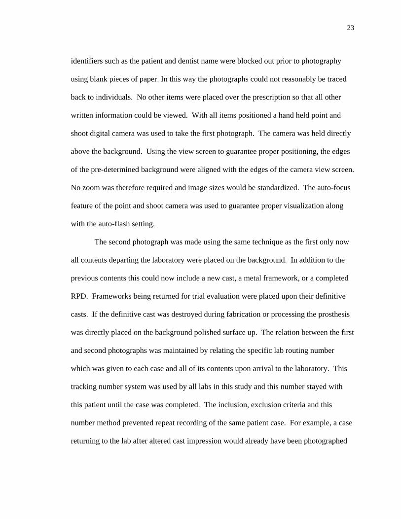
identifiers such as the patient and dentist name were blocked out prior to photography
using blank pieces of paper. In this way the photographs could not reasonably be traced
back to individuals. No other items were placed over the prescription so that all other
written information could be viewed. With all items positioned a hand held point and
shoot digital camera was used to take the first photograph. The camera was held directly
above the background. Using the view screen to guarantee proper positioning, the edges
of the pre-determined background were aligned with the edges of the camera view screen.
No zoom was therefore required and image sizes would be standardized. The auto-focus
feature of the point and shoot camera was used to guarantee proper visualization along
with the auto-flash setting.
The second photograph was made using the same technique as the first only now
all contents departing the laboratory were placed on the background. In addition to the
previous contents this could now include a new cast, a metal framework, or a completed
RPD. Frameworks being returned for trial evaluation were placed upon their definitive
casts. If the definitive cast was destroyed during fabrication or processing the prosthesis
was directly placed on the background polished surface up. The relation between the first
and second photographs was maintained by relating the specific lab routing number
which was given to each case and all of its contents upon arrival to the laboratory. This
tracking number system was used by all labs in this study and this number stayed with
this patient until the case was completed. The inclusion, exclusion criteria and this
number method prevented repeat recording of the same patient case. For example, a case
returning to the lab after altered cast impression would already have been photographed
24
during initial receipt to the lab and this could be confirmed by review of the lab tracking
number.
Calibration of labs was conducted on two separate occasions, during the pilot
study and again after study methodology was confirmed. Calibration consisted of
describing the objectives and methods of the photography process to those making the
photographs. After demonstration, the lab person in charge of photographing the cases
was observed during one session of photographs to assure the technique described above
was followed accurately. A single photographer from each lab was selected and trained;
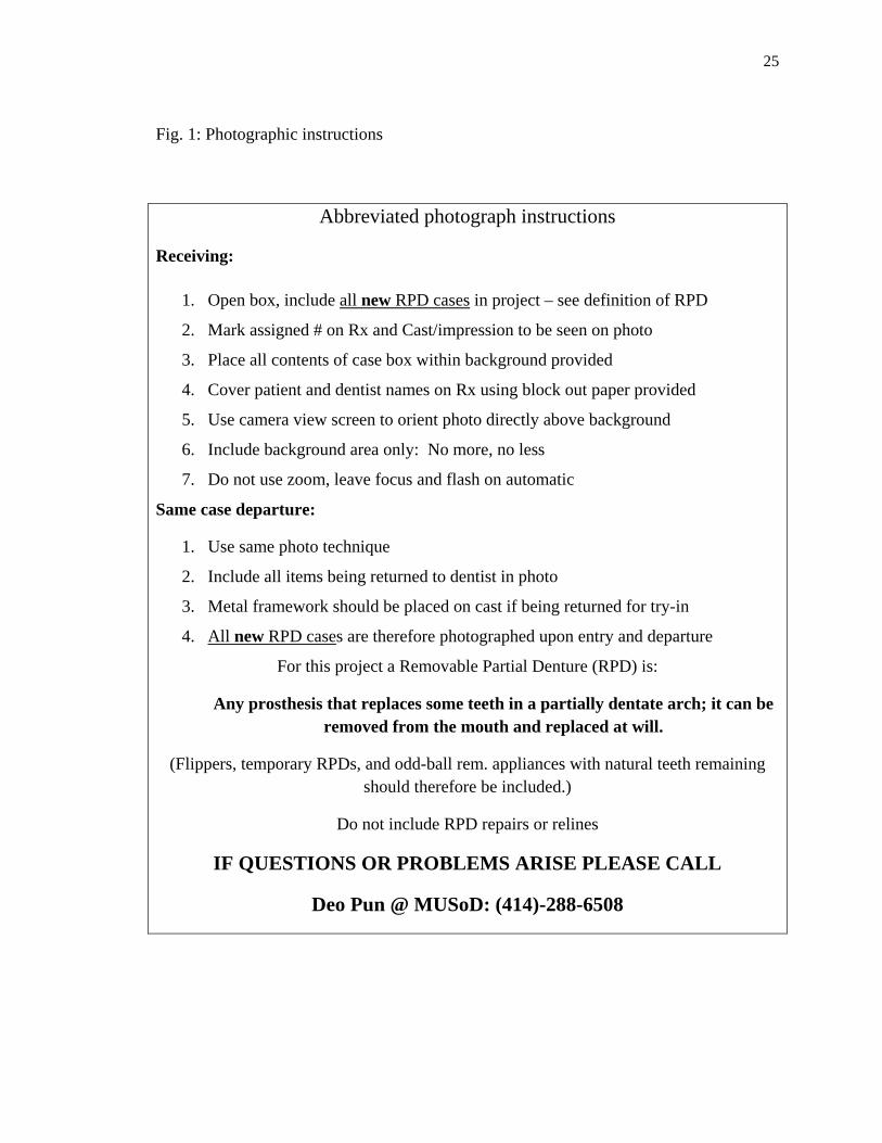
no other photographers were used during the study period. Once calibrated a summary of
instructions was left for each photographer to reference (Figure 1). Since each lab has
different receiving times established for in-coming cases, it was determined that
photographs should be taken immediately upon opening of received case boxes. This
assured that cases would not by-pass the initial photographs and be missed. For out-
going cases, photographs were taken just prior to packaging.
Once study methodology was confirmed a minimum of 1 follow-up visit to each
laboratory was conducted during the initial 2 weeks of data collection. Calibration of the
photographer was confirmed, data was reviewed for accuracy, and camera maintenance
was performed. Based upon the case volume during this first week camera maintenance
was customized for each camera and each laboratory. Camera maintenance consisted of
downloading of digital photos to clear the memory card, recharging or replacing batteries,
cleaning the lens, and any other procedure required to continue efficient and accurate data
collection. Data collection was planned to continue for a minimum of 4 weeks with a
minimum sample size goal of 500 cases.
25
Fig. 1: Photographic instructions
Abbreviated photograph instructions
Receiving:
1. Open box, include all new RPD cases in project – see definition of RPD
2. Mark assigned # on Rx and Cast/impression to be seen on photo
3. Place all contents of case box within background provided
4. Cover patient and dentist names on Rx using block out paper provided
5. Use camera view screen to orient photo directly above background
6. Include background area only: No more, no less
7. Do not use zoom, leave focus and flash on automatic
Same case departure:
1. Use same photo technique
2. Include all items being returned to dentist in photo
3. Metal framework should be placed on cast if being returned for try-in
4. All new RPD cases are therefore photographed upon entry and departure
For this project a Removable Partial Denture (RPD) is:
Any prosthesis that replaces some teeth in a partially dentate arch; it can be removed from the mouth and replaced at will.
(Flippers, temporary RPDs, and odd-ball rem. appliances with natural teeth remaining should therefore be included.)
Do not include RPD repairs or relines
IF QUESTIONS OR PROBLEMS ARISE PLEASE CALL
Deo Pun @ MUSoD: (414)-288-6508
26
As photographed cases were received, the first and second photographs of each
case were matched and then numbered as previously described. Once matched the first
ten cases from each lab were viewed by three of the investigatorsa-c. Each investigator
simultaneously and independently collected information regarding that case with the data
collection form (Figure 2). After completion the three authors’ forms were reviewed and
compared for calibration purposes. Any final changes in data collection criteria or
definitions were made at this time. Once agreement on data collection criteria were
confirmed 10 new cases from each lab were independently reviewed by three of the
investigatorsa-c. This group of data collection forms was tested for reliability and
accuracy. In addition, the lead investigatora and data collectorb selected and reviewed an
additional 10 random cases. One week later these same cases were again reviewed and
the data collection forms were compared to confirm repeatability.
The laboratory name, laboratory case serial number, and arch were recorded at the
top of each form. Whether the definitive cast or impression to create the definitive cast
was included in the box was determined from the initial photograph. The prescription
was used as confirmation. In addition, the basic type of impression material used was
visually determined to be either irreversible hydrocolloid or a ‘rubber’ material. Question
4 gathered information regarding the amount of design information given by the dentist
in the prescription. In essence it was an attempt to determine the level of design input by
the dentist and therefore how much decision making was delegated to the laboratory
technician. The design information was obtained directly from the dentist’s written
prescription. If the written prescription referenced a design cast or design drawing this
27
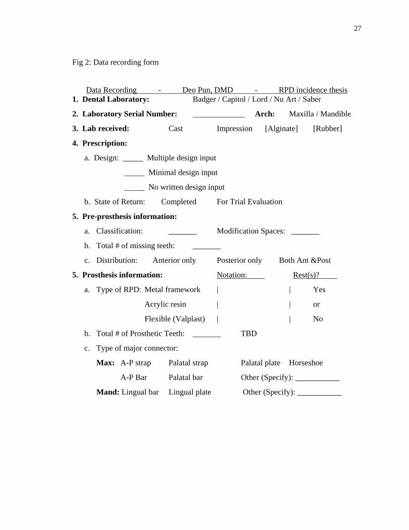
Fig 2: Data recording form
Data Recording - Deo Pun, DMD - RPD incidence thesis 1. Dental Laboratory: Badger / Capitol / Lord / Nu Art / Saber
2. Laboratory Serial Number: _____________ Arch: Maxilla / Mandible
3. Lab received: Cast Impression [Alginate] [Rubber]
4. Prescription:
a. Design: _____ Multiple design input
_____ Minimal design input
_____ No written design input
b. State of Return: Completed For Trial Evaluation
5. Pre-prosthesis information:
a. Classification: _______ Modification Spaces: _______
b. Total # of missing teeth: _______
c. Distribution: Anterior only Posterior only Both Ant &Post
5. Prosthesis information: Notation: Rest(s)?
a. Type of RPD: Metal framework | | Yes
Acrylic resin | | or
Flexible (Valplast) | | No
b. Total # of Prosthetic Teeth: _______ TBD
c. Type of major connector:
Max: A-P strap Palatal strap Palatal plate Horseshoe
A-P Bar Palatal bar Other (Specify): ___________
Mand: Lingual bar Lingual plate Other (Specify): ___________
28
was considered written information as well. Information from a design cast was not
considered if there was no written notice of such information. This protocol was
developed due to inconsistencies encountered during pilot data collection. Comparisons
were made between the written prescription and the returned laboratory work to assist
with this evaluation. Design input was considered to have four basic areas of
information: major connector, guide planes, rests, and clasps. Data collection including
other information was attempted during the pilot testing. While information beyond
these 4 basic criteria is often required and/or helpful, it was not able to be consistently
and reliably obtained. This was primarily due to the data collection method, differences
in laboratory prescription forms, and the wide variety of design prescription methods
used. The following three levels of design input were considered:
a) Multiple design input: At least 2 of the 4 basic areas of design were
communicated via the written prescription.
b) Minimal design input: 1 of the 4 basic areas of design was communicated via
the written prescription.
c) No written design input – 0 of the 4 basic areas of design were communicated
via the written prescription.
The state of return of the prescribed prosthesis was recorded next. The RPD was
considered completed if it was returned after processing and/or with all prescribed
prosthetic teeth attached. It was considered returned for trial evaluation if it was returned
incomplete. Lack of prosthetic teeth or certain components was considered incomplete.
If helpful, the prescription was also used to confirm the state of return of the RPD in
question.
29
Pre-prosthesis information was collected next. The Kennedy classification with
appropriate modification space enumeration was listed according to Applegate’s
modifications.18 In those instances where implants were present as part of the abutment
layout, the implants were considered as abutments when considering the Kennedy
classifciation.84 Whether anterior only, posterior only, or both anterior and posterior teeth
were missing was recorded. If assessment of missing anterior teeth was in question,
anterior teeth were considered missing if the prosthesis consisted of a prosthetic anterior
tooth. This avoided misrepresentation of large diastema’s or spaces closed due to tooth
migration. The total number of missing teeth was recorded. Third molars, fixed
prosthodontic pontics, and closed spaces were not considered missing teeth. If an
immediate prosthesis was requested on the laboratory prescription the total number of
missing teeth was recorded based on the modified cast. Therefore, if an unmodified cast
was sent to the laboratory the number of missing teeth were recorded from the
information in the second photograph.
Information on the prosthesis itself was now recorded. The type of RPD was
determined using information from the prescription and or visualization of the prosthesis
itself. A RPD was considered to be a metal framework if the major connector was cast.
The RPD was considered to be acrylic resin if the major connector was processed in
acrylic resin. This type of RPD may or may not have included wire clasps, rests, or
reinforcement. If a RPD consisted of a combination of metal components and acrylic
resin the final decision was based upon the material used for the major connector. The
flexible type RPD was determined mainly from the laboratory prescription. This selection
was confirmed by visualization of non-metallic clasp assemblies or the differing
30
appearance of the vinyl-acrylic material. In addition, if a specific type of complex RPD
was found a notation was made. For example; swing-locks were noted under metal
framework. It was not expected that many of these types of RPDs would be seen and
therefore a specific check box was not included. After framework determination,
information from either photograph was used to determine whether or not a partial
denture rest was present. A partial denture rest, as defined by the 8th edition of the
Glossary of Prosthodontic Terms, is a rigid extension of a fixed or removable dental
prosthesis that prevents movement towards the mucosa and transmits functional forces to
the teeth or dental implant.83 Therefore, an extension of the RPD onto either a prepared
rest seat or over the occlusal or incisal surface of a tooth or dental implant was considered
to meet this definition. Lingual plates, minor connectors, and clasp arms were not
considered rests unless they involved an actual rest, rest seat, or they involved an occlusal
or incisal surface. At that point they could also be described in terms of the above
definition; “preventing movement towards the mucosa.” Total number of prosthetic
teeth was determined by counting the teeth present on the completed prosthesis. If only a
metal framework existed the number of prosthetic teeth was either estimated based upon
size and position of remaining teeth or recorded as to-be-determined.
The major connector was visualized and recorded. The expected types were listed
using the frequency of major connectors in previous research. The visualized major
connector was classified as that which came closest to one of the following definitions:
a) Palatal strap: a maxillary major connector having an anterior/posterior
dimension of 13-20 mm that directly or obliquely traverse the palate and is
31
generally located in the area of the second premolar and first molar. This type of
major connector leaves the anterior and posterior palatal surfaces uncovered.83
b) Palatal plate: a maxillary major connector that covers the palate completely or
partially but extends posterior to junction of the hard and soft palates.21,83
c) A-P strap: a palatal plate type major connector with an opening in the middle.
d) Horseshoe (U-shaped): a maxillary major connector consisting of a band of
metal running along the lingual surface of the posterior teeth and extending onto
the palate to cover the entire rugae area.21
e) Palatal bar (or A/P palatal bar): a maxillary major connector characterized by
being relatively narrow but of increased thickness while traversing the mid-palatal
region.83
f) Lingual bar: a mandibular major connector located lingual to the dental arch
with visible gingival tissue lingual to any anterior tooth.83
g) Lingual plate: a mandibular major connector contacting the lingual surfaces of
all remaining anterior teeth.83
Less frequent designs were to be listed as other with a written description if
discovered.
After establishing reliability of data collection procedures the lead investigator
completed review of the remaining photographed cases. Cases were organized digitally
according to the previously mentioned lab initials and the serial numbers that were
assigned to the case by each laboratory. Photographs were viewed digitally using an over
head projector 92”x70”. Images were opened on-screen using a standard image viewing
program (Microsoft Office Picture Manager). If helpful, the zoom feature of this
32
program was used in order to accurately collect all data. Any questionable or difficult to
analyze photos were reviewed by the same previous three investigators to determine
exclusion or inclusion. Once all information was transferred to the data collection forms
these forms were tabulated using a computerized spread sheet (Excel, Microsoft).
a = Deo Pun b = Michael Waliszewski c = Kenneth Waliszewski

33
CHAPTER IV
RESULTS
34
Three investigators were evaluated using Fisher’s exact test to analyze the
accuracy of the data collection form. Similarly, Fisher’s exact test was conducted to test
the repeatability of the data collection forms of two investigators. The Chi-squared test
was used compare the frequency of different Kennedy classification among three studies
of RPDs incidence (p<0.05).
A total of 1502 images were received from the laboratories. Of these, 1140
images were matched photographs resulting in a total of 570 cases (N=570). Only these
matched images were included in the study analysis. The remaining 362 individual
images were excluded. Of this excluded total 347 images were unable to be matched and
the other 15 were unmatched and unreadable. This was either due to errant omissions by
the laboratory photographer or one of the two images being outside the time frame of
data collection. Due partly to the ability to procure information from either of the two
photos, none of the matching pairs of images had to be excluded.
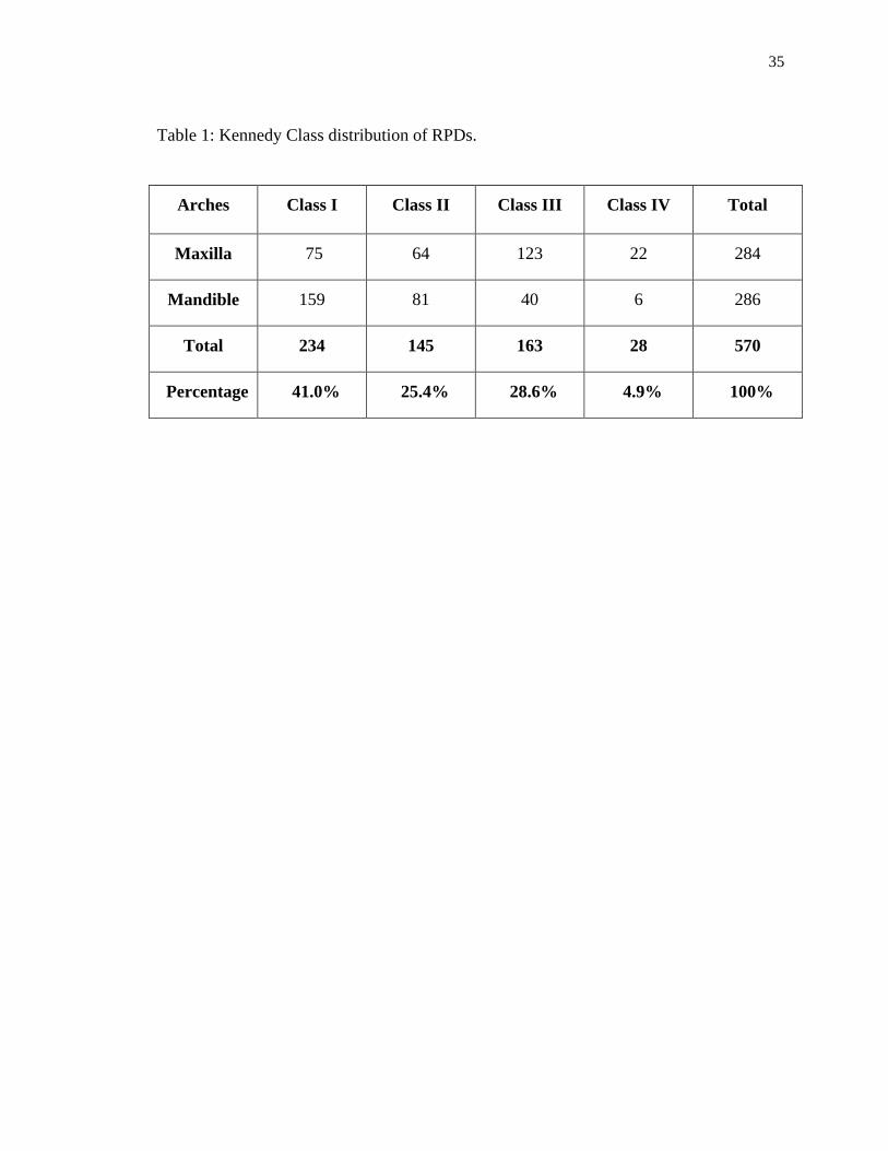
Of the 570 included cases, 284 were maxillary and 286 were mandibular. The
distribution of RPDs based on Kennedy Class is shown in Table 1. The most frequently
fabricated RPD was Class I (41.0%) followed by Class III (28.6%), Class II (25.4%) and
finally Class IV (4.9%). Class III’s predominated in the maxillary arch (43.3%), while
Class I’s dominated the mandibular arch (55.6%).
The break-down of modification spaces for each classification are summarized in
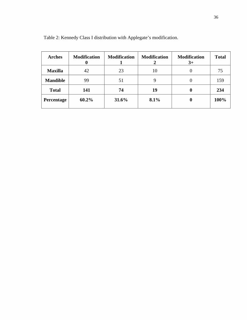
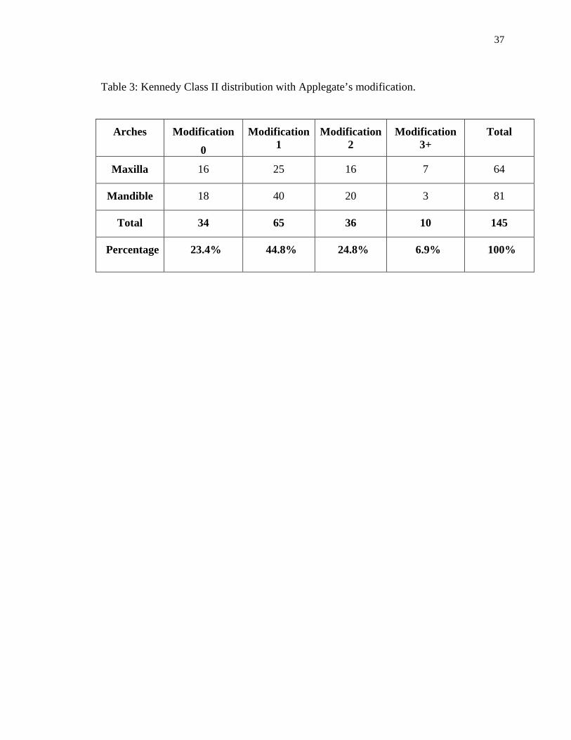
Tables 2-4. 60.2% of Class I RPDs lacked any modification spaces while only 23.4% of
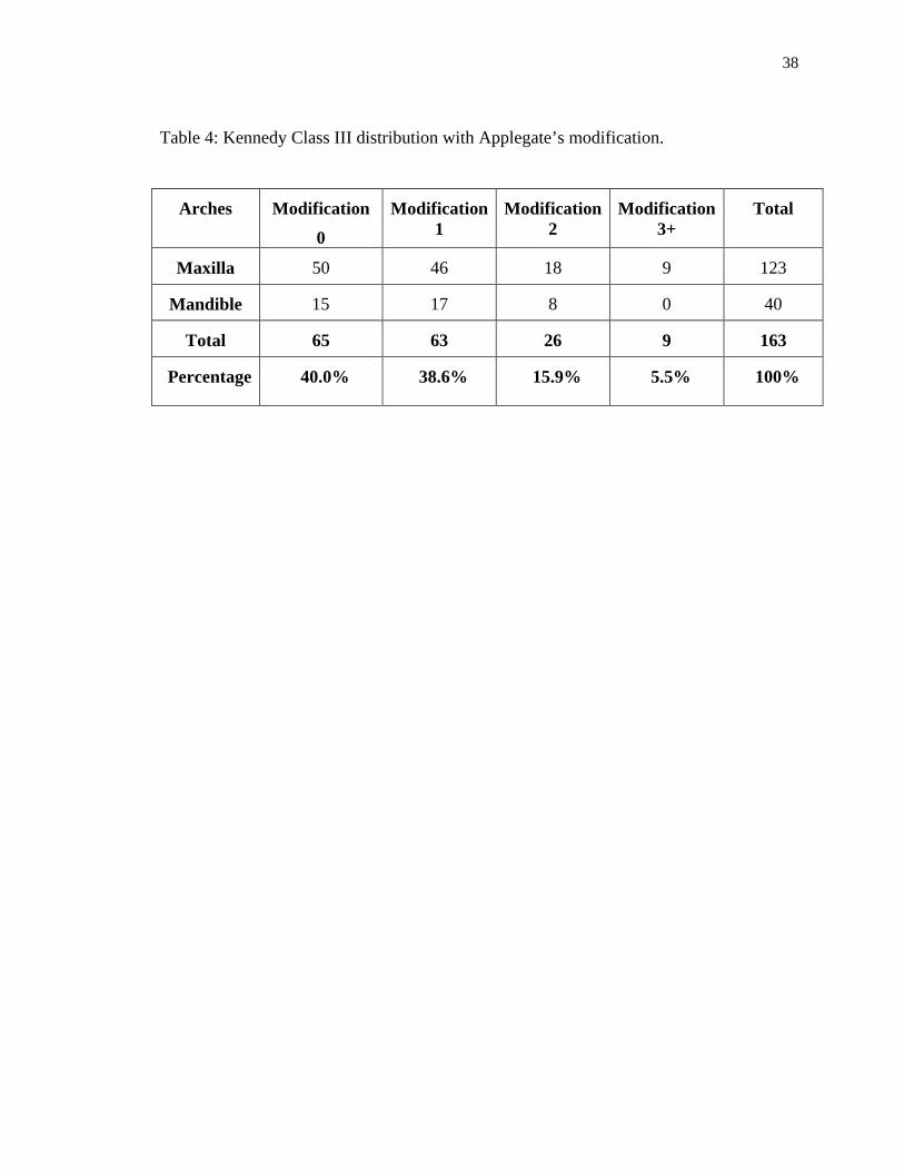
Class II RPD’s lacked the same. 78.5% of Class III RPD’s had either no or a single
modification space indicating a large majority of either unilateral or single bilateral
edentulous spaces. Overall, there were 268 RPDs fabricated without any modifications
35
Table 1: Kennedy Class distribution of RPDs.
Arches Class I Class II Class III Class IV Total
Maxilla 75 64 123 22 284
Mandible 159 81 40 6 286
Total 234 145 163 28 570
Percentage 41.0% 25.4% 28.6% 4.9% 100%
36
Table 2: Kennedy Class I distribution with Applegate’s modification.
Arches Modification 0
Modification 1
Modification 2
Modification 3+
Total
Maxilla 42 23 10 0 75
Mandible 99 51 9 0 159
Total 141 74 19 0 234
Percentage 60.2% 31.6% 8.1% 0 100%
37
Table 3: Kennedy Class II distribution with Applegate’s modification.
Arches Modification 0
Modification 1
Modification 2
Modification 3+
Total
Maxilla 16 25 16 7 64
Mandible 18 40 20 3 81
Total 34 65 36 10 145
Percentage 23.4% 44.8% 24.8% 6.9% 100%
38
Table 4: Kennedy Class III distribution with Applegate’s modification.
Arches Modification 0
Modification 1
Modification 2
Modification 3+
Total
Maxilla 50 46 18 9 123
Mandible 15 17 8 0 40
Total 65 63 26 9 163
Percentage 40.0% 38.6% 15.9% 5.5% 100%
39
while 302 RPDs required at least one modification. According to Applegate’s rules, a
Class IV arch may not have any modification spaces.
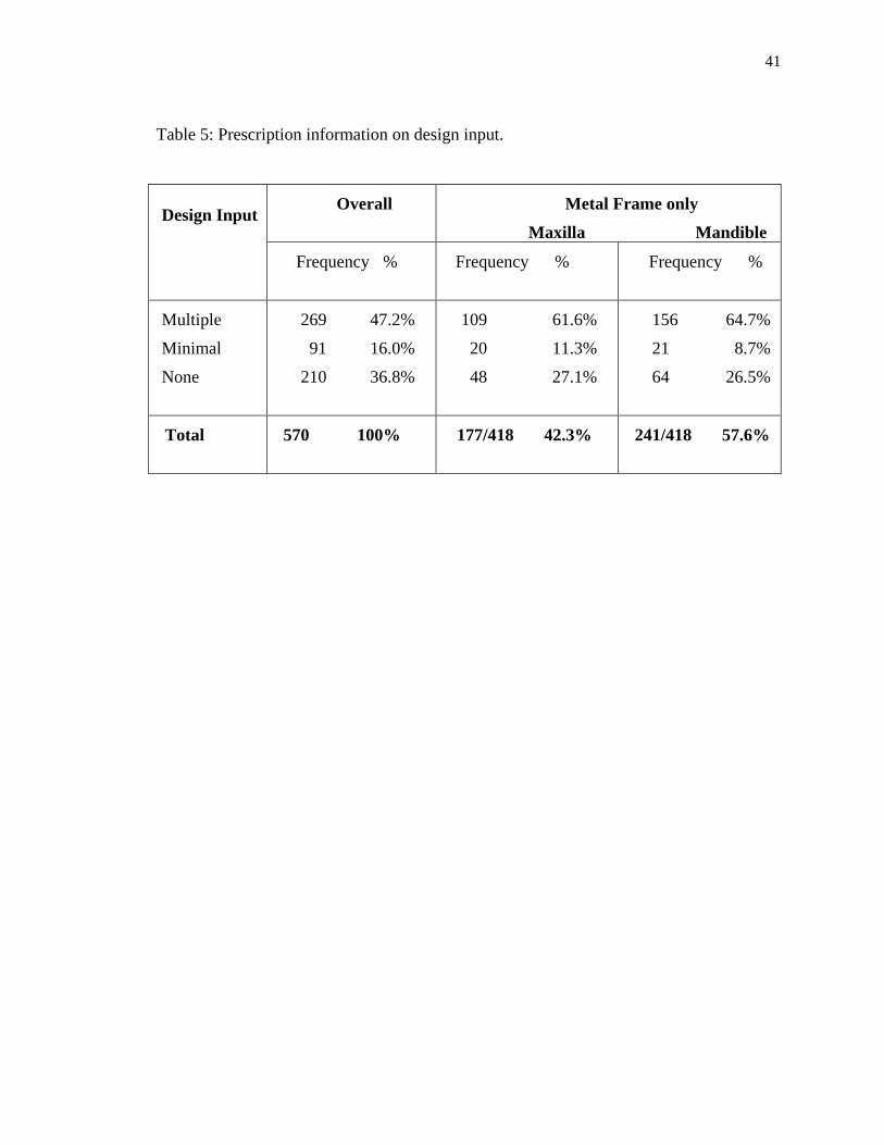
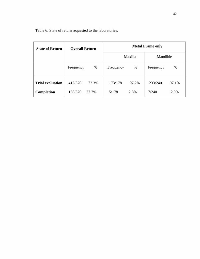
Information regarding the Dentist prescription is presented in Tables 5 and 6.
47% of prescriptions were classified as having ‘multiple design input’, 16% as having
‘minimal design input,’ and 37% as having ‘no design input’. 72% of cases requested
return for trial evaluation prior to completion. 97% of metal frameworks requested return
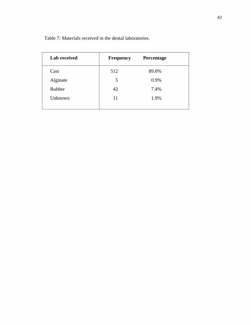
for trial evaluation. According to the images; 512 casts and 58 impressions were
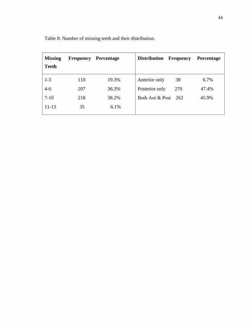
received at the lab (Table 7). The mean number of teeth replaced in this sample was 6. It
was further noted that 47.4% of cases replaced posterior teeth only, 45.9% replaced both
anterior and posterior, and 6.7% replaced anterior teeth only (Table 8).
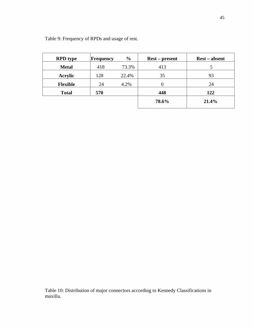
Data on RPD framework details are presented in Tables 9-14. 73.3% of the cases
were metal, 22.5% acrylic, and 4.2% flexible. According to the criteria discussed, 84%
of cases were considered to have rests. Overall the horseshoe (72.5%) was the most
frequently used maxillary major connector while the lingual plate (59.4%) was the most
frequent mandibular major connector. Tables 12, 13 and 14 show the frequency of
different major connectors among the different RPD material types. When analyzed
separately nearly all (95.3%) non-metal maxillary major connectors were a horseshoe
design. Likewise, nearly all (91.1%) of non-metal mandibular major connectors were a
lingual plate.
Some additional findings of interest included 22 cases using attachments
within some portion of the design or framework. One frame was fabricated from yellow
gold. Four metal frameworks could not be classified based on the definitions used in this
study. These cases presented as large Kennedy Class IV arches with remaining molars

40
only. These frameworks consisted of clasp assemblies over the molars (generally
bilateral) and lengthy cross-arch meshwork extending along the lengthy edentulous span.
7 unilateral prostheses (Nesbit type) were also found. Four of the unilateral prostheses
were made from flexible materials and three were metal.
41
Table 5: Prescription information on design input. Design Input
Overall Metal Frame only Maxilla Mandible
Frequency %
Frequency %
Frequency %
Multiple
Minimal
None
269 47.2%
91 16.0%
210 36.8%
109 61.6%
20 11.3%
48 27.1%
156 64.7%
21 8.7%
64 26.5%
Total 570 100% 177/418 42.3% 241/418 57.6%
42
Table 6: State of return requested to the laboratories.
State of Return
Overall Return Metal Frame only
Maxilla Mandible
Frequency %
Frequency %
Frequency %
Trial evaluation Completion
412/570 72.3% 158/570 27.7%
173/178 97.2% 5/178 2.8%
233/240 97.1% 7/240 2.9%
43
Table 7: Materials received in the dental laboratories.
Lab received Frequency Percentage
Cast
Alginate
Rubber
Unknown
512 89.8%
5 0.9%
42 7.4%
11 1.9%
44
Table 8: Number of missing teeth and their distribution.
Missing Frequency Percentage Teeth
Distribution Frequency Percentage
1-3 110 19.3%
4-6 207 36.3%
7-10 218 38.2%
11-13 35 6.1%
Anterior only 38 6.7%
Posterior only 270 47.4%
Both Ant & Post 262 45.9%
45
Table 9: Frequency of RPDs and usage of rest.
Table 10: Distribution of major connectors according to Kennedy Classifications in maxilla.
RPD type Frequency % Rest – present Rest – absent
Metal 418 73.3% 413 5
Acrylic 128 22.4% 35 93
Flexible 24 4.2% 0 24
Total 570 448 122
78.6% 21.4%
46
Table 11: Distribution of major connectors according to Kennedy Classifications in mandible.
Maxilla
Class I Class II Class III Class IV Frequency Percentage
AP strap
Palatal strap
Palatal plate
Horseshoe
AP bar
Unilateral
Others
8 8 8 3 27 9.5%
3 13 11 0 27 9.5%
9 3 2 2 16 5.6%
54 39 97 16 206 72.5%
0 1 1 0 2 0.7%
0 0 4 0 4 1.4%
1 0 0 1 2 0.7%
Total
75 64 123 22 284 100%
47
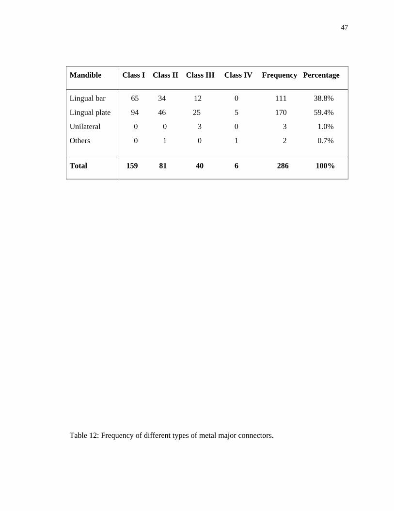
Table 12: Frequency of different types of metal major connectors.
Mandible Class I Class II Class III Class IV Frequency Percentage
Lingual bar
Lingual plate
Unilateral
Others
65 34 12 0 111 38.8%
94 46 25 5 170 59.4%
0 0 3 0 3 1.0%
0 1 0 1 2 0.7%
Total 159 81 40 6 286 100%
48
Maxilla Total Mandible Total
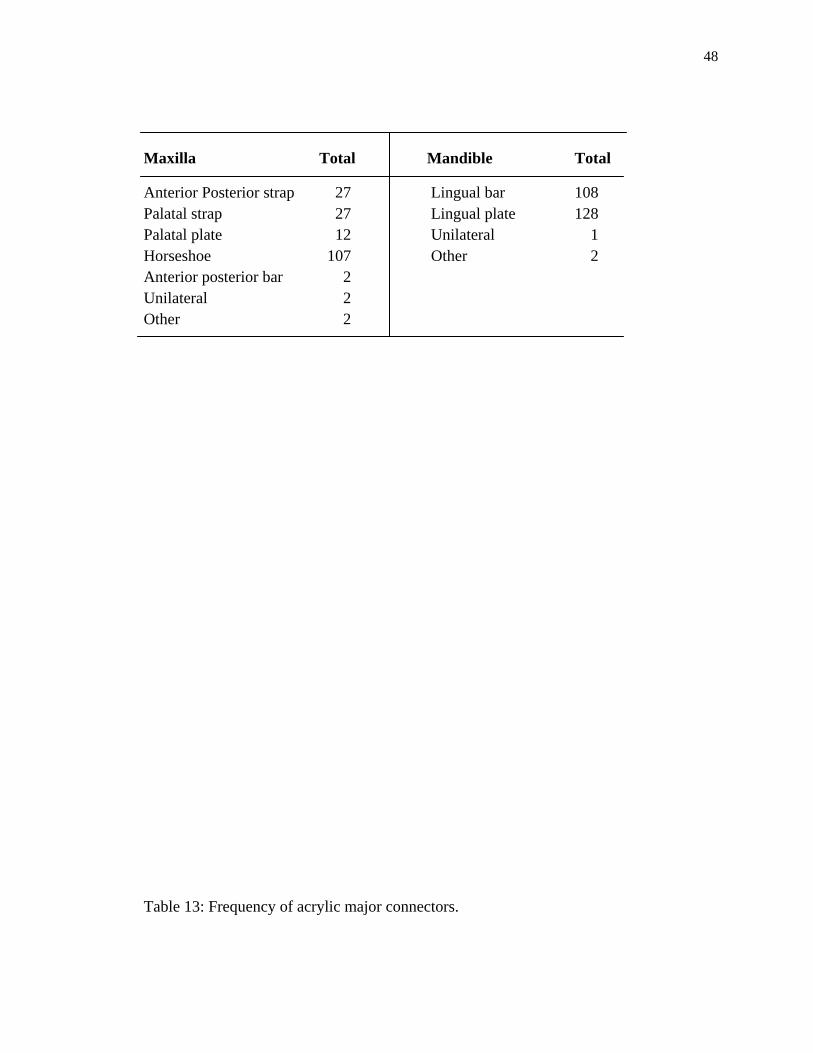
Anterior Posterior strap 27 Lingual bar 108 Palatal strap 27 Lingual plate 128 Palatal plate 12 Unilateral 1 Horseshoe 107 Other 2 Anterior posterior bar 2 Unilateral 2 Other 2 Table 13: Frequency of acrylic major connectors.
49
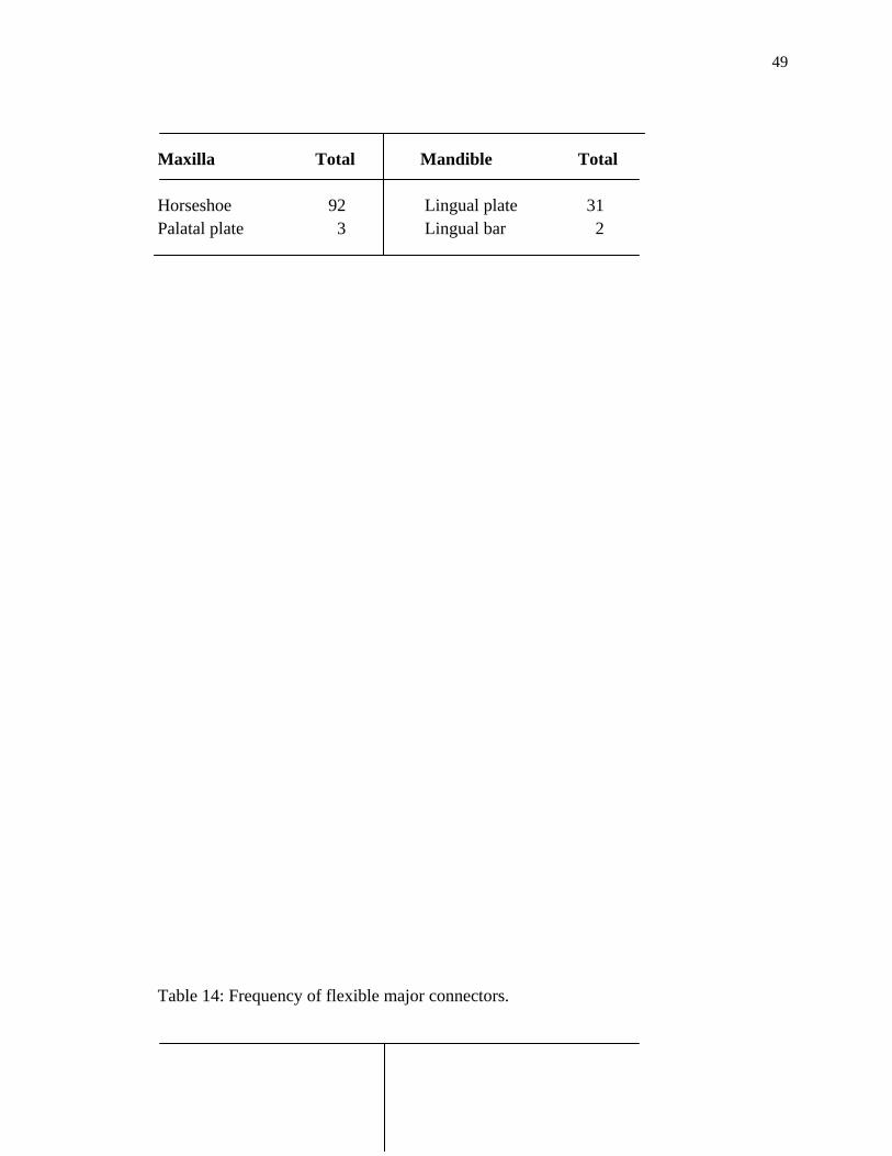
Maxilla Total Mandible Total Horseshoe 92 Lingual plate 31 Palatal plate 3 Lingual bar 2 Table 14: Frequency of flexible major connectors.

50
Maxilla Total Mandible Total Horseshoe 10 Lingual plate 10 Unilateral 2 Unilateral 2
CHAPTER V

51
Discussion
The laboratories were asked to provide consecutive RPDs until a minimum of 500
matched photographs were collected. Despite attempts at calibrating the sequence and
52
format of the photographs numerous images were found that did not follow the originally
designed protocol. In particular, there were 362 unmatched photographs. Dental
laboratories are busy places and it is not surprising that many cases were not
photographed twice. One of the laboratories which provided most of the samples also had
the most unmatched images. The most likely missing images were second photographs.
In addition, it was strongly suspected that the written methods were not consistently
followed. It was asked that the contents of the incoming case boxes be photographed as
received. However, in several instances it was found that the lab poured up impressions
to create a cast prior to taking the first photograph. It was also noted that the lab would
often take the supposed ‘incoming’ photograph after completion of the case. Therefore,
the first image in essence became the definitive cast without the framework in the image.
This was discovered by the time stamp on the images as well as by the fact that many
images had laboratory design drawings on the casts for the ‘incoming’ images. It was
also suspicious that one of the dental laboratories only had images of metal frameworks.
It is known that this lab does, on occasion create non-metallic frameworks. To have none
of these within a three month time frame may be due to their decision to not include
these. When the principal investigator inquired of the lab which provided only metal
RPDs, it was admitted that only 3-4 non-metal RPDs were missed. The reason for these
omissions and changes is suspected to be inadequate calibration and laboratory time
constraints. If the explanation to the photographers had been improved they may have
had a better understanding of what the purpose of the distinctly different images was. If a
better explanation had been given and more time had been spent on preparation of the
laboratory they may have realized that simply taking an image of the definitive cast
53
without framework was not sufficient to gather the planned data. One solution would
have been to have the research investigators collect the images themselves. This was not
considered logistically possible when using multiple laboratories. It was originally
discussed that a single image would have been sufficient to collect certain data. This
would have been easier for the laboratory but would have not been an attempt to gather
information on the desired questions. With this in mind it is understood that the 347
accepted but unmatched images hold a great deal of reliable and usable information.
They were not included in this analysis since they did not meet the original inclusion
criteria but will be included in subsequent analyses. So while much of the data is usable it
is admitted here that any data collected relying on the original protocol of two
specifically timed images is not reliable and therefore should not be used for any
definitive conclusions. This would include information on design input and what
materials the laboratory received.
Since the data was collected at the laboratory only cases already in treatment were
tabulated. Therefore, these numbers do not reflect the prevalence of RPDs or tooth
distribution within the general population. However, by including all RPD types a broad
picture of the partially edentulous population seeking this type of care was analyzed.
Despite the previously mentioned methodological issues it is believed that very few RPD
cases were not photographed at least once during this time frame. Since the major RPD
producing dental laboratories in the Milwaukee area were all included. It is believed that
the totals presented here are a reliable estimate of the metropolitan areas RPD fabrication
incidence over the 4 month time frame of the data collection. Overall the encountered
54
limitations proved minor due to the large number of cases collected and the simplistic
nature of the critical data analyzed.
For this study, a removable partial denture was defined as any prosthesis that
replaces a tooth or teeth in a partially dentate arch; it can be removed from the mouth and
replaced at will.83 This was a much more strict use of the definition than previous
analyses of RPD incidence which appear to have only collected data on metal framework
RPDs. Despite this, Kennedy Class I cases continued to be the most common RPD
configuration.24,40-42 Class I was followed in order by Class III, Class II and Class IV. A
nearly identical number of maxillary (284) and mandibular (286) cases were found.
While it had previously been speculated that a greater number of mandibular RPDs was
being fabricated due to difficulties with mandibular complete denture use, this was not
found to be the case in this sample.42 With dental implant use now a mainstream
treatment alternative the fear of denture adaption may be reduced for those with the
economic means to afford this alternative. In addition, improved knowledge regarding
RPD success and treatment of dental disease may have encouraged maintenance of more
maxillary teeth. It is interesting to compare the incidence of the various classifications to
that discovered in other decades.24, 42 The incidence of Class I RPDs found by Curtis et
al. at a single regional dental laboratory in northern California in 1986 (40%) is nearly
identical to that found here (41%). In contrast to that consistency is the increased
frequency of Class III RPDs that was found. Curtis et al. found only 18% of the total
cases to be Class III while 28.6% were in the current study. This increase was offset by a
decrease in both Class II and Class IV cases in our data. Other major differences
between the incidences of various classifications can be found when looking at specific
55
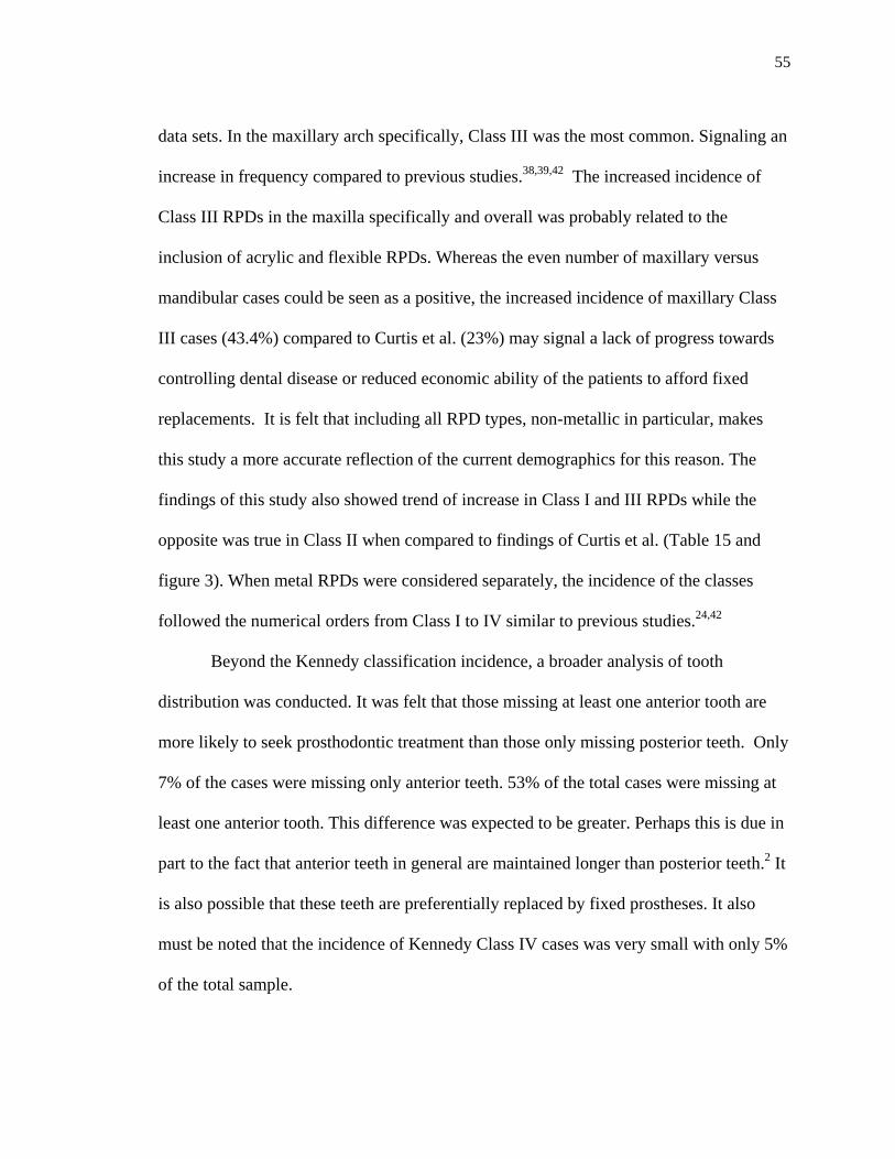
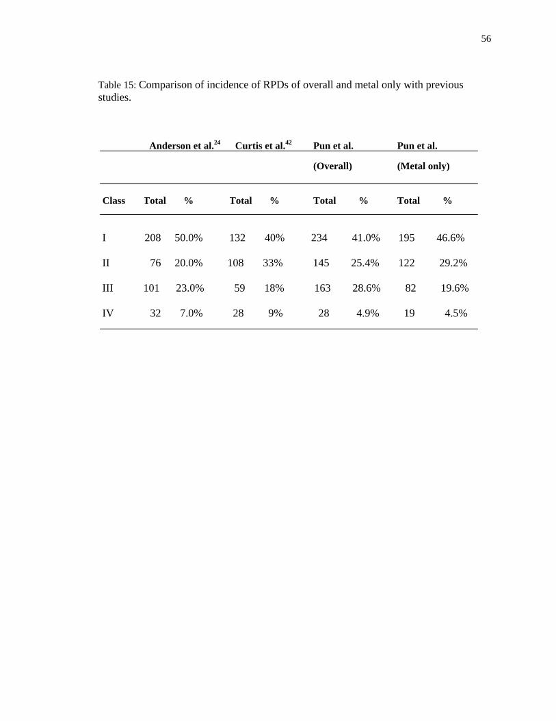
data sets. In the maxillary arch specifically, Class III was the most common. Signaling an
increase in frequency compared to previous studies.38,39,42 The increased incidence of
Class III RPDs in the maxilla specifically and overall was probably related to the
inclusion of acrylic and flexible RPDs. Whereas the even number of maxillary versus
mandibular cases could be seen as a positive, the increased incidence of maxillary Class
III cases (43.4%) compared to Curtis et al. (23%) may signal a lack of progress towards
controlling dental disease or reduced economic ability of the patients to afford fixed
replacements. It is felt that including all RPD types, non-metallic in particular, makes
this study a more accurate reflection of the current demographics for this reason. The
findings of this study also showed trend of increase in Class I and III RPDs while the
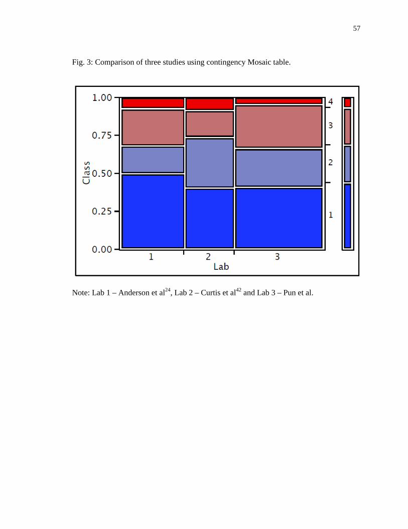
opposite was true in Class II when compared to findings of Curtis et al. (Table 15 and
figure 3). When metal RPDs were considered separately, the incidence of the classes
followed the numerical orders from Class I to IV similar to previous studies.24,42
Beyond the Kennedy classification incidence, a broader analysis of tooth
distribution was conducted. It was felt that those missing at least one anterior tooth are
more likely to seek prosthodontic treatment than those only missing posterior teeth. Only
7% of the cases were missing only anterior teeth. 53% of the total cases were missing at
least one anterior tooth. This difference was expected to be greater. Perhaps this is due in
part to the fact that anterior teeth in general are maintained longer than posterior teeth.2 It
is also possible that these teeth are preferentially replaced by fixed prostheses. It also
must be noted that the incidence of Kennedy Class IV cases was very small with only 5%
of the total sample.
56
Table 15: Comparison of incidence of RPDs of overall and metal only with previous studies.
Anderson et al.24 Curtis et al.42 Pun et al. Pun et al.
(Overall) (Metal only)
Class Total % Total % Total % Total %
I 208 50.0% 132 40% 234 41.0% 195 46.6%
II 76 20.0% 108 33% 145 25.4% 122 29.2%
III 101 23.0% 59 18% 163 28.6% 82 19.6%
IV 32 7.0% 28 9% 28 4.9% 19 4.5%
57
Fig. 3: Comparison of three studies using contingency Mosaic table.
Note: Lab 1 – Anderson et al24, Lab 2 – Curtis et al42 and Lab 3 – Pun et al.
58
This study revealed that 73% of all RPDs were fabricated with cast metal
frameworks. The remaining RPDs (27%) were fabricated with non-metal major
connectors. These were made from either acrylic or flexible materials. The incidence of
non-metal RPDs found in our study is much higher than the 5% found by Öwall and
Taylor using similar inclusion/exclusion criteria in 1989.43 However, this number is
much less than that reported by several recent international studies.46,48, 49 The high
incidence of non-metallic RPDs found in this study was suspected. The laboratory which
provided 45% of the total sample fabricates RPDs for Marquette University School of
Dentistry. Only metal frameworks are fabricated by the dental school except in rare
instances. It can be speculated that one laboratory who admitted not taking non-metal
RPDs could have reduced the frequency of acrylic or flexible RPDs.
Despite the lack of comparative studies between metallic and non-metallic
framework RPDs several major shortcomings of non-metallic RPDs exist. This material
is often chosen for economic reasons but it is reported that it is poor economy in the long
run to choose the cheaper construction.55-57 This is primarily due to high rates of non-
metallic framework fracture but may also be due to the reported soft tissue damage and
potential for other periodontal complications.53,54,55-57 The standard design feature meant
to minimize soft tissue damage from RPDs is the rest. Despite our broad definition of a
rest only 78.6% of cases used at least one rest. Therefore, 1 in 5 cases were completely
tissue borne prostheses. When one looks at only non-metallic RPDs a mere 23% were
considered to have at least one rest. Very often acrylic RPDs were given credit for a rest
due to an embrasure style wire clasp extending across the occlusal surfaces of posterior
teeth. Even though these were credited as a rest, 77% of non-metallic cases were
59
considered completely tissue borne. This evidence lends some insight to the frequent
observation of periodontal tissue damage with these prostheses.
With the amount of advertising promoting flexible RPD frameworks it was
somewhat surprising to find only 4% of these within the total sample. None of these cases
were considered to have a rest of any kind. Perhaps this was due to the manufacturer
claims of superior stability and retention of frameworks made from these materials. The
authors were unable to locate any peer-reviewed or company sponsored research in
regards to the clinical performance of these materials.
The majority of maxillary major connectors in this study (72.5%) fell under the
definition of a horseshoe. A mere 27 (9.5%) palatal straps were found. In North America,
one previous study found 56% of maxillary major connectors to be horseshoes while
another did not find any.42, 43 It is suspected that one study used the term anterior strap to
describe a horseshoe type connector.42 If this is in fact the case they found a mere 12.3%
of their sample of this type. This demonstrates the necessity in clearly defining each
major connector type as well as potential regional differences in RPD design. It is felt
that the definitions used in this study were clear and easy to use. This statement is
supported by the excellent accuracy and repeatability achieved by the calibrated
instructors. There remains room for debate regarding the definitions. Our selected
definition for a lingual bar was a mandibular major connector located lingual to the dental
arch with visible gingival tissue lingual to any anterior tooth. This meant that a case that
plated the canines but not the incisors was considered a lingual plate. Essentially, a
lingual plate was required to cover the gingiva lingual to all remaining anterior teeth.
Despite this, a majority of lingual plate major connectors was still found (59.4%). This is
60
much greater than in other research.42,43 Part of the reason for the large majority of
horseshoe major connectors and lingual plate major connectors was likely due to the
material selection. Non-metallic RPDs generally require greater dimensions for adequate
strength. Therefore, a bulky wide version of the horseshoe and lingual plate were selected
nearly all of the time when non-metallic major connectors were used.
After attempting several different methods of collecting prescription information
given by the dentist, a method similar to that used by Allen and Lynch was used.75 Other
methods left too much room for interpretation of drawings, writing, or the various
prescription forms themselves. The accuracy and repeatability of our three choices was
positive. By intentionally giving the dentist much credit for very little information we
were still able to demonstrate how little input the majority of dentists have upon the
prosthesis they are supposedly in charge of creating. 53% of the total prescriptions either
did not provide any design information or prescribed one of the four components deemed
necessary. It must also be noted that the cases from the lab servicing the dental school
had a strong bias towards ‘multiple design input.’ This is because of the quality control
procedures in place at the school. No case can be sent to the laboratory without a
complete design, complete prescription form, and completed design cast with drawing.
Considering that many of the cases from this lab were dental school cases the bias
towards ‘multiple input’ was significant. Despite these limitations, the numbers and
method still demonstrated a significant lack of design input by the majority of dentists.
Many studies both old and new have highlighted this lack of prescribing dentist input as
well as the varying RPD design philosophies.70

61
CHAPTER VI
SUMMARY AND CONCLUSIONS
62
This study evaluated 570 pairs of digital images taken at 5 dental laboratories
located in the state of Wisconsin, USA. Images were made of incoming and then out-
going RPD cases only. The Kennedy classification according to Applegate’s
modification, major connector type, as well as other information was identified. The
following conclusions were drawn from the study:
1) Kennedy Class I RPD is still the most commonly fabricated RPD.
2) Class I distal extension RPD was most commonly fabricated in the mandible (55.6%) while it was Class III RPD in the Maxillary arch (43.3%).
3) 73% of RPDs in this sample had a metal framework, while 27% utilized non-metallic frameworks.
4) Dentist prescribed multiple design input in 47% of the RPDs while 53% of the time no design information or only one of the 4 components was prescribed.
5) At least 20% of RPDs from this sample did not provide any rests.
6) The most common major connectors were the horseshoe for the maxilla and the
lingual plate for the mandible.
63
BIBLIOGRAPHY
1. Douglas CW, Watson AJ: Future needs for fixed and removable partial dentures in the United States. J Prosthet Dent 2002;87:9-14
2. Marcus SE, Drury TF, Brown LJ, et al: Tooth retention and tooth loss in the
permanent dentition of adults: United States, 1988-1991. J Dent Res 1996;75(Spec Iss):684-695
3. Hillyer E. The retention of partial dentures. Dent Cosmos 1915;57:1019-1022
4. Ettinger RL, Beck JD, Jakobsen J: Removable prosthodontic treatment needs: a survey. J Prosthet Dent 1984;51:419-427
5. Hunt RJ, Srisilapanan P, Beck JD: Denture-related problems and prosthodontic
treatment needs in the elderly. Gerodontics 1985;1:226-230 6. Redford M, Drury TF, Kingman A, et al: Denture use and the technical quality of
dental prostheses among persons 18-74 years of age: United States, 1988-1991. J Dent Res 1996;75(Spec Iss):714-725
7. Harvey WL, Hoffman WM. Ten-year study of trends in removable prosthodontic
service. J Prosthet Dent 1989;62:644-6 8. Joshi A, Douglas CW, Feldman H, et al: Consequences of success: do more teeth translate into more disease and utilization? J Public Health Dent 1996;56:190- 197 9. Manski RJ, Moeller JF, Maas WR: Dental services: an analysis of utilization over 20 years. J Am Dent Assoc 2001;132:655-664 10. Burt BA, Ismail AI, Morrison EC, et al: Risk factors for tooth loss over a 28-year period. J Dent Res 1990;69:1126-1130 11. Eklund SA, Burt BA. Risk factors for total tooth loss in the United States; longitudinal analysis of national data. J Public Health Dent 1994;54:5-14 12. Dolan TA, Gilbert GH, Duncan RP, et al: Risk indicators of edentulism, partial tooth loss, and prosthetic status among black and white middle-aged older adults. Comm Dent Oral Epidemiol 2001;29:329-340 13. Janus CE, Hunt RJ, Unger JW: Survey of prosthodontic service provided by general dentists in Virginia. J Prosthet Dent 2007;97:287-291
64
14. Bailyn M: Tissue support in partial denture construction. Dent Cosmos 1928;70:988- 997
15. Kennedy E. Partial denture construction Brooklyn: Dental Items of Interest
Publishing Co. 1928;3-8 16. Cummer W: Partial Denture Service, in Anthony LP: American Textbook of
Prosthetic Dentistry. Philadelphia, PA, Lea & Febiger, 1942, pp. 339-452 17. Skinner C: A classification of removable partial dentures based upon the principles
of anatomy and physiology. J Prosthet Dent 1959;9:240-246 18. Applegate DC. The rationale of partial denture choice. J Prosthet Dent
1960;10:891-907 19. Miller EL: Systems for classifying partially dentulous arches. J Prosthet Dent
970;24:25-40 20. McGivney GP, Carr AB: McCracken’s removable partial prosthodontics (edn 10). St. Louis, MO, 2000, p 19-23 21. Phoenix RD, Cagna DR, DeFreest CF: Stewart’s clinical removable partial prosthodontics. (edn 4) Hanover Park, IL, Quintessence, 2008, p 8-17 22. Academy of Prosthodontics. Principles, concepts, and practices in prosthodontics. J
Prosthet Dent 1995;73:73-94 23. McGarry TJ, Nimmo A, Skiba JF, et al: Classification system for partial
edentulism. J Prosthodont 2002;11:181-93 24. Anderson JN, Lammie GA. A clinical survey of partial dentures. Br Dent J
1952;92:59-67 25. Anderson JN, Bates JF. The cobalt-chromium partial denture: A clinical survey. Br
Dent J 1959;107:57-62 26. Tomlin HR, Osborne J. Cobalt-chromium partial dentures: A clinical survey. Br
Dent J 1961;110:307-10 27. Basker RM, Davenport JC. A survey of partial denture design in general dental
practice. J Oral Rehabil 1978;5:215-222 28. Basker RM, Harrison A, Davenport JC, et al: Partial denture design in general
dental practice – 10 years on. Br Dent J 1988:165:245
65
29. Derry A, Bertram V. A clinical survey of removable partial dentures after 2 years usage. Acta Odontol Scand 1970;28:581-98.
30. Axéll T, Öwall B. Prevalences of removable dentures and edentulousness in an adult
Swedish population. Swed Dent J 1979;3:129-137 31. Björn A, Öwall B. Partial edentulism and its prosthetic treatment: A frequency
study within a Swedish population. Swed Dent J 1979;3:315-25 32. Laine P, Murtomaa H. Frequency and suppliers of removable dentures in Finland in
1983. Community Dent Oral Epidemiol 1985;13:47-50 33. Tervonen T, Bergenholtz A, Nordling H, Ainamo A, Ainamo J. Edentulousness and
the use of removable dentures among people 25, 35, 50 and 65 years old in Ostrobothnia, Finland. Proc Finn Dent Soc 1985;81:264-70
34. Bergman B, Hugoson A, Olsson C. Periodontal and prosthetic conditions in patients
treated with removable partial dentures and artificial crowns. A longitudinal two-year study. Acta Odont Scand 1971;29:621-38
35. Yeung ALP, Lo ECM, Clark RKF et al: Usage and status of cobalt-chromium
removable partial dentures 5-6 years after placement. J Oral Rehab 2002;29:127-32 36. Wetherell JD, Smales RJ. Partial denture failures: A long term clinical survey. J
Dent 1980;8:333-40 37. Keyf F. Frequency of the various classes of removable partial dentures and
selection of major connector and direct/indirect retainers. Turk J Med Sci 2001;31:445-9
38. Sadig WM, Idowu AT. Removable partial denture design: A study of a selected
population in Saudi Arabia. J Contemp Dent Pract 2002;3:40-53 39. Al-Dwairi ZN. Partial edentulism and removable denture construction: a frequency study in Jordanians. Eur J Prosthodont Restor Dent 2006;14:13-7 40. Schwalm CA, Smith DE, Erickson JD. A clinical study of patients 1 to 2 years after
placement of removable partial dentures. J Prosthet Dent 1977;38:380-91 41. Chandler JA, Brudvik JS. Clinical evaluation of patients eight t nine years after
placement of removable partial dentures. J Prosthet Dent 1984;51:736-43 42. Curtis DA, Curtis TA, Wagnild GW, Finzen FC. Incidence of various classes of
removable partial dentures. J Prosthet Dent 1992;67:664-7
66
43. Öwall BE, Taylor RL. A survey of dentitions and removable partial dentures constructed for patients in North America. J Prosthet Dent 1989;61:465-70
44. Walmsley AD: Acrylic partial dentures. Dent update 2003;30:424-429
45. Allen PF, Jepson NJ, Doughty J et al: Attitudes and practice in the provision of removable partial dentures. Br Dent J 2008;204:1-5
46. Lewandowska A, Speichowicz E, Öwall BE. Removable partial denture treatment
in Poland. Quintessence Int 1989;20:353-8 47. Thean HP, Payne JA, Jeganathan S. The use of removable partial dentures amongst
private dental practitioners in Singapore. Singapore Dent J 1996;21:26-30 48. Radhi A, Lynch CD, Hanningan A. Quality of written communication and master impressions for fabrication of removable partial prostheses in the Kingdom of
Bahrain. J Oral Rehabil 2007;34:153-7 49. Schwarz W, Barsby M. A survey of the practice of partial denture prosthetics in the
United Kingdom. J Dent 1980;8:95-101 50. Lynch CD, Allen PF. The teaching of removable partial dentures in Ireland and the
United Kingdom. Br Dent J 2007;203:1-5 51. McCartney JW, Fiks S. The all-acrylic resin mandibular removable partial denture:
design considerations. J Prosthet Dent 1997;77:638 52. Smith RA, Rymarz FP. Cast clasp transitional removable partial dentures. J Prosthet Dent 1969;22:381-5 53. Zlatarić DK, Čelebić A, Valentić-Peruzović M. The Effect of Removable Partial
dentures on Periodontal Health of Abutment and Non-Abutment Teeth. J Periodontol 2002;73:137-44
54. Tuominen R, Ranta K, Paunio I. Wearing of removable partial dentures in relation
to periodontal pockets. J Oral Rehab 1989;16:119-26 55. Carlsson GE, Hedegård B, Koivumaa KK. Studies in partial dental prosthesis III. A
longitudinal study of mandibular partial dentures with double extension saddles. Acta Odontol Scand. 1962 Jun;20:95-119
56. Carlsson GE, Hedegård B, Koivumaa KK Studies in partial dental prosthesis. IV.
Final results of a 4-year longitudinal investigation of dentogingivally supported partial dentures. Acta Odont Scand 1965;23:443-72
67
57. Carlsson GE, Hedegård B, Koivumaa KK. Late results of treatment with partial dentures. An investigation by questionnaire and clinical examination 13 years after treatment. J Oral Rehab 1976;3:267-72
58. Bergman B, Hugoson A, Olsson CO. Caries, periodontal and prosthetic findings in patients
with removable partial dentures: A ten-year longitudinal study. J Prosthet Dent 1982;48:506-14
59. Bergman B, Hugoson A, Olsson CO. A 25 year longitudinal study of patients treated
with removable partial dentures. J Oral Rehab 1995;22:595-9 60. Bissada NF, Ibrahim S, Brasoum WM. Gingival response to various types of removable
partial dentures. J Periodontol 1974;45:651-9 61. Stern MA, Brudvik JS, Frank RP. Clinical evaluation of removable partial denture rest seat adaptation. J Prosthet Dent 1985;53:658-62. 62. Dunham D, Brudvik JS, Morris WJ, et al: A clinical investigation of fit of removable partial dental prosthesis clasp assemblies. J Prosthet Dent 2006;95:323-326 63. Firtell DN, Muncheryan AM, Green AJ: Laboratory accuracy in casting removable partial denture frameworks. J Prosthet Dent 1985;54:856-862 64. Brudvik JS, Reimers D: The tooth-removable partial denture interface. J Prosthet Dent1992;68:924-927 65. Keltjens HM, Mulder J, Kayser AF, et al: Fit of direct retainers in removable partial dentures after 8 years of use. J Oral Rehab 1997;24:138-142 66. Akers PE: A new and simplified method of partial denture prosthesis. J Am Dent
Assoc 1925;12:711-717 67. van Minden F: Fitting, placing, and maintenance of cast partial dentures. J Amer Dent Assoc 1931;18:442-449 68. Lynch CD, Allen PF. Why do dentists struggle with removable partial denture
design? An assessment of financial and educational issues. Br Dent J 2006;200:277-81
69. Taylor TD, Aquilino SA, Matthews AC, Logan NS. Prosthodontic survey. Part II:
Removable prosthodontic curriculum survey. J Prosthet Dent 1984;52:747-749 70. Sykora O, Calikkocaoglu S. Maxillary removable partial denture designs by
commercial dental laboratories. J Prosthet Dent 1970;23:633-40
68
71. McCracken WI. Survey of partial denture designs by commercial dental laboratories. J Prosthet Dent 1962;12:1089-1110
72. Frantz WR. Variability in dentists’ designs of a removable maxillary partial
denture. J Prosthet Dent 1973;29:172-82 73. Frantz WR. Variations in a removable maxillary partial denture design by dentists. J
Prosthet Dent 1975;34:625-033 74. Schwarz W, Barsby M. Design of partial dentures in dental practice. J Dent
1978;6:166-170 75. Lynch D, Allen PF. Quality of written prescriptions and master impressions for
fixed and removable prosthodontics: a comparative study. Br Dent J 2005;198:17-21
76. Taylor TD, Matthews AC, Aquilino SA, Logan NS. Prosthodontic survey. Part I:
Removable prosthodontic laboratory survey. J Prosthet Dent 1984;52:598-601 77. Cotmore JM, Mingledorf EB Pomerantz JM et al: Removable partial denture
survey: Clinical practice today. J Prosthet Dent 1983;49:321-7 78. Hardy F, Stuart LM. A critique of materials submitted by dentists to dental
laboratories for the fabrication of removable partial dentures. Quintessence Dent Technol 1983;7:93-5
79. Burns DR, Ward JE, Nance GL. Removable partial denture design and fabrication
survey of the prosthodontic specialist. J Prosthet Dent 1989;62:303-307 80. Hummel SK, Wilson MA, Marker VA, Nunn ME. Quality of removable partial
dentures worn by the adult U.S. population. J Prosthet Dent 2002;88:37-43 81. Frank RP, Brudvik JS, Leroux B, Milgrom P, Hawkins N. Relationship between
the standards of removable partial denture construction, clinical acceptability, and patient satisfaction. J Prosthet Dent 2000;83:521-527
82. Frank RP, Milgrom P, Leroux BG, Hawkins N. Treatment outcomes with
mandibular removable partial dentures: A population-based study of patient satisfaction. J Prosthet Dent 1998;80:36-45
83. The Academy of Prosthodontics. Glossary of Prosthodontic Terms. J Prosthet Dent
2005;94:68 84. Al-Johany SS, Andres C. ICK classification system for partially edentulous arches.
J Prosthodont. 2008;17:502-7.


![Denture Delivery [Removable Prosthodontics Seminar @AmCoFam]](https://img.dokumen.tips/doc/110x75/543eb8c3b1af9f700b8b47f1/denture-delivery-removable-prosthodontics-seminar-amcofam.jpg)