Embed Size (px)
DESCRIPTION
IN VIVO STUDIES IN ANIMALS : PRECLINICAL PHARMACOLOGY. An essential step between molecular and human studies for understanding the pathophysiological mechanisms involved in human diseases and the mechanisms of action of pharmacological agents. DEVELOPMENT OF EXPERIMENTAL MODELS - PowerPoint PPT Presentation
Citation preview
IN VIVO STUDIES IN ANIMALS :PRECLINICAL PHARMACOLOGY
An essential step between molecular and human studies for
understanding the pathophysiological mechanisms involved in human
diseases and the mechanisms of action of pharmacological agents.
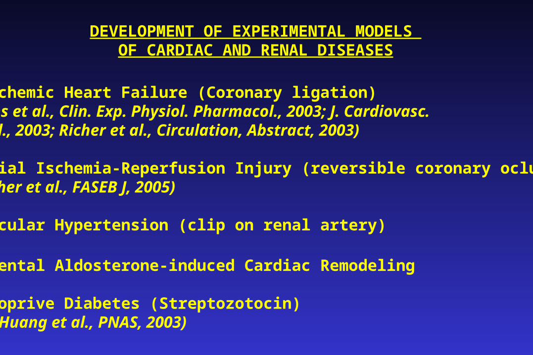
DEVELOPMENT OF EXPERIMENTAL MODELS OF CARDIAC AND RENAL DISEASES
•Post-Ischemic Heart Failure (Coronary ligation) (Pons et al., Clin. Exp. Physiol. Pharmacol., 2003; J. Cardiovasc. Pharmacol., 2003; Richer et al., Circulation, Abstract, 2003)
•Myocardial Ischemia-Reperfusion Injury (reversible coronary oclusion)(Richer et al., FASEB J, 2005)
•Renovascular Hypertension (clip on renal artery)
•Experimental Aldosterone-induced Cardiac Remodeling
•Insulinoprive Diabetes (Streptozotocin)(Huang et al., PNAS, 2003)
ANIMAUX TRANSGENIQUES ET MALADIES CARDIOVASCULAIRES
Ce sont des modèles avec une ou plusieurs modifications ciblées d’une hormone ou d’un second messager impliquésdans la régulation cardiovasculaire
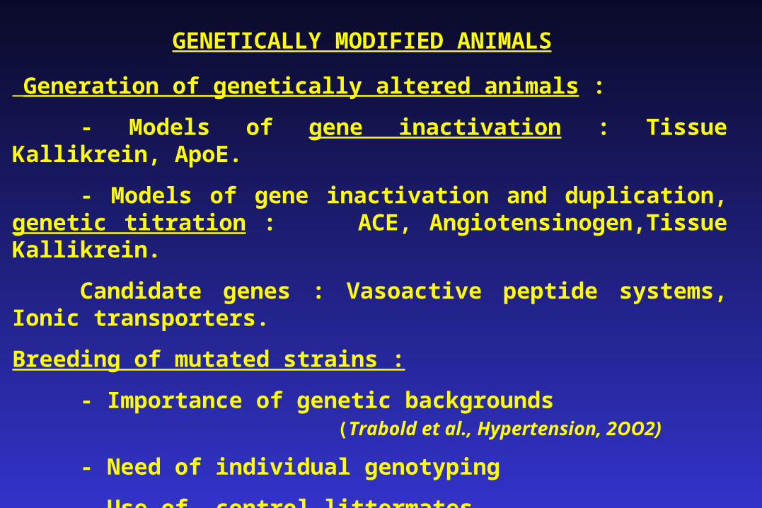
GENETICALLY MODIFIED ANIMALS
Generation of genetically altered animals :
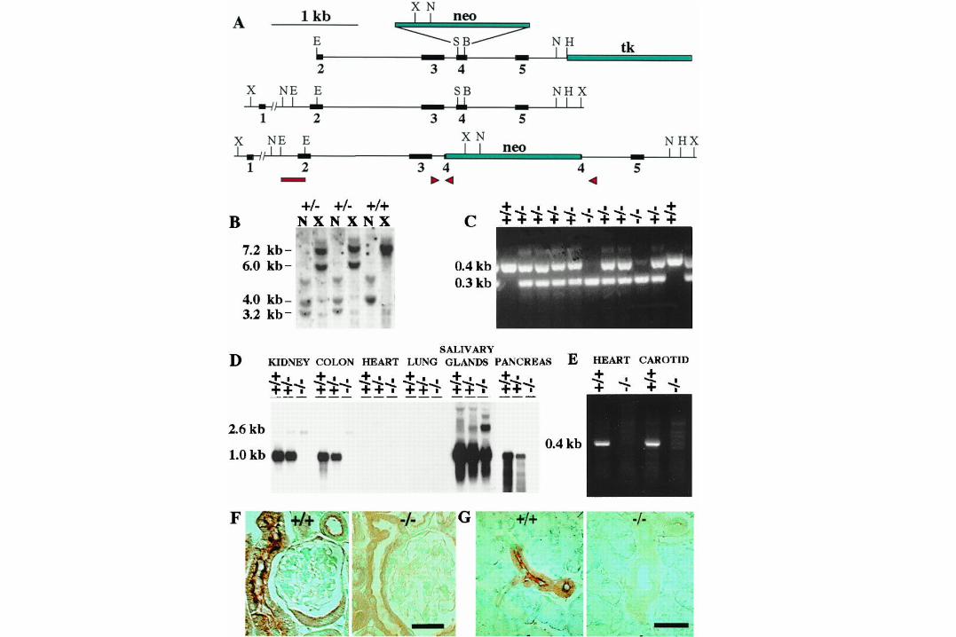
- Models of gene inactivation : Tissue Kallikrein, ApoE.
- Models of gene inactivation and duplication, genetic titration :ACE, Angiotensinogen,Tissue Kallikrein.
Candidate genes : Vasoactive peptide systems, Ionic transporters.
Breeding of mutated strains :
- Importance of genetic backgrounds (Trabold et al., Hypertension, 2OO2)
- Need of individual genotyping
- Use of control littermates
- Role of environmental factors : housing, diet.
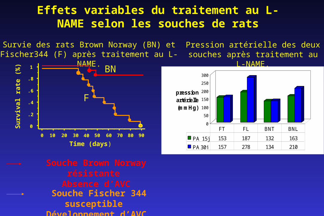
Survie des rats Brown Norway (BN) et Fischer344 (F) après traitement au L-NAME.
Souche Fischer 344 susceptible
Développement d’AVC
Effets variables du traitement au L-NAME selon les souches de rats
Souche Brown Norway résistante
Absence d’AVC
BN
F
0 10 20 30 40 50 60 70 80 90
Time (days)
0
.2
.4
.6
.8
1
Su
rviv
al r
ate
(%)
Pression artérielle des deux souches après traitement au L-
NAME.
0
50
100
150
200
250
300
pression artérielle (mmHg)
PA 15j 153 187 132 163
PA 30j 157 278 134 210
FT FL BNT BNL
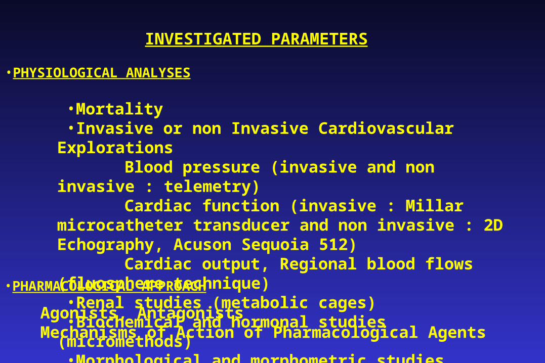
INVESTIGATED PARAMETERS
•PHYSIOLOGICAL ANALYSES
•Mortality •Invasive or non Invasive Cardiovascular Explorations Blood pressure (invasive and non invasive : telemetry) Cardiac function (invasive : Millar microcatheter transducer
and non invasive : 2D Echography, Acuson Sequoia 512) Cardiac output, Regional blood flows (fluosphere technique)•Renal studies (metabolic cages)•Biochemical and hormonal studies (micromethods)•Morphological and morphometric studies
•PHARMACOLOGICAL APPROACH
• Agonists, Antagonists• Mechanisms of Action of Pharmacological Agents
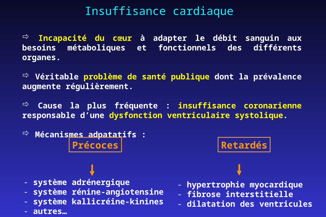
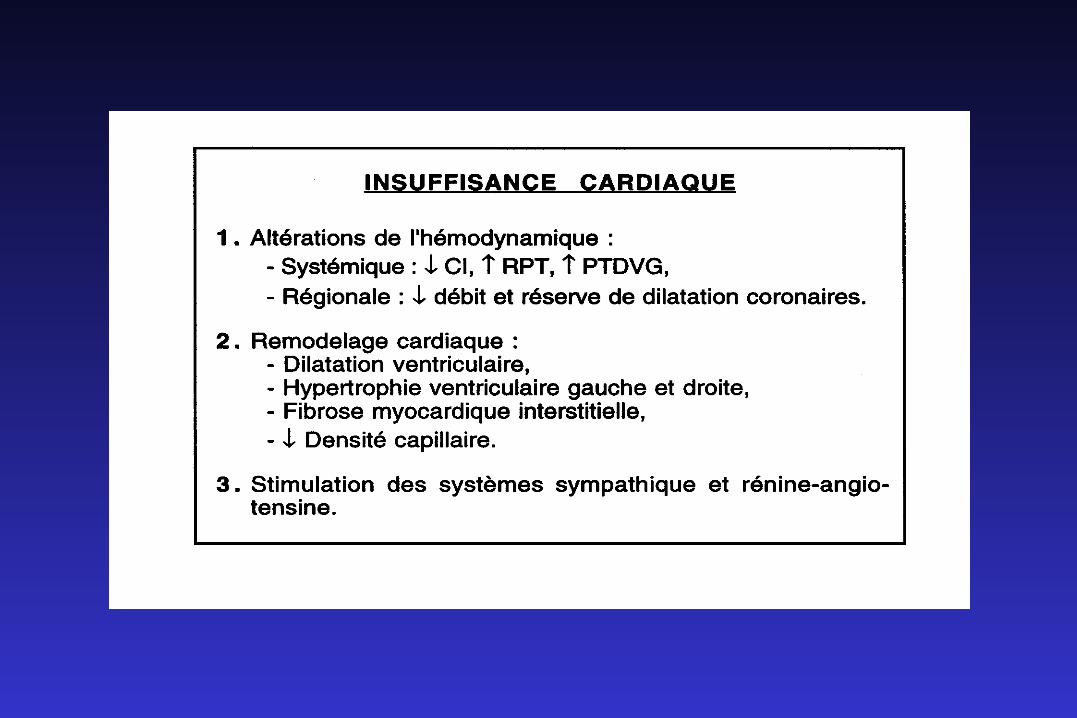
Insuffisance cardiaque
Incapacité du cœur à adapter le débit sanguin aux besoins métaboliques et fonctionnels des différents organes.
Véritable problème de santé publique dont la prévalence augmente régulièrement.
Cause la plus fréquente : insuffisance coronarienne responsable d’une dysfonction ventriculaire systolique.
Mécanismes adpatatifs :
Précoces Retardés
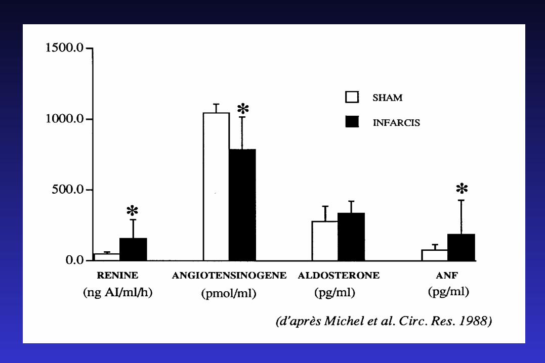
- système adrénergique- système rénine-angiotensine- système kallicréine-kinines- autres…
- hypertrophie myocardique- fibrose interstitielle- dilatation des ventricules
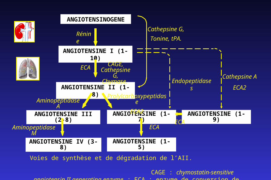
ANGIOTENSINOGENE
ANGIOTENSINE I (1-10)
ANGIOTENSINE II (1-8)
ANGIOTENSINE III (2-8)
ANGIOTENSINE IV (3-8)
ANGIOTENSINE (1-7)
Endopeptidases
Cathepsine G,
Tonine, tPA.
Aminopeptidase M
Rénine
ECACAGE,
Cathepsine G, Chymase.
Aminopeptidase AProlylcarboxypeptidase
ECA2
ANGIOTENSINE (1-5)
ECA
ANGIOTENSINE (1-9)
Cathepsine A
ECA2
Voies de synthèse et de dégradation de l’AII. CAGE : chymostatin-sensitive angiotensin II generating enzyme ;
ECA : enzyme de conversion de l’angiotensine ; tPA : tissue plasminogen activator.
ECA
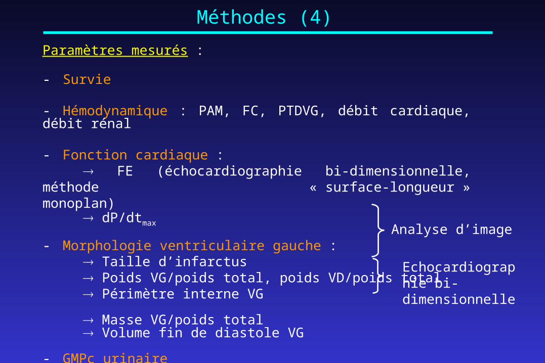
Méthodes (4)
Paramètres mesurés :
- Survie
- Hémodynamique : PAM, FC, PTDVG, débit cardiaque, débit rénal
- Fonction cardiaque : FE (échocardiographie bi-dimensionnelle, méthode
« surface-longueur » monoplan) dP/dtmax
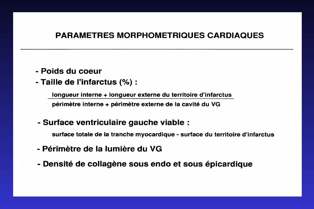
- Morphologie ventriculaire gauche : Taille d’infarctus Poids VG/poids total, poids VD/poids total Périmètre interne VG
Masse VG/poids total Volume fin de diastole VG
- GMPc urinaire
Analyse d’image
Echocardiographie bi-dimensionnelle
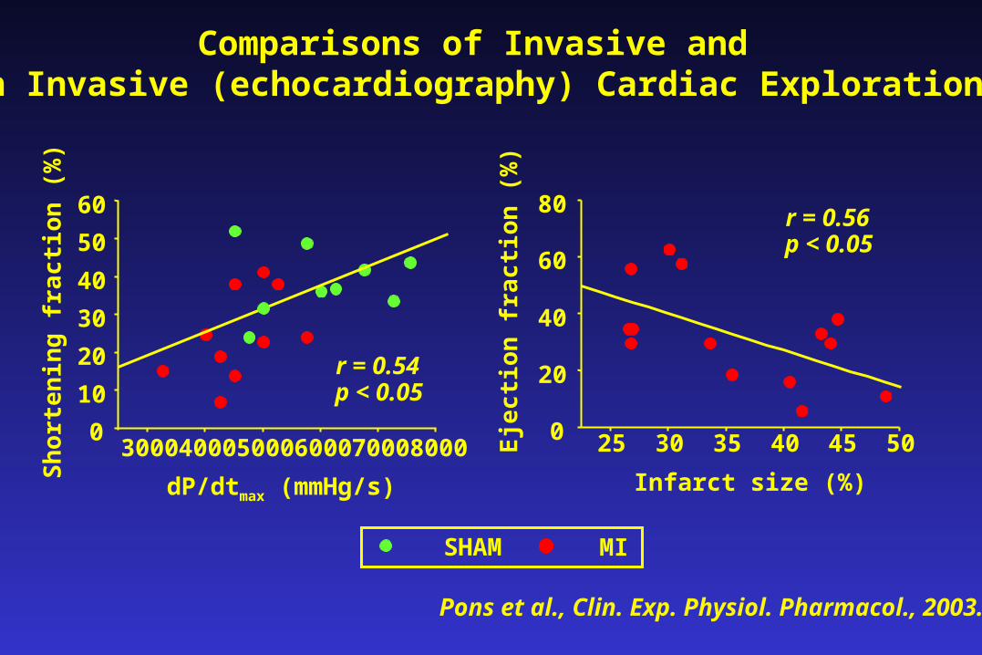
dP/dtmax (mmHg/s)
25 30 35 40 45 500
20
40
60
80r = 0.56p < 0.05
Eje
ctio
n f
ract
ion
(%
)Infarct size (%)
3000 4000 5000 6000 7000 80000
10
20
30
40
50
60
r = 0.54p < 0.05
Sh
orte
nin
g fr
acti
on (
%)
SHAM MI
Comparisons of Invasive and non Invasive (echocardiography) Cardiac Explorations
Pons et al., Clin. Exp. Physiol. Pharmacol., 2003.
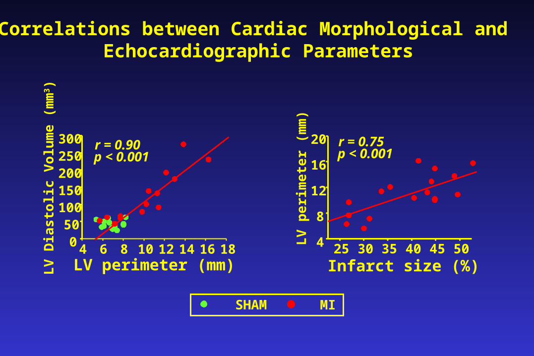
Correlations between Cardiac Morphological and Echocardiographic Parameters
4 6 8 10 12 14 16 18050
100150200250300 r = 0.90
p < 0.001
LV
Dia
stol
ic V
olu
me
(mm
3 )
LV perimeter (mm)25 30 35 40 45 504
8
12
16
20
LV
per
imet
er (
mm
)
Infarct size (%)
r = 0.75p < 0.001
SHAM MI
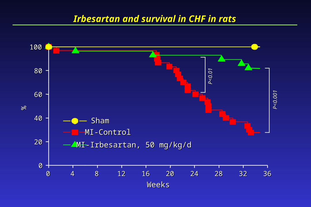
Irbesartan and survival in CHF in ratsIrbesartan and survival in CHF in rats
00 44 88 1212 1616 2020 2424 2828 3232 363600
2020
4040
6060
8080
100100
ShamSham
MI-ControlMI-Control
MI-Irbesartan, 50 mg/kg/dMI-Irbesartan, 50 mg/kg/d
%%
WeeksWeeks
P<0.001
P<0.001
P<0.01
P<0.01
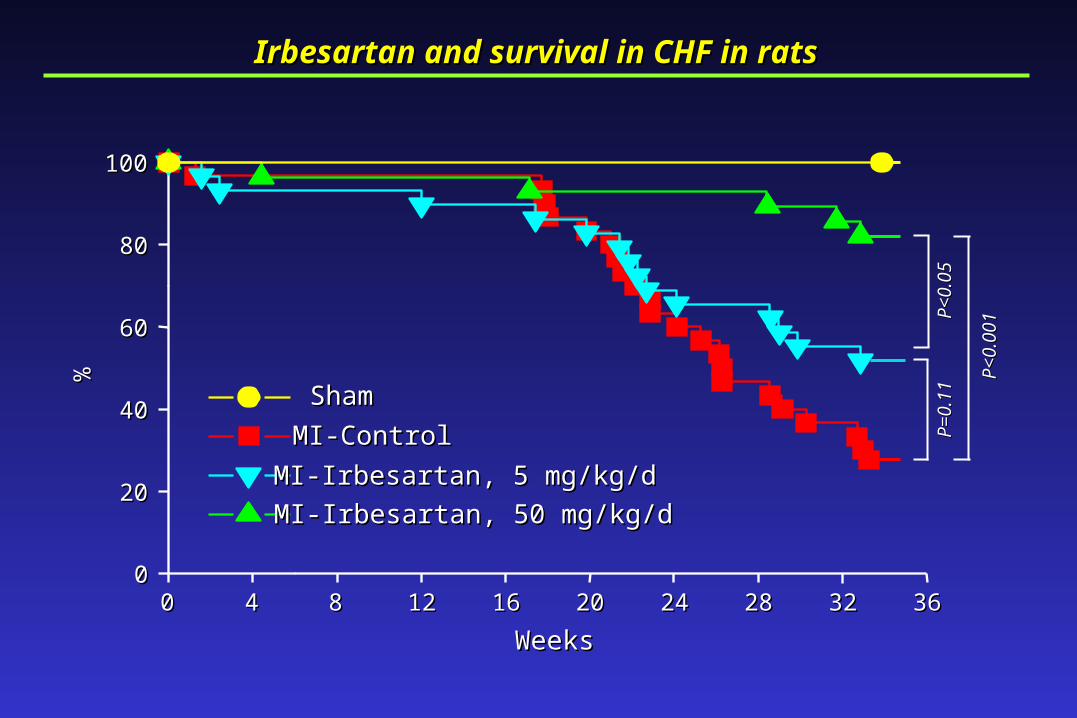
Irbesartan and survival in CHF in ratsIrbesartan and survival in CHF in rats
00 44 88 1212 1616 2020 2424 2828 3232 363600
2020
4040
6060
8080
100100
ShamSham
MI-ControlMI-Control
MI-Irbesartan, 5 mg/kg/dMI-Irbesartan, 5 mg/kg/d
MI-Irbesartan, 50 mg/kg/dMI-Irbesartan, 50 mg/kg/d
%%
WeeksWeeks
P=0.11
P=0.11
P<0.05
P<0.05
P<0.001
P<0.001
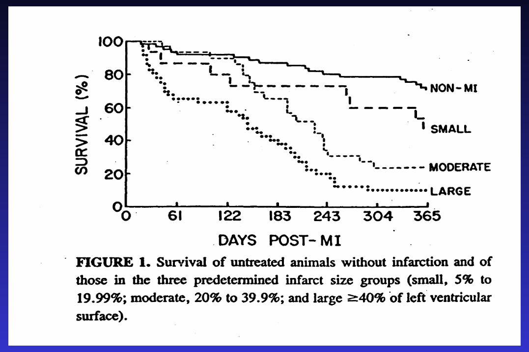
1. Up- regulation of the renin- angiotensin system (RAS) is a primary feature associated with chronic heart
failure (CHF) in animal models and in humans. 2. Angiotensin I- converting enzyme inhibitors (ACEIs)
prolong survival and prevent cardiac remodeling in the rat model of post- -ischemic heart failure (Pfeffer et al., 1985).
ACEIs reduce morbidity and mortality and improve quality of life in patients with CHF and in patients who have had a myocardial infarction (Consensus,
Solvd, Save, Aire, Trace, etc...).
3. Angiotensin II AT1 receptor blockers (ARBs) also prolong survival and prevent cardiac remodelingin the rat model of post- -ischemic heart failure.
In patients with CHF, Losartan was effective at reducing mortality and morbidity but its efficacy
was not greater than that of Captopril (Elite II). .
RATIONALE FOR ACEIs -ARBs COMBINATION IN CHF
1. ACEIs alone do not completely and permanently
suppress Ang II production (reactive rise in renin, chymase). Addition of ARBs should result in a more complete inhibition of the RAS.
2. In hypertensi on, ACEIs and ARBs prove to be
synergistic and , the greater the RAS blockade achieved, the greater the antihypertensive effect.
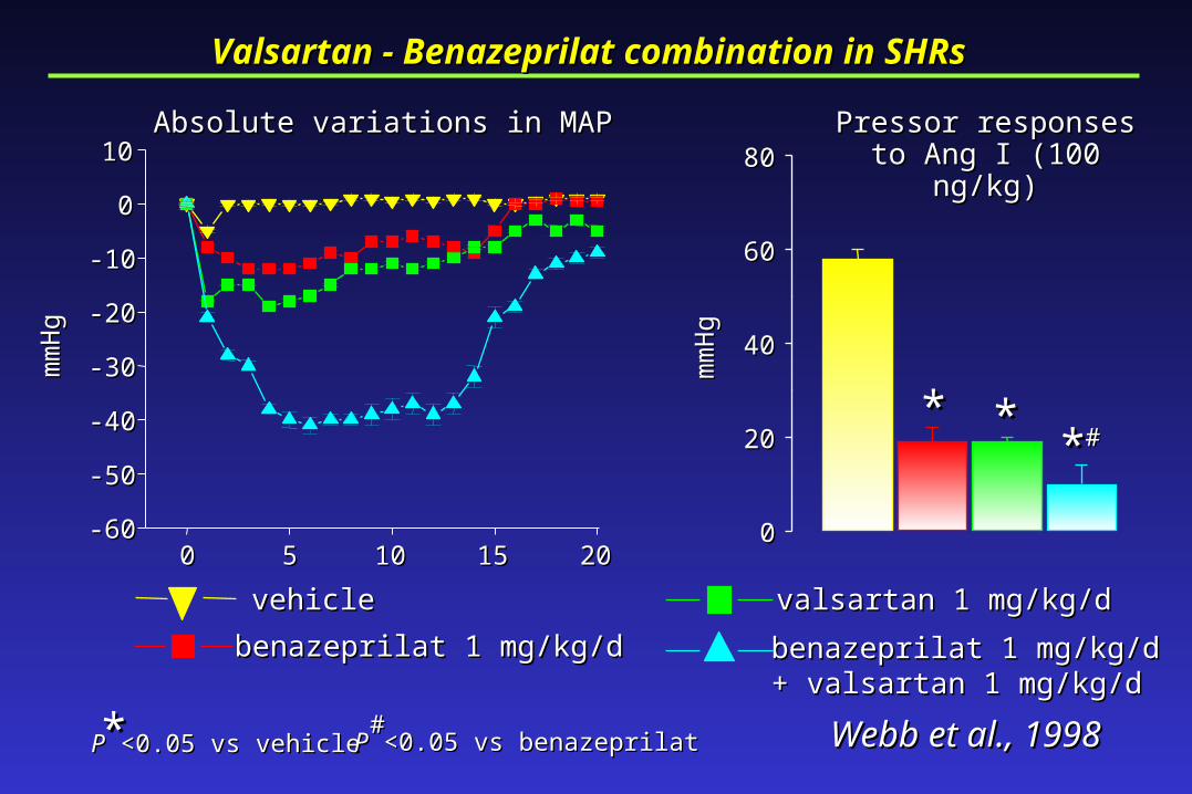
Pressor responsesPressor responsesto Ang I (100 ng/kg)to Ang I (100 ng/kg)
00 55 1010 1515 2020-60-60
-50-50
-40-40
-30-30
-20-20
-10-10
00
1010
mm
Hg
mm
Hg
Absolute variations in MAPAbsolute variations in MAP
vehiclevehicle
benazeprilat 1 mg/kg/dbenazeprilat 1 mg/kg/d
valsartan 1 mg/kg/dvalsartan 1 mg/kg/d
benazeprilat 1 mg/kg/dbenazeprilat 1 mg/kg/d+ valsartan 1 mg/kg/d+ valsartan 1 mg/kg/d
PP <0.05 vs vehicle <0.05 vs vehicle** PP <0.05 vs benazeprilat <0.05 vs benazeprilat## Webb et al., 1998Webb et al., 1998
Valsartan - Benazeprilat combination in SHRs Valsartan - Benazeprilat combination in SHRs
00
2020
4040
6060
8080
mm
Hg
mm
Hg
** **** ##
Absolute variations in Absolute variations in sitting DBPsitting DBP
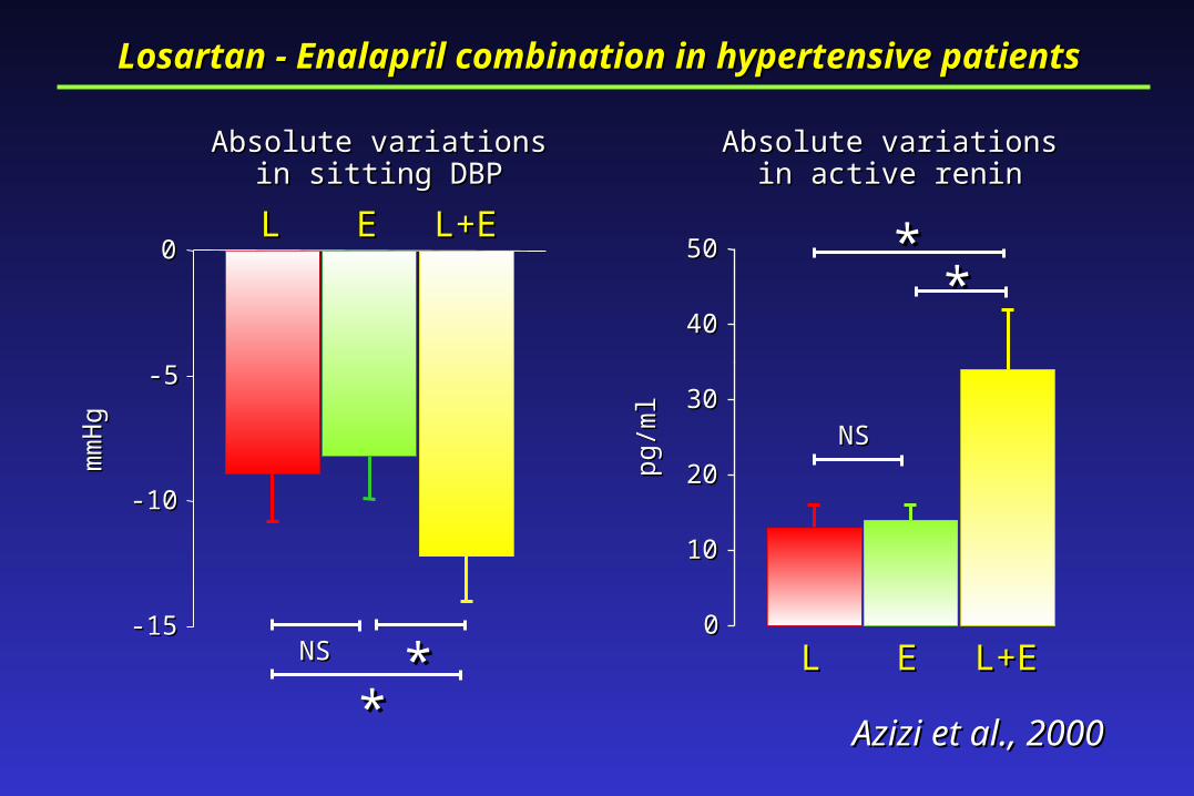
Azizi et al., 2000Azizi et al., 2000
Losartan - Enalapril combination in hypertensive patients Losartan - Enalapril combination in hypertensive patients
Absolute variations in Absolute variations in active reninactive renin
00
1010
2020
3030
4040
5050
-15-15
-10-10
-5-5
00
LL EE L+EL+E
LL EE L+EL+ENSNS ****
NSNS
****
mm
Hg
mm
Hg
pg/m
lpg
/ml
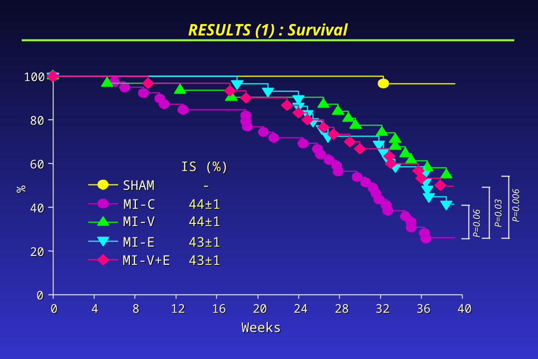
RESULTS (1) : SurvivalRESULTS (1) : Survival
00 44 88 1212 1616 2020 2424 2828 3232 3636 404000
2020
4040
6060
8080
100100
SHAMSHAM
MI-CMI-C--
4444±±11MI-VMI-V 4444±±11
MI-EMI-E 4343±±11
MI-V+EMI-V+E 4343±±11
IS (%)IS (%)
%%
WeeksWeeks
P=0.06
P=0.06
P=0.03
P=0.03
P=0.006
P=0.006
SHAMSHAM
MI-CMI-C
MI-VMI-V
MI-EMI-E
MI-V+EMI-V+E
PP <0.05 vs SHAM <0.05 vs SHAM**
RESULTS (2) : Cardiovascular parametersRESULTS (2) : Cardiovascular parameters
00
50005000
1000010000
1500015000
00
5050
100100
150150
200200
00
1010
2020
3030
** ** ** **
** ** ** ** **** **
mm
Hg
mm
Hg
mm
Hg
mm
Hg
mm
Hg/
sm
mH
g/s
SAPSAP LVEDPLVEDP
dP/dtdP/dt
00
11
22
33
44
55
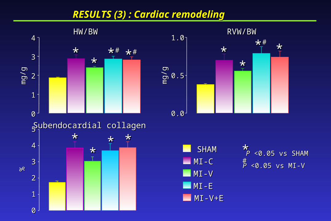
Subendocardial collagenSubendocardial collagen
%%
SHAMSHAM
MI-CMI-C
MI-VMI-V
MI-EMI-E
MI-V+EMI-V+E
PP <0.05 vs SHAM <0.05 vs SHAM**PP <0.05 vs MI-V <0.05 vs MI-V
##
00
11
22
33
44
0.00.0
0.50.5
1.01.0
RESULTS (3) : Cardiac remodelingRESULTS (3) : Cardiac remodeling
mg/
gm
g/g
mg/
gm
g/g
HW/BWHW/BW RVW/BWRVW/BW
****
** ## ** ##** ##
****
**
****
** **
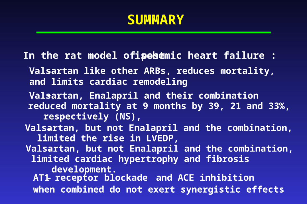
SUMMARY
In the rat model of post-ischemic heart failure :
-
Valsartan, Enalapril and their combination reduced mortality at 9 months by 39, 21 and 33%, respectively (NS),
- Valsartan, but not Enalapril and the combination, limited the rise in LVEDP,
-
Valsartan, but not Enalapril and the combination, limited cardiac hypertrophy and fibrosis development.
Valsartan like other ARBs, reduces mortality,and limits cardiac remodeling
-
AT1 receptor blockade and ACE inhibition when combined do not exert synergistic effects
-
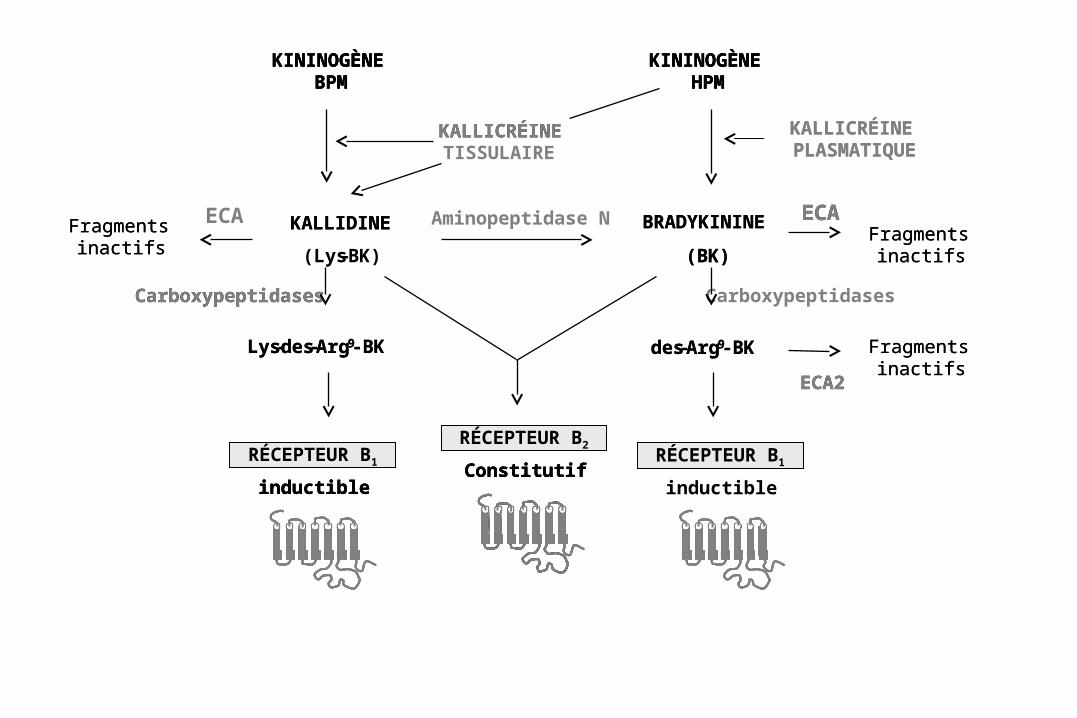
KININOGÈNE BPM
KININOGÈNE HPM
KININOGÈNE BPM
KININOGÈNE BPM
KININOGÈNE HPM
KININOGÈNE HPM
RÉCEPTEUR B2
ConstitutifRÉCEPTEUR B1
inductibleConstitutifConstitutif
inductibleinductible
Lys-des-Arg9-BK
Carboxypeptidases
Fragments inactifs
ECA ECAFragments
inactifs
des-Arg9-BK Fragments inactifs
ECA2
Carboxypeptidases
Lys-des-Arg9-BK
Carboxypeptidases
Aminopeptidase NFragments inactifs
ECAFragments
inactifs
des-Arg9-BK Fragments inactifs
ECA2
KALLIDINE
(Lys-BK)
KALLICRÉINE PLASMATIQUE
BRADYKININE
(BK)
KALLICRÉINE TISSULAIRE
KALLICRÉINE
KALLIDINE
KALLICRÉINE PLASMATIQUE
BRADYKININE
(BK)
RÉCEPTEUR B1
inductible
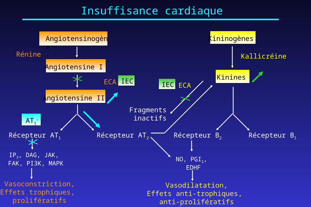
Angiotensinogène
Angiotensine I
Angiotensine II
Rénine
ECA
Kininogènes
Kinines
Fragments inactifs
Kallicréine
Récepteur AT2Récepteur AT1 Récepteur B1Récepteur B2
ECA
IP3, DAG, JAK, FAK, PI3K, MAPK
NO, PGI2, EDHF
Vasoconstriction,Effets trophiques,
prolifératifs
Vasodilatation,Effets anti-trophiques,
anti-prolifératifs
IEC IEC
AT1-
Insuffisance cardiaque
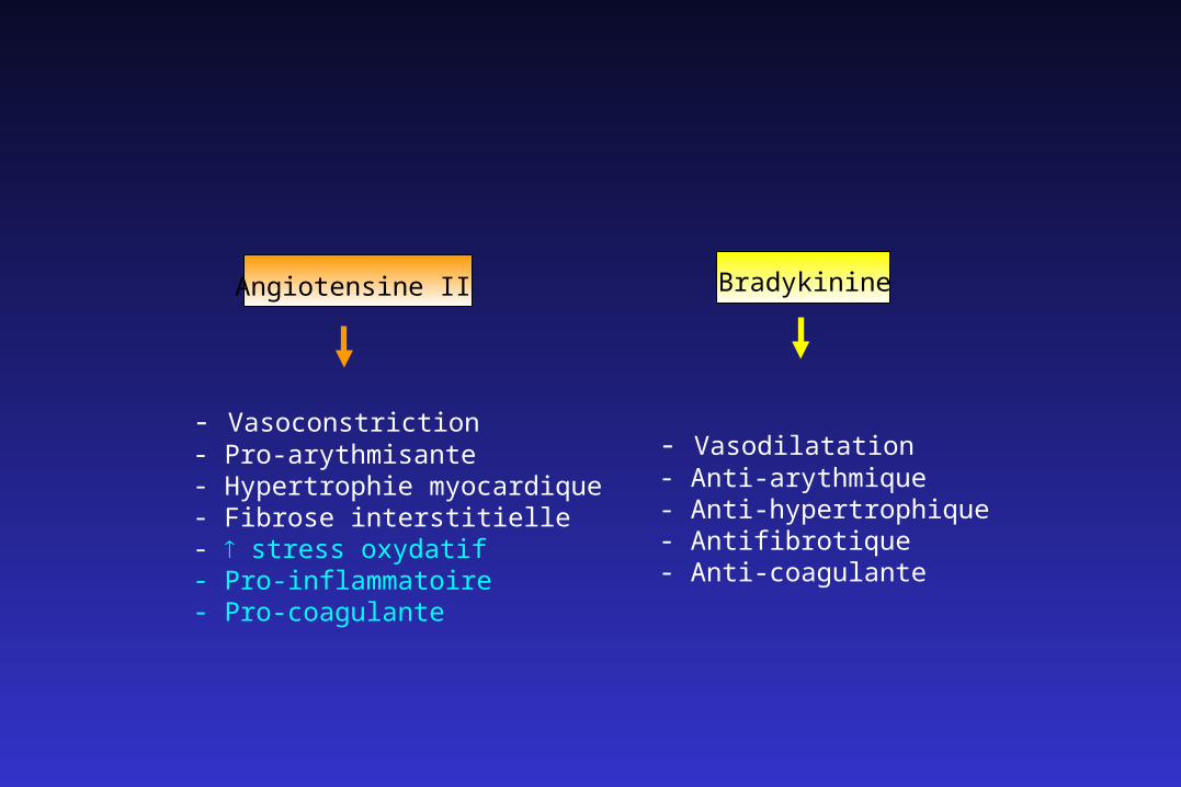
- Vasoconstriction- Pro-arythmisante- Hypertrophie myocardique- Fibrose interstitielle- stress oxydatif- Pro-inflammatoire- Pro-coagulante
Bradykinine
- Vasodilatation- Anti-arythmique- Anti-hypertrophique- Antifibrotique- Anti-coagulante
Angiotensine II
Le modèle d’insuffisance cardiaque post-ischémique peut être appliqué à des souris génétiquement modifiées pour étudier :
• l’implication d’un ou plusieurs gènes dans le développement de la maladie,
• le rôle de certains peptides dans le mécanisme d’action d’agents pharmacogiques utilisés dans le traitement de cette maladie.
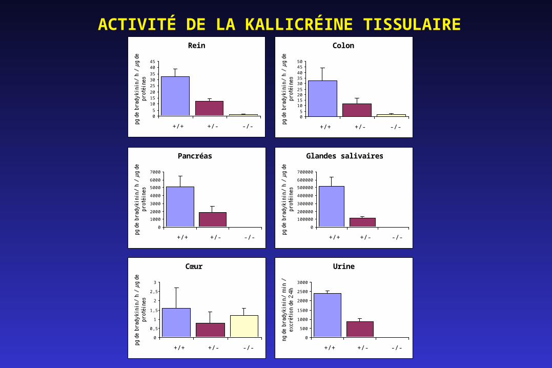
ACTIVITÉ DE LA KALLICRÉINE TISSULAIRERein
05
101520
253035
4045
+/+ +/- -/-
Colon
05
101520253035404550
+/+ +/- -/-
Pancréas
0
1000
2000
3000
4000
5000
6000
7000
+/+ +/- -/-
Glandes salivaires
0
100000
200000
300000
400000
500000
600000
700000
+/+ +/- -/-
Cœur
0
0,5
1
1,5
2
2,5
3
+/+ +/- -/-
Urine
0
500
1000
1500
2000
2500
3000
+/+ +/- -/-
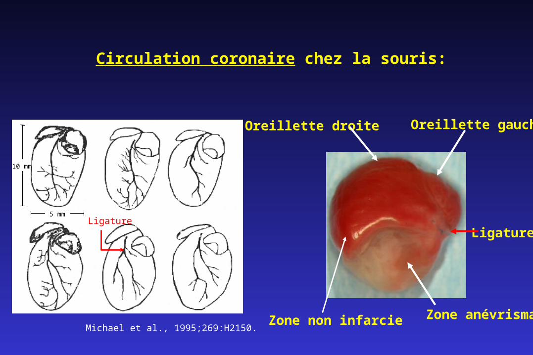
Oreillette gaucheOreillette droite
Zone anévrismale
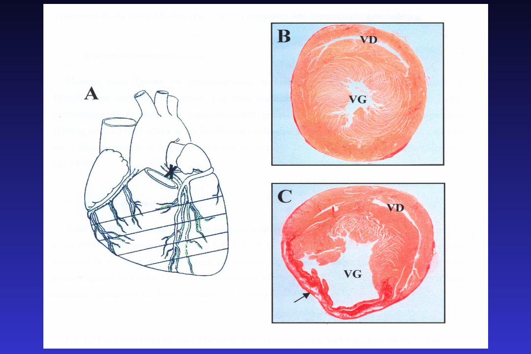
Ligature
Zone non infarcieMichael et al., 1995;269:H2150.
Circulation coronaire chez la souris:
10 mm
5 mmLigature

Sham
IM-Témoin *p<0.05 vs valeur sham correspondante
Fonction cardiaque
0
10
20
30
40
50
60
70
80FE
(%
) * *
0
1000
2000
3000
4000
5000
6000
7000
dP/dtmax
(mm
Hg/s
)
**
3 mois 12 mois6 mois 12 mois6 mois
Sham
IM-Témoin
*p<0.05 vs valeur sham correspondante
†p<0.05 vs valeur 3 mois correspondante
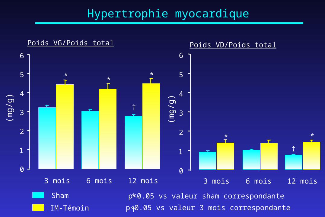
Hypertrophie myocardique
0
1
2
3
4
5
6
Poids VG/Poids total
(mg
/g)
3 mois 12 mois6 mois
†
***
0
1
2
3
4
5
6
†
**
Poids VD/Poids total
(mg
/g)
3 mois 12 mois6 mois
Sham
IM-Témoin *p<0.05 vs valeur sham correspondante
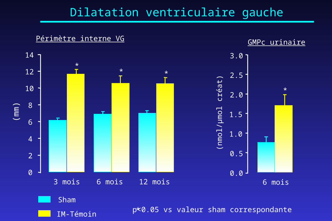
Dilatation ventriculaire gauche
0.0
0.5
1.0
1.5
2.0
2.5
3.0
6 mois
(nm
ol/µm
ol cr
éat)
GMPc urinaire
*
0
2
4
6
8
10
12
14
** *
3 mois 12 mois6 mois
Périmètre interne VG
(mm
)
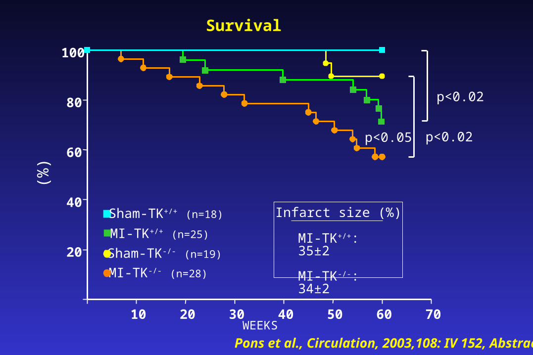
Survival
p<0.02
10 20 30 40 50 60 70WEEKS
(%)
20
40
60
80
100
p<0.05
Sham-TK-/- (n=19)
MI-TK-/- (n=28)
p<0.02
MI-TK+/+ (n=25)
Sham-TK+/+ (n=18) Infarct size (%)
MI-TK+/+: 35±2
MI-TK-/-: 34±2
Pons et al., Circulation, 2003,108: IV 152, Abstract
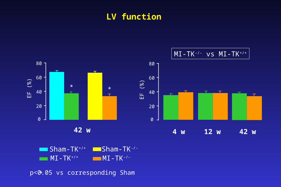
LV function
Sham-TK+/+
MI-TK+/+
Sham-TK-/-
MI-TK-/-
EF
(%
)
0
20
40
60
80
**
4 w 12 w 42 wE
F (
%)
42 w
p<0.05 vs corresponding Sham *
0
20
40
60
80
MI-TK-/- vs MI-TK+/+
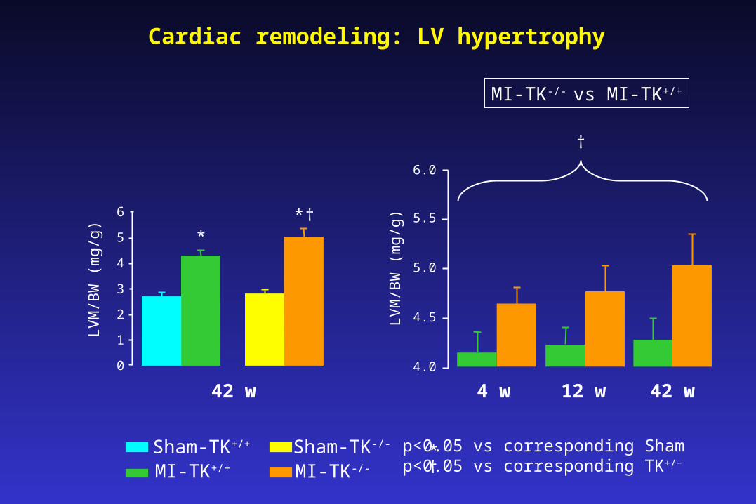
Cardiac remodeling: LV hypertrophy
0
1
2
3
4
5
6
LV
M/B
W (
mg/
g)
†**
LV
M/B
W (
mg/
g)
4.0
4.5
5.0
5.5
6.0
4 w 12 w 42 w42 w
Sham-TK+/+
MI-TK+/+
Sham-TK-/-
MI-TK-/-
* p<0.05 vs corresponding Sham p<0.05 vs corresponding TK+/+†
†
MI-TK-/- vs MI-TK+/+
0
40
80
120
160
200
240
LV
dia
stol
ic v
olum
e (m
m3 )
†*
*
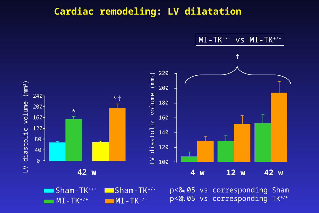
Cardiac remodeling: LV dilatation
100
120
140
160
180
200
220
LV
dia
stol
ic v
olum
e (m
m3 )
4 w 12 w 42 w42 w
Sham-TK+/+
MI-TK+/+
Sham-TK-/-
MI-TK-/-
* p<0.05 vs corresponding Sham p<0.05 vs corresponding TK+/+†
†
MI-TK-/- vs MI-TK+/+
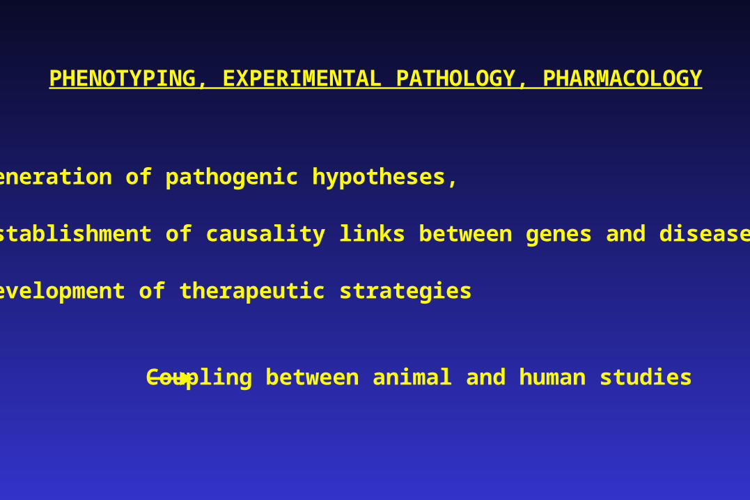
PHENOTYPING, EXPERIMENTAL PATHOLOGY, PHARMACOLOGY
• Generation of pathogenic hypotheses,
• Establishment of causality links between genes and diseases,
• Development of therapeutic strategies
Coupling between animal and human studies