Embed Size (px)
Citation preview
Volume 61(1):1-11, 2017Acta Biologica Szegediensis
http://www2.sci.u-szeged.hu/ABS
ARTICLE
School of Science, Monash University Malaysia, 47500 Bandar Sunway, Petaling Jaya, Malaysia
In vitro endophyte-host plant interaction study to hypothetically describe endophyte survival and antifungal activities in plantaHye Seung Yoo, Adeline Su Yien Ting*
ABSTRACT This study is the first to adopt a hypothetical approach to establish the influence of the complex endophyte-host interaction on endophyte survival and antifungal expression. Three key interactions were evaluated; (I) influence of host-induced enzymes on endophyte growth (biomass) and colonization, (II) link between endophyte-produced cellulase, their growth and colonization, and (III) the influence of host environment on antifungal expression of endophytes. The interactions with the host were performed using plant slurry (PS) to mimic in planta (host) environment with analysis on interactions evaluated using Pearson correlation coefficients (r). Results revealed that host induced enzymes may be a limiting factor to coloniza-tion of endophytes with inverse correlations observed (-0.046 ≤ r ≤ -0.7164). These enzymes may also limit growth of endophyte although, PAL (r = 0.536) and TPC (r = 0.8894) appeared contrary. Results were also suggestive that endophytes produced cellulase to aid in colonization in host plants (r = 0.7073 in PS), and cellulase activities are continuously produced even when growth of endophytes are limited (r = -0.314 in PS). Endophytes are presumed to produce antifungal compounds in planta (r = 0.2760 in PS), and these compounds may be secondary metabolites, which are primarily produced under nutrient-depleted conditions where growth is poor (in host plant). The superior growth of endophytes in synthetic PDB media has an inverse correlation to antifungal activity (r = -0.5129), confirming that secondary metabolites are involved in antifungal activities. This study clearly presents that success of inoculated endophytes in colonizing, grow-ing and expressing antifungal activities is dependent on the host plant.Acta Biol Szeged 61(1):1-11 (2017)
KEy WoRdS
antifungal activityendophyte-host interactionendophyte survivalFusarium oxysporum f. sp. cubense tropical race 4simulation study
Submitted January 3, 2017; Accepted April 10, 2017*Corresponding author. E-mail: [email protected]; [email protected]
1
Introduction
Endophytes are microorganisms that exist or spend part of their life cycle in the host plant. Their roles as biocontrol agents of various diseases are attributed to several mecha-nisms; direct inhibitory effect towards the pathogen (Liu et al. 2007; Ting et al. 2010a, 2011a), indirect disease suppression via plant growth improvement (Ting et al. 2008, 2009a), and induced host resistance against pathogens (Sundaramoorthy et al. 2012; Ting et al. 2009b, 2010b, 2012a). Endophytic biocontrol agents are typically isolated from an array of host species, and screened for bioactivity prior to introduction to commercially important crops. Although, the approach in using biocontrol agents is more environment-friendly, field applications seldom yield satisfactory biocontrol results (Ting
et al. 2009a). For many years, poor soil conditions have been identified as the primary factor in contributing the absence of disease control in the field. The survival of introduced bio-control agents was impeded by the intense competition with indigenous microflora in the soil, as well as physicochemi-cal soil conditions (extreme pH, poor oxygen level/aeration, poor moisture level) (Sylvia and Neal 1990; Donnison et al. 2000). This has led to the emergence of studies in microbial bioformulations, to innovate and improve delivery and ap-plication of biocontrol agents in the field (Ting et al. 2009c; Manikandan et al. 2010; Shanmugam and Kanoujia 2011; Ting et al. 2011b).
In this study, we proposed that the complex endophyte-host plant interaction may be a contributing factor that in-fluences endophyte survival in planta and their subsequent biocontrol activity towards the pathogen. This hypothesis is a refreshing perspective, which suggests that introduced endophytes are influenced by host plant factors and not solely dependent on soil factors. To establish this, an in vitro study was designed to determine interactions of the growth
2
Yoo, Ting
of introduced endophytes (biomass, colonization extent, cellulase activities for colonization) with host plant factors (host defense enzymes) and their subsequent influence on the antifungal activity of endophytes against the pathogen (Fusarium oxysporum f. sp. cubense tropical race 4, FocR4). The in vitro study was performed using plant slurry derived from banana plantlets to mimic in planta conditions. The links between the factors are validated by Pearson correlation coefficients (r).
To the best of our knowledge, this study is the first to test several key novelties. Firstly, we explored the influence of host-induced enzymes on endophyte growth and their colo-nization. This is an atypical approach to link and evaluate in-duced host defense enzymes as a factor influencing endophyte colonization in planta. Secondly, we explored the influence of endophyte-produced cellulase, a cell wall-degrading enzyme (CWDE) on endophyte growth and colonization. Finally, this in vitro study also examined the antifungal activities of en-dophytes expressed under an artificial in planta environment (using plant slurry) to mimic host plant environment. The varying degree of inhibition suggests the possible potency of antifungal compounds produced in planta. Therefore, this in vitro study provides new insights on the endophyte-host plant interaction and its influence on endophyte survival and antifungal expression towards the pathogenic F. oxysporum f. sp. cubense tropical race 4.
Materials and Methods
Culture establishment
The fungal pathogen F. oxysporum f. sp. cubense race 4 (FocR4) (VCG 01213/16) (Bentley et al. 1998) was obtained from Prof. Dr. Sariah Meon from University Putra Malaysia as cultures on Potato Dextrose Agar (PDA, Merck). Five endophytic isolates were selected; two isolates from weeds (Diaporthe sp. MIF01; Diaporthe sp. WAA02) and three isolates from banana plants (Penicillium citrinum BTF08; Cladosporium sp. BTF21; Trichoderma sp. T2), and all cul-tures maintained on PDA (Merck) at 25 ± 2 ºC. These five endophytes have demonstrated antagonistic properties against FocR4 in plate screenings (dual culture assays) in our previ-ous studies (Ting et al. 2009d, 2010a, 2012a).
Assays for host-induced enzymes
Tissue-cultured banana plantlets (Pisang berangan cv. Intan, 3-4 leaf staged) were obtained from United Plantations Pte. Ltd. To induce production of host-induced enzymes, the plantlets were inoculated by immersing (root-dipping) into 30 ml of endophyte suspension (inoculum concentration of 106
cfu mL-1) (Ting et al. 2012a). The inoculated plantlets were maintained in sterilized soil, watered daily with sterile dis-tilled water, and kept isolated in a growth chamber from other plants. At a daily interval (for the next 7 days), plantlets were harvested and assayed for the following enzymes; peroxidase (PO), polyphenol oxidase (PPO), phenylalanine ammonia lyase (PAL) and total phenolic content (TPC). Triplicates were prepared for each isolate at every sampling interval.
To detect PO and polyphenol oxidase PPO levels, crude extracts were first prepared by grinding 1 g (fresh weight) of the tissues in 1 mL of chilled 0.05 M Sodium Acetate Buffer (SAB) (pH 5) supplemented with 5 mg of polyvinylpyrroli-done (PVP) (British Drug Houses, BDH). The slurry was centrifuged (20 min, 4 ºC, 14 000 rpm) and the supernatant (50 µL) incubated with 0.75 mL of reaction substrate [80 mL Sodium Phosphate Buffer (SPB, pH 6), 1 mL of 30% H2O2 (Merck), 20 mL of guaiacol (2-methoxyphenol, Merck)] at room temperature (25 ± 2 ºC). Blanks were prepared by sub-stituting the supernatant with distilled water. The absorbance was measured (470 nm) using a spectrophotometer (Genesys™
20; Thermo Fisher Scientific) after 5 min of reaction. The total amount of PO was calculated using the equation as described by Ting et al. (2012a):Units g-1 fresh weight of tissues = (absorbance at 470 nm )(dilution factor )
Amount of tissue assayed , g x 1000
For PPO, 50 µL of the supernatant was mixed with 750 µL of the reaction substrate [SPB (50 mM) with 5 x 10⁻⁴ M chlorogenic acid (Aldrich)] and allowed to stand at room temperature for 20 min. The absorbance was read at 410 nm and the PPO levels calculated (as for PO) and expressed as total PPO produced (units g-1 fresh weight of tissues). Blanks were prepared by substituting the supernatant with distilled water (Ting et al. 2012a).
For PAL assay, 1 g fresh weight of the plant tissues were ground in 5 mL of 0.1 M Sodium Borate Buffer (SBB) (pH 8.8), supplemented with 5 mM mercaptoethanol (Merck). The slurry was centrifuged (10 min, 4 ºC, 15 000 rpm) and the supernatant collected. The supernatant (1 mL) was mixed with 3 mL of 300 mM SBB amended with 30 mM L-pheny-lalanine (Alfa Aesar). The reaction mixture was incubated in a water bath at 40 ºC for 1 h. Spectroscopic measurement was made at 290 nm, and the PAL activity was expressed as mg of cinnamic acid produced per g fresh weight of tissues. Blanks were prepared by substituting the enzyme extract with distilled water (Podille and Laxmi 1998). Trans-cinnamic acid (Aldrich) (0 to 0.5 mg mL-1) was used to construct the standard curve.
To detect TPC, 1 g (fresh weight) of plant tissues were ground in 9 mL of distilled water. The mixture was transferred to a universal bottle (wrapped in aluminum foil), and agitated at 150 rpm for 1 h. For colorimetric analysis, 100 µL of the plant extract was mixed with 0.75 mL of 1/10th diluted Folin-Ciocalteu reagent (FS Chem), and after 5 min, 0.75 mL of

3
Hypothetical study on endophyte-host interaction
6% Na2CO3 (Amresco) solution and 5 mL of distilled water were added. The solution was mixed for reaction to occur at room temperature for 90 min. The absorbance was gradually read at 725 nm and TPC quantified based on standard curve plotted using gallic acid (FS Chem) of 0.01 to 0.5 mg mL-1 (Amin et al. 2004).
Determining colonization extent and growth of endophytes
Banana plantlets were inoculated by root-dipping into 30 mL fungal suspensions (106 cfu mL-1 of MIF01, WAA02, BTF08, BTF21, T2 or FocR4 separately). At a daily interval (for the next 7 days), the plantlets were sampled and cut into root, corm, pseudostem and leaf tissues (of approximately 1 cm2 each), and surfaced disinfected (1 min in 5% NaOCl, 1 min in 75% ethanol, 1 min in 95% ethanol, rinsed with sterile dis-tilled H2O) (Schulz et al. 1993). The disinfected tissues were subsequently cut longitudinally to expose the internal tissues, which were then plated on PDA and supplemented with 50 mg L-1 streptomycin sulfate (Sigma) (to inhibit bacterial growth). The seeded plates (containing 3 pieces each of root, corm, pseudostem and leaf tissues per plate) were incubated at room temperature for 7 days. The extent of colonization is expressed as the percentage of colonization according to Akello et al. (2009), where the number of tissue pieces with endophyte growth detected is compared against total number of tissue pieces plated (12 pieces).
To determine the growth (proliferation) of endophytes in host tissues, endophytes were first inoculated into three different media: plant slurry (PS), a diluted Potato Dextrose Broth (PDB), and a mixture of PS + PDB. Plant slurry me-dium (PS) was designed to mimic conditions in the plant. It was prepared by blending fresh banana plantlets (150 g fresh weight) in 100 mL of sterile distilled water, then 3 mL of this homogenized plant material was added to 10 mL of sterile distilled H2O. When PDB was prepared, 3 mL of sterile distilled H2O was added to 10 mL of Potato Dextrose Broth. PS + PDB medium was prepared by mixing 3 mL of homogenized plant material with 10 mL of Potato Dextrose Broth. Two agar plugs (5 mm diameter) were then inoculated into each medium, and incubated at room temperature, with 100 rpm for 30 days. At every 10-day interval, the fungal biomass was filtered with a cheesecloth and oven-dried (60 ºC) overnight prior to weighing.
Determining cellulase activities by endophytes
Endophytes were inoculated (5 mL, 106 cfu mL-1) into 100 mL of PS, PDB and PS + PDB. At a daily interval (for the next 7 days), 50 μL of the culture filtrate was sampled and mixed into 200 μL acetate buffer solution (125 mM, pH 5) contain-ing 1.25% w/v carboxymethyl cellulose (CMC). The mixture
was incubated at room temperature (25 ± 2 ºC) for 15 min. The reaction was terminated by adding 250 μL of reaction solution [1% DNS reagent (dinitrosalicylic acid solution: 1% DNS (Bio Basic Inc), 0.05% sodium sulphite (Bendosen), 1% sodium hydroxide (Riendemann Schmidt), 2% sodium tartrate (Riendemann Schmidt)] and boiled in water bath for 5 min. The absorbance was measured at 575 nm (Tecan Infite M200 Multi detection Microplate reader) (Ting et al. 2012b), and the cellulase activities derived from a standard curve (0-100 μmol mL-1 of glucose).
Antifungal activities of endophytes
Five mL of fungal inoculum (106 cfu mL-1) was first inocu-lated to PS, PDB and PS + PDB. After 2 weeks, 30 mL of the culture was pipetted, centrifuged (9000 rpm, 10 min) and the supernatant filtered through sterile filter syringe (0.45 µm, MCE membrane, Biofil). The filtrate (6.5 mL) was then mixed with 6.5 mL of sterile double-strength molten PDA in Petri dishes. After the agar has solidified, mycelial plug of FocR4 was inoculated at the center of the plate and their growth was measured after 10 days. The antifungal activity is expressed as Percentage Inhibition of Diameter Growth (PIDG, %), where D1: diameter of FocR4 in control plates, D2: diameter of FocR4 in agar plates supplemented with filtrate (Zacky and Ting 2013):
PIDG (%) = D1-D2 x 100% D1
Statistical analysis
The experiments were carried out in a Complete Randomized Design (CRD). Data was analyzed using One-way Analysis of Variance (ANOVA) at the significance level of p<0.05. Tukey’s test (HSD (0.05)) was used to compare the means of the triplicate. Data analysis was performed using the soft-ware SPSS (Statistical Program for Social Science) ver. 20.0 (IBM). The Pearson correlation coefficients (r) were derived and the relationships/interactions between key areas was interpreted based on the following coefficient values; where 0.5 ≤ r ≤ 1.0 indicates high correlation, 0.3 ≤ r ≤0.5 indicates moderate correlation, 0.1 ≤ r ≤0.3 indicates weak correlation and r ≤ 0.1 indicates no correlation.
Results
Production of host-induced enzymes elicited by endophytes
The PO, PAL and TPC levels in plantlets upon infection by various endophytes (MIF01, WAA02, BTF08, BTF21, T2)
4
Yoo, Ting
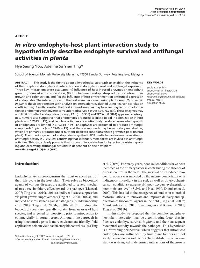
and pathogenic FocR4 were not significantly different from one another; with PO levels of 171.54 to 285.96 units g-1 fresh weight of tissues, PAL levels of 0.63 to 0.78 mg cinnamic acid g-1 fresh weight of tissues, and TPC levels of 0.08 to 0.14 mg mL-1 (Fig. 1A, 1C, 1D). The PO and TPC levels were however, significantly different between isolates BTF08 and FocR4 for PO, and isolates WAA02 and FocR4 for TPC. This suggested that different species may elicit different response from the host plant. On the contrary, the PPO levels varied in plants induced by different endophytic isolates. PPO levels were the highest in plants triggered by BTF21 (62.11 units g-1 fresh weight of tissues), while BTF08 (14.00 units g-1 fresh weight of tissues) resulted in the least PPO produced. The pathogenic FocR4 was observed to elicit comparable levels of PO (171.54 units g-1 fresh weight of tissues), PPO (39.53 units g-1 fresh weight of tissues), PAL (0.63 mg cinnamic acid g-1 fresh weight of tissues) and TPC (0.08 mg mL-1), when inoculated to plants (Fig. 1A-D).
Colonization extent and growth of endophytes
Isolate T2 had the highest percentage of tissue colonization (60.42%), followed by isolates WAA02, FocR4, BTF08, and
MIF01 with 41.37, 31.85, 29.76, and 22.62%, respectively (Fig. 2). Isolate BTF21 was not detected in any of the plant tissues sampled (root, corm, pseudostem, and leaf). Isolates T2, WAA02, MIF01, BTF08 and FocR4, demonstrated bet-ter colonization in the corm tissues (65.08%), compared to pseudostem (29.56%), root (26.98%) and leaf tissues (2.38%) (data not shown). For fungal growth (proliferation), higher biomass was recovered from isolates cultured in PDB and PS + PDB compared to PS. Cultures from PDB weighed between 0.032-0.051 g, which were significantly higher than biomass collected from cultures in PS (0.003-0.110 g) (Fig. 3). An atypical growth response was demonstrated by isolate WAA02, where similar biomass was recovered from PS (0.030 g), PDB (0.047 g) and PS + PDB (0.028 g) (Fig. 3). This suggested that among the isolates tested, isolate WAA02 may have better survival potential in planta, while other isolates grew better in synthetic media (PDB).
Cellulase production by endophytes
Isolates inoculated in PDB produced higher cellulase levels compared to their production in the presence of PS. In PS, cellulase production is severely limited especially for isolates
Figure 1. Plant defence response, when inoculated with various fungal isolates. (A) PO activity, (B) PPO activity, (C) PAL activity, and (D) TPC. Means with the same letters are not significantly different according to Tukey’s test (HSD (0.05)). Bars represent standard error of means.

5
Hypothetical study on endophyte-host interaction
BTF21, BTF08 and WAA02 as the differences of cellulase levels in PS and PDB were 19.56, 19.36 and 12.04 μmol mL-1
cellulase, respectively. Isolates BTF21, BTF08 and WAA02 produced only 0.35, 0.41, and 0.07 μmol mL-1 cellulase com-pared to 19.90, 19.77, and 12.11 μmol mL-1, respectively, when inoculated in PDB (Fig. 4). On the contrary, cellulase levels by isolate T2 were the least affected by growth condi-tions (PS, PDB, PS + PDB), with similar cellulase levels derived from cultures cultivated in PS (4.09 μmol mL-1), PDB (14.84 μmol mL-1) and PS + PDB (9.03 μmol mL-1) (Fig. 4). This suggested that endophytic isolates could produce cellu-lase in nutrient-deprived conditions (in PS) to possibly aid in colonization, but the cellulase levels were inferior compared to nutrient-rich conditions (PDB).
Antifungal activities by endophytes
The antifungal activities by endophytes cultured in PS were relatively stronger than, if not comparable, to those derived from PDB and PS + PDB. Filtrate derived from isolates BTF21 (50.6% PIDG) and T2 (34.8%) cultured in PS showed stronger inhibitory effect compared to filtrates of the same isolates derived from PDB (7% for BTF21, 0.9% for BTF08) (Fig. 5). For isolates WAA02 (28.3%), BTF08 (12.0%), and MIF01 (10.7%), their respective filtrates from PS demon-strated similar inhibitory effect as filtrates derived from
Figure 2. Overall colonisation (%) of the banana plantlets by FocR4 and the five antagonistic endophytes. Means with the same letters are not significantly different according to Tukey’s test (HSD (0.05)).
Figure 3. Mean biomass (g) of six investigated fungal isolates cultured in PS, PDB and PS + PDB. Comparison of mean dry mass was determined among the culture media for each fungal isolate. Means with the same letters are not significantly different according to Tukey’s test (HSD
(0.05)). Bars represent standard error of means.
Figure 4. Cellulase activities (µmol mL-1) of fungal isolates cultured in plant slurry (PS), PDB and PS + PDB. Means with the same letters are not significantly different according to Tukey’s test (HSD (0.05)). Tukey’s grouping was determined for each fungal isolate among the culture media. Bars represent standard error of means.
Figure 5. Percentage inhibition of diameter growth (PIDG, %) of FocR4 under the influence of cell-free extracts of respective endo-phytes cultured in PS, PDB and PS + PDB. Means with the same letters are not significantly different according to Tukey’s test (HSD (0.05)). Tukey’s grouping was determined across the different culture media within each of the fungal isolate. Bars represent the standard error of means. PS: plant slurry; PDB: Potato Dextrose Broth; PS + PDB: plant slurry + PDB.

6
Yoo, Ting
PDB (34.7, 15.8, and 4.8%, respectively). The potential of endophytes in producing potent antifungal compounds, while cultured in PS strongly suggests that this may also occur in planta (host environment), which is a valuable insight on antifungal expression by endophytes in host tissues. The observations here are also suggestive that despite the nutrient-depleted host environment, endophytes are still capable of producing antifungal compounds.
Establishing endophyte-host interactions based on correlation coefficients (r)
The first endophyte-host interaction established is the possible influence of host-induced enzymes on endophyte growth and colonization. Correlation analysis using data from 3.1 and 3.2 revealed that growth (biomass) of endophytes was generally inversely correlated to host-induced enzymes (except for PAL and TPC). The inverse correlation ranges from poor/weak correlation, i.e. for PO (r = -0.2404) and PPO (r = -0.001) on growth, and PO (r = -0.0406), PAL (r = -0.1246) and TPC (r = -0.1392) on colonization extent; to strong/high correlation such as for PAL and colonization extent (r = -0.7164) (Fig. 6, 7). The inverse correlations are suggestive that the higher levels of enzymes, lesser the growth and colonization extent
of the endophytes were detected. On the contrary, enzymes PAL and TPC have strong, positive correlations to growth of endophytes with r = 0.5366 and 0.8894, respectively (Fig. 6, 7). This indicated that host-induced enzymes triggered by en-dophytes, may be a limiting factor to the colonization extent (and possibly the growth to a certain degree) of endophytes in the host plant.
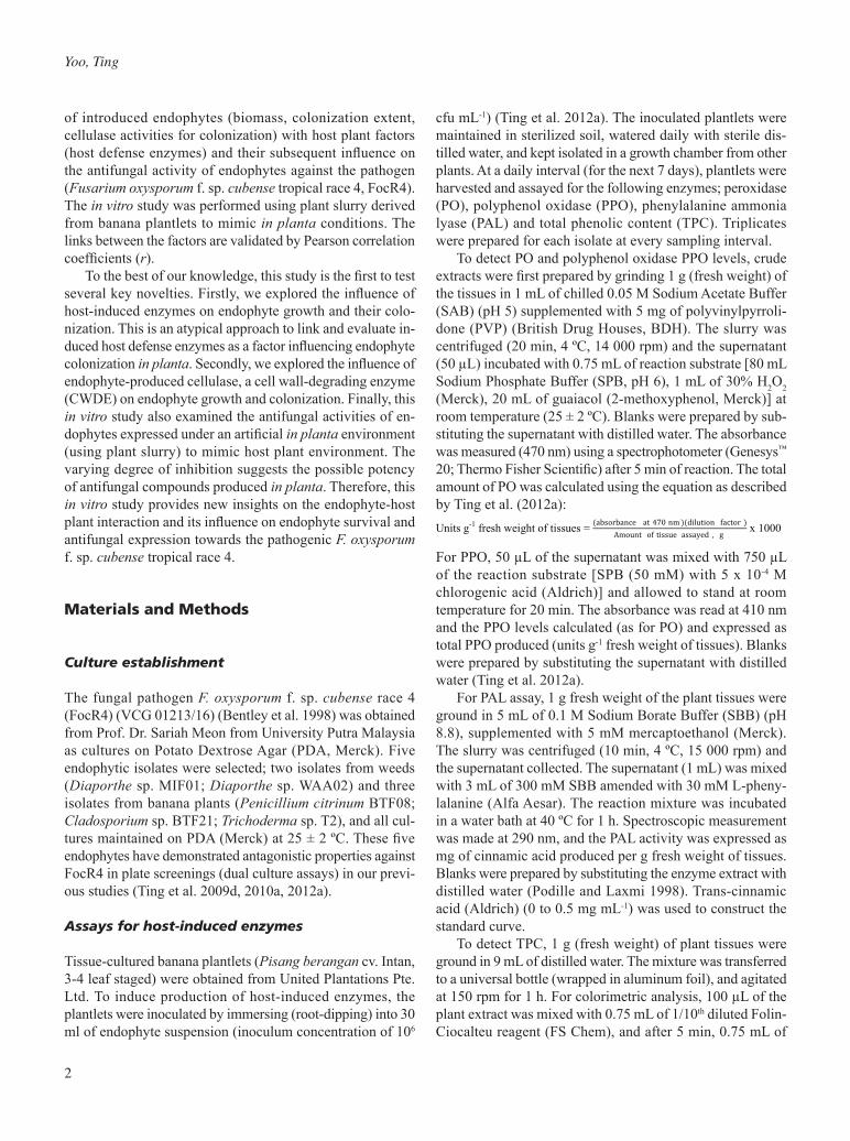
The second endophyte-host interaction validated is the association of endophyte growth and colonization extent to cellulase levels produced by endophytes in artificial host environment (achieved using PS). Correlation analysis was derived from data in sections 3.2 and 3.3. Endophyte growth was observed to have positive correlations to cellulase pro-duction in PDB (r = 0.5454) and PS + PDB (r = 0.53054), but an inverse correlation when cultured in PS (r = -0.3140) (Fig. 8). This suggested that growth of endophytes may po-tentially influence the cellulase production in nutrient-rich conditions (PDB, PS + PDB), but not in nutrient-depleted conditions (PS). The biomass and cellulase production in PS is inversely correlated, allowing the presumption that although growth may occur in PS, cellulase production is nevertheless limited and possibly vice versa. The interaction between endophyte colonization extent to cellulase levels were positively correlated for conditions in PS (r = 0.7073)
Figure 6. Correlation coefficient (r) from Pearson correlation test, for interactions between host-induced enzymes (PO, PPO, PAL, TPC) and endophyte growth (biomass).

7
Hypothetical study on endophyte-host interaction
and PS + PDB (r = 0.0778), but inversely correlated for PDB (r = -0.1906) (Fig. 9). This result illustrates the importance of cellulase production in aiding colonization of endophytes as the extent of colonization is strongly correlated to cellulase activities by the endophytes. This strong correlation observed in PS, further suggests that this phenomenon is typical in host environments.
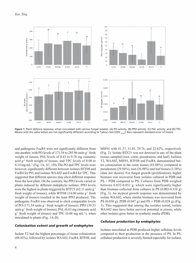
The third endophyte-host interaction studied the influence of endophyte growth on antifungal activity towards FocR4. Correlation analysis was performed using data from sections 3.2 and 3.4. Endophyte growth in PDB has an inverse correla-tion to antifungal activities (r = -0.5129), while positive cor-relations were obtained for growth and antifungal activities in PS (r = 0.2760) and PS + PDB (r = 0.4381) (Fig. 10). The
Figure 7. Correlation coefficient (r) from Pearson correlation test, for interactions between host-induced enzymes (PO, PPO, PAL, and TPC) and colonization extent for endophytes.
Figure 8. Correlation coefficient (r) from Pearson correlation test, for interactions between endophyte growth (biomass) and cellulase activi-ties.
8
Yoo, Ting
inverse correlation strongly suggests that antifungal activi-ties are compromised by the abundant growth of isolates. On the contrary, in nutrient-depleted conditions such as in host tissues (PS), the correlation is present where good fungal growth may appear to be necessary for effective production of antifungal compounds.
discussion
We obtained several profound insights from this study. Firstly, the inverse correlation between colonization extent (and to a certain degree for growth) of endophytes and induced host enzymes provides hypothetical assumptions, that the
rate and efficiency of endophyte colonization may be lim-ited by the enzymes produced. These enzymes, produced as a natural response to presence or infection by biotic or abiotic stimulators, have conventionally been thought to be advantages as it forms mechanisms to disease resistance. These enzymes are typically studied for their roles in the production of antimicrobial compounds (quinone molecules) and the reinforcement of cell walls (lignification) to prevent spread of pathogens (Raju et al. 2008; Naveen et al. 2013). Nevertheless, here we discovered that the induction of these enzymes may also limit endophyte colonization as the en-zymes are non-specific in their effect towards endophytes. The influence of induced host defense enzymes on endophytic colonization is demonstrated particularly for isolates BTF21 and T2. Although, we did not further determine the extent of
Figure 9. Correlation coefficient (r) from Pearson correlation test, for interactions between endophyte colonization with cellulase activities.
Figure 10. Correlation coefficient (r) from Pearson correlation test, for interactions between endophyte growth and antifungal activities.

9
Hypothetical study on endophyte-host interaction
structural lignification, elevated levels of PO, PPO and PAL suggests that lignification may have occurred and restricted endophyte colonization (Ting 2014). The interaction of fac-tors here revealed that although, BTF21 would have been a desirable biocontrol agent (high levels of induced enzymes), the endophyte is highly susceptible to induced-host enzymes where endophytic colonization is compromised. On the con-trary, isolate T2, which elicited the least levels of enzymes, demonstrated better colonization. Endophytes also revealed that they are not immune to these induced enzymes, despite being natural colonizers of the host tissues.
The influence of induced host-defense enzymes on endo-phyte growth was tested in PS, PDB and PS + PDB. In this experiment, PS hypothetically mimics the host tissues (host environment) due to the presence of blended tissue extracts (slurry). This approach is novel and takes the lead from a study by Stevens et al. (1988). In their study, they used diced cortical tissues of apples to determine the degradation of cell wall of apple by human fecal bacteria. Similarly, the blended plant slurry used in this study mimics the possible in planta condition. We do not deny that this may not accurately dem-onstrate in planta conditions, especially in replicating all the major substrates present in the plant tissues; nevertheless, the use of plant slurry is deemed the closest possible approach to creating a mimicry of in planta environment. Blended plant slurry provided at least some of the plant metabolites, which benefited the experiment especially when it is almost impossible to replicate in planta conditions, definitely. The PS used in this experiment was also sterilized, therefore induc-tion of defense enzymes in PS is not possible. As such, the growth of endophytes particularly BTF21 and WAA02 in PS, which produced relatively higher biomass compared to other isolates, clearly suggested that better growth (of BTF21 and WAA02) in planta (PS) was possible and achievable when in-dependent of the presence of induced host-defense enzymes. Hence, induced enzymes not only influence colonization extent, but may also influence the growth and proliferation of endophytes in planta.
This study also revealed that cellulase activities have valid interactions with colonization extent and growth of endophytes. Cellulase activities are found to have a key role in influencing endophyte colonization with strong correlation detected in conditions using PS. This illustrates the coloniza-tion potential of endophytes in plants (PS), where higher cel-lulase levels will lead to better colonization. On the contrary, cellulase activities have an inverse correlation with endophyte growth (biomass) in PS. The production of cellulase enzymes in PS by isolate T2, corresponded with the hypothesis sug-gesting endophytes that colonize to great extents in planta, do so by producing significant amounts of cell wall degrading enzymes (CWDEs) such as cellulase to degrade plant cell wall for colonization (Deshmukh et al. 2006; Gao et al. 2010). The variations in cellulase levels produced by isolates suggest that
cellulase production may be species specific, influenced by their mode of infection (penetration into cell wall) and colo-nization (intercellular or intracellular) (Yedidia et al. 1999; Tucker and Talbot 2001; Moy et al. 2002; Gao and Mendgen 2006; Rosenblueth and Martinez-Romero 2006; Vargas et al. 2009). For PDB, the nutrient-rich condition elicited different sets of observations. We do not rule out the possibility that high cellulase levels may be the consequence of growth. Cel-lulase levels are bound to or excreted by cell surface (Hurek and Reinhold-Hurek 2003), hence abundant biomass would naturally lead to higher cellulase levels.
In the final part of the study, growth/biomass of endo-phytes have contrasting correlation responses to antifungal activity in PS and in PDB. In PS, growth somewhat influenced antifungal activities, whereby better growth leads to stronger antifungal activities. On the contrary, an inverse correlation was detected in growth and antifungal activities in PDB. This suggested that the probable reason could be attributed to the fact that antifungal compounds are usually secondary metabolites produced under nutrient-depleted (or nutrient stress) conditions, where growth of fungi is compromised (Pavithra et al. 2012). As such, growth in PS is important to ensure the production of antifungal compounds. In PDB, where a nutrient-rich condition is present, growth is abundant and secondary metabolite production may be absent. Hence, the more growth detected in PDB, lesser the antifungal compounds were produced. Our results on the response of isolates in PS (simulation study) are of interest as they may indicate the possible antifungal activity connected to endo-phytes in planta. Although, we did not quantify the antifungal compounds produced, the results suggest that antifungal activities are expressed in planta and may be influenced by other factors.
In conclusion, this study has revealed some interesting insights. Firstly, we established that colonization and growth of introduced endophytes is influenced by the host plant, par-ticularly, the role of induced defense enzymes. Low levels of plant defense enzymes led to better endophytic colonization and vice versa. The host enzymes also influenced growth of endophytes and their colonization within specific plant tis-sues. Therefore, host-defense enzymes, which are typically important to curb pathogen spread, have now been revealed to restrict endophyte colonization. The limitations on the growth may also lead to poorer cellulase levels that hamper its colonization activities. However, endophytes retained their antifungal activity in planta, despite poor growth. This indicates that endophytes can be introduced as biocontrol agents as they have ability to express antifungal activities and colonize the plant. Nevertheless, the degree of success-ful establishment and biocontrol expression is linked to the host plant. From this study, we do not propose a solution to managing endophyte-host plant interactions, but merely of-fering a better understanding on factors that may influence the

10
Yoo, Ting
colonization of endophytes in host plants. The complexity of the endophyte-host-pathogen factors remained to be further explored. In future, investigations into the association of host-defense enzymes with endophyte colonization and their subsequent biocontrol efficacy can be performed to enhance understanding on using endophytes as BCAs.
Acknowledgements
The authors express their gratitude to the Malaysian Min-istry of Higher Education (MOHE) for the Exploratory Re-search Grant Scheme (ERGS/1/2013/STWN03/MUSM/02/1) awarded and to Monash University Malaysia for the facilities to complete the project.
References
Akello J, Dubois T, Coyne D, Kyamanywa S (2009) The ef-fect of Beauveria bassiana dose and exposure duration on colonization and growth of tissue cultured banana (Musa sp.) plants. Biol Contr 49:6-10.
Amin I, Norazaidah Y, Emmy-Hainida KI (2004) Antioxidant activity and phenolic content of raw and blanched Ama-ranthus species. Food Chem 94(1):47-52.
Bentley S, Pegg KG, Moore NY, Davis RD, Buddenhagen IW (1998) Genetic variation among vegetative compatibility group of Fusarium oxysporum f. sp. cubense analyzed by DNA fingerprinting. Phytopathology 88:1283-1293.
Deshmukh S, Huckelhoven R, Schaefer P, Imani J, Sharma M, Weiss M, Waller F, Kogel KH (2006) The root endophytic fungus Piriformospora indica requires host cell death for proliferation during mutualistic symbiosis with barley. Proc Natl Acad Sci USA 103(49):18450-18457.
Donnison LM, Griffith GS, Hedger J, Hobbs PJ, Bardgett RD (2000) Management influences on soil microbial communities and their function in botanically diverse hay meadows of northern England and Wales. Soil Biol Biochem 32:253-263.
Gao FK, Dai CC, Liu XZ (2010) Mechanisms of fungal endophytes in plant protection against pathogens. Afr J Microbiol Res 4(13):1346-1351.
Gao K, Mendgen K (2006) Seed transmitted beneficial en-dophytic Stagonospora sp. can penetrate the walls of the root epidermis, but does not proliferate in the cortex of Phragmites australis. Can J Bot 84:981-988.
Hurek T, Reinhold-Hurek B (2003) Azoarcus sp. strain BH72 as a model for nitrogen-fixing grass endophytes. J Bio-technol 106:169-178.
Liu YF, Chen ZY, Ng TB, Zhang J, Zhou MG, Song FP, Lu
F, Liu YZ (2007) Bacisubin, an antifungal protein with ribonuclease and hemagglutinating activities from Bacil-lus subtilis strain B-916. Peptides 28:553-559.
Manikandan R, Saravanakumar D, Rajendran L, Raguchander T, Samiyappan R (2010) Standardization of liquid formu-lation of Pseudomonas fluorescens Pf1 for its efficacy against Fusarium wilt of tomato. Biol Contr 54:83-89.
Moy M, Li HM, Sullivan R, White Jr JF, Belanger FC (2002) Endophytic fungal β-1,6-glucanase expression in the in-fected host grass. Plant Physiol 130:1298-1308.
Naveen J, Hariprasad P, Chandra-Nayaka S, Niranjana SR (2013) Cerebroside mediated elicitation of defense re-sponse in chilli (Capsicum annuum L.) against Colletotri-chum capsici infection. J Plant Interact 8(1):65-73.
Pavithra N, Sathish L, Ananda K (2012) Antimicrobial and enzyme activity of endophytic fungi isolated from Tulsi. J Pharm Biomed Sci 16(12):1-6.
Podille AR, Laxmi VDV (1998) Seed bacterization with Bacillus subtilis AF1 increases phenylalanine ammonia lyase and reduces the incidence of fusarial wilt in pigeon pea. J Phytopathol 146(5-6):255-259.
Raju S, Jayalakshmi SK, Sreeramulu K (2008) Compara-tive study on the induction of defense related enzymes in two different cultivars of chickpea (Cicer arietinum L) genotypes by salicylic acid, spermine and Fusarium oxysporum f. sp. ciceri. Aust J Crop Sci 2(3):121-140.
Rosenblueth M, Martinez-Romero E (2006) Bacterial en-dophytes and their interactions with hosts. Mol Plant Microbe Interact 19:827-837.
Schulz B, Wanke U, Draeger S, Aust HJ (1993) Endophytes from herbaceous plants and shrubs: effectiveness of surface sterilization methods. Mycol Res 97(12):1447-1450.
Shanmugam V, Kanoujia N (2011) Biocontrol of vascular wilt and corm rot of gladiolus caused by Fusarium oxysporum f. sp. gladioli using plant growth promoting rhizobacterial mixture. Crop Prot 30:807-813.
Stevens BJH, Selvendran RR, Bayliss CE, Turner R (1988) Degradation of cell wall material of apple and wheat bran by human faecal bacteria in vitro. J Sci Food Agric 44:151-166.
Sundaramoorthy S, Raguchander T, Ragupathi N, Samiyap-pan R (2012) Combinatorial effect of endophytic and plant growth promoting rhizobacteria against wilt disease of Capsicum annuum L. caused by Fusarium solani. Biol Contr 60:59-67.
Sylvia DM, Neal LH (1990) Nitrogen affects the phosphorus response of VA mycorrhiza. New Phytol 115:303-310.
Ting ASY, Meon S, Kadir J, Radu S, Singh G (2008) Endo-phytic microorganisms as potential growth promoters of banana. Biocontrol 53:541-553.
Ting ASY, Meon S, Kadir J, Radu S, Singh G (2009a) Field evaluation of non-pathogenic Fusarium oxysporum iso-

11
Hypothetical study on endophyte-host interaction
lates UPM31P1 and UPM39B3 for the control of Fusari-um wilt in ‘Pisang Berangan’ (Musa, AAA). Acta Hortic 828:139-144.
Ting ASY, Meon S, Kadir J, Radu S, Singh G (2009b) Induced host resistance by non-pathogenic Fusarium endophyte as a potential defense mechanism in Fusarium wilt manage-ment of banana. Pest Technol 3(1):67-72.
Ting ASY, Fang MT, Tee CS (2009c) Assessment on the effect of formulative materials on the viability and effi-cacy of Serratia marcescens - a biocontrol agent against Fusarium oxysporum f. sp. cubense race 4. Am J Agric Biol Sci 4(4):283-288.
Ting ASY, Mah SW, Tee CS (2009d) Prevalence of endo-phytes antagonistic towards Fusarium oxysporum f. sp. cubense Race 4 in various plants. Am Eur J Sustain Agric 3(3):399-406.
Ting ASY, Mah SW, Tee CS (2010a) Identification of volatile metabolites from fungal endophytes with biocontrol po-tential towards Fusarium oxysporum f. sp. cubense race 4. Am J Agric Biol Sci 5(2):177-182.
Ting ASY, Meon S, Kadir J, Radu S, Singh G (2010b) Induc-tion of host defense enzymes by the endophytic bacterium Serratia marcescens in banana plantlets. Int J Pest Man-age 56:183-188.
Ting ASY, Mah SW, Tee CS (2011a) Detection of potential volatile inhibitory compounds produced by endobacteria with biocontrol properties towards Fusarium oxysporum f. sp. cubense race 4. World J Microbiol Biotechnol 27:229-235.
Ting ASY, Fang MT, Tee CS (2011b) Efficacy of clay based
formulated Serratia in reducing inoculum of Fusarium ox-ysporum f. sp. cubense race 4. Acta Hortic 897:421-426.
Ting ASY, Mah SW, Tee CS (2012a) Evaluating the feasibility of induced host resistance by endophytic isolate Penicilli-um citrinum BTF08 as a control mechanism for Fusarium wilt in banana plantlets. Biol Contr 61:155-159.
Ting ASY, Tay HQ, Peh KL, Tan WS, Tee CS (2012b) Novel isolation of thermophilic Ureibacillus terrenus from compost of Empty Fruit Bunches (EFB) of oil palm and its enzymatic activities. Biocatal Agric Biotechnol 2(2):162-164.
Ting ASY (2014) Biosourcing endophytes as biocontrol agents of wilt diseases. In Verma VC, Gange AC (eds.), Advances in Endophytic Research. Springer, India. Pp. 283-300.
Tucker SL, Talbot NJ (2001) Surface attachment and pre-penetration stage development by plant pathogenic fungi. Ann Rev Phytopathol 39:385-417.
Vargas WA, Mandawe JC, Kenerley CM (2009) Plant-derived sucrose is a key element in the symbiotic association be-tween Trichoderma virens and maize plants. Plant Physiol 151:792-808.
Yedidia I, Benhamou N, Chet I (1999) Induction of defense responses in cucumber plants (Cucumis sativus L.) by the biocontrol agent Trichoderma harzianum. Appl Environ Microbiol 65:1061-1070.
Zacky FA, Ting ASY (2013) Investigating the bioactivity of cells and cell-free extracts of Streptomyces griseus towards Fusarium oxysporum f. sp. cubense race 4. Biol Contr 66:204-208.