Embed Size (px)
Citation preview
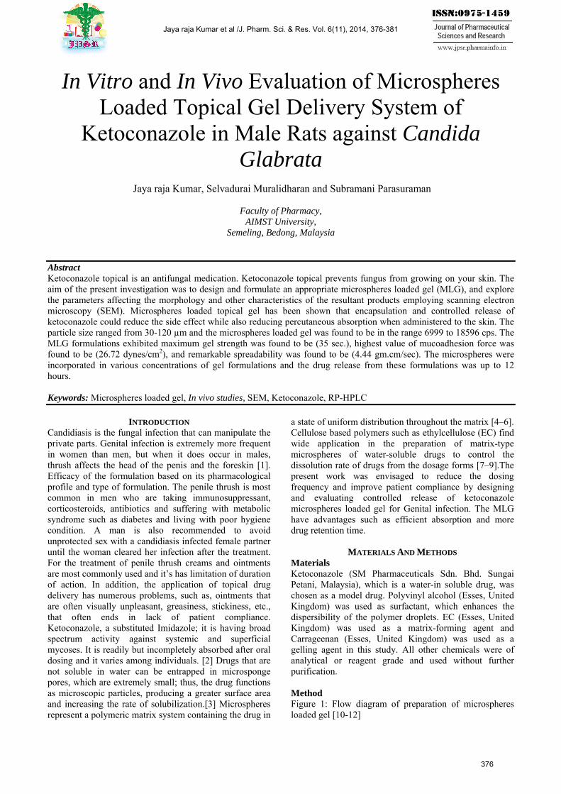
In Vitro and In Vivo Evaluation of Microspheres Loaded Topical Gel Delivery System of
Ketoconazole in Male Rats against Candida Glabrata
Jaya raja Kumar, Selvadurai Muralidharan and Subramani Parasuraman
Faculty of Pharmacy,
AIMST University, Semeling, Bedong, Malaysia
Abstract Ketoconazole topical is an antifungal medication. Ketoconazole topical prevents fungus from growing on your skin. The aim of the present investigation was to design and formulate an appropriate microspheres loaded gel (MLG), and explore the parameters affecting the morphology and other characteristics of the resultant products employing scanning electron microscopy (SEM). Microspheres loaded topical gel has been shown that encapsulation and controlled release of ketoconazole could reduce the side effect while also reducing percutaneous absorption when administered to the skin. The particle size ranged from 30-120 µm and the microspheres loaded gel was found to be in the range 6999 to 18596 cps. The MLG formulations exhibited maximum gel strength was found to be (35 sec.), highest value of mucoadhesion force was found to be (26.72 dynes/cm2), and remarkable spreadability was found to be (4.44 gm.cm/sec). The microspheres were incorporated in various concentrations of gel formulations and the drug release from these formulations was up to 12 hours. Keywords: Microspheres loaded gel, In vivo studies, SEM, Ketoconazole, RP-HPLC
INTRODUCTION
Candidiasis is the fungal infection that can manipulate the private parts. Genital infection is extremely more frequent in women than men, but when it does occur in males, thrush affects the head of the penis and the foreskin [1]. Efficacy of the formulation based on its pharmacological profile and type of formulation. The penile thrush is most common in men who are taking immunosuppressant, corticosteroids, antibiotics and suffering with metabolic syndrome such as diabetes and living with poor hygiene condition. A man is also recommended to avoid unprotected sex with a candidiasis infected female partner until the woman cleared her infection after the treatment. For the treatment of penile thrush creams and ointments are most commonly used and it’s has limitation of duration of action. In addition, the application of topical drug delivery has numerous problems, such as, ointments that are often visually unpleasant, greasiness, stickiness, etc., that often ends in lack of patient compliance. Ketoconazole, a substituted Imidazole; it is having broad spectrum activity against systemic and superficial mycoses. It is readily but incompletely absorbed after oral dosing and it varies among individuals. [2] Drugs that are not soluble in water can be entrapped in microsponge pores, which are extremely small; thus, the drug functions as microscopic particles, producing a greater surface area and increasing the rate of solubilization.[3] Microspheres represent a polymeric matrix system containing the drug in
a state of uniform distribution throughout the matrix [4–6]. Cellulose based polymers such as ethylcellulose (EC) find wide application in the preparation of matrix-type microspheres of water-soluble drugs to control the dissolution rate of drugs from the dosage forms [7–9].The present work was envisaged to reduce the dosing frequency and improve patient compliance by designing and evaluating controlled release of ketoconazole microspheres loaded gel for Genital infection. The MLG have advantages such as efficient absorption and more drug retention time.
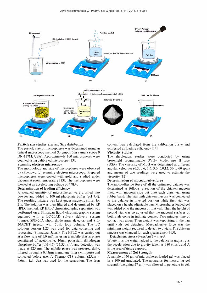
MATERIALS AND METHODS Materials Ketoconazole (SM Pharmaceuticals Sdn. Bhd. Sungai Petani, Malaysia), which is a water-in soluble drug, was chosen as a model drug. Polyvinyl alcohol (Esses, United Kingdom) was used as surfactant, which enhances the dispersibility of the polymer droplets. EC (Esses, United Kingdom) was used as a matrix-forming agent and Carrageenan (Esses, United Kingdom) was used as a gelling agent in this study. All other chemicals were of analytical or reagent grade and used without further purification. Method Figure 1: Flow diagram of preparation of microspheres loaded gel [10-12]
Jaya raja Kumar et al /J. Pharm. Sci. & Res. Vol. 6(11), 2014, 376-381
376
Particle size studies Size and Size distribution The particle size of microspheres was determined using an optical microscopy method (Olympus 70g camera scope 9 DN-117M, USA). Approximately 100 microspheres were counted using calibrated microscope [13]. Scanning electron microscopy The morphology and size of microspheres were observed by (Phenoworld) scanning electron microscopy. Prepared microspheres were coated with gold and studied under vacuum at room temperature [13]. The microspheres were viewed at an accelerating voltage of 4.8kV. Determination of loading efficiency A weighed quantity of microspheres were crushed into powder and added to 100 ml phosphate buffer (pH 7.4). The resulting mixture was kept under magnetic stirrer for 2 h. The solution was then filtered and determined by RP HPLC method. RP HPLC chromatographic separation was performed on a Shimadzu liquid chromatographic system equipped with a LC-20AD solvent delivery system (pump), SPD-20A photo diode array detector, and SIL-20ACHT injector with 50μL loop volume. The LC solution version 1.25 was used for data collecting and processing (Shimadzu, Japan). The HPLC was carried out at a flow rate of 1.0 ml/min using a mobile that is phase constituted of acetonitrile, 10mm potassium dihydrogen phosphate buffer (pH 4.5) (65:35, v/v), and detection was made at 225 nm. The mobile phase was prepared daily, filtered through a 0.45μm membrane filter (Millipore) and sonicated before use. A Thermo C18 column (25cm × 4.6mm i.d., 5μ) was used for the separation. The drug
content was calculated from the calibration curve and expressed as loading efficiency [14]. Viscosity Studies The rheological studies were conducted by using brookfield programmable DVII+ Model pro II type (USA). The viscosity of MLG was determined at different angular velocities (0.3, 0.6, 1.5, 3.0, 6.0,12, 30 to 60 rpm) and means of two readings were used to estimate the viscosity [12]. Determination of mucoadhesive force
The mucoadhesive force of all the optimized batches was determined as follows, a section of the chicken mucosa fixed with mucosal side out onto each glass vial using rubber band. The vial with chicken mucosa was connected to the balance in inverted position while first vial was placed on a height adjustable pan. Microspheres loaded gel was added onto the mucosa of first vial. Then the height of second vial was so adjusted that the mucosal surfaces of both vials come in intimate contact. Two minutes time of contact was given. Then weight was kept rising in the pan until vials get detached. Mucoadhesive force was the minimum weight required to detach two vials. The chicken mucosa was changed for each measurement [15].
Detachment stress (dynes/cm2) = m g/A Where m is the weight added to the balance in grams; g is the acceleration due to gravity taken as 980 cm/s2; and A is the area of tissue exposed. Measurement of Gel Strength A sample of 50 gm of microspheres loaded gel was placed in a 100 ml graduated. The apparatus for measuring gel strength (weighing 27 gm) was allowed to penetrate in gel.
Jaya raja Kumar et al /J. Pharm. Sci. & Res. Vol. 6(11), 2014, 376-381
377
The gel strength, which means the viscosity of the gels was determined by the time (seconds), the apparatus took to sink 5cm down through the prepared gel [15]. Spreadability For the determination of spreadability, excess of sample was applied in between two glass slides and was compressed to uniform thickness by placing 1000g weight for 5 min. weight (50 g) was added to the pan. The time in which the upper glass slide moves over to the lower plate was taken as measure of spreadability [15]. S= ML/T Where, M = weight tide to upper slide (g) L = length moved on the glass slide (cm) T = time taken (sec) Diffusion studies The in vitro release of MLG formulations were studied using cellophane membrane using modified apparatus. The dissolution medium used was phosphate buffer, freshly prepared (pH 7.4). Cellophane membrane previously soaked overnight in the dissolution medium, was tied to one end of a specifically designed glass cylinder (open at both ends). One gram of MLG (equivalent to 1% w/w of ketoconazole) was accurately placed into this assembly. The cylinder was attached to stand and suspended in 200 ml of dissolution medium maintained at 37 ± 1°C, the membrane just touching the receptor medium surface. The dissolution medium was stirred at 100 RPM speed using teflon coated magnetic bead. Aliquots, each of 5 ml volume were withdrawn periodically at predetermined time interval of 0.15, 0.30, 1.0, 2.0, 3.0, 4.0, 5.0 up to 12 hours and replaced by an equal volume of the receptor medium. The samples were appropriately diluted and measured by using RP HPLC method. In vivo evaluation of therapeutic efficacy Animals: Adult Wistar rats (280 ± 10 g) of either gender were obtained from SCS College of pharmacy, Harapanahalli. The animals were housed in large, spacious polyacrylic cages at an ambient room temperature with 12-h light/12-h dark cycle. Rats had free access to water and rodent pellets diet. The study was approved by the Institute Animal Ethics Committee of the SCS College of pharmacy, Harapanahalli and all the animal experiments were carried out according to the Committee for the Purpose of Control and Supervision of Experiments on Animals (CPCSEA) guidelines, Ministry of Environment and Forests, Government of India. Acute toxicity testing: The female rats were used for the acute toxicity testing. Hair present in the dorsal surface of the animal (2 X 2 cm) was removed by applying hair remover and cleaned with alcohol. The screening are was marked (1 X 1 cm) and 0.5 g of a microspheres loaded gel was applied to the surface of an animal's skin. During the observation period (14 days), signs such as erythema and edema were assessed [16]. Evaluation of therapeutic efficacy: The male rats were used for the experiment. The rats were divided into the four groups viz., normal control (group I), Candida glabrata control (group II), standard treatment group
(group III) and microsponges enriched gel treatment group (group IV). Group II to IV animals were changed with intravenous methylprednisolone (5mg/kg) for 3 days for induction and maintenance of cell-mediated immunosuppression (Organisms from stock isolates were stored in nutrient agar at 27°C, streaked onto nutrient broth, and incubated at 37°C for 24 h and included culture was used for further experiment). Candida glabrata culture was diluted with PBS and swabbed in smooth muscle of rat pennies and allowed to grow for 3 days until the growth of Candida was observed on ischiocavernosus smooth muscle. The colony growth was confirmed by counting colony-forming-unit. The animals which as CFU value of more than 3 CFU/ml ware included in the study. The animals were treated for week period and visually observed its physical changes. The swab culture was collected on initial day, 4th and 7th day of the experiment for microscopical evaluation. End of the experiment the animals were sacrificed and ischiocavernosus smooth muscle was collected from all the experimental animals and preserved in 10% formalin. Microscopical evaluation: The colony was collected in sterile cotton swab and transferred into 0.5 ml sterile phosphate buffer saline (PBS). The mixture was diluted 10 fold and inculcated in nutrient agar media, incubated for 48 h at 37°C. The yeast count was expressed as log10 of CFU/ml of PBS. Histopathologic analysis: The liver and pancreas were dehydrated with alcohol for 12 h each and cleaned with xylene for 15-20 min. The tissue blocks were prepared and the blocks were cut using microtome to get sections of thickness 5 μm. The sections were taken on a microscopic slide on which egg albumin (sticky substance) was applied and allowed for drying. Finally, Serial cross sections of the tissues were obtained and stained with periodic acid-Schiff (PAS) for fungal visualization [17]. Statistical analysis All the data were expressed as mean ± SEM. Statistical significance between the groups were tested using one-way analysis of variance (ANOVA) followed by Dunnett's t-test post-hoc test. A P less than 0.5 were considered significant.
RESULTS AND DISCUSSION Ethyl cellulose microspheres were prepared by an emulsion/solvent evaporation method, using water as non-solvent. Microencapsulated techniques have frequently been used for lipophilic drugs since hydrophilic drugs showed low loading efficiency [18]. Ketoconazole, due to its lipophilicity is likely to preferentially no partition out into the aqueous medium, leading to high loading efficiency. The effects of numerous process and formulation limitations on the drug loading efficiency of microspheres are shown in Table 1. The highest (85.67%) entrapment loading was achieved with polymer-drug ratio (1:1) and further increase in polymer-drug ratio from1:1 to 1:2 and 1:3 shown decrease in loading efficiency of ketoconazole. As the concentration of ethyl cellulose increased the viscosity of the solution increased resulting in the
Jaya raja Kumar et al /J. Pharm. Sci. & Res. Vol. 6(11), 2014, 376-381
378
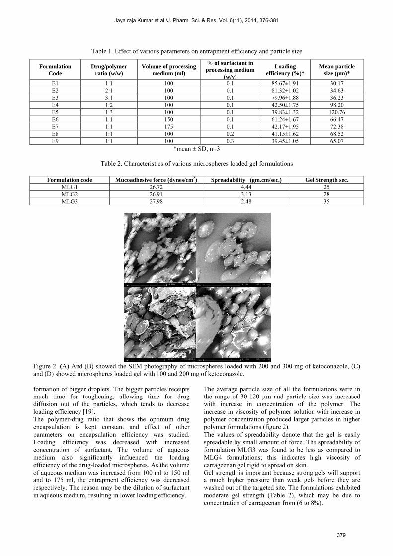
Table 1. Effect of various parameters on entrapment efficiency and particle size
*mean ± SD, n=3
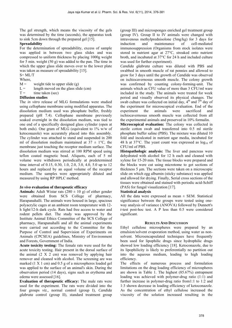
Table 2. Characteristics of various microspheres loaded gel formulations
Figure 2. (A) And (B) showed the SEM photography of microspheres loaded with 200 and 300 mg of ketoconazole, (C) and (D) showed microspheres loaded gel with 100 and 200 mg of ketoconazole. formation of bigger droplets. The bigger particles receipts much time for toughening, allowing time for drug diffusion out of the particles, which tends to decrease loading efficiency [19]. The polymer-drug ratio that shows the optimum drug encapsulation is kept constant and effect of other parameters on encapsulation efficiency was studied. Loading efficiency was decreased with increased concentration of surfactant. The volume of aqueous medium also significantly influenced the loading efficiency of the drug-loaded microspheres. As the volume of aqueous medium was increased from 100 ml to 150 ml and to 175 ml, the entrapment efficiency was decreased respectively. The reason may be the dilution of surfactant in aqueous medium, resulting in lower loading efficiency.
The average particle size of all the formulations were in the range of 30-120 µm and particle size was increased with increase in concentration of the polymer. The increase in viscosity of polymer solution with increase in polymer concentration produced larger particles in higher polymer formulations (figure 2). The values of spreadability denote that the gel is easily spreadable by small amount of force. The spreadability of formulation MLG3 was found to be less as compared to MLG4 formulations; this indicates high viscosity of carrageenan gel rigid to spread on skin. Gel strength is important because strong gels will support a much higher pressure than weak gels before they are washed out of the targeted site. The formulations exhibited moderate gel strength (Table 2), which may be due to concentration of carrageenan from (6 to 8%).
Formulation Code
Drug/polymer ratio (w/w)
Volume of processing medium (ml)
% of surfactant in processing medium
(w/v)
Loading efficiency (%)*
Mean particle size (µm)*
E1 1:1 100 0.1 85.67±1.91 30.17 E2 2:1 100 0.1 81.32±1.02 34.63 E3 3:1 100 0.1 79.96±1.88 36.23 E4 1:2 100 0.1 42.50±1.75 98.20 E5 1:3 100 0.1 39.83±1.32 120.76 E6 1:1 150 0.1 61.24±1.67 66.47 E7 1:1 175 0.1 42.17±1.95 72.38 E8 1:1 100 0.2 41.15±1.62 68.52 E9 1:1 100 0.3 39.45±1.05 65.07
Formulation code Mucoadhesive force (dynes/cm2) Spreadability (gm.cm/sec.) Gel Strength sec. MLG1 26.72 4.44 25 MLG2 26.91 3.13 28 MLG3 27.98 2.48 35
Jaya raja Kumar et al /J. Pharm. Sci. & Res. Vol. 6(11), 2014, 376-381
379
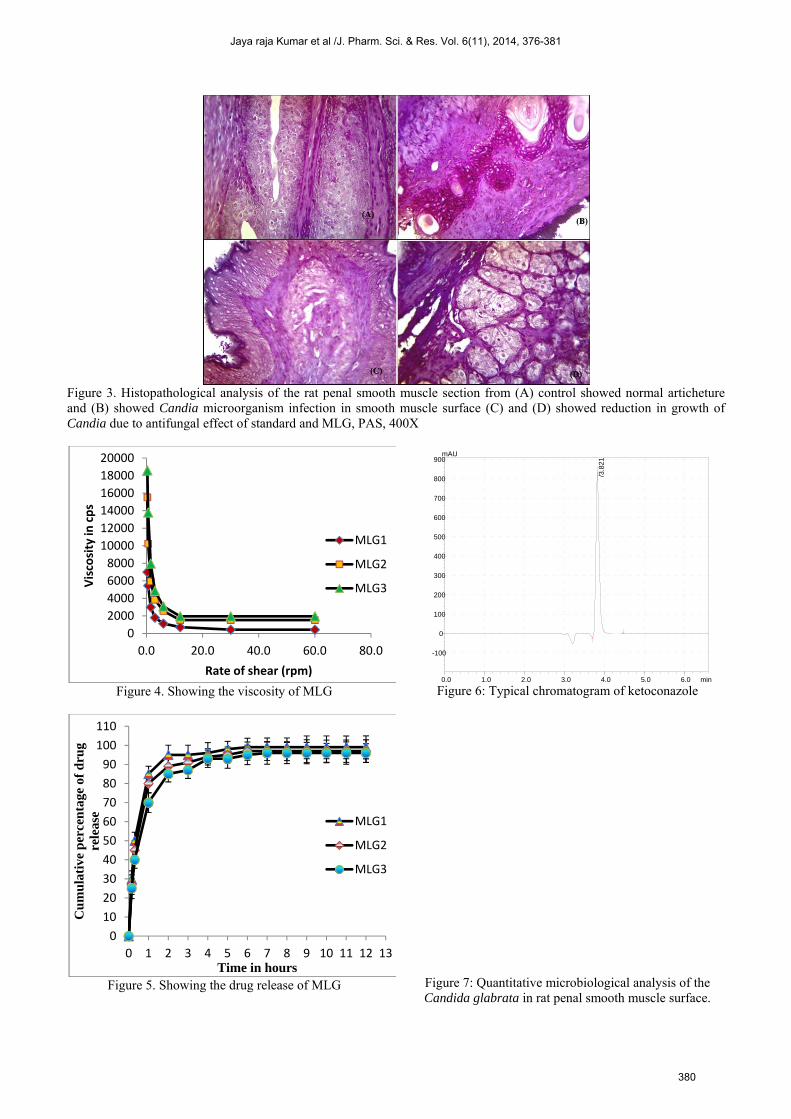
Figure 3. Histopathological analysis of the rat penal smooth muscle section from (A) control showed normal articheture and (B) showed Candia microorganism infection in smooth muscle surface (C) and (D) showed reduction in growth of Candia due to antifungal effect of standard and MLG, PAS, 400X
Figure 4. Showing the viscosity of MLG
Figure 5. Showing the drug release of MLG
Figure 6: Typical chromatogram of ketoconazole
Figure 7: Quantitative microbiological analysis of the Candida glabrata in rat penal smooth muscle surface.
02000400060008000
100001200014000160001800020000
0.0 20.0 40.0 60.0 80.0
Visc
osity
in c
ps
Rate of shear (rpm)
MLG1
MLG2
MLG3
0102030405060708090100110
0 1 2 3 4 5 6 7 8 9 10 11 12 13
Cu
mu
lati
ve p
erce
nta
ge o
f d
rug
rele
ase
Time in hours
MLG1
MLG2
MLG3
0.0 1.0 2.0 3.0 4.0 5.0 6.0 min
-100
0
100
200
300
400
500
600
700
800
900mAU
/3.8
21
Jaya raja Kumar et al /J. Pharm. Sci. & Res. Vol. 6(11), 2014, 376-381
380
Mucoadhesive drug delivery system to mucosal membrane leads to an increase in the drug concentration at the absorption site and therefore improved bioavailability of systemic delivery drugs [20]. The mucoadhesive force is an important physic-chemical parameter for topical application. The formulations showed maximum mucoadhesive force, these may be due to increase in concentration of carrageenan in the formulations (Table 2). All the MLG formulations were behaving as shear thinning systems (Figure 4). The formulation MLG 3 having the maximum concentration of carrageenan (8% w/v) showed maximum viscosity in gels forms and hence was not suitable for application. The drug release was decreased with increase in concentration of the carrageenan as increase in the gel viscosity has effect on drug bust from the microspheres. It is understood that higher viscosity of gelling agent results in a longer diffusional path length, so drug release is extended for 12 hours (Figure 5-6). The therapeutic efficacy of MLG was compared with Candida glabrata control by quantitative microbiological analysis and histopathological evaluations (Figure 3). MLG and standard marketed formulation treated animals showed significant reduction of CFU count on 4th day of the treatment onwards. The efficacy of the MLG is comparable with standard marketed formulation (Figure 7).
CONCLUSION Microspheres loaded gel of ketoconazole was successfully prepared by emulsion solvent diffusion method using EC polymer and carrageenan as a gelling agent. The concentration of the EC influenced in all the evaluated parameters. The above findings revealed that Microspheres loaded gel of ketoconazole offer a convenient dosage form for achieving better release profiles and improved patient compliance. The in vivo animal studies conducted on adult wistar rats against Candida glabrata in penile candidiasis revealed that the MLG formulation made with ketoconazole were eradicating completely the fungal burden in comparison with standard.
REFERENCE 1. Cdc. Genital Candidiasis: Statistics. Centers For Disease Control
And Prevention. Published Online, Accessed 7th October 2013. 2. Hardman Jg, Limbird Le. Goodman And Gilman's. The
Pharmacological Basis Of Therapeutics. 10th Ed. New York: Mcgraw-Hill; 2001. Anti-Microbial Agents: Anti-Fungal Agents; Pp. 1301–2.
3. Chadawar V, Shaji J. Microsponge Delivery System. Curr Drug Deliv. 2007; 4 (2):123–129.
4. Andreani L, Cercena R, Ramos Bgz, Soldi V. Development And Characterization Of Wheat Gluten Microspheres For Use In A Controlled Release System. Mat Sci And Eng C. 2009; 29:524–31.
5. Mateovic T, Kriznar B, Bogataj M, Mrhar A. The Influence Of Stirring Rate On Biopharmaceutical Properties Of Eudragit Rs Microspheres. J Microencapsul. 2002;19:29–36.
6. Nordstierna L, Abdalla Aa, Nordin M, Nyden M. Comparison Of Release Behaviour From Microcapsules And Microspheres. Progress In Org Coat. 2010; 69:49–51.
7. Kangarlou S, Haririan I, Gholipour Y. Physico-Mechanical Analysis Of Freeethylcellulose Films Comprised With Novel Plast I C Izers Of Vitamin Resources. Int J Pharm. 2008; 356:153–66.
8. Lai Hl, Pitt K, Craig Dqm. Characterization Of The Thermal Properties Of Ethylcellulose Using Differential Scanning And Quasi-Isothermal Calorimetric Approaches. Int J Pharm. 2010; 386:178–84.
9. Sogol K, Ismaeil H. Mechanical Influence Of Static Versus Dynamic Loadings On Parametrical Analysis Of Plasticized Ethyl Cellulose Films. Int J Pharm. 2011; 408:1–8.
10. D’souza, J.I., Jagdish, K., Saboji, S.G. And Killedar,H.N.: More “Design And Evaluation Of Benzoyl Peroxide Microsponges To Enhance Therapeutic Efficacy In Acne Treatment”, Accepted For Presentation In 20th Fapa Congress, Bangkok , Thailand, Nov30-Dec 3 (2004).
11. Jaya Raja Kumar, Selvadurai Muralidharan, Sokkalingam Arumugam Dhanaraj. Anti-Fungal Activity Of Microemulsion Based Fluconazole Gel For Onychomycosis Against Aspergillus Niger, International Journal Of Pharmacy And Pharmaceutical Sciences 11/2012; Vol 5(1):96-102.
12. Jaya Raja Kumar, Selvadurai Muralidharan And Sanggetha Ramasamy .Microsponges Enriched Gel (Megs): A Novel Strategy For Opthalmic Drug Delivery System Containing Ketotifen, Journal Of Pharmacy Research 2013,5(4),97-102
13. Jaya Raja Kumar, Muralidharan S, Ramasamy S. Microsponges Enriched Gel (Megs): A Novel Strategy For Opthalmic Drug Delivery System Containing Ketotifen. J. Pharm. Sci. & Res 2013; 5: 97-102.
14. D’souza Ji, More Hn. Topical Anti-Inflammatory Gels Of Fluocinolone Acetonide Entrapped In Eudragit Based Microsponge Delivery System. Res J Pharm Tech 2008; 1:502-506
15. Development And In Vitro Evaluation Of Guar Gum Based Fluconazloe In Situ Gel For Oral Thrush, J.Pharm.Sci. & Res. Vol.4 (12), 2012, 2009-2014.
16. Parasuraman S. Toxicological Screening. J Pharmacol Pharmacother 2011; 2:74-9.
17. Giuseppe Intini, Alfredo Aguirre, Libuse Anna Bobek. Efficacy Of Human Salivary Mucin Muc7-Derived Peptide And Histatin 5 In A Murine Model Of Candidiasis, International Journal Of Antimicrobial Agents 22 (2003) 594 /600.
18. Jung-Hwa L, Tae Gwan P, Hoo-Kyan C. Effect Of Formulation And Processing Variable On The Characteristics Of Microspheres For Water Soluble Drugs Prepared By W/O/O Double Emulsion Solvent Diffusion Method. International Journal Of Pharmaceutics 2000; 196:75-78
19. Naveen Chella, Kiran Kumar Yada, Rashmi Vempati. Preparation And Evaluation Of Ethyl Cellulose Microspheres Containing Diclofenac Sodium By Novel W/O/O Emulsion Method, J. Pharm. Sci. & Res. Vol.2 (12), 2010, 884-888
20. Jaya Raja Kumar, Selvadurai Muralidharan, Subramani Parasuraman, Sokkalingam Arumugam Dhanaraj, Development Of Microparticle Loaded Gel (Mplgs) For Prolong Ocular Drug Delivery Containing Ketorolac Tromethamine, J. Pharm. Sci. & Res. Vol. 6(3), 2014, 148-152
Jaya raja Kumar et al /J. Pharm. Sci. & Res. Vol. 6(11), 2014, 376-381
381