Embed Size (px)
Citation preview
1
IN VITRO AND IN VIVO EFFECTS OF α7 NICOTINIC ACETYLCHOLINE RECEPTOR GENE DELIVERY ON NEUROPROTECTIVE PATHWAYS
By
YAN REN
A DISSERTATION PRESENTED TO THE GRADUATE SCHOOL OF THE UNIVERSITY OF FLORIDA IN PARTIAL FULFILLMENT
OF THE REQUIREMENTS FOR THE DEGREE OF DOCTOR OF PHILOSOPHY
UNIVERSITY OF FLORIDA
2011

2
© 2011 Yan Ren

3
To my parents, Bao’er Ren and Qiaozhen Pu, and my grandmother, Fenlan Ge

4
ACKNOWLEDGMENTS
The research described in this dissertation is made possible through the help and
support from the faculty, staff and fellow students at the University of Florida. First of all,
I would like to express my sincere appreciation to my mentors, Dr. Sihong Song and Dr.
Jeffrey A. Hughes, for their invaluable guidance and support throughout my graduate
education. I would also like to thank Dr. Edwin M. Meyer for his care along every step of
my graduate study. Not only has he mentored me in developing research skills, Dr.
Meyer also took the patience to read my presentations and manuscripts and made great
effort to help improve my English speaking and writing. I am also grateful to Dr. Michael
A. King, for his generosity in sharing ideas and experience. He is always there when I
need advice and has been my constant source of neuroscience knowledge.
I also thank all my committee members for their helpful comments and
suggestions. Dr. Hartmut Derendorf has an amazing knowledge of pharmacometrics,
and has inspired my interest in this area with his brilliant teaching skills. Dr. Veronika
Butterweck is always very kind and willing to help. Dr. Marta L. Wayne used to be my
director in Genetics and Genomics Graduate Program. She provided me the opportunity
to pursue my Ph.D degree at University of Florida; her thoughtfulness and consideration
have accompanied me through all these years.
This project could not have been completed without the help and encouragement
of many other people. Dr. Aaron C. Hirko, my current labmate, consistently helps me
with my experiments and I enjoyed every passionate discussion with him. Dr. Ke Ren,
my previous labmate and dear “sister”, have made my first year enjoyable both in lab
and in personal life. Dr. Roger L. Papke, professor in Department of Pharmacology and
Therapeutics, inspired me a lot with his talent and pursuit in science. He is also my

5
supplier of experimental drug. Mike Matheny, working next door to us, is an excellent
lab manager and generously lends me anything that I cannot find in our lab. I would also
like to thank all the staff and fellow graduate students in the Department of
Pharmaceutics as well as the Genetics and Genomics Program for their friendship and
support.
Last but not least, I thank my parents, for the loneliness they endured and the love
they constantly offered during my study oversea. I am grateful to my husband for his
listening and encouragement through good times and bad times. I would also like to
thank all my friends, in the United States and back in China, for making my ordinary life
extraordinary.
6
TABLE OF CONTENTS page
ACKNOWLEDGMENTS .................................................................................................. 4
LIST OF TABLES ............................................................................................................ 8
LIST OF FIGURES .......................................................................................................... 9
LIST OF ABBREVIATIONS ........................................................................................... 11
ABSTRACT ................................................................................................................... 13
CHAPTER
1 INTRODUCTION .................................................................................................... 15
Alzheimer Disease .................................................................................................. 15 Cholinergic Hypothesis ........................................................................................... 16 Neuronal Nicotinic Acetylcholine Receptor ............................................................. 17 α7 nAChRs in Alzheimer Disease ........................................................................... 19
Identification ..................................................................................................... 19 Localization ...................................................................................................... 19 Function............................................................................................................ 20
α7 nAChRs Mediated Neuroprotection ................................................................... 20 Effects on in Vitro and in Vivo Models .............................................................. 20 α7 nAChRs and Cellular Signaling Pathway .................................................... 21 α7 nAChRs and TrkA ....................................................................................... 22 α7 nAChRs and β Amyloid ............................................................................... 23
Therapeutic Approaches Targeting α7 nAChRs ..................................................... 25 Agonist Therapy ............................................................................................... 25 Gene Therapy .................................................................................................. 26
Specific Aims and Significance ............................................................................... 28
2 MATERIALS AND METHODS ................................................................................ 30
Reagents ................................................................................................................ 30 Plasmid Preparations .............................................................................................. 30 Cell Culture ............................................................................................................. 31
Cell Gene Delivery, Differentiation and Drug Treatments ................................. 32 Cell Viability ...................................................................................................... 32
α7 Nicotinic Receptor Binding Assay ...................................................................... 33 Enzyme-Linked Immunosorbent Assay................................................................... 33 Western Blot ........................................................................................................... 34 Caspase Assay ....................................................................................................... 35 Immunofluorescence............................................................................................... 35 Virus Vector Packaging and Titration ...................................................................... 36
7
Transfection ...................................................................................................... 36 Purification ........................................................................................................ 36 Dot-Blot Assay .................................................................................................. 38
Stereotaxic Surgery ................................................................................................ 39 Morris Water Maze Tests ........................................................................................ 40 Immunohistochemistry ............................................................................................ 41 Statistical Analysis .................................................................................................. 42
3 EFFECTS OF α7 RECEPTOR GENE DELIVERY ON PROCESSES UNDERLYING CELL VIABILITY ............................................................................. 43
Introduction ............................................................................................................. 43 Results .................................................................................................................... 46
Gene Delivery System Resulted Functional α7 Receptor Expression .............. 46 Effects of 4OH-GTS-21 on p-ERK2 and p-Akt in Transfected Cells: ................ 50 Effects of 4OH-GTS-21 on Caspase Activity: ................................................... 51
Discussion .............................................................................................................. 55
4 EFFECTS OF α7 RECEPTOR GENE DELIVERY ON NEUROTROPHIN PATHWAY .............................................................................................................. 61
Introduction ............................................................................................................. 61 Results .................................................................................................................... 65
Effects of α7 nAChRs Overexpression on TrkA In Vitro ................................... 65 Effects of AAV8-α7 Gene Delivery on TrkA in Vivo .......................................... 70 Effects of Septal AAV8-α7 Gene Delivery on Memory Related Behavior ......... 72
Discussion .............................................................................................................. 75
5 CONCLUSION AND FUTURE DIRECTION ........................................................... 80
Conclusion .............................................................................................................. 80 Future Direction ...................................................................................................... 82
LIST OF REFERENCES ............................................................................................... 85
BIOGRAPHICAL SKETCH .......................................................................................... 102

8
LIST OF TABLES
Table page 1-1 FDA approved drugs for treating Alzheimer disease. ......................................... 17
9
LIST OF FIGURES
Figure page 1-1 Structure of α7 and α4β2 nAChRs. ..................................................................... 18
1-2 Cellular signaling pathways involved in α7 nAChRs mediated neuroprotection. .................................................................................................. 22
1-3 Chemical structure of GTS-21 and 4OH-GTS-21. .............................................. 26
3-1 Transfection efficiency in PC12 cells. ................................................................. 47
3-2 Effects of α7 and ric-3 gene delivery on α7 receptor density in PC12 cells. ....... 48
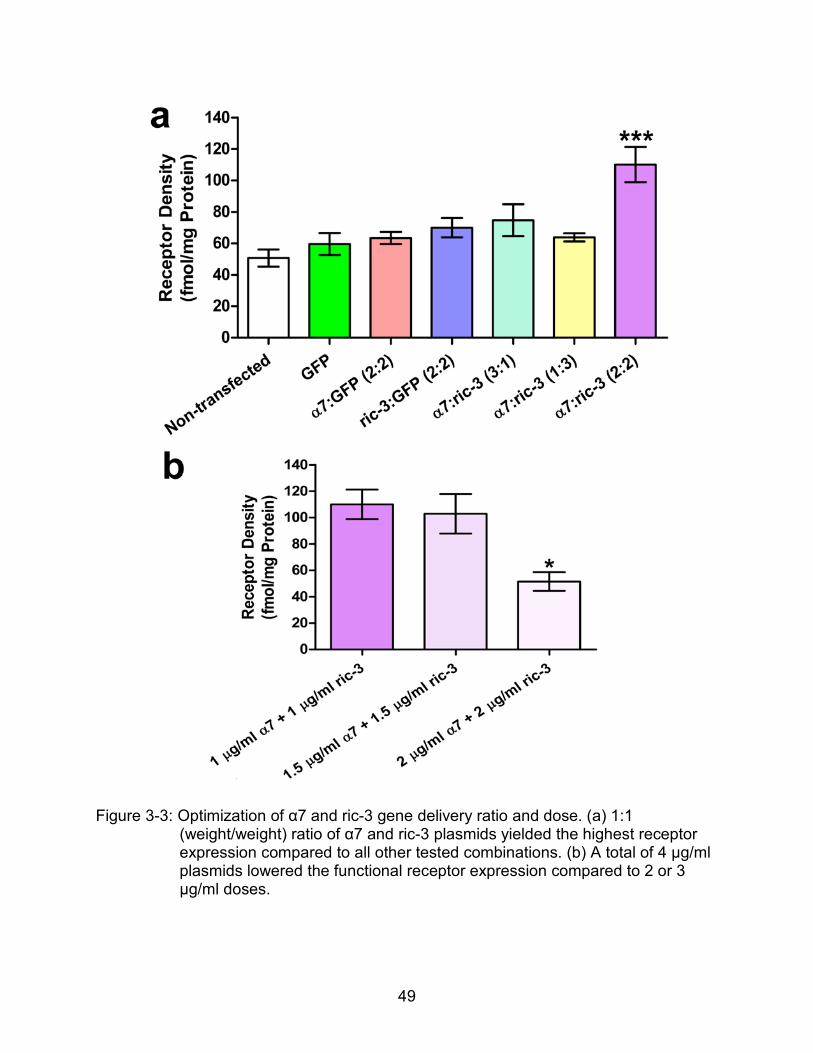
3-3 Optimization of α7 and ric-3 gene delivery ratio and dose.. ................................ 49
3-4 Effects of α7 overepxression on 4OH-GTS-21 induced ERK2 response.. .......... 52
3-5 Effects of α7 overepxression on 4OH-GTS-21 induced AKT response. ............. 53
3-6 AKT phosphorylation is not affected by ERK inhibitor in the case of α7 activaton.. ........................................................................................................... 54
3-7 Effects of α7 overexpression and activation on JNK phosphorylation. ............... 55
3-8 Effects of α7 overexpression on 4OH-GTS-21 induced caspase 3/7 response.. ........................................................................................................... 56
4-1 Sample pictures for PC12 differentiation experiment.. ........................................ 66
4-2 Effects of α7 and ric-3 co-transfection on NGF mediated PC12 cell differentiation.. .................................................................................................... 68
4-3 Immunoactivity of TrkA in PC12 cells after α7 and ric-3 co-transfection.. ........... 69
4-4 Immunoactivities of α7, TrkA and ChAT in septum, left side hippocampus and right hippocampus after α7 overexpression. ................................................ 71
4-5 Immunofluorescence staining of TrkA and α7 protein in left septum after α7 overexpression. .................................................................................................. 72
4-6 Training curves resulted from Morris water maze platform navigation task.. ...... 73
4-7 Swimming tracks of individual animal in probe test. ........................................... 74
4-8 Memory related parameters resulted from probe test.. ....................................... 75
4-9 API-2 blocked cytoprotective effects seen in α7 up-regulated groups.. .............. 77

10
5-1 Pathways regulated by α7 and TrkA receptors in PC12 cells.. ........................... 81
5-2 Septohippocampal reciprocal loop that may involve in α7 mediated TrkA elevation. ............................................................................................................ 82
11
LIST OF ABBREVIATIONS
AAV Adeno associate virus
Aβ β amyloid
ACh Acetylcholine
AChE Acetylcholinesterase
AD Alzheimer disease
APP Amyloid precursor protein
Bcl-2 B -cell lymphoma
BDNF Brain derived neurotrophic factor
BH Bcl-2 homology
CaMKII Calcium / calmodulin-dependent protein kinase II
cAMP Cyclic adenosine monophosphate
CBA Chicken β-actin
ChAT Choline acetyltransferase
CMV Cytomegalovirus
CNS Central nervous system
CREB cAMP response element-binding protein
DMXB 3-(2, 4)-Dimethoxybenzylidine anabaseine
ER Endoplasmic reticulum
ERK Extracellular signal regulated kinase
FDA Food and Drug Administration
GDP Gross domestic product
JAK2 Janus kinase 2
JNK c-jun-N-terminal kinase
KRH Krebs Ringer buffer
12
MAPK mitogen-activated protein kinases
MLA Methyllycacotinine
MRI Magnetic resonance imaging
nAChR Nicotinic acetylcholine receptor
NBM Nucleus basalis of meynert
NGF Nerve growth factor
NT-3 Neurotrophin-3
PBS Phosphate buffered saline
PC12 Rat phaeochromocytoma cells
PET Positron emission tomography
PKC protein kinase C
PI3K Phosphoinositide 3-kinase
STAT3 Signal transducer and activator of transcription protein 3
TrkA Tyrosine kinase receptor type 1
13
Abstract of Dissertation Presented to the Graduate School of the University of Florida in Partial Fulfillment of the Requirements for the Degree of Doctor of Philosophy
IN VITRO AND IN VIVO EFFECTS OF α7 NICOTINIC ACETYLCHOLINE RECEPTOR
GENE DELIVERY ON NEUROPROTECTIVE PROCESSES
By
Yan Ren
May 2011
Chair: Sihong Song, Ph.D Major: Genetics and Genomics
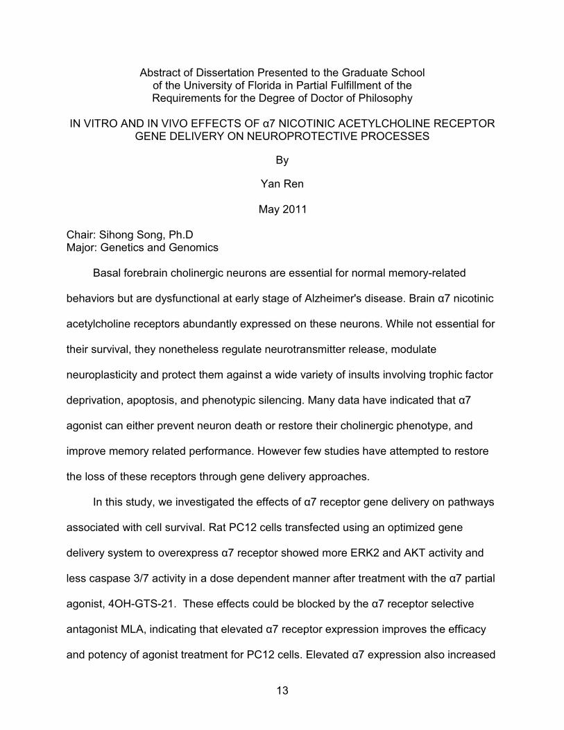
Basal forebrain cholinergic neurons are essential for normal memory-related
behaviors but are dysfunctional at early stage of Alzheimer's disease. Brain α7 nicotinic
acetylcholine receptors abundantly expressed on these neurons. While not essential for
their survival, they nonetheless regulate neurotransmitter release, modulate
neuroplasticity and protect them against a wide variety of insults involving trophic factor
deprivation, apoptosis, and phenotypic silencing. Many data have indicated that α7
agonist can either prevent neuron death or restore their cholinergic phenotype, and
improve memory related performance. However few studies have attempted to restore
the loss of these receptors through gene delivery approaches.
In this study, we investigated the effects of α7 receptor gene delivery on pathways
associated with cell survival. Rat PC12 cells transfected using an optimized gene
delivery system to overexpress α7 receptor showed more ERK2 and AKT activity and
less caspase 3/7 activity in a dose dependent manner after treatment with the α7 partial
agonist, 4OH-GTS-21. These effects could be blocked by the α7 receptor selective
antagonist MLA, indicating that elevated α7 receptor expression improves the efficacy
and potency of agonist treatment for PC12 cells. Elevated α7 expression also increased
14
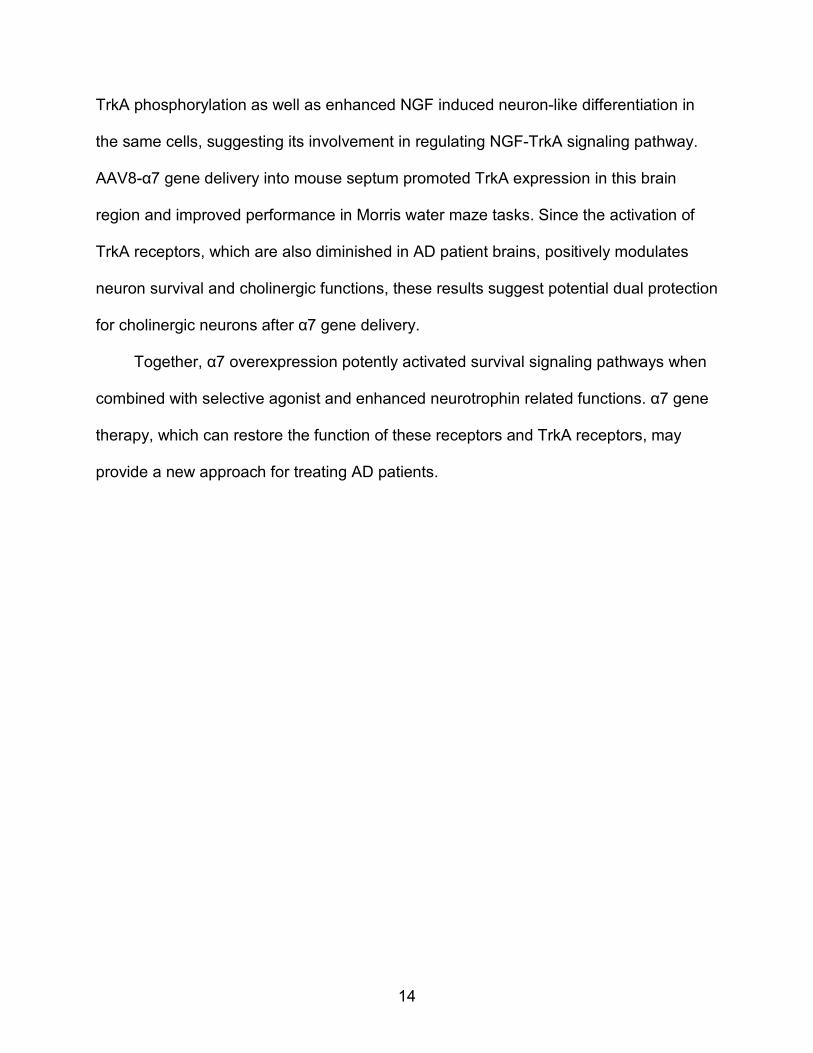
TrkA phosphorylation as well as enhanced NGF induced neuron-like differentiation in
the same cells, suggesting its involvement in regulating NGF-TrkA signaling pathway.
AAV8-α7 gene delivery into mouse septum promoted TrkA expression in this brain
region and improved performance in Morris water maze tasks. Since the activation of
TrkA receptors, which are also diminished in AD patient brains, positively modulates
neuron survival and cholinergic functions, these results suggest potential dual protection
for cholinergic neurons after α7 gene delivery.
Together, α7 overexpression potently activated survival signaling pathways when
combined with selective agonist and enhanced neurotrophin related functions. α7 gene
therapy, which can restore the function of these receptors and TrkA receptors, may
provide a new approach for treating AD patients.
15
CHAPTER 1 INTRODUCTION
Alzheimer Disease
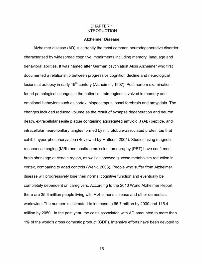
Alzheimer disease (AD) is currently the most common neurodegenerative disorder
characterized by widespread cognitive impairments including memory, language and
behavioral abilities. It was named after German psychiatrist Alois Alzheimer who first
documented a relationship between progressive cognition decline and neurological
lesions at autopsy in early 19th century (Alzheimer, 1907). Postmortem examination
found pathological changes in the patient’s brain regions involved in memory and
emotional behaviors such as cortex, hippocampus, basal forebrain and amygdala. The
changes included reduced volume as the result of synapse degeneration and neuron
death, extracellular senile plaque containing aggregated amyloid β (Aβ) peptide, and
intracellular neurofibrillary tangles formed by microtubule-associated protein tau that
exhibit hyper-phosphorylation (Reviewed by Mattson, 2004). Studies using magnetic
resonance imaging (MRI) and positron emission tomography (PET) have confirmed
brain shrinkage at certain region, as well as showed glucose metabolism reduction in
cortex, comparing to aged controls (Wenk, 2003). People who suffer from Alzheimer
disease will progressively lose their normal cognitive function and eventually be
completely dependent on caregivers. According to the 2010 World Alzheimer Report,
there are 35.6 million people living with Alzheimer’s disease and other dementias
worldwide. The number is estimated to increase to 65.7 million by 2030 and 115.4
million by 2050. In the past year, the costs associated with AD amounted to more than
1% of the world's gross domestic product (GDP). Intensive efforts have been devoted to

16
find promising therapeutic approaches for AD, yet its pathogenesis has not been fully
elucidated. To data, several hypotheses have been proposed and studied in depth.
Cholinergic Hypothesis
The cholinergic hypothesis was initially presented in 1970s, which suggested that
dysfunction of cholinergic neurotransmitter system in certain brain regions contributes to
the cognitive decline (Bartus et al., 1982; Coyle et al., 1983). The cholinergic system is
one of the most important nervous pathways. Acetylcholine (ACh) is the
neurotransmitter that is synthesized, stored and released by cholinergic neurons.
Reductions in the activity of the cholinergic enzymes choline acetyl transferase (ChAT)
were identified as being most severe in the cortex and hippocampus (Davies, 1979) in
AD patients’ brain compared to age-matched controls, along with descending ACh
synthesis and release (Bowen et al., 1981). A loss of cholinergic forebrain neurons of
the nucleus basalis of meynert (NBM) was then shown to be the first evidence of
neurotransmitter-specific neuronal loss in AD (Whitehouse et al., 1982). Since NBM
projects the majority of cholinergic input to hippocampus and cortex (Mesulam and Van
Hoesen, 1976; Wenk et al., 1980), it suggested that selective loss of cholinergic
neurons in basal forebrain resulted in reduced cholinergic input in these two regions
reflecting decreased ChAT activity. Disrupting cholinergic function by using antagonists
(anti-muscarnic or anti-nicotinic receptors) or fimbria-fonix lesion (which damages the
cholinergic input to cortex or hippocampus from the basal forebrain) revealed similar
memory deficits in animals and young humans as those in AD animal models / patients
(Terry and Buccafusco, 2003). In contrast, appropriately enhancing cholinergic activity
by providing anti-acetylcholinesterase (AChE) inhibitor or receptor agonists reduced
memory impairments in aged subjects. Based on the cholinergic hypothesis, four
17
among the five Food and Drug Administration (FDA) recognized drugs for AD are AChE
inhibitors (Table 1-1): tacrine, donepezil, rivastigmine and galantamine. They have
provided convincing therapeutic effects balanced with an acceptable burden of side
effects. Memantine is a glutamate receptor antagonist, which reduces the excitotoxicity
induced by glutamate receptor over activation in AD (Molinuevo et al., 2005). All of
these currently available medications, however, can only help control symptoms of the
disease. Studies focusing on halting or preventing the disease process are pursued.
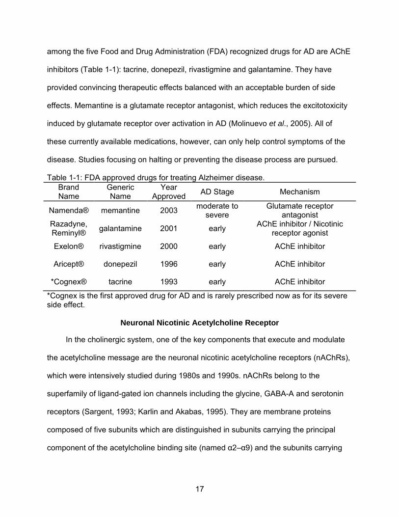
Table 1-1: FDA approved drugs for treating Alzheimer disease. Brand Name
Generic Name
Year Approved AD Stage Mechanism
Namenda® memantine 2003 moderate to severe
Glutamate receptor antagonist
Razadyne, Reminyl® galantamine 2001 early AChE inhibitor / Nicotinic
receptor agonist
Exelon® rivastigmine 2000 early AChE inhibitor
Aricept® donepezil 1996 early AChE inhibitor
*Cognex® tacrine 1993 early AChE inhibitor
*Cognex is the first approved drug for AD and is rarely prescribed now as for its severe side effect.
Neuronal Nicotinic Acetylcholine Receptor
In the cholinergic system, one of the key components that execute and modulate
the acetylcholine message are the neuronal nicotinic acetylcholine receptors (nAChRs),
which were intensively studied during 1980s and 1990s. nAChRs belong to the
superfamily of ligand-gated ion channels including the glycine, GABA-A and serotonin
receptors (Sargent, 1993; Karlin and Akabas, 1995). They are membrane proteins
composed of five subunits which are distinguished in subunits carrying the principal
component of the acetylcholine binding site (named α2–α9) and the subunits carrying
18
the complementary component of the acetylcholine binding site (named non α or β2–β4)
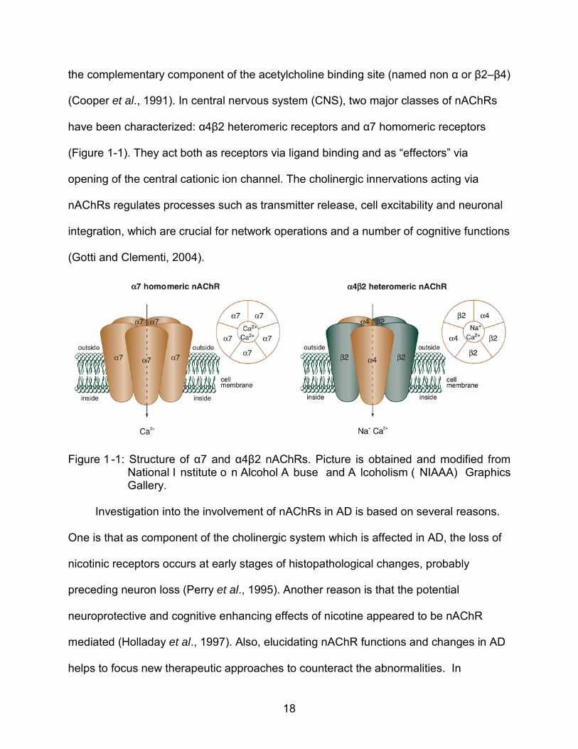
(Cooper et al., 1991). In central nervous system (CNS), two major classes of nAChRs
have been characterized: α4β2 heteromeric receptors and α7 homomeric receptors
(Figure 1-1). They act both as receptors via ligand binding and as “effectors” via
opening of the central cationic ion channel. The cholinergic innervations acting via
nAChRs regulates processes such as transmitter release, cell excitability and neuronal
integration, which are crucial for network operations and a number of cognitive functions
(Gotti and Clementi, 2004).
Figure 1 -1: Structure of α7 and α4β2 nAChRs. Picture is obtained and modified from National I nstitute o n Alcohol A buse and A lcoholism ( NIAAA) Graphics Gallery.
Investigation into the involvement of nAChRs in AD is based on several reasons.
One is that as component of the cholinergic system which is affected in AD, the loss of
nicotinic receptors occurs at early stages of histopathological changes, probably
preceding neuron loss (Perry et al., 1995). Another reason is that the potential
neuroprotective and cognitive enhancing effects of nicotine appeared to be nAChR
mediated (Holladay et al., 1997). Also, elucidating nAChR functions and changes in AD
helps to focus new therapeutic approaches to counteract the abnormalities. In

19
particular, the homomeric α7 receptors acquired particular attention because they
exhibited special role in memory enhancing and neuroprotective actions as discussed
below.
α7 nAChRs in Alzheimer Disease
Identification
Historically, α7 subtypes of nAChRs were distinguished from α4β2 nAChRs by its
high affinity to α-bungarotoxin (Kd 0.65~1.7 nM) and relatively low affinity to nicotine
(Clarke et al 1985, Marks et al., 1986, Schoepfer et al., 1990). α-bungarotoxin is a
peptide isolated from a species of East Asian snake (Bungarus multicinctus), and has
been recognized for its high affinity to muscle-type nicotinic receptors (Changeux et al.,
1970). These results were supported by the observation that α-bungarotoxin but not
nicotine binding sites were absent in α7 knockout mice (Orr-Urtreger et al., 1997). α7
genes of different species were then cloned and proven to encode homomeric
functional nicotinic channels, in which the β subunit is not needed (Couturier et al.,
1990; Seguela et al., 1993).
Localization
α-bungarotoxin binding sites are found in many brain regions and are especially
concentrated in hippocampus, cerebral cortex, hypothalamus (Domingues del Toro et
al., 1994; Bina et al., 1995), where they locate presynaptically to facilitate the release of
transmitters such as glutamate, GABA or dopamine (ALkondon et al., 1996;
MacDermott et al., 1999), as well as exist postsynaptically to mediate fast synaptic
responses (Jones et al., 1999, Broide and Leslie, 1999). Hippocampal and septal α7
receptor density is decreased at early stages of AD (Perry et al. 1995). Normal aging
may be associated with reductions in α7 subunit mRNA as well as protein expression
20
(Court et al. 2001), while in AD α7 protein loss (Engidawork et al. 2001; Perry et al.
2001) is observed without significant mRNA level change (Wevers et al. 2000) or even
increased α7 mRNA levels (Hellstrom-Lindahl et al. 1999), possibly in a compensatory
manner.
Function
The α7 nAChRs have an unusually high permeability to calcium compared to other
subtypes (Fucile et al., 2003) and exhibit exceptionally rapid desensitization following
exposure to agonists (Couturier et al., 1990; Castro and Albuquerque, 1993). Ca2+
influx can facilitate transmitter release when presynaptic a7 receptors are activated,
depolarize postsynaptic cells and act as a second messenger to initiate many cell
processes, including those promoting neuronal survival (Messi et al., 1997; Role and
Berg, 1996). The particularly rapid receptor desensitization could be beneficial in terms
of preventing the excitotoxicity of an excessive Ca2+ influx.
These emerging evidences suggested roles for α7 subtype nAChR in maintaining
the cholinergic synaptic function, regulating neuronal plasticity and modulating
septohippocampal cholinergic phenotype. Along with their special pharmacological
behaviors, these functions of α7 nAChRs illuuminate useful therapeutic target for
Alzheimer disease.
α7 nAChRs Mediated Neuroprotection
Effects on in Vitro and in Vivo Models
Activation of α7 nAChRs triggers neuroprotective effects have. In vitro studies
showed α7 nAChR activation protected cells in models of neuronal apoptosis, at least in
part: trophic factor and serum deprivation in rat phaeochromocytoma (PC12) cells (Li et
al., 1999b), glutamate-induced excitotoxicity in primary rat brain neuronal cultures

21
(Shimohama et al., 1998), Aβ amyloid exposure in neurons and cell lines (Meyer et al.,
1998a; Marrero et al., 2004), and ethanol toxicity in primary neuronal cultures and PC12
cells (Li et al., 1999a and 2002). In vivo, activation of α7 nAChR promoted survival of
neurons from trophic factor deprivation (Messi et al., 1997), ischemic damage
(Shimohama et al., 1998), oxygen/glucose deprivation (Rosa et al., 2006; Egea et al.,
2007) and fimbria-fornix Lesions lesions which damage the septohippocampal
connection (Meyer et al., 1998b; Ren et al., 2007a).
α7 nAChRs and Cellular Signaling Pathways
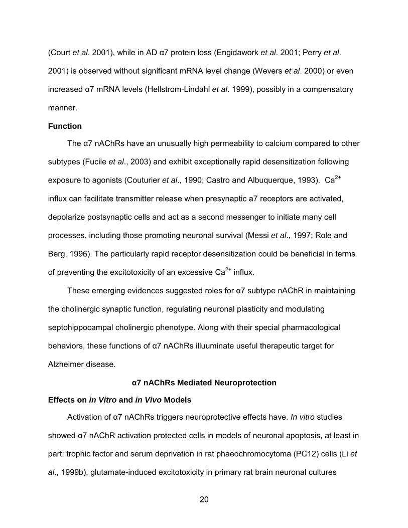
One mechanism underlying α7 nicotinic receptor mediated neuroprotection
involves several intracellular processes triggered by receptor activation.
Phosphorylation of calcium/calmodulin-dependent protein kinase II (CaMKII), protein
kinase C (PKC), mitogen-activated protein kinases (MAPK), phosphoinositide 3-kinase
(PI3K) / AKT and Janus kinase 2 (JAK2) have been found in response to α7 nAChRs
activation. These kinases transduce signals by further activating cyclic adenosine
monophosphate (cAMP) response element-binding protein (CREB) and signal
transducer and activator of transcription protein 3 (STAT3), which regulate new gene
synthesis (Figure 1-2). Mitochondria membrane stabilization, reduced release of
mitochondria cytochrome oxidase and increased B-cell lymphoma 2 (Bcl-2) protein
expression are also found to be involved in these protective effects (Kihara et al., 2001;
Dajas-Bailador et al., 2002a; Li et al., 2002; Ren et al., 2005; Marrero and Bencherif,
2009).
22
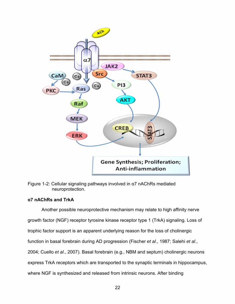
Figure 1-2: Cellular signaling pathways involved in α7 nAChRs mediated neuroprotection.
α7 nAChRs and TrkA
Another possible neuroprotective mechanism may relate to high affinity nerve
growth factor (NGF) receptor tyrosine kinase receptor type 1 (TrkA) signaling. Loss of
trophic factor support is an apparent underlying reason for the loss of cholinergic
function in basal forebrain during AD progression (Fischer et al., 1987; Salehi et al.,
2004; Cuello et al., 2007). Basal forebrain (e.g., NBM and septum) cholinergic neurons
express TrkA receptors which are transported to the synaptic terminals in hippocampus,
where NGF is synthesized and released from intrinsic neurons. After binding
23
extracellular NGF, membrane-spanning TrkA is endocytosed and transported
retrogradely to cell bodies in basal forebrain. This trophic complex modulates
cholinergic neuron morphology and function by enhancing sprouting, ACh synthesis,
cholinergic neuronal firing and transmitter release. In AD brain, NGF levels are normal
or even elevated in hippocampus and neocortex, but are reduced in the basal forebrain
(Scott et al., 1995), suggesting reduced retrograde transport of this peptide to basal
forebrain via TrkA receptors (Cooper et al., 2001), due to a combination of lower TrkA
levels (Kerwin et al., 1992) and altered TrkA isoform expression (Dubus et al., 2000). It
has been found that α7 nAChRs agonists increased NGF as well as TrkA levels in
multiple brain regions (Garrido et al., 2003; Martinez-Rodriguez et al., 2003; Jonnala et
al., 2002; French et al., 1999); while the α7 selective antagonist methyllycacotinine
(MLA) blocks TrkA elevation (Jonnala et al., 2002). The anti-apoptotic effect of nicotine
in spinal cord neurons was blocked with TrkA inhibition (Garrido et al., 2003). These
studies with nicotinic agonists and antagonists suggested α7 activation induced TrkA
and NGF up-regulation, which protects the neurons in complement to direct α7 effects.
α7 nAChRs and β Amyloid
In recent years, a significant body of evidence suggested the role of α7 nAChRs in
the processing and clearance of Aβ peptide, another rational target for treating AD. Aβ
represents the key molecule of the “amyloid hypothesis”, which states that the
aggregation of Aβ in memory related brain regions is toxic to the neurons in either
soluble or insoluble forms and contributes to the progress of cognitive decline (Hardy
and Selko, 2002). Aβ competitively binds to α7 receptors with high affinity, impairs their
normal functions, increase tau-hyperphosphorylation, potentiates cholinergic deficits
and neuron death (Liu et al., 2001; Wang et al., 2000a and 2000b; Wang et al., 2003).
24
Dissociating Aβ from α7 by selective partial agonist attenuated these effects (Wang et
al., 2009). Activating α7 receptors decreased Aβ products (Utsuki et al., 2002; Mousavi
et al., 2009), attenuates Aβ-induced tau phosphorylation both in vitro and in vivo (Hu et
al., 2008; Bitner et al., 2009) and ameliorated the cognitive deficits induced by Aβ
toxicity in a mouse model (Chen et al., 2010). Despite controversial data showing that
α7 nAChR knock-out mutation rescued cognitive decline in an APP-over expressing
mouse model (Dziewczapolski et al., 2009), a more recent study using a mouse model
crossed from APP mutant mice and α7 knockout mice showed that loss of α7
accelerated cholinergic dysfunction in basal forebrain and hippocampus at an early
preplaque stage, as well as exacerbated Aβ accumulation, early stage cognitive decline
and septohippocampal pathology (Hernandez et al., 2010).
A possible underlying mechanism might be that α7 activation induced signaling
pathways regulate amyloid precursor protein (APP) metabolism via secretase activities.
APP is processed by α-, β-, and γ-secretases. α-form of secreted APP (αAPPs)
resulting from α-secretase cleavage is believed to be non-toxic, while the product from
the collaboration of β- and γ-secretases, Aβ, is neuron toxic (Esler and Wolfe, 2001). In
vitro studies have shown that α7 activation decreases Aβ production by suppressing γ-
scretase activity, and increased αAPPs (Nie et al., 2010). Besides, microglia α7
activation may facilitate Aβ clearance in CNS (Takata et al., 2010). A recent clinical
study reported that CNS Aβ clearance rate is decreased in AD patient’s brain compared
to elderly controls, while the Aβ production rate is not significantly differed
(Mawuenyega et al., 2010). Therefore, microglia α7 could also be targeted to counteract
the altered Aβ metabolism.
25
Together, α7 nAChRs have special functions in neuroprotection and cognitive
improvement through activating cell viability associated cellular kinases, promoting TrkA
related neuron tropic pathways and possibly preventing Aβ induced toxicity. More
details from future studies will help us understand systemically about α7 receptor
function in maintaining septohippocampal cholinergic phenotype and preserving
hippocampal integrity at early stage of dementia.
Therapeutic Approaches Targeting α7 nAChRs
Agonist Therapy
No new AD drug has been approved by FDA since 2003. α7 receptors have been
a favorable target for their special effects in memory enhancing and neuroprotection.
During past decade, hundreds of compounds functioning as selective α7 agonists have
been synthesized and studied in lab models. Some of them, such as 3-(2,4)-
dimethoxybenzylidine anabaseine (DMXB; also known as GTS-21), have been applied
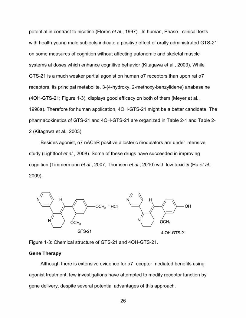
to clinical research. GTS-21 (Figure 1-3) is a selective α7 receptor partial agonist
(Meyer et al., 1998a) that has been widely studied. It has displayed promising function
in improving cell viability in vitro and enhancing a variety of cognitive behaviors,
including spatial memory in the Morris water task (Meyer et al., 1998a), passive and
active avoidance behaviors in rats (Meyer et al., 1997), radial arm maze in aged rats
(Arendash et al., 1995), delayed eye blink behavior in rabbits (Woodruff-Pak, 2003),
hippocampal gating behavior in mice (Simosky et al., 2001), and delayed pair matching
and word recall (Briggs et al., 1997). In contrast, the selective antagonist MLA reduces
performance in radial arm maze when injected directly into the hippocampus at low
concentrations (Bettany and Levin, 2001). Chronic administration of GTS-21 does not
lead to significant receptor up-regulation or possess significant drug dependence
26
potential in contrast to nicotine (Flores et al., 1997). In human, Phase I clinical tests
with health young male subjects indicate a positive effect of orally administrated GTS-21
on some measures of cognition without affecting autonomic and skeletal muscle
systems at doses which enhance cognitive behavior (Kitagawa et al., 2003). While
GTS-21 is a much weaker partial agonist on human α7 receptors than upon rat α7
receptors, its principal metabolite, 3-(4-hydroxy, 2-methoxy-benzylidene) anabaseine
(4OH-GTS-21; Figure 1-3), displays good efficacy on both of them (Meyer et al.,
1998a). Therefore for human application, 4OH-GTS-21 might be a better candidate. The
pharmacokinetics of GTS-21 and 4OH-GTS-21 are organized in Table 2-1 and Table 2-
2 (Kitagawa et al., 2003).
Besides agonist, α7 nAChR positive allosteric modulators are under intensive
study (Lightfoot et al., 2008). Some of these drugs have succeeded in improving
cognition (Timmermann et al., 2007; Thomsen et al., 2010) with low toxicity (Hu et al.,
2009).
Figure 1-3: Chemical structure of GTS-21 and 4OH-GTS-21.
Gene Therapy
Although there is extensive evidence for α7 receptor mediated benefits using
agonist treatment, few investigations have attempted to modify receptor function by
gene delivery, despite several potential advantages of this approach.
27
α7 receptors display a high degree of concentration-dependent kinetics of agonist-
evoked responses (Seguela et al., 1993; Uteshev et al. 2003; Papke et al. 2009). They
desensitize very quickly; at high agonist concentrations there is little receptor function
following this desensitization phase. One component of this loss of receptor function at
high agonist concentrations may also be due to the receptor staying in a dysfunctional
state when too many subunits are agonist-bound. Therefore, in order to obtain a long
term agonist-response, the concentration must be maintained at low enough
concentrations for a long term response, but not too low to elicit virtually no response. In
vivo, particularly, this may be a difficult goal. Besides, drug treatment alone may not be
sufficient due to the loss of α7 receptors in some parts of the AD brain. Increasing α7
receptor density by gene therapy provides an approach to increase receptor function
that is likely either independent of drug-concentration (hence does not require higher
drug doses) or perhaps even leads to more potent agonist responses (i.e. broadening
the dose-response nature of the receptor). In addition, it provides a mechanism to
counteract the effects of AD on receptor density. Of course, gene delivery can be
combined with agonist treatment to optimize the α7 receptor responses, depending on
the availability of the endogenous agonists--choline and acetylcholine.
With gene therapy, a single injection could give a long term, perhaps permanent
effect that could be localized to those the brain regions affected by the disease. This is
in contract to drugs that spread throughout the brain and body and may cause
unwanted side effects. Gene delivery may also mediate the expression of several
genes, which may be an advantage for AD, since it involves multiple pathological
mechanisms.
28
Functional α7 gene expression involves the chaperon protein RIC-3 (Millar 2008).
RIC-3 appears to act by interacting with unassembled receptor subunits within the
endoplasmic reticulum (ER), thereby facilitating subunit folding and receptor assembly
(Lansdell et al. 2005; Alexander et al., 2010). Heterologous expression studies
performed in Xenopus oocytes have demonstrated that co-expression of C. elegans
RIC-3 causes enhanced levels of functional expression of α7 receptor (Halevi et al.
2002), and similar results have been reported with a human homologue of RIC-3 (Halevi
et al. 2003; Williams et al. 2005). In the absence of RIC-3, little or no specific binding of
nicotinic radioligands is detected when the α7 subunit is expressed in mammalian cell
lines, which lack endogenous RIC-3 (such as simian COS cells and some human
HEK293- derived cell lines) (Millar 2008). In contrast, co-expression of RIC-3 with α7
facilitates both high levels of receptor-specific [125I] α-bungarotoxin binding and the
expression of functional α7 receptors (Williams et al., 2005). These findings support the
idea that genetic modulation of RIC-3 expression could also increase α7 maturation and
expression. However, to what extent RIC-3 versus α7 receptor synthesis is limiting for
functional α7 receptor function in neuronal type cells (e.g., PC-12 cells) has not been
well characterized, which is one of the aims of this thesis.
Specific Aims and Significance
In this study, we investigated the effects of α7 together with ric-3 gene delivery: 1)
on agonist sensitivity relative to intracellular processes associated with cell survival and
differentiation; 2) on TrkA receptor levels and activities.
29
Aim 1: Test the hypothesis that α7 and ric-3 gene delivery are both important for
increasing functional α7 receptors in PC12 cells; and set up a in vitro model for studying
underlying processes of up-regulated α7 activation.
Aim 2: Test the hypothesis that increased α7 receptor expression elevates the
agonist-induced intracellular responses associated with cell viability.
Aim 3: Test the hypothesis that α7 receptor expression increases TrkA activities.
The study will help to elucidate the benefits of α7 gene delivery in terms of
cytoprotection and promote the application of α7 gene therapy in Alzheimer’s disease.
The combination of pharmacological and genetic approaches employed in this study will
reduce the likelihood that the drugs will act through unexpected, non-α7 receptor
mechanisms. The long term goal of this study is to develop effective neuroprotective
treatments for AD and other degenerative conditions affecting basal forebrain neurons
expressing α7 nicotinic receptors.
30
CHAPTER 2 MATERIALS AND METHODS
Reagents
Except where noted, all chemicals used were purchased from Fisher Scientific
(Hampton, NH).
Plasmid Preparations
Three plasmids were used in this study: rat α7 and algae green fluorescent protein
(GFP) genes were inserted in pUF12 vector; human ric-3 gene was inserted in pCDNA3
vector, each under control of the truncated cytomegalovirus (CMV) / chicken β-actin
(CBA) hybrid promoter. For plasmid preparation, a single E. coli colony was grown for 6-
8 hr in 5 ml of NZY broth (Fisher Scientific, Pittsburgh, PA) containing 100 µg/ml of
ampicillin (NZY/Amp) at 37°C on a shaking platform. The mixture was then inoculated
into 2 L of NZY/Amp and incubated for 16~17 hr. The overnight culture was centrifuged
at 4,000 g for 15 min in a Sorvall® RC-5B refrigerated super-speed centrifuge (DuPont
Instruments). The pellet was resuspended in 20 ml of lysozyme buffer (10 mM Tris-HCl,
10 mM EDTA and 50 mM sucrose, pH 8.0). Lysis was initiated with 12 mg/ml lysozyme
(Sigma Aldrich, St. Louis, MO) followed by NaOH/SDS solution (0.5 N NaOH and 1%
SDS in water). Genomic DNA was precipitated with 3M NaAc (pH4.6-5.2) and pelleted
by centrifuging at 16,000 g for 20 min. Plasmid DNA in the supernatant was precipitated
with 40% PEG on ice for 10 min, pelleted at 16, 000 g for 10 min and dissolved in water.
LiCl (5.5 M; 1:1, volume/volume of the solution) was added, followed by centrifugation at
16,000 g for 10 min. Plasmid DNA was precipitated from the supernatant by ice-cold
isopropanol and pelleted at 16,000 g for 10 min. The pellet was dissolved in 5 M CsCl
medium with ethidium bromide (0.5 mg/ml) in Optiseal Beckman tube (Beckman
31
Instruments, Brea, CA). Gradients were formed ultracentrifuging at 200,000 g over 19 hr
at 20°C in Beckman L8-70M Ultracentrifuge. The concentrated plasmid DNA band was
detected by a hand held UV lamp (366 nm). The lower band of the two bands was
collected with 16 G needle (BD biosciences, San Diego, CA) and transferred to a 15 ml
tube. The samples were extracted 3-4 times with equal volume isoamyl alcohol (Fisher
Scientific, Pittsburgh, PA) until they are clear and then transferred to a new tube. 2.5
volume of water and the combined volume of ethanol were mixed with the sample to
precipitate plasmid DNA by icing for 30 min, following with 15 min centrifuge at 5,000 g.
The pellet was dissolved in TE buffer (10 mM Tris-HCl, 1 mM EDTA, PH 8.0) and then
purified by 2 times phenol/chloroform extraction and 1 time chloroform extraction. The
purified plasmid DNA was precipitated with absolute ethanol and NaAc (0.08 M),
centrifuged at 16,000 g for 20 min, washed once with 75% ethanol, air dried and
dissolved in TE. The DNA concentration was detected at 260/280 nm in a Beckman DU
650 Spectrophotometer. The DNA quality was confirmed by restriction digestion and gel
electrophoresis.
Cell Culture
Rat PC12 cells (ATCC, Rockville, MD) were cultured and maintained in F12k
medium (ATCC) containing 2.5% fetal bovine serum (Sigma Aldrich) and 15% horse
serum (ATCC) at 37°C in a humidified atmosphere of 5% CO2. They were typically split
using 0.25% Trypsin (Fisher Scientific, Pittsburgh, PA) at a 1:4 ratio every 4 days for up
to 20 passages before use. The cell culture medium was removed and cells exposed to
trypsin for 1~2 min and observed under microscope. The trypsin solution was removed
when cell morphology became round but still attached to the culture dish. Cells were
then washed from the bottom with fresh medium, completely separated by titration and
32
transferred into new culture dishes. Before transfection / transduction, cells were
transferred to 6-well, 24-well or 96-well Costar® tissue culture treated plates (Corning
Inc., Corning, NY) at approximately 70% confluence if not indicated otherwise.
Cell Gene Delivery, Differentiation and Drug Treatments
A variety of GFP expression vectors were evaluated for gene-transfer efficiency in
PC12 cells: recombinant adeno associated virus (rAAV) using either serotype #2, #5, or
#8, each with terminal repeats (TR) from serotype #2; calcium phosphate mediated
plasmid transfection; and Lipofectamine 2000TM (Invitrogen, Carlsbad, CA) / Lipo293TM
(SignaGen, Ijamsville, MD) mediated plasmid transfection. The percent of cells
expressing GFP was determined by fluorescence microscopy.
For kinase assays, cells were incubated 24 hr post transfection with a specified
concentration of 4OH-GTS-21 in fresh medium for 30 min, with or without a 30 min pre-
treatment with 50 nM of the selective α7 nicotinic receptor antagonist methyllycaconitine
(Sigma Aldrich), 50 µM ERK phosphorylation inhibitor PD 98059 (Calbiochem, La Jolla,
CA) or 20 µM AKT phosphorylation inhibitor API-2 (Calbiochem).
For differentiation studies, ~40% confluenced cells were treated 24 hr after
transfection with 50 ng/ml nerve growth factor (BD biosciences, San Diego, CA) for 4
days, with or without 30 min pre-treatment of MLA, PD 98059 or API-2. Nerve growth
factor was added repeatedly to maintain the working concentration.
Cell Viability
Cell viability was estimated with the CyQuantTM Cell Proliferation assay kit
(Molecular Probes, Invitrogen, Carlsbad, CA) according to the manufacturer’s
instructions. Fluorescence was measured directly on a FL600 Microplate Fluorescence
33
Reader (Bio-Tek Instruments, Inc, Burlington, VT) to determine the percentage of viable
cells.
α7 Nicotinic Receptor Binding Assay
One day after transfection with specified plasmids, cultures were washed twice
with phosphate buffered saline (PBS) and harvested in ice-cold Krebs Ringer buffer
(KRH; 118 mM NaCl, 5 mM KCl, 10 mM glucose, 1 mM MgCl2, 2.5 mM CaCl2, 20 mM
Hepes; pH 7.5). Cells were homogenized in ice-cold KRH buffer with a Polytron at
setting 4 for 10 s. After two 1 ml washes with KRH at 20,000g, the membranes were
incubated in 0.5 ml KRH with 50 nM [3H] MLA (100 Ci/mmol; Tocris, Ellisville, MO), for
60 min at room temperature, plus or minus 5 mM nicotine. Tissues were washed three
times with 5 ml ice-cold KRH buffer by filtration through Whatman GF/C glass microfiber
paper (Brandel, Gaithersburg, MD) that was pre-incubated for 30 min with 0.5%
polyethylenimine using M-24R. S Cell Harvester (Brandel Inc., Gaithersburg, MD).
Liquid scintillation counting of radioactivity was conducted in a Beckman LS1800 using
EcoLite (Fisher Pharmaceuticals, Hampton, NH). Nicotine-displaceable binding was
calculated for each sample in triplicate in each experiment, and normalized for total
protein concentrations measured with Pierce® BCA protein Assay Kit (Thermo
Scientific, Rockford, IL).
Enzyme-Linked Immunosorbent Assay
Treated cells were harvested in RIPA buffer (Cell Signaling, Danvers, MA)
containing protease inhibitor cocktail and phosphatase inhibitor cocktail (Thermo
Scientific, Rockford, IL), sonicated and centrifuged at 4oC. Supernatant was collected
and frozen at -80oC till use. For ERK and JNK, ELISA antibodies and standard proteins
were purchased from R&D systems (Minneapolis, MN). Immulon 4HBX microplates
34
were coated with ERK2, p-ERK2, JNK or p-JNK capture antibody (1, 3, 3 and 2 µg/ml,
respectively) overnight followed by blocking with 1% BSA in PBS/0.05% NaN3. 100 µl
cell samples or diluted standard proteins were added into the wells and incubated for 2
hr. Targeted proteins were probed by detection antibodies (0.5 µg/ml) for 2 hr and then
incubated with Streptavidin-HRP (1:200) for 20min. Washes were conducted in PBS
with 0.02% Tween 20. Substrate solutions (R&D Systems, Minneapolis, MN) were
added for color reaction and stopped with 2 N H2SO4 after 20 min. AKT and p-AKT
were evaluated using ELISA kits purchased from Invitrogen by following manufacture’s
protocol. Optical densities were determined by a microplate reader (Bio-Rad, Hercules,
CA) set to 450 nm. All results were normalized for total protein concentrations and
expressed as % of GFP-transfected control values.
Western Blot
Treated cells or dissected brain tissue samples were homogenized in RIPA buffer
containing protease and phosphatase inhibitor cocktails and centrifuged at 4oC.
Supernatant was collected for protein assay or western blots. After boiling with loading
buffer (Laemmli buffer containing 2% 14.2 M 2-Mercaptoethanol) for 5 min, proteins
were separated on 12% Tris-HCl gels (Sigma Aldrich) and transferred to PVDF
membranes (Bio-rad). The membranes were blocked by incubation in 5% skim milk in
TBS buffer (0.5% Tween 20, 10 mM Tris, 50 mM NaCl, pH 8.8) at room temperature for
1 hr. All washes were conducted with TBS buffer. The membranes were incubated at
4°C overnight with the following antibodies (dilutions given in parentheses): anti-p-ERK,
anti-ERK, anti-p-TrkA, anti-β actin (Cell Signaling, Danvers, MA, 1:1000), anti-TrkA
(Upstate Biotechnology, New York City, NY), anti-α7 (Covance, Princeton, NJ, 1:500)
35
and anti-ChAT (Chemicon Inc., Billerica, MA; 1:2000). Incubation with the horseradish
peroxidate-conjugated anti-mouse or anti-rabbit IgG (GE, Piscataway, NJ, 1:10000) was
conducted in TBS buffer containing 5% skim milk for 45 min at room temperature. The
signal was detected by the ECL detection system (PerkinElmer, Waltham, MA),
according to the manufacturer’s instructions, with Kodak BioMax MR film (Sigma
Aldrich). The bands were quantified with ImageJ.
Caspase Assay
Cells were grown and treated in 96 well plates. Caspase activities were evaluated
with an Apo-ONE® homogeneous caspase-3/7 assay kit (Promega, Madison, WI)
following the manufacturer’s protocol. 100 µl of Apo-ONE® were added to each well of
the 96-well plate containing 100 µl of blank (culture medium), control or cells in culture.
The mixture was sealed and incubated for 2 hr. Fluorescence of each well was read
(excitation/emission: 485 nm / 530 nm) using Synergy 2 plate reader (BioTek
Instruments Inc. Highland Park, VT) and normalized to untreated cell culture medium.
Immunofluorescence
Cells grown and transfected in 24 well plates were washed with PBS 3 times, fixed
in 3% ice cold paraformaldehyde in PBS with 1 mM MgCl2 at room temperature for 15
min and permeabilized with 1.0% Trition X-100 for 5 min. Non specific signals were
blocked with 2% BSA for 1 hr. Either anti-α7 antibody (Sigma) or anti-p-TrkA antibody
(Sigma Aldrich) was added in and incubated with cells for at least 1 hr. Both of the
antibodies were 1:400 diluted in PBS with 2% BSA. Controls without primary antibody
reaction were incubated in PBS with 2% BSA for the same interval. Signals were
visualized by either Alexa Fluor 488 or Alexa Fluor 594 (1: 400 dilutions, Invitrogen,
36
Carlsbad, CA). Fluorescence pictures were taken with Axiovert 135 microscope (Zeiss,
Thornwood, NY).
Virus Vector Packaging and Titration
Transfection
Plasmids were packaged into rAAV8 using the adenovirus-free method developed
by Zolotukhin et al. (1999). HEK 293 cells at 70% confluence were transfected by a
calcium phosphate method with pUF12-rat α7 / pUF12-GFP and rAAV8 helper plasmid
pXYZ8 / rAAV5 helper plasmid pXYZ5 in an equal molar ratio. 10 cell culture dishes of
15 cm diameter were used. 1.25 ml of 2 M CaCl2, 0.6 mg helper plasmid, 0.3 mg of
pUF12-rat α7 or pUF12 and sterile water were mixed to the total volume of 10 ml for ten
dishes. This mixture was added dropwise into equal volumes of 2X HBA while
vortexing. This transfection mixture was added to 200 ml of warmed DMEM, which
contained 10% FBS and 1% penicillin/streptomycin. 22 ml of this medium mixture were
added to each dish. 6-hour after transfection, the mixture was removed and replaced
with fresh DMEM. The cells were incubated for 3 days to reach 100% confluence and
then harvested using cell scraper (Corning Inc.) and centrifuged at 4oC and 3,000 g for
15 min. The cells were resuspended in AAV lysis buffer (150 mM NaCl, 50 mM Tris, pH
8.5).
Purification
The cell mix was frozen and thawed 3 times in a mixture of dry ice/ethanol (10 min
freeze, 15 min thaw, vortexing every 5 min). The lysate were incubated with benzonase
at 37oC for 30 min to digest unpackaged DNA followed by centrifuging in 4oC at 3,000 g
for 30 min. The supernatant was transferred into 39 ml Optiseal tube (Beckman) with
16G syringe/needle. Pump Pro (Watson-Marlow, UK) was set up as follows: 200 μl
37
glass pipettes were used for intake and 100 μl glass pipettes were used for output.
Pump speed was set at 37 rpm counterclockwise. The tube was rinsed with water and
15% iodixanol (IOD). The output pipette was loaded into Optiseal tubes containing
samples. Pumping was started with 15% IOD (1:47 min), followed by 25% IOD (1:15
min), 40% IOD (1:47 min) and 60% IOD (1:50 min). (180 ml of 15% IOD contained 45
ml of OptiPrep (Axis-Shield Poc AS, Norway), 36 ml of 5 M NaCl, 36 ml of 5x TD (5x
PBS, 5 mM MgCl 2, 12.5 mM KCl) and 63 ml water. 120 ml of 25% IOD contained 50 ml
of OptiPrep, 24 ml of 5x TD, 46 ml of water and 300 μl of 0.5 % phenol red solution. 100
ml of 40% IOD solution contained 68 ml of OptiPrep, 20 ml of 5x TD and 12 ml of water;
100 ml of 60% IOD contained 100 ml of OptiPrep and 250 μl of 0.5% phenol red
solution). The tubes were topped off with rAAV lysis buffer, heat-sealed and
untracentrifuged at 100,000 g in Beckman L8-70M Ultracentrifuge for 2 hour at 18oC.
To collect the virus, first interphase (between 60% and 40%) from bottom of tube and up
to but not including second interphase was collected using 16 G syringe/needle.
Q sepharose was used for rAAV5 and rAAV8. The bottom of the Bio-Rad Econo-
pac disposable chromatography column was snapped off and 5 ml of well-mixed Q
sepharose (Sigma) were added into the column. The Q sepharose column was
equilibrated with 20 ml of solution A (20 mM Tris/15 mM NaCl, pH 8.5), washed with 20
ml solution B (20 mM Tris/1M NaCl, pH 8.5) and again with 30 ml solution A. AAV
samples was diluted with two times solution A and loaded to the column. After loading
the sample, 50 ml of solution A was added to the column. The sample was eluted with
20 ml of solution C (20 mM Tris/355 mM NaCl, pH 8.5) and collected into a 50 ml
conical tube. The sample was concentrated using Biomax-100K NMWL membrane
38
concentrator (Millipore, Billerica, MA) to 1 ml. The concentrate was then diluted with 9
ml of Ringer’s solution and concentrated to 1 ml twice in order to reduce the salt
concentration in virus solution. Typical final virus sample volume was approximately 300
μl. Samples was collected into siliconized tubes and stored at -20oC.
Dot-Blot Assay
A dot-blot assay was used to determine the total number of genomic particles of
rAAV virus (Kube and Srivastava, 1997). A 4 μl aliquot of the virus stock was treated
with DNAse I (Roche, Mannheim, Germany) for 1 hour at 37oC, followed by incubating
with Proteinase K (Boehinger Mannheim, Germany) for another hour at 37oC to obtain
the encapsidated DNA. Samples were extracted with equal volume of phenol-
chloroform twice and equal volume of chloroform once. The aqueous layer was
transferred into a new tube and mixed with 1/10 volume of 3 M NaAc (pH 5.2) and 2.5
volumes of 100% ethanol. DNA was precipitated overnight at -80oC, centrifuged at
14,000 g for 20 min, washed with 75% ethanol, air dried and dissolved in 40 μl of water.
The sample was quantified by a DNA slot blot assay using 1.7 kb EcoRI segment
of pUF12 as probe and a series of dilutions of pUF12 (0.1 ng ~ 100 ng in alkaline buffer)
as standard curve. For virus samples, 10 μl of 1:10 dilution and 1:1 dilution each were
added into 200 μl of alkaline buffer tube. The standards and samples were boiled for 10
minutes and loaded into each well of the dot blotter. The samples were aspirated slowly
with vacuum until fully soaked. The vacuum was disconnected. 400 μl alkaline buffer
was added to each well and allowed to stand for 5 min, followed by vacuuming away
any remaining solution. The membrane from the dot blotter was crosslinked by a UV
Statalinker 1800 crosslinker (Strategene, La Jolla, CA) and placed into a small Biometra
bottle, which was filled with prehybridyzation buffer (7% SDS, 0.25 M NaHPO4 pH 7.2, 1
39
mM EDTA pH 8.0), and incubated at 65°C for 1 hr. 6 μl of biotinylated probe was diluted
with 54 μl of 10 mM EDTA and denatured at 90°C for 10 min. The total 60 μl of
denatured probe were added to the hybridization buffer, quickly mixed and incubated
with the membrane overnight at 65 °C in Biometra oven.
A Brightstar Bio Detect kit (Ambion, TX) was used for signal detection according to
the manufacture’s manual. The intensities of the standard DNA bands were used to
build a standard curve. The virus titers were calculated using the coefficients: 1 ng DNA
= 4 x 10¹¹ particles/ml. Vector stocks were ranged from 1012-1013genomic particles per
ml.
Stereotaxic Surgery
All procedures were approved and overseen by the University of Florida
Institutional Animal Use and Care Committee. B6C3F1/J wild type male mouse (~25 g,
2 months old, The Jackson Laboratory, Bar Harbor, ME) were housed and bred the in
an AAALAC-accredited animal facility at the Health Science Center. Animals were
warmed under a heat lamp, anesthetized with 4% isoflourane/oxygen and injected
subcutaneously with carprofen (5 mg/kg) before the surgery to minimize pain and
infection. rAAV8-rat α7 or rAAV8-GFP vectors (109 genomic particles) were injected into
septum through a 27-gauge cannula connected via 26 gauge I.D. polyethylene tubing to
a 10 μl syringe mounted to a CMA/100 microinjection pump at a rate of 0.15 μl /min.
The injection coordinates for the left lateral septum were: +0.25 mm from Bregma;
+0.35 mm from the midsaggital suture and -2.5 mm from the skull surface according to
the atlas of Paxinos & Franklin (2001). The cannula was left at the injection site for 5
min and then removed slowly. The skin was sutured. Mice were warmed at 37oC until
recovery from anesthesia, and returned to home cages. Two weeks after injections,

40
mice were trained with Morris water maze tasks as described below. After behavioral
evaluation, mice were deeply anesthetized with 4% isoflourane/oxygen. Half of each
group was perfused with PBS; brains were excised, fixed for 48 hr in 4%
paraformaldehyde and equilibrated in 30% surcrose for immunohistochemistry and
immunefluorescence. The other half of them in each group were euthanized by
decapitation, and the left hippocampus, right hippocampus and septum were dissected
quickly, snap frozen in liquid nitrogen and stored at -80°C for biochemical procedures.
Morris Water Maze Tests
Morris water maze tests were performed to test the spatial learning and memory of
mice injected with rAAV8-rat α7 or rAAV8-GFP vectors. These tests were conducted
using a specially designed circular tank (75 cm interior diameter) with a white interior
filled to a depth of ~34 cm with room temperature water, which was made opaque by
the addition of powdered white paint. An escape platform (5.5 cm diameter), made of
Plexiglas and covered with a coarse material that provided grip for climbing onto the
platform, was located approximately 1.0 cm below the water surface. Various geometric
high-contrast images (e.g., circles, squares, triangles) were hung above the water
surface as navigational references. Mice received 8 trials daily for 2 days in order to
memorize the location of the submerged platform. The mice had 60 sec to search for
the platform (and were hand guided to the platform if they did not reach it during that
interval). They were then allowed 30 seconds to stay on the platform. Probe test was
followed with the 2-day training, in which, the platform was removed and the mice had
60 sec to search for the platform. The swim distance and percentage of time spent in
each quadrant were recorded by video camera and analyzed with Image-Pro software
(Media Cybernetics, Bethesda, MD).
41
Immunohistochemistry
Fixed forebrains blocks were cut into coronal sections (50 µm) on a sliding
microtome with freezing stage. Antigen retrieval was done by incubating the sections
with 50 mM sodium citrate (pH 8.5-9.0) at 80oC for 30 min (Jiao et al., 1999). Antigen
detection was conducted on free-floating sections by incubating the sections at 4°C in
blocking solution (3% goat serum, 0.3% Triton X-100, 0.05% azide in PBS) for 1 hr at
room temperature, followed by primary antibody incubation overnight at 4°C. Primary
antibodies used were: anti-α7 (1:200, Covance), anti-TrkA (1:500, Upstate
Biotechnology), and anti-ChAT (1:1000, Chemicon). Sections were incubated with
primary antibodies diluted in blocking solution at 4°C for 3 days. Sections were then
washed with PBS three times for 5 minutes each wash. Then the sections were
incubated overnight at 4°C with secondary antibody (biotinylated anti-mouse IgG or
biotinylated anti-rabbit IgG, 1:1000, Dako, CA) diluted in blocking solution. Sections
were again washed 3 times in PBS. Next the sections were incubated for two hours at
room temperature in PBS with ExtrAvidin peroxidase (HRP) conjugate (1:1000, Sigma).
Washing was performed again. Development of tissue labeled with HRP was performed
with a solution of 0.67 mg/mL diaminobenzidine (DAB, Sigma) / 0.003% H2O2/ 8 mM
imidazole / 2% NiSO4. The sections were mounted on Superfrost plus microscopic
slides (Fisher Scientific), air dried and dehydrated by passing through water, followed by
70%, 95%, 100%, 100% ethanol (5 min each). Then they were passed through two
changes of xylene, and coverslipped with Eukitt (Calibrated Instruments, Hawthorne,
NY).
Some sections were used for immunofluorescence staining. After incubating with
mouse anti-α7 and rabbit anti-TrkA primary antibodies, sections were washed in PBS 3
42
times and incubated Alex Fluo 488 labled anti-mouse IgG and Alexa Fluor 594 labled
anti-rabbit IgG. Sections were washed in PBS and then mounted on Fisher Superfrost
Plus slides, air-dried and coverslipped with glycerol gelatin (Sigma).
Statistical Analysis
Statistical analyses involved one-way or two-way ANOVA for main effects
hypothesis testing using Prism (GraphPad software, a Jolla, CA). Post-hoc comparisons
of parametric populations were made using Bonferroni’s tests. Statistical analyses for
mice water maze effects were conducted in SAS (SAS Institute, Cary, NC) General
Linear Models procedure.
43
CHAPTER 3 EFFECTS OF α7 RECEPTOR GENE DELIVERY ON PROCESSES UNDERLYING
CELL VIABILITY
Introduction
Cholinergic neurons are essential for normal memory-related behaviors but are
dysfunctional in AD. Studies suggest that α7 nAChRs expressed on these and other
neurons regulate their normal physiologic function and positively modulate their viability
under apoptotic conditions. These neuroprotective functions of α7 receptor appeared to
be attenuated in AD, making them suitable targets for treating this progressive memory-
related disorder. Selective agonist treatments have shown success in a variety of
models, however, these receptors exhibit highly dose-dependent properties. They
experience fast desensitization at high agonist concentrations. An alternative approach
would be increasing the receptor level by gene delivery. α7 nicotinic receptor gene
delivery into mouse hippocampus by adeno-associated virus was recently shown to
improve spatial memory performance (Ren et al., 2007b). While this improvement was
likely mediated by altered receptor response to endogenous agonists, no study has
attempted to evaluate the processes underlying combined effects of gene delivery with
selective agonist.
Activation of α7 receptors can increase calcium accumulation both directly as well
as through indirect activation of downstream L-type voltage sensitive channels, IP-3
channels, and ryanodine channels (Vijayaraghavan et al., 1992; Gueorguiev et al.,
2000; Shoop et al., 2001; Dajas-Bailador et al., 2002b). Several groups including ours
have demonstrated that GTS-21 and 4OH-GTS-21 α7 agonists increased intracellular
calcium concentrations (Li et al., 2002), activated the calcium-sensitive transduction

44
processes such as PKA, PKC, PI3K, MAPK and JAK kinases, each is essential for α7
mediated protection against one or more apoptotic insults.
It has been well documented that extracellular signal regulated kinase 1 and 2
(ERK 1/2), member of the MAPK signaling pathway, are involved in α7 activation
mediated neuroprotection (Toborek et al., 2007) and long term potentiation
enhancement (Welsby et al., 2009). Data suggested that α7 nAChR agonist-triggered
Ca2+ transients in PC12 cells induce activation of CaMKII, leading to sequential
phosphorylation of MEK1/2, ERK1/2 and CREB (Nakayama et al., 2001; Gubbins et al.,
2010). This pathway regulates new gene expression (Curtis and Finkbeiner, 1999) and
is involved in cellular survival, synaptic plasticity and long term memory (Orban et al.,
1999).
Another MAPK cascade member c-jun-N-terminal kinase (JNK) is more frequently
involved in downstream pro-apoptosis pathways (Manning and Davis, 2003; Nishina et
al., 2004). Knock-out mouse studies have demonstrated that removing particular JNK
genes can reduce the severity in various disease scenarios, including those which are
used to model neurodegenerative diseases (Bonny et al., 2005). There are data
showing that JNK is involved in Tau and APP protein metabolism, leading to
hyperphosphorylated Tau and overproduced Aβ (Reynolds et al., 1997; Philpott and
Facci, 2008). Therefore, blocking JNK pathway may be a valuable approach for AD and
other neurodegenerative disease research.
PI3K/AKT is another cellular signaling pathway responsive to α7 nAChRs
activation. Nicotine induced protection in rat-cultured primary neurons was blocked by
either an α7 nAChR antagonist, a PI3K inhibitor or a Src inhibitor. Levels of
45
phosphorylated Akt, an effector of PI3K, and anti-apoptotic protein Bcl-2 and Bcl-x were
increased by nicotine administration (Shimohama et al., 2009). AChE inhibitor drugs
used for AD treatment, e.g., donepezil and galanathamine, prevent glutamate
neurotoxicity and inflammation via up-regulated AKT phosphorylation (Takada-Takatori
et al., 2006; Shen et al., 2010; Tyagi et al., 2010) and were blocked by α7 selective
antagonist MLA and JAK2 inhibitor. Therefore, it has been proposed that the α7 nAChR
activation stimulates the Src family and JAK2, which activate PI3K to phosphorylate
AKT and subsequently transmits the survival signal to up-regulate Bcl-2 and Bcl-x. This
pathway could prevent cells from neuronal death induced by Aβ (Kihara et al., 2001;
Shaw et al., 2002).
Besides ERK1/2 and AKT activation, the ability of caspase inhibitors to block
neuronal cell death induced by trophic factor deprivation and other cytotoxic conditions
including amyloid β exposure has provided evidence for a crucial role of caspases in
apoptotic neuronal cell death (Deshmukh et al. 1997; Youdim et al. 2001; Eckert et al.
2003; Yuan et al. 2000). In addition to activating death programs, caspase activated by
amyloid β can cleave tau to generate a proteolytic product and promote pathological tau
filament assembly in neurons (Gamblin et al. 2003). Regulation of caspase activity
involves MAPK / ERK1/2 and the PI3K / AKT pathways. Therefore in this study, we
investigated the effects of α7 nAChR gene delivery on agonist sensitivity relative to
intracellular signal processes associated with cell survival. We hypothesized that α7
receptor overexpression with the assistance of chaperone RIC-3 will increase the
agonist effects on ERK phosphorylation, AKT phosphorylation and caspase activity in
PC12 cells.
46
Results
Gene Delivery System Resulted Functional α7 Receptor Expression
Pheochromocytoma PC12 cells express α7 nAChRs and the endogenous agonists
acetylcholine and choline. The signaling pathways that respond to α7 nAChR activation
in these cells have been well characterized using cell lines with high intrinsic α7
receptor level (e.g., over 100 fmol/mg protein). However, the PC12 cells used in the
present study was selected for its relatively low levels of endogenous functional α7
receptors expression according to ligand binding assay. This provided a more suitable
model for basal forebrain cholinergic neurons in AD brain, where α7 receptor density is
decreased during the disease progression.
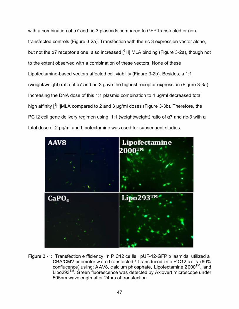
PC12 cells are refractory to gene delivery by most vehicles, with no successful
procedure reported in the literature. Therefore, a variety of gene delivery methods were
evaluated initially for GFP expression efficiency in PC12 cells (Figure 3-1): rAAV, using
either serotype coats #2, #5, or #8, each with terminal repeats from serotype #2);
calcium phosphate, that precipitate plasmid DNA via interaction with calcium ions; and
cationic lipid vectors (Lipofectamine 2000TM from Invitrogen and Lipo293TM from
SignaGen). Among these approaches, cationic lipid vectors consistently yielded the
highest transfection efficiency (15~20%) based on GFP expression, with no apparent
cytotoxicity. Therefore these vectors were used for all the following in vitro gene
delivery.
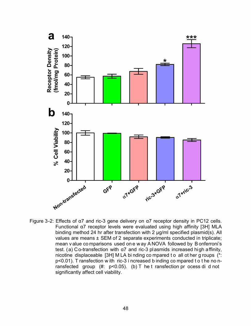
pUF-12-α7 plasmids were transfected into PC12 cells together with pcDNA-ric-3
plasmids using Lipofactamine 2000TM. The functional α7 receptor density was evaluated
by high affinity [3H] MLA binding assay. 24 hr after transfection, the functional
membrane α7 receptor level was increased by around two fold in the cells transfected
47
with a combination of α7 and ric-3 plasmids compared to GFP-transfected or non-
transfected controls (Figure 3-2a). Transfection with the ric-3 expression vector alone,
but not the α7 receptor alone, also increased [3H] MLA binding (Figure 3-2a), though not
to the extent observed with a combination of these vectors. None of these
Lipofectamine-based vectors affected cell viability (Figure 3-2b). Besides, a 1:1
(weight/weight) ratio of α7 and ric-3 gave the highest receptor expression (Figure 3-3a).
Increasing the DNA dose of this 1:1 plasmid combination to 4 μg/ml decreased total
high affinity [3H]MLA compared to 2 and 3 μg/ml doses (Figure 3-3b). Therefore, the
PC12 cell gene delivery regimen using 1:1 (weight/weight) ratio of α7 and ric-3 with a
total dose of 2 µg/ml and Lipofectamine was used for subsequent studies.
Figure 3 -1: Transfection e fficiency i n P C12 ce lls. pUF-12-GFP p lasmids utilized a CBA/CMV pr omoter w ere t ransfected / t ransduced i nto P C12 c ells (60% conflucence) using: AAV8, calcium ph osphate, Lipofectamine 2000TM, and Lipo293TM. Green fluorescence was detected by Axiovert microscope under 505nm wavelength after 24hrs of transfection.
48
Figure 3-2: Effects of α7 and ric-3 gene delivery on α7 receptor density in PC12 cells. Functional α7 receptor levels were evaluated using high affinity [3H] MLA binding method 24 hr after transfection with 2 μg/ml specified plasmid(s). All values are means ± SEM of 2 separate experiments conducted in triplicate; mean v alue co mparisons used on e w ay A NOVA followed by B onferroni’s test. (a) Co-transfection with α7 and ric-3 plasmids increased high affinity, nicotine displaceable [3H] M LA bi nding co mpared t o all ot her g roups (*: p<0.01). T ransfection w ith ric-3 i ncreased b inding co mpared t o t he no n-ransfected group (#: p<0.05). (b) T he t ransfection pr ocess di d not significantly affect cell viability.
49
Figure 3-3: Optimization of α7 and ric-3 gene delivery ratio and dose. (a) 1:1 (weight/weight) ratio of α7 and ric-3 plasmids yielded the highest receptor expression compared to all other tested combinations. (b) A total of 4 μg/ml plasmids lowered the functional receptor expression compared to 2 or 3 μg/ml doses.

50
Effects of 4OH-GTS-21 on p-ERK2 and p-Akt in Transfected Cells:
α7 receptors are highly permeable to calcium (Fucile et al., 2003). Receptor
activation by agonist can induce ERK and AKT phosphorylation via calcium influx and
PI3K activation. Between the two major ERK isoforms ERK1 (p44) and ERK2 (p42), the
role of ERK2 seems preeminent over that of ERK1 despite sharing the same activators
and substrates (Lefloch et al. 2008). A pharmacology experiment was designed to
investigate the response of ERK2 to α7 activation: PC12 cells were transfected with
α7+ric-3 or an equal amount of GFP as control. 24 hr after transfection, the cells were
treated with or without 100 nM α7 antagonist MLA for 30 min and then incubated with 1
µM, 3 µM, 10 µM or 30 µM 4OH-GTS-21 for another 30 min. The agonist concentrations
chosen were broad enough to cover the range from cytoprotection to toxicity observed
in previous PC12 studies (Li et al., 1999b). Cell samples were collected to evaluate the
total protein, total ERK2 and p-ERK2 levels. ERK2 and p-ERK2 values were normalized
to total protein concentrations and then to GFP transfected control without 4OH-GTS-21
or MLA treatment.
Total ERK levels were not affected by gene delivery or by a 30 min treatment with
any concentration of 4OH-GTS-21 (Figure 3-4a). The p-ERK2/ERK2 ratio, an estimate
of ERK2 activation, was neither attenuated by MLA pretreatment nor activated by a
broad range of 4OH-GTS-21 concentrations in GFP-transfected cells (Figure 3-4b). In
α7 + ric-3 transfected cells, however, 4OH-GTS-21 increased p-ERk2/ERK2 ratios in an
inverted U-shaped, concentration-dependent manner compared to GFP-treated controls
(Figure 3-4b). MLA sensitive, agonist-induced elevations in p-ERK2/ERK2 ratios were
observed at 1-10 μM 4OH-GTS-21 concentrations (Figure 3-4c); at the 30 µM agonist
51
concentration, there was no activation of ERK2 phosphorylation, consistent with the
rapid α7 receptor desensitization reported at this elevated 4OH-GTS-21 concentration.
Similarly, total AKT levels were not changed within 30 min of 4OH-GTS-21
treatment in any transfection group (Figure 3-5a). In the α7+ric-3 transfected group, p-
AKT/AKT ratios were significantly higher than in the GFP-transfected group at the 1 µM
and 3 μM 4OH-GTS-21 concentrations (Figure 3-5b). Like ERK2, AKT did not respond
at the 30 µM 4OH-GTS-21 concentration. MLA pretreatment attenuated the increase in
p-AKT/AKT ratios seen with 1 µM 4OH-GTS-21 in the α7 + ric-3 group (Figure 3-5c).
In order to understand the relationship between ERK and AKT activation evoked
through α7 receptors, ERK phosphorylation inhibitor PD 98059 (Alessi et al. 1995) was
preincubated with cells before agonist treatment; and p-AKT levels were evaluated
afterwards. The results showed that PD 98059 blocked ERK phosphorylation (Figure 3-
6a), but did not influence AKT phosphorylation either in α7 + ric-3 (Figure. 3-6b) or
GFP-transfected groups (Figure 3-6c).
In contrast to the ERK and AKT systems, p-JNK/JNK ratios were not affected by
either transfection with α7 + ric-3 or subsequent treatment with 4OH-GTS-21 (Figure 3-
7). Total JNK levels were also unaffected by these treatments (not shown).
Effects of 4OH-GTS-21 on Caspase Activity:
In order to evaluate the effect of α7 and ric-3 gene delivery and activation on cell
apoptosis, we analyzed the activity of central caspase cascade executers: caspase 3/7
(Riedl et al. 2004). Caspase 3/7 activities were significantly reduced after 1-10 µM 4OH-
GTS-21 treatment in the α7 + ric-3 transfected groups compared to GFP-transfected
controls (Figure 3-8a). This effect was potently blocked by MLA (Figure 3-8a and 3-8b).
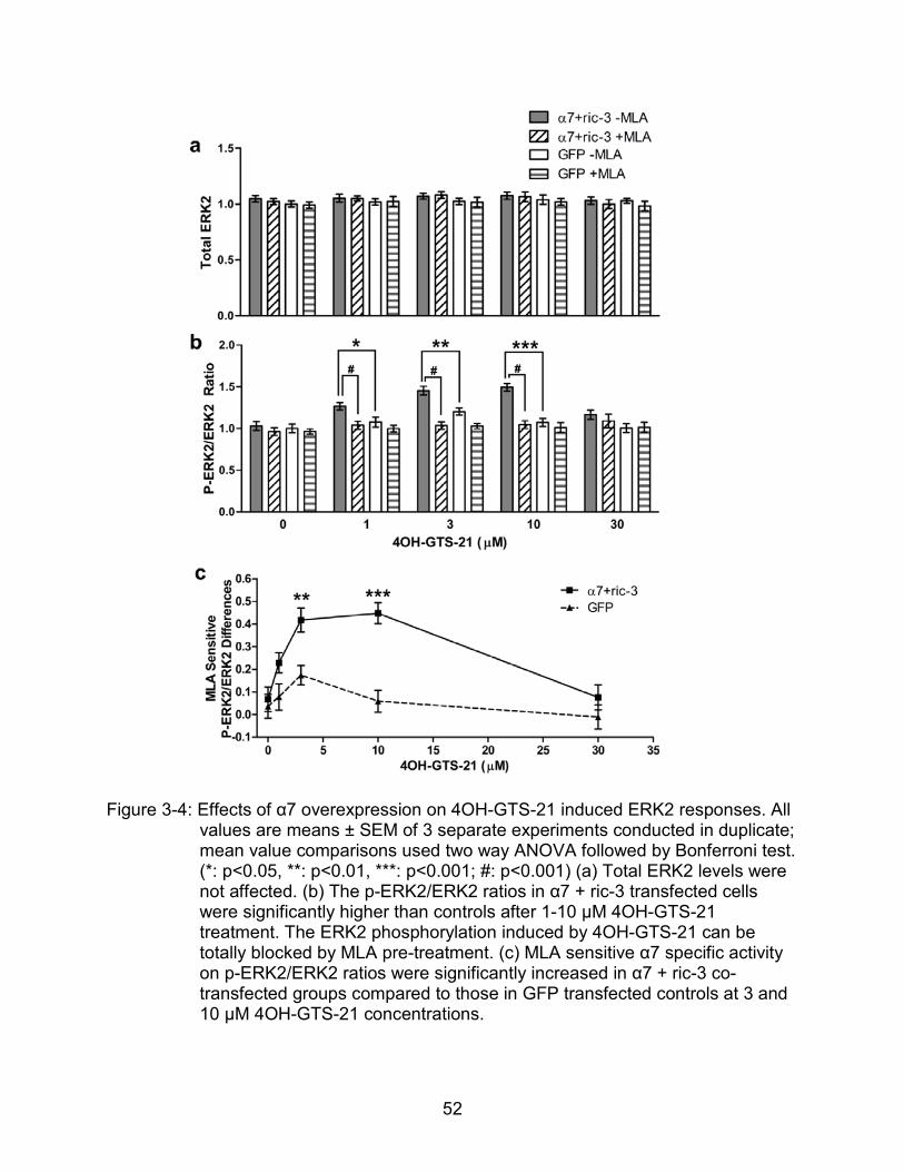
52
Figure 3-4: Effects of α7 overexpression on 4OH-GTS-21 induced ERK2 responses. All values are means ± SEM of 3 separate experiments conducted in duplicate; mean value comparisons used two way ANOVA followed by Bonferroni test. (*: p<0.05, **: p<0.01, ***: p<0.001; #: p<0.001) (a) Total ERK2 levels were not affected. (b) The p-ERK2/ERK2 ratios in α7 + ric-3 transfected cells were significantly higher than controls after 1-10 µM 4OH-GTS-21 treatment. The ERK2 phosphorylation induced by 4OH-GTS-21 can be totally blocked by MLA pre-treatment. (c) MLA sensitive α7 specific activity on p-ERK2/ERK2 ratios were significantly increased in α7 + ric-3 co-transfected groups compared to those in GFP transfected controls at 3 and 10 µM 4OH-GTS-21 concentrations.
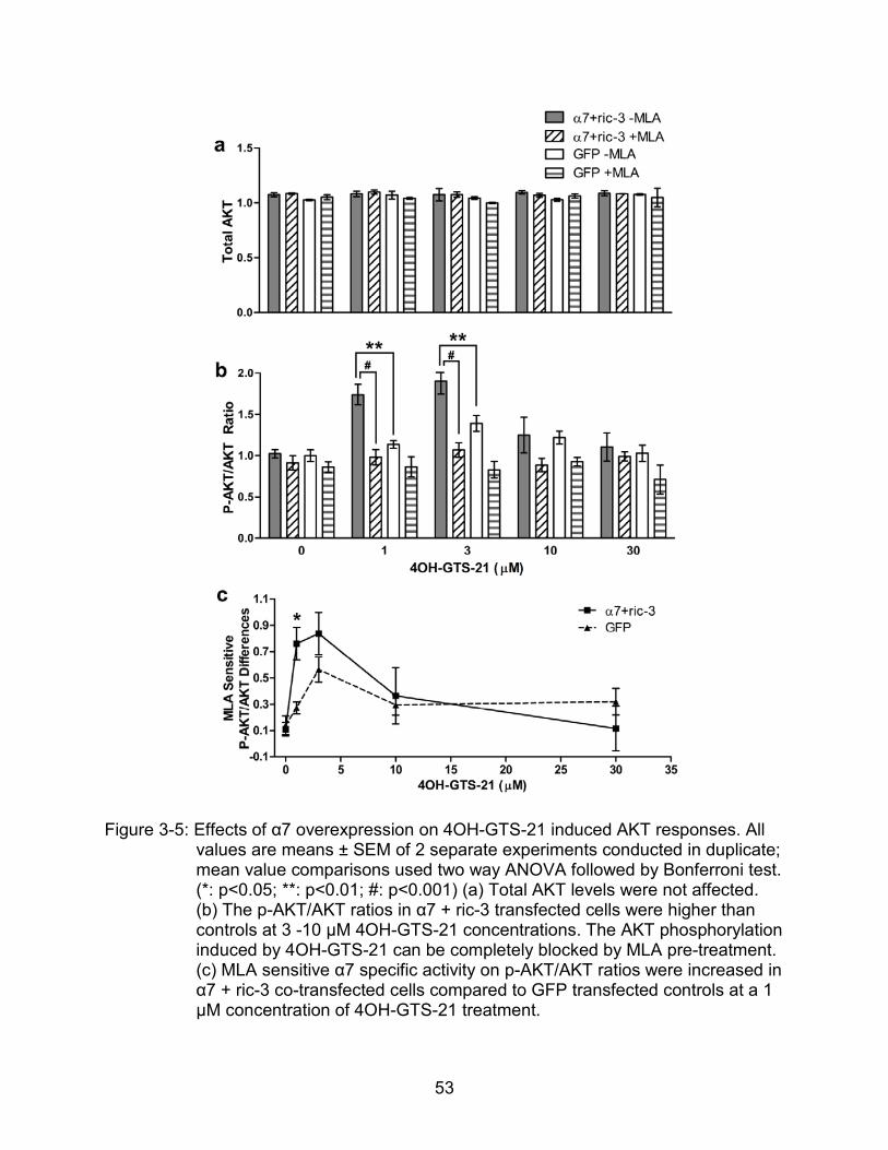
53
Figure 3-5: Effects of α7 overexpression on 4OH-GTS-21 induced AKT responses. All values are means ± SEM of 2 separate experiments conducted in duplicate; mean value comparisons used two way ANOVA followed by Bonferroni test. (*: p<0.05; **: p<0.01; #: p<0.001) (a) Total AKT levels were not affected. (b) The p-AKT/AKT ratios in α7 + ric-3 transfected cells were higher than controls at 3 -10 µM 4OH-GTS-21 concentrations. The AKT phosphorylation induced by 4OH-GTS-21 can be completely blocked by MLA pre-treatment. (c) MLA sensitive α7 specific activity on p-AKT/AKT ratios were increased in α7 + ric-3 co-transfected cells compared to GFP transfected controls at a 1 µM concentration of 4OH-GTS-21 treatment.
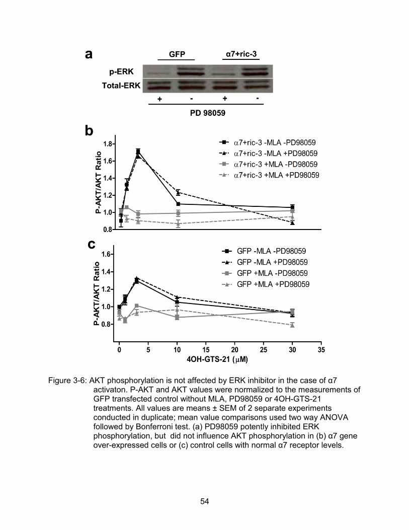
54
Figure 3-6: AKT phosphorylation is not affected by ERK inhibitor in the case of α7 activaton. P-AKT and AKT values were normalized to the measurements of GFP transfected control without MLA, PD98059 or 4OH-GTS-21 treatments. All values are means ± SEM of 2 separate experiments conducted in duplicate; mean value comparisons used two way ANOVA followed by Bonferroni test. (a) PD98059 potently inhibited ERK phosphorylation, but did not influence AKT phosphorylation in (b) α7 gene over-expressed cells or (c) control cells with normal α7 receptor levels.
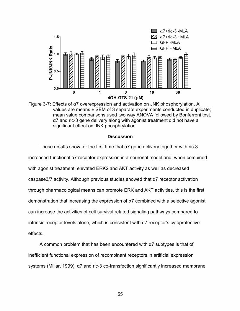
55
Figure 3-7: Effects of α7 overexpression and activation on JNK phosphorylation. All
values are means ± SEM of 3 separate experiments conducted in duplicate; mean value comparisons used two way ANOVA followed by Bonferroni test. α7 and ric-3 gene delivery along with agonist treatment did not have a significant effect on JNK phosphrylation.
Discussion
These results show for the first time that α7 gene delivery together with ric-3
increased functional α7 receptor expression in a neuronal model and, when combined
with agonist treatment, elevated ERK2 and AKT activity as well as decreased
caspase3/7 activity. Although previous studies showed that α7 receptor activation
through pharmacological means can promote ERK and AKT activities, this is the first
demonstration that increasing the expression of α7 combined with a selective agonist
can increase the activities of cell-survival related signaling pathways compared to
intrinsic receptor levels alone, which is consistent with α7 receptor’s cytoprotective
effects.
A common problem that has been encountered with α7 subtypes is that of
inefficient functional expression of recombinant receptors in artificial expression
systems (Millar, 1999). α7 and ric-3 co-transfection significantly increased membrane
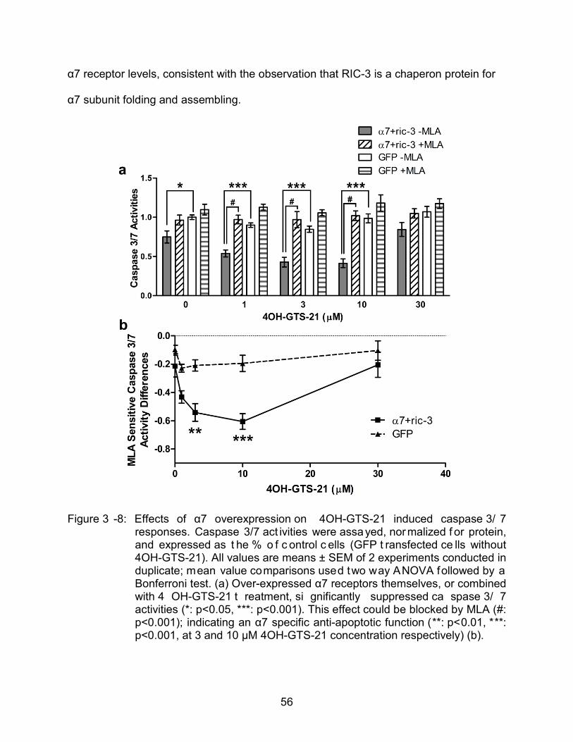
56
α7 receptor levels, consistent with the observation that RIC-3 is a chaperon protein for
α7 subunit folding and assembling.
Figure 3 -8: Effects of α7 overexpression on 4OH-GTS-21 induced caspase 3/ 7 responses. Caspase 3/7 act ivities were assayed, normalized f or protein, and expressed as t he % o f c ontrol c ells (GFP t ransfected ce lls without 4OH-GTS-21). All values are means ± SEM of 2 experiments conducted in duplicate; mean value comparisons used two way ANOVA followed by a Bonferroni test. (a) Over-expressed α7 receptors themselves, or combined with 4 OH-GTS-21 t reatment, si gnificantly suppressed ca spase 3/ 7 activities (*: p<0.05, ***: p<0.001). This effect could be blocked by MLA (#: p<0.001); indicating an α7 specific anti-apoptotic function (**: p<0.01, ***: p<0.001, at 3 and 10 µM 4OH-GTS-21 concentration respectively) (b).
57
RIC-3 it was originally identified in the nematode Caenorhabditis elegans (C.
elegans) as the protein encoded by the gene ric-3 (resistance to inhibitors of
cholinesterase) and has subsequently been cloned and characterized from mammalian
and insect species (Nguyen et al., 1995).It is interesting to note that Ric-3 gene delivery
alone also elevated the membrane α7 receptor expression to some extent, while α7
gene delivery alone did not show a significant effect, suggesting ric-3 expression is
more limiting than α7 translation for the number of functional α7 receptors in plasma
membrane in this PC12 cell line. In contrast to normal aging, where both α7 mRNA and
protein levels were decreased (Court et al. 2001), in AD α7 protein loss is observed
without significant mRNA level change or even increased α7 mRNA level (Engidawork
et al. 2001; Perry et al. 2001; Wevers et al. 2000; Hellstrom-Lindahl et al. 1999). These
data pointed to a possibility that the α7 receptor expression deficit in AD is related with
translational or post-translational problems rather than transcriptional insufficiency. Thus,
RIC-3 modulation itself may be important for AD pathogenesis and a valuable target for
future studies.
Selective α7 agonists have been intensively studied and broadly used for
therapeutic purposes. However, α7 receptors desensitize rapidly and persistently at
relatively low steady-state agonist concentrations (Seguela et al., 1993; Uteshev et al.
2003; Papke et al. 2009). Therefore a low agonist concentration needs to be maintained
for long-term treatment. In our study, receptors still desensitize at high 4OH-GTS-21
concentrations, but the agonist efficacy at lower concentrations was increased reflecting
on ERK2 and AKT phosphorylation, which play important roles in cell proliferation,
differentiation and anti-apoptosis (Pettmann et al. 1998; Traverse et al. 1992; Franke et
58
al. 1997). Considering the 15~20% of plasmid transfection efficiency, the true effects
might be even higher, which is quite promising for protection purposes. Although a
previous study showed that α7 gene overexpression in PC12 cells alone resulted in
sustained ERK phosphorylation (Utsugisawa et al., 2002), other studies suggested α7
induced ERK phosphorylation is a transient event (Ren et al., 2005; El Kouhen et al.,
3009; Gubbins et al., 2010), consistant with our results. ERK phosphorylation inhibitor
PB98059 did not influence AKT phosphorylation, suggesting no crosstalk between
these two pathways (at least at the pathways before AKT and ERK) when induced by
α7 receptor activation. Some studies showed AKT and ERK converged downstream at
the level of Bcl family proteins and ribosomal s6 kinase (RSK) (Creson et al., 2009). The
activation of JNK, another MAPK pathway kinase, might be necessary but not sufficient,
to induce neuronal apoptosis (Manning et al. 2003; Yuan et al. 2000). In our results,
JNK activity is not significantly influenced by α7 gene delivery combined with agonist
treatment. However, there was a tendency for reduced JNK activation at certain 4OH-
GTS-21 concentrations in the α7 and ric-3 transfected group.
Both ERK and AKT survival pathways regulate Bcl family proteins (Almeida et al.,
2005; Shen et al., 2010), which have a crucial role in intracellular apoptotic signal
transduction. This gene family includes both anti-apoptotic and pro-apoptotic proteins
that contain one or more bcl-2 homology (BH) domains. The major anti-apoptotic
members of the Bcl-2 family, Bcl-2 and Bcl-xl, are localized to the mitochondrial outer
membrane and to the endoplasmic reticulum and perinuclear membrane.
Overexpression of Bcl-2 attenuated the pathogenesis in a triple transgenic mice model
for AD, suggesting the activation of apoptotic pathways may be an early event
59
contributing to the progress of AD (Rohn et al., 2008). Studies have shown that ERK
and AKT may regulate Bcl-2 by activating CREB and enhance Bcl-2 synthesize (Creson
et al., 2009) or by directly phosporylating Bcl-2 (Ito et al., 1997). Blocking ERK or AKT
phosphorylation prevented the anti-apoptosis effects in neurons (Laing et al., 2008;
Namikawa et al., 2000). ERK can also protect cortical neurons via inactivating Bad, the
pro-apoptotic Bcl family protein, by phosphorylate it (Jin et al., 2002). Our studies
showed Bcl-2 levels were not significantly changed after α7 and ric-3 gene delivery
compared to controls. One possible reason might be that 30 min agonist treatment is
not sufficient for detectable changes in Bcl-2 transcription and translation.
Bcl-2 inhibits mitochondra cytochrome C release triggered by apoptotic stimuli,
thereby consequently blocking the activation of caspase cascade (Riedl and Shi, 2004).
The present results showed that up-regulating α7 receptors triggered a decrease in
caspase 3/7 activities. It was interesting to observe that, with no 4OH-GTS-21 present,
over-expressed α7 receptors alone were sufficient to induce caspase 3/7 response
compared to the GFP controls (Figure 3-8a, at 0 µM 4OH-GTS-21) but not in the case
of ERK/AKT responses (Figure 3-4a and 3-4b at 0 µM 4OH-GTS-21). This suggested
that over-expressed α7 receptors may regulate the caspase activity both directly and
indirectly from α7 receptor mediated signaling pathways. A possible indirect pathway is
that elevated α7 receptors increase the expression of nerve growth factor (Jonnala et al.
2002; Hernandez et al. 2005) which helps to down-regulate caspase activities by
activating TrkA receptors. Mounting evidence suggested the involvement of caspases in
the disease processes associated with AD. The activation of caspases not only triggers
the apoptosis cascade, but also cleavage of APP and tau to facilitate the production of
60
neurotoxic Aβ and neurofibrilary tangles. Therefore, caspase inhibition may be a
potential future approach for treating neurodegeneration (Rohn and Head, 2009).
In conclusion, α7 and ric-3 co-transfection increased α7 nicotinic receptor
expression in PC12 cells, which, combined with selective agonist 4OH-GTS-21,
increased α7 receptor mediated ERK and AKT activation as well as decreased caspase
3/7 activities. The combination of pharmacological and genetic approaches employed in
this study may be useful for treating conditions such as AD which are characterized by
low levels of endogenous α7 receptor expression in brain. The mechanistic studies
focusing on the roles of ERK, JNK MAPKs as well as caspases are expected to reveal
future targets and approaches for drug development in an AD-like milieu.
61
CHAPTER 4 EFFECTS OF α7 RECEPTOR GENE DELIVERY ON NEUROTROPHIN PATHWAY
Introduction
The basal forebrain cholinergic neurons are largely comprised of the nucleus
basalis magnocellularis, the horizontal and vertical diagonal bands of Broca and the
medial septal nucleus. Cholinergic neurons within the nucleus basalis and the septal
complex provide the major source of cholinergic innervation to the cerebral cortex and
hippocampus, respectively, contributing to the memory and attentional functions of
those regions (Niewiadomska 2010). Cholinergic neurons are particularly vulnerable
during AD, and emerging evidence shows NGF to be an essential maintenance factor
for their normal function (Cuello et al., 2010).
NGF is one of the neurotrophic factors that stimulate axonal growth and maintain
neuronal phenotype and promote neuronal survival (Siegel and Chauhan, 2000). During
development, growing axons compete for limited amounts of neurotrophic factors
produced by target tissues (Yuen et al., 1996). In adult brain, neurotrophic factors are
required to maintain normal functions and specific phenotype (Sofroniew et al., 2001).
NGF mRNA and protein are highly concentrated within basal forebrain targeted regions,
such as the hippocampus and cerebral cortex (Large et al., 1986) and released from
postsynaptic terminals. Once bound to its receptors existing on cholinergic presynatic
terminals, the NGF-receptor complex is internalized and retrogradely transported from
these regions to basal forebrain neuron somas (DiStefano et al., 1992). A wealth of data
demonstrated that basal forebrain cholinergic neurons are exquisite sensitive to NGF;
insufficient NGF supply may be responsible for the consistent atrophy and neuron loss
seen in AD. Transection of fimbria cuts the connection between septal basal forebrain
62
and hippocampus, blocks NGF transportation, results in cholinergic neuron loss and
mimics AD-like behavioral deficits (Lapchak 1993). NGF treatment in septum rescued
cholinergic neurons that undergo degeneration following septohippocampal axotomy in
rodents and primates (Hefti 1986; Williams et al., 1986; Kordower et al., 1994).
Therefore, initially it was suggested that alterations in NGF synthesis within cortical
production sites may be responsible for the basal forebrain cholinergic neuron death
during early AD (Hefti et al., 1989).
However, studies with AD patients found that NGF mRNA levels in cholinergic
targeted regions are unaltered, and its protein levels are even increased (Mufson et al.,
1996). These data suggested that despite normal NGF synthesis, NGF decreasing in
basal forebrain as a result of defective receptor binding and / or retrograde transport of
NGF from the hippocampus / cortex to consumer neuron somas within the basal
forebrain.
Basal forebrain cholinergic neurons express two types of NGF receptors: the pan-
neurotrophin receptor p75NTR and the NGF-specific receptor tyrosine kinase TrkA
(Counts and Mufson, 2005). p75NTR binds NGF with low affinity (Kd=nM); while TrkA
binds NGF with high affinity (Kd=pM) (Lapchak 1993) and mediates the binding,
internalization and retrograde transportation of target derived NGF to basal forebrain
(Kaplan and Miller, 1997; Mufson et al., 1999). It has been found that TrkA mRNA within
individual nucleus basalis neurons was significantly reduced (66%) in early AD cases
relative to aged controls while p75NTR is not affected (Mufson et al., 1996; Boissiere
1997; Mufson 1997; Counts et al., 2004); indicating that there is a selective defect in
TrkA expression in AD brain. Besides, TrkA receptor antagonist produces a depletion
63
of pre-exisitng cholinergic boutons (Debeir et al., 1999); and activation of TrkA alone
prevented the degeneration of axotomized cholinergic neurons in rats (Mufson et al.,
1996). These data suggested that signaling via TrkA receptors is a pivotal trophic event
underlying the viability of these cells and reduced NGF transport may be secondary to
diminished TrkA expression.
The mechanisms underlying TrkA protein reduction in cholinergic neurons in early
AD are unclear. However, TrkA gene expression is under positive feedback from NGF
signaling (Holtzman et al. 1992). Despite retrograde transportation, NGF also activates
MAPK and PI3K pathway that are related to cholinergic neuron survival; results in
CREB mediated transcription and new protein synthesis (Counts and Mufson, 2005).
Basal forebrain cholinergic neurons exhibited rapid increases in TrkA immunostaining in
15 min of NGF application to the hippocampus (Williams et al., 2005), probably through
the MAPK pathway. This rapid signaling pathway is diminished in aged rodents
compared to young ones (Williams et al., 2006).
TrkA mediated NGF signaling also enhances cholinergic innervation by promoting
ACh synthesis, storage and release (Lapchak 1993), which, in turn, enhances the
neuronal activity and NGF synthesis in hippocampus and cortex. The impairment of this
reciprocal signaling between basal forebrain cholinergic neurons and
hippacampal/cortex neurons may underlie the eventual cholinergic deficits seen in the
progression of AD. Therefore, as a potential therapeutic target, many studies have
targeted NGF and have shown that exogenous NGF administration reversed both age-
related and lesion-induced cholinergic neuronal degeneration as well as spatial memory
deficits in animal models. In human, NGF gene therapy is associated with potential
64
risks. A Phase 1 clinical study using AAV-NGF vector with 6 subjects showed cognitive
improvement in 2 subjects but revealed back pain which might due to diffused NGF at
peripheral nerve system (Tuszynski et al., 2005).
Besides targeting NGF directly, several studies have shown that nicotine
exposure selectively increased NGF levels in multiple brain regions for prolonged
intervals (Garrido et al., 2003; Martinez-Rodriguez et al., 2003; Jonnala et al., 2002) and
led to astrocytic NGF expression that was undetectable in control tissues (Martinez-
Rodriguez et al., 2003). No increase is seen in other neurotrophins such as brain-
derived neurotrophic factor (BDNF) or neurotrophin-3 (NT-3) levels (French et al.,
1999). Also, nicotine administration increased TrkA expression in vitro and in vivo
(French et al., 1999; Formaggio et al., 2010). These effects of nicotine were blocked by
α7 antagonist (Jonnala et al., 2002; Li et al., 2005), suggesting the role of α7 nAChRs in
modulating TrkA and NGF expression.
However, no direct evidence has shown that α7 activation will increase TrkA
levels. In this study, we investigated whether α7 receptor overexpression and activation
will have cellular actions that both involve TrkA receptor expression as well as mimic it.
If successful, the α7 gene delivery approach will not only counteract the α7 receptor
loss in basal forebrain cholinergic neurons, but also benefit the reciprocal loop of the
septohippacampal pathway by elevating NGF-TrkA survival signaling. The activation of
α7 and TrkA receptors transduce the message to similar cellular kinase cascades
including MAPK and PI3K-AKT; which may achieve “double protection” for degenerating
cholinergic neurons during AD.
65
Results
Effects of α7 nAChRs Overexpression on TrkA In Vitro
Like basal forebrain cholinergic neurons affected by AD, rat PC12 cells
endogenously express both α7 and TrkA proteins and are a valuable model for studying
the relationship between the two receptors. In the first report of this cell line, PC12 cell
differentiation triggered NGF exposure was described, characterized by halted
proliferation and extended neurite outgrowth (Greene and Tischler, 1976). The cellular
signaling pathways involved in NGF mediated PC12 cell differentiation have been
intensively studied; at least three independent pathways now known to contribute to this
response, including ras/raf-MEK-ERK, cAMP-PKA and PI3K-AKT (Vaudry et al., 2002;
Counts and Mufson, 2005; Yung et al., 2010).
In order to evaluate the effects of α7 overexpression on TrkA expression, we
transfected the cells at ~40% confluence with the same doses of α7+ric-3, α7+GFP, ric-
3+GFP or GFP plasmids (2 µg/ml) based on previously established gene delivery
system as described in Chapter 3. 24 hr after transfection, the following antagonists was
added in with fresh medium into cell culture: anti-α7, MLA; anti-ERK, PD98059 or anti-
AKT, API-2 (Yang et al., 2004), separately or in combination, for 30 min and then
removed. NGF was added to the medium repeatedly to maintain a concentration of ~50
ng/ml for 4 days, which induced neurite growth and halt proliferation. Cells were then
fixed with 4% paraformaldehyde; and random pictures of each well were taken under
microscope. Cells with neuron-like process(es) longer than its body length were
considered as differentiated (Figure 4-1). Numbers of differentiated and total cells
numbers were counted in a blinded manner. The differentiation rate of each group is
summarized in Figure 4-2.
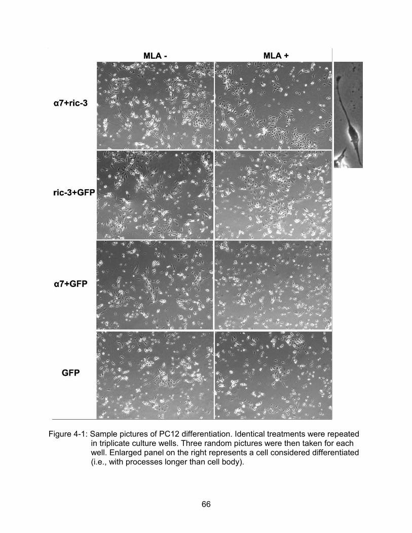
66
Figure 4-1: Sample pictures of PC12 differentiation. Identical treatments were repeated
in triplicate culture wells. Three random pictures were then taken for each well. Enlarged panel on the right represents a cell considered differentiated (i.e., with processes longer than cell body).
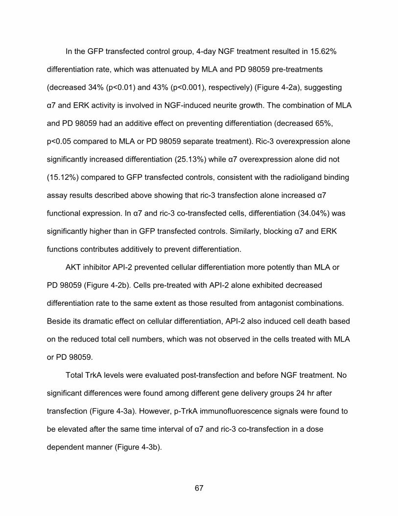
67
In the GFP transfected control group, 4-day NGF treatment resulted in 15.62%
differentiation rate, which was attenuated by MLA and PD 98059 pre-treatments
(decreased 34% (p<0.01) and 43% (p<0.001), respectively) (Figure 4-2a), suggesting
α7 and ERK activity is involved in NGF-induced neurite growth. The combination of MLA
and PD 98059 had an additive effect on preventing differentiation (decreased 65%,
p<0.05 compared to MLA or PD 98059 separate treatment). Ric-3 overexpression alone
significantly increased differentiation (25.13%) while α7 overexpression alone did not
(15.12%) compared to GFP transfected controls, consistent with the radioligand binding
assay results described above showing that ric-3 transfection alone increased α7
functional expression. In α7 and ric-3 co-transfected cells, differentiation (34.04%) was
significantly higher than in GFP transfected controls. Similarly, blocking α7 and ERK
functions contributes additively to prevent differentiation.
AKT inhibitor API-2 prevented cellular differentiation more potently than MLA or
PD 98059 (Figure 4-2b). Cells pre-treated with API-2 alone exhibited decreased
differentiation rate to the same extent as those resulted from antagonist combinations.
Beside its dramatic effect on cellular differentiation, API-2 also induced cell death based
on the reduced total cell numbers, which was not observed in the cells treated with MLA
or PD 98059.
Total TrkA levels were evaluated post-transfection and before NGF treatment. No
significant differences were found among different gene delivery groups 24 hr after
transfection (Figure 4-3a). However, p-TrkA immunofluorescence signals were found to
be elevated after the same time interval of α7 and ric-3 co-transfection in a dose
dependent manner (Figure 4-3b).
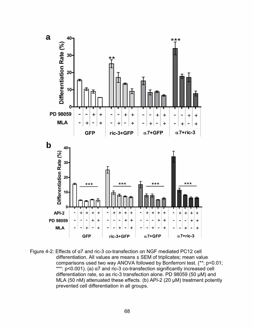
68
Figure 4-2: Effects of α7 and ric-3 co-transfection on NGF mediated PC12 cell differentiation. All values are means ± SEM of triplicates; mean value comparisons used two way ANOVA followed by Bonferroni test. (**: p<0.01; ***: p<0.001). (a) α7 and ric-3 co-transfection significantly increased cell differentiation rate, so as ric-3 transfection alone. PD 98059 (50 µM) and MLA (50 nM) attenuated these effects. (b) API-2 (20 µM) treatment potently prevented cell differentiation in all groups.
a
b
69
Figure 4-3: Immunoactivity of TrkA in PC12 cells after α7 and ric-3 co-transfection. (a)
western blot of TrkA levels after different types of transfection. The TrkA was detected by anti-pan Trk antibody; the bands of TrkA (~135kD) were quantified by ImageJ and normalized with corresponding β-actin levels in each sample. All data were normalized to non-transfected control and expressed as mean ± SEM; N=3. (b) immunostaining for α7 and phospho-TrkA 24 hr after transfection with different α7+ric-3 doses. Secondary antibody only control (pictures not shown here) showed very low non-specific signals.
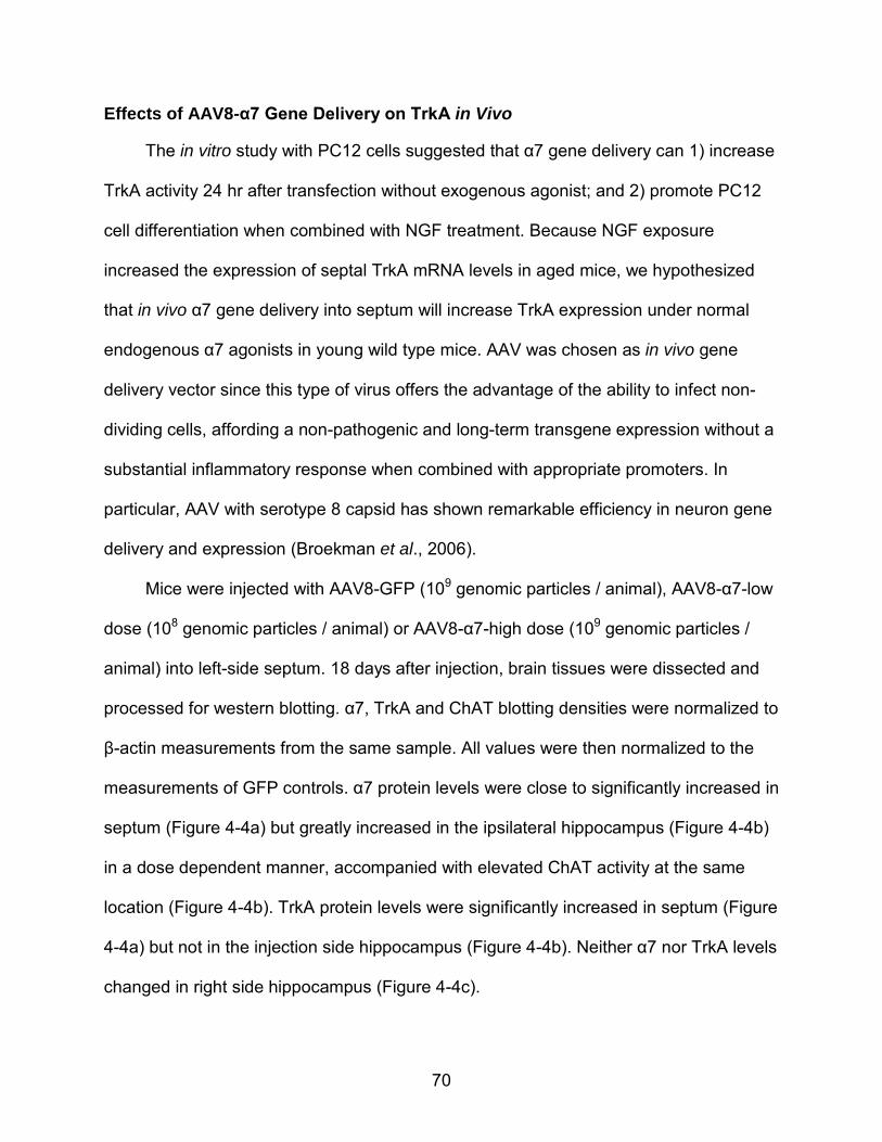
70
Effects of AAV8-α7 Gene Delivery on TrkA in Vivo
The in vitro study with PC12 cells suggested that α7 gene delivery can 1) increase
TrkA activity 24 hr after transfection without exogenous agonist; and 2) promote PC12
cell differentiation when combined with NGF treatment. Because NGF exposure
increased the expression of septal TrkA mRNA levels in aged mice, we hypothesized
that in vivo α7 gene delivery into septum will increase TrkA expression under normal
endogenous α7 agonists in young wild type mice. AAV was chosen as in vivo gene
delivery vector since this type of virus offers the advantage of the ability to infect non-
dividing cells, affording a non-pathogenic and long-term transgene expression without a
substantial inflammatory response when combined with appropriate promoters. In
particular, AAV with serotype 8 capsid has shown remarkable efficiency in neuron gene
delivery and expression (Broekman et al., 2006).
Mice were injected with AAV8-GFP (109 genomic particles / animal), AAV8-α7-low
dose (108 genomic particles / animal) or AAV8-α7-high dose (109 genomic particles /
animal) into left-side septum. 18 days after injection, brain tissues were dissected and
processed for western blotting. α7, TrkA and ChAT blotting densities were normalized to
β-actin measurements from the same sample. All values were then normalized to the
measurements of GFP controls. α7 protein levels were close to significantly increased in
septum (Figure 4-4a) but greatly increased in the ipsilateral hippocampus (Figure 4-4b)
in a dose dependent manner, accompanied with elevated ChAT activity at the same
location (Figure 4-4b). TrkA protein levels were significantly increased in septum (Figure
4-4a) but not in the injection side hippocampus (Figure 4-4b). Neither α7 nor TrkA levels
changed in right side hippocampus (Figure 4-4c).
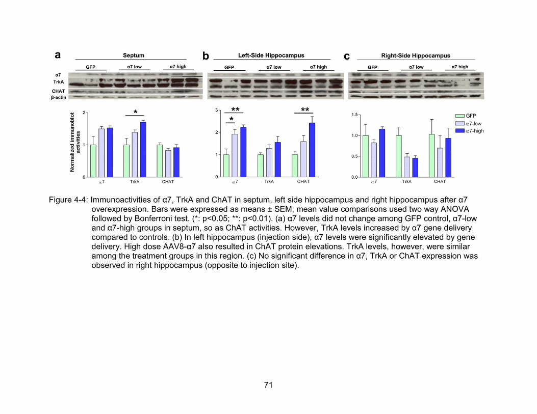
71
Figure 4-4: Immunoactivities of α7, TrkA and ChAT in septum, left side hippocampus and right hippocampus after α7
overexpression. Bars were expressed as means ± SEM; mean value comparisons used two way ANOVA followed by Bonferroni test. (*: p<0.05; **: p<0.01). (a) α7 levels did not change among GFP control, α7-low and α7-high groups in septum, so as ChAT activities. However, TrkA levels increased by α7 gene delivery compared to controls. (b) In left hippocampus (injection side), α7 levels were significantly elevated by gene delivery. High dose AAV8-α7 also resulted in ChAT protein elevations. TrkA levels, however, were similar among the treatment groups in this region. (c) No significant difference in α7, TrkA or ChAT expression was observed in right hippocampus (opposite to injection site).
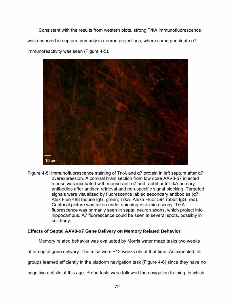
72
Consistent with the results from western blots, strong TrkA immunofluorescence
was observed in septum, primarily in neuron projections, where some punctuate α7
immunoreactivity was seen (Figure 4-5).
Figure 4-5: Immunofluorescence staining of TrkA and α7 protein in left septum after α7
overexpression. A coronal brain section from low dose AAV8-α7 injected mouse was incubated with mouse-anti-α7 and rabbit-anti-TrkA primary antibodies after antigen retrieval and non-specific signal blocking. Targeted signals were visualized by fluorescence labled secondary antibodies (α7: Alex Fluo 488 mouse IgG, green; TrkA: Alexa Fluor 594 rabbit IgG, red). Confocal picture was taken under spinning-disk microscopy. TrkA fluorescence was primarily seen in septal neuron axons, which project into hippocampus. Α7 fluorescence could be seen at several spots, possibly in cell body.
Effects of Septal AAV8-α7 Gene Delivery on Memory Related Behavior
Memory related behavior was evaluated by Morris water maze tasks two weeks
after septal gene delivery. The mice were ~12 weeks old at that time. As expected, all
groups learned efficiently in the platform navigation task (Figure 4-6) since they have no
cognitive deficits at this age. Probe tests were followed the navigation training, in which
73
the escape platform was removed from the water maze. 60 min swimming period of
each mouse was recorded by video camera, and swimming tracks were captured and
analyzed for times passing the platform area, percent time spent / swimming distance /
swimming speed in each quadrant, etc (Figure 4-7).
The percent time spent in the target quadrant (where the platform was on training
trials) (Figure 4-7 pie chart, Figure 4-8b) was not different among the treatment groups,
nor were the swimming distances. The α7 low dose group showed a tendency to cross
the platform region more times than GFP injected mice (p=0.07 by Student’s T-test)
(Figure 4-8a). They also swam faster in the opposite quadrant compared to controls
(Figure 4-8c).
Figure 4-6: Training curves resulting from Morris water maze platform navigation task.
Mice were trained for 2 days with 8 trails per day. Mice were placed into water facing the wall of the tank, each time at a different site. They were allowed to up to 60 sec to search for the escape platform, followed by 30 sec on the platform to enhance the memory. Geometric signs were hung on the tank to help navigation. Time spent to find the platform of each trial was plotted to represent a learning curve. All groups succeeded in fast locating of the platform after 2 days’ training.
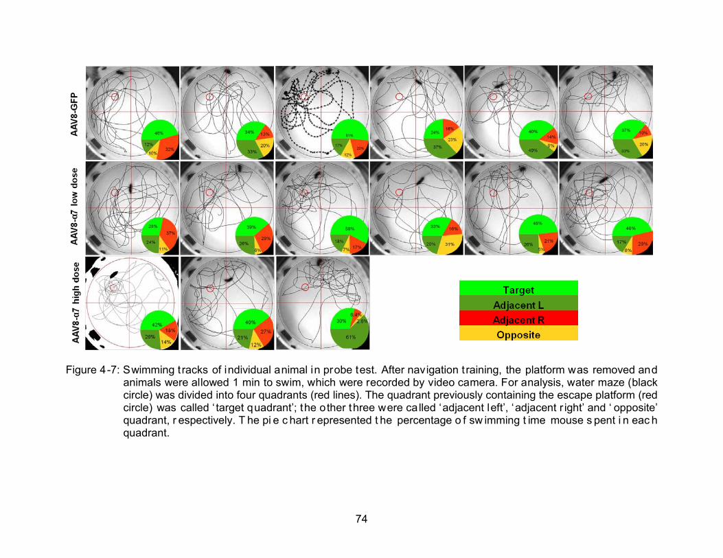
74
Figure 4-7: Swimming tracks of individual animal in probe test. After navigation t raining, the platform was removed and
animals were allowed 1 min to swim, which were recorded by video camera. For analysis, water maze (black circle) was divided into four quadrants (red lines). The quadrant previously containing the escape platform (red circle) was called ‘ target quadrant’; the other three were called ‘adjacent left’, ‘adjacent r ight’ and ‘ opposite’ quadrant, r espectively. T he pi e c hart r epresented t he percentage o f sw imming t ime mouse s pent i n eac h quadrant.
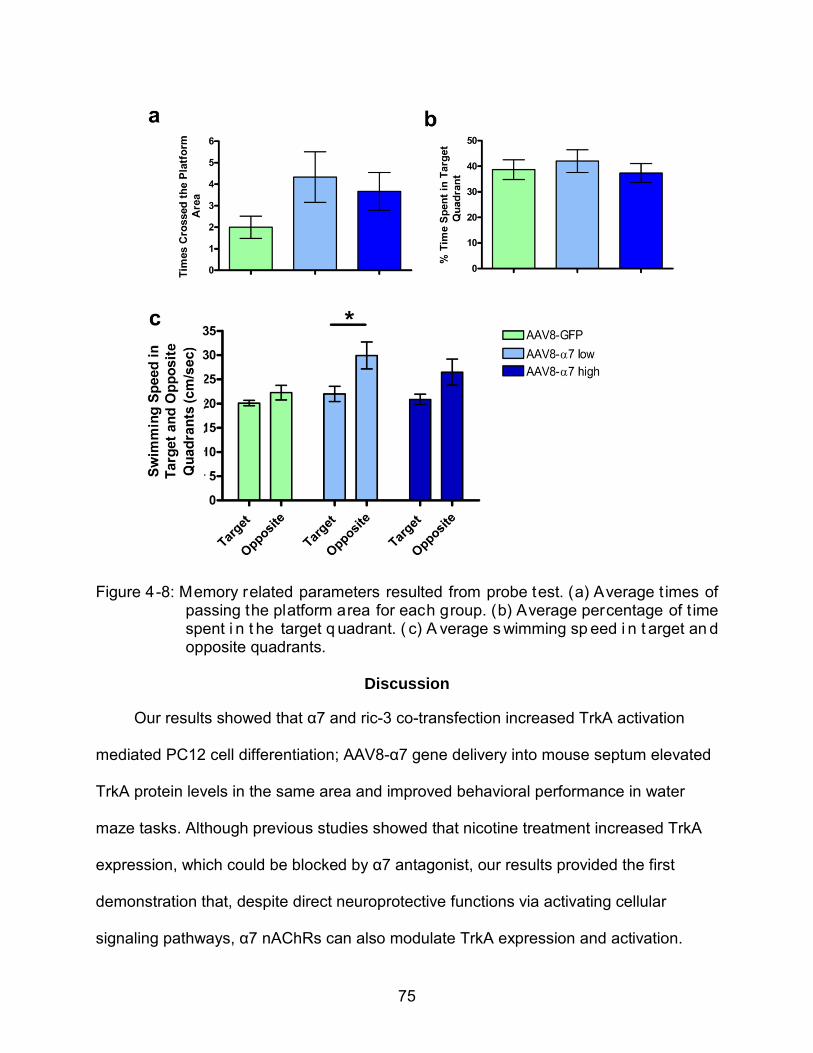
75
Figure 4-8: Memory related parameters resulted from probe test. (a) Average t imes of
passing the platform area for each group. (b) Average percentage of t ime spent i n t he target q uadrant. ( c) A verage s wimming sp eed i n t arget an d opposite quadrants.
Discussion
Our results showed that α7 and ric-3 co-transfection increased TrkA activation
mediated PC12 cell differentiation; AAV8-α7 gene delivery into mouse septum elevated
TrkA protein levels in the same area and improved behavioral performance in water
maze tasks. Although previous studies showed that nicotine treatment increased TrkA
expression, which could be blocked by α7 antagonist, our results provided the first
demonstration that, despite direct neuroprotective functions via activating cellular
signaling pathways, α7 nAChRs can also modulate TrkA expression and activation.
76
Since the NGF-TrkA pathway tranduce differentiation and survival messages, α7 gene
delivery approach may potentially result in combined protective effects from α7 nAChRs
themselves as well as TrkA receptors.
NGF induces PC12 cell differentiation reflecting decreased proliferation and
increased neuritic extention. Interestingly, we found that α7 and ric-3 co-transfection
significantly increased the differentiation rate of the NGF treated PC12 cells, and this
effect was attenuated by 30 min MLA treatment post transfection and before NGF
incubation, suggesting that α7 nAChRs were partially involved in the differentiation
process. Considering that the p-TrkA level but not the total TrkA protein level was
elevated 24 hr post transfection, α7 possibly modulated cell differentiation via increasing
NGF release, which form a complex with TrkA and induced TrkA phosphorylation. ERK
inhibitor PD 98059 also attenuated PC12 cell differentiation, consistent with the fact that
ERK is one of the downstream kinases responsive to NGF-induced TrkA activation. Our
previous data showed that in PC12 cells the endogenous activation of up-regulated α7
nAChRs was not sufficient to induce detectable change in ERK phosphorylation (Figure
3-3 at 0 µM 4OH-GTS-21). In this study, the effects of MLA and PD 98059 were
additive. These two pieces of information suggested that α7 nAChRs may be regulating
PC12 cell differentiation through a non-ERK pathway(s). AKT inhibitor API-2 also
decreased differentiation rate, but more effectively than either MLA or PD 98059,
indicating NGF-TrkA induced differentiation is more dependent on AKT signaling
pathway.
In addition, cells transfected with ric-3 or α7+ric-3 over-expressed functional α7
nAChRs (Figure 3-2a), contributing to enhanced differentiation after NGF treatment
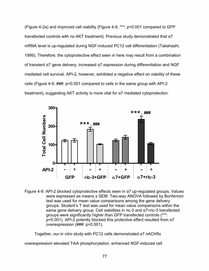
77
(Figure 4-2a) and improved cell viability (Figure 4-9, ***: p<0.001 compared to GFP
transfected controls with no AKT treatment). Previous study demonstrated that α7
mRNA level is up-regulated during NGF-induced PC12 cell differentiation (Takahashi,
1999). Therefore, the cytoprotective effect seen in here may result from a combination
of transient α7 gene delivery, increased α7 expression during differentiation and NGF
mediated cell survival. API-2, however, exhibited a negative effect on viability of these
cells (Figure 4-9, ###: p<0.001 compared to cells in the same group with API-2
treatment), suggesting AKT activity is more vital for α7 mediated cytoprotection.
Figure 4-9: API-2 blocked cytoprotective effects seen in α7 up-regulated groups. Values were expressed as means ± SEM. Two-way ANOVA followed by Bonferroni test was used for mean value comparisons among the gene delivery groups. Student’s T test was used for mean value comparisons within the same gene delivery group. Cell viabilities in ric-3 and α7+ric-3 transfected groups were significantly higher than GFP transfected controls (***: p<0.001). API-2 potently blocked this protective effect resulted from α7 overexpression (###: p<0.001).
Together, our in vitro study with PC12 cells demonstrated α7 nAChRs
overexpression elevated TrkA phosphorylation, enhanced NGF-induced cell
78
differentiation and improved cell viability. The AKT signaling pathway presented pre-
eminent function in conducting signals related with differentiation and survival, and can
potentially be targeted in future studies.
The in vivo gene delivery study showed that AAV8-α7 injection into mouse left
septum resulted in α7 protein increasing in left hippocampus compared to controls
injected with AAV8-GFP. ChAT, which is considered to be the marker for cholinergic
activity, was also elevated along with the α7 overexpression, indicating that septal
cholinergic neurons projecting into hippocampus are likely responsible for at least some
of the increased expression. Increased α7 nAChR expression therefore led to up-
regulated TrkA protein levels in the septum, supporting our hypothesis and can be
explained by the septohippocampal reciprocal loop. Two bands were observed in TrkA
western blots (Figure 4-4). The bottom band represented the molecular weight of TrkA
at ~140 Kd; and the top band was ~160 Kd. Interestingly, the densities of the 160 Kd
bands were lower in septum (Figure 4-4a) but higher in hippocampus (Figure 4-4b and
4-4c) compared to the 140 Kd bands in the same areas. Because in hippocampus two
NGF molecules (13 Kd) form dimers (26 Kd) via disulfide bridge as the active form
(Wiesmann et al., 1999), we speculate that the 160 Kd band represented a NGF dimer-
TrkA complex. In septum, endocytosed TrkA is predominantly sorted for recycling back
to cell surface (Chen et al., 2005), which may account for why less of the higher
molecular weight ‘NGF-TrkA’ was found.
Surprisingly, improved performance in Morris water maze tasks was observed
after α7 overexpression, which is usually not easy to see in young wild type mice. Times
passing the platform region were close to significantly increased in low dose AAV8-α7
79
injected group compared to controls, and the same mice swam faster in the opposite
quadrant, which partly indicated better memory for the platform location.
In conclusion, α7 receptors were found to be involved in modulating neurotrophin
pathways both in vitro and in vivo. For therapeutic application, it is possible that α7
overexpression could compensate for α7 and TrkA receptor loss early in AD, and
enhance the function of the septohippocampal reciprocal loop between cholinergic and
hippocampal neurons. It can also magnify the neuroprotective function by up-regulating
endogenous NGF synthesis, which can avoid the adverse side effects in more systemic
NGF therapy. No exogenous agonist was required in these studies, affording potential
advantage in utilizing endogenous α7 agonists.
80
CHAPTER 6 CONCLUSION AND FUTURE DIRECTIONS
Conclusion
The cholinergic hypothesis for AD has been studied for almost 40 years.
Cholinergic deterioration is one of the most consistent pathological features of all types
of AD. α7 nAChRs are a favorable target for protecting the cholinergic neuron as well as
improving cognitive functions in a variety of models. Although α7 selective agonists
have been intensively studied and displayed promising therapeutic effects, gene
therapy may provide more advantages including locally restoring the receptor loss and
avoiding fast desensitization. The goal of our research is to investigate the benefits of
α7 overexpression, improve our understanding in underlying neuroprotective
mechanisms, and test whether α7 gene delivery could be potential approach to treat
AD. Several hypotheses were involved, including: 1) up-regulated α7 receptors can
increase the agonist efficacy and potency on cellular signaling pathways; 2) α7
receptors can modulate neurotrophin induced cell differentiation; and 3) AAV mediated
α7 gene delivery in vivo will enhance TrkA expression.
In PC12 cells we found α7 and ric-3 co-transfection significantly increased
functional α7 nAChRs, which not only elevated the responses of cellular kinases and
reduced caspase 3/7 activities in response to 4OH-GTS-21, but also enhanced the cell
differentiation mediated by NGF, possibly via modulating TrkA expression (Figure 6-1).
All these effects were blocked by selective antagonist MLA. In mouse septum, AAV8-α7
gene delivery resulted in enhanced α7 receptor expression in the transduced side of the
hippocampus and TrkA expression in septum; improved behavioral performance was
observed even in young mice without cognition deficits. These effects may involve α7

81
activation modulated NGF synthesis, which is vital for reciprocally maintaining
cholinergic functions (Figure 6-2).
Figure 5-1: Pathways regulated by α7 and TrkA receptors in PC12 cells. Both kinds of receptors activate MAPK and PI3K/AKT pathways, which promote new gene synthesis through transcription factors like CREB. JAK2 and ERK5 are also found to be regulated by α7 and TrkA, respectively. Bcl-2 expression is increased in response to receptor activation. Bcl-2 blocks cytochrome C release from mitochondria, which induces caspase cascade. These effects regulate cell proliferation and differentiation.
Young wild type mice are a good model to investigate the effects of α7
overexpression under normal physiological conditions and endogenous
neurotransmitter levels. Α7 gene delivery into left side septum increased the receptor
expression in left side hippocampus but not right side hippocampus, consistent with the
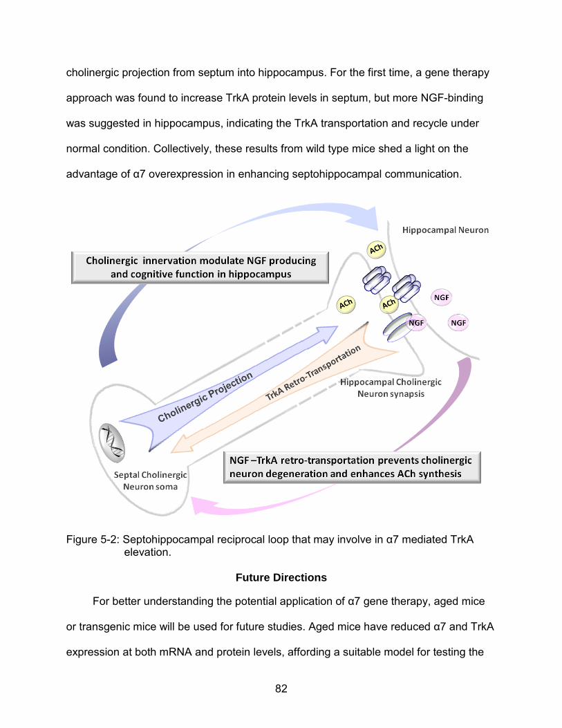
82
cholinergic projection from septum into hippocampus. For the first time, a gene therapy
approach was found to increase TrkA protein levels in septum, but more NGF-binding
was suggested in hippocampus, indicating the TrkA transportation and recycle under
normal condition. Collectively, these results from wild type mice shed a light on the
advantage of α7 overexpression in enhancing septohippocampal communication.
Figure 5-2: Septohippocampal reciprocal loop that may involve in α7 mediated TrkA elevation.
Future Directions
For better understanding the potential application of α7 gene therapy, aged mice
or transgenic mice will be used for future studies. Aged mice have reduced α7 and TrkA
expression at both mRNA and protein levels, affording a suitable model for testing the
83
α7 gene delivery and its effect on TrkA. Memory related behavioral deficits are more
significantly observed in these mice. Proper transgenic mouse models for AD should be
carefully selected according to specified purposes since there is no rodent model that
recapitulates all of the symptoms and neuropathological hallmarks of AD.
In particular, NGF deficient mice exhibit cholinergic loss in the basal forebrain and
neurofibrillary tangles / Aβ accumulation in the hippocampus, representing a useful
model for studying the function of α7 nAChRs in modulating NGF expression as
hypothesized from our research. Aβ overexpression mouse models (carry mutant APP,
γ-secretase component presenilin 1 or presenilin 2) could be used for investigating the
effects of α7 gene therapy on Aβ metabolism. Aβ is considered as the major toxic
peptide in AD brain and the processing of its precursor protein APP has been found to
be modulated by α7 nAChR activation. In turn, Aβ can competitively bind to α7
receptors and result in conflicting observations. Some groups found Aβ blocked α7
function while others found it activated α7 and induced downstream kinase
phosphorylation. Previous studies in our laboratory found α7 activation by 4OH-GTS-21
protected neurons from Aβ toxicity in APP/PS1 mouse model, and α7 gene delivery into
hippocampus improved special memory. Future studies that deliver α7 genes in Aβ
overexpressing mouse septum will help us better characterize the correlation between
α7 nAChRs and Aβ, which will benefit the application of α7 gene therapy in AD caused
by abnormal Aβ accumulation. Transgenic mice carrying mutant Tau gene exhibit
another major pathologic feature of AD: neurofibrillary tangles formed by hyper-
phophorylated Tau. Tau is a microtubule associated protein that functioning in
stabilizing cytoskeleton. AD patients have impaired septohippocampal transportation of
84
TrkA via cholinergic projections, implying potential deficits in normal Tau function.
Therefore investigation in whether α7 overexpression affects Tau pathology in those
mouse models will also be a interesting future direction. In addition, the crossed mouse
models may be helpful for cocktail gene therapy, which has not been attempted yet for
treating AD.
In conclusion, α7 gene delivery represents a potential approach for protecting
cholinergic neurons and improving cognition. Our data characterized the effects of α7
overexpression on neuroprotective pathways including MAPK, PI3K/AKT and NGF-
TrkA. Studies with specific animal models will help promote the therapeutic application
of α7 gene therapy for AD as well as other α7 nAChRs involved neurodegenerative
diseases.
85
LIST OF REFERENCES
Alessi D. R., Cuenda A., Cohen P., Dudley D. T. and Saltiel A. R. (1995) PD 098059 is a specific inhibitor of the activation of mitogen-activated protein kinase kinase in vitroand in vivo. J. Biol. Chem. 270, 27489–27494.
Alexander J. K ., Sagher D., Krivoshein A . V ., Criado M., Jefford G. and G reen W. N. (2010) Ric-3 promotes alpha7 nicotinic receptor assembly and trafficking through the ER subcompartment of dendrites. J Neurosci. 30, 10112–10126.
Alkondon M., Pereira E. F. and Albuquerque E. X. (1996) Mapping the location of functional nicotinic and gamma-aminobutyric acid A receptors on hippocampal neurons. J Pharmacol Exp Ther. 279, 1491–506.
Almeida R. D., Manadas B. J., Melo C. V., Gomes J. R., Mendes C. S., Grãos M. M., Carvalho R. F., Carvalho A. P. and Duarte C. B. (2005) Neuroprotection by BDNF against glutamate-induced apoptotic cell death is mediated by ERK and PI3-kinase pathways. Cell Death Differ. 12, 1329–1343.
Alzheimer A. (1907) A characteristic disease of the cerebral cortex. In: Bick K., Amaducci L. and Pepeu G. The early story of Alzheimer’s Diseae. Liviana Press, Padava Italy, pp 1–3
Arendash G.W., Sengstoch G.J., Sanberg P.R., and Kem W.R. (1995). Improved learning and memory in aged rats with chronic administration of the nicotinic receptor agonist GTS-21. Brain Res. 674, 252–259.
Bartus R. T., Dean R. L. 3rd, Beer B. and Lippa A. S. (1982) The cholinergic hypothesis of geriatric memory dysfunction. Science 217, 408–414.
Bettany J.H. and Levin E.D. (2001). Ventral hippocampal alpha7 nicotinic receptor blockade and chronic nicotine effects on memory performance in the radial-arm maze. Pharmacol. Biochem. Behav. 70, 467–474.
Bina K. G., Guzman P., Broide R. S., Leslie F. M., Smith M. A. and O’Dowd D. K. (1995) Localization of alpha 7 nicotinic receptor subunit mRNA and alpha-bungarotoxin binding sites in developing mouse somatosensory thalamocortical system. J. Comp. Neurol. 363:321–332.
Bitner R. S., Nikkel A. L., Markosyan S., Otte S., Puttfarcken P. and Gopalakrishnan M. (2009) Selective alpha7 nicotinic acetylcholine receptor activation regulates glycogen synthase kinase 3 beta and decreases tau phosphorylation in vivo. Brain Res. 1265, 65–74.
86
Boissiere F., Faucheux B., Ruberg M., Agid Y. and Hirsch E. C. (1997) Decreased TrkA gene expression i n c holinergic neurons of the st riatum a nd b asal f orebrain of patients with Alzheimer's disease. Exp. Neurol. 145, 245–252.
Bonny C., Borsello T. and Zine A. (2005) Targeting the JNK pathway as a therapeutic protective strategy for nervous system diseases. Rev. Neurosci. 16, 57–67.
Bowen D. M., Sims N. R., Benton J. S., Curzon G., Davison A. N., Neary D. and Thomas D. J. (1981) Treatment of Alzheimer's disease: a cautionary note. N. Engl. J. Med. 305, 1016.
Briggs C. A., Anderson, D. J., Brioni J. D., Buccafusco J. J., Buckley M. J. and AL. E. (1997) Functional characterization of the novel neuronal nicotinic acetylcholine receptor ligand GTS-21 in vitro and in vivo. Pharmacol. Biochem. Behav. 57, 231–241.
Broekman M . L ., Comer L . A ., Hyman B . T. an d Sena-Esteves M. ( 2006) A deno-associated v irus vectors serotyped w ith A AV8 c apsid ar e m ore e fficient than AAV-1 or -2 serotypes for widespread gene delivery to the neonatal mouse brain. Neuroscience 138, 501–510.
Broide R. S. and Leslie F. M. (1999) The alpha7 nicotinic acetylcholine receptor in neuronal plasticity. Mol Neurobiol. 20, 1–16.
Castro N. G. and Albuquerque E. X. (1993) Brief-Lifetime, fast-inactivating ion channels account for the r-bungarotoxin-sensitive nicotinic response in hippocampal neurons. Neurosci. Lett. 164, 137–140.
Changeux J. P., Kasai M. and Lee C. Y. (1970) Use of a snake venom toxin to characterize the cholinergic receptor protein. Proc. Natl. Acad. Sci. U S A. 67, 1241–1247.
Chen L., Wang H., Zhang Z., Li Z., He D., Sokabe M. and Chen L. (2001) DMXB (GTS-21) ameliorates the cognitive deficits in beta amyloid(25-35(-) ) injected mice through preventing the dysfunction of alpha7 nicotinic receptor. J. Neurosci. Res. 88, 1784–1794.
Chen Z. Y., Ieraci A., Tanowitz M. and Lee F. S. (2005) A novel endocytic recycling signal distinguishes biological responses of Trk neurotrophin receptors. Mol. Biol. Cell 16, 5761–5772.
Clarke P. B., Schwartz R. D., Paul S. M., Pert C. B. and Pert A. (1985) Nicotinic binding in rat brain: autoradiographic comparison of [3H]acetylcholine, [3H]nicotine, and [125I]-alpha-bungarotoxin. J. Neurosci. 5, 1307–1315.
87
Cooper E., Couturier S. and Ballivet M. (1991) Pentameric structure and subunit stoichiometry of a neuronal nicotinic acetylcholine receptor. Nature 350, 235–238.
Cooper J. D., Salehi A., Delcroix J. D., Howe C. L., Belichenko P. V., Chua-Couzens J. Kilbridge J. F., Carlson E. J., Epstein C. J. and Mobley W. C. (2001) Failed retrograde transport of NGF in a mouse model of Down's syndrome: reversal of cholinergic neurodegenerative phenotypes following NGF infusion. Proc. Natl. Acad. Sci. USA 98, 10439–10444.
Counts S . E ., Nadeem M ., Wuu J. , Ginsberg S . D ., Saragovi H . U. an d Mufson E . J. (2004) Reduction of co rtical T rkA but not p75(NTR) pr otein in e arly-stage Alzheimer's disease. Ann. Neurol. 56, 520–531.
Counts S . E . an d Mufson E. J . ( 2005) The r ole of nerve g rowth factor r eceptors i n cholinergic basal forebrain d egeneration i n pr odromal Alzheimer di sease. J Neuropathol. Exp. Neurol. 64, 263–272.
Court J., Martin-Ruiz C., Piggott M., Spurden D., Griffiths M. and Perry E. (2001) Nicotinic receptor abnormalities in Alzheimer's disease. Biol. Psychiatry. 49, 175–184.
Couturier S., Bertrand D., Matter J. M., Hernandez M. C., Bertrand S., Millar N., Valera S., Barkas T. and Ballivet M. (1990) A neuronal nicotinic acetylcholine receptor subunit (alpha 7) is developmentally regulated and forms a homo-oligomeric channel blocked by alpha-BTX. Neuron 5, 847–856.
Coyle J. T ., P rice D . L. and DeLong M. R . ( 1983) A lzheimer’s disease: a di sorder of cortical cholinergic innervation. Science 219, 1184–1190.
Creson T. K., Yuan P., Manji H. K. and Chen G. (2009) Evidence for involvement of ERK, PI3K, and RSK in induction of Bcl-2 by valproate. J. Mol. Neurosci. 37, 123–134.
Cuello A. C., Bruno M. A. and Bell K. F. (2007) NGF-cholinergic dependency in brain aging, MCI and Alzheimer's disease. Curr. Alzheimer Res. 4, 351–358.
Cuello A . C. , Bruno M. A., Allard S ., Leon W . and Iulita M . F . ( 2010) C holinergic involvement in Alzheimer's disease. A link with NGF maturation and degradation. J. Mol. Neurosci. 40, 230–235.
Curtis J. and Finkbeiner S. (1999) Sending Signals from the Synapse to the Nucleus: Possible Roles for CaMK, Ras/ERK, and SAPK Pathways in the Regulation of synaptic Plasticity and Neuronal Growth. J. Neurosci. Res. 58, 88–95.
88
Dajas-Bailador F. A., Soliakov L. and Wonnacott S. (2002a) Nicotine activates the extracellular signal regulated kinase 1/2 via the alpha7 nicotinic acetylcholine receptor and protein kinase A, in SH-SY5Y cells and hippocampal neurones. J. Neurochem. 80, 520–530.
Dajas-Bailador F., Mogg A. J. and Wonnacott S. (2002b) Intracellular Ca2+ channels evoked by stimulation of nicotinic acetylcholine receptors in SH-SY5Y cells: contribution of voltage-operated Ca2+
channels and Ca2+ stores. J. Neurochem.
81, 606–619.
Davies P. (1979) Neurotransmitter-related enzymes in senile dementia of the Alzheimer type. Brain Res. 171, 319–327.
Debeir T., Saragovi H. U. and Cuello A. C. (1999) A nerve growth factor mimetic TrkA antagonist ca uses withdrawal o f co rtical c holinergic boutons in t he a dult r at. Proc. Natl. Acad. Sci. U S A 96, 4067–4072.
Deshmukh M. and Johnson E. M. Jr. (1997) Programmed cell death in neurons: focus on the pathway of nerve growth factor deprivation-induced death of sympathetic neurons. Mol. Pharmacol. 51, 897–906.
DiStefano P . S ., Friedman B ., Radziejewski C. , Alexander C ., Boland P ., Schick C . M., Lindsay R. M. and Wiegand S. J. (1992) The neurotrophins BDNF, NT-3, and NGF d isplay di stinct pat terns of r etrograde ax onal t ransport i n peripheral an d central neurons. Neuron 8, 983–993.
Dominguez del Toro E., Juiz J. M., Peng X., Lindstrom J. and Criado M. (1994) Immunocytochemical localization of the alpha 7 subunit of the nicotinic acetylcholine receptor in the rat central nervous system. J. Comp. Neurol. 349, 325–342.
Dubus P., Faucheux B., Boissiere F., Groppi A., Vital C., Vital A., Agid Y., Hirsch E. C. and Merlio J. P. (2000) Expression of Trk isoforms in brain regions and in the striatum of patients with Alzheimer's disease. Exp. Neurol. 165, 285–294.
Dziewczapolski G., Glogowski C. M., Masliah E. and Heinemann S. F. (2009) Deletion of the α 7 nicotinic acetylcholine receptor gene improves cognitive deficits and synaptic pathology in a mouse model of Alzheimer's disease. J Neurosci. 29, 8805–8815.
Eckert A., M arques C. A ., Keil U ., S chüssel K . a nd M üller W. E. ( 2003) I ncreased apoptotic cell death in sporadic and genetic Alzheimer's disease. Ann. N. Y. Acad. Sci. 1010, 604–609.
89
Egea J., Rosa A. O., Sobrado M., Gandía L., López M. G. and García A. G. (2007) Neuroprotection afforded by nicotine against oxygen and glucose deprivation in hippocampal slices is lost in alpha7 nicotinic receptor knockout mice. Neuroscience. 145, 866–72.
El Kouhen R., Hu M., Anderson D. J., Li J. and Gopalakrishnan M. (2009) Pharmacology of alpha7 nicotinic acetylcholine receptor mediated extracellular signal-regulated kinase signalling in PC12 cells. Br. J. Pharmacol. 156, 638–648.
Engidawork E., Gulesserian T ., Balic N., Cairns N. a nd Lubec G. ( 2001) C hanges in nicotinic acetylcholine receptor subunits expression in br ain o f pat ients with Down syndrome an d A lzheimer's disease. J. Neural. Transm. Suppl. 61, 2 11–222.
Esler W. P. and Wolfe M. S. (2001) A portrait of Alzheimer secretases--new features and familiar faces. Science 293, 1449–1454.
Fischer W., Wictorin K., Björklund A., Williams L. R., Varon S. and Gage FH. (1987) Amelioration of cholinergic neuron atrophy and spatial memory impairment in aged rats by nerve growth factor. Nature 329, 65–68.
Flores C. M., Davila-Garcia I., U lrich Y. M., Kellar K. J. (1997) Differential regulation of neuronal nicotinic receptor binding sites following chronic nicotine administration. J. Neurosci. 69, 2216–2219.
Formaggio E ., Fazzini F ., Dalfini A . C. , Di Ch io M ., Cantù C ., Decimo I ., Fiorini Z., Fumagalli G. and Chiamulera C. (2010) Nicotine increases the expression of neurotrophin r eceptor tyrosine ki nase r eceptor A i n b asal forebrain ch olinergic neurons. Neuroscience 166, 580–589.
Franke T. F., Kaplan D. R. and Cantley L. C. (1997) PI3K downstream AKT ion blocks apoptosis. Cell 88, 435–437.
French S. J., Humby T., Horner C. H., Sofroniew M. V. and Rattray M. (1999) Hippocampal neurotrophic and trk receptor mRNA levels are altered by local administration of nicotine, carbachyol and pilocarpine. Brain Res. Mo. Brain Res. 67, 124–136.
Fucile S., Renzi M., Lax P. and Eusebi F. (2003) Fractional Ca(2+) current through human neuronal alpha7 nicotinic acetylcholine receptors. Cell Calcium. 34, 205–209.
Gamblin T . C. , Chen F ., Zambrano A . et al. (2003) C aspase cl eavage of t au: l inking amyloid and neurofibrillary tangles in Alzheimer's disease. Proc. Natl. Acad. Sci. 100, 10032–10037.
90
Garrido R., King-Pospisil K., Son K. W., Hennig B. and Toborek M. (2003) Nicotine upregulates nerve growth factor expression and prevents apoptosis of cultured spinal cord neurons. Neurosci. Res. 47, 349–355.
Gotti C. and Clementi F. (2004) Neuronal nicotinic receptors: from structure to pathology. Prog Neurobiol. 74, 363–396.
Gubbins E. J., Gopalakrishnan M. and Li J. (2010) Alpha7 nAChR-mediated activation of MAP kinase pathways in PC12 cells. Brain Res. 1328, 1–11.
Gueorguiev V. D., Zeman R. J., Meyer E. M. and Sabban E. L. (2000) Involvement of alpha7 nicotinic acetylcholine receptors in activation of tyrosine hydroxylase and dopamine beta-hydroxylase gene expression in PC12 cells. J. Neurochem. 75, 1997–2005.
Halevi S., McKay J., Palfreyman M., Yassin L., Eshel M. and Jorgensen E. (2002) The C. elegans ric-3 gene is required for maturation of nicotinic acetylcholine receptors. EMBO J. 21, 1012–1020
Halevi S ., Y assin L. , E shel M ., S ala F ., S ala S ., C riado M . a nd T reinin M . ( 2003) Conservation w ithin t he R IC-3 ge ne family: ef fectors of mammalian n icotinic acetylcholine receptor expression. J. Biol. Chem. 278, 34411–34417.
Hardy J. and Selkoe D. J. (2002) The amyloid hypothesis of Alzheimer's disease: progress and problems on the road to therapeutics. Science 297, 353–356.
Hefti F. (1986) Nerve growth factor promotes survival of septal cholinergic neurons after fimbrial transections. J. Neurosci. 6, 2155–2162.
Hefti F ., Hartikka J. and Knusel B. (1989) Function of neurotrophic factors in the adult and ag ing br ain and t heir possi ble us e i n t he t reatment o f n eurodegenerative diseases. Neurobiol. Aging. 10, 515–533.
Hellström–Lindahl E., Mousavi M., Zhang X., Ravid R. and Nordberg A. (1999) Regional distribution of ni cotinic receptor su bunit mRNAs in hum an br ain: co mparison between Alzheimer and normal brain. Brain Res. Mol. Brain Res. 66, 94–103.
Hernandez C. M. and Terry A. V. Jr. (2005) Repeated nicotine exposure in rats: effects on memory function, cholinergic markers and nerve growth factor. Neuroscience. 130, 997–1012.
Hernandez C. M., Kayed R., Zheng H., Sweatt J. D. and Dineley K. T. (2010) Loss of alpha7 nicotinic receptors enhances beta-amyloid oligomer accumulation, exacerbating early-stage cognitive decline and septohippocampal pathology in a mouse model of Alzheimer's disease. J. Neurosci. 30, 2442–2453.
91
Holladay M. W., Dart M. J. and Lynch J. K. (1997) Neuronal nicotinic acetylcholine receptors as targets for drug discovery. J. Med. Chem. 40, 4169–4194.
Holtzman D. M ., Li Y. , Parada L. F ., Kinsman S. , Chen C. K ., Valletta J. S ., Zhou J., Long J. B ., Mobley W. C . ( 1992) p140trk mRNA m arks NGF-responsive forebrain neurons: evidence that trk gene expression is induced by NGF. Neuron 9, 465–478.
Hu M., Waring J. F., Gopalakrishnan M. and Li J. (2008) Role of GSK-3beta activation and alpha7 nAChRs in Abeta(1-42)-induced tau phosphorylation in PC12 cells. J. Neurochem. 106, 1371–1377.
Hu M ., G opalakrishnan M . an d Li J. ( 2009) P ositive al losteric modulation o f al pha7 neuronal nicotinic acetylcholine receptors: lack of cytotoxicity in PC12 cells and rat primary cortical neurons. Br. J. Pharmacol. 158, 1857–1864.
Ito T., Deng X., Carr B. and May W. S. (1997) Bcl-2 phosphorylation required for anti-apoptosis function. J. Biol. Chem. 272, 11671–11673.
Jiao Y., Sun Z., Lee T., Fusco F. R., Kimble T. D., Meade C. A., Cuthbertson S. and Reiner A. (1999) A simple and sensitive antigen retrieval method for free-floating and slide-mounted tissue sections. J. Neurosci. Methods 93, 149–162.
Jin K., Mao X. O., Zhu Y. and Greenberg D. A. (2002) MEK and ERK protect hypoxic cortical neurons via phosphorylation of Bad. J. Neurochem. 80, 119–125.
Jones S., Sudweeks S. and Yakel J. L. (1999) Nicotinic receptors in the brain: correlating physiology with function. Trends Neurosci. 22, 555–561.
Jonnala R.R., Terry A.V. Jr. and Buccafusco J.J. (2002) Nicotine increases the expression of high affinity nerve growth factor receptors in vitro and in vivo. Life Sci. 70, 1543–1554.
Kaplan D. R. and Miller F. D. (1997) Signal transduction by the neurotrophin receptors. Curr. Opin. Cell Biol. 9, 213–221.
Karlin A. and Akabas M. H. (1995) Toward a structural basis for the function of nicotinic acetylcholine receptors and their cousins. Neuron 15, 1231–1244.
Kerwin J. M., Morris C. M., Perry R. H. and Perry E. K. (1992) Hippocampal nerve growth factor receptor immunoreactivity in patients with Alzheimer's and Parkinson's disease. Neurosci. Lett. 143, 101–104.
92
Kihara T., Shimohama S., Sawada H., Honda K., Nakamizo T., Shibasaki H., Kume T. and A kaike A . ( 2001) A lpha7 N icotinic r eceptor t ransduces si gnals t o phosphatidylinositol 3 -kinase t o bl ock a b eta a myloid-induced ne urotoxicity. J. Biol. Chem. 276, 541–546.
Kitagawa H., Takenouchi T., Azuma R., Wesnes K. A., Kramer W. G., Clody D. E. and Burnett A. L. (2003). Safety, pharmacokinetics, and effects on cognitive function of m ultiple dos es of G TS-21 i n heal thy, m ale v olunteers. Neuropsychopharmacology 28, 542–551.
Kordower J. H ., Winn S . R ., Liu Y . T ., Mufson E . J ., Sladek J .R. Jr , Hammang J. P., Baetge E . E . an d Emerich D . F . ( 1994) The aged m onkey basal f orebrain: rescue a nd s prouting o f ax otomized bas al f orebrain ne urons after g rafts o f encapsulated cells secreting human nerve growth factor. Proc. Natl. Acad. Sci. U S A 91, 10898–10902.
Kube D. M. and Srivastava A . (1997) Quantitative DNA slot blot analysis: inhibition o f DNA binding to membranes by magnesium ions. Nucleic. Acids Res. 25, 3375–3376.
Laing J. M., Golembewski E. K., Wales S. Q., Liu J., Jafri M. S., Yarowsky P. J. and Aurelian L. (2008) Growth-compromised HSV-2 vector Delta RR protects from N-methyl-D-aspartate-induced neuronal degeneration through redundant activation of the MEK/ERK and PI3-K/Akt survival pathways, either one of which overrides apoptotic cascades. J. Neurosci. Res. 86, 378–391.
Lansdell S . J. , G ee V. J. , H arkness P . C ., Doward A . I ., B aker E. R . and G ibb A . J. (2005) R IC-3 enhances functional expression o f multiple ni cotinic acetylcholine receptor subtypes in mammalian cells. Mol. Pharmacol. 68, 1431–1438.
Lapchak P. A. (1993) Nerve growth factor pharmacology: application to the treatment of cholinergic neurodegeneration in Alzheimer's disease. Exp. Neurol. 124, 16–20.
Large T . H., Bodary S . C., Clegg D. O ., Weskamp G ., Otten U. and Reichardt L . F . (1986) Nerve growth factor gene expression in the developing rat brain. Science 234, 352–355.
Lefloch R., Pouysségur J. and Lenormand P. (2008) Single and Combined Silencing of ERK1 and E RK2 R eveals Their P ositive C ontribution to G rowth Signaling Depending on Their Expression Levels. Mol. Cell Biol. 28, 511–527.
Li X . D. , Arias E., Jonnala R . R ., Mruthinti S . and Buccafusco J. J. ( 2005) E ffect o f amyloid peptides on the increase in TrkA receptor expression induced by nicotine in vitro and in vivo. J. Mol. Neurosci. 27, 325–336.
93
Li Y., King M. A., Grimes J., Smith N., De Fiebre C. M. and Meyer E.M. (1999a). Alpha7 nicotinic receptor mediated protection against ethanol-induced cytotoxicity in PC12 cells. Brain Res. 816, 225–228.
Li Y ., P apke R . L ., Martin E . J. , H e Y . J., M illard W. J. an d Meyer E . M . ( 1999b) Characterization o f t he ne uroprotective an d t oxic effects of al pha7 nicotinic receptor activation in PC12 cells. Brain Res. 830, 218–225.
Li Y., Meyer E. M., Walker D. W., Millard W. J., He Y. J. and King M. A. (2002). Alpha7 nicotinic receptor activation inhibits ethanol-induced mitochondrial dysfunction, cytochrome c release and neurotoxicity in primary rat hippocampal neuronal cultures. J. Neurochem. 81, 853–858.
Lightfoot A . P ., Kew J. N. and Skidmore J. ( 2008) A lpha7 nicotinic acetylcholine receptor agonists and positive allosteric modulators. Prog. Med. Chem. 46, 131–171.
Liu Q. S., Kawai H. and Berg D. K. (2001) Beta-Amyloid peptide blocks the response of α7-containing nicotinic receptors on hippocampal neurons. Proc. Natl. Acad. Sci. U S A 48, 4734–4739.
MacDermott A. B., Role L. W. (1999) and Siegelbaum SA. Presynaptic ionotropic receptors and the control of transmitter release. Annu. Rev. Neurosci. 22, 443–485.
Manning A. M. and Davis R. J. (2003) Targeting JNK for therapeutic benefit: from junk to gold? Nat. Rev. Drug Discov. 2, 554–565.
Marks M. J., Stitzel J. A., Romm E., Wehner J. M. and Collins AC. (1986) Nicotinic binding sites in rat and mouse brain: comparison of acetylcholine, nicotine, and alpha-bungarotoxin. Mol. Pharmacol. 30, 427–436.
Marrero M. B., Papke R. L., Bhatti B. S., Shaw S. and Bencherif M. (2004) The neuroprotective effect of 2-(3-pyridyl)-1-azabicyclo[3.2.2 ]nonane (TC-1698), a novel alpha7 ligand, is prevented through angiotensin II activation of a tyrosine phosphatase. J. Pharmacol. Exp. Ther. 309, 16–27.
Marrero M. B. and Bencherif M. (2008) Convergence of alpha 7 nicotinic acetylcholine receptor-activated pathways for anti-apoptosis and anti-inflammation: central role for JAK2 activation of STAT3 and NF-kappaB. Brain Res. 1256, 1–7.
Martinez–Rodriguez R., Toledano A., Alvarez M. I., Turegano L., Colman O., Roses P., Gomez de Segura I. and De Miguel E. (2003) Chronic nicotine administration increases NGF-like immunoreactivity in frontoparietal cerebral cortex. J. Neurosci. Res. 173, 708–716.
94
Mattson M. P. (2004) Pathways towards and away from Alzheimer's disease. Nature 430, 631–639.
Mawuenyega K. G., Sigurdson W., Ovod V., Munsell L., Kasten T., Morris J. C., Yarasheski K. E. and Bateman RJ. (2010) Decreased clearance of CNS beta-amyloid in Alzheimer's disease. Science 330, 1774.
Messi M. L., Renganathan M., Grigorenko E. and Delbono O. (1997) Activation of alpha7 nicotinic acetylcholine receptor promotes survival of spinal cord motoneurons. FEBS Lett. 411, 32–38.
Mesulam M. M. and Van Hoesen G. W. (1976) Acetylcholinesterase-rich projections from the basal forebrain of the rhesus monkey to neocortex. Brain Res. 109, 152–157.
Meyer E. M., Tay E. T., Papke R. L., Meyers C., Huang G. and De Fiebre C. (1997). 3-[2,4-Dimethoxybenzylidene] anabaseine (DMXB) selectively activates rat alpha7 receptors and improvees memory-related behaviors in a mecamylamine-sensitive manner. Brain Res. 768, 49–56.
Meyer E. M., Kuryatov A., Gerzanich V., Lindstrom J., Papke R. L. (1998a) Analysis of 3-(4-hydroxy, 2-Methoxybenzylidene)anabaseine selectivity and activity at human and rat alpha-7 nicotinic receptors. J. Pharmacol. Exp. Ther. 287, 918–925.
Meyer E. M., Meyers C. and King M. A. (1998b) The selective alpha7 nicotinic receptor agonist DMXB protects against neocortical neuron loss after nucleus basalis lesions. Brain Res. 786, 152–154.
Millar N. S. (1999) Heterologous expression of mammalian and insect neuronal nicotinic acetylcholine receptors in cultured cell lines. Biochem. Soc. Trans. 27, 944–950.
Millar N. S . ( 2008) RIC-3: a nicotinic acetylcholine r eceptor ch aperone. Br. J. Pharmacol. 153, S177–183.
Molinuevo J. L., Lladó A. and Rami L. (2005) Memantine: targeting glutamate excitotoxicity in Alzheimer's disease and other dementias. Am. J. Alzheimers Dis. Other Demen. 20, 77–85.
Mousavi M. and Hellström-Lindahl E. (2008) Nicotinic receptor agonists and antagonists increase sAPPalpha secretion and decrease Abeta levels in vitro. Neurochem. Int. 54, 237–244.
Mufson E. J., Li J. M., Sobreviela T . and Kordower J. H . (1996) Decreased t rkA gene expression within basal forebrain neurons in Alzheimer's disease. Neuroreport. 8, 25–29.

95
Mufson E. J., Lavine N., Jaffar S., Kordower J. H., Quirion R. and Saragovi H. U. (1997) Reduction in p140-TrkA receptor protein within the nucleus basalis and cortex in Alzheimer's disease. Exp. Neurol. 146, 91–103.
Mufson E . J. , Kroin J . S ., Sendera T. J . a nd Sobreviela T . ( 1999) D istribution a nd retrograde t ransport o f t rophic factors in t he central nervous system: functional implications for t he t reatment o f neurodegenerative di seases. Prog. Neurobiol. 57, 451–484.
Nakayama H., Numakawa T., Ikeuchi T. and Hatanaka H. (2001) Nicotine-induced phosphorylation of extracellular signal-regulated protein kinase and CREB in PC12h cells. J. Neurochem. 79, 489–98.
Namikawa K., Honma M., Abe K., Takeda M., Mansur K., Obata T., Miwa A., Okado H. and Kiyama H. (2000) Akt/protein kinase B prevents injury-induced motoneuron death and accelerates axonal regeneration. J. Neurosci. 20, 2875–2886.
Nguyen M., Alfonso A., Johnson C. D. and Rand J. B. (140) Caenorhabditis elegans mutants resistant to inhibitors of acetylcholinesterase. Genetics 140, 527–535.
Nie H. Z., Shi S., Lukas R. J., Zhao W. J., Sun Y. N. and Yin M. (2010) Activation of α7 nicotinic receptor affects APP processing by regulating secretase activity in SH-EP1-α7 nAChR-hAPP695 cells. Brain Res. 1356, 112–120.
Niewiadomska G., Mietelska-Porowska A. and Mazurkiewicz M. (2010) The cholinergic system, n erve g rowth factor an d t he cytoskeleton. Behav. Brain Res. [Epub ahead of print]
Nishina H., Wada T. and Katada T. (2004) Physiological Roles of SAPK/JNK Signaling Pathway. J. Biochem. 136, 123–126.
Orban P. C., Chapman P. F. and Brambilla. (1999) Is the Ras-MAPK signaling pathway necessary for long-term memory formation? Trends Neurosci. 22, 38–44.
Orr-Urtreger A., Göldner F. M., Saeki M., Lorenzo I., Goldberg L., De Biasi M., Dani J. A., Patrick J. W. and Beaudet AL. (1997) Mice deficient in the alpha7 neuronal nicotinic acetylcholine receptor lack alpha-bungarotoxin binding sites and hippocampal fast nicotinic currents. J. Neurosci. 17, 9165–9171.
Papke R. L., Kem W. R., Soti F., López-Hernández G. Y. and Horenstein N. A. (2009) Activation and desensitization of nicotinic Alpha7-type acetylcholine receptors by benzylidene anabaseines and nicotine. J. Pharmacol. Exp. Ther. 329, 791–807.
Paxinos G. and F ranklin K. B. J. (2001) The Mouse Brain in Stereotaxic Coordinates. Second Edition, Academic Press, San Diego.
96
Perry E. K., Morris C. M., Court J. A., Cheng A., Fairbairn A. F., McKeith I. G., Irving D., Brown A . and P erry R. H . ( 1995) A lteration i n nicotine bi nding si tes in Parkinson’s disease, Lewy body dem entia and A lzheimer’s disease: possi ble index of early neuropathology. Neuroscience 64, 85–95.
Perry E. K. , Martin-Ruiz C. M . a nd Court J. A . ( 2001) N icotinic receptor su btypes in human brain related to aging and dementia. Alcohol. 24, 63–68.
Pettmann B. and Henderson C. E. (1998) Neuronal cell death. Neuron. 20, 633–647.
Philpott K. L. and Facci L. (2008) MAP kinase pathways in neuronal cell death. CNS Neurol. Disord. Drug Targets. 7, 83–97.
Ren K., Puig V ., Papke R. L. , I toh Y ., Hughes J. A . and Meyer E . M. (2005) Multiple calcium channels and ki nases mediate a7 nicotinic receptor n europrotection i n PC12 cells. J. Neurochem. 94, 926–933.
Ren K., King M. A., Liu J., Siemann J., Altman M., Meyers C., Hughes J. A. and Meyer E. M. (2007a) The alpha7 nicotinic receptor agonist 4OH-GTS-21 protects axotomized septohippocampal cholinergic neurons in wild type but not amyloid-overexpressing transgenic mice. Neuroscience 148, 230–237.
Ren K., Thinschmidt J., Liu J., Ai L., Papke R. L., King M. A., Hughes J. A. and Meyer E. M. (2007b) α7 Nicotinic receptor gene delivery into mouse hippocampal neurons leads to functional receptor expression, improved spatial memory-related performance, and tau hyperphosphorylation. Neuroscience 145, 314–322.
Reynolds C. H., Utton M. A., Gibb G. M., Yates A. and Anderton B. H. (1997) Stress-activated protein kinase/c-Jun N-terminal kinase phosphorylates Tau protein. J. Neurochem. 68, 1736–1744.
Riedl S. J. and Shi Y. (2004) Molecular mechanisms of caspase regulation during apoptosis. Nat. Rev. Mol. Cell Biol. 5, 897–907.
Rohn T. T., Vyas V., Hernandez-Estrada T., Nichol K. E., Christie L. A. and Head E. (2008) Lack of pathology in a triple transgenic mouse model of Alzheimer's disease after overexpression of the anti-apoptotic protein Bcl-2. J. Neurosci. 28, 3051–3059.
Rohn T. T. and Head E. (2009) Caspases as therapeutic targets in Alzheimer's disease: is it time to "cut" to the chase? Int. J. Clin. Exp. Pathol. 2, 108–118.
Role L. W. and Berg D. K. (1996) Nicotinic receptors in the development and modulation of CNS synapses. Neuron 16, 1077–1085.
97
Rosa A. O., Egea J., Gandía L., López M. G. and García A. G. (2006) Neuroprotection by nicotine in hippocampal slices subjected to oxygen-glucose deprivation: involvement of the alpha7 nAChR subtype. J. Mol. Neurosci. 30, 61–62.
Salehi A., Delcroix J. D. and Swaab D. F. (2004) Alzheimer's disease and NGF signaling, J. Neural. Transm. 111, 323–345.
Sargent P. B. (1993) The diversity of neuronal nicotinic acetylcholine receptors. Annu. Rev. Neurosci.16, 403–443.
Schoepfer R., Conroy W. G., Whiting P., Gore M. and Lindstrom J. (1990) Brain alpha-bungarotoxin binding protein cDNAs and MAbs reveal subtypes of this branch of the ligand-gated ion channel gene superfamily. Neuron 5, 35–48.
Scott S. A., Mufson E. J., Weingartner J. A., Skau K. A. and Crutcher K. A. (1995) Nerve growth factor in Alzheimer's disease: increased levels throughout the brain coupled with declines in nucleus basalis. J. Neurosci. 15, 6213–6221.
Séguéla P., Wadiche J., Dineley-Miller K., Dani J. A. and Patrick J. W. (1993) Molecular cloning, functional properties, and distribution of rat brain alpha 7: a nicotinic cation channel highly permeable to calcium. J. Neurosci. 13, 596–604.
Shaw S., Bencherif M. and Marrero M. B. (2002) Janus kinase 2, an early target of alpha 7 nicotinic acetylcholine receptor-mediated neuroprotection against Abeta-(1-42) amyloid. J. Biol. Chem. 277, 44920–44924.
Shen H., Kihara T., Hongo H., Wu X., Kem W. R., Shimohama S., Akaike A., Niidome T. and Sugimoto H. (2010) Neuroprotection by donepezil against glutamate excitotoxicity involves stimulation of alpha7 nicotinic receptors and internalization of NMDA receptors. Br. J. Pharmacol. 161, 127–139.
Shen J., Wu Y., Xu J. Y., Zhang J., Sinclair S. H., Yanoff M., Xu G., Li W. and Xu G. T. (2010) ERK- and Akt-dependent neuroprotection by erythropoietin (EPO) against glyoxal-AGEs via modulation of Bcl-xL, Bax, and BAD. Invest. Ophthalmol. Vis. Sci. 51, 35–46.
Shimohama S., Greenwald D. L., Shafron D. H., Akaika A., Maeda T., Kaneko S., Kimura J., Simpkins C. E., Day A. L. and Meyer EM. (1998) Nicotinic alpha 7 receptors protect against glutamate neurotoxicity and neuronal ischemic damage. Brain Res. 779, 359–363.
Shimohama S. (2009) Nicotinic receptor-mediated neuroprotection in neurodegenerative disease models. Biol. Pharm. Bull. 32, 332–336.
98
Shoop R. R., Chang K. T., Ellisman M. H. and Berg D. K. (2001) Synaptically driven calcium transients via nicotinic receptors on somatic spiness. J. Neurosci. 21, 771–781.
Siegel G . J. and Chauhan N B. ( 2000) Neurotrophic factors in A lzheimer's and Parkinson's disease brain. Brain Res. Brain Res. Rev. 33, 199–227.
Simosky J. K., Stevens K. E. and Kem W. R. and Freedman R. (2001). Intragastric DMXB-A, an alpha7 nicotinic agonist, improves deficient sensory inhibition in DBA/2 mice. Biol. Psychiatry. 50, 493–500.
Sofroniew M. V ., Howe C. L. and Mobley W. C . (2001) Nerve g rowth f actor signaling, neuroprotection, and neural repair. Annu. Rev. Neurosci. 24, 1217–1281.
Takada-Takatori Y., Kume T., Sugimoto M., Katsuki H., Sugimoto H. and Akaike A. (2006) Acetylcholinesterase inhibitors used in treatment of Alzheimer's disease prevent glutamate neurotoxicity via nicotinic acetylcholine receptors and phosphatidylinositol 3-kinase cascade. Neuropharmacology 51, 474–86.
Takahashi T., Yamashita H., Nakamura S., Ishiguro H., Nagatsu T. and Kawakami H. (1999) Effects of nerve growth factor and nicotine on the expression of nicotinic acetylcholine receptor subunits in PC12 cells. Neurosci. Res. 35, 175–181.
Takata K., Kitamura Y., Saeki M., Terada M., Kagitani S., Kitamura R., Fujikawa Y., Maelicke A., Tomimoto H., Taniguchi T. and Shimohama S. (2010) Galantamine-induced amyloid-{beta} clearance mediated via stimulation of microglial nicotinic acetylcholine receptors. J. Biol. Chem. 285, 40180–40191.
Terry A. V. Jr and Buccafusco J. J. (2003) The cholinergic hypothesis of age and Alzheimer's disease-related cognitive deficits: recent challenges and their implications for novel drug development. J. Pharmacol. Exp. Ther. 306, 821–827.
Thomsen M. S., Hansen H. H., Timmerman D. B. and Mikkelsen J. D. (2010) Cognitive improvement by activation o f al pha7 nicotinic acetylcholine receptors: f rom animal models to human pathophysiology. Curr. Pharm. Des. 16, 323–343.
Timmermann D. B., Grønlien J. H., Kohlhaas K. L., Nielsen E. Ø., Dam E., Jørgensen T. D., Ahring P. K. , Peters D., Holst D. , Christensen J. K ., Malysz J. , Briggs C. A., Gopalakrishnan M . an d Olsen G . M . ( 2007) A n a llosteric m odulator of t he alpha7 ni cotinic acetylcholine r eceptor poss essing co gnition-enhancing properties in vivo. J. Pharmacol. Exp. Ther. 323, 294–307.
Toborek M., Son K. W., Pudelko A., King-Pospisil K., Wylegala E. and Malecki A. (2007) ERK 1/2 signaling pathway is involved in nicotine-mediated neuroprotection in spinal cord neurons. J. Cell. Biochem. 100, 279–292.
99
Traverse S., Gomez N., Paterson H., Marshall C. and Cohen P. (1992) Sustained activation of the mitogen-activated protein (MAP) kinase cascade may be required for differentiation of PC12 cells. Comparison of the effects of nerve growth factor and epidermal growth factor. J. Biochem. 288, 351–355
Tuszynski M . H ., Thal L. , Pay M ., Salmon D . P., U H. S ., Bakay R ., Patel P ., Blesch A., Vahlsing H. L., Ho G., Tong G., Potkin S. G., Fallon J., Hansen L., Mufson E. J., Kordower J. H., Gall C. and Conner J. (2005) A phase 1 clinical trial of nerve growth factor gene therapy for Alzheimer disease. Nat. Med. 11, 551–555.
Tyagi E., Agrawal R., Nath C. and Shukla R. (2010) Cholinergic protection via alpha7 nicotinic acetylcholine receptors and PI3K-Akt pathway in LPS-induced neuroinflammation. Neurochem. Int. 56, 135–142.
Uteshev V. V., Meyer E. M. and Papke R. L. (2003) Regulation of neuronal function by choline and 4O H-GTS-21 through al pha 7 nicotinic r eceptors. J. Neurophysiol. 89, 1797–1806.
Utsugisawa K., Nagane Y., Obara D. and Tohgi H. (2002) Over-expression of alpha7 nicotinic acetylcholine receptor induces sustained ERK phosphorylation and N-cadherin expression in PC12 cells. Brain Res. Mol. Brain Res. 106, 88–93.
Utsuki T., Shoaib M., Holloway H., Ingram D., Wallace W., Haroutunian V., Sambamurti K., Lahiri D. and Greig N. (2002). Nicotine lowers the secretion of the Alzheimer's amyloid beta-protein precursor that contains amyloid beta-peptide in rat. J. Alzheimers Dis. 4, 405–415.
Vijayaraghavan S., Pugh P. C., Zhang Z.W., Rathouz M. M. and Berg D.K. (1992) Nicotinic receptors that bind alpha-bungarotoxin on neuerons raise intracellular free Ca2+. Neuron 8, 353–362.
Wang H. Y., Lee D. H., D'Andrea M. R., Peterson P. A., Shank R. P. and Reitz A. B. (2000a) β-Amyloid1-42 binds to α7 nicotinic acetylcholine receptor with high affinity: implications for Alzheimer's disease pathology. J. Biol. Chem. 275, 5626–5632.
Wang H. Y., Lee D. H., Davis C. B. and Shank R. P. (2000b) Amyloid peptide Aβ1-42 binds selectively and with pico-molar affinity to α7 nicotinic acetylcholine receptors. J. Neurochem. 75, 1155–1161.
Wang H. Y., Li W., Benedetti N. J. and Lee D. H. (2003) α7 Nicotinic acetylcholine receptors alphpa7 nicotinic acetylcholine receptors mediate β-amyloid peptides-induced tau protein phosphorylation. J. Biol. Chem. 278, 31547–31553.
100
Wang H. Y., Stucky A., Liu J., Shen C., Trocme-Thibierge C. and Morain P. (2009) Dissociating beta-amyloid from α 7 nicotinic acetylcholine receptor by a novel therapeutic agent, S 24795, normalizes α 7 nicotinic acetylcholine and NMDA receptor function in Alzheimer's disease brain. J. Neurosci. 29, 10961–10973.
Welsby P. J., Rowan M. J. and Anwyl R. (2009) Intracellular mechanisms underlying the nicotinic enhancement of LTP in the rat dentate gyrus. Eur. J. Neurosci. 29, 65–75.
Wenk H., Bigl V. and Meyer U. (1980) Cholinergic projections from magnocellular nuclei of the basal forebrain to cortical areas in rats. Brain Res. 2, 295–316.
Wenk G. L. (2003) Neuropathologic changes in Alzheimer's disease. J. Clin. Psychiatry 64 Suppl 9, 7–10.
Wevers A., Burghaus L. , Moser N ., Witter B ., Steinlein O . K., Schütz U ., Achnitz B., Krempel U ., Nowacki S ., Pilz K ., Stoodt J. , Lindstrom J. , De V os R. A., Jansen Steur E . N . and Schröder H . (2000) E xpression o f ni cotinic acetylcholine r eceptors in A lzheimer's disease: postmortem i nvestigations and experimental approaches. Behav. Brain Res. 113, 207–215.
Whitehouse P. J., Struble R. G., Clark A. W. and Price D. L. (1982) Alzheimer disease: plaques, tangles, and the basal forebrain. Ann. Neurol. 12, 494.
Wiesmann C., Ultsch M. H., Bass S. H. and de. Vos. A. M. (1999) Crystal structure of nerve growth factor in complex with the ligand-binding domain of the TrkA receptor. Nature 401, 184–188.
Williams L. R. , Varon S ., Peterson G. M ., Wictorin K ., Fischer W ., Bjorklund A . and Gage F. H. (1986) Continuous infusion of nerve growth factor prevents basal forebrain neuronal death after fimbria fornix transection. Proc. Natl. Acad. Sci. U S A 83, 9231–9235.
Williams M. E., Burton B., Urrutia A., Shcherbatko A., Chavez-Noriega L. E., Cohen C. J. and Aiyar J. ( 2005) R ic-3 promotes functional expression of t he ni cotinic acetylcholine receptor α7 subunit in mammalian cells. J. Biol. Chem. 280, 1257–1263.
Woodruff-Pak, D.S. (2003). Mecamylamine reversal by nicotine and by a partial alpha7 nicotinic acetylcholine receptor agonist (GTS-21) in rabbits tested with delay eyeblink classical conditioning. Behav. Brain Res. 143, 159–167.
101
Yang L., Dan H. C., Sun M., Liu Q., Sun X. M., Feldman R. I., Hamilton A. D., Polokoff M., Nicosia S. V., Herlyn M., Sebti S. M. and Cheng J. Q. (2004) Akt/protein kinase B signaling inhibitor-2, a selective small molecule inhibitor of Akt signaling with antitumor activity in cancer cells overexpressing Akt. Cancer Res. 64, 4394–4399.
Youdim M. B. and Weinstock M. (2001) Molecular basis of neuroprotective activities of rasagiline and the anti-Alzheimer drug TV3326 [(N-propargyl-(3R)aminoindan-5-YL)-ethyl methyl carbamate]. Cell Mol. Neurobiol. 21, 555–573.
Yuan J. Y . a nd Y ankner B . A . ( 2000) A poptosis in the n ervous system. Nature 407, 802–809.
Yuen E. C., Howe C. L., Li Y., Holtzman D. M. and Mobley W. C. (1996) Nerve growth factor and the neurotrophic factor hypothesis. Brain Dev. 18, 362–368.
Zolotukhin S., Byrne B. J., Mason E., Zolotukhin I., Potter M., Chesnut K., Summerford C., Samulski R. J. and Muzyczka N. (1999) Recombinant adeno-associated virus purification using novel methods improves infectious titer and yield. Gene Ther. 6, 973–985.
102
BIOGRAPHICAL SKETCH
Yan Ren was born in Huzhou, China in November 1984, to Qiaozhen Pu and
Bao’er Ren. Starting at an early age, Yan was immersed in different interests and
hobbies. Her favorite activities at that time were painting, reading and gardening. After
high school, Yan matriculated at Fudan University for her undergraduate study,
majoring in life sciences. Yan started with her first research project when she was a
sophomore, studying gene mutation related with carcinogenesis. It was in Fudan
University that she met and fell love with Lin Zhang, who became her husband later. In
summer 2006, Yan received her Bachelor of Science degree in life sciences and was
accepted into Graduate Program of Genetics and Genomic of University of Florida,
Gainesville. In 2007, Yan joined the laboratory of Jeffrey A. Hughes in the Department
of Pharmaceutics, and pursued her doctoral research in Alzheimer disease under
supervision of Dr. Jeffrey A. Hughes and Dr. Edwin M. Meyer. In 2009, Yan continued
her research project under supervision of Dr. Sihong Song after Dr. Jeffrey A. Hughes
leaving the University of Florida for Roche. Outside of lab, Yan was interested in
cooking, traveling and studying Chinese traditional medicine. She received her Ph.D.
from the University of Florida in the spring of 2011. In the future, Yan hopes to pursue a
career in pharmaceutical research, as well as caring for her family.