Embed Size (px)
Citation preview
Neuron
Previews
In Search of the Ever-Elusive Positive Endozepine
Stephen C. Harward1 and James O. McNamara1,2,3,*1Department of Neurobiology2Department of Medicine (Neurology)3Department of Pharmacology and Molecular Cancer BiologyDuke University Medical Center, Durham, NC 27710, USA*Correspondence: [email protected]://dx.doi.org/10.1016/j.neuron.2013.06.004
In this issue of Neuron, Christian et al. (2013) provide functional evidence for positive endozepines (positiveallosteric modulators of GABAARs) within the thalamic reticular nucleus. These molecules are encoded bythe Dbi gene and modulate thalamocortical oscillations.
Since their initial discovery over 50 years
ago, benzodiazepines have become one
of the most commonly prescribed medi-
cations in the fields of Psychiatry and
Neurology. Thanks to their ease of admin-
istration (orally), potency, efficacy, and
low toxicity, benzodiazepines are widely
used as anti-anxiety, anticonvulsant,
sedative, and muscle-relaxing agents.
One mechanism by which these medi-
cations mediate their effect involves
increasing the duration of inhibitory
postsynaptic currents (IPSCs) through
GABAARs, thereby enhancing inhibitory
synaptic transmission (Mody et al., 1994).
Biochemical studies have revealed the
presence of a benzodiazepine binding
site, termed the benzodiazepine receptor
(BR), within GABAARs to which benzodi-
azepines can bind and mediate their
pharmacologic effects (Braestrup and
Squires, 1977; Mohler and Okada, 1977).
It turns out that benzodiazepines are
not the only molecule able to bind to the
BR within GABAARs. In fact, a diversity
of small molecules can bind this site and
produce a wide array of effects. Classi-
cally, these effects are divided into three
categories: (1) positive allosteric modula-
tors (PAMs) like the traditional benzodiaz-
epines that enhance GABAR-mediated
function; (2) negative allosteric modula-
tors (NAMs), such as beta-carbolines,
that reduce GABAR-mediated function;
and (3) antagonists, such as flumazenil,
that block the actions of both PAMs and
NAMs by competing with them for access
to the BR (Braestrup et al., 1980; Hunkeler
et al., 1981; Mody et al., 1994).
The discovery of the BR within
GABAARs led to the hypothesis that the
CNS produces endogenous molecules
that bind to this site and serve as allosteric
modulators of GABAARs—molecules that
have been referred to as ‘‘endozepines’’
(Iversen, 1977). This hypothesis in turn
led to the discovery of a 10 kDa protein
termed diazepam binding inhibitor (DBI),
also known as acyl-CoA binding protein
(Knudsen, 1991). Elimination of the gene
encoding this protein has been linked to
negative allosteric modulatory effects on
GABAARs, one consequence of which is
to promote neurogenesis postnatally in
the subventricular zone (Alfonso et al.,
2012). This success in identification of
endogenous NAMs notwithstanding,
discovery of endogenous PAMs has
proven more challenging. Antagonists of
the BR reduce GABA-mediated IPSCs
recorded from acutely isolated hippo-
campal slices and cultured cortical
neurons (King et al., 1985; Vicini et al.,
1986). These findings are consistent with
the presence of an endogenous PAM.
However, these results could also be
explained by negative modulatory effects
of these compounds on GABAARs, thus
precluding a definitive conclusion.
In this issue of Neuron, Christian et al.
(2013) continue the search for an endo-
genous PAM. Christian et al. (2013) focus
their search within a single thalamic
nucleus—the reticular nucleus (nRT). The
nRT plays a critical gating role in oscilla-
tory firing between thalamic and cortical
circuits (Steriade et al., 1993). Synaptic
inhibition intrinsic to nRT functions to
control these oscillations and a reduction
of such inhibition manifests as epilepti-
form oscillations that promote absence
seizures (Sohal and Huguenard, 2003).
Interestingly, benzodiazepines can sup-
press these thalamocortical oscillations
Neuron
by enhancing inhibition within nRT
(Sohal et al., 2003). Furthermore, humans
with a mutation of the g2 subunit of
GABAARs that disrupts the BR commonly
develop absence seizures (Wallace et al.,
2001). Together, these observations led
Christian et al. (2013) to hypothesize that
a PAM of GABAARs resides within the
nRT and that it functions to enhance
synaptic inhibition, thereby limiting thala-
mocortical oscillations.
In pursuit of this hypothesis, several key
findings emerged. First, Christian et al.
(2013) studied mutant animals with a
point mutation of the a3 subunit of
GABAAR (a3(H126R)) which disrupts the
BR. Whole-cell recordings from neurons
within nRT revealed reduced duration of
both spontaneous ISPCs (sIPSCs) and
evoked IPSCs (eIPSCs) in slices from
mutant animals compared to wild-type
controls. Responses of outside-out
patches from WT and mutant nRT cells
to laser-evoked GABA uncaging were
similar, arguing that differences in
GABA affinity, chloride conductance, or
GABAAR expression did not account
for the differences observed in IPSCs.
Moreover, a BR antagonist reduced
duration of IPSCs in nRT cells of slices
of wild-type but not mutant animals.
These findings are consistent with the
presence of an endogenous PAM within
nRT of wild-type mice.
Christian et al. (2013) provide additional
convincing evidence of a PAM residing
within nRT by examining an adjacent
thalamic nucleus—the ventrobasal (VB)
nucleus. In contrast to neurons within
nRT, a BR antagonist had no effect on
the duration of IPSCs of neurons within
VB. Might this be due to differences in
78, June 19, 2013 ª2013 Elsevier Inc. 951
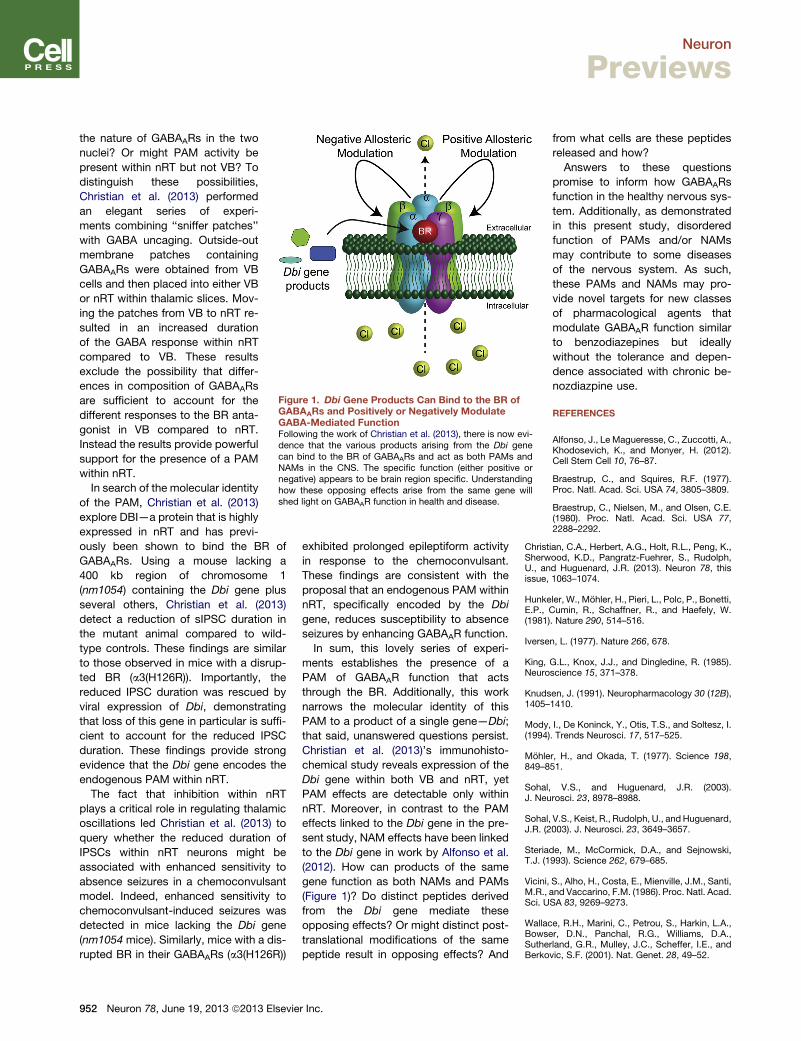
Figure 1. Dbi Gene Products Can Bind to the BR ofGABAARs and Positively or Negatively ModulateGABA-Mediated FunctionFollowing the work of Christian et al. (2013), there is now evi-dence that the various products arising from the Dbi genecan bind to the BR of GABAARs and act as both PAMs andNAMs in the CNS. The specific function (either positive ornegative) appears to be brain region specific. Understandinghow these opposing effects arise from the same gene willshed light on GABAAR function in health and disease.
Neuron
Previews
the nature of GABAARs in the two
nuclei? Or might PAM activity be
present within nRT but not VB? To
distinguish these possibilities,
Christian et al. (2013) performed
an elegant series of experi-
ments combining ‘‘sniffer patches’’
with GABA uncaging. Outside-out
membrane patches containing
GABAARs were obtained from VB
cells and then placed into either VB
or nRT within thalamic slices. Mov-
ing the patches from VB to nRT re-
sulted in an increased duration
of the GABA response within nRT
compared to VB. These results
exclude the possibility that differ-
ences in composition of GABAARs
are sufficient to account for the
different responses to the BR anta-
gonist in VB compared to nRT.
Instead the results provide powerful
support for the presence of a PAM
within nRT.
In search of the molecular identity
of the PAM, Christian et al. (2013)
explore DBI—a protein that is highly
expressed in nRT and has previ-
ously been shown to bind the BR of
GABAARs. Using a mouse lacking a
400 kb region of chromosome 1
(nm1054) containing the Dbi gene plus
several others, Christian et al. (2013)
detect a reduction of sIPSC duration in
the mutant animal compared to wild-
type controls. These findings are similar
to those observed in mice with a disrup-
ted BR (a3(H126R)). Importantly, the
reduced IPSC duration was rescued by
viral expression of Dbi, demonstrating
that loss of this gene in particular is suffi-
cient to account for the reduced IPSC
duration. These findings provide strong
evidence that the Dbi gene encodes the
endogenous PAM within nRT.
The fact that inhibition within nRT
plays a critical role in regulating thalamic
oscillations led Christian et al. (2013) to
query whether the reduced duration of
IPSCs within nRT neurons might be
associated with enhanced sensitivity to
absence seizures in a chemoconvulsant
model. Indeed, enhanced sensitivity to
chemoconvulsant-induced seizures was
detected in mice lacking the Dbi gene
(nm1054 mice). Similarly, mice with a dis-
rupted BR in their GABAARs (a3(H126R))
952 Neuron 78, June 19, 2013 ª2013 Elsevie
exhibited prolonged epileptiform activity
in response to the chemoconvulsant.
These findings are consistent with the
proposal that an endogenous PAM within
nRT, specifically encoded by the Dbi
gene, reduces susceptibility to absence
seizures by enhancing GABAAR function.
In sum, this lovely series of experi-
ments establishes the presence of a
PAM of GABAAR function that acts
through the BR. Additionally, this work
narrows the molecular identity of this
PAM to a product of a single gene—Dbi;
that said, unanswered questions persist.
Christian et al. (2013)’s immunohisto-
chemical study reveals expression of the
Dbi gene within both VB and nRT, yet
PAM effects are detectable only within
nRT. Moreover, in contrast to the PAM
effects linked to the Dbi gene in the pre-
sent study, NAM effects have been linked
to the Dbi gene in work by Alfonso et al.
(2012). How can products of the same
gene function as both NAMs and PAMs
(Figure 1)? Do distinct peptides derived
from the Dbi gene mediate these
opposing effects? Or might distinct post-
translational modifications of the same
peptide result in opposing effects? And
r Inc.
from what cells are these peptides
released and how?
Answers to these questions
promise to inform how GABAARs
function in the healthy nervous sys-
tem. Additionally, as demonstrated
in this present study, disordered
function of PAMs and/or NAMs
may contribute to some diseases
of the nervous system. As such,
these PAMs and NAMs may pro-
vide novel targets for new classes
of pharmacological agents that
modulate GABAAR function similar
to benzodiazepines but ideally
without the tolerance and depen-
dence associated with chronic be-
nozdiazpine use.
REFERENCES
Alfonso, J., Le Magueresse, C., Zuccotti, A.,Khodosevich, K., and Monyer, H. (2012).Cell Stem Cell 10, 76–87.
Braestrup, C., and Squires, R.F. (1977).Proc. Natl. Acad. Sci. USA 74, 3805–3809.
Braestrup, C., Nielsen, M., and Olsen, C.E.(1980). Proc. Natl. Acad. Sci. USA 77,2288–2292.
Christian, C.A., Herbert, A.G., Holt, R.L., Peng, K.,Sherwood, K.D., Pangratz-Fuehrer, S., Rudolph,U., and Huguenard, J.R. (2013). Neuron 78, thisissue, 1063–1074.
Hunkeler, W., Mohler, H., Pieri, L., Polc, P., Bonetti,E.P., Cumin, R., Schaffner, R., and Haefely, W.(1981). Nature 290, 514–516.
Iversen, L. (1977). Nature 266, 678.
King, G.L., Knox, J.J., and Dingledine, R. (1985).Neuroscience 15, 371–378.
Knudsen, J. (1991). Neuropharmacology 30 (12B),1405–1410.
Mody, I., De Koninck, Y., Otis, T.S., and Soltesz, I.(1994). Trends Neurosci. 17, 517–525.
Mohler, H., and Okada, T. (1977). Science 198,849–851.
Sohal, V.S., and Huguenard, J.R. (2003).J. Neurosci. 23, 8978–8988.
Sohal, V.S., Keist, R., Rudolph, U., and Huguenard,J.R. (2003). J. Neurosci. 23, 3649–3657.
Steriade, M., McCormick, D.A., and Sejnowski,T.J. (1993). Science 262, 679–685.
Vicini, S., Alho, H., Costa, E., Mienville, J.M., Santi,M.R., and Vaccarino, F.M. (1986). Proc. Natl. Acad.Sci. USA 83, 9269–9273.
Wallace, R.H., Marini, C., Petrou, S., Harkin, L.A.,Bowser, D.N., Panchal, R.G., Williams, D.A.,Sutherland, G.R., Mulley, J.C., Scheffer, I.E., andBerkovic, S.F. (2001). Nat. Genet. 28, 49–52.