Embed Size (px)
Citation preview
lable at ScienceDirect
Experimental Parasitology 157 (2015) 117e123
Contents lists avai
Experimental Parasitology
journal homepage: www.elsevier .com/locate /yexpr
Research brief
Immunoblotting using Strongyloides venezuelensis larvae,parthenogenetic females or eggs extracts for the diagnosis ofexperimentally infected immunosuppressed rats
Edson Fernando Goulart de Carvalho, Jos�e Eduardo Neto de Sousa,Ana Lúcia Ribeiro Gonçalves, Jair Pereira da Cunha-Junior, Julia Maria Costa-Cruz*
Department of Immunology, Microbiology and Parasitology, Institute of Biomedical Sciences, Federal University of Uberlandia, Av. Par�a 1720, CEP 38400-902, Uberlandia, MG, Brazil
h i g h l i g h t s
* Corresponding author. Departamento de Imunositologia, Instituto de Ciencias Biom�edicas, Laborat�orioses, Universidade Federal de Uberlandia, AvenidaUberlandia, Minas Gerais, Brazil.
E-mail address: [email protected] (J.M. Costa-Cruz
http://dx.doi.org/10.1016/j.exppara.2015.07.0090014-4894/© 2015 Elsevier Inc. All rights reserved.
g r a p h i c a l a b s t r a c t
� L3 larval extract was more effectivein detection of anti-Strongyloidesvenezuelensis IgG.
� IgG was detected early in immuno-suppressed group when compared toimmunocompetent.
� Fraction of 17 kDa is possible markerof infection in immunosuppressedrats.
a r t i c l e i n f o
Article history:Received 19 November 2014Received in revised form25 June 2015Accepted 24 July 2015Available online 26 July 2015
Keywords:RatsStrongyloides venezuelensisImmunoblottingImmunossupression
a b s t r a c t
The nematode Strongyloides stercoralis is responsible for strongyloidiasis in humans. Diagnosis ofinfection occurs through detection of larvae in feces, but low elimination of larvae often hampers thedetection of disease, particularly in cases of patient immunosuppression. Immunodiagnostic tests havebeen developed; however obtaining S. stercoralis larvae for the production of homologous antigen extractis technically difficult. Thus, the use different developmental forms of Strongyloides venezuelensis hasbecome an alternative method for the production of antigen extracts. The aim of this study was toevaluate immunoblotting using alkaline extracts from S. venezuelensis L3 larvae, parthenogenetic femalesor eggs to test detection of experimental strongyloidiasis associated with immunosuppression. Immu-nocompetent and immunosuppressed male rats were experimentally infected, and serum sample fromall animals were obtained at 0, 5, 8 13, and 21 days post infection (d.p.i.). Immunoblotting was evaluatedfor use in detection of anti-S. venezuelensis IgG in both experimental rat groups. The larval extractimmunoblotting profile had the most immunoreactive fractions in the immunosuppressed groupbeginning at 5 d.p.i., while the immunocompetent group reactivity began on 8 d.p.i. Immunoreactiveprotein fractions of 17 kDa present in larval alkaline extract presented as possible markers of infection in
logia, Microbiologia e Para-o de Diagn�ostic.o de Parasit-Par�a 1720, CEP 38400-902,
).
E.F.G. Carvalho et al. / Experimental Parasitology 157 (2015) 117e123118
immunosuppressed rats. It is concluded that all extracts using immunoblotting have diagnostic potentialin experimental strongyloidiasis, particularly larval extract in immunosuppressed individuals.
© 2015 Elsevier Inc. All rights reserved.
1. Introduction
Strongyloides stercoralis is widespread throughout tropical andsubtropical regions. Geohelminthiasis can occur asymptomatically,as a potentially fatal hyperinfection, or as disseminated infection(Al-Hasan et al., 2007; Puthiyakunnon et al., 2014). Immunosup-pression has been associated with cases of hyperinfection syn-drome and studies of disseminated disease in transplant patients,asthmatics, and patients with chronic lung or autoimmune diseaseshave identified corticosteroid therapy as a common denominatorfor developing serious infection (Mejia and Nutman, 2012; Bollelaet al., 2013; Toledo et al., 2015).
Strongyloidiasis is difficult to diagnose because parasite load islow and larval output is irregular (Siddiqui and Berk, 2001; Khieuet al., 2013; Sch€ar et al., 2014). S. stercoralis infective larvae aredifficult to obtain, and thus so are sufficient quantities of antigensto enable further fractionation and analysis; this technical chal-lenge limits the development and standardization of serologicaltests with greater sensitivity and specificity (Feliciano et al., 2010;Gonçalves et al., 2012a).
Strongyloides venezuelensis is a nematode that infects wild ro-dents, and is often used as a model organism for studies of stron-gyloidiasis. It has also been used to standardize new immunologicaltechniques for improving diagnosis of human strongyloidiasis(Machado et al., 2003; Marra et al., 2011; Gonçalves et al., 2010,2012a). S. venezuelensis release eggs in experimentally infectedrats, which in stool culture produce a large quantity infective larvae,simplifying antigen production for accurate and specific recogni-tion of IgG against strongyloidiasis (Machado et al., 2003, 2008;Gonçalves et al., 2008). Other life stages of the parasite, such asparthenogenetic females and eggs may also serve as potentialsources of diagnostic antigens. Female parasite and eggs are bothfound in human intestinal mucosa in infected persons, and contactwith host systems may lead to the development of immuneresponse; for this reason it is prudent to test the efficacy of femaleparasite and egg extracts for use in immunodiagnosis (Gonçalveset al., 2012b).
The aim of this study was to evaluate immunoblotting usingalkaline extracts from S. venezuelensis L3 larvae, parthenogeneticfemales or eggs to test detection of experimental strongyloidiasisassociated with immunosuppression.
2. Material and methods
2.1. Animals
Male Rattus norvegicus (Wistar) rats weighing 100e120 g withage between 6 and 8 week were used in e experiments. Rats werebred in a conventional manner at the Centro de Experimentaç~ao An-imal (CBEA) of the Universidade Federal de Uberlandia (UFU). Ratswere kept in cages with a maximum density of four rats per cagelined with bed shavings, with access to water and fed with industrialfeed. Colony room temperature was 22 ± 2 �C, and artificial lightingconsisted of a 12 h:12 h lightedark cycle. All experiments wereconducted in accordance with animal ethics guidelines and wereapproved by e Comite de �Etica na Utilizaç~ao de Animais of the Uni-versidade Federal de Uberlandia (CEUA/UFU 096/10).
2.2. Parasites
The L-2 strains of S. venezuelensis used in this study were ob-tained from feces from the wild rodent of the species Bolomyslasiurus (April, 1986), isolated and retained in Rattus norvergicusWistar at the Institute of Biology at Universidade Estadual deCampinas (UNICAMP), S~ao Paulo, Brazil. The strain ofS. venezuelensis were kindly provided by the Laborat�orio de Diag-n�ostico de Parasitoses, Universidade Federal de Uberlandia andmaintained in R. norvergicus.
S. venezuelensis third-stage infective larvae (L3) were obtainedfrom charcoal cultures of infected rat faeces. The cultures werestored at 28 �C for 48 h, and the infective larvae were collected andconcentrated using the Rugai method (Rugai et al., 1954). The pellet(3 mL) from e conical cup were diluted 10 times in distilled waterand larvae were counted using stereomicroscopy. For infection,1.500 S. venezuelensis L3 larvae were inoculated subcutaneouslyinto each of the rats.
To recover parthenogenetic females, rats were anesthetized(ketamine 60 mg/kg e Syntec do Brasil Ltda, Cotia, S~ao Paulo andxylazine 7 mg/kg e Syntec do Brasil Ltda, Cotia, S~ao Paulo) andsacrificed on 8 day post-infection (d.p.i.), the small intestines wereremoved from animals and placed in Petri dishes containing salinesolution, longitudinally sectioned and incubated at 37 �C for twohours. Parthenogenetic females were counted following method-ology by Sato and Toma (1990).
To obtain eggs, three rats experimentally infected withS. venezuelensiswere placed on clean, damp paper towel to defecatein 7 and 8 d.p.i. and feces were collected. Feces were recoveredmoistened with distilled water, macerated and adjusted withdistilled water (v/v). The solution was passed in sieve plot0.300 mm, 0.149 mm and 0.47 mm, respectively. The collected fluidwas centrifuged at 13,000� g for 10min, the supernatant discardedand the pellet resuspended in 0.9% saline. The saline faeces werethen centrifuged at 300� g for 5min and the supernatant collected.To the supernatant was added distilled water v/v and centrifuged at13,000 � g for 10 min. The supernatant was discarded and thepellet resuspended in PBS. The estimated number of eggs per gramof feces was performed using the method of Cornell-McMaster(Gordon and Whitlock, 1939) and eggs were stored at �20 �C forlater use.
2.3. Experimental groups
Rats were divided into two groups: immunocompetent (n ¼ 30)and immunosuppressed (n ¼ 30) rats. Prior to infection, immuno-suppressed groups received 5 mg/mL of dexamethasone disodiumphosphate (Medley Indústria Farmaceutica Ltda, Campinas, S~aoPaulo, Brazil) diluted inwater for 5 days, as previously described byRomand et al. (1998). Animals from both groups were inoculatedsubcutaneously in the abdominal regionwith 1.500 S. venezuelensislarvae, except immunocompetent or immunosuppressed animalsof day 0 (negative control of experiments).
At each time point (day 0 and 5, 8, 13 and 21 d.p.i.), 6 rats fromeach group were anesthetized with ketamine 60 mg/kg (Syntec doBrazil Ltda, Cotia, S~ao Paulo) and xylazine 7 mg/kg (Syntec do BrazilLtda, Cotia, S~ao Paulo) s.c, and blood samples were collected by

E.F.G. Carvalho et al. / Experimental Parasitology 157 (2015) 117e123 119
cardiac puncture. The blood collected (volume of 3 mL to each rat)was centrifuged and the serum aliquots were stored at �20 �C.Later it was made a pool of sera from rats as points of kinetics ofinfection for use in experiments. The pool of sera from day 0 of eachgroup was used as negative controls.
2.4. Control of infection
Eggs per gram of feces were counted on days 5, 8, 13 and 21 d.p.i.in order to certify the animal infection. The parasitological exami-nation was performed three times, and the average of the threeresults was recorded.
2.5. Alkaline extract from S. venezuelensis
Alkaline extracts were prepared, as previously described(Machado et al., 2003) with modifications. Briefly, 300,000S. venezuelensis larvae, 500 parthenogenetic females or 300,000eggs were resuspended in PBS (0.01 mol/L, pH 7.2) containingprotease inhibitors (benzamidine 1 mmol/L, aprotinin 1 mg/mL, andleupeptin 2 mg/mL) and disrupted in cycles of freezing(1 min, �196 �C). After disruption, 1 mL of 0.15 M NaOH was addedin each of the antigens and was maintained under gentle agitationfor 2 h at 4 �C. Subsequently, 0.3 M HCl was added until a pH of 7.0was reached. These preparations were then centrifuged at12,400� g for 30 min at 4 �C. Supernatants (alkaline extracts) wereanalyzed for protein content according to Lowry et al. (1951),subdivided into aliquots and stored at �20 �C until use.
2.6. Sodium dodecyl sulfateepolyacrylamide gel electrophoresis(SDS-PAGE)
Sodium dodecyl sulfateepolyacrylamide gel electrophoresis(SDS-PAGE) was performed as described by Laemmli (1970) underreducing conditions. Briefly, larvae, adult female worms, and eggextracts were submitted for electrophoresis using a 15% acrylamideseparation gel. Samples were diluted in 10 mmol/L Tris/HCl buffer(pH 8.0) containing 1 mmol/L EDTA, 2% SDS, 10% glycerol, and 2.5%(v/v) b-mercaptoethanol, and boiled for 5 min (100 �C). Sampleswere run simultaneously with molecular weight Marker Recom™Blue wide Range Prestain Marker (cat. No. RE001, Real Biotech Cor-poration e RBC, Taiwan.) on equipment SE-300-10A-10 miniVEvertical electrophoresis system (Hoefer, Inc., San Francisco, USA.) andgels were analyzed by silver nitrate (Friedman, 1982). The electro-phoresis was performed three times.
2.7. Immunoblotting
A sample of 250 mg of each antigen preparationwas submitted toelectrophoresis SDS-PAGE 12% and transferred to nitrocellulosemembranes nitropure 0.45 mm (cod. WP4HY00010, GE-Osmonics,Inc., Trerose, USA.) as described by Towbin et al. (1979) usingSE302 miniVE blotter (Hoefer, Inc., Richmond, USA). The nitrocel-lulose strips were blocked with phosphate buffered saline with0.01% of Tween20 added of non-fat milk (PBS-TM) 5% for 2 h at roomtemperature and incubated overnight at 4 �C with pool serumsamples diluted 1:30 in PBS-TM 1%. After washing with PBS-TM0.05%, strips were incubated for 2 h at room temperature withperoxidase-labeled goat anti-rat IgG conjugate (cod. A9037, Sig-maeAldrich Co., St. Louis, USA), diluted at 1:500 in PBS-TM 1%. Thestrips were washed in PBS-TM 0.05%, and e assay was developed byadding hydrogen peroxide and 3.30-diaminobenzidine tetra hy-drochloride (cod 1001232725, DAB-SigmaFast tablets, Sigma-eAldrich Co., St. Louis, USA). The reaction was stopped by addingdistilled water, and positive reactions were determined by e
appearance of clearly defined protein fractions. The immunoblot-ting was performed three times. The relative molecular masses ofthe recognized protein fractions were determined by comparisonwith molecular markers (Recom Blue - RBC).
2.8. Data analysis
The molecular weight of proteic fractions was estimated fromthe linear regression curve constructed by values of the molecularweights from markers in relation to Rf (relative motility) byGraphPad Prism version 5.0 (GraphPad Software, La Jolla, USA)software. The Rf was obtained from the following formula:Rf ¼ distance from source migration/distance from source to pointof reference. The protein fractions were analyzed accordingGassmann et al. (2009) using the software of image analysis Image Jversion 1.47 (National Institutes of Health, Bethesda, USA.).
3. Results
3.1. Monitoring of infection
The eggs releasing began at 5 d.p.i. in both groups. The numberof eggs recovered in feces at 8 d.p.i. was significantly higher forimmunosuppressed (8000 eggs per gram of feces) and immuno-competent (3000 eggs per gram of feces) rats. After 13 d.p.i., thenumber of eggs decreased in both groups. However, it was observedthat on the 21 d.p.i. the infection was eliminated in immunocom-petent group, while in the immunocompromised group, theinfection persisted.
3.2. Electrophoretic profile of the alkaline extracts
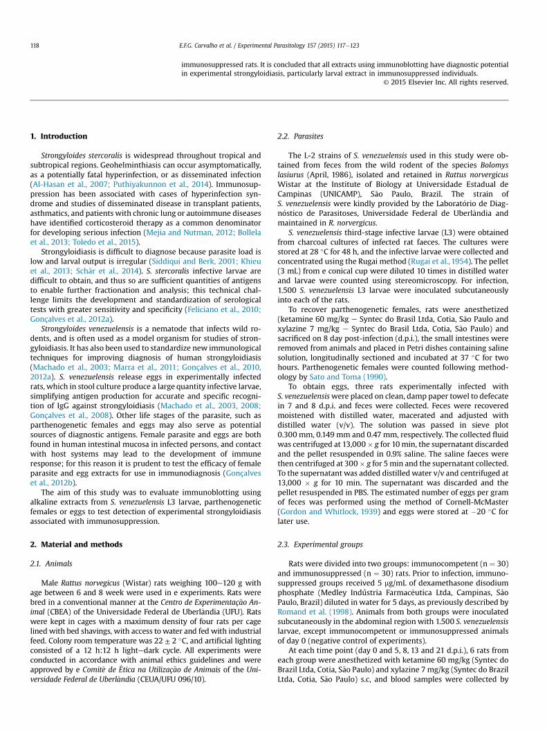
Alkaline extracts of the different developmental forms of theparasite subjected to SDS-PAGE (12%) show distinct electrophoreticprofiles with molecular weights ranging from 14 to 91 kDa, butwith some patterns of representative protein peaks indicating closemolecular weight (Fig. 1). Larval alkaline extract showed 8 bands,including protein fractions of 14, 17, 21, 25, 50, 70 and 91 kDa.Parthenogenetic female alkaline extract produced fractions of 14,25, 31, 32, 50, 68 and 70 kDa. Egg extracts produced five fractionswith molecular weights of 25, 35, 36, 50 and 70 kDa (Fig. 1).
3.3. Detection of anti-IgG antibodies by immunoblotting
Detection of anti-IgG antibodies by immunoblotting with larvalalkaline extract in the immunocompetent rat group presented laterreactivity compared to the immunosuppressed rat group. Reactive17 kDa protein fractions were present in the immunocompetentgroup at 13 and 21 d.p.i., were expressed in the immunosuppressedgroup from the 5 d.p.i. and persisting for remaining days of infec-tion. Both groups showed similar protein fractions for detection ofIgG. Fractions of 50 kDa were reactive at 8, 13 and 21 d.p.i. Day zeroof infection showed nonspecific protein fractions of 91, 75 and35 kDa in both groups, which remained throughout the infectionperiod (Fig. 2 A, B). The analysis of 8 d.p.i. showed higher reactivityin the immunosuppressed group, with protein fractions of 71, 60,50, 42, 38, 37 and 30e17 kDa (Fig. 2 A, B).
Parthenogenetic female alkaline extract showed IgG reactivityin protein fractions of high molecular weight, from 116 to 71 kDa.We observed a pattern of early reactivity in the immunosuppressedgroup compared to the immunocompetent group. Protein fractionsof 71 and 80e85 kDawere present in the immunosuppressed groupat 5 and 8 d.p.i, respectively, while these same fractions were onlypresent at 21, and 13 and 21 d.p.i. respectively in the immuno-competent group. Controls showed a non-specific fraction of
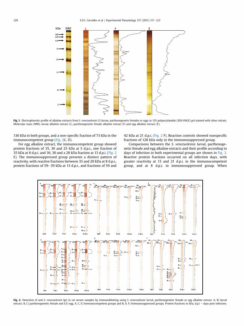
Fig. 1. Electrophoretic profile of alkaline extracts from S. venezuelensis L3 larvae, parthenogenetic females or eggs in 12% polyacrylamide (SDS-PAGE) gel stained with silver nitrate.Molecular mass (MM), larvae alkaline extract (L), parthenogenetic female alkaline extract (F) and egg alkaline extract (E).
E.F.G. Carvalho et al. / Experimental Parasitology 157 (2015) 117e123120
136 kDa in both groups, and a non-specific fraction of 73 kDa in theimmunocompetent group (Fig. 2C, D).
For egg alkaline extract, the immunocompetent group showedprotein fractions of 35, 30 and 25 kDa at 5 d.p.i., one fraction of35 kDa at 8 d.p.i. and 36, 30 and a 28 kDa fraction at 13 d.p.i. (Fig. 2E). The immunosuppressed group presents a distinct pattern ofreactivity, with reactive fractions between 35 and 20 kDa at 8 d.p.i.,protein fractions of 59e39 kDa at 13 d.p.i., and fractions of 59 and
Fig. 2. Detection of anti-S. venezuelensis IgG in rat serum samples by immunoblotting usiextract; B, C) parthenogenetic female and E,F) egg. A, C, E) Immunocompetent groups and B
42 kDa at 21 d.p.i. (Fig. 2 F). Reaction controls showed nonspecificfractions of 128 kDa only in the immunosuppressed group.
Comparisons between the S. venezuelensis larval, parthenoge-netic female and egg alkaline extracts and their profile according todays of infection in both experimental groups are shown in Fig. 3.Reactive protein fractions occurred on all infection days, withgreater reactivity at 13 and 21 d.p.i. in the immunocompetentgroup, and at 8 d.p.i. in immunosuppressed group. When
ng S. venezuelensis larval, parthenogenetic female or egg alkaline extract. A, B) larval, D, F) Immunosuppressed groups. Protein fractions in kDa, d.p.i e days post-infection.
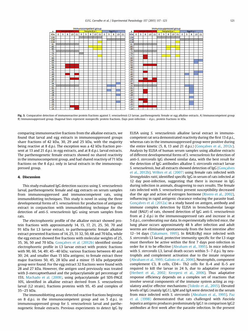
Fig. 3. Comparative detection of immunoreactive protein fractions against S. venezuelensis L3 larvae, parthenogenetic female or egg alkaline extracts. A) Immunocompetent groupB) Immunosuppressed group. Diagonal lines represent nonspecific protein fractions. Days post-infection e d.p.i., protein fractions in kDa.
E.F.G. Carvalho et al. / Experimental Parasitology 157 (2015) 117e123 121
comparing immunoreactive fractions from the alkaline extracts, wefound that larval and egg extracts in immunosuppressed groupsshare fractions of 42 kDa, 30, 29 and 25 kDa, with the majoritybeing reactive at 8 d.p.i. The exception was a 42 kDa fraction pre-sent at 13 and 21 d.p.i. in egg extracts, and at 8 d.p.i. larval extracts.The parthenogenetic female extracts showed no shared reactivityin the immunocompetent group, and had shared reactiviy of 71 kDafractions on the 8 d.p.i. only in larval extracts in the immunosup-pressed group.
4. Discussion
This study evaluated IgG detection success using S. venezuelensislarval, parthenogenetic female and egg extracts on serum samplesfrom immunosuppressed and immunocompetent rats, usingimmunoblotting techniques. This study is novel in using the threedevelopmental forms of S. venezuelensis for production of antigenicextract in conjunction with immunoblotting technique for thedetection of anti-S. venezuelensis IgG using serum samples fromrats.
The electrophoretic profile of the alkaline extract showed pro-tein fractions with approximately 14, 15, 17, 21, 25, 50, 70 and91 kDa for L3 larvae extract, to parthenogenetic female alkalineextract presented fractions of 14, 25, 3132, 50, 68 and 70 kDa, whilethe egg extract showed five fractions with molecular weights of 25,35, 36, 50 and 70 kDa. Gonçalves et al. (2012b) identified similarelectrophoretic profile in L3 larvae extract with proteic fractionswith 90, 60, 54, 49; 45e40 kDa; various fractions between 40 and30; 24; and smaller than 15 kDa antigens; to female extract threemajor fractions 50, 45, 28 kDa and a minor 15 kDa polypeptidegroup were found. For the egg extract 32 fractions were found with28 and 27 kDa. However, the antigen used previously was treatedwith b-mercaptoethanol and the polyacrylamide gel percentage of15%. Machado et al. (2008), using polyacrylamide gel SDS-PAGE10%, identified in alkaline extract derived from S. venezuelensislarval (L2 strain), fractions proteins with 95, 45 and complex of35e25 kDa.
The immunoblotting assay detected reactive fractions beginningon 8 d.p.i. in the immunocompetent group and on 5 d.p.i. inimmunosuppressed group for S. venezuelensis larval and parthe-nogenetic female extracts. Previous experiments to detect IgG by
ELISA using S. venezuelensis alkaline larval extract in immuno-competent rat sera demonstrated reactivity during the first 13 d.p.i.,whereas rats in the immunosuppressed group were positive duringthe entire kinetic (5, 8, 13 and 21 d.p.i.) (Gonçalves et al., 2012c).Analysis by ELISA of human serum samples using alkaline extractsof different developmental forms of S. venezuelensis for detection ofanti-S. stercoralis IgG showed similar data, with the best result forthe detection of IgG antibodies alkaline S. stercoralis extract larvaeS. venezuelensis, but all extracts showed detection of IgG (Gonçalveset al., 2012b). Wilkes et al. (2007) using female rats infected withStrongyloides ratti, identified specific IgG in serum of rats infected at12 day post-infection, suggesting that there is increase in IgGduring infection in animals, disagreeing to ours results. The femalerats infected with S. venezuelensis present susceptibility decreaseddue the age and action of estrogen hormone (Rivero et al., 2002),influencing in rapid antigenic clearance reducing the parasite load.Gonçalves et al. (2012a) in a study based on antigen, antibody andimmune complex detection by ELISA in bronchoalveolar lavagefluid (BALF) of rats, showed detection of IgG anti-S. venezuelensisfrom at 2 d.p.i in the immunosuppressed rats and increase in at5 d.p.i., corroborating our data. In experimentally infected mice, thelung phase occurs approximately 48 h after infection and adultworms are eliminated spontaneously from the host intestine after12e14 days (Takamure, 1995). In BALB/cByJ mice infected withS. stercoralis L3 larval, protective immunity specific for the L3 stagemust therefore be active within the first 7 days post-infection inorder for it to be effective (Abraham et al., 1995). In mice infectedwith S. stercoralis L3, larval death occurs within 5e7 d.p.i. by neu-trophils and complement activation due to the innate response(Abraham et al., 1995; Galioto et al., 2006). Neutrophils, componentcomplement C3, B cells, CD4þ Th2 cells for IL-4 and IL-5 arerequired to kill the larvae in 24 h, due to adaptative response(Herbert et al., 2002; Kerepesi et al., 2006). Thus adaptativeresponse efficiency depends on a complex set of reactions thatinvolve several components of the immune system acting as reg-ulatory and/or effector mechanisms (Toledo et al., 2015). Elevatedlevels of IgG (mainly IgG1), IgM and IgAwere detected in the serumfrom mice infected with S. stercoralis (Abraham et al., 1995). Pazet al. (1998) demonstrated that rats challenged with Fasciolahepatica antigens produces predominately IgG1 in comparison IgG2antibodies at first week after the parasite infection. In the present
E.F.G. Carvalho et al. / Experimental Parasitology 157 (2015) 117e123122
study, using anti-rat IgG, it was observed an early reactivity toS. venezuelensis antigens in immunosuppressed rats, indicating thatat least part of this phenomenon of reactivity may be due the IgG1isotype detection.
The immunosuppressed group was more efficient in detectingIgG antibodies against S. venezuelensis alkaline extract. This may beexplained by dexamethasone administration, which promoteshyperinfection in animals, and can result in a higher antigenic loaddue to abundance of infective larvae and increased number ofparthenogenetic females (i.e., producingmore eggs). Machado et al.(2011) demonstrated that when administered to BALB/c miceinfected with S. venezuelensis, dexamethasone promotes anincreased parasitic burden and subsequent hyperinfection. In arecent review, Yasuda et al. (2014) highlighted the importance ofinnate and acquired immunity in rapid expulsion of S. venezuelensisin rodents, demonstrating the role of these two pathways infighting infection highlighting the contribution of Th2, togetherwith IgG and IgE in the expulsion.
In the present study, serum samples from rats showed a patternwith immunoreactive protein fractions of 50, 38 and 17 kDa for thelarval alkaline extract in both groups. Parthenogenetic femalealkaline extract presented fractions of 116, 85, 80 and 71 kDa inboth experimental groups, and egg alkaline extract showed frac-tions of 35, 30 and 25 kDa in both group. Levenhagen and Costa-Cruz (2014) in a recent review of the diagnosis of human strongy-loidiasis reported previous studies using extracts of Strongyloidessp. at different developmental stages using immunoblot tech-niques, and the immunoreactive protein fractions had molecularweights between 26 and 28, 31e33, 41e45, 120e160 and 205 kDa.The three alkaline extracts showed some nonspecific fractions onday 0 (control experiments) possibly due to the use of conventionalrats or presence of commensal agents that showed cross-reactivitywith antigens of S. venezuelensis.
Immunoreactive protein fraction of 26 kDa is present only inlarval alkaline extracts in immunosuppressed group on 5 and8 d.p.i. Sato et al. (1990) used the immunoblotting technique todetect anti-S. stercoralis IgG in human serum samples and fractions97, 66, 41 and 26 kDa have been identified in most patient serasamples. Study involving epitope identification using larva ofS. stercoralis in human serum samples showed a 26 kDa immuno-reactive protein fraction with high frequency in the group withstrongyloidiasis (Sudr�e et al., 2007). In the immunosuppressedgroup, fractions of 17 kDa present only in the larval alkaline extractmay present a potential diagnostic tool. In a study conducted foridentification of heat-shock proteins (HSPs) during processing ofinfective S. venezuelensis larvae, therewas an increase in 70 kDa and16e22 kDa protein fraction complexes in immunoblotting; theseproteins are related to transformation between developmentalstages of the parasite (Tsuji et al., 1996). These protein fractionsmaybe useful in diagnosis due to reactivity beginning on 5 d.p.i. in theimmunosuppressed group. The use of these fractions as possiblemarkers of early infection in immunocompromised individualsrequires additional study.
5. Conclusions
We conclude that alkaline larval extract was more effective inthe detection of anti-S. venezuelensis IgG than extracts from otherparasite developmental stages, with better reactivity in immuno-suppressed group. Detection of protein fractions was earlier andbetter in the immunosuppressed group compared to the immu-nocompetent group for the three alkali extracts. Larval alkalineextract immunoreactive protein fractions of 17 kDa were reactivebeginning on 5 d.p.i. in the immunosuppressed group, representinga possible infection marker in cases of immunosuppression.
Conflicts of interest
The authors declare that there is no conflict of interest.
Acknowledgments
This study was supported by Conselho Nacional de Desenvol-vimento Científico e Tecnol�ogico (CNPq e 302426/2012-4), Coor-denaç~ao de Aperfeiçoamento de Pessoal de Nível Superior (CAPESeN� 3200601004M-8) and by Fundaç~ao de Amparo �a Pesquisa doEstado de Minas Gerais (FAPEMIG e CBB-PPM-00396-13), Brazil.
References
Al-Hasan, M.N., McCormick, M., Ribes, J.A., 2007. Invasive enteric infections inhospitalized patients with underlying strongyloidiasis. Am. J. Clin. Pathol. 128,622e627.
Abraham, D., Rotman, H.L., Haberstroth, H.F., Yutanawiboonchai, W., Brigandi, R.A.,Leon, O., Nolan, T.J., Schad, A., 1995. Strongyloides stercoralis: protective im-munity to third-stage larvae in BALB/cByJ mice. Exp. Parasitol. 80, 297e307.
Bollela, V.R., Feliciano, C., Teixeira, A.C., Junqueira, A.C.R., Rossi, M.A., 2013. Fulmi-nant gastrointestinal hemorrhage due to Strongyloides stercoralis hyperinfectionin an AIDS patient. Rev. Soc. Bras. Med. Trop. 46, 111e113.
Feliciano, N.D., Gonzaga, H.T., Gonçalves-Pires Mdo, R., Gonçalves, A.L.,Rodrigues, R.M., Ueta, M.T., Costa-Cruz, J.M., 2010. Hydrophobic fractions fromStrongyloides venezuelensis for use in the human immunodiagnosis of stron-gyloidiasis. Diagn. Microbiol. Infect. Dis. 67, 153e161.
Friedman, R.D., 1982. Comparison of four different silver-staining techniques forsalivary protein detection in alkaline polyacrylamide gels. Anal. Biochem. 126,346e349.
Gassmann, M., Grenacher, B., Rohde, B., Voge, J., 2009. Quantifying western blots:pitfalls of densitometry. Electrophoresis 30, 1845e1855.
Galioto, A.M., Hess, J.A., Nolan, T.J., Schad, G.A., Lee, J.J., Abraham, D., 2006. Role ofeosinophils and neutrophils in innate and adaptive protective immunity tolarval Strongyloides stercoralis in mice. Infect. Immun. 74, 5730e5738.
Gonçalves, A.L., Ribeiro, T.S., Silva, C.V., Ueta, M.T., Costa-Cruz, J.M., 2012a. A novelapproach based on antigen, antibody and immune complex detection inbronchoalveolar lavage fluid samples from rats experimentally infected withStrongyloides venezuelensis. Acta Trop. 124, 166e169.
Gonçalves, A.L., Rocha, C.A., Gonzaga, H.T., Gonçalves-Pires Mdo, R., Ueta, M.T.,Costa-Cruz, J.M., 2012b. Specific IgG and IgA to larvae, parthenogenetic females,and eggs of Strongyloides venezuelensis in the immunodiagnosis of humanstrongyloidiasis. Diagn. Microbiol. Infect. Dis. 72, 79e84.
Gonçalves, A.L., Rodrigues, R.M., Silva, N.M., Gonçalves, F.A., Cardoso, C.R.,Beletti, M.E., Ueta, M.T., Silva, J.S., Costa-Cruz, J.M., 2008. Immunolocalizationand pathological alterations following Strongyloides venezuelensis infection inthe lungs and the intestine of MHC class I or II deficient mice. Vet. Parasitol. 158,319e328.
Gonçalves, A.L., Silva, C.V., Ueta, M.T., Costa-Cruz, J.M., 2010. A new faecal antigendetection system for Strongyloides venezuelensis diagnosis in immunosup-pressed rats. Exp. Parasitol. 125, 338e341.
Gonçalves, A.L., Silva, C.V., Ueta, M.T., Costa-Cruz, J.M., 2012c. Antigen, antibody andimmune complex detection in serum samples from rats experimentally infectedwith Strongyloides venezuelensis. Exp. Parasitol. 130, 205e208.
Gordon, H.M., Whitlock, H.V.A., 1939. New technique for counting nematode eggs insheep feces. J. Council Sci. Ind. Res. 12, 17e18.
Herbert, D.R., Nolan, T.J., Schad, G.A., Abraham, D., 2002. The role of B cells in im-munity against larval Strongyloides stercoralis in mice. Parasite Immunol. 24,95e101.
Kerepesi, L.A., Hess, J.A., Nolan, T.J., Schad, G.A., Abraham, D., 2006. Complementcomponent C3 is required for protective innate and adaptive immunity to larvalStrongyloides stercoralis in mice. J. Immunol. 176, 4315e4322.
Khieu, V., Sch€ar, F., Marti, H., Sayasone, S., Duong, S., Muth, S., Odermatt, P., 2013.Diagnosis, treatment and risk factors of Strongyloides stercoralis in school-children in Cambodia. PLoS Negl. Trop. Dis. 7, e2035.
Laemmli, U.K., 1970. Cleavage of structural proteins during the assembly of the headof bacteriophage T4. Nature 227, 680e685.
Levenhagen, M.A., Costa-Cruz, J.M., 2014. Update on immunologic and moleculardiagnosis of human strongyloidiasis. Acta Trop. 135, 33e43.
Lowry, O.H., Rosebrough, N.J., Farr, A.L., Randall, R.J., 1951. Protein measurementwith the folin phenol reagent. J. Biol. Chem. 193, 265e275.
Machado, E.R., Ueta, M.T., de Fatima Gonçalves-Pires Mdo, R., Alves de Oliveira, J.B.,Faccioli, L.H., Costa-Cruz, J.M., 2003. Strongyloides venezuelensis alkaline extractfor the diagnosis of human strongyloidiasis by enzyme-linked immunosorbentassay. Mem. Inst. Oswaldo Cruz 98, 849e851.
Machado, E.R., Faccioli, L.H., Costa-Cruz, J.M., Lourenço, E.V., Roque-Barreira, M.C.,Gonçalves-Pires, M.R.F., Ueta, M.T., 2008. Strongyloides venezuelensis: the anti-genic identity of eight strains for the immunodiagnosis of human strongyloi-diasis. Exp. Parasitol. 119, 7e14.
Machado, E.R., Carlos, D., Sorgi, C.A., Ramos, S.G., Souza, D.I., Soares, E.G., Costa-
E.F.G. Carvalho et al. / Experimental Parasitology 157 (2015) 117e123 123
Cruz, J.M., Ueta, M.T., Aronoff, D.M., Faccioli, L.H., 2011. Dexamethasone effectsin the Strongyloides venezuelensis infection in a murine model. Am. J. Trop. Med.Hyg. 84, 957e966.
Marra, N.M., Chiuso-Minicucci, F., Machado, G.C., Zorzella-Pezavento, S.F.,Franca, T.G., Ishikawa, L.L., Amarante, A.F., Sartori, A., Amarante, M.R., 2011.Migratory route of Strongyloides venezuelensis in Lewis rats: comparison ofhistological analyses and PCR. Exp. Parasitol. 127, 334e339.
Mejia, R., Nutman, T.B., 2012. Screening, prevention, and treatment for hyper-infection syndrome and disseminated infections caused by Strongyloides ster-coralis. Curr. Opin. Infect. Dis. 25, 458e463.
Paz, A., S�anchez-Andrade, R., Panadero, R., Díez-Ba~nos, P., Morrondo, P., 1998. IgGisotype specific immune response in rats infected with Fasciola hepatica. Vet.Parasitol. 79, 229e237.
Puthiyakunnon, S., Boddu, S., Li, Y., Zhou, X., Wang, C., Li, J., Chen, X., 2014.StrongyloidiasisdAn insight into its global prevalence and management. PLoSNegl. Trop. Dis. 8, e3018.
Rivero, J.C., Inoue, Y., Murakami, N., Horii, Y., 2002. Age- and sex-related changes insusceptibility of Wistar rats to Strongyloides venezuelensis infection. J. Vet. Med.Sci. 64, 519e521.
Romand, S., Thulliez, P., Dubey, J.P., 1998. Direct agglutination test for serologicdiagnosis of Neospora caninum infection. Parasitol. Res. 84, 50e53.
Rugai, E., Mattos, T., Brisola, A.P., 1954. A new technic for the isolation of nematodelarvae from feces; modification of Baermann's method. Rev. Inst. Adolfo Lutz 14,5e8.
Sato, Y., Toma, H., 1990. Strongyloides venezuelensis infections in mice. Int. J. Para-sitol. 20, 57e62.
Sato, Y., Inoue, F., Matsuyama, R., Shiroma, Y., 1990. lmmunoblot analysis of anti-bodies in human strongyloidiasis. Trans. R. Soc. Trop. Med. Hyg. 84, 403e406.
Sch€ar, F., Hattendorf, J., Khieu, V., Muth, S., Char, M.C., Mart, H.P., Odermatt, P., 2014.Strongyloides stercoralis larvae excretion patterns before and after treatment.Parasitology 141, 892e897.
Siddiqui, A.A., Berk, S.L., 2001. Diagnosis of Strongyloides stercoralis infection. Clin.Infect. Dis. 33, 1040e1047.
Sudr�e, A.P., Siqueira, R.C., Barreto, M.G.M., Peralta, R.H.S., Macedo, H.W., Peralta, J.M.,2007. Identification of a 26-kDa protein fraction as an important antigen forapplication in the immunodiagnosis of strongyloidiasis. Parasitol. Res. 101,1117e1123.
Takamure, A., 1995. Migration route of Strongyloides venezuelensis in rodents. Int. J.Parasitol. 25, 907e911.
Toledo, R., Mu~noz-Antoli, C., Esteban, J.G., 2015. Strongyloidiasis with emphasis onhuman infections and its different clinical forms. Adv. Parasitol. 88, 165e241.
Towbin, H., Staehelin, T.,, Gordon, J., 1979. Electrophoretic transfer of proteins frompolyacrylamide gels to nitrocellulose sheets: procedures and some applications.Proc. Natl. Acad. Sci. U. S. A. 76, 4350e4354.
Tsuji, N., Ohta, M., Fujisaki, K., 1996. Expression of a 70-kDa heat-shock-relatedprotein during transformation from free-living infective larvae to the parasiticstage in Strongyloides venezuelensis. Parasitol. Res. 83, 99e102.
Wilkes, C.P., Bleay, C., Paterson, S., Viney, M.E., 2007. The immune response during aStrongyloides ratti infection of rats. Parasite Immunol. 29, 339e346.
Yasuda, K., Matsumoto, M., Nakanishi, K., 2014. Importance of both innate immunityand acquired immunity for rapid expulsion of S. venezuelensis. Front. Immunol.5, 1e5.