Embed Size (px)
Citation preview
Immunity
Article
Intestinal Bacteria Trigger T Cell-IndependentImmunoglobulin A2 Class Switching by InducingEpithelial-Cell Secretion of the Cytokine APRILBing He,1 Weifeng Xu,1 Paul A. Santini,1,3 Alexandros D. Polydorides,1 April Chiu,1 Jeannelyn Estrella,1
Meimei Shan,2 Amy Chadburn,1 Vincenzo Villanacci,4 Alessandro Plebani,5 Daniel M. Knowles,1
Maria Rescigno,6 and Andrea Cerutti1,3,*1 Department of Pathology and Laboratory Medicine2 Department of Immunology and Microbiology
Weill Medical College of Cornell University, 1300 York Avenue, New York, NY 10021, USA3 Weill Graduate School of Medical Sciences of Cornell University, 1300 York Avenue, New York, NY 10021, USA4 Secondo Servizio di Anatomia Patologica, Spedali Civili di Brescia, P.le Spedali Civili 1, Brescia 25123, Italy5 Clinica Pediatrica e Istituto di Medicina Molecolare ‘‘A. Nocivelli,’’ Universita’ di Brescia, P.le Spedali Civili 1, Brescia 25123, Italy6 European Institute of Oncology (IEO), Via Ripamonti 435, Milano 20141, Italy
*Correspondence: [email protected]
DOI 10.1016/j.immuni.2007.04.014
SUMMARY
Bacteria colonize the intestine shortly after birthand thereafter exert several beneficial func-tions, including induction of protective immuno-globulin A (IgA) antibodies. The distal intestinecontains IgA2, which is more resistant to bacte-rial proteases than is IgA1. The mechanism bywhich B cells switch from IgM to IgA2 remainsunknown. We found that human intestinal epi-thelial cells (IECs) triggered IgA2 class switchingin B cells, including IgA1-expressing B cells ar-riving from mucosal follicles, through a CD4+
T cell-independent pathway involving a prolifer-ation-inducing ligand (APRIL). IECs releasedAPRIL after sensing bacteria through Toll-likereceptors (TLRs) and further increased APRILproduction by activating dendritic cells via thy-mic stromal lymphopoietin. Our data indicatethat bacteria elicit IgA2 class switching by link-ing lamina propria B cells with IECs througha TLR-inducible signaling program requiringAPRIL. Thus, mucosal vaccines should activateIECs to induce more effective IgA2 responses.
INTRODUCTION
Antibody diversity is essential for protective immunity.
Immature B cells generate antigen recognition diversity
by assembling the antigen-binding variable region of im-
munoglobulins (Igs) from individual variable (V), diversity
(D), and/or joining (J) gene segments through recombina-
tion-activating gene proteins (Schlissel, 2003). Mature B
cells further diversify the antibody repertoire through
V(D)J gene somatic hypermutation (SHM) and heavy-
chain (H) class switch DNA recombination (CSR), two
812 Immunity 26, 812–826, June 2007 ª2007 Elsevier Inc.
processes that require activation-induced cytidine deam-
inase (AID) (Honjo et al., 2002). While SHM introduces
point mutations in the V(D)J exon, thereby providing the
structural correlate for selection by antigen of higher-affin-
ity mutants (Odegard and Schatz, 2006), CSR modulates
the antibody effector functions by substituting IgM and
IgD with IgG, IgA, or IgE (Chaudhuri and Alt, 2004).
IgA class switching enables antibody secretion onto
mucosal surfaces (Brandtzaeg et al., 2001). In addition
to targeting dietary antigens, toxins, and pathogenic mi-
croorganisms, IgA controls the growth of commensal
bacteria and prevents their adhesion to intestinal epithelial
cells (IECs) (Fagarasan et al., 2002; Macpherson and Uhr,
2004; Mestecky et al., 1999). Although coated with IgA,
commensal bacteria survive and exert several beneficial
functions, including synthesis of essential vitamins and
protection against pathogens (Nagler-Anderson, 2001).
In addition, commensal bacteria induce mucosal IgA class
switching through a mechanism that remains poorly
understood (Macpherson, 2006).
In general, IgA class switching requires the stimulation of
B cells by CD4+ T cells through CD40 ligand (CD40L) and
cytokines, including interleukin (IL)-4, IL-10, and trans-
forming growth factor (TGF)-b (Coffman et al., 1989;
Fayette et al., 1997; Stavnezer, 1996). These signals in-
duce AID expression and subsequent IgA CSR in B cells
within the germinal center (GC) of mucosal inductive sites,
including Peyer’s patches (PPs) (Fagarasan and Honjo,
2003). Then, class-switched B cells differentiate into IgA-
secreting plasmacytoid B cells, which migrate to the intes-
tinal lamina propria (LP) under the influence of IEC-derived
chemokines (Mora et al., 2006; Wilson and Butcher, 2004).
Ultimately, IgA released at mucosal effector sites binds to
a polymeric Ig receptor (pIgR) on the basolateral surface of
IECs and translocates onto the mucosal surface to exert its
protective function (Brandtzaeg et al., 2001).
IECs further promote mucosal immunity by cross talking
with dendritic cells (DCs) (Neutra et al., 2001). While
Immunity
Intestinal Epithelial Cells Induce IgA2 via APRIL
sampling antigen in the intestinal lumen (Rescigno et al.,
2001), DCs receive signals from thymic stroma lympho-
poietin (TSLP), an IL-7-like cytokine released by IECs
(Ziegler and Liu, 2006). In addition to secreting plasma
cell-inducing IL-6 (Sato et al., 2003), TSLP-conditioned
DCs prime CD4+ T helper (Th) cells to undergo type-2
(Th2) differentiation and IL-4 and IL-10 secretion (Rimoldi
et al., 2005). Together with TGF-b, these mediators enable
mucosal B cells to skew their antibody repertoire toward
IgA (Coffman et al., 1989; Fagarasan and Honjo, 2003).
In addition to initiating T cell-dependent (TD) IgA re-
sponses to pathogens, DCs trigger T cell-independent
(TI) IgA responses to commensals. In mice, this TI pathway
involves activation of broadly reactive CD5+ B-1 cells in
the intestinal LP by commensal-loaded DCs (Fagarasan
et al., 2001; Macpherson and Uhr, 2004; Mora et al.,
2006). Humans lack canonical B-1 cells but undergo TI
IgA CSR in response to DCs expressing B cell-activation
factor of the tumor necrosis factor family (BAFF) and a pro-
liferation-inducing ligand (APRIL) (Litinskiy et al., 2002).
These CD40L-related molecules are also produced by
mucosal epithelial cells and may account for IgA produc-
tion in children with defective CD40L signaling (Jain et al.,
2004; Kato et al., 2006; Xu et al., 2007). Accordingly, de-
fects of BAFF and APRIL signaling cause IgA deficiency
(Castigli et al., 2004, 2005; Salzer et al., 2005).
Human B cells produce two IgA subclasses (Stavnezer,
1996). Whereas systemic B cells produce mostly IgA1,
mucosal B cells produce both IgA1 and IgA2. The latter
is very abundant in the distal intestine and is more resis-
tant than IgA1 to degradation by bacterial proteases
(Mestecky et al., 1999). The regulation of IgA2 CSR re-
mains elusive. We hypothesized that intestinal IgA2
production occurs in a TI fashion because IgA2 can be in-
duced by DCs (Fayette et al., 1997) and, similar to B-1-
derived IgA (Fagarasan and Honjo, 2003), can recognize
multiple bacterial products (Lue et al., 1988; Tarkowski
et al., 1990).
We found that APRIL was essential to trigger IgA2 class
switching in human B cells, including IgA1-producing
effector B cells. IECs released APRIL after sensing bacte-
ria through Toll-like receptors (TLRs) and further aug-
mented APRIL production by stimulating DCs through
TSLP. Our data suggest that intestinal bacteria induce
IgA2 diversification of LP B cells arriving from mucosal
lymphoid follicles by linking them with IECs via a TI signal-
ing program involving APRIL.
RESULTS
IgA2-Producing B Cells Are Abundant
in the Intestinal LP
IgA2 production occurs mainly in the lower intestinal tract
and positively correlates with the heavier bacterial load of
this mucosal district (Crago et al., 1984; Kett et al., 1986;
Mestecky et al., 1999). Thus, we wondered whether
IgA2-expressing B cells are more abundant near the
bacteria-colonized lumen of the colon. To address this
question, we took advantage of an immunofluorescence-
based approach to determine the presence of IgA1 and
IgA2 in human colon tissue samples. We found that the
LP contained more IgA2+ B cells than IgA1
+ B cells (Figures
1A and 1B; Figure S1 in the Supplemental Data available
online). Similarly, IECs contained more IgA2 than IgA1. In
contrast, PP B cells expressed more IgA1 than IgA2 (Fig-
ures 1C and 1D). Some of these PP IgA1+ B cells had
a GC phenotype as they expressed AID, a B cell-restricted
protein associated with ongoing CSR (Fagarasan et al.,
2002), in addition to B cell-specific activation protein
(BSAP), a nuclear transcription factor essential for Ig
gene transcription (Stavnezer, 1996). In general, LP and
PPs from the colon included more IgA2+ B cells than LP
and PPs from the jejunum (Figure 1E). These intestinal dis-
tricts contained more IgA2+ B cells than the aerodigestive
mucosa of palatine tonsils and nonmucosal districts, such
as peripheral blood (PB), spleen, and systemic lymph no-
des. Thus, our data indicate that the LP of the distal gut
provides a niche favorable for IgA2 production.
IgA2 CSR Requires APRIL
Human B cells undergo IgA CSR in response to CD40L,
BAFF, or APRIL and IL-10 (Fayette et al., 1997; Litinskiy
et al., 2002; Xu et al., 2007). The requirements for IgA2
CSR remain elusive. To elucidate these requirements, we
purified preswitched IgD+ B cells and cultured them with
various IgA-inducing stimuli. Then, we determined the in-
duction of IgA1 and IgA2 CSR byproducts at appropriate
time points through standard techniques. Purified IgD+ B
cells upregulated surface IgA1 upon exposure to IL-10
together with BAFF, APRIL, or CD40L, the last being more
efficient than BAFF or APRIL (Figure 1F; Figure S2A). Upre-
gulation of surface IgA1 was associated with induction of
postswitch Im-Ca1 transcripts, a hallmark of IgA1 CSR (Fig-
ures S2B and S2C). In addition to IgA1, APRIL and, to
a lesser extent, BAFF induced surface IgA2 as well as post-
switch Im-Ca2 transcripts (Figure 1F; Figures S2B and S2C),
a hallmark of IgA2 CSR. Although capable of delivering
powerful survival and proliferation signals, CD40L did not
induce surface IgA2 and postswitch Im-Ca2 transcripts. In-
duction of IgA1 and IgA2 secretion required an additional
signal from surface Ig, at least in B cells exposed to
BAFF or APRIL. Of note, some APRIL-induced IgA2 re-
acted against commensals, such as Lactobacillus plancta-
rum, LPS, and flagellin (Figure S3A). Although it had poor
IgA2-inducing activity, BAFF enhanced APRIL-induced
IgA2 secretion (Figure S3B), perhaps by augmenting IgA2+
B cell survival (Schneider, 2005). These data indicate that
APRIL is essential to initiate IgA2 CSR.
IECs Express APRIL
Having shown that APRIL is required for IgA2 production,
we wished to identify the source of APRIL in the gut. To
address this point, we performed APRIL-specific immuno-
fluorescence on human colon tissue samples and Caco-2,
a human colon IEC line that recapitulates most of the
properties of primary IECs, including the ability to form
a polarized epithelial monolayer with tight junctions
(Rescigno et al., 2001; Rimoldi et al., 2005). We found
Immunity 26, 812–826, June 2007 ª2007 Elsevier Inc. 813
Immunity
Intestinal Epithelial Cells Induce IgA2 via APRIL
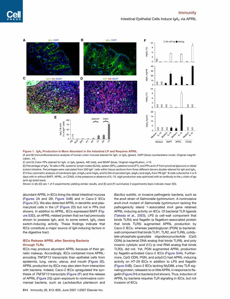
Figure 1. IgA2 Production Is More Abundant in the Intestinal LP and Requires APRIL
(A and B) Immunofluorescence analysis of human colon mucosa stained for IgA1 or IgA2 (green). DAPI (blue) counterstains nuclei. Original magnifi-
cation, 35.
(C and D) Colon PPs stained for IgA1 or IgA2 (green), AID (red), and BSAP (blue). Original magnification, 310.
(E) Percentage of IgA2+ B cells in PB, systemic lymph nodes (SLNs), spleen (SPL), palatine tonsil (PT), and PPs and LP from proximal (jejunum) or distal
(colon) intestine. Percentages were calculated from 200 IgA+ cells within tissue sections from three different donors double-stained for IgA and IgA2.
(F) Flow cytometric analysis of membrane IgA1 (mIgA1) and mIgA2 and ELISA of secreted IgA1 (sIgA1) and sIgA2 from PB IgD+ B cells cultured for 4 or 8
days with or without BAFF, APRIL, or CD40L in the presence or absence of IL-10. sIgA production was optimized with an antibody to the m chain of Igs
(anti-Ig) (solid bars).
Shown in (A)–(D) are 1 of 5 experiments yielding similar results, and (E) and (F) summarize 3 experiments (bars indicate mean SD).
abundant APRIL in IECs lining the distal intestinal mucosa
(Figures 2A and 2B; Figure S4B) and in Caco-2 IECs
(Figure 2C). We also detected APRIL in dendritic and plas-
macytoid cells in the LP (Figure 2D) but not in PPs (not
shown). In addition to APRIL, IECs expressed BAFF (Fig-
ure S3D), an APRIL-related protein that we had previously
shown to possess IgA1 and, to some extent, IgA2 class
switch-inducing activity. These findings indicate that
IECs constitute a major source of IgA-inducing factors in
the digestive tract.
IECs Release APRIL after Sensing Bacteria
through TLRs
IECs may produce abundant APRIL because of their ge-
netic makeup. Accordingly, IECs contained more APRIL-
encoding TNFSF13 transcripts than epithelial cells from
epidermis, lung, cervix, uterus, and mouth (Figure 2E).
APRIL production by IECs may also stem from interaction
with bacteria. Indeed, Caco-2 IECs upregulated the syn-
thesis of TNFSF13 transcripts (Figure 2F) and the release
of APRIL (Figure 2G) upon exposure to noninvasive com-
mensal bacteria, such as Lactobacillus plantarum and
814 Immunity 26, 812–826, June 2007 ª2007 Elsevier Inc.
Bacillus subtilis, or invasive pathogenic bacteria, such as
the aroA strain of Salmonella typhimurium. A noninvasive
aroA-invA mutant of Salmonella typhimurium lacking the
pathogenicity island 1-associated invA gene retained
APRIL-inducing activity on IECs. Of bacterial TLR ligands
(Takeda et al., 2003), LPS (a cell-wall component that
binds TLR4) and flagellin (a flagellum-associated protein
that binds TLR5) augmented APRIL production by
Caco-2 IECs, whereas peptidoglycan (PGN) (a bacterial-
wall component that binds TLR1, TLR2, and TLR6), cytidy-
late-phosphate-guanylate oligodeoxynucleotide (CpG
ODN) (a bacterial DNA analog that binds TLR9), and poly
inosinic cytidylic acid (I:C) (a viral RNA analog that binds
TLR3), did not. Yet, PGN augmented APRIL production
by flagellin-activated Caco-2 IECs (Figure S4A). Further-
more, CpG ODN, PGN, and poly(I:C) had APRIL-inducing
activity on HT-29 IECs in addition to LPS and flagellin
(Figure S4B). Caco-2 IECs lacking MyD88, a key TLR sig-
naling protein, released no or little APRIL in response to fla-
gellin (Figure 2H) or bacteria (not shown). Thus, induction of
APRIL by bacteria requires TLR signaling in IECs, but not
invasion of IECs.
Immunity
Intestinal Epithelial Cells Induce IgA2 via APRIL
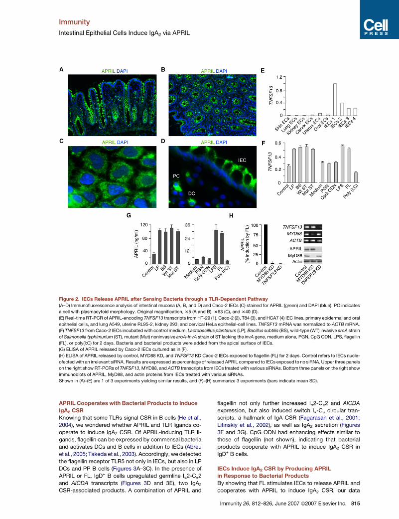
Figure 2. IECs Release APRIL after Sensing Bacteria through a TLR-Dependent Pathway
(A–D) Immunofluorescence analysis of intestinal mucosa (A, B, and D) and Caco-2 IECs (C) stained for APRIL (green) and DAPI (blue). PC indicates
a cell with plasmacytoid morphology. Original magnification, 35 (A and B), 363 (C), and 340 (D).
(E) Real-time RT-PCR of APRIL-encoding TNFSF13 transcripts from HT-29 (1), Caco-2 (2), T84 (3), and HCA7 (4) IEC lines, primary epidermal and oral
epithelial cells, and lung A549, uterine RL95-2, kidney 293, and cervical HeLa epithelial-cell lines. TNFSF13 mRNA was normalized to ACTB mRNA.
(F) TNFSF13 from Caco-2 IECs incubated with control medium, Lactobacillus plantarum (LP), Bacillus subtilis (BS), wild-type (WT) invasive aroA strain
of Salmonella typhimurium (ST), mutant (Mut) noninvasive aroA-InvA strain of ST lacking the invA gene, medium alone, PGN, CpG ODN, LPS, flagellin
(FL), or poly(I:C) for 2 days. Bacteria and bacterial products were added from the apical surface of IECs.
(G) ELISA of APRIL released by Caco-2 IECs cultured as in (F).
(H) ELISA of APRIL released by control, MYD88 KD, and TNFSF13 KD Caco-2 IECs exposed to flagellin (FL) for 2 days. Control refers to IECs nucle-
ofected with an irrelevant siRNA. Results are expressed as percentage of released APRIL compared to IECs exposed to no siRNA. Upper three panels
on the right show RT-PCRs of TNFSF13, MYD88, and ACTB transcripts from IECs treated with various siRNAs. Bottom three panels on the right show
immunoblots of APRIL, MyD88, and actin proteins from IECs treated with various siRNAs.
Shown in (A)–(E) are 1 of 3 experiments yielding similar results, and (F)–(H) summarize 3 experiments (bars indicate mean SD).
APRIL Cooperates with Bacterial Products to Induce
IgA2 CSR
Knowing that some TLRs signal CSR in B cells (He et al.,
2004), we wondered whether APRIL and TLR ligands co-
operate to induce IgA2 CSR. Of APRIL-inducing TLR li-
gands, flagellin can be expressed by commensal bacteria
and activates DCs and B cells in addition to IECs (Abreu
et al., 2005; Takeda et al., 2003). Accordingly, we detected
the flagellin receptor TLR5 not only in IECs, but also in LP
DCs and PP B cells (Figures 3A–3C). In the presence of
APRIL or FL, IgD+ B cells upregulated germline Ia2-Ca2
and AICDA transcripts (Figures 3D and 3E), two IgA2
CSR-associated products. A combination of APRIL and
flagellin not only further increased Ia2-Ca2 and AICDA
expression, but also induced switch Ia-Cm circular tran-
scripts, a hallmark of IgA CSR (Fagarasan et al., 2001;
Litinskiy et al., 2002), as well as IgA2 secretion (Figures
3F and 3G). CpG ODN had enhancing effects similar to
those of flagellin (not shown), indicating that bacterial
products cooperate with APRIL to induce IgA2 CSR in
IgD+ B cells.
IECs Induce IgA2 CSR by Producing APRIL
in Response to Bacterial Products
By showing that FL stimulates IECs to release APRIL and
cooperates with APRIL to induce IgA2 CSR, our data
Immunity 26, 812–826, June 2007 ª2007 Elsevier Inc. 815
Immunity
Intestinal Epithelial Cells Induce IgA2 via APRIL
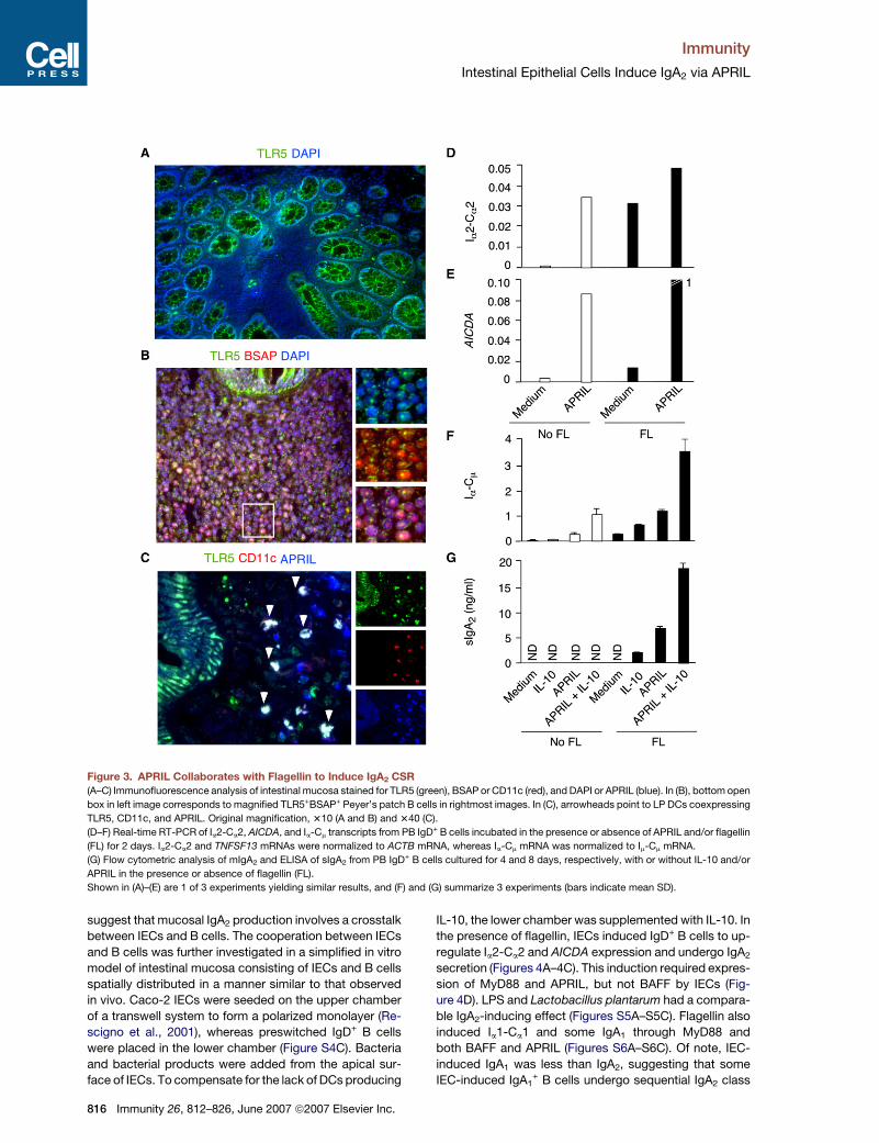
Figure 3. APRIL Collaborates with Flagellin to Induce IgA2 CSR
(A–C) Immunofluorescence analysis of intestinal mucosa stained for TLR5 (green), BSAP or CD11c (red), and DAPI or APRIL (blue). In (B), bottom open
box in left image corresponds to magnified TLR5+BSAP+ Peyer’s patch B cells in rightmost images. In (C), arrowheads point to LP DCs coexpressing
TLR5, CD11c, and APRIL. Original magnification, 310 (A and B) and 340 (C).
(D–F) Real-time RT-PCR of Ia2-Ca2, AICDA, and Ia-Cm transcripts from PB IgD+ B cells incubated in the presence or absence of APRIL and/or flagellin
(FL) for 2 days. Ia2-Ca2 and TNFSF13 mRNAs were normalized to ACTB mRNA, whereas Ia-Cm mRNA was normalized to Im-Cm mRNA.
(G) Flow cytometric analysis of mIgA2 and ELISA of sIgA2 from PB IgD+ B cells cultured for 4 and 8 days, respectively, with or without IL-10 and/or
APRIL in the presence or absence of flagellin (FL).
Shown in (A)–(E) are 1 of 3 experiments yielding similar results, and (F) and (G) summarize 3 experiments (bars indicate mean SD).
suggest that mucosal IgA2 production involves a crosstalk
between IECs and B cells. The cooperation between IECs
and B cells was further investigated in a simplified in vitro
model of intestinal mucosa consisting of IECs and B cells
spatially distributed in a manner similar to that observed
in vivo. Caco-2 IECs were seeded on the upper chamber
of a transwell system to form a polarized monolayer (Re-
scigno et al., 2001), whereas preswitched IgD+ B cells
were placed in the lower chamber (Figure S4C). Bacteria
and bacterial products were added from the apical sur-
face of IECs. To compensate for the lack of DCs producing
816 Immunity 26, 812–826, June 2007 ª2007 Elsevier Inc.
IL-10, the lower chamber was supplemented with IL-10. In
the presence of flagellin, IECs induced IgD+ B cells to up-
regulate Ia2-Ca2 and AICDA expression and undergo IgA2
secretion (Figures 4A–4C). This induction required expres-
sion of MyD88 and APRIL, but not BAFF by IECs (Fig-
ure 4D). LPS and Lactobacillus plantarum had a compara-
ble IgA2-inducing effect (Figures S5A–S5C). Flagellin also
induced Ia1-Ca1 and some IgA1 through MyD88 and
both BAFF and APRIL (Figures S6A–S6C). Of note, IEC-
induced IgA1 was less than IgA2, suggesting that some
IEC-induced IgA1+ B cells undergo sequential IgA2 class
Immunity
Intestinal Epithelial Cells Induce IgA2 via APRIL
switching. These data indicate that IECs induce IgA2 CSR
via APRIL after sensing bacteria through TLRs.
Intestinal LP B Cells Actively Undergo IgA CSR In Situ
Having shown that IECs induce IgA2 CSR in vitro, we won-
dered whether LP B cells undergo IgA2 CSR in vivo. Ongo-
ing CSR is associated with expression of AID (Fagarasan
et al., 2001). We identified AICDA transcripts and AID pro-
tein in LP B cells, including IgA1+ B cells lodged in the
proximity of or within the intestinal epithelium (Figures
4E and 4F; Figure S7A). AID, IgA2, and APRIL had similar
expression patterns in both intestinal and aerodigestive
mucosae (Figures S7B and S8A–S8C). AID was also de-
tected in IgD+ and a few IgG+ LP B cells (Figure 4G). In ad-
dition to AICDA transcripts, ex vivo isolated LP B cells
contained excised switch a (Sa)-Sm switch circles (Fig-
ure 5A; Figures S7C), a hallmark of ongoing IgM-to-IgA
CSR. Because of the high degree of sequence identity
between Sa1 and Sa2 regions, we could not distinguish
Sa1-Sm from Sa2-Sm switch circles, nor could we detect
Sa2-Sa1 switch circles originating from IgA1-to-IgA2 CSR.
Some LP IgA1+AID+ B cells expressed not only AID and
BSAP, as actively class-switching GC B cells do, but
also B-lymphocyte activation-induced maturation protein
1 (Blimp-1) and CD138 (Figure 5B), as plasma cells do
(Calame, 2001). LP IgA1+AID+ B cells also expressed inter-
feron regulatory factor 4 (IRF4), a CSR-inducing transcrip-
tion factor associated with GC, extrafollicular, and plas-
macytoid B cells (Cattoretti et al., 2006; He et al., 2004;
Klein et al., 2006). These data show that the human LP is
a site of active IgA CSR and suggest that LP IgA1+ effector
B cells arriving from mucosal GCs undergo IgA2 CSR
in situ.
Intestinal LP B Cells Undergo IgA2 CSR
in a T Cell- and CD40-Independent Fashion
To elucidate the nature of CSR events in LP IgA2+ B cells,
we analyzed the composition of their chromosomal S-S
DNA junctions. LP IgA2+ B cells contained chromosomal
Sm-Sa2, Sm-Sa1-Sa2 and, less frequently, Sm-Sg1-Sa2 junc-
tions originating from direct or sequential IgA2 CSR events
(Figure 5C; Figures S9A). Similar S-S junctions were de-
tected in tonsillar IgA2+ B cells (Figures S9B and S9C). In
additional experiments, we took advantage of mucosal
specimens from a patient with CD40 deficiency resulting
from congenital type-3 hyper-IgM syndrome (HIGM3)
and a patient with CD4+ T cell deficiency resulting from
acquired immunodeficiency syndrome (AIDS) to verify
whether IgA2 CSR requires help from CD4+ T cells
in vivo (Ferrari et al., 2001; Hel et al., 2006). The intestinal
LP from both HIGM3 and AIDS patients showed con-
served expression of IgA1, IgA2, AID, AICDA, and APRIL
(Figures 6A and 6B; Figures S10A–S10C). Considering
that CD40 deficiency results in a lack of systemic and mu-
cosal GCs (Castigli et al., 1994; Ferrari et al., 2001), these
data confirm that LP B cells can undergo IgA2 CSR in situ
through a TI- and CD40-independent mechanism involv-
ing APRIL.
APRIL Induces Sequential IgA2 CSR in IgA1+
or IgG1+ B Cells
Having detected chromosomal Sm-Sa1-Sa2 and Sm-Sg1-
Sa2 junctions in LP B cells, we wondered whether IgA1+
or IgG1+ B cells undergo sequential IgA2 CSR in response
to APRIL. In the presence of IL-10, APRIL augmented
chromosomal DNA recombination to Sa2 in IgG1+ or
IgA1+ B cells (Figure 6C; Figures S11A and S11B). This
effect was associated with induction of germline Ia2-Ca2,
AICDA and postswitch Im-Ca2 transcripts as well as mem-
brane and secreted IgA2 proteins (Figures 6D and 6E;
Figure S11C). CD40L induced similar effects, although
less than APRIL and only in IgG1+ B cells. Unlike APRIL,
CD40L did not induce IgA2 secretion. These data indicate
that APRIL is required for IgA1+ and IgG1
+ B cells to un-
dergo sequential IgA2 CSR as well as IgA2 secretion.
IECs Stimulate APRIL Production by DCs
through TSLP
In agreement with prior studies on in vitro generated
myeloid DCs (Litinskiy et al., 2002), we detected APRIL in
myeloid CD11c+ DCs within the intestinal LP (Figure 7A).
Some of these DCs were proximal to IgA1+ B cells,
whereas others were intercalated between IECs and em-
anated projections into the intestinal lumen. Thus, we hy-
pothesized that mucosal DCs express APRIL upon expo-
sure to bacterial and epithelial products, including TSLP
(Rescigno et al., 2001; Rimoldi et al., 2005). Consistent
with this possibility, flagellin upregulated the expression
of TSLP transcripts by Caco-2 IECs (Figures 7B and 7C).
Furthermore, flagellin cooperated with TSLP to upregulate
the expression of TSLP receptor-encoding cytokine
receptor-like factor 2 (CRFL2) and TNFSF13 transcripts as
well as the release of APRIL and IL-10 by myeloid DCs
(Figures 7D–7F). In the presence of flagellin, TSLP-suffi-
cient Caco-2 IECs upregulated the expression of
TNFSF13, APRIL, and IL-10 by myeloid DCs, whereas
TSLP-deficient Caco-2 IECs did not (Figures 7G–7I).
LPS, PGN, and Lactobacillus plantarum recapitulated
the effects of flagellin on IECs and myeloid DCs (not
shown). Thus, bacteria and IECs cooperatively induce
APRIL in mucosal DCs through TSLP.
TSLP-Conditioned DCs Enhance IEC-Induced IgA2
CSR through APRIL
Having shown that IECs increase APRIL production by
myeloid DCs via TSLP, we verified whether DCs augment
IEC-induced IgA2 class switching. IECs were placed in the
upper chamber of a transwell system, whereas DCs and
IgD+ B cells were placed in the lower chamber. In the pres-
ence of flagellin, DCs induced as much IgA2 production as
Caco-2 IECs (Figure 7J). IgA2 production was further upre-
gulated by TSLP, and this upregulation could be pre-
vented by B cell maturation-antigen (BCMA)-Ig, a soluble
APRIL decoy receptor that prevents activation of B cells
by APRIL (Litinskiy et al., 2002). In the presence of flagellin,
TSLP-sufficient IECs and DCs induced as much IgA2 as
DCs exposed to both flagellin and TSLP. Lack of TSLP
in IECs or addition of BCMA-Ig abolished induction of
Immunity 26, 812–826, June 2007 ª2007 Elsevier Inc. 817
8
Immunity
Intestinal Epithelial Cells Induce IgA2 via APRIL
18 Immunity 26, 812–826, June 2007 ª2007 Elsevier Inc.
Immunity
Intestinal Epithelial Cells Induce IgA2 via APRIL
IgA2 by IECs and DCs. Thus, in addition to directly inducing
IgA2 class switching through APRIL, bacteria-activated
IECs enhance the APRIL-dependent IgA2-inducing activity
of myeloid DCs via TSLP (Figure S12).
DISCUSSION
We have reported here that APRIL is essential to trigger
IgA2 CSR in B cells, including IgA1-expressing effector B
cells arriving from mucosal lymphoid follicles. IECs re-
leased APRIL after sensing bacteria through TLRs and fur-
ther augmented APRIL production by activating myeloid
DCs via TSLP. Our data suggest that intestinal bacteria
elicit IgA2 class switching by linking LP B cells with IECs
through a CD4+ T cell- and CD40L-independent pathway
involving APRIL.
The intestine is the largest lymphoid organ of our body,
and every day intestinal plasma cells produce more anti-
bodies than all other lymphoid organs combined, includ-
ing spleen, lymph nodes, and bone marrow (Brandtzaeg
et al., 2001; Mestecky et al., 1999). Although the remark-
able dominance of IgA in the intestine has been known
for decades, fine details of the mechanism underlying
IgA class switching, mucosal homing of IgA-producing
plasma cells, and transepithelial IgA transport continue
to emerge with an impressive frequency (Mora et al.,
2006). Nonetheless, many aspects of mucosal immunity
remain unclear, and in particular scarce data are available
on IgA2. This IgA subclass is predominant in the lower hu-
man intestinal tract, is more resistant than IgA1 to bacterial
degradation, and is often reactive against highly con-
served bacterial products, thus resembling polyreactive
IgA antibodies released by mouse B-1 cells (Crago et al.,
1984; Fagarasan and Honjo, 2003; Kett et al., 1986; Mac-
pherson, 2006; Mestecky et al., 1999; Tarkowski et al.,
1990). The present data indicate that, similar to mouse
B-1 cells (Fagarasan et al., 2001), human IgA2 B cell pre-
cursors undergo TI class switching in the intestinal LP.
One feature unique to mucosal surfaces is the crucial
role of epithelial cells in the initiation and modulation of
immune responses (Neutra et al., 2001; Xu et al., 2007).
Functional interdependence of IECs and B cells has
been amply documented and entails release of chemo-
kines that promote recruitment of IgA-producing plasma
cells to the intestinal LP (Wilson and Butcher, 2004), pro-
duction of cytokines that enhance the terminal differentia-
tion of arriving IgA-producing plasma cells (Mestecky
et al., 1999), and transportation of IgA onto the mucosal
surface through pIgR (Brandtzaeg et al., 2001). Our data
indicate that IECs intersect humoral immunity by inducing
IgA2 CSR in LP B cells via APRIL.
We identified APRIL as an essential IgA2 CSR-inducing
factor. In agreement with recent studies showing pro-
duction of APRIL and BAFF by epithelial cells from the
respiratory tract (Kato et al., 2006; Xu et al., 2007), we
found that IECs are a major source of APRIL in the gut.
Production of APRIL by IECs is at least in part genetically
determined as IECs expressed more APRIL-encoding
TNFSF13 transcripts than epithelial cells from nonintesti-
nal districts. Commensal bacteria further enhance APRIL
expression by activating IECs via TLRs. Thus, steady-
state TLR signaling as induced by commensal bacteria
could maintain intestinal homeostasis not only by promot-
ing IEC growth and repair (Rakoff-Nahoum et al., 2004),
but also by eliciting production of protective IgA2 via
APRIL. In addition to APRIL, IECs released BAFF, which
not only triggered IgA1 CSR, but also augmented APRIL-
induced IgA2 secretion, perhaps by delivering prosurvival
signals to IgA2-producing B cells.
Our findings suggest that bacteria-activated IECs utilize
APRIL to generate IgA2-inducing signals in LP B cells
arriving from PPs, including effector IgA1+ B cells. Several
lines of evidence support this model. First, the intestinal
LP contained more IgA2 and less IgA1 than PPs. Second,
some LP IgA1+ B cells expressed AID, a hallmark of ongo-
ing CSR, along with phenotypic traits evocative of late GC
B cells-early plasma cells. Third, LP IgA2+ plasmablasts
harbored genomic Sa1-Sa2 DNA junctions originating
from sequential IgA2 CSR events. Fourth, APRIL, a key
IEC-derived factor, elicited sequential IgA2 CSR in purified
IgA1+ B cells. Thus, effector IgA1
+ B cells emerging from
the GC of PPs may undergo APRIL-dependent sequential
CSR as mechanism to acquire protease-resistant IgA2,
which is more suited than IgA1 to cope with the dense
commensal microflora of the distal intestine.
IgG+ effector B cells may be also targeted by IEC-
derived APRIL to undergo sequential IgG1-to-IgA2 CSR
in the LP, because we could detect AID in LP IgG+ B cells
as well as genomic Sg1-Sa2 DNA junctions in LP IgA2+
plasmablasts. In addition, APRIL triggered IgA2 CSR in
purified IgG1+ B cells. Of note, Sg1-Sa2 junctions were
less frequent than Sa1-Sa2 and Sm-Sa2 junctions, which
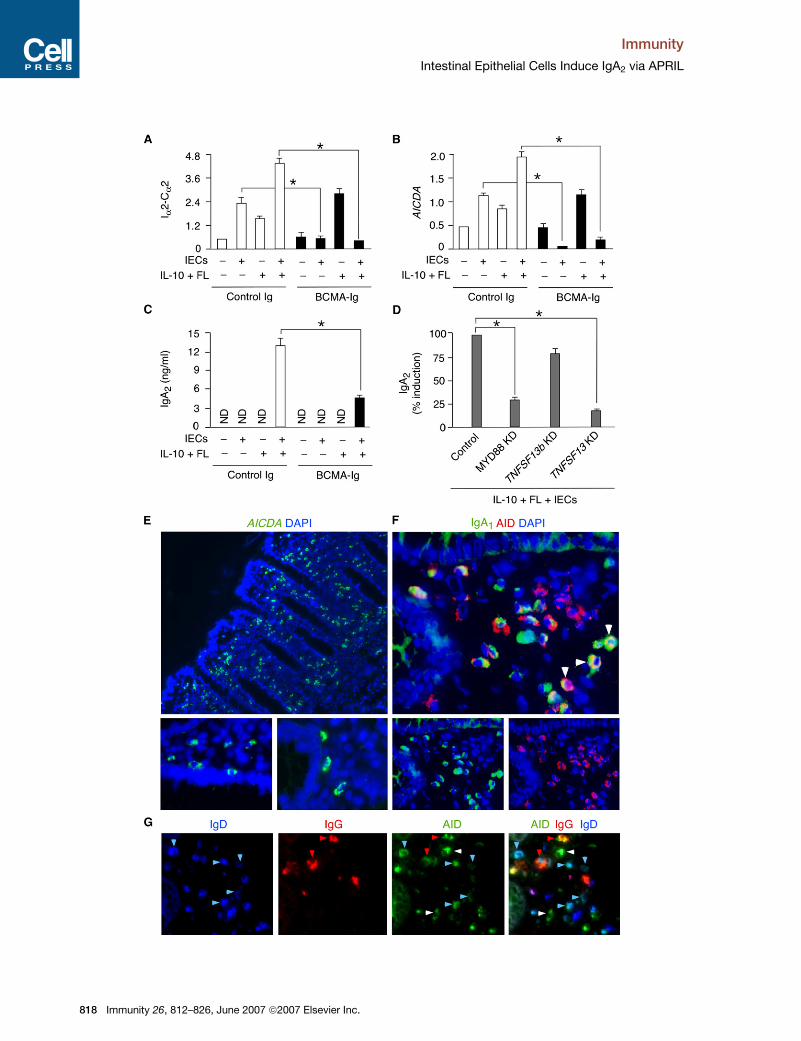
Figure 4. IECs Induce IgA2 CSR by Releasing APRIL after Sensing Bacteria
(A and B) Real-time RT-PCR of Ia2-Ca2 and AICDA transcripts from PB IgD+ B cells incubated with or without IECs, IL-10, and/or FL in the presence of
control Ig or BCMA-Ig for 2 days. FL was added from the apical surface of IECs. Ia2-Ca2 and AICDA mRNAs were normalized to ACTB mRNA.
(C) ELISA of sIgA2 from PB IgD+ B cells cultured for 8 days as in (A). ND, not detected.
(D) sIgA2 from PB IgD+ B cells cultured for 8 days with control, MYD88 KD, and TNFSF13 KD Caco-2 IECs and in the presence of FL and IL-10. Control
refers to Caco-2 IECs nucleofected with an irrelevant siRNA. Results are expressed as percentage of released sIgA2 compared to Caco-2 IECs
exposed to no siRNA.
(E) In situ hybridization of AICDA (green). DAPI stains intestinal nuclei (blue). Bottom images show periepithelial AICDA+ B cells. Original magnification,
310 (top) and 340 (bottom).
(F) Intestinal mucosa stained for IgA1 (green), AID (red), and DAPI (blue). Arrowheads point to IgA1+AID+ B cells. Original magnification, 363.
(G) Intestinal mucosa stained for AID (green), IgG (red), and IgD (blue). Blue, red, and white arrowheads point to IgD+ (blue), IgG1+, and double-
negative B cells expressing AID, respectively. Original magnification, 340.
Three experiments are summarized in (A)–(D) (bars indicate mean SD; *p < 0.05), and (E)–(G) show 1 of 3 experiments yielding similar results.
Immunity 26, 812–826, June 2007 ª2007 Elsevier Inc. 819
Immunity
Intestinal Epithelial Cells Induce IgA2 via APRIL
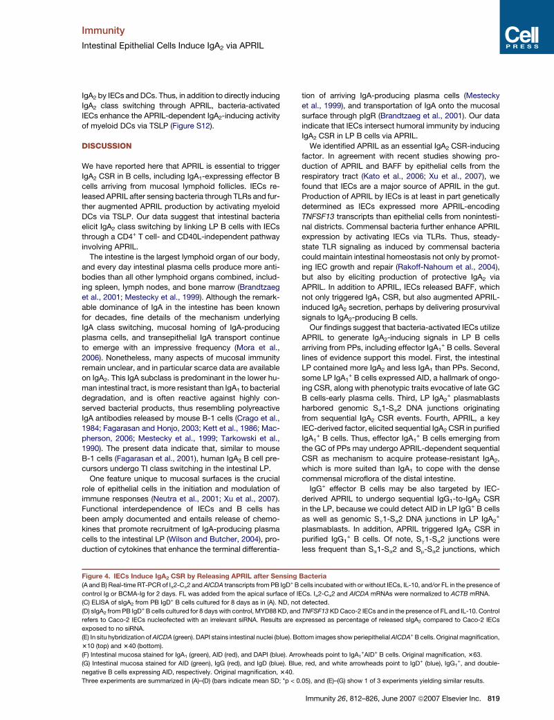
Figure 5. Intestinal LP B Cells Actively Undergo IgA CSR
(A) Extrachromosomal Sa-Sm switch circles PCR amplified from tonsillar or colon LP B cells (top) and Southern hybridized with an appropriate Sm probe
(bottom). Leftmost and rightmost lanes contain a 1 Kb DNA marker and no genomic DNA, respectively.
(B) Immunofluorescence analysis of intestinal LP stained for IgA1 (green), AID (red), and BSAP, IRF4, Blimp-1, CD138, and/or DAPI (blue). Arrowheads
point to IgA1+AID+ LP B cells expressing a transitional GC-plasmacytoid phenotype. Original magnification, 363.
(C) Sequences of chromosomal Sm-Sa2 and Sm-Sa1-Sa2 junctions from LP IgA2+ B cells obtained after intestinal tissue digestion. The Sm portion of
Sm-Sa1-Sa2 junctions is not shown for clarity. Dashes indicate identities. Arrows indicate the breakpoint site within the Sm, Sa1, or Sa2 region (top
and bottom sequences) as given in the EMBL/GenBank/DDBJ database.
Shown in (A) and (B) are 1 of 3 experiments yielding similar results.
820 Immunity 26, 812–826, June 2007 ª2007 Elsevier Inc.
Immunity
Intestinal Epithelial Cells Induce IgA2 via APRIL
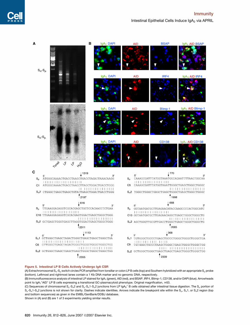
Figure 6. Intestinal LP B Cells Undergo IgA2 CSR without Help from CD4+ T Cells or CD40L
(A) Immunofluorescence analysis of intestinal mucosa from a CD40-deficient HIGM3 patient stained for IgA1 (green), AID (red), and DAPI (blue). Upper
right inset shows AID and DAPI only. Original magnification, 340.
(B) Immunofluorescence analysis and in situ hybridization of intestinal mucosa from a CD4+ T cell-deficient AIDS patient stained for IgA1 (green),
AICDA (red), and DAPI (blue). Open box in leftmost image corresponds to magnified AICDA+ or IgA1+ B cells in rightmost panels. Original magnifi-
cation, 310 (left) and 320 (right).
(C) Chromosomal Sm-Sa1 and Sm-Sa2 junctions were PCR amplified from PB IgG1+ or IgA1
+ B cells incubated with or without IL-10, APRIL, and/or
CD40L for 4 days and then Southern hybridized with appropriate radiolabeled Sm or Sa2 probes. In the rightmost lane, Sm-Sa2 DNA junctions from
sorted colon IgA2+ B cells are a positive control. Genomic ACTB is a loading control. Asterisk indicates spontaneous IgA2 CSR, whereas arrowheads
indicate bands subjected to cloning and sequencing.
(D) Real-time RT-PCR of Ia2-Ca2 and AICDA transcripts from PB IgG1+ or IgA1
+ B cells incubated as in (C) for 8 days. Ia2-Ca2 and AICDA mRNAs were
normalized to ACTB mRNA.
(E) Flow cytometric analysis of mIgA2 and ELISA of sIgA2 from PB IgG1+ or IgA1
+ B cells incubated as in (C) for 4 or 8 days.
Shown in (A)–(C) are 1 of 3 experiments yielding similar results, and (D) and (E) summarize 3 experiments (bars indicate mean SD;*p < 0.05,**p < 0.005
versus medium alone).
is consistent with the relative rarity of IgG in the intestinal
LP (Brandtzaeg et al., 2001). It is tempting to speculate
that some IgG+ B cells colonize the LP to amplify the local
IgA2 repertoire via sequential IgG-to-IgA2 CSR.
A large fraction of intestinal IgA2+ plasmablasts derives
from local IgD+ precursors, because LP B cells contained
AID and extrachromosomal Sm-Sa switch circles, two hall-
marks of ongoing IgA CSR, in addition to chromosomal
Sm-Sa2 DNA junctions. Accordingly, APRIL triggered IgA2
CSR in IgD+ B cells. Unlike LP IgA1+ and IgG1
+ B cells,
which likely derive from PPs (Brandtzaeg et al., 2001), at
least some LP IgD+ B cells may originate from the circula-
tion and could include marginal zone-like B cells (Suzuki
et al., 2005; Weller et al., 2004). In agreement with this
Immunity 26, 812–826, June 2007 ª2007 Elsevier Inc. 821
Immunity
Intestinal Epithelial Cells Induce IgA2 via APRIL
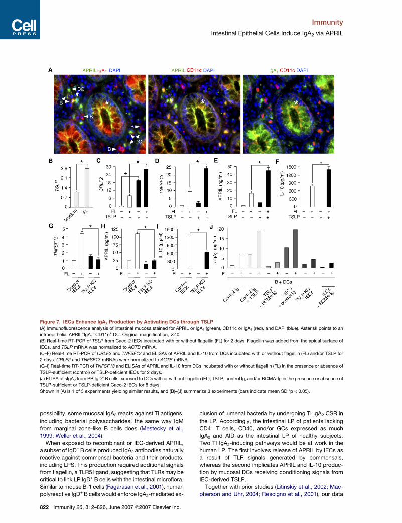
Figure 7. IECs Enhance IgA2 Production by Activating DCs through TSLP
(A) Immunofluorescence analysis of intestinal mucosa stained for APRIL or IgA1 (green), CD11c or IgA1 (red), and DAPI (blue). Asterisk points to an
intraepithelial APRIL+IgA1�CD11c+ DC. Original magnification, 340.
(B) Real-time RT-PCR of TSLP from Caco-2 IECs incubated with or without flagellin (FL) for 2 days. Flagellin was added from the apical surface of
IECs, and TSLP mRNA was normalized to ACTB mRNA.
(C–F) Real-time RT-PCR of CRLF2 and TNFSF13 and ELISAs of APRIL and IL-10 from DCs incubated with or without flagellin (FL) and/or TSLP for
2 days. CRLF2 and TNFSF13 mRNAs were normalized to ACTB mRNA.
(G–I) Real-time RT-PCR of TNFSF13 and ELISAs of APRIL and IL-10 from DCs incubated with or without flagellin (FL) in the presence or absence of
TSLP-sufficient (control) or TSLP-deficient IECs for 2 days.
(J) ELISA of sIgA2 from PB IgD+ B cells exposed to DCs with or without flagellin (FL), TSLP, control Ig, and/or BCMA-Ig in the presence or absence of
TSLP-sufficient or TSLP-deficient Caco-2 IECs for 8 days.
Shown in (A) is 1 of 3 experiments yielding similar results, and (B)–(J) summarize 3 experiments (bars indicate mean SD;*p < 0.05).
possibility, some mucosal IgA2 reacts against TI antigens,
including bacterial polysaccharides, the same way IgM
from marginal zone-like B cells does (Mestecky et al.,
1999; Weller et al., 2004).
When exposed to recombinant or IEC-derived APRIL,
a subset of IgD+ B cells produced IgA2 antibodies naturally
reactive against commensal bacteria and their products,
including LPS. This production required additional signals
from flagellin, a TLR5 ligand, suggesting that TLRs may be
critical to link LP IgD+ B cells with the intestinal microflora.
Similar to mouse B-1 cells (Fagarasan et al., 2001), human
polyreactive IgD+ B cells would enforce IgA2-mediated ex-
822 Immunity 26, 812–826, June 2007 ª2007 Elsevier Inc.
clusion of lumenal bacteria by undergoing TI IgA2 CSR in
the LP. Accordingly, the intestinal LP of patients lacking
CD4+ T cells, CD40, and/or GCs expressed as much
IgA2 and AID as the intestinal LP of healthy subjects.
Two TI IgA2-inducing pathways would be at work in the
human LP. The first involves release of APRIL by IECs as
a result of TLR signals generated by commensals,
whereas the second implicates APRIL and IL-10 produc-
tion by mucosal DCs receiving conditioning signals from
IEC-derived TSLP.
Together with prior studies (Litinskiy et al., 2002; Mac-
pherson and Uhr, 2004; Rescigno et al., 2001), our data
Immunity
Intestinal Epithelial Cells Induce IgA2 via APRIL
suggest that intestinal DCs acquire B cell-licensing func-
tions after sampling bacteria from the lumen. In particular,
DCs produce IgA2 CSR-inducing factors, such as APRIL
and IL-10, after receiving instructing signals from bacteria
and IECs via TLR ligands and TSLP, respectively. Overall,
our data indicate that intestinal bacteria trigger IgA2 class
switching via a TI pathway linking IECs with DCs and B
cells through an APRIL-mediated TLR-dependent signal-
ing program. Thus, mucosal vaccines should activate
IECs to stimulate more effective production of protease-
resistant IgA2 antibodies.
EXPERIMENTAL PROCEDURES
B Cells
PB and tonsillar B cells were purified as reported (Cerutti et al., 2002;
Litinskiy et al., 2002; Xu et al., 2007). PB IgD+, PB IgG1+, PB IgA1
+, and
tonsillar IgA2+ B cells were magnetically sorted with biotinylated mouse
mAbs 2032-08 to IgD, 4E3 to IgG1, B3506B4 IgA1, and A9604D2
to IgA2 (Southern Biotec), respectively, and anti-biotin MicroBeads
(Miltenyi Biotec). Purity after sorting was consistently R97%. LP
CD19+ and IgA2+ B cells were purified from LP mononuclear cells
obtained upon enzymatic digestion of nonneoplastic specimens of
patients undergoing resection for colon carcinoma (distance from the
neoplasm >8 cm) as described in the Supplemental Experimental
Procedures. LP CD19+ B cells ranged from 6 to 15 3 106, whereas LP
IgA2+ B cells ranged from 4 to 6 3 106. Both cell types were >95%
pure. The Institutional Review Board of Weill Medical College of Cornell
University approved studies with tonsillar and LP cells, and patients
provided informed consent. The cell line Ramos was used as a positive
control for AICDA expression.
DCs
Myeloid dendritic cells were obtained from PB monocytes. In brief,
monocytes were sorted with a biotinylated mAb to CD14 (Serotec)
and streptavidin MicroBeads and then cultured for 6–8 days in medium
RPMI 1640 (Invitrogen) supplemented with 5% human AB serum
(Sigma), 1000 U/ml of granulocyte monocyte-colony stimulating factor
(Berlex Laboratories), and 1000 U/ml of IL-4 (R&D Systems). Every
2 days, 400 ml of medium was removed from each well and replaced
by 500 ml of fresh medium with cytokines. After 6 days, more than
95% of the cells in culture expressed DC-specific antigens, including
CD11c, CD40, CD83, CD86, HLA-II, DEC-205, mannose receptor,
and DC-specific intercellular adhesion molecule-3 grabbing noninte-
grin (DC-SIGN), but less than 10% expressed CD14.
IECs and Other Epithelial Cells
HT-29, Caco-2, T84, and HCA7 IEC lines were derived from human co-
lon carcinoma. All functional assays involving IECs were performed
with Caco-2 IECs, which recapitulate most of the properties of primary
human IECs, including formation of a polarized monolayer connected
by tight junctions (Rescigno et al., 2001; Rimoldi et al., 2005). Lung
A549, uterine RL95-2, kidney 293, and cervical HeLa epithelial cell
lines as well as primary epidermal (Cambrex Bio Science Walkersville)
and oral (MatTek Corporation) epithelial cells were used for control
studies. HT-29, Caco-2, T84, HCA7, A549, RL95-2, 293, and HeLa
epithelial cell lines were cultured in RPMI 1640 medium or Dulbecco’s
Modified Eagle’s Medium supplemented with 10% fetal calf serum.
Primary epidermal and oral ECs were cultured as reported (Xu et al.,
2007).
Coculture Systems
Caco-2 IECs were cultured in the upper chamber of 3.0 mm pore Trans-
well filters (BD PharMingen) for 7 days in a 24-well plate until a transe-
pithelial resistance of �300 U/cm2 was achieved. B cells (1.0 3 106)
and DCs (0.2 3 106) were seeded in the lower chamber. Filters were
treated with bacteria (10 bacteria to 1 IEC) or bacterial products from
the apical surface. At 1 hr after incubation, bacteria were washed out
and the medium was replaced with medium containing 100 mg/ml of
gentamycin. Cocultures were carried out in complete RPMI 1640
medium supplemented with 10% bovine serum.
Bacterial Strains and Products
The following Salmonella typhimurium strains on SL1344 background
were provided by G. Dougan (Imperial College, London, UK): metabol-
ically defective invasive strain (aroA), with attenuated ability to repli-
cate in vivo in mice, and metabolically defective noninvasive strain
(aroA-invA mutant), defective in the invA gene and therefore unable
to form productive type-three secretion system. These strains were
grown with appropriate antibiotics to preserve carried mutations.
Lactobacillus plantarum NCIMB882 was grown at 37�C without agitation
in MRS broth (Biokar Diagnostic). The Bacillus subtilis 168-derivative
JH642 was obtained from the Bacillus Genetic Stock Center (Colum-
bus, Ohio) and grown at 37�C in 2X YT medium (GIBCO-BRL).
Poly(I:C), PGN from Escherichia coli 0111:B4 (InvivoGen), flagellin
from Bacillus subtilis, LPS from Escherichia coli 0111:B4 (Sigma),
and CpG ODN (Operon Technologies) were used at 20 mg/ml,
20 mg/ml, 0.5 mg/ml, 20 mg/ml, and 20 mg/ml, respectively.
Other Reagents
APRIL (R&D Systems), BAFF (Alexis), CD40L (Immunex), IL-10 (Scher-
ing-Plough), and TSLP (R&D Systems) were used at 500ng/ml, 500 ng/ml,
200 ng/ml, 50 ng/ml, and 30 ng/ml, respectively. APRIL was blocked
with 5 mg/ml of BCMA-Ig (Ancell). Mouse MOPC21 IgG1 with irrelevant
binding activity was used at 5 mg/ml to control BCMA-Ig. In IgD+ B cell
cultures, an H15100 polyclonal antibody (pAb) to the m chain of Igs
(Caltag Laboratories) was added at 2 mg/ml to optimize IgA secretion.
Flow Cytometry
The following fluorescein (FITC)-, phycoerythrin or allophycocyanin-
conjugated mAbs were used: IADB6 to IgD, SA-DA4 to IgM,
A9604D2 to IgA, H2 to IgG, B3506B4 to IgA1, A9604D2 to IgA2 (South-
ern Biotech), and F3637 to IgE (Sigma-Aldrich). At least 1 3 104 viable
cells were acquired with a FACScalibur analyzer (BD PharMingen).
Enzyme-Linked Immunoadsorbent Assays
ELISAs for total IgA antibodies and IL-10 were described previously
(Xu et al., 2007). To measure IgA1 and IgA2, Immulon 4 HBX micro-
plates (Thermo Electron Corp.) were coated with a goat F(ab’)2 poly-
clonal antibody (pAb) to human Igs (Cappel) and with a mouse mAb
14A to human IgA2 (Serotec), respectively, in carbonate-bicarbonate
buffer. Then, a biotin-labeled mouse mAb B3506B4 to human IgA1
or a biotin-labeled mouse A9604D2 mAb to human IgA2 (Southern Bio-
tech) were added followed by peroxidase-conjugated streptavidin
(Vector Laboratories) and TMB Microwell Peroxidase Substrate Kit
(Kirkegaard and Perry). Readings were done at 450 nm. A similar strat-
egy was employed to measure IgA2 to LPS and flagellin, except that
microplates were coated with 20 mg/ml of LPS (Sigma) and 10 mg/ml
of flagellin (Sigma), respectively. IgA2 to Lactobacillus plantarum
were measured by coating microplates with mAb 14A and by sequen-
tially adding bacteria and mAb A9604D2. To measure APRIL, micro-
plates were reacted with a rabbit ED2 mAb (ProSci Inc.) and a biotin-
labeled goat BAF884 pAb (R&D Systems).
Immunofluorescence and Immunohistochemistry
Caco-2 IECs stained with an unconjugated rabbit pAb ED2 to APRIL
(Pro Sci) and an appropriate secondary reagent were analyzed by im-
munofluorescence as reported (Litinskiy et al., 2002). Intestinal tissue
samples were obtained from five non-AIDS patients and one AIDS pa-
tient undergoing resection for colon carcinoma (distance from the neo-
plasm was more than 8 cm). The PB CD4+ T cell count of the AIDS pa-
tient was 2 cells/ml. Additional intestinal tissue sections were obtained
from a HIGM3 patient carrying a mutated CD40 gene. The immunological
and molecular features of this case, which corresponds to ‘‘Patient 1’’
Immunity 26, 812–826, June 2007 ª2007 Elsevier Inc. 823
Immunity
Intestinal Epithelial Cells Induce IgA2 via APRIL
described in a recent report (Ferrari et al., 2001), are summarized in
the Supplemental Experimental Procedures. The Institutional Review
Board of Weill Medical College of Cornell University approved the
study and patients provided informed consent. Tissue sections 5 mm
in thickness were paraformaldehyde fixed and then stained with the
following primary antibodies: biotin-conjugated mouse mAbs F(ab’)2B3506B4 to IgA1, F(ab’)2 A9604D2 to IgA2 (Southern Biotech), 1D6 to
BAFF (eBioscience), and 3.9 to CD11c (Ancell); goat F(ab’)2 pAbs
2032-02 to IgD, 2042-08 to IgG (Southern Biotech), and sc-6059 to
IRF4 (Santa Cruz); unconjugated mouse mAbs A-11 to BSAP (Santa
Cruz), PG-B6p to Bcl6 (Dako), 3H2-E8 to Blimp-1 (Novus Biologicals),
and RPA-T4 to CD4 (Research Diagnostic, Inc.); unconjugated rabbit
pAbs H-240 to pan-cytokeratin (Santa Cruz) and ED2 to APRIL (Pro
Sci); unconjugated rat mAb EK2-5G9 to AID (Ascenion GmbH, Ger-
many); and, finally, FITC-conjugated mouse mAb 85B152.5 to TLR5
(Imgenex). Control primary antibodies included FITC-conjugated, bio-
tin-conjugated, or unconjugated mouse IgG1 mAbs, unconjugated rat
IgG2a mAb, biotin-conjugated goat F(ab’)2 pAb, and unconjugated
rabbit pAb. Slides were incubated with the following secondary re-
agents: indodicarbocyanine-conjugated anti-mouse pAb, rhoda-
mine-conjugated anti-mouse pAb (Jackson ImmunoResearch Labora-
tories), Alexa Fluor 546/488-conjugated anti-goat pAb, Alexa Fluor
647-conjugated anti-rabbit pAb, and cyanine 3/5-conjugated or rho-
damine-conjugated streptavidin (Molecular Probes). Nuclei were visu-
alized with DAPI, 4’,6-diamidine-20-phenylindole dihydrochloride
(Boehringer Mannheim). Images were acquired with an Axiovert
200M microscope (Carl Zeiss).
In Situ RNA Hybridization
Full-length AICDA cDNA under the control of the SP6 promoter was
used as template to transcribe an antisense RNA probe with a commer-
cial riboprobe generation kit containing biotin-labeled UTP (Roche Ap-
plied Science) as previously reported (Xu et al., 2007). A sense RNA
probe was used as negative control. Tissue sections were fixed, hy-
bridized, and stained as described in the Supplemental Experimental
Procedures.
Conventional and Quantitative Real-Time RT-PCRs
TNFSF13, AICDA, and ACTB transcripts were RT-PCR amplified as re-
ported (Litinskiy et al., 2002). MYD88, Im-Ca1, and Im-Ca2 were RT-PCR
amplified with MYD88 forward 50-GAGCGTTTCGATGCCTTCAT-30
plus MYD88 reverse 50-CGGATCATCTCCTGCACAAA-30 and Im for-
ward 50-GTGATTAAGGAGAAACACTTTGAT-30 plus either Ca1 reverse
50-GGGTGGCGGTTAGCGGGGTCTTGG-30 or Ca2 reverse 50-
TGTTGGCGGTTAGTGGGGTCTTGCA-30. AICDA, Ia-Cm, Im-Cm, TSLP,
CRFL2, and ACTB were quantitated through real-time RT-PCR as de-
scribed (Xu et al., 2007). Ia2-Ca2 and TNFSF13 were quantitated with
Ia2-Ca2 forward 50-CTCAGCACTGCGGGCCCTCCA-30 plus Ia2-Ca2
reverse 50-GTTCCCATCTTGGGGGGTGCTGTC-30 and with TNFSF13
forward 50-GTGATGTGGCAACCAGCTCTT-30 plus TNFSF13 reverse
50-CCCTTGGTGTAAATGGAAGAC-30, respectively.
Microdissection
Intestinal specimens were frozen in Tissue-Tek optimum cutting tem-
perature compound (Sakura). Cryostat sections were affixed to glass
foil slides for membrane-based laser microdissection (Leica), allowed
to dry for 30 min at 20�C–25�C, fixed for 1 min in 70% ethanol, washed
for 30 s in distilled water, stained for 1 min with 0.6% (weight/volume)
methyl green (Fluka), rinsed in distilled water, dehydrated by a graded
ethanol series (70%, 95%, and 100%) for 1 min each, and allowed to
air dry at 20�C–25�C for 7–12 hr. LP regions containing at least 200
IgA+ B cells were isolated by laser-capture microdissection on a Leica
AS LMD and collected in a 2 ml microcentrifuge tube for DNA extrac-
tion and amplification.
Genomic DNA PCR and Southern Blots
Genomic DNA was extracted from B cells with miniDNA preparation kit
(Qiagen) and from microdissected LP specimens with QIAamp DNA
824 Immunity 26, 812–826, June 2007 ª2007 Elsevier Inc.
micro kit (Qiagen). Chromosomal Sm-Sa1 and Sm-Sa2 junctions were
amplified with an Sm forward primer 50-CTTGTTAATGGACTTGGAG
GAATGATTCC-30 and either a Sa1 reverse primer 50-ACTGTGAGG
ACGCGGCCCTCTCCT-30 or a Sa2 reverse primer 50-AATGCGCTGT
GAGGACGCGGCCCTCATGC-30. Elongase (Invitrogen) was used un-
der the following conditions: denaturation 1 min at 94�C, annealing
2 min at 62�C, and extension 3 min at 72�C for 30 cycles. A second
PCR was performed on the DNA product of the first PCR under similar
conditions, but with the nested Sm forward primer 50-GCTGCTG
CATTTGCTTCTCTTAAAAC-30 and the nested Sa2 reverse primer 50-
AGCCCACCCCAGGGCAGCTCAGTAC-30. To verify their identity to
Sm-Sa2 junctions, PCR amplicons were fractionated onto a 0.8% aga-
rose gel, transferred overnight onto nylon membranes, and hybridized
with a radiolabeled probe spanning residues 274–497 of the 50 portion
of Sm. Then, membranes were stripped and hybridized with a radiola-
beled probe spanning residues 3199–3238 of the 30 portion of Sa2.
Extrachromosomal Sa-Sm junctions were PCR amplified as reported
(Litinskiy et al., 2002) and hybridized with a radiolabeled probe re-
cognizing the 30 portion of Sm as described in the Supplemental Ex-
perimental Procedures. Genomic ACTB and Im-Cm transcripts were
detected as reported (Litinskiy et al., 2002). Im-Ca1 and Im-Ca2 tran-
scripts were hybridized with a radiolabeled consensus Ca 50-CGAC
ACGGGTCGGTACCTTGGTACCC-30 oligoprobe.
Cloning and Sequencing
0.5–3 Kb PCR products hybridizing with both Sm and Sa2 probes were
gel purified (Qiagen) and cloned into a PCR2.1 TOPO vector with a TA
cloning kit (Invitrogen). Positive clones were PCR identified with the Sm
forward primer 50-CCACTAGAAGGGGAACTGGTCTTA-30 and the Sa2
reverse primer 50-AGCCCACCCCAGGGCAGCTCAGTAC-30. The per-
centage of positive clones ranged from 90% to 94%. Positive clones
were purified and sequenced as described in the Supplemental Exper-
imental Procedures.
RNA Interference and Nucleofection
TSLP-deficient Caco-2 IECs were generated as described (Rimoldi
et al., 2005). To knock down MYD88, a psiRNA-hMyD88 plasmid
expressinga MYD88-targetingsmall interfering RNA (siRNA)ora plasmid
expressing control siRNA (Invivogen) was resuspended with 1.0 3 106
IECs into 100 ml of human keratinocyte nucleofector solution (Amaxa).
Plasmids were nucleofected with an appropriate device (Amaxa). To
knock down TNFSF13, 0.4 ml of a 20 mM Hs_ATGGCTCTGCTGACCC
AACAA_1_HP APRIL siRNA or control siRNA (Qiagen) were incubated
with 6 ml of HiPerFect Transfection Reagent (Qiagen) in 200 ml of se-
rum-free DMEM medium for 5–10 min at room temperature. This mix-
ture was added drop-wise to 3 3 105 EC cells, which were then incu-
bated at 37�C. Expression of targeted mRNA and proteins was
evaluated after 24 and 48 hr, respectively.
Western Blot
Protein extracts were transferred to nylon membranes as reported (Xu
et al., 2007). After blocking, membranes were probed with primary
goat pAbs to APRIL (R-15), MyD88 (N-19), or actin (I-19) (Santa Cruz
Biotechnologies). Membranes were washed and incubated with an ap-
propriate secondary antibody (Santa Cruz). Proteins were detected
with an enhanced chemiluminescence detection system (Amersham).
Statistical Analysis
For immunoglobulin secretion, proliferation, survival, and reporter as-
says, values were calculated as mean standard deviation for at least
three separate experiments done in triplicate. The significance of dif-
ferences between experimental variables was determined with the
Student’s t test.
Supplemental Data
Twelve figures and Experimental Procedures are available at http://
www.immunity.com/cgi/content/full/26/6/812/DC1/.
Immunity
Intestinal Epithelial Cells Induce IgA2 via APRIL
ACKNOWLEDGMENTS
We thank M. Rimoldi (European Institute of Oncology, Milan, Italy) for
preparing Caco-2 cells and bacteria and A. Dannenberg (Weill Medical
College of Cornell University, New York, NY) for generating dendritic
cells and providing HT-29 cells. This work was supported by NIH grant
AI057653 (to A. Cerutti), funds from NIH T32 grant AI07621 (to W.X.),
and funds from Fondazione C. Golgi and Centro Immunodeficienze
Mario Di Martino (A.P.). B.H. and W.X. equally contributed to this
work by performing research and analyzing and discussing data;
P.A.S., J.E., and M.S. performed research; A. Chiu, A. Chadburn,
A.D.P., C.R.R., V.V., A.P., and D.M.K. provided tissue samples and
discussed data; M.R. provided essential reagents, discussed data,
and critically read the paper; and A. Cerutti designed research, ana-
lyzed data, and wrote the paper.
Received: December 10, 2006
Revised: April 2, 2007
Accepted: April 25, 2007
Published online: June 14, 2007
REFERENCES
Abreu, M.T., Fukata, M., and Arditi, M. (2005). TLR signaling in the gut
in health and disease. J. Immunol. 174, 4453–4460.
Brandtzaeg, P., Baekkevold, E.S., and Morton, H.C. (2001). From B to
A the mucosal way. Nat. Immunol. 2, 1093–1094.
Calame, K.L. (2001). Plasma cells: finding new light at the end of B cell
development. Nat. Immunol. 2, 1103–1108.
Castigli, E., Alt, F.W., Davidson, L., Bottaro, A., Mizoguchi, E., Bhan,
A.K., and Geha, R.S. (1994). CD40-deficient mice generated by recom-
bination-activating gene-2-deficient blastocyst complementation.
Proc. Natl. Acad. Sci. USA 91, 12135–12139.
Castigli, E., Scott, S., Dedeoglu, F., Bryce, P., Jabara, H., Bhan, A.K.,
Mizoguchi, E., and Geha, R.S. (2004). Impaired IgA class switching in
APRIL-deficient mice. Proc. Natl. Acad. Sci. USA 101, 3903–3908.
Castigli, E., Wilson, S.A., Scott, S., Dedeoglu, F., Xu, S., Lam, K.P.,
Bram, R.J., Jabara, H., and Geha, R.S. (2005). TACI and BAFF-R me-
diate isotype switching in B cells. J. Exp. Med. 201, 35–39.
Cattoretti, G., Buttner, M., Shaknovich, R., Kremmer, E., Alobeid, B.,
and Niedobitek, G. (2006). Nuclear and cytoplasmic AID in extrafollic-
ular and germinal center B cells. Blood 107, 3967–3975.
Cerutti, A., Zan, H., Kim, E.C., Shah, S., Schattner, E.J., Schaffer, A.,
and Casali, P. (2002). Ongoing in vivo immunoglobulin class switch
DNA recombination in chronic lymphocytic leukemia B cells. J. Immu-
nol. 169, 6594–6603.
Chaudhuri, J., and Alt, F.W. (2004). Class-switch recombination: inter-
play of transcription, DNA deamination and DNA repair. Nat. Rev. Im-
munol. 4, 541–552.
Coffman, R.L., Lebman, D.A., and Shrader, B. (1989). Transforming
growth factor beta specifically enhances IgA production by lipopoly-
saccharide-stimulated murine B lymphocytes. J. Exp. Med. 170,
1039–1044.
Crago, S.S., Kutteh, W.H., Moro, I., Allansmith, M.R., Radl, J., Haaij-
man, J.J., and Mestecky, J. (1984). Distribution of IgA1-, IgA2-, and
J chain-containing cells in human tissues. J. Immunol. 132, 16–28.
Fagarasan, S., and Honjo, T. (2003). Intestinal IgA synthesis: regulation
of front-line body defences. Nat. Rev. Immunol. 3, 63–72.
Fagarasan, S., Kinoshita, K., Muramatsu, M., Ikuta, K., and Honjo, T.
(2001). In situ class switching and differentiation to IgA-producing cells
in the gut lamina propria. Nature 413, 639–643.
Fagarasan, S., Muramatsu, M., Suzuki, K., Nagaoka, H., Hiai, H., and
Honjo, T. (2002). Critical roles of activation-induced cytidine deami-
nase in the homeostasis of gut flora. Science 298, 1424–1427.
Fayette, J., Dubois, B., Vandenabeele, S., Bridon, J.M., Vanbervliet,
B., Durand, I., Banchereau, J., Caux, C., and Briere, F. (1997). Human
dendritic cells skew isotype switching of CD40-activated naive B cells
towards IgA1 and IgA2. J. Exp. Med. 185, 1909–1918.
Ferrari, S., Giliani, S., Insalaco, A., Al-Ghonaium, A., Soresina, A.R.,
Loubser, M., Avanzini, M.A., Marconi, M., Badolato, R., Ugazio, A.G.,
et al. (2001). Mutations of CD40 gene cause an autosomal recessive
form of immunodeficiency with hyper IgM. Proc. Natl. Acad. Sci.
USA 98, 12614–12619.
He, B., Qiao, X., and Cerutti, A. (2004). CpG DNA induces IgG class
switch DNA recombination by activating human B cells through an in-
nate pathway that requires TLR9 and cooperates with IL-10. J. Immu-
nol. 173, 4479–4491.
Hel, Z., McGhee, J.R., and Mestecky, J. (2006). HIV infection: first bat-
tle decides the war. Trends Immunol. 27, 274–281.
Honjo, T., Kinoshita, K., and Muramatsu, M. (2002). Molecular mecha-
nism of class switch recombination: linkage with somatic hypermuta-
tion. Annu. Rev. Immunol. 20, 165–196.
Jain, A., Ma, C.A., Lopez-Granados, E., Means, G., Brady, W., Orange,
J.S., Liu, S., Holland, S., and Derry, J.M. (2004). Specific NEMO muta-
tions impair CD40-mediated c-Rel activation and B cell terminal differ-
entiation. J. Clin. Invest. 114, 1593–1602.
Kato, A., Truong-Tran, A.Q., Scott, A.L., Matsumoto, K., and
Schleimer, R.P. (2006). Airway epithelial cells produce B cell-activating
factor of TNF family by an IFN-beta-dependent mechanism. J. Immu-
nol. 177, 7164–7172.
Kett, K., Brandtzaeg, P., Radl, J., and Haaijman, J.J. (1986). Different
subclass distribution of IgA-producing cells in human lymphoid organs
and various secretory tissues. J. Immunol. 136, 3631–3635.
Klein, U., Casola, S., Cattoretti, G., Shen, Q., Lia, M., Mo, T., Ludwig,
T., Rajewsky, K., and Dalla-Favera, R. (2006). Transcription factor
IRF4 controls plasma cell differentiation and class-switch recombina-
tion. Nat. Immunol. 7, 773–782.
Litinskiy, M.B., Nardelli, B., Hilbert, D.M., He, B., Schaffer, A., Casali,
P., and Cerutti, A. (2002). DCs induce CD40-independent immuno-
globulin class switching through BLyS and APRIL. Nat. Immunol. 3,
822–829.
Lue, C., Tarkowski, A., and Mestecky, J. (1988). Systemic immuniza-
tion with pneumococcal polysaccharide vaccine induces a predomi-
nant IgA2 response of peripheral blood lymphocytes and increases
of both serum and secretory anti-pneumococcal antibodies. J. Immu-
nol. 140, 3793–3800.
Macpherson, A.J. (2006). IgA adaptation to the presence of commen-
sal bacteria in the intestine. Curr. Top. Microbiol. Immunol. 308, 117–
136.
Macpherson, A.J., and Uhr, T. (2004). Induction of protective IgA by in-
testinal dendritic cells carrying commensal bacteria. Science 303,
1662–1665.
Mestecky, J., Russell, M.W., and Elson, C.O. (1999). Intestinal IgA:
novel views on its function in the defence of the largest mucosal sur-
face. Gut 44, 2–5.
Mora, J.R., Iwata, M., Eksteen, B., Song, S.Y., Junt, T., Senman, B.,
Otipoby, K.L., Yokota, A., Takeuchi, H., Ricciardi-Castagnoli, P.,
et al. (2006). Generation of gut-homing IgA-secreting B cells by intes-
tinal dendritic cells. Science 314, 1157–1160.
Nagler-Anderson, C. (2001). Man the barrier! Strategic defences in the
intestinal mucosa. Nat. Rev. Immunol. 1, 59–67.
Neutra, M.R., Mantis, N.J., and Kraehenbuhl, J.P. (2001). Collabora-
tion of epithelial cells with organized mucosal lymphoid tissues. Nat.
Immunol. 2, 1004–1009.
Odegard, V.H., and Schatz, D.G. (2006). Targeting of somatic hyper-
mutation. Nat. Rev. Immunol. 6, 573–583.
Immunity 26, 812–826, June 2007 ª2007 Elsevier Inc. 825
Immunity
Intestinal Epithelial Cells Induce IgA2 via APRIL
Rakoff-Nahoum, S., Paglino, J., Eslami-Varzaneh, F., Edberg, S., and
Medzhitov, R. (2004). Recognition of commensal microflora by toll-like
receptors is required for intestinal homeostasis. Cell 118, 229–241.
Rescigno, M., Urbano, M., Valzasina, B., Francolini, M., Rotta, G., Bo-
nasio, R., Granucci, F., Kraehenbuhl, J.P., and Ricciardi-Castagnoli, P.
(2001). Dendritic cells express tight junction proteins and penetrate gut
epithelial monolayers to sample bacteria. Nat. Immunol. 2, 361–367.
Rimoldi, M., Chieppa, M., Salucci, V., Avogadri, F., Sonzogni, A., Sam-
pietro, G.M., Nespoli, A., Viale, G., Allavena, P., and Rescigno, M.
(2005). Intestinal immune homeostasis is regulated by the crosstalk
between epithelial cells and dendritic cells. Nat. Immunol. 6, 507–514.
Salzer, U., Chapel, H.M., Webster, A.D., Pan-Hammarstrom, Q.,
Schmitt-Graeff, A., Schlesier, M., Peter, H.H., Rockstroh, J.K.,
Schneider, P., Schaffer, A.A., et al. (2005). Mutations in TNFRSF13B
encoding TACI are associated with common variable immunodefi-
ciency in humans. Nat. Genet. 37, 820–828.
Sato, A., Hashiguchi, M., Toda, E., Iwasaki, A., Hachimura, S., and Ka-
minogawa, S. (2003). CD11b+ Peyer’s patch dendritic cells secrete IL-
6 and induce IgA secretion from naive B cells. J. Immunol. 171, 3684–
3690.
Schlissel, M.S. (2003). Regulating antigen-receptor gene assembly.
Nat. Rev. Immunol. 3, 890–899.
Schneider, P. (2005). The role of APRIL and BAFF in lymphocyte acti-
vation. Curr. Opin. Immunol. 17, 282–289.
Stavnezer, J. (1996). Antibody class switching. Adv. Immunol. 61, 79–
146.
826 Immunity 26, 812–826, June 2007 ª2007 Elsevier Inc.
Suzuki, K., Meek, B., Doi, Y., Honjo, T., and Fagarasan, S. (2005). Two
distinctive pathways for recruitment of naive and primed IgM+ B cells
to the gut lamina propria. Proc. Natl. Acad. Sci. USA 102, 2482–2486.
Takeda, K., Kaisho, T., and Akira, S. (2003). Toll-like receptors. Annu.
Rev. Immunol. 21, 335–376.
Tarkowski, A., Lue, C., Moldoveanu, Z., Kiyono, H., McGhee, J.R., and
Mestecky, J. (1990). Immunization of humans with polysaccharide
vaccines induces systemic, predominantly polymeric IgA2-subclass
antibody responses. J. Immunol. 144, 3770–3778.
Weller, S., Braun, M.C., Tan, B.K., Rosenwald, A., Cordier, C., Conley,
M.E., Plebani, A., Kumararatne, D.S., Bonnet, D., Tournilhac, O., et al.
(2004). Human blood IgM ‘‘memory’’ B cells are circulating splenic
marginal zone B cells harboring a prediversified immunoglobulin reper-
toire. Blood 104, 3647–3654.
Wilson, E., and Butcher, E.C. (2004). CCL28 controls immunoglobulin
(Ig)A plasma cell accumulation in the lactating mammary gland and IgA
antibody transfer to the neonate. J. Exp. Med. 200, 805–809.
Xu, W., He, B., Chiu, A., Chadburn, A., Shan, M., Buldys, M., Ding, A.,
Knowles, D.M., Santini, P.A., and Cerutti, A. (2007). Epithelial cells trig-
ger frontline immunoglobulin class switching through a pathway regu-
lated by the inhibitor SLPI. Nat. Immunol. 8, 294–303.
Ziegler, S.F., and Liu, Y.J. (2006). Thymic stromal lymphopoietin in nor-
mal and pathogenic T cell development and function. Nat. Immunol. 7,
709–714.